screws type ii(c) fracture treated with headless a … 4: the post-op x-ray images of the...
TRANSCRIPT
Received 09/05/2016 Review began 09/12/2016 Review ended 09/14/2016 Published 09/23/2016
© Copyright 2016Kapoor et al. This is an open accessarticle distributed under the terms ofthe Creative Commons AttributionLicense CC-BY 3.0., which permitsunrestricted use, distribution, andreproduction in any medium, providedthe original author and source arecredited.
A Case of Distal Femur Medial Condyle HoffaType II(C) Fracture Treated with HeadlessScrewsChirag Kapoor , Aditya Merh , Malkesh Shah , Paresh Golwala
1. Orthopaedics, Sumandeep Vidyapeeth, Vadodara, Gujarat
Corresponding author: Chirag Kapoor, [email protected] Disclosures can be found in Additional Information at the end of the article
AbstractCoronal plane fractures of the distal femur are less frequent compared to sagittal plane fractures.They were described by Hoffa in 1904 and are known as Hoffa fractures (AO type B3). They areisolated fractures of the femoral condyle and rare in occurrence. The objective in the treatment ofthese fractures is to achieve anatomical reduction of the articular surface and a stable fixation toprevent joint damage in future and prevent post-traumatic arthritis of the joint. We report the caseof a young male patient who had a rare type of medial Hoffa fracture which was treated by openreduction and internal fixation using headless Herbert screws using a posterior approach. Thefracture was united in eight weeks, and the patient had a full range of knee movement. We advocatethis approach and modality of treatment for Hoffa type II(C) fractures.
Categories: Physical Medicine & Rehabilitation, Orthopedics, OtherKeywords: hoffa, herbert screw, posterior approach
IntroductionCoronal plane fractures of the distal femur are less frequent compared to sagittal plane fractures andwere described by Hoffa in 1904 and are known as Hoffa fractures (AO type B3). They are isolatedfractures of the femoral condyle and rare in occurrence [1]. Lateral condyle Hoffa fractures are threetimes more common than medial condyle fractures [2]. Because of the rarity of medial condyle Hoffafractures, not much is reported about this injury and its management.
The mechanism of injury has been reported to be a direct anteroposterior force to the flexed andabducted knee for lateral condylar fractures and a direct impact on the medial side of the knee inflexion for a medial condylar fracture. The objective in the treatment of these fractures is to achieveanatomical reduction of the articular surface and a stable fixation to prevent joint damage and post-traumatic arthritis in future [3].
We report the case of a young male patient who had a rare type of medial Hoffa fracture type II(C)which was managed surgically by using a posterior approach. Informed consent was obtained fromthe subject used in the study.
Case PresentationA 38-year-old male patient had a road traffic accident and presented to us one day later in thecasualty department. He was not able to walk and had pain in the right knee joint posteriorly. Therewas a tense hemarthrosis of the right knee joint with overlying shiny skin. The knee range ofmovement was severely restricted due to pain. The patient was vitally stable and had no otherassociated injury.
1 1 1 1
Open Access CaseReport DOI: 10.7759/cureus.802
How to cite this articleKapoor C, Merh A, Shah M, et al. (September 23, 2016) A Case of Distal Femur Medial Condyle HoffaType II(C) Fracture Treated with Headless Screws. Cureus 8(9): e802. DOI 10.7759/cureus.802

Plain radiographs (anteroposterior, lateral, and oblique views of the right knee joint) showed a smallosteochondral fragment of the posterior aspect of medial condyle which was displaced and rotatedand was situated just proximal to the intercondylar notch [Figure 1]. A three-dimensional CT scanwas done for better visualization of the fracture configuration which confirmed that it was a medialHoffa type II(C) fracture [Figure 2].
FIGURE 1: The pre-op Xray images of the anteroposterior & lateralviews showing the distal femur medial condyle Hoffa type II(C)fracture
FIGURE 2: A three-dimensional computed tomography (CT) scanimage showing the distal femur medial condyle Hoffa type II(C)fracture
2016 Kapoor et al. Cureus 8(9): e802. DOI 10.7759/cureus.802 2 of 7
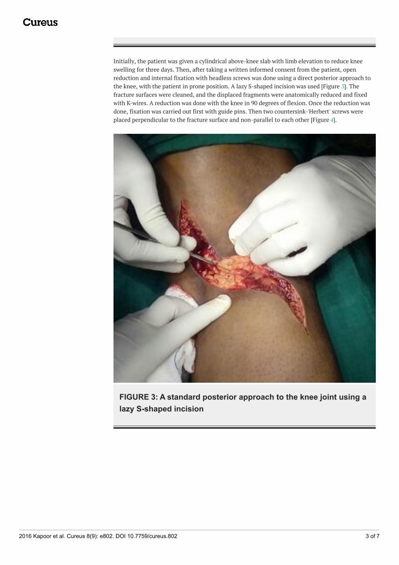
Initially, the patient was given a cylindrical above-knee slab with limb elevation to reduce kneeswelling for three days. Then, after taking a written informed consent from the patient, openreduction and internal fixation with headless screws was done using a direct posterior approach tothe knee, with the patient in prone position. A lazy S-shaped incision was used [Figure 3]. Thefracture surfaces were cleaned, and the displaced fragments were anatomically reduced and fixedwith K-wires. A reduction was done with the knee in 90 degrees of flexion. Once the reduction wasdone, fixation was carried out first with guide pins. Then two countersink-'Herbert' screws wereplaced perpendicular to the fracture surface and non-parallel to each other [Figure 4].
FIGURE 3: A standard posterior approach to the knee joint using alazy S-shaped incision
2016 Kapoor et al. Cureus 8(9): e802. DOI 10.7759/cureus.802 3 of 7
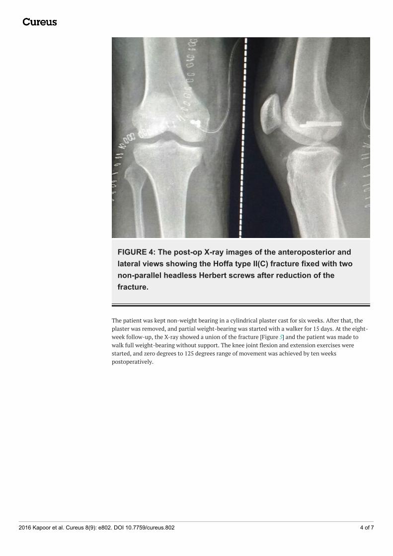
FIGURE 4: The post-op X-ray images of the anteroposterior andlateral views showing the Hoffa type II(C) fracture fixed with twonon-parallel headless Herbert screws after reduction of thefracture.
The patient was kept non-weight bearing in a cylindrical plaster cast for six weeks. After that, theplaster was removed, and partial weight-bearing was started with a walker for 15 days. At the eight-week follow-up, the X-ray showed a union of the fracture [Figure 5] and the patient was made towalk full weight-bearing without support. The knee joint flexion and extension exercises werestarted, and zero degrees to 125 degrees range of movement was achieved by ten weekspostoperatively.
2016 Kapoor et al. Cureus 8(9): e802. DOI 10.7759/cureus.802 4 of 7

FIGURE 5: A 10-week follow-up Xray image of the anteroposteriorand lateral views showing the union of the fracture with in-situHerbert screws
DiscussionIntra-articular coronal plane “Hoffa” fractures of the distal femur medial condyle are rare injuriesand difficult to treat. Conservative management often leads to unsatisfactory results and nonunionas many undisplaced fractures get displaced after conservative treatment. This makes the prognosisof such injuries worse.
If the fragment is not reduced properly, roughening of the articular surface and osteonecrosis occur,which may produce a marked disability in future. Moreover, if the fragment is small, it should not beremoved because it is an important part of the articular surface when the knee is flexed at 90degrees. So, it is mandatory to fix this fragment anatomically to achieve a good outcome.
Lewis, et al. [4] classified the Hoffa fractures into three types. In Type I and Type III Hoffa fracturesthere are some soft tissue elements attached to the fractured condylar fragment to provide bloodsupply to this fragment. But in Type II Hoffa fractures there is no soft tissue element attached to thefractured condylar fragment [Figure 6].
[4]"> [4]" itemprop="image"src="https://assets.cureus.com/uploads/figure/file/7262/article_river_317d7380734d11e6b52dff9eb7a0ecaa-
Figure_6.png" title="Classification-of-Hoffa-fracture-by-Lewis-et-al.[4]" />
FIGURE 6: Classification of Hoffa fracture by Lewis et al.[4]Classification of Hoffa fracture by Lewis et al. [4]
2016 Kapoor et al. Cureus 8(9): e802. DOI 10.7759/cureus.802 5 of 7
Up to 30% of these fractures get overlooked on plain radiographs and require computerizedtomography to assess the fracture pattern and plan the surgical fixation [5]. We also preferred to geta three-dimensional CT done so as to assess the fracture configuration properly.
It is accepted that screw fixation is a good fixation method for treating Hoffa fractures. There is nostandardized surgical approach in treating these fractures, and usually, an anterolateral oranteromedial incision is used (depending on the condyle involved) for Type I and III fractures andtwo anteroposteriorly placed lag screws are inserted from the non-articular area just proximal to thepatella-femoral joint to engage the fractured condylar fragment. In Type II Hoffa fractures, becausethe fracture line is near the articular cartilage of the posterior condyle, a posterior approach and twopostero-anteriorly placed lag screws are preferred [6, 7]. However, the screws are inserted throughthe articular surface, so the screw heads should be countersunk or headless Herbert screws shouldbe used instead of compression screws.
In our case it was a Type II(C) fracture, so we preferred a posterior approach as the fragment was toosmall to get held by anteroposterior screws, was rotated and displaced posterior to the intercondylarnotch, and was difficult to reduce using the anteromedial approach. So we preferred to approach thefracture posteriorly and fix it with posteroanterior Herbert screws in a non-parallel fashion ratherthan anteroposterior screws. Lag screws placed in a posteroanterior direction are more stable thananteroposteriorly placed lag screws [8] and crossed screws are more rigid than the parallel screws[9].
ConclusionsWe believe that these rare injuries should be identified in time and treated aggressively by an earlyopen reduction and an anatomically rigid internal fixation, to achieve a good recovery of thefunction of the joint.
Additional InformationDisclosuresHuman subjects: Consent was obtained by all participants in this study. Conflicts of interest: Incompliance with the ICMJE uniform disclosure form, all authors declare the following:Payment/services info: All authors have declared that no financial support was received from anyorganization for the submitted work. Financial relationships: All authors have declared that theyhave no financial relationships at present or within the previous three years with any organizationsthat might have an interest in the submitted work. Other relationships: All authors have declaredthat there are no other relationships or activities that could appear to have influenced thesubmitted work.
References1. Manfredini M, Alessandro G, Leo M, et al.: Unicondylar femoral fractures: Therapeutic strategy and
long-term results. A review of 23 patients. Acta orthopaedica Belgica. 2001, 67(2):132-138.2. Holmes SM, Bomback D, Baumgaertner MR: Coronal fractures of the femoral condyle: a brief report
of five cases. J Orthop. 2004, 18:316–319.3. Ostermann PA, Neumann K, Ekkernkamp A, et al.: Long term results of unicondylar fractures of
the femur. J Orthop. 1994, 8:142–146.4. Lewis SL, Pozo JL, Muirhead-Allwood WF: Coronal fracture of the lateral femoral condyle . J Bone
Joint Surg. 1989, 71(1):118-120.5. Nork SE, Segina DN, Aflatoon K, et al.: The association between supracondylar intercondylar distal
femoral fractures and coronal plane fractures. J Bone Joint Surg Am. 2005, 87(3):564-569.6. Medvecky MJ, Noyes FR: Surgical approaches to the posteromedial and posterolateral aspects of
the knee. J Am Acad Orthop Surg. 2005, 13(2):121–128.7. Tan Y, Li H, Zheng Q: A modified posterolateral approach for Hoffa fracture . Eur J Orthop Surg
Traumatol. 2013, 24(7):1321–1323.8. Jarit GJ, Kummer FJ, Gibber MJ, et al.: A mechanical evaluation of two fixation methods using
2016 Kapoor et al. Cureus 8(9): e802. DOI 10.7759/cureus.802 6 of 7

cancellous screws for coronal fractures of the lateral condyle of the distal femur (OTA type 33B). JOrthop Trauma. 2006, 20(4):273–276.
9. Friedman RL, Glisson RR, Nunley JA, et al.: A biomechanical comparative analysis of twotechniques for tibiotalar arthrodesis. Foot Ankle Int. 1994, 15(6):301–305.
2016 Kapoor et al. Cureus 8(9): e802. DOI 10.7759/cureus.802 7 of 7