screening of north african medicinal plant extracts for ... · organic extracts and fractions of...
TRANSCRIPT

____________________________________________________________________________________________
*Corresponding author: Email: [email protected];
European Journal of Medicinal Plants3(3): 310-332, 2013
SCIENCEDOMAIN internationalwww.sciencedomain.org
Screening of North African Medicinal PlantExtracts for Cytotoxic Activity Against Tumor
Cell Lines
Lamiae Belayachi1,3, Clara Aceves-Luquero1, Nawel Merghoub3,Youssef Bakri3, Silvia Fernández de Mattos1,2, Saaïd Amzazi3
and Priam Villalonga1,2*
1Cancer Cell Biology Group, Institut Universitari d’Investigació en Ciències de la Salut,(IUNICS), Spain.
2Departament de Biologia Fonamental, Universitat de les Illes Balears, Illes Balears, Spain.3Biochemistry-Immunology Laboratory, Faculty of Sciences, Mohammed V-Agdal University,
Rabat, Morocco.
Authors’ contributions
This work was carried out in collaboration between all authors. Author LB performed allexperiments and participated in the experimental design, analysis of the data and redactionof the manuscript. Author CAL participated in the experimental design, analysis of the data
and preparation of the manuscript. The plants harvest, identification and extraction weredone by authors NM and YB. Author SA participated in the analysis of the data and the
preparation of the manuscript. Author SFM participated in the experimental design and theanalysis of the data. Author PV directed the research and supervised the preparation of the
manuscript. All authors have contributed and approved the manuscript.
Received 24th February 2013Accepted 17th April 2013
Published 27th April 2013
ABSTRACT
Aims: The aim of this study was to evaluate the in vitro cytotoxic activity and cellulareffects of organic extracts and fractions of four plants; Inula viscosa, Ormenis eiriolepis(Asteraceae), Retama monosperma (Fabaceae) and Marrubium vulgare (Lamiaceae), allof them used in Moroccan traditional medicine.Methodology: The four plants were extracted using organic solvents and screened on apanel of human cancer cell lines including cell types from both solid and haematologicalcancer origin as well as non-transformed murine fibroblasts. Cell viability assays were
Research Article
European Journal of Medicinal Plants, 3(3): 310-332, 2013
311
performed with sixteen plant extracts. Sensitive cell lines were then exposed to increasingconcentrations of the most efficient extracts in order to calculate IC50 values. Microscopy,flow cytometry and caspase activity assays were then performed in LN229, SW620 andPC-3 cell lines upon treatment to investigate the cell morphology, cell cycle distributionand cell death.Results: cell viability assays reveals that at least one extract from each plant was able toexert cytotoxic activity against the majority of cell lines tested, the IC50 values of the activeextracts were in most cases ≤ 30 µg/ml. the study of the cellular effects of the most activeextracts on LN229, SW620 and PC-3 cell lines shows their ability to promote cell cyclearrest and cell death. The data obtained herein support strongly the use of these plants bytraditional healers for the treatment of cancer patients and could have some scientificsupport indicating the presence of bioactive compounds.Conclusion: The reported biological activity of these four medicinal plants used intraditional Moroccan medicine provides a starting point for forthcoming studies todetermine the molecular basis of their activity and to identify the chemical compoundswithin the most active extracts responsible for their antitumoral effects.
Keywords: Inula viscose; Retama monosperma; Ormenis eriolepis; Marrubium vulgare;Moroccan pharmacopeia; anticancer agents; cell proliferation; cell death.
1. INTRODUCTION
Numerous plant extracts have been used by several civilizations for their scent andmedicinal properties. Active compounds within those extracts constitute the basis oftraditional medicine systems that have been in existence for thousands of years andcontinue to provide mankind with new remedies [1]. According to the World HealthOrganization (WHO) about 65–80% of the world’s population in developing countriesdepends essentially on plants for their primary health care due to poverty and lack of accessto modern medicine [2]. Medicinal plants have thus a long history of use for almost everygroup of human diseases including cancer, one of the major causes of mortality throughoutthe world. Statistics have estimated that cancer will cause 83.2 million deaths between 2005and 2015 [3]. Drug discovery from medicinal plants plays an important role in the treatmentof cancer and, indeed, most new clinical applications of plant secondary metabolites andtheir actives compounds derivatives over the last half century have been applied againstcancer [4]. Of all available anticancer drugs between 1940 and 2002, 40% were naturalproducts or natural product-derived mimics, including Vinca alkaloids, Taxusditerpenes,Camptotheca alkaloids, and Podophyllumlignans [5]. Currently, there are 16 new plantderived compounds being tested in clinical trials and 13 of these compounds are beingtested in phase I or II, and 3 are in phase III. Among these compounds, flavopiridol, isolatedfrom the Indian tree Dysoxylum binectariferum and mesoindigo, isolated from the Chineseplant Indigo feratinctoria, have been shown to exhibit anti-cancer effects with lesser toxicitythan conventional chemotherapeutic drugs [6]. Ethnomedical knowledge is one of the mainstrategies to select plant species in cancer drug discovery [7]. In Morocco, traditionalmedicine belongs to one of the world's oldest pharmacopoeia; in fact it disposes of a widearsenal of plants remedies because of the natural diversity of the environment and flora [8].The aim of this study was to evaluate the in vitro cytotoxic activity and cellular effects oforganic extracts and fractions of four plants; Inula viscosa, Ormenis eiriolepis (Asteraceae),Retama monosperma (Fabaceae) and Marrubium vulgare (Lamiaceae), all of them used inMoroccan traditional medicine. The plant selection was based on their reputation as folk

European Journal of Medicinal Plants, 3(3): 310-332, 2013
312
medicines in the treatment of tumors [9]; and related diseases and also on their previouslyreported phytochemical and biological properties (Table 1).
2. MATERIALS AND METHODS
2.1 Plant Material
The selected plants were collected in different areas of Morocco during the period 2007-2011 and were identified by Dr. M. Fennane from the Scientific Institute of Rabat (Table 1).
Table 1. Ethnobotanical data and some reported pharmacological activities of plantsspecies used in this study
Plantsspecies(family)
Trivialname
Place ofcollection
Partplantcollected
Traditional use Pharmacologicalactivitiesand references
Inula viscosaL. Ait.(Asteraceae)
Magramane-Terhala
Ain atikTemara
Leaves Skin diseases,treat cutaneousabscesses,wound healing,Tuberculosis,bronchialinfections [9]
Antimicrobialactivity [11].Antifungal activity Ḹ[12]Anti inflammatoryeffects [13].Antitumoral activity[14], [15], [16].
RetamamonospermaL. Bois(Fabaceae)
R’tm Sidi-BoughabaMahdia
Leaves Purgative,vermifuge,antihelmintic,abortive anddisinfectant [10]
Antitumoral activity[15], [16]
OrmeniseriolepisCoss.(Asteraceae)
Hellala Ouarzazat Aerial part Stomachic,anthelminticand antidiabetic[9]
Antibacterialactivities [17]Antileishmaniaactivities [18]Antifungic activity[19]
Marrubiumvulgare L.(Lamiaceae)
MerriwutMerriwa
Temara Aerial part Antidiabetic,antidiarrheal,febrifuge,expectorant,antiicteric,treatophthalmia,otitis,abscessesfeversandboils[9]
Antispasmodic [20]Antinociceptive [21]Analgesic [22]Antioxidant, Anticholesterol [23]Antioeudematogenic [24]Antidiabetic [25],[26], [27], [28].
2.2 Experimental Design
Plant extracts were screened across a panel of human cancer and a non-tumoral mousefibroblast cell lines from different tumor origin (Table 2) to analyze the cytotoxic activity anddetermines the IC50 of the most active extracts. In order to examine the cellular effects of the

European Journal of Medicinal Plants, 3(3): 310-332, 2013
313
most active extracts, we analyzed cell morphology, cell cycle distribution, cell deathinduction and caspase activity in LN229, SW620 and PC-3 cell lines.
Table 2. Cell lines used in the study related to the cells type and diseases
Cell lines Cell type Disease
Hum
an
LN-229 epithelial GlioblastomaT98G fibroblast GlioblastomamultiformeU-87 MG epithelial Glioblastoma; Astrocytoma grade IVJurkat T lymphocyte Acute T cell LeukemiaJeKo-1 lymphoblast non-Hodgkin lymphomasSW620 epithelial Colorectal adenocarcinomaSW480 epithelial Colorectal adenocarcinomaPC-3 epithelial Prostate adenocarcinomaU2OS epithelial Osteosarcoma
Mice NIH/3T3 Embryonic fibroblast Normal
2.3 Preparation of Organic Extract
The collected parts of the selected plants were air-dried and mechanically ground to producea fine powder. 200g of each plant powder was successively extracted using a Soxhletapparatus with n-hexane (1.3L) and methanol (1.3L) to obtain hexanic extract andmethanolic extract the resulting extracts were then evaporated by a Rotavapor to give driedextracts. The methanol concentrated extract was dissolved in distilled water and wassuccessively extracted with dichloromethane (1.3L) and ethyl acetate (1.3L) to obtaindichloromethane fraction and ethyl acetate fraction. All extracts were evaporated by aRotavapor and kept at -20ºC until use.
2.4 Cell Culture
Jurkat and Jeko-1 cells were maintained in RPMI1640 with L-Glutamine and HEPES(Invitrogen, Carlsbad, CA). LN229, T98G, U87MG, SW620, SW480, U2OS, PC-3 andNIH3T3 cells were maintained in DMEM High Glucose (4.5 g/l) with L-glutamine (Invitrogen,Carlsbad, CA). All cells were grown in a humidified incubator at 37ºC with 5% CO2. RPMIand DMEM were supplemented with 10% heat inactivated foetal bovine serum and 100units/ml penicillin/streptomycin (Invitrogen, Carlsbad, CA). All cell lines were subconfluentlygrown and passaged, routinely tested for mycoplasma contamination and subjected tofrequent morphological tests and growth curve analysis as quality-control assessments. Allcell lines were treated at a prophylactic concentration of 5 μg/ml with Plasmocin™(InvivoGen, San Diego, CA).
2.5 Cell Viability Assays
The number of viable cells in culture was determined based on quantification of ATP, whichsignals the presence of metabolically active cells, using the Cell Titer-Glo Luminiscent assaykit (Promega, Madison, WI, USA) following the manufacturer’s instructions. Briefly, cellswere plated in 96-well plates, treated 24h later with 50 µg/ml of extracts dissolved in DMSO

European Journal of Medicinal Plants, 3(3): 310-332, 2013
314
for 48h followed by addition of Cell Titer-Glo reagent. Luminiscence was detected using amulti-well Synergy Mx scanning spectrophotometer (Biotek, Winooski, VT).
2.6 Cellular Morphology Analysis
Cells were plated in 6-well plates and treated with 50 µg/ml of the indicated extract for 24hand 48h. Representative images were collected under a light optic microscope (LeicaMicrosystems, Wetzlar, Germany).
2.7 Cell Cycle Analysis
Cell cycle analysis was performed following propidium iodide staining. Briefly, trypsinizedcells were washed in phosphate-buffered saline (PBS) and fixed in 70% ethanol. Fixed cellswere then washed twice in PBS and DNA was stained with propidium iodide (50 µg/ml) inthe presence of 50 µg/ml RNase A (Sigma-Aldrich, Saint Louis, MO), then analysed by flowcytometry using a FACS can (Coulter Epics XL-MSL; Beckman Coulter, Fullerton, CA, USA)and winMDI software.
2.8 Annexin V‐FITC/Propidium Iodide FLow Cytometric Analysis
Analysis of phosphatidylserine externalization in apoptotic cells was determined by Annexin-V-FITC (Invitrogen, Carlsbad, CA) staining, according to the manufacturer’s instructions.Briefly, 2x10⁵ cells were seeded in 6-well plates and treated with 50 μg/ml of the extracts for48h. They were then collected and suspended in 100μl of Annexin Vbinding buffer.Following incubation with Annexin-V-FITC and propidium iodide for 15 min at roomtemperature in the dark, flow cytometry analysis was carried out using a FACScan (CoulterEpics XL-MSL; Beckman Coulter, Fullerton, CA, USA) and analyzed with the winMDIsoftware.
2.9 Caspase Activity Assays
Caspase activity was determined by measurement of caspases 3 and 7 activity in controland treated cells with 50 µg/ml of indicated extracts plated in 96-well plates for 48h usingthe luminometric Caspase-Glo 3/7 assay (Promega) according to the manufacturer’sprotocol, using a Synergy HT multidetection microplate reader (Bio- Tek, Winooski, VT).
2.10 Statistical Analysis
Data are presented as means ± SD of at least three different assays performed in triplicate.IC50 values and the statistical significance of differences by Student’s t test were assessedusing GraphPad Prism (GraphPad Software Inc. La Jolla, CA). Statistically significantdifferences are indicated by ***P,0.001, **P,0.01 and *P,0.05.
3. RESULTS AND DISCUSSION
For this study, four medicinal plants used for various diseases were harvested in differentareas of Morocco. Four organic extracts of Inula viscosa, Ormenis eiriolepis (Asteraceae),Retama monosperma (Fabaceae) and Marrubium vulgare (Lamiaceae) were prepared.Table 1 summarizes the ethnobotanical data including botanical and local names,ethnomedical uses and the relevant plant parts employed together with their reported

European Journal of Medicinal Plants, 3(3): 310-332, 2013
315
pharmacological activity. The cytotoxic screening was performed on a panel of establishedhuman cancer cell lines including Glioblastoma Multiforme, Acute T cell Leukemia, MantleCell Lymphoma, Colon cancer, Prostate Cancer and Osteosarcoma cells. The NIH3T3nontransformed mouse fibroblast cell line was employed as a control cell line to explore thecytotoxic effects of the extracts on nontumoral cells. Table 2 provides an overview of thecharacteristics of the cell lines used. In our screening program, all cell lines were initiallytreated with all sixteen extracts at a starting concentration of 50μg/ml to determine theselective activity of the extracts.
3.1 Selected Plant Extracts Show Cytotoxicity against Human GlioblastomaCell Lines
The human glioblastoma cell lines LN229, T98G and U87MG were treated with the completeselection of plants extracts and fractions at 50μg/ml for 48h. Fig. 1 shows the percentage ofcell viability compared to vehicle-treated cells. The results revealed that cell lines respondeddifferently and each one was selectively sensitive to specific extracts. A significant reductionin cell viability was particularly observed in LN229 cells which were sensitive to Iv-DF(37.72%), Iv-HE (25.93%), Oe-DF (35.87%) and Oe-HE (17.81%) extracts (Fig. 1A), whileT98G (Fig. 1B) and U87MG cell lines (Fig. 1c) were sensitive to Iv-DF (34.01%).and Rm-DF(34.01%).

European Journal of Medicinal Plants, 3(3): 310-332, 2013
316
Fig. 1. Cell viability analysis of Glioblastoma cell linesLN229 (A), T98G (B) and U87MG (C) cell lines were treated for 48 h with extracts from Inula viscosa(Iv), Retama monosperma (Rm), Ormenis eiriolepis (Oe) and Marrubium vulgare (Mv) with 50μg/ml ofmethanol (ME) and hexane extracts (HE) and dichloromethane (DF) and ethyl acetate (AF) fractionsResults represent the mean ± SD of at least 3 independent experiments indicating the percentage of
viable cells relative to vehicle-treated (control) cells. The differences between control and the indicatedplants extracts treatement are statistically significant (Student t-test *P<0.05; **P<0.01 and ***P<0.001
respectively).
In Glioblastoma cells several key regulatory elements of cell homeostasis and apoptosis arealtered at different levels, such as the status of the tumor suppressor p53 [29]. Previouscharacterization of p53 genotype and drug sensitivity of human cancer cell lines hasrevealed that cells with mutant or absent p53 are less sensitive than cells with wild-type p53to the majority of clinically-used anticancer agents [30]. Interestingly, our results show thatp53-mutant LN229 cells are the most sensitive glioblastoma cell line to the tested plantextracts, in comparison with U87MG cells, expressing wild-type p53.
3.2 Acute T-cell Leukemia and Mantle Cell Lymphoma Cell Lines ShowIncreased Sensitivity to the Tested Extracts
Jurkat (Acute T-cell leukemia) and Jeko-1 (Mantle Cell Lymphoma) cells were nextevaluated with the panel of extracts. Both cell lines were very sensitive to at least one extractof each of the four medicinal plants selected. Fig. 2 reveals that, particularly, thedichloromethane fraction and hexanic extract of each plant are very effective on Jurkat (Fig.2A) and Jeko-1 cells (Fig. 2B) with a percentage of viability lower than 6%.

European Journal of Medicinal Plants, 3(3): 310-332, 2013
317
Fig. 2. Cell viability analysis of acute T-cell leukemia and mantle cell lymphoma celllines
Jurkat (A) and Jeko-1 (B) cells were treated and the data obtained represented as in Fig. 1. Resultsrepresent the mean ± SD of at least 3 independent experiments indicating the percentage of viablecells relative to vehicle-treated (control) cells. The differences between control and the indicated
plants extracts treatement are statistically significant (Student t-test *P<0.05; **P<0.01 and***P<0.001 respectively).
Both hematological malignancies are characterized by a deregulated expression ofapoptosis-related molecules. Overexpression of anti-apoptotic oncogenes is one of themechanisms that decrease the efficacy of therapeutic agents [31]. Our results indicate thatboth oncohematological cell lines tested were very sensitive to several extracts. Morestudies are required to elucidate which survival pathways are affected.
3.3 Effects on Colorectal Cancer Cell Lines
The cell viability analysis of colorectal cancer cell lines (Fig. 3) indicates that three extractsinduced clear significant effects on SW620 cells. Iv-DF (28, 35%), Iv-HE (35, 38%) and Mv-DF (33, 11%) (Fig. 3A), whereas Iv-HE (22, 63%) and Oe-HE (35, 38%) were active againstSW480 cells (Fig. 3B). Colon cancer becomes resistant to apoptosis as it acquiresmetastatic potential [32]. SW480 and SW620 colon cancer cells were established from thesame patient at different stages of tumor progression [32]. Metastatic SW620 colon cancercells have undergone multiple gene product modifications resulting in resistance to celldeath [32]. Interestingly, our data indicate that SW620 cells were more sensitive, suggesting

European Journal of Medicinal Plants, 3(3): 310-332, 2013
318
that our extracts may contain molecules with high therapeutic potential for multidrug resistantcell lines.
Fig. 3. Cell viability analysis of colon cancer cell linesSW620 and SW480 colorectal cancer cells were were treated and the data obtained represented as in
Fig. 1. Results represent the mean ± SD of at least 3 independent experiments indicating thepercentage of viable cells relative to vehicle-treated (control) cells. The differences between control
and the indicated plants extracts treatement are statistically significant (Student t-test *P<0.05;**P<0.01 and ***P<0.001 respectively).
3.4 Analysis of Extract Activity against Prostate Cancer and OsteosarcomaCells
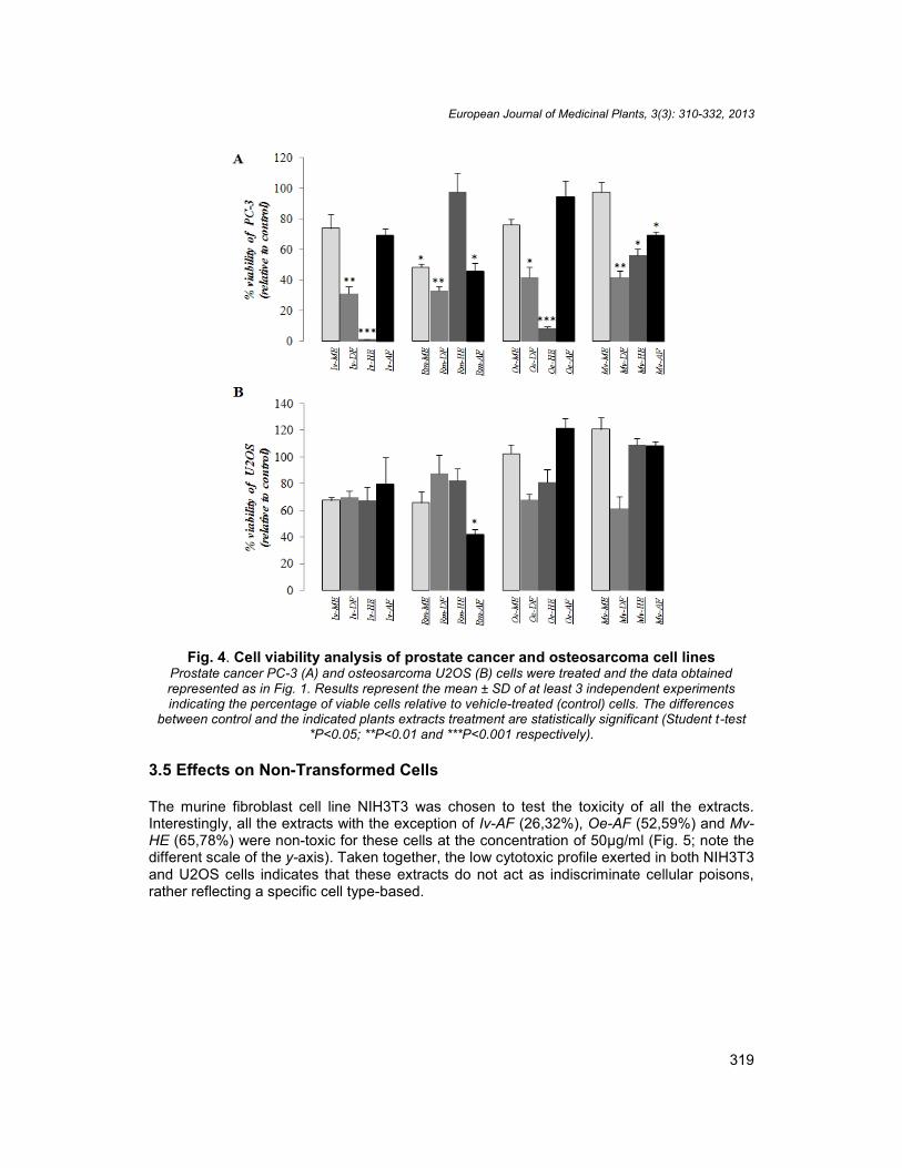
The effect of the different extracts was also tested in the PC-3 Prostate cancer cell line andin U2OS Osteosarcoma cells, which were exposed to the same conditions described above(Fig. 4). Fig. 4A illustrates that only Iv-HE (1,2%) and Oe-HE (8,2%) were active against PC-3 cells, although they both induced a dramatic effect on these cells. Unfortunately,conventional chemotherapeutic drugs have a discreet effect on prostate cancers and do notprovide a marked survival advantage for patients [33]. In contrast, in the Osteosarcoma cellline U2OS (Fig. 4B) almost none of the extracts exerted a significant activity at the testedconcentration, except Rm-AF (42, 3%). This could be explained by the fact that U2OS cellline is chromosomally highly altered.
European Journal of Medicinal Plants, 3(3): 310-332, 2013
319
Fig. 4. Cell viability analysis of prostate cancer and osteosarcoma cell linesProstate cancer PC-3 (A) and osteosarcoma U2OS (B) cells were treated and the data obtainedrepresented as in Fig. 1. Results represent the mean ± SD of at least 3 independent experimentsindicating the percentage of viable cells relative to vehicle-treated (control) cells. The differences
between control and the indicated plants extracts treatment are statistically significant (Student t-test*P<0.05; **P<0.01 and ***P<0.001 respectively).
3.5 Effects on Non-Transformed Cells
The murine fibroblast cell line NIH3T3 was chosen to test the toxicity of all the extracts.Interestingly, all the extracts with the exception of Iv-AF (26,32%), Oe-AF (52,59%) and Mv-HE (65,78%) were non-toxic for these cells at the concentration of 50μg/ml (Fig. 5; note thedifferent scale of the y-axis). Taken together, the low cytotoxic profile exerted in both NIH3T3and U2OS cells indicates that these extracts do not act as indiscriminate cellular poisons,rather reflecting a specific cell type-based.

European Journal of Medicinal Plants, 3(3): 310-332, 2013
320
Fig. 5. Cell viability analysis of mouse embryonic fibroblast cell lineNIH3T3 mouse fibroblasts were treated and the data obtained represented as in Fig. 1. Results
represent the mean ± SD of at least 3 independent experiments indicating the percentage of viablecells relative to vehicle-treated (control) cells. The differences between control and the indicated plants
extracts treatment are statistically significant (Student t-test *P<0.05).
3.6 Analysis of the IC50 of the Most Active Extracts in Sensitive Cell Lines
According to NCI (National Cancer Institute, U.S.A.) recommendations to consider a crudeextract promising for further purification, IC50 values need to be lower than 30μg/mL in orderto discover and develop potential anticancer natural compounds [35,36]. For this purposeincreasing concentrations ranging from 1μg/ml to 50μg/ml of selected extracts were thentested and the IC50 values were calculated. All tested extracts were able to reduce cellviability in a dose-response manner after treatment. All determined IC50 values for the mostactive extracts in sensitive cell lines are included in Table 3. In most cases the IC50 valueswere under 30μg/ml, and specifically Iv-DF, Iv- HE, Rm-DF; Rm-HE; Oe-DF; Oe-HE and Mv-DF showed the most promising activities in this assay.

European Journal of Medicinal Plants, 3(3): 310-332, 2013
321
Table 3. IC50 values of extracts (ME, HE) and fractions (DF, AF) of Inulaviscosa (Iv), Retamamonosperma (Rm),Ormeniseiriolepis (Oe) and Marrubiumvulgare (Mv) against selected cell lines. Jurkat, Jeko-1, LN229, T98G, U87MG, SW620,SW480, U2OS and PC-3 were exposed to increasing concentration fromdifferent extracts range 1µg/mlto 50 µg/ml for 48h.The IC50 values were determined only for the most active and non toxic extracts exhibiting a significant cell death effect
usingGraphPad Prism 5 software and Data are expressed as IC50 ± SD of three independent experiments
Organic Extracts IC 50(µg/ml) ±SDJurkat Jeko-1 LN229 T98 U87 SW620 SW480 U2OS PC-3
Inula viscose L. Ait.
Iv-MEIv-DFIv-HEIv-AF
25,81±2,9211,16±6,957,52±4,57NA
>508,68±3,1<1NA
NA17,66±5,45,9±3,57NA
NA20,50±2,21>50NA
>50>50NANA
NA23,30±6,338,40±3,31NA
>50>5010,36±6,59NA
>50>50>50NA
NA20,15±9,096,53±1,42NA
Retama monospermaL.Bois.Rm-MERm- DFRm- HERm- AF
>509,12±4,2034,44±3,3820,22±10,51
21,47±3,0524,77±4,85NA12,01±5,14
NA>50NA>50
NA>50NA>50
NA47,85±4,08NA>50
NANANA>50
NA>50NA>50
>50NANANA
>5045,41±4,6NA>50
Ormenis eriolepisCoss.Oe-MEOe- DFOe- HEOe- AF
27,45±4,0311, 33±5,379,81±8,44NA
<113,63±1,67<1NA
NA21,70±2,3741,67±1,98NA
NA>50>50NA
NA>50>50NA
>50>50NANA
>50>5019,31±4,88NA
NA>50NANA
NA>5012,59±4,69NA
Marrubium vulgare L.Mv- MEMv- DFMv- HEMv- AF
NA11,54±2,83NANA
>506,04±2,77NANA
NA>50NANA
NA>50NANA
NA>50NANA
NA10,58±4,37NANA
NANANANA
NA>50NANA
NA>50NANA
NA: Not active; >50: above 50µg /ml; <1: below 1µg/ml

European Journal of Medicinal Plants, 3(3): 310-332, 2013
322
3.7 Analysis of the Cellular Effects of the Most Active Extracts in LN229Glioblastoma Cells
Four extracts have shown a robust growth inhibitory activity against LN229 cells: Iv- DF, Iv-HE, Oe-DF and Oe-HE. We used these extracts at 50 μg/ml to treat LN229 cells for 24 and48h and investigate their cellular effects. We observed that all extracts clearly induced cellshrinkage and floating cell formation, which are suggestive of cell death (Fig. 6A). We nextperformed flow cytometry following propidium iodide staining to quantitate the cell cycledistribution after treatment (Fig. 6B). Iv-DF causes a cell cycle arrest in G1 at 24h with asignificant increase (46,17%) of the sub- G1 population at 48h. Similarly, Iv-HE induced acell cycle arrest in G1 at 24h and increased the proportion of sub-G1 cells (43,22%) at 48h.Interestingly, Oe-DF seems to induce a G2/M arrest at 24h, followed by an increase in sub-G1 cell population (24,2%) at 48h (Fig. 6B), whereas Oe-HE mainly exerts a combination ofG1 arrest and sub- G1 increase at 24h, with this latter population further increasing at 48hup to 42% (Fig. 6B). We next performed double Annexin V/Propidium Iodide stainingfollowing treatment with the selected extracts for 24h and 48h. In agreement with ourprevious results, cells shifted upon treatment in a time-dependent manner to regions mainlyindicative of early apoptosis, late apoptosis and necrosis, showing a total percentage of celldeath higher than 50% (Fig. 6C). We next performed caspase activity assays upon treatmentof LN229 cells with 50 μg/ml of each extract for 48h. Oe-DF seemed to induce the higherincrease in caspase activity, although a parallel increase in the proportion of apoptotic, asopposed to necrotic, cells was not detected by flow cytometry. This discrepancy may reflectan underestimation of the percentage of apoptotic cells detected by Annexin V staining.Altogether, the results obtained with LN229 cells are in agreement with our previous datashowing that these cells were the most sensitive Glioblastoma cell line, compared with T98Gand U87MG cells. Therefore, active compounds from Inula viscosa and Ormenis eiriolepisextracts may be attractive therapeutic agents; specially in Glioblastoma cells that presentmultiple alterations at different levels including loss of heterozygoty (LOH), inactivatingmutations, methylation or altered expression of cancer-relevant signaling intermediates suchas bcl-2 family members, inhibitors of apoptosis (IAPs) or receptor tyrosine kinases like theepidermal growth factor receptor(EGFR) and their down-stream effectors [30].

European Journal of Medicinal Plants, 3(3): 310-332, 2013
323
Fig. 6. Cellular effects analysis of the most active extracts on LN229 glioblastoma cellline
(A) images of morphological changes of LN229 cells under treatment with 50µg/ml of Iv-DF, Iv-HE, Oe-DF and Oe-HE for 24h and 48h (magnification 5×) (B) cell cycle analysis of LN229 cells treated same
as in (A). (C) cell death analysis of LN229 at 24h and 48h under the same conditions as in (A) and(B).(D) caspase activity analysis of LN229 cells treated with Iv-DF, Iv-HE, Oe-DF and Oe-HE for 48h.
Results show fold induction respective to untreated cells. Data is means ± SEM from threeindependent determinations performed in duplicate.
C

European Journal of Medicinal Plants, 3(3): 310-332, 2013
324
3.8 Analysis of the Cellular Effects of the Most Active Extracts in SW620Colon Cancer Cells
Viability assays in colon cancer cells indicated that SW620 cells were the most sensitive toIv-DF, Iv-HE and Mv-DF, which all exhibited clear cytotoxic effects (Fig. 3A). We thus usedthe same experimental approach as with LN229 cells to investigate if those extracts mayaffect SW620 cell morphology, cell cycle distribution and cellular death. Cell shrinkage andfloating cell formation were observed upon treatment suggesting that Iv-DF, Iv-HE and Mv-DF were inducing cell death (Fig. 7A). Cell cycle analysis upon propidium iodide stainingrevealed that Iv-DF, Iv-HE and Mv-DF were able to induce cell cycle arrest in G1 at 24h anda clear increase in the sub-G1 population at 48h, with 39%, 12% and 21% cells in sub-G1,respectively (Fig. 7B). Annexin V/Propidium Iodide staining was then performed to betteranalyze cellular death. Treatment with all extracts dramatically increased cellular necrosis,without the presence of a detectable apoptotic cell fraction as indicated by the low numbersof Annexin V-positive cells (Fig. 7C). The percentages of necrotic cells at 48h were 42%,79% and 22% for Iv-DF, Iv-HE and Mv-DF, respectively. Surprisingly, however, Iv-HEspecifically induced a dramatic induction of caspase activity (30-fold) which again suggeststhat the detection of Annexin V-positive cells in these assays may not accurately reflectcaspase-dependent apoptosis (Fig. 7D). In comparison with SW480 cells, their nonmetastatic counterpart, SW620 cells have acquired genetic defects both in the intrinsic andextrinsic pathways of apoptosis and are thus more resistant to apoptosis induced by CH-11,cisplatin, and ionizing radiation, respectively [32]. In our study SW620 cells were sensitive toInula viscosa and Marrubium vulgare extracts, suggesting that those plants may lead to newtherapeutic molecules that could challenge the acquired mechanisms leading to tumor cellresistance to current cytotoxic drugs in colon cancer.

European Journal of Medicinal Plants, 3(3): 310-332, 2013
325
European Journal of Medicinal Plants, 3(3): 310-332, 2013
325
European Journal of Medicinal Plants, 3(3): 310-332, 2013
325

European Journal of Medicinal Plants, 3(3): 310-332, 2013
326
Fig. 7. Cellular effects analysis of the most active extracts on SW620 colon cancercells
(A) images of morphological changes of SW620 cells under treatment with 50µg/ml of Iv-DF, Iv-HE andMv-DF for 24h and 48h (magnification 5×) (B) cell cycle analysis of SW620 cells treated same as in
(A).(B) cell cycle analysis of SW620 cells treated same as in (A). (C) cell death analysis of SW620 at24h and 48h under the same conditions as in (A) and (B). (D) caspases activity analysis of SW620
cells treated with Iv-DF, Iv-HE and Mv-DF for 48h. Results show fold induction respective to untreatedcells. Data is means ± SEM from three independent determinations performed in duplicate.
3.9 Analysis of the Cellular Effects of the Most Active Extracts in PC-3Prostate Cancer Cells
The same experiments were next performed in PC-3 prostate cancer cells to figure out if themost active extracts in viability assays could also affect cell morphology, cell cycleprogression and cell death. Morphological changes were observed in PC-3 cells upontreatment with Iv-DF, Iv-HE, Rm-DF and Oe-HE extracts for 24h and 48h. Rounding up andcell shrinking, with a clear reduction of cellular and nuclear volume were observed (Fig. 8A).Flow cytometry analysis of cell cycle distribution and cell death upon Iv- DF, Iv-HE, Rm-DFand Oe-HE treatment revealed that the four extracts induced a clear G2/M arrest which wasalready detectable at 24h leading at 48h to a G2/M cell population of 41,83%, 53,75%,48,67% and 44.11%, respectively (Fig. 8B). The results obtained with Annexin V/Propidiumeiodide indicate that only Iv-HE and, more robustly, Oe-HE were able to induce cell death inPC-3 cells, reaching at 48h a percentage of 25% and 29% of cell death, respectively.Interestingly, in Oe-HE-treated cells the results primarily indicate apoptotic cell death.Accordingly, the results of caspase assays showed the highest increase of caspase activityin Iv-HE and Oe-HE treated cells. PC-3 prostate cancer cells are androgen-insensitive andapoptosis-resistant; they survive and eventually develop into androgen-refractory andmetastatic clones [33]. Our data suggest that Inula viscosa and Ormenis eiriolepis extractsexert growth inhibition, cell cycle arrest and apoptosis induction and could contain activecompounds with the potential to effectively target apoptosis extrinsic and intrinsic pathways.Therefore, more studies are clearly needed to clarify the molecular mechanisms involved.

European Journal of Medicinal Plants, 3(3): 310-332, 2013
327

European Journal of Medicinal Plants, 3(3): 310-332, 2013
328
Fig. 8. Cellular effects analysis of the most active extracts on PC-3 prostate cancercells
(A) images of morphological changes of PC-3 cells under treatment with 50µg/ml of Iv-DF,Iv-HE , Rm-DF and Oe-HE for 24h and 48h (magnification 5×). (B) cell cycle analysis of PC-
3 cells treated same as in (A). (C) cell death analysis of PC-3 at 24h and 48h under thesame conditions as in (A) and (B). (D) caspases activity analysis of PC-3 cells treated with
Iv-DF, Iv-HE , Rm-DF and Oe-HE for 48h. Results show fold induction respective tountreated cells. Data is means ± SEM from three independent determinations performed in
duplicate.
4. CONCLUSION
In agreement with our data, the antitumoral activity of the hexanic extract and thedichloromethane fraction of Inula viscosa and Retama monosperma were also observed inthe cervical cancer cell lines HeLa and SiHa, where they exhibited a high level of cytotoxityby inducing apoptosis following pro-caspase activation, Bcl-2 expression and PARPcleavage [16]. The chemical composition of Inula viscosa and Retama monosperma extractshas already been investigated. Numerous studies have reveale d the presence of asesquiterpene acid (isocostic acid) and two sesquiterpenes lactones: tomentosin andinuviscolide, as major compounds in Inula viscosa extract [16]. Indeed, this plant is a sourceof a number of bioactive compounds as well as flavonoids [13] and sesquiterpenederivatives [37]. However, the dichloromethane fraction of Retama monosperma revealedthe presence of five known quinolizidine alkaloids together with sparteine, L-methyl cytisine,17-oxosparteine, lupanine and anagyrine [16]. The Retama species have been reported tocontain alkaloids [38] and flavonoids [39]. Accordingly, 15 quinolizidine and 3 dipiperidinealkaloids were isolated from the leaves of flowering plants of R. monosperma collected fromMorocco [40]. The compositional analysis of the aqueous infusion of Marrubium vulgare hasrevealed the presence of fifteen metabolites, all belonging to the class of polyphenols.Particularly, seven flavonoids have been detected, together with 5-caffeoylquinic(chlorogenic) acid in small amounts; the extract is dominated by the presence of a series ofcomplex molecules, characterized as verbascoside (acteoside) derivatives [26]. However,the chemical constituents of Ormenis eiriolepis still remain to be investigated. The selectedspecies are reported to have both preventive and therapeutic properties. According to ourfindings, the use of these plants by traditional healers for the treatment of cancer patients
European Journal of Medicinal Plants, 3(3): 310-332, 2013
329
could have some scientific support. Because cancer drug-resistance is a significant healthproblem, the screening of medicinal plants for new anticancer agents remains a priority indrug discovery programs. The herein reported biological activity of medicinal plants used intraditional Moroccan medicine provides a starting point for forthcoming studies to determinethe molecular basis of their activity and to identify the chemical compounds. Within the mostactive extracts responsible for their antitumoral effects.
CONSENT
Not applicable.
ETHICAL APPROVAL
Not applicable.
ACKNOWLEDGEMENTS
We are grateful to Joan Seoane (Institut de Recerca Hospital Vall d’Hebron, Barcelona), forthe gift of human glioma cell lines and to Beatriz Martínez (CNIO, Madrid) and Joan Gil(IDIBELL, Barcelona) for the gift of Jeko-1 and Jurkat cells, respectively. L.B. is a fellow ofthe Averroes mobility Programme (European Comission). P.V. is a “Ramón y Cajal” Fellow(Ministerio de Ciencia e Innovación, Spain).
DECLARATION OF COMPETING INTEREST
The authors declare that there are no competing interests.
REFERENCES
1. Ameenah Gurib-Fakim. Medicinal plants: Traditions of yesterday and drugs oftomorrow. Molecular Aspects of Medicine. 2006;27(1):1-93.
2. João B. Calixto, Twenty-five years of research on medicinal plants in Latin America: Apersonal view, Journal of Ethnopharmacology. 2005;100(1–2):131-134.
3. Kabbaj F, Meddah B, Cherrah Y, Faouzi E. Ethnopharmacological profile of traditionalplants used in Morocco by cancer patients as herbal therapeutics.Phytopharmacology. 2012;2(2):243-256.
4. Mark SB. The role of natural product chemistry in drug discovery. Journal of NaturalProducts. 2004;67(12):2141–2153.
5. David JN, Gordon MC, Kenneth MS. The influence of natural products upon drugdiscovery. Natural Product Reports. 1999;17(3):215–234.
6. Arvind Saklani, Samuel K. Kutty. Plant-derived compounds in clinical trials. DrugDiscovery Today. 2008;13(3–4):161-171.
7. Lautié E, Quintero R, Fliniaux M-A, Villarreal M-L. Selection methodology with scoringsystem: Application to Mexican plants producing podophyllotoxin related lignans.Journal of Ethnopharmacology. 2008;120(3):402-412.
8. Jouad H, Haloui M, Rhiouani H, El Hilaly J, Eddouks M. Ethnobotanical survey ofmedicinal plants used for the treatment of diabetes, cardiac and renal diseases in theNorth centre region of Morocco (Fez–Boulemane), Journal of Ethnopharmacology,October 2001;77 (2–3):175-182.

European Journal of Medicinal Plants, 3(3): 310-332, 2013
330
9. Jamal Bellakhdar, Renée Claisse, Jacques Fleurentin, Chafique Younos, Repertory ofstandard herbal drugs in the Moroccan pharmacopoeia. Journal ofEthnopharmacology. 1991;35(2):123-143.
10. Benrahmoune I.Z. &Dubruille C., 2003. Invitation à l'amour des plantes. Guidefloristique illustré de la réserve biologique de Sidi Bou Ghaba. Editions Scriptura,Rabat; 2003.
11. Maoz M, Neeman I. Effect of Inula viscosa extract on chitin synthesis indermatophytes and Candida albicans. Journal of Ethnopharmacology. 2000;71(3):479-482.
12. Cafarchia C, De Laurentis N, Milillo MA, Losacco V, Puccini V, Antifungal activity ofessential oils from leaves and flowers of Inula viscosa (Asteraceae) by Apulian region.Parassitologia. 2002;44:153-156.
13. Hernández V, Carmen Recio M, Máñez S, Rosa Giner M, Ríos J-L, Effects of naturallyoccurring dihydroflavonols from Inulaviscosa on inflammation and enzymes involved inthe arachidonic acid metabolism, Life Sciences. 2007;81(6):480-488.
14. Rozenblat S, Grossman S, Bergman M, Gottlieb H, Cohen Y, Dovrat S, Induction ofG2/M arrest and apoptosis by sesquiterpene lactones in human melanoma cell lines,Biochemical Pharmacology. 2008;75(2):369-382.
15. Merghoub N, Benbacer L, Amzazi S, Morjani H, El mzibri M, Cytotoxic effect of someMoroccan medicinal plant extracts on human cervical cell lines, Journal of MedicinalPlants Research. 2009;3:1045–1050.
16. Benbacer L, Merghoub N, El Btaouri H, Gmouh S, Attaleb M, Morjani H, Amzazi S andEl Mzibri M. Antiproliferative Effect and Induction of Apoptosis by Inulaviscosa L. andRetamamonosperma L. Extracts in Human Cervical Cancer Cells, Topics on CervicalCancer With an Advocacy for Prevention, Dr. R. Rajamanickam (Ed.), 2012; ISBN:978-953-51-0183-3.
17. El Hanbali F, Mellouki F, Akssira M, Balasquez A. Etude Comparative desCompositions Chimiques et de l’Activité Antibactérienne des Huiles Essentielles deChamaemelumeriolepis Maire et ChamaemelumAfricanaJ. & Four. 1erCongrèsMaroco-Espagnol sur la Chimie Organique et 4ème Rencontre Andalou-Marocaine sur la Chimie des Produits Naturels 16-18 sept 2004.
18. El Hanbali F, Mellouki F, Akssira M, Balasquez A. Etude Activité antileishmaniennedeshuiles essentielles de deux camomilles:Ormeniseriolepis et Ormenisafricana.International Congress On Medicinal Plants, Errachidia16-19 March 2005.
19. Hamani, Rouhi R, Idrissi Hassani LM. Étude anatomique et screening phytochimiquede trois espèces du sud marocain: Chamaecytisus mollis, Retama monosperma etHesperolaburnum platycarpum. Détermination du potentiel antifongique des extraitsaqueux. The 3rd International Symposium on Aromatic and Medicinal Plants(SIPAM3).The first International symposium on Bioactive Molecules (CIMB1) Oujda –Morocco 29- 30 May 2008.
20. Schlemper V, Ribas A, Nicolau M, CechinelFilho V, Antispasmodic effects ofhydroalcoholic extract of Marrubiumvulgare on isolated tissues, Phytomedicine.1996;3(2):211-216.
21. De Jesus R.A.P, Cechinel-Filho V, Oliveira A.E, Schlemper V, Analysis of theantinociceptive properties of marrubiin isolated from Marrubiumvulgare,Phytomedicine. 2000;7(2):111-115.
22. Meyre-Silva C, Yunes R.A, Schlemper V, Campos-Buzzi F, Cechinel-Filho V,Analgesic potential of marrubiin derivatives, a bioactive diterpene present inMarrubium vulgare (Lamiaceae), Il Farmaco. 2005;60(4):321-326.

European Journal of Medicinal Plants, 3(3): 310-332, 2013
331
23. Berrougui H, Isabelle M, Cherki M, Khalil A, Marrubium vulgare extract inhibits human-LDL oxidation and enhances HDL-mediated cholesterol efflux in THP-1 macrophage,Life Sciences. 2006;80(2):105-112.
24. Stulzer HK, Tagliari MP, Zampirolo JA, Cechinel-Filho V, Schlemper V,Antioedematogenic effect of marrubiin obtained from Marrubiumvulgare, Journal ofEthnopharmacology. 2006;108(3):379-384.
25. Elberry AA, Harraz FM, Ghareib SA, Gabr SA, Nagy AA, Methanolic extract ofMarrubiumvulgare ameliorates hyperglycemia and dyslipidemia in streptozotocin-induced diabetic rats, International Journal of Diabetes Mellitus, Available online 52011; ISSN 1877-5934, 10.1016/j.ijdm.2011.01.004.
26. Boudjelal A, Henchiri C, Siracusa L, Sari M, Ruberto G, Compositional analysis and invivo anti-diabetic activity of wild Algerian Marrubiumvulgare L. infusion, Fitoterapia.2012;83(2):286-292.
27. Erdogan Orhan I, Belhattab R, Şenol FS, Gülpinar AR, Hoşbaş S, Kartal M. Profiling ofcholinesterase inhibitory and antioxidant activities of Artemisia absinthium, A. herba-alba, A. fragrans, Marrubium vulgare, M. astranicum, Origanum vulgare subsp.glandulossum and essential oil analysis of two Artemisia species, Industrial Crops andProducts. 2010;32(3):566-571.
28. Pukalskas A, Rimantas Venskutonis P, Salido S, de Waard P, van Beek T A, Isolation,identification and activity of natural antioxidants from horehound (Marrubium vulgareL.) cultivated in Lithuania, Food Chemistry. 2012;130(3):695-701.
29. Eisele G, Weller M. Targeting apoptosis pathways in glioblastoma, Cancer Letters.2013;332(2):335-45.
30. Baguley BC, Hicks KO, Wilson WR, Chapter 15 - Tumor cell cultures in drugdevelopment, In: Bruce C. Baguley and David J. Kerr, Editor(s), Anticancer DrugDevelopment, Academic Press, San Diego. 2002;269-284.
31. Nathalie Droin, Leslie Guéry, Naïma Benikhlef, Eric Solary, Targeting apoptosisproteins in hematological malignancies, Cancer Letters.2013;332(2):325-34.
32. Huerta S, Heinzerling JH, Anguiano-Hernandez YM, Huerta-Yepez S, Lin J, Chen D,Bonavida B, Livingston EH. Modification of Gene Products Involved in Resistance toApoptosis in Metastatic Colon Cancer Cells: Roles of Fas, Apaf-1, NFκB, IAPs,Smac/DIABLO, and AIF, Journal of Surgical Research. 2007;142(1):184-194.
33. Huang YT, Huang DM, Chueh SC, Teng CM, Guh JH, Alisol B acetate, a triterpenefrom Alismatis rhizoma, induces Bax nuclear translocation and apoptosis in humanhormone-resistant prostate cancer PC-3 cells, Cancer Letters. 2006;231(2):270-278.
34. Jain R, Kumar Jain S, Screening of in vitro cytotoxic activity of some medicinal plantsused traditionally to treat cancer in Chhattisgarh state, India, Asian Pacific Journal ofTropical Biomedicine. 2011;1(2):147-150.
35. de Mesquita M L, Ede Paula J, Pessoa C, de Moraes MO, L Veras, Grougnet R, et al,Cytotoxic activity of Brazilian Cerrado plants used in traditional medicine againstcancer cell lines, Journal of Ethnopharmacology. 2009;123(3):439-445.
36. Steenkamp V, Gouws MC, Cytotoxicity of six South African medicinal plant extractsused in the treatment of cancer, South African Journal of Botany. 2006;72(4):630-633.
37. Fontana G, La Rocca S, Passannanti S, Paternostro MP. Sesquiterpene compoundsfrom Inulaviscosa, Natural Product Research. 2007;20-21(9):824-827.
38. Abdel Halim OB, Abdel Fattah H, Halim A.F, Murakoshi I. Comparative chemical andbiological studies of the alkaloidal content of Lygos species and varieties growing inEgypt, Acta Pharmceutica Hungarica. 1997;67:241-247.

European Journal of Medicinal Plants, 3(3): 310-332, 2013
332
39. Kassem M, Mosharrafa SA, Saleh NA, Abdel-Wahab SM. Two new flavonoids fromRetama raetam. Fitoterapia. 2000;71:649-654.
40. Touati D, Allain P, Pellecuer J, Fkih-Tetouani SA. Alkaloids from Retama monospermassp. Eumonosperma. Fitoterapia. 1996;67:49-52.
_________________________________________________________________________© 2013 Belayachi et al.; This is an Open Access article distributed under the terms of the Creative CommonsAttribution License (http://creativecommons.org/licenses/by/3.0), which permits unrestricted use, distribution, andreproduction in any medium, provided the original work is properly cited.
Peer-review history:The peer review history for this paper can be accessed here:
http://www.sciencedomain.org/review-history.php?iid=223&id=13&aid=1293