screening for lung cancer: chest guideline and expert
TRANSCRIPT

Accepted Manuscript
Screening for Lung Cancer: CHEST Guideline and Expert Panel Report
Peter J. Mazzone, MD, MPH, FCCP, Gerard A. Silvestri, MD, MS, FCCP, SheenaPatel, MPH, Jeffrey P. Kanne, MD, FCCP, Linda S. Kinsinger, MD, Renda SoylemezWiener, MD, MPH, Guy Soo Hoo, MD, FCCP, Frank C. Detterbeck, MD, FCCP
PII: S0012-3692(18)30094-1
DOI: 10.1016/j.chest.2018.01.016
Reference: CHEST 1523
To appear in: CHEST
Received Date: 24 October 2017
Revised Date: 20 December 2017
Accepted Date: 10 January 2018
Please cite this article as: Mazzone PJ, Silvestri GA, Patel S, Kanne JP, Kinsinger LS, Wiener RS, SooHoo G, Detterbeck FC, Screening for Lung Cancer: CHEST Guideline and Expert Panel Report, CHEST(2018), doi: 10.1016/j.chest.2018.01.016.
This is a PDF file of an unedited manuscript that has been accepted for publication. As a service toour customers we are providing this early version of the manuscript. The manuscript will undergocopyediting, typesetting, and review of the resulting proof before it is published in its final form. Pleasenote that during the production process errors may be discovered which could affect the content, and alllegal disclaimers that apply to the journal pertain.
MANUSCRIP
T
ACCEPTED
ACCEPTED MANUSCRIPT
Word Count: 14,116
Screening for Lung Cancer: CHEST Guideline and Expert Panel Report
Peter J Mazzone, MD, MPH, FCCP; Gerard A Silvestri, MD, MS, FCCP; Sheena Patel, MPH; Jeffrey
P Kanne, MD, FCCP; Linda S Kinsinger, MD; Renda Soylemez Wiener, MD, MPH; Guy Soo Hoo,
MD, FCCP; Frank C. Detterbeck, MD, FCCP
Affiliations: Respiratory Institute, Cleveland Clinic (Dr Mazzone), Cleveland, OH; Medical
University of South Carolina (Dr Silvestri), Charleston, SC; CHEST (Ms Patel), Glenview, IL;
University of Wisconsin School of Medicine and Public Health (Dr Kanne), Madison, WI; VHA
National Center for Health Promotion and Disease Prevention (Dr Kinsinger), Durham, NC;
Center for Healthcare Organization & Implementation Research, Edith Nourse Rogers Memorial
VA Hospital, Bedford, MA and Boston University School of Medicine (Dr Wiener), Boston, MA;
VA Greater Los Angeles Healthcare System (Dr Soo Hoo), Los Angeles, CA; Yale University (Dr
Detterbeck), New Haven, CT
Conflicts of Interest: (see e-Table 1)
Funding/Support: This study was funded in total by internal funds from the American College of Chest Physicians.
Disclaimer: CHEST Guidelines are intended for general information only, are not medical
advice, and do not replace professional medical care and physician advice, which always should
be sought for any medical condition. The complete disclaimer for this guideline can be accessed
at http://www.chestnet.org/Guidelines-and-Resources
Correspondence to: Peter J Mazzone, MD, MPH, FCCP, Respiratory Institute, Cleveland Clinic,
9500 Euclid Ave, A90, Cleveland, OH 44195; e-mail: [email protected]

MANUSCRIP
T
ACCEPTED
ACCEPTED MANUSCRIPT
Abstract
Background: Low-dose chest CT screening for lung cancer has become a standard of care in the United
States in the past few years, in large part due to the results of the National Lung Screening Trial. The
benefit and harms of low-dose chest CT screening differ in both frequency and magnitude. The
translation of a favorable balance of benefit and harms into practice can be difficult. Here, we update
the evidence base for the benefit, harms, and implementation of low radiation dose chest CT screening.
We use the updated evidence base to provide recommendations where the evidence allows, and
statements based on experience and expert consensus where it does not.
Methods: Approved panelists developed key questions using the PICO (population, intervention,
comparator, and outcome) format to address the benefit and harms of low-dose CT screening, as well as
key areas of program implementation. A systematic literature review was conducted using MEDLINE via
PubMed, Embase, and the Cochrane Library. Reference lists from relevant retrievals were searched, and
additional papers were added. The quality of the evidence was assessed for each critical or important
outcome of interest using the GRADE approach. Important clinical questions were addressed based on
the evidence developed from the systematic literature review. Graded recommendations and un-
graded statements were drafted, voted on, and revised until consensus was reached.
Results: The systematic literature review identified 59 studies that informed the response to the 12
PICO questions that were developed. Key clinical questions were addressed resulting in 6 graded
recommendations and 9 ungraded consensus based statements.
Conclusions: Evidence suggests that low-dose CT screening for lung cancer results in a favorable but
tenuous balance of benefit and harms. The selection of screen-eligible patients, the quality of imaging
and image interpretation, the management of screen detected findings, and the effectiveness of
smoking cessation interventions, can impact this balance. Additional research is needed to optimize the
approach to low-dose CT screening.

MANUSCRIP
T
ACCEPTED
ACCEPTED MANUSCRIPT
Abbreviations ACR = American College of Radiology AHRQ = Agency for Healthcare Research and Quality CCI = Charlson Comorbidity Index CHEST = American College of Chest Physicians CISNET = Cancer Intervention and Surveillance Modeling Network CMS = Centers for Medicare and Medicaid Services COI = Conflict of interest COPD = Chronic obstructive pulmonary disease CT = Computed Tomography CXR = Chest radiograph (x-ray) DANTE = Detection and Screening of Early Lung Cancer by Novel Imaging Technology and Molecular Essays Trial DLCST = Danish Lung Cancer Screening Trial FDG-PET = Fluorodeoxyglucose – Positron emission tomography GDT = Guideline Development Tool GOC = Guidelines Oversight Committee GRADE = Grading of Recommendations, Assessment, Development, and Evaluation HR = Hazard ratio ITALUNG = Italian Lung Cancer Screening Trial LDCT = Low-Dose Computed Tomography LUSI = German Lung Cancer Screening Intervention Trial LSS = Lung Screening Study MD = Mean difference MILD = Multi-centric Italian Lung Detection Trial NELSON = Nederlands-Leuvens Longkanker Screenings Onderzoek Study NLST = National Lung Screening Trial NSCLC = Non-small Cell Lung Cancer PICO = Population, Intervention, Comparator, Outcome PSC = Professional Standards Committee QALY = Quality-adjusted life year RCT = Randomized controlled trial RR = Risk ratio SEER = Surveillance, Epidemiology, and End Results STR = Society of Thoracic Radiology UKLS = United Kingdom Lung Screening Study USPSTF = United States Preventative Services Task Force VA = Veterans Affairs

MANUSCRIP
T
ACCEPTED
ACCEPTED MANUSCRIPT
SUMMARY OF RECOMMENDATIONS
1. For asymptomatic smokers and former smokers age 55 to 77 who have smoked 30 pack years or
more and either continue to smoke or have quit within the past 15 years, we suggest that annual
screening with low-dose CT should be offered. (Weak recommendation, moderate-quality evidence)
Remark: Age 77 represents the oldest age of participants in the NLST at the end of the screening period.
Age 77 also matches the oldest age of CMS coverage for low-dose CT screening. Age 80 has been
recommended by the USPSTF based on modeling studies. Recommendation #2 can be applied to
individuals age 78 to 80.
Remark: Asymptomatic refers to the absence of symptoms suggesting the presence of lung cancer.
2. For asymptomatic smokers and former smokers who do not meet the smoking and age criteria in
Recommendation #1 but are deemed to be at high risk of having/developing lung cancer based on
clinical risk prediction calculators, we suggest that low-dose CT screening should not be routinely
performed. (Weak recommendation, low-quality evidence)
Remark: It is recognized that clinical risk prediction calculators may be slightly more efficient at
identifying individuals who have or will develop lung cancer than the eligibility criteria listed in
Recommendation #1. It is also recognized that the variables included in the clinical risk prediction
calculators are risk factors for morbidity from the evaluation and treatment of screen detected findings,
and death from any cause. Thus a cohort at high risk for lung cancer based on a clinical risk prediction
calculator may be less likely to benefit and more likely to be harmed by lung cancer screening than the
cohort identified by the eligibility criteria listed in Recommendation #1. Thus, we do not believe the
evidence supports a policy to screen this group.
Remark: It is also recognized that there will be individuals within the cohort deemed to be at high risk
for lung cancer from a clinical risk prediction calculator who are healthy enough to benefit from lung
cancer screening, and that low-dose CT screening could be considered in these individuals.
Remark: A risk threshold of 1.51% over 6 years on the PLCOm2012 calculator is an example of high risk.
Remark: In the United States, health insurance providers may not pay for low-dose CT screening for
those who do not meet the eligibility criteria listed in Recommendation #1.
Remark: Additional lung cancer screening trials that include patients who do not meet the eligibility
criteria listed in Recommendation #1 but have a high risk of having/developing lung cancer based on
clinical risk prediction calculators are needed.
3. For individuals who have accumulated fewer than 30 pack years of smoking or are younger than age
55 or older than 77, or have quit smoking more than 15 years ago, and do not have a high risk of
having/developing lung cancer based on clinical risk prediction calculators, we recommend that low-
dose CT screening should not be performed. (Strong recommendation, moderate-quality evidence)
4. For individuals with comorbidities that adversely influence their ability to tolerate the evaluation
of screen detected findings, or tolerate treatment of an early stage screen detected lung cancer, or

MANUSCRIP
T
ACCEPTED
ACCEPTED MANUSCRIPT
that substantially limit their life expectancy, we recommend that low-dose CT screening should not be
performed. (Strong recommendation, low-quality evidence)
Remark: At very severe stages of a comorbid condition it can be clear that low-dose CT screening is not
indicated (e.g. advanced liver disease, COPD with hypoventilation and hypoxia, NYHA class IV heart
failure) because competing mortality limits the potential benefit, and harms are magnified. At less
severe stages it can be difficult to determine if an individual’s comorbidities are significant enough that
they should not receive low-dose CT screening. Further research is required to assist clinicians with this
decision.
5. We suggest that low-dose CT screening programs develop strategies to determine whether patients
have symptoms that suggest the presence of lung cancer, so that symptomatic patients do not enter
screening programs but instead receive appropriate diagnostic testing, regardless of whether the
symptomatic patient meets screening eligibility criteria. (Ungraded Consensus-Based Statement)
Remark: In centralized low-dose CT screening programs, the provider that meets with the patient prior
to the low-dose CT should ask about symptoms that would suggest diagnostic testing is indicated.
Remark: In de-centralized low-dose CT screening programs, the screening program should assist the
ordering provider through educational outreach and/or the provision of clinical tools (e.g. reminders
built into electronic medical records).
6. We suggest that screening programs define what constitutes a positive test on the low-dose CT
based on the size of a detected solid or part-solid lung nodule, with a threshold for a positive test that
is either 4 mm, 5 mm, or 6 mm in diameter. (Weak recommendation, low-quality evidence)
Remark: A positive test is defined as a test that leads to a recommendation for any additional testing
other than to return for the annual screening exam.
Remark: Nodule diameter is the average of long- and short-axis diameters obtained on the same
sagittal, coronal, or transverse image. For part-solid nodules, nodule diameter should be based on the
size of the solid component of the nodule.
Remark: An equivalent volumetric threshold can also be considered.
Remark: The LungRADS structured reporting system currently uses 6 mm at the baseline scan and 4 mm
if a new nodule is found on the annual scan for solid nodules; and 6 mm at the baseline scan and any
size if a new nodule is found on the annual scan for part-solid nodules.
7. We suggest that low-dose CT screening programs develop strategies to maximize compliance with
annual screening exams. (Ungraded Consensus-Based Statement)
Remark: Additional research is needed to better understand the factors that influence compliance, and
to develop tools to help screening programs maximize compliance with annual screening exams.
8. We suggest that low-dose CT screening programs develop a comprehensive approach to lung
nodule management, including multi-disciplinary expertise (Pulmonary, Radiology, Thoracic Surgery,
Medical and Radiation Oncology), and algorithms for the management of small solid nodules, larger
solid nodules, and sub-solid nodules. (Ungraded Consensus-Based Statement)

MANUSCRIP
T
ACCEPTED
ACCEPTED MANUSCRIPT
Remark: For programs without lung nodule management expertise available on site, collaborations with
centers capable of high quality lung nodule management can be formed (e.g. referral, distance
evaluation).
9. We suggest that low-dose CT screening programs develop strategies to minimize overtreatment of
potentially indolent lung cancers. (Ungraded Consensus-Based Statement)
Remark: It is important to educate patients about the potential to detect an indolent lung cancer to help
mitigate the psychological distress that could result from living with an indolent untreated lung cancer.
Remark: For malignant nodules, pure ground glass is the nodule morphology most likely to represent an
indolent cancer.
10. For current smokers undergoing low-dose CT screening, we recommend that screening programs
provide evidence-based tobacco cessation treatment as recommended by the US Public Health
Service. (Strong recommendation, low-quality evidence)
Remark: Further research about the ideal approach to tobacco treatment specific to the lung cancer
screening setting is needed.
11. We suggest that low-dose CT screening programs develop strategies to provide effective
counseling and shared decision-making visits prior to the performance of the LDCT screening exam.
(Ungraded Consensus-Based Statement)
Remark: Components of the counseling and shared decision making visit include a determination of
screening eligibility (e.g. age, smoking history, the absence of symptoms, confirmation of overall health),
the use of decision aids with information about benefits and harms of screening, a discussion about the
potential CT findings and need for follow-up testing, the need for annual screening exams, confirmation
of the willingness to accept treatment for a screen detected cancer, and counseling about smoking
cessation.
Remark: In centralized low-dose CT screening programs, a screening program provider may meet with
the patient prior to the low-dose CT to perform the counseling and shared decision-making visit.
Remark: In de-centralized low-dose CT screening programs, the screening program should ensure that
ordering providers are trained, and/or have the tools necessary, to deliver an effective counseling and
shared decision-making visit. These tools may include decision aids, information brochures, videos, and
links to electronic resources.
Remark: Additional research about the most effective way to conduct counseling and shared decision-
making visits is needed.
12. We suggest that low-dose CT screening programs follow the ACR/STR protocols for performing low
radiation dose chest CT scans. (Ungraded Consensus-Based Statement)
Remark: An awareness of the potential for radiation related harm can help programs thoughtfully plan
ways to minimize this risk through proper patient selection, the performance of the CT scan, and
appropriate management of screen detected findings.

MANUSCRIP
T
ACCEPTED
ACCEPTED MANUSCRIPT
13. We suggest that low-dose CT screening programs use a structured reporting system to report the
exam results. (Ungraded Consensus-Based Statement)
Remark: The structured reporting system should include a description of the number, location, size, and
characteristics of all lung nodules, guideline based recommendations for surveillance of small lung
nodules, and a description of other incidental findings.
Remark: The ACR LungRADS structured report is the most prevalent system used today. LungRADS
categories translate directly into data requests from the ACR National Registry.
14. We suggest that low-dose CT screening programs develop strategies to guide the management of
non-nodule findings. (Ungraded Consensus-Based Statement)
Remark: Examples include coronary artery calcification, thyroid nodules, adrenal nodules, kidney and
liver lesions, thoracic aortic aneurysms, pleural effusions, and parenchymal lung disease.
Remark: A lung cancer screening program should anticipate such incidental findings and have a system
in place to address them. Examples include evidence based guidance within the structured report to
assist the ordering provider, or centralized management of all incidental findings by the screening
program. Clear communication between providers is important to prevent misunderstandings about
who will assume responsibility for deciding what needs attention and ensuring appropriate follow-up
evaluation.
Remark: The wording of how incidental findings are reported should be systematically developed to
minimize anxiety and misunderstanding.
15. We suggest that low-dose CT screening programs develop data collection and reporting tools
capable of assisting with quality improvement initiatives and reporting to the current National
Registry. (Ungraded Consensus-Based Statement)
Remark: Data categories include patient eligibility criteria, imaging findings and their evaluation, results
of the evaluation of imaging findings including complications, smoking cessation interventions, and lung
cancer diagnoses including histology, stage, treatment, and outcomes.
BACKGROUND
The benefit of cancer screening is a reduction in the number of cancer related deaths in the group that is
screened. Even within groups at high risk of developing a cancer, only a small fraction of those screened
will benefit, while everyone screened is exposed to potential harms. The benefit and harms of screening
differ in both frequency and magnitude. This makes it difficult to determine an acceptable balance of
benefit and harms at the population level. For an individual patient, it highlights the importance of
education to foster informed, value based decisions about whether to be screened.
Even when large studies suggest that the value of the benefit of screening outweighs identified harms,
the translation of this favorable balance into practice can be difficult. In lung cancer screening, the
selection of screen-eligible patients, the quality of imaging and image interpretation, the management
of screen detected findings and the effectiveness of smoking cessation interventions can impact this
balance.
MANUSCRIP
T
ACCEPTED
ACCEPTED MANUSCRIPT
In this manuscript, we update the evidence base for the benefit, harms, and implementation of low
radiation dose chest CT (LDCT) screening. We use the updated evidence base to provide
recommendations where the evidence allows, and statements based on experience and expert
consensus where it does not. We have not provided updates for other forms of lung cancer screening
(i.e. chest radiography (CXR), sputum analysis) as the evidence base and recommendations related to
chest radiography and sputum analysis have not changed since the previous iteration of these
guidelines.1 The intended audience for this guideline is practicing clinicians, administrators, and policy
makers.
METHODS
Expert Panel Composition
The chair of the panel (P.M.) was appointed by CHEST’s Lung Cancer Guideline Executive Committee and
subsequently reviewed and approved by CHEST’s Professional Standards Committee (PSC). Panelists
were nominated by the chair based on their expertise relative to potential guideline questions. The final
panel consisted of the guideline chair, 5 panelists (F.D., J.K., L.K., G.S., and R.W.), a methodologist (S.P.),
and a member (G.S.H.) serving as a liaison to CHEST’s Guidelines Oversight Committee (GOC).
Conflicts of Interest
All panel nominees were reviewed for their potential conflicts of interest (COI) by CHEST’s PSC. After
review, nominees who were found to have no substantial COIs were approved, whereas nominees with
potential intellectual and financial COIs that were manageable were “approved with management”.
Panelists approved with management were prohibited from participating in discussions or voting on
recommendations in which they had substantial COIs. A grid was created listing panelists’ COIs for each
recommendation for use during voting. The COI grid can be found in e-Table 1.
Formulation of Key Questions
The expert panel drafted a total of 19 key clinical questions in a PICO (population, intervention,
comparator, outcome) format (6 related to questions from the 3rd Edition of the Lung Cancer Screening
Guidelines1 and 13 new questions). The panel independently assessed, then discussed and reached
consensus about which of the PICO questions to pursue. This resulted in 12 PICO questions (9 of which
were new questions) (Table 1). The panel organized the manuscript in sections to help frame the
presentation of data. Where the evidence review from the PICO questions did not fully address the
considerations of a particular section, the expert panel supplemented the evidence review with relevant
literature.
Literature Search
CHEST partnered with Doctor Evidence LLC (Doctor Evidence: Library Management System. Santa
Monica, CA: Doctor Evidence, LLC) to conduct components of the systematic review process including
literature searches, study selection and data abstraction. Systematic searches were conducted in August
2016 using the following databases: MEDLINE via PubMed, Embase, and the Cochrane Library. Searches
were conducted using a combination of the National Library of Medicine’s Medical Subject Headings and
other key words specific to each topic. Reference lists from relevant retrievals were also searched, and
MANUSCRIP
T
ACCEPTED
ACCEPTED MANUSCRIPT
additional papers were manually added if needed through 8/2017. Studies were limited to English
language, but no other restrictions (i.e. publication date, study design) were put on the searches.
Additional details on the literature searches and the selection of studies can be found in e-Figure 1
(PRISMA diagram).
Study Selection and Data Extraction
Studies retrieved from the completed literature searches were reviewed for relevance through two
rounds of screening. During the first round, screening was performed against the predefined PICO
selection criteria using the Doctor Evidence Library Management System (Doctor Evidence: Library
Management System. Santa Monica, CA: Doctor Evidence, LLC). The Library Management System (LMS)
is a web-based software platform featuring keyword emphasis (coloring or bolding of keywords), search,
and ranking functionalities, as well as the ability to assign and manage the reasons references were
rejected at all stages of screening, resulting in generation of a PRISMA diagram. Title/abstract screening
was initially performed by a single reviewer with subsequent quality control by an independent
reviewer. Additional quality control was performed by an independent methodologist validating all
included abstracts and a random sample of excluded abstracts. All quality control was performed using
the tools and functions available in the LMS. Systematic reviews or meta-analyses of studies meeting the
selection criteria were hand-checked and individual studies were included for extraction if they met the
selection criteria. The reference lists of individual studies were also manually checked for relevant
studies.
Studies that met the inclusion criteria based on the population, intervention, and study design reported
in the title/abstract were retrieved for full-text review to determine their final inclusion. Members of the
guideline panel were divided into pairs, with each pair assigned a portion of the included studies to
review. Disagreements were resolved through discussion.
Data extraction was conducted using the DOCTM Data version 2.0 software platform (Doctor Evidence,
LLC, Santa Monica, CA, USA) and its universal electronic extraction form. Before data extraction began, a
standardized Data Configuration Protocol (DCP), completed by the panel, was used to define the study
level variables, intervention variables, patient characteristics, and specific outcomes to be digitized from
eligible studies. Data and meta-data (variables that characterize numerical data points) were obtained
from text manually and digitizer software was used to capture relevant data points from figures, charts,
and tables. Data integrity was supported by automated DOC Data quality control features such as the
prevention of incorrect data-type entry into incompatible fields. Each collected data point was extracted
by two highly trained and proctored evidence analysts.
Risk of Bias Assessment
The methodologist assessed the risk of bias in all included studies. The Cochrane Risk of Bias tool was
used to assess the risk of bias for randomized controlled trials2 and the Risk of Bias in Non-randomized
Studies of Interventions (ROBINS-I) tool to evaluate risk of bias for observational studies.3 In cases in
which existing systematic reviews were available, we used the Documentation and Appraisal Review
Tool to assess methodological quality.4
Meta-Analysis
MANUSCRIP
T
ACCEPTED
ACCEPTED MANUSCRIPT
When individual studies were available or a meta-analysis needed to be updated, we used the Cochrane
Collaboration Review Manager, version 5.25 as well as the DocData platform using the open-source R
Project for Statistical Computing through a proprietary user interface. We used a random-effects model
and the method of DerSimonian and Laird to pool the individual estimates.6 Relative risk (RR) was used
to report results of dichotomous outcomes and mean difference (MD) for continuous outcomes. A p-
value less than 0.05 was considered statistically significant for all tests. Statistical heterogeneity was
assessed using the Higgins I2 test and a X2 P < 0.05 was considered to represent significant heterogeneity.
For analyses on harms due to screening with binary data (i.e., complications due to invasive procedures,
surgery for benign disease, etc.), the number, proportion or percentage of events was used to generate
an overall summary measure of effect using the DerSimonian and Laird random effects model.
Assessing the Overall Quality of the Evidence
The overall certainty (quality) of the evidence was assessed for each critical or important outcome of
interest using the GRADE approach.7 Evidence profiles were created using the Guideline Development
Tool (GDT), which categorized the overall quality of the body of evidence into one of four levels: high,
moderate, low, or very low. Each level represented the confidence in the estimated effects for a specific
outcome (Table 2).
Recommendations
The panel drafted and graded recommendations based on the results of the meta-analyses and evidence
profiles. Recommendations were graded according to CHEST’s grading system which uses the GRADE
approach.8,9 The recommendations were either “strong” or “weak” according to this approach. Strong
recommendations use the wording “we recommend” and weak recommendations use the wording “we
suggest”. The implications of the strength of recommendation are summarized in e-Table 2.
In instances in which there was insufficient evidence, but a clinically relevant area was felt to require a
guiding comment, a weak suggestion was developed and “Ungraded Consensus-Based Statement”
replaced the grade.10
Consensus Development
All drafted recommendations and suggestions were presented to the panel in an anonymous online
voting survey to reach consensus and gather feedback. Panelists were requested to indicate their level
of agreement on each statement based on a five-point Likert scale derived from the GRADE grid.11
Panelists with COIs related to the individual recommendations were not allowed to vote (per the terms
of management). According to CHEST policy, each recommendation and statement required a 75%
voting participation rate and at least 80% consensus to “pass”. Any recommendation or suggestion that
did not meet these criteria was revised by the panel based on the feedback, and a new survey that
incorporated those revisions was completed.
Peer Review Process
Reviewers from the GOC, the CHEST Board of Regents, and the CHEST journal reviewed the methods
used and the content of the manuscript for consistency, accuracy and completeness. The manuscript
was revised according to feedback from the reviewers.

MANUSCRIP
T
ACCEPTED
ACCEPTED MANUSCRIPT
RESULTS
The literature search identified a total of 3,081 eligible studies. After two rounds of study screening, 59
were selected for the final evidence review. Ten trials (with multiple publications) and 13 cohort studies
of LDCT screening that address the benefits and harms of screening were included. Table 3 describes the
study design of the 10 lung cancer screening trials and Tables 5 and 6 the relevant results of these trials.
Table 4 describes the study design of the 13 cohort studies.
Benefit of Screening for Lung Cancer
Lung Cancer Mortality Reduction
PICO 1. What is the rate of death from lung cancer (i.e. lung cancer mortality) among individuals at
elevated risk of lung cancer who undergo screening with LDCT, compared to either no screening or
screening with another modality?
Five randomized controlled trials address the benefit of screening though only the National Lung
Screening Trial (NLST) was adequately powered to answer the question of whether a mortality benefit
from screening can be achieved.12-16 The NLST included 53,452 current or former smokers age 55-74
with at least a 30-pack year history of cigarette use. Former smokers had to have quit within the past 15
years. Participants were randomized to a baseline and two annual LDCT scans or CXRs. The results, as
initially reported, showed a 20% reduction in lung cancer specific mortality and 7% reduction in overall
mortality, favoring LDCT screening.17 In a subsequent report that used a later follow-up date for lung
cancer deaths, the reduction in lung cancer specific mortality (per 100,000 person years) was 16%.12 In
absolute terms, for every 1000 persons screened approximately 3 lung cancer deaths were prevented.
The other 4 trials randomized 12,673 patients to either annual LDCT or usual care. None of these trials
were individually powered to adequately address a mortality benefit (smaller size, screened a lower risk
group than the NLST). Several explicitly stated that they expected to pool their data with other European
trials. 13-16 None of these trials showed a benefit to screening (Figure 1, e-Table 3). An additional 1,186
patients were randomized to biennial LDCT (i.e. every 2 years) versus usual care within the MILD trial.15
Again, no benefit was seen with screening on an every other year basis. The Dutch-Belgian randomized
LDCT screening trial (NELSON trial), which has yet to report final results, may have adequate power to
assess the mortality benefit of screening. This study differs from the NLST by risk group assessed (age
50-75, 15 cigarettes per day for 20 years or 10 cigarettes per day for 30 years, smoked within the past 10
years), screening interval (baseline, year 1, year 3, and year 5.5), and nodule identification strategy
(volumetric).18
PICO 2. What is the rate of death from lung cancer (i.e. lung cancer mortality) among individuals at
elevated risk of lung cancer with different clinical phenotypes (sex, age, race, risk, COPD, comorbidities)
who undergo screening with LDCT, compared to either no screening or screening with another
modality?
The NLST was the only study from which reports of lung cancer mortality stratified by sex, age, race, and
cancer risk were identified. A non-significant trend toward women benefiting more than men was seen
(RR 0.73 vs. 0.92, p = 0.08).12 Similarly, a non-significant trend towards black individuals benefiting more
MANUSCRIP
T
ACCEPTED
ACCEPTED MANUSCRIPT
than white individuals was reported (HR 0.61 vs. 0.86, p = 0.29).19 There were no significant differences
between those age < 65 and those ≥ 65 (RR 0.82 vs. 0.87, p = 0.60), or between current and former
smokers (RR 0.81 vs. 0.91, p = 0.40).12,20 Patients diagnosed with squamous cell carcinoma did not seem
to benefit whether male (RR 1.31) or female (RR 1.04). The reduction in relative risk of lung cancer
mortality was similar among lung cancer risk quintiles in the NLST, though the number needed to screen
to avert a lung cancer death was much higher in the lowest compared to the highest risk quintile (5,276
vs. 161).21
In the DLCST there was no difference in lung cancer mortality in those with a < 35 pack year smoking
history compared to a ≥ 35 pack year smoking history (RR 1.26 vs. 0.92, p = 0.52) or between those with
or without COPD (RR 0.85 vs. 1.38, p = 0.30).16 In the NLST-ACRIN subgroup, patients with COPD had an
increase in lung cancer incidence (IRR 2.15), no excess lung cancers in the LDCT arm, and a more
favorable stage shift.22
Harms of Screening for Lung Cancer
Harms in lung cancer screening are related to the performance of the screening test and the
consequences of evaluating abnormal test results. A taxonomy of screening harms categorizes harms as
either physical, psychological, financial, or related to opportunity costs.23 Commonly discussed harms
from LDCT screening include the physical and psychological consequences of identifying and evaluating
lung nodules, the impact of the cumulative radiation exposure on cancer risk, and the potential for
overdiagnosis and over-treatment of lung cancer. The cost-effectiveness of lung cancer screening is an
important societal consideration that we have positioned in the harms section, though it could fit
elsewhere. A final potential harm is the consequence of evaluating other imaging findings, unrelated to
lung cancer (e.g. coronary artery calcification). Little is known about whether this evaluation is more
likely to be an added harm or benefit of LDCT screening.
Here, the evidence collected from LDCT screening studies on each of these potential harms is described
in turn. While these results provide the best available evidence, it is critical to acknowledge that the
impact of these harms may be magnified or minimized based on the quality of LDCT screening
implementation outside the auspices of well-supported trials. Careful attention to patient selection,
effective communication about the results of screening, and the judicious use of invasive procedures to
evaluate and treat screen-detected nodules and cancers is required to meet or improve on the results of
reported studies.
Death and Complications Resulting from Biopsies
PICO 3. What is the rate of death or complications resulting from biopsies of detected lesions among
individuals at elevated risk of lung cancer who undergo screening with LDCT, compared to either no
screening or screening with another modality?
Lung nodules are commonly found at the time of LDCT screening for lung cancer (Table 5). The
frequency of nodule detection is impacted by the criteria used to label the finding positive (e.g. nodule
size, or a nodule resulting in additional testing), the imaging slice thickness, the duration of screening,
and the geographic location of the screening program. In the NLST 39.1% of those in the LDCT arm had a
nodule identified by the end of the screening period.17 In total, 2,033 procedures were performed for a
screen detected finding in 26,722 patients in the LDCT arm compared to 758 procedures in 26,732

MANUSCRIP
T
ACCEPTED
ACCEPTED MANUSCRIPT
patients in the chest x-ray arm.17 A Veterans Administration (VA) demonstration project found 59.7% of
those screened had any size nodule on the prevalence screen, with 12.7% > 8 mm in diameter. The
number of patients screened who underwent further diagnostic evaluation for screen detected benign
nodules (42, 2% of all patients screened) was higher than the number of patients with screen detected
lung cancer (31, 1.5% of all patients screened).24 Procedure rates in other reviewed studies varied in part
based on trial length and design (1.2 to 6.8%).25-29 In total, 3 studies described procedure rates in those
screened with CXR, and 17 studies in those screened with LDCT. 2.7% of those screened with CXR and
5.1% with LDCT had an invasive procedure performed (e-Figure 2a, 2b). A balance must be considered
when reviewing data about procedures for screen detected nodules. Ideally procedures should be
minimized in those with benign nodules without avoiding procedures and thus delaying treatment in
those with malignant nodules.
The most serious concern is the risk of death as a result of the evaluation of a screen detected nodule.
As reported in the studies reviewed, it is difficult to determine if death soon after a procedure was the
result of the procedure or was an unrelated event that occurred shortly after the procedure was
performed. Limited data are available that carefully assess this (Table 6). In the LDCT screening arms of
six studies, 19 deaths were reported after invasive procedures performed for screen detected findings,
corresponding to an absolute number of 7.7 deaths per 1,000 patients undergoing invasive procedures
(e-Figure 3a, e-Figure 3b, e-Table 4).17,25-29 The length of time after a procedure in which death was
considered peri-procedural varied among the studies. The NLST provides the highest quality data at this
time. In the NLST the rate of death within 2 months of the most invasive procedure performed to
evaluate a screen detected finding during the entire screening period was 6 per 10,000 individuals
screened by LDCT and 4 per 10,000 individuals screened by CXR.17 This corresponds to 0.8% of
procedures performed in individuals screened by LDCT and 1.3% of procedures performed in individuals
screened by CXR. Focusing only on patients who had detected nodules eventually found to be benign,
the risk of death following invasive procedures in the NLST was 2.2 per 10,000 screening participants in
the LDCT arm. It is not clear if the deaths reported in the NLST were related to the procedure.
Rates of major complications were higher among patients who underwent LDCT compared with CXR
screening in the NLST (3.1 vs. 0.9 per 1,000 screened; 7.8% of procedures vs. 6.3%).17,26,28 Two additional
studies of LDCT alone, with less inclusive definitions of major complications, were reviewed. Rates were
0.8 and 1.9 per 1,000 screened (3.7% and 8.2% of procedures).26,28 Focusing only on those patients who
had detected nodules eventually found to be benign, the risk of major complications following invasive
procedures in the NLST was 4.1 per 10,000 screening participants in the LDCT arm and 0.37 per 10,000
screening participants in the CXR arm.17 This evidence is summarized in e-Figure 4a. and 4b. and graded
in e-Table 5.
In summary, LDCT screening led to an appreciable increase in the frequency of invasive procedures, the
number of deaths soon after an invasive procedure, and the number of major complications resulting
from invasive procedures compared to the control arms.
PICO 4. What is the rate of death or complications resulting from biopsies of screen detected lesions
among individuals at elevated risk of lung cancer with different clinical phenotypes (sex, age, race, risk,
COPD, comorbidities) who undergo screening with LDCT, compared to either no screening or screening
with another modality?

MANUSCRIP
T
ACCEPTED
ACCEPTED MANUSCRIPT
There were no studies identified that described complications from biopsies of screen detected lesions
within different clinical phenotypes. Further research in this area is warranted.
Surgery and Non-Surgical Procedures for Benign Disease
PICO 5. What is the rate of surgery for benign disease among individuals at elevated risk of lung cancer
who undergo screening with LDCT, compared to either no screening or screening with another
modality?
Some of the physical harms occur in patients who could not have benefited from the procedure, as their
screen-detected nodules were ultimately found to be benign. The rate of surgical procedures for benign
disease varied across studies but was consistently higher among patients who underwent LDCT versus
CXR screening. The rate of surgery (any surgical resection by thoracotomy or video-assisted
thoracoscopic surgery) for benign disease was 4.7 per 1000 screened in those screened by LDCT (17
studies), as compared with a rate of 2.6 per 1000 screened by CXR (3 studies).13,15,17,25,26,30-41 This
comparison is influenced by the length of the screening period of the studies included. A direct
comparison in the 3 studies that included both LDCT and CXR showed rates of surgery for benign disease
of 6.1 vs. 1.7, 13.4 vs. 4.2, and 11.3 vs. 3.9 per 1000 screened respectively.13,17,33 In the LDCT and CXR
studies 22.9% and 20.1% of surgeries were performed for benign disease respectively (e-Figure 5a and
5b, e-Table 6). In LDCT and CXR studies 30.3% and 18.5% of non-surgical procedures were performed for
benign disease respectively (e-Figure 6a. and 6b. and graded in e-Table 7).
Psychosocial Impact
PICO 6. What is the psychosocial impact (including distress, anxiety, depression, and quality of life) on
individuals at elevated risk of developing lung cancer who undergo screening with LDCT and are found
to have a screen detected lung nodule, compared to either no screening or no nodule detected on LDCT
screening?
Three randomized trials examined the potential for an adverse psychological impact among those
patients found to have a screen-detected nodule.42-44 Participants in the NELSON trial with an
indeterminate result experienced an increase in lung cancer specific distress, as measured by the impact
of events scale, which persisted up to their follow-up exam.42 Similarly, participants in the UKLS trial with
an indeterminate nodule experienced an increase in lung-cancer specific distress, measured by the
Cancer Worry Scale, that had resolved at the time of a follow-up survey (mean 16 months, range 10-29
months).44 In the NLST and UKLS trials, no clinically significant difference was found in either short-term
or long-term anxiety among those with indeterminate versus negative results.43,44 Neither the NELSON
trial nor the NLST found a difference in health-related quality of life among those with indeterminate
versus normal results.42,43 In summary, clinical trials suggest that finding a screen-detected nodule may
transiently increase distress, but does not adversely affect anxiety levels or quality of life.
Overdiagnosis
PICO 7. What is the rate of overdiagnosis among individuals at elevated risk of lung cancer who undergo
screening with LDCT, compared to either no screening or screening with another modality?
The debate about the impact of overdiagnosis is in part related to how it is defined. Traditionally,
overdiagnosis has been defined as the discovery of a cancer that is so indolent that it is clinically

MANUSCRIP
T
ACCEPTED
ACCEPTED MANUSCRIPT
insignificant (i.e., it would not have caused symptoms or presented clinically had screening not been
undertaken). Alternatively, one may extend this definition to include any lung cancer diagnosed,
whether indolent or aggressive, in a patient with a comorbid condition that leads to their death before
the cancer would have affected their well-being. As the risk factors for lung cancer are shared with other
potentially serious conditions, it is natural for a portion of screen eligible patients to die of other causes
while enrolled in a screening program. The overall 5-year survival of NLST eligible, USPSTF eligible, and
Medicare eligible patients in the general population has been estimated to be 89%, 87%, and 80%
respectively.45 By extension, early stage screen detected lung cancers may not have impacted the lives
of those who died of other causes within the asymptomatic lung cancer phase. This definition of
overdiagnosis highlights the importance of selecting patients for screening who are without comorbid
conditions that carry a risk of death that overshadows the risk of death from lung cancer.
Overdiagnosis is associated with the harm of overtreatment, exposing patients to invasive procedures
including surgeries that are essentially unnecessary, and the psychological impact of living after a cancer
diagnosis. Overdiagnosis is difficult to quantify as a tumor cannot truly be called “clinically insignificant”
unless it is observed indefinitely without treatment, causes no symptoms, and the patient ultimately
dies of another cause. Pragmatically, and from multiple investigations, the slow growth rate of tumors
that begin as pure ground glass nodules (often lepidic predominant adenocarcinomas histologically)
makes them more likely to represent overdiagnosed tumors.46-50
Investigators from the NLST attempted to quantify rates of overdiagnosis by calculating the excess lung
cancers detected by LDCT (compared with CXR) screening divided by all lung cancers detected by
screening in the LDCT arm.46 They concluded that among all LDCT screen-detected tumors 18.5% (95%
CI, 5.4%-30.6%) were overdiagnosed, and that 78.9% (95% CI, 62.2%-93.5%) of lepidic predominant
adenocarcinomas detected by LDCT were overdiagnosed. It was estimated that 1.38 lung cancers were
overdiagnosed for every lung cancer death averted. Grading of this evidence is provided in e-Table 8.
Cost-effectiveness
PICO 8. What is the cost-effectiveness of LDCT screening of individuals at elevated risk of lung cancer,
compared to either no screening or screening with another modality?
By most currently used standards in the United States, LDCT screening is considered cost-effective.
Results from a recent systematic review that included data from 13 studies found that cost-effectiveness
estimates for LDCT screening range from $18,452 to $66,480 per life year gained and $27,756 to
$243,077 per quality-adjusted life-year (QALY) gained.51 A study published after the systematic review
used microsimulation modeling to estimate the cost-effectiveness of lung cancer screening in a
population-based setting in Ontario, Canada.52 Several models were tested with the optimal scenario for
screening identified as current and former smokers age 55-75 with > 40 pack-years of smoking, who
were active smokers or had quit smoking <10 years ago, screened annually. In this group, the
incremental cost-effectiveness ratio was $41,136 Canadian dollars ($33,825 US dollars) per life-year
gained. A cost-effectiveness analysis performed using data from the NLST showed an overall cost-
effectiveness of $81,000 per QALY while highlighting that cost-effectiveness varies by sex, smoking
status, and the risk of having lung cancer.53 For example, the cost per QALY was between $123,000 and
$269,000 in the lowest three quintiles of lung cancer risk and between $32,000 and $52,000 in the
highest two quintiles of lung cancer risk. Cost-effectiveness of LDCT screening could vary substantially as
it is implemented in real-world settings depending on patient selection, false positive rate, and rates of

MANUSCRIP
T
ACCEPTED
ACCEPTED MANUSCRIPT
invasive procedures. The cost of evaluating and managing other findings on the LDCT (i.e. not lung
nodules) has not been completely factored into cost-effectiveness analyses54,55
Radiation Exposure from the LDCT
Although a LDCT is a noninvasive procedure, patients are exposed to ionizing radiation during the scan.
Patients enrolled in a lung cancer screening program may undergo many LDCT scans during long-term
enrollment, as well as diagnostic CT and FDG PET/CT scans for the evaluation of screen detected
findings.
The risk of ionizing radiation to an individual patient undergoing LDCT screening depends on the age at
which screening begins, patient gender, number of CT scans received, and exposure to other sources of
ionizing radiation, particularly other medical imaging tests. Assessing the risks to patients from ionizing
radiation from lung cancer screening is challenging because of limited data that relies on modeling, and
the unknown effects of estimated effective doses under 100 mSv (single exposure or cumulative). The
average estimated effective dose of one LDCT in the NLST was 1.5 mSv.56 Lower average estimated
effective doses can be achieved on currently available CT scanners. In one analysis, authors estimated
the lifetime attributable risk of radiation related lung cancer mortality, assuming annual LDCT
examinations from age 55 to age 74, with technique like that of the NLST, to be approximately 0.07% for
males and 0.14% for females.57 Other estimates of cumulative radiation exposure and health impact
include: one cancer death caused by radiation per 2,500 persons screened with the NLST protocol58;
cumulative radiation doses exceeding lifetime radiation exposures of nuclear power workers and atomic
bomb survivors59; lower expected lung cancer mortality reduction when radiation risk is incorporated
into models of the benefit of LDCT screening60; and the need for substantial mortality reduction from
LDCT screening to overcome the radiation risk (e.g. 25% for female never smokers age 50-52, 2% for
male active smokers age 50-52).61
What to Consider when Implementing a High-Quality Lung Cancer Screening Program
It is critical that high quality screening programs are developed that can optimize the tenuous balance of
benefit and harms from LDCT screening described above. Several manuscripts have outlined phases of
program development, implementation considerations, and key program components.62,63 Each
program will need to develop approaches to screening that fit their local environment. Questions will
include who to screen, how to identify and schedule appropriate patients, how to conduct a shared-
decision-making visit, how to perform the LDCT, how to communicate the results of the LDCT, how to
manage abnormal findings, how to assure compliance with annual screening, how to incorporate
smoking cessation guidance, and how to collect, report and use data for program improvement. We
have attempted to develop recommendations that are applicable regardless of program design. In the
remarks of some of the recommendations we comment on implementation within a spectrum of
program structures ranging from decentralized to centralized. In this context, decentralized is defined as
allowing the ordering provider to perform the key program functions – final arbiter of patient eligibility,
performance of the counseling and shared decision making visit, provision of smoking cessation
guidance, communication of the LDCT results, and management of the findings. In contrast, centralized
is defined as a program structure where the ordering provider may identify potentially eligible patients
but program personnel perform the key program functions. We do not recommend one program

MANUSCRIP
T
ACCEPTED
ACCEPTED MANUSCRIPT
structure over the other, recognizing that local resources and health system designs will influence the
structure, and tradeoffs of quality and access must be considered. In this section, we describe some of
the evidence available to help guide the implementation of high quality programs, regardless of their
structure.
Eligibility for Low-Dose CT Screening for Lung Cancer
PICO 9. What is the rate of lung cancer detection when clinical risk assessment tools are applied for the
selection of individuals at elevated risk of lung cancer for LDCT screening, compared to the use of the
NLST or USPSTF criteria?
The ability to predict which individuals are at high risk for developing lung cancer using age and smoking
history criteria alone is limited. Adding additional risk factors may improve risk prediction and thus
screening efficiency. Three studies were identified that addressed the use of risk assessment tools for
selecting individuals at elevated risk of lung cancer for LDCT screening.64-66 Tammemagi et al. developed
the PLCOm2012 model, which includes age, race/ethnicity, education level, body mass index, the presence
of COPD, a personal history of cancer, a family history of lung cancer, smoking status (current vs.
former), smoking intensity, smoking duration, and smoking quit time. The accuracy of this model was
compared with the NLST criteria (age and smoking history) by selecting the same number of individuals
for lung cancer screening from the PLCO data set with the model as met the NLST criteria (required a
model threshold of 1.35% probability of lung cancer over a 6 year period).64 The model showed
improved sensitivity (83.0% vs. 71.1%; p<0.001) and positive predictive value (4.0% vs. 3.4%, p=0.01)
compared to the NLST criteria, without decreasing specificity (62.9% vs. 62.7%; p=0.54). More recently,
they found that the PLCOm2012 model (at a threshold of 1.51% probability of lung cancer over a 6 year
period) performed better than US Preventive Services Task Force (USPSTF) criteria (sensitivity 80.1% vs.
71.2%, specificity 66.2% vs. 62.7%, and positive predictive value 4.2% vs. 3.4%).65 Application of the
model to the intervention arm of the PLCO trial, compared to use of the USPSTF criteria, would have
resulted in 8.8% fewer patients being screened with the model and 12.4% more lung cancers being
identified. A study by Katki et al. applied a risk-based model to NLST data and estimated that the use of
model-based criteria to identify individuals with a predicted 5-year lung cancer risk of ≥1.9% would lead
to a 17% reduction in the number needed to screen to prevent one lung cancer death.66 Studies
investigating the use of these models in clinical practice are not yet available. The UKLS trial identified
studied participants through use of the Liverpool Lung Project risk calculator version 2 (≥ 5% 5-year lung
cancer risk).67 This was not compared to other eligibility criteria.
A fundamental question when applying these models is whether the identification of patients for
screening based on risk factors other than age and smoking history would lead to changes in patient or
cancer phenotype that would affect the balance of benefit and harms of screening. The risk models
include variables that impact nodule presence68, the risk of nodule evaluation69, the risk of lung cancer
treatment70, survival after lung cancer treatment71, and overall survival.72 It is thus important to pursue
clinical utility studies of the application of these models in clinical practice.
The inclusion criteria, interval and duration of screening were also explored in a sophisticated study
conducted by the Cancer Intervention and Surveillance Modeling Network (CISNET) group to inform the
USPSTF.73-76 Five centers built independent models that were calibrated to the NLST and PLCO data. The
models yielded similar predictions which were then averaged and coalesced in an AHRQ summary
report.74 The models explored 576 permutations of the screening interval (every year, every 2 years,

MANUSCRIP
T
ACCEPTED
ACCEPTED MANUSCRIPT
every 3 years), age to begin screening (45, 50, 55, 60), age to end screening (75, 80, 85), minimum
smoking history (10, 20, 30, 40 pack-years) and the duration since quitting (10, 15, 20, 25 years).
The CISNET models73 provide some insights into the interrelationships and inherent trade-offs of lung
cancer screening. Directly related to the inclusiveness of the eligibility criteria are the proportion of the
population cohort ever screened (ranging from ~13 to ~30%), the number of scans done (ranging from
~170,000 to ~600,000 per 100,000 population cohort) and the rate of radiation-induced lung cancers
(ranging from 17-37 per 100,000). The number of lung cancer deaths averted increases with more
inclusive eligibility (range ~11-21%). This is also true for the number of life-years gained (range ~ 4000-
9000 per 100,000). The trade-off between greater lung cancer mortality reduction and the harm of a
greater number of screens is not linear. Decreasing the minimum smoking exposure from 30 to 20 pack-
years increases the lung cancer mortality reduction (from about ~14% to ~19%), at the cost of a larger
increase in the number of screens (from ~300,000 to ~425,000 per 100,000). Increasing the minimum
smoking exposure from 30 to 40 pack-years has less effect (approximately 1% less lung cancer mortality
reduction with a slightly larger decrease in the number of screens). Increasing the time since smoking
cessation from 15 to 25 years resulted in ~10% greater lung cancer mortality reduction and ~20% more
scans. This modeling was used by the USPSTF to make a judgment about a set of criteria that reflects the
best balance of mortality reduction for the number of scans performed. The criteria selected was annual
screening, for ages 55-80, with a 30+ pack-year smoking history, who were either active smokers or
former smokers who quit ≤ 15 years ago.73,77
Other estimates of the risk of lung cancer in individuals currently ineligible for screening based on
smoking histories have been reported. Active smokers of 20-29 pack-years had a risk equal to former
smokers in the NLST (HR 1.07).78 Never smokers were found to require a relative risk 15-35 times that of
the average never smoker to have the potential to benefit from screening.79
Impact of comorbidity and quality of life
For lung cancer screening to be effective earlier stage lung cancer must be discovered than would have
been without screening, the patient must be healthy enough to undergo treatment of early stage
disease, and not have competing causes of death which would substantially diminish the effect of
screening. The population enrolled in the NLST met this basic tenant. So much so that of the 347 stage 1
lung cancers discovered during screening only 7 (2%) were treated with radiation alone, suggesting the
population was largely able to tolerate surgery. The surgical mortality for those undergoing resection for
a screen detected cancer in the NLST was extremely low (1%) whereas national data on surgical
mortality for stage 1 disease reports mortality rates between 2 and 5%.17
One study assessed the generalizability of the NLST surgical outcomes in a cohort of elderly patients
using SEER-Medicare data to create NLST eligible (defined as a Charlson Comorbidity Index (CCI) of 0 or
1) and ineligible cohorts (CCI 2 or more).80 When compared to the NLST group undergoing surgery for
stage 1 disease, those in the SEER-Medicare NLST eligible group had no difference in 30, 60 and 90-day
surgical mortality or 5-year cancer specific survival. Patients in the SEER-Medicare NLST ineligible cohort
had significantly worse surgical outcomes and 5-year overall survival, suggesting competing causes of
death played a role. Patients who did not receive surgery for early stage disease (radiotherapy with
curative intent) had vastly worse early and late outcomes. Similarly, using NLST data, it was found that
LDCT screening was efficacious in those with 0 or 1 coexisting pulmonary conditions (6.2 and 9.6
prevented lung cancer deaths per 10,000 person-years respectively), while it was not efficacious in

MANUSCRIP
T
ACCEPTED
ACCEPTED MANUSCRIPT
those with two or more pulmonary conditions (-0.5 prevented lung cancer deaths per 10,000 person-
years).21
Those participating in the NLST were healthier than the general population of patients who meet NLST
eligibility criteria (see PICO 7 above). If comorbidities suggest a high risk from surgical resection,
competing causes of death may diminish the benefit garnered from screening. When considering
screening on an individual basis, balancing the risk of developing lung cancer versus the risk of dying of
competing causes of death is an area that deserves further study.
Symptoms that suggest the presence of lung cancer
New symptoms that are poorly explained, such as coughing, hemoptysis, shortness of breath, chest
pain, unintentional weight loss, hoarseness, bone pains, headaches and vision changes, should make
one consider lung cancer in the proper clinical setting.81,82 Symptoms and signs related to paraneoplastic
syndromes (confusion, nausea, constipation, weakness, clubbing) may also be part of the initial
presentation. Individuals who present with these symptoms should have diagnostic testing performed
unrelated to their screening eligibility.
PICO 10. What is the rate of lung cancer detection when molecular biomarker results are applied to the
selection of individuals at elevated risk of lung cancer for LDCT screening, compared to the use of the
NLST or USPSTF criteria?
There is growing interest in investigating the use of molecular biomarkers to improve the sensitivity and
specificity of lung cancer screening eligibility criteria. An accurate molecular biomarker could identify
individuals who are more likely to benefit from lung cancer screening and/or reduce the harms of LDCT
screening. Despite their potential promise, evidence that using such biomarkers would improve the
efficiency of lung cancer screening is lacking. No applicable studies comparing molecular biomarkers to
NLST or USPSTF criteria were found that could be included in the systematic review for this guideline.
One study assessed the accuracy of a microRNA signature classifier in 939 participants in the MILD
screening trial (69 with cancer). The signature had a sensitivity of 87% and specificity of 81%. This was
not compared to the NLST or USPSTF criteria.83 Further research in this field has the potential to
optimize and expand the impact of lung cancer screening.
Frequency and Duration of LDCT Screening for Lung Cancer
As detailed above, the interval and duration of screening were explored in the CISNET modeling study
that informed the USPSTF.74-76,84 For the duration of LDCT screening, the models indicate that as the age
to begin screening is increased the lung cancer mortality reduction decreases (about one quarter of the
mortality reduction is lost by increasing the age from 50 to 60). Concomitantly, the number of scans
(and the radiation induced lung cancers) decreases by a similar amount. As the age to end screening is
increased the mortality reduction as well as the number of scans increases slightly (~10% increase in
both for a 5-year jump in the age at which screening is ended).
The models also show an effect on lung cancer mortality and the number of scans performed from
altering the interval between LDCT exams. Screening every 2 or 3 years appears to lower both the
number of scans performed and the expected lung cancer mortality reduction to ½ or 1/3 that of annual
screening. The number of radiation-induced deaths also decreases by ½ or 1/3. As described above, the
details of the modeling efforts and a judgment about the tradeoff of mortality reduction and harm led
MANUSCRIP
T
ACCEPTED
ACCEPTED MANUSCRIPT
the USPSTF to recommend an annual screening interval up until age 80, assuming one remains healthy
enough to benefit from treatment for a screen detected cancer.
Another important consideration, affected by the interval and duration of lung cancer screening, is cost
and cost effectiveness. A detailed model (described in the cost-effectiveness section above) suggested
annual screening was more cost-effective than longer screening intervals.52
A final consideration, described in detail above, is the rate of overdiagnosis. As the interval between
screening examinations increases the proportion of screen-detected tumors that have low
aggressiveness increases. With a longer interval between screens, fewer cancers will be screen-detected
and more will be interval-detected (symptomatic). A recent modeling study of the impact of
overdiagnosis on screening effectiveness85 found that the rate of overdiagnosis is higher in patients with
higher smoking rates (pack-years) and in older patients (older starting age and older stopping age). This
can be explained by a greater rate of competing causes of death in such individuals. In addition, the
study found that overdiagnosis was lower with longer intervals between screening examinations. The
models used did not account for a shift in tumor aggressiveness with screening, and assumed that the
rate of non-lung cancer causes of death was like a general population with similar age and smoking
histories. Hence the models minimized the type of overdiagnosis due to detection of indolent tumors
and accentuated the type of overdiagnosis related to competing causes of death.85
1. For asymptomatic smokers and former smokers age 55 to 77 who have smoked 30 pack years or
more and either continue to smoke or have quit within the past 15 years, we suggest that annual
screening with low-dose CT should be offered. (Weak recommendation, moderate-quality evidence)
Remark: Age 77 represents the oldest age of participants in the NLST at the end of the screening period.
Age 77 also matches the oldest age of CMS coverage for low-dose CT screening. Age 80 has been
recommended by the USPSTF based on modeling studies. Recommendation #2 can be applied to
individuals age 78 to 80.
Remark: Asymptomatic refers to the absence of symptoms suggesting the presence of lung cancer.
2. For asymptomatic smokers and former smokers who do not meet the smoking and age criteria in
Recommendation #1 but are deemed to be at high risk of having/developing lung cancer based on
clinical risk prediction calculators, we suggest that low-dose CT screening should not be routinely
performed. (Weak recommendation, low-quality evidence)
Remark: It is recognized that clinical risk prediction calculators may be slightly more efficient at
identifying individuals who have or will develop lung cancer than the eligibility criteria listed in
Recommendation #1. It is also recognized that the variables included in the clinical risk prediction
calculators are risk factors for morbidity from the evaluation and treatment of screen detected findings,
and death from any cause. Thus a cohort at high risk for lung cancer based on a clinical risk prediction
calculator may be less likely to benefit and more likely to be harmed by lung cancer screening than the
cohort identified by the eligibility criteria listed in Recommendation #1. Thus, we do not believe the
evidence supports a policy to screen this group.
Remark: It is also recognized that there will be individuals within the cohort deemed to be at high risk
for lung cancer from a clinical risk prediction calculator who are healthy enough to benefit from lung
cancer screening, and that low-dose CT screening could be considered in these individuals.
MANUSCRIP
T
ACCEPTED
ACCEPTED MANUSCRIPT
Remark: A risk threshold of 1.51% over 6 years on the PLCOm2012 calculator is an example of high risk.
Remark: In the United States, health insurance providers may not pay for low-dose CT screening for
those who do not meet the eligibility criteria listed in Recommendation #1.
Remark: Additional lung cancer screening trials that include patients who do not meet the eligibility
criteria listed in Recommendation #1 but have a high risk of having/developing lung cancer based on
clinical risk prediction calculators are needed.
3. For individuals who have accumulated fewer than 30 pack years of smoking or are younger than age
55 or older than 77, or have quit smoking more than 15 years ago, and do not have a high risk of
having/developing lung cancer based on clinical risk prediction calculators, we recommend that low-
dose CT screening should not be performed. (Strong recommendation, moderate-quality evidence)
4. For individuals with comorbidities that adversely influence their ability to tolerate the evaluation
of screen detected findings, or tolerate treatment of an early stage screen detected lung cancer, or
that substantially limit their life expectancy, we recommend that low-dose CT screening should not be
performed. (Strong recommendation, low-quality evidence)
Remark: At very severe stages of a comorbid condition it can be clear that low-dose CT screening is not
indicated (e.g. advanced liver disease, COPD with hypoventilation and hypoxia, NYHA class IV heart
failure) because competing mortality limits the potential benefit, and harms are magnified. At less
severe stages it can be difficult to determine if an individual’s comorbidities are significant enough that
they should not receive low-dose CT screening. Further research is required to assist clinicians with this
decision.
5. We suggest that low-dose CT screening programs develop strategies to determine whether patients
have symptoms that suggest the presence of lung cancer, so that symptomatic patients do not enter
screening programs but instead receive appropriate diagnostic testing, regardless of whether the
symptomatic patient meets screening eligibility criteria. (Ungraded Consensus-Based Statement)
Remark: In centralized low-dose CT screening programs, the provider that meets with the patient prior
to the low-dose CT should ask about symptoms that would suggest diagnostic testing is indicated.
Remark: In de-centralized low-dose CT screening programs, the screening program should assist the
ordering provider through educational outreach and/or the provision of clinical tools (e.g. reminders
built into electronic medical records).
Lung Nodule Size Threshold (i.e. nodule size that triggers additional testing prior to an annual LDCT
screening exam)
PICO 11. What is the stage distribution of lung cancer, the rate of death from lung cancer (i.e. lung
cancer mortality), and the portion of positive scans, among individuals at elevated risk of lung cancer
who undergo annual screening with LDCT with a 4 mm nodule size threshold for defining a positive
LDCT, compared to other definitions of a positive LDCT?
In lung cancer screening, the lung cancer mortality rate, stage distribution, and portion of positive scans
may depend on the size of pulmonary nodules deemed appropriate for follow-up or further
investigation. Nine LDCT screening trials have published results related to these outcomes. Patient

MANUSCRIP
T
ACCEPTED
ACCEPTED MANUSCRIPT
eligibility criteria (age, smoking history, and years since quitting) varied among the trials but generally
focused on older individuals with substantial smoking exposure. The trials also varied in the size of
nodules found on low dose CT scans that were defined as “positive”, ranging from ≥4 mm in the NLST
and LSS trials to ≥5 mm for solid nodules in the DANTE, LUSI, ITALUNG and UKLS trials, to size and
growth based on volumetric measurements in the MILD, DLCST, and NELSON trials.
Only the NLST, which used a nodule size of 4 mm or larger as a positive finding, has reported a
statistically significant reduction in lung cancer mortality. Stage distribution ranged from 58-62% stage 1
and 12-13% stage IV in the 2 studies with the ≥4 mm nodule size definition to 30-69% stage 1 and 5-36%
stage IV in the studies with a larger nodule size definition. Likewise, the portion of positive scans varied
from 34.5-39.1% in the NLST and LSS studies to 2.0-39.7% in the other studies (Table 5). Owing to the
number of differences in these studies, not only the varying definitions of a “positive” nodule size,
drawing a conclusion about the optimal nodule size to label the screening test as “positive” is not
possible.
The challenge with identifying an ideal cutoff for nodule size is the tradeoff of fewer false positives with
the potential for delayed cancer diagnosis as the “positive” nodule size threshold increases. Using
LungRADS criteria of a 6 mm nodule size threshold on the baseline scan investigators assessed this
tradeoff against NLST criteria (4 mm nodule size threshold). At baseline and during the incidence screens
respectively the 6 mm threshold would have led to a reduction in false positives of 52.1% and 76.1%
with a potential delay in cancer diagnosis in 9.2% and 16.2% of those with lung cancer.86 The impact of
increasing the threshold for a positive nodule on the baseline CT was also evaluated in the iELCAP study.
The percentage of positive scans for thresholds of 5, 6, 7, 8, and 9 mm were 16.1, 10.2, 7.1, 5.1, and 4.0
respectively. Potential delays in cancer diagnoses would not have occurred with an increase to the 6 mm
threshold.87 Similarly, the NLST reported nodule frequencies on the baseline scan at thresholds of 4, 7,
and 11 mm of 26.7, 12.6, and 4.6% respectively. Potential delays in cancer diagnosis with a threshold of
7 and 11 mm were 6.7% and 19.9% of all lung cancers respectively.88 The impact of potential delays in
diagnosis would be magnified by poor compliance with annual follow-up.
6. We suggest that screening programs define what constitutes a positive test on the low-dose CT
based on the size of a detected solid or part-solid lung nodule, with a threshold for a positive test that
is either 4 mm, 5 mm, or 6 mm in diameter. (Weak recommendation, low-quality evidence)
Remark: A positive test is defined as a test that leads to a recommendation for any additional testing
other than to return for the annual screening exam.
Remark: Nodule diameter is the average of long- and short-axis diameters obtained on the same
sagittal, coronal, or transverse image. For part-solid nodules, nodule diameter should be based on the
size of the solid component of the nodule.
Remark: An equivalent volumetric threshold can also be considered.
Remark: The LungRADS structured reporting system currently uses 6 mm at the baseline scan and 4 mm
if a new nodule is found on the annual scan for solid nodules; and 6 mm at the baseline scan and any
size if a new nodule is found on the annual scan for part-solid nodules.
Maximizing Compliance with Annual Screening

MANUSCRIP
T
ACCEPTED
ACCEPTED MANUSCRIPT
For a screening program to be effective, participants must return for yearly follow-up screening if they
continue to meet eligibility criteria. Further, when positive findings are discovered, compliance with
follow-up testing is important. Many of the available clinical trials had high adherence rates for repeat
screens. The NLST and the Mayo LDCT screening project reported 95% and 98% compliance over three
years of annual screening, respectively.17,89 Generalizing these high adherence rates is problematic for
several reasons. First, patients in these studies received their scans at no cost. An analysis of 2 cohorts
screened in the Early Lung Cancer Action Project (ELCAP) found that while adherence was 88% in those
who did not pay for their LDCT it dropped to 62% in those who had to pay for their scan. 90 Second,
patients enrolled in the NLST were better educated, > 90% were white, had a higher socio-economic
status (SES), and were more likely to be former smokers when compared to the population of Americans
eligible for screening. Patients with these attributes are far more likely to adhere to their screening
regimen. In studies of other commonly screened for cancers (e.g. colorectal, breast, cervical) the factors
associated with poor adherence include being unmarried, lower SES, black or Hispanic race, not having a
primary care provider, and being a smoker.91-93
While there is very little data on adherence for lung cancer screening in community settings, data from
other established cancer screening programs highlight potential challenges. A meta-analysis of
adherence in cervical cancer screening which included 24 studies and more than 400,000 people
showed mean adherence rate of 65% (24% - 84%).91 A study of colorectal cancer screening assessing
more than 35,000 patients, found fewer than 50% were compliant with screening recommendations
over the study period.92 Given the potential for poor adherence with annual testing in the demographic
eligible for LDCT screening, it is important that patients are informed about the value of annual testing,
and that further research is performed to better understand the factors that influence compliance,
which can then be used to inform the development of tools to assist screening programs.
7. We suggest that low-dose CT screening programs develop strategies to maximize compliance with
annual screening exams. (Ungraded Consensus-Based Statement)
Remark: Additional research is needed to better understand the factors that influence compliance, and
to develop tools to help screening programs maximize compliance with annual screening exams.
Managing Screen Detected Lung Nodules
Given the frequency with which lung nodules are identified on LDCT screening exams, the knowledge
that the vast majority of screen detected nodules are benign, and the implications of nodule
management decisions on the benefit and harms of screening, nodule management strategies are a
critical component of LDCT screening. It is essential that nodule management strategies are in place to
avoid overreacting to inconsequential nodules, and underreacting to malignant nodules.
Conceptually, one can categorize pulmonary nodules into several types: clearly benign (e.g. calcified
nodules, subpleural lymph nodes), solid nodules ≤ 8 mm diameter, solid nodules > 8 mm diameter, part-
solid and pure ground glass nodules. Clearly benign nodules do not require additional surveillance. Solid
nodules ≤ 8 mm in diameter may be followed with serial imaging at intervals based on the size of the
nodule. Solid nodules > 8 mm in diameter are evaluated by first estimating the probability of
malignancy. Several nodule risk prediction calculators are available that use clinical and imaging features
to assist with nodule malignancy probability estimates.94-97 Nodules with a very low probability of
malignancy are monitored with serial imaging, those with a high probability of malignancy may proceed

MANUSCRIP
T
ACCEPTED
ACCEPTED MANUSCRIPT
directly to resection (if the patient is otherwise fit), and those with a low to moderate probability of
malignancy are assessed with FDG-PET imaging and/or non-surgical biopsy if feasible. Part-solid nodules
may be evaluated based on the size of the solid portion of the nodule. These nodules have a higher
probability of malignancy than an equally sized solid nodule. Pure ground glass nodules are evaluated
based on their size and an understanding of the indolent nature of the malignancy they may represent.
It is worth noting that lung cancers with a predominantly ground glass appearance account for the
majority of overdiagnosed lung cancers detected by screening.98
Specific recommendations for nodule management are beyond the scope of this guideline. An excellent
resource for the management of all nodule types and sizes can be found in the ACCP lung cancer
guidelines.99 Other resources include the Fleischner Society recommendations which focus on the
surveillance frequency of smaller solid and sub-solid nodules, and LungRADS which focuses on the
screening setting.100 One of the nodule risk prediction calculators, developed in the screening setting,
can also be incorporated into screen detected nodule management algorithms.97
As described in the harms section above, despite the high rate of identifying lung nodules, clinical trials
have reported a low rate of procedures for lung nodules, major complications from procedures, and
death potentially related to procedures. Most of the trials that informed this section were performed at
large institutions with experience in lung nodule management, tools available to assess lung nodules,
and a nodule evaluation policy and system in place. The majority of nodules found were managed
without an invasive procedure. This reflects the experience of well-organized prospective screening
studies, conducted mostly at major medical centers. By contrast, surveys indicate that systems and
processes of care to facilitate nodule evaluation have not been consistently adopted in US medical
facilities.101,102 Studies that include more diverse practice settings have reported higher and more
variable rates of biopsy and complications.69
8. We suggest that low-dose CT screening programs develop a comprehensive approach to lung
nodule management, including multi-disciplinary expertise (Pulmonary, Radiology, Thoracic Surgery,
Medical and Radiation Oncology), and algorithms for the management of small solid nodules, larger
solid nodules, and sub-solid nodules. (Ungraded Consensus-Based Statement)
Remark: For programs without lung nodule management expertise available on site, collaborations with
centers capable of high quality lung nodule management can be formed (e.g. referral, distance
evaluation).
9. We suggest that low-dose CT screening programs develop strategies to minimize overtreatment of
potentially indolent lung cancers. (Ungraded Consensus-Based Statement)
Remark: It is important to educate patients about the potential to detect an indolent lung cancer to help
mitigate the psychological distress that could result from living with an indolent untreated lung cancer.
Remark: For malignant nodules, pure ground glass is the nodule morphology most likely to represent an
indolent cancer.
Incorporating Smoking Cessation into Lung Cancer Screening
PICO 12. What is the rate of smoking cessation among active smokers at elevated risk of lung cancer
who receive smoking cessation counseling as part of a LDCT screening program, compared to those who

MANUSCRIP
T
ACCEPTED
ACCEPTED MANUSCRIPT
do not receive smoking cessation counseling, and compared to those who do not participate in LDCT
screening?
LDCT screening represents a potential teachable moment to counsel current smokers about smoking
cessation. The Center for Medicare and Medicaid Services policy requires smoking cessation counseling
to be delivered at the time of LDCT screening. This is particularly important given that some smokers
believe that LDCT screening will protect them from developing lung cancer and they therefore feel little
urgency to quit smoking.103 Current evidence is conflicting regarding whether undergoing LDCT
screening in and of itself motivates smokers to quit. While the DLCST (11.9% vs. 11.8%) and NELSON
(13.7% vs. 15.5%) trials found no difference in smoking cessation rates between LDCT screening and
control groups, the UKLS trial found higher rates of smoking cessation at up to two years in the LDCT
screening versus the control group (15% vs 10%; adjusted OR 1.60, 95% CI 1.17 to 2.18, p=0.003).104-106
Trials do suggest that patients with a screen-detected nodule are more likely to quit smoking than
patients with negative screening results.107
The most effective intervention to promote smoking cessation in the setting of lung cancer screening is
currently unknown and is an area of active research. There are well-established smoking cessation
interventions that have been studied in other settings that provide a basis for establishing a smoking
cessation component to a lung cancer screening program.108 109
10. For current smokers undergoing low-dose CT screening, we recommend that screening programs
provide evidence-based tobacco cessation treatment as recommended by the US Public Health
Service. (Strong recommendation, low-quality evidence)
Remark: Further research about the ideal approach to tobacco treatment specific to the lung cancer
screening setting is needed.
Counseling and Shared-Decision-Making Visits
One of the requirements for Medicare coverage of lung cancer screening is that a beneficiary has a “lung
cancer screening counseling and shared decision-making visit.”110 The visit is to include: determination
of eligibility for lung cancer screening; shared decision-making, using decision aids with information
about benefits and harms of screening, follow-up testing, false positive rate, and radiation exposure;
counseling on the need for repeated annual screening and possible diagnostic testing and treatment;
and counseling on smoking cessation or maintaining abstinence. The goal of shared decision-making
between providers and patients is to increase the likelihood that patients understand the screening
options, benefits, and harms, and can make decisions that are aligned with their preferences and values.
Decision aids are usually print or video materials that provide information for patients, often in graphic
and/or numeric formats, that may help them in reaching their decisions about screening. The optimal
design of such aids is an area of active research.
The extent to which such visits improve patients’ knowledge and satisfaction with screening decisions is
not certain. A recent study reported on the experience of a lung cancer screening counseling and shared
decision-making visit within a lung cancer screening program at one major medical center.111 All but
5.4% (23) of 423 patients who had a shared decision-making visit went on to have a low-dose CT
screening test (9 of the 23 did not meet eligibility criteria). Most patients did not have a good level of
understanding of the screening criteria, benefits, or harms before the visit. Knowledge levels showed

MANUSCRIP
T
ACCEPTED
ACCEPTED MANUSCRIPT
some improvement immediately after the visit but declined modestly at one month, suggesting that a
counseling and shared decision-making visit may be useful prior to each annual round of screening to
reassess eligibility, reinforce knowledge about screening, and provide further smoking cessation
counseling. In a recent report from the Veterans Health Administration, the authors noted that only
58% of veterans who met screening criteria and were approached about lung cancer screening agreed
to undergo screening.24 The reasons for patients’ declining screening were not recorded. One of the
proposed components of the shared decision-making visit, the use of lung cancer screening decision
aids, has been shown to increase patient knowledge about LDCT screening and its tradeoffs.112-115
Further study of patient experiences with making shared decisions about screening and about the most
effective way to conduct these visits are needed.
11. We suggest that low-dose CT screening programs develop strategies to provide effective
counseling and shared decision-making visits prior to the performance of the LDCT screening exam.
(Ungraded Consensus-Based Statement)
Remark: Components of the counseling and shared decision making visit include a determination of
screening eligibility (age, smoking history, the absence of symptoms, confirmation of overall health), the
use of decision aids with information about benefits and harms of screening, a discussion about the
potential CT findings and need for follow-up testing, the need for annual screening exams, confirmation
of the willingness to accept treatment for a screen detected cancer, and counseling about smoking
cessation.
Remark: In centralized low-dose CT screening programs, a screening program provider may meet with
the patient prior to the low-dose CT to perform the counseling and shared decision-making visit.
Remark: In de-centralized low-dose CT screening programs, the screening program should ensure that
ordering providers are trained, and/or have the tools necessary, to deliver an effective counseling and
shared decision-making visit. These tools may include decision aids, information brochures, videos, and
links to electronic resources.
Remark: Additional research about the most effective way to conduct counseling and shared decision-
making visits is needed.
Lung Cancer Screening Program Personnel
A high-quality lung cancer screening program requires a complex set of health care personnel,
components, and processes to effectively maximize the benefits and minimize the harms for the
population being screened. Key professional groups, including the American College of Radiology and
the American College of Chest Physicians/American Thoracic Society, have identified several essential
components of lung cancer screening programs.62,116
Delivering a high quality LDCT screening program requires close teamwork and effective communication
among many stakeholders, including primary care physicians, pulmonologists, radiologists, thoracic
surgeons, medical and radiation oncologists, nursing staff, information technology staff, and
administrative staff (e-Table 9). Having dedicated clinicians, such as registered nurses or advanced
practice providers, who interact with screening patients and assist with the management of screening
findings, may be especially important for ensuring that patients’ participation in all aspects of the
screening program goes smoothly.
MANUSCRIP
T
ACCEPTED
ACCEPTED MANUSCRIPT
Only a few reports on real-world implementation of lung cancer screening programs have been
published to date.24,117,118 Implementation challenges identified in these reports have included difficulty
identifying eligible patients due to incomplete smoking history information, the time and effort required
for shared decision-making, the inconsistent use of electronic tools and standardized templates in
medical records, the capacity of clinical services to manage potentially large numbers of patients being
screened, and the need for accurate data capture. Some primary care physicians and pulmonologists
have questioned whether it is practical to implement lung cancer screening programs in their practice
setting.119-121
LDCT Parameters
Appropriate technique is necessary to ensure that LDCT scans are obtained in a manner that produces
high quality images while minimizing patient exposure to ionizing radiation. Images should be optimized
to avoid artifacts and provide high spatial resolution while maintaining a CT dose volume index (CTDIvol)
≤ 3.0 mGy for average size patients, adjusted accordingly for larger or smaller patients. To maintain a
standardized approach to LDCT screening, a dedicated LDCT protocol should be developed and reviewed
annually by the supervising radiologist, medical physicist, and radiology technologist.
While specific LDCT protocols will vary across manufacturers and even individual scanner models,
certain general principles apply to all LDCT protocols (e-Table 10). The American Association of Physicists
in Medicine provides a free library of optimized protocols for LDCT screening scans for the most
commonly installed CT scanners.
12. We suggest that low-dose CT screening programs follow the ACR/STR protocols for performing low
radiation dose chest CT scans. (Ungraded Consensus-Based Statement)
Remark: An awareness of the potential for radiation related harm can help programs thoughtfully plan
ways to minimize this risk through proper patient selection, the performance of the CT scan, and
appropriate management of screen detected findings.
Structured Radiology Reporting
The American College of Radiology and Society of Thoracic Radiology (STR) Practice Parameter for the
Performance and Reporting of Lung Cancer Screening Thoracic Computed Tomography provides
guidance about how to report the LDCT screening exam.122 Current CMS requirements include the use of
a standardized lung nodule identification, classification, and reporting system for all lung cancer
screening LDCT scans as well as participation in a CMS approved registry. The rationales for such
practices are to reduce variability, minimize additional imaging, and limit potential overdiagnosis.
Whether standardized classification and reporting systems improve outcomes has yet to be determined.
The most prevalent structured reporting system, called LungRADS, was developed and described by the
ACR and STR.122 The ACR hosts the only national data registry, which accepts data on imaging findings
based on the LungRADS system, making this a practical choice for most programs. The structured report
categorizes lung nodules based on size/risk, provides recommendations for surveillance intervals for
small nodules, and can be used to report other incidental findings.
13. We suggest that low-dose CT screening programs use a structured reporting system to report the
exam results. (Ungraded Consensus-Based Statement)

MANUSCRIP
T
ACCEPTED
ACCEPTED MANUSCRIPT
Remark: The structured reporting system should include a description of the number, location, size, and
characteristics of all lung nodules, guideline based recommendations for surveillance of small lung
nodules, and a description of other incidental findings.
Remark: The ACR LungRADS structured report is the most prevalent system used today. LungRADS
categories translate directly into data requests from the ACR National Registry.
Managing “Other Findings”
A chest CT scan does not image only the lungs, but everything from the lower neck to the upper abdomen. The cohort eligible for LDCT screening, based on smoking history and age, has been shown to frequently have comorbidities (e.g. HTN in ~60%, hyperlipidemia in ~50%, COPD in ~30%, coronary artery disease in 15%, DM in 15%).123 As such, it is not surprising that many LDCT screening scans reveal incidental findings (other than pulmonary nodules). 54,55,123-126 The value of what amounts to screening for other findings is undefined; the balance of benefits and harms of lung cancer screening is impacted if a significant portion of those screened undergo investigation of incidental findings. Therefore, management of incidental findings is an important part of implementation of a screening program.
The prevalence of incidental findings has varied, with most studies reporting high rates on baseline scans (41% to 94%). 24,54,55,123,124,127 The definition of an incidental finding affects the prevalence. Reported rates of further investigation prompted by incidental findings on a baseline CT range from 9% to 15%.54,55,123,124 In the majority of these instances a consultation and additional imaging or other non-invasive testing was involved.54,123 Few patients (<5%) underwent invasive procedures either for diagnosis or as part of a therapeutic intervention.54,123 The rate of eventually identifying conditions that lead to a therapeutic intervention is estimated to be <1% (0.3%, 0.4%, 3%, and 0.2% in referenced work).54,55,123,124 Finally, while incidental findings are very common on the baseline scan, new incidental findings are uncommon on subsequent scans (~5% per year).54,55
It may be practical to organize incidental findings into 3 categories: not clinically relevant, possibly clinically relevant, and concerning (e-Table 11). These can be thought of in terms of next steps that might be considered: no investigation is necessary (in the context of annual screening), further investigation may be indicated (clinical judgment), and therapeutic intervention is likely to be indicated. These categories include an assumption of patient age and smoking status, the lack of significant acute symptoms, generally good health, and compliance with annual LDCT screening. These categories are also developed with an awareness of formal guidelines for investigation and treatment of relevant conditions, as is discussed in the e-Supplement (and in e-Table 12).
The evaluation of incidental findings accounts for about 50% of the reimbursement from LDCT screening.54,55,123 Studies have estimated that costs arising from additional investigations of incidental findings amount to about $10-20 US dollars per screened individual at baseline;54,55,128 when the reimbursement for interventions is included, it is approximately $800 per screened individual.123
14. We suggest that low-dose CT screening programs develop strategies to guide the management of
non-nodule findings. (Ungraded Consensus-Based Statement)
Remark: Examples include coronary artery calcification, thyroid nodules, adrenal nodules, kidney and
liver lesions, thoracic aortic aneurysms, pleural effusions, and parenchymal lung disease.
Remark: A lung cancer screening program should anticipate such incidental findings and have a system
in place to address them. Examples include evidence based guidance within the structured report to

MANUSCRIP
T
ACCEPTED
ACCEPTED MANUSCRIPT
assist the ordering provider, or centralized management of all incidental findings by the screening
program. Clear communication between providers is important to prevent misunderstandings about
who will assume responsibility for deciding what needs attention and ensuring appropriate follow-up
evaluation.
Remark: The wording of how incidental findings are reported should be systematically developed to
minimize anxiety and misunderstanding.
Minimizing Disparities
Among patients enrolled in the NLST, current smokers and blacks experienced the highest lung cancer
mortality, and the greatest benefit from LDCT screening. Yet minorities and those with low SES (who are
more likely to be current smokers) often experience disparities in receiving appropriate preventive
healthcare. LDCT screening has been slow to be implemented and is underused nationally despite
coverage by private and public insurers. Lower rates of screening uptake have been found among
minorities and individuals with low SES.129,130 As screening is implemented more widely, outreach to
underserved populations to ensure that eligible individuals receive LDCT screening will be of critical
importance to prevent disparities. Little work has been done to establish the most effective strategies.
Attention may need to be paid to addressing cultural beliefs about lung cancer and its treatment to
reduce barriers to screening acceptance.131,132 Smaller or geographically isolated locations may struggle
to provide all the components of high quality lung cancer screening. Linking with larger centers through
emerging distance health tools may help to facilitate high quality screening in under serviced
communities.
Data Collection, Reporting, and Review
Data collection, reporting, and review helps screening programs reflect on their performance, and
design and implement plans for improvement. Similarly, data reporting and review helps inform the
screening community and policy makers about the current state of lung cancer screening, aspects of
screening that would benefit from additional research, and the policy level support required to expand
access to high quality screening. Data collection and reporting to a national registry is currently
mandated by CMS. The only available national registry is run by the ACR.
There are requirements for the reporting of patient information related to eligibility criteria and other
lung cancer risk factors. Patient compliance with the follow-up of screen detected findings and with
annual screening are important data elements that could help to uncover quality issues that a program
may not be aware of.
Data on LDCT imaging technique and findings are part of mandatory data collection. Details about the
presence, size/category, and features of lung nodules may help in planning for their evaluation.
Reporting key findings in a way that conforms to a standardized system promotes uniformity in
interpretation and comparison between programs.
Data on testing performed for the management of lung nodules and incidental findings may help
programs make improvements to internal care pathways, and garner support for program
infrastructure. While there are various approaches to lung nodule management, important elements of
data collection include the number of surveillance and diagnostic imaging studies, non-surgical and
surgical biopsies for screen detected nodules, procedure related adverse events (hospitalization,

MANUSCRIP
T
ACCEPTED
ACCEPTED MANUSCRIPT
mortality) and cancer diagnoses. Data should also be collected on the impact of smoking cessation
interventions managed by the screening program (types of program; utilization, success). Data collection
requirements from CMS and the ACR national registry can be found in e-Tables 13 and 14.
15. We suggest that low-dose CT screening programs develop data collection and reporting tools
capable of assisting with quality improvement initiatives and reporting to the current National
Registry. (Ungraded Consensus-Based Statement)
Remark: Data categories include patient eligibility criteria, imaging findings and their evaluation, results
of the evaluation of imaging findings including complications, smoking cessation interventions, and lung
cancer diagnoses including histology, stage, treatment, and outcomes.
SUMMARY
In this document, we have provided an update of the evidence related to the benefit and harms of lung
cancer screening, as well as evidence that assists programs to implement high quality LDCT screening.
Based on this review we have developed recommendations where evidence allowed and consensus
based statements in areas that we felt warranted comment despite a lack of high quality evidence.
Future updates to this guideline are planned as new evidence becomes available.
REFERENCES
1. Detterbeck FC, Mazzone PJ, Naidich DP, Bach PB. Screening for lung cancer: Diagnosis and management of lung cancer, 3rd ed: American College of Chest Physicians evidence-based clinical practice guidelines. Chest. 2013;143(5 Suppl):e78S-e92S.
2. Higgins J, Altman D, Sterne J, (editors). Chapter 8: Assessing risk of bias in included studies. In: Higgins JPT, Green S (editors). Cochrane Handbook for Systematic Reviews of Interventions Version 5.1.0 (updated March 2011). The Cochrane Collaboration, 2011. Available from www.cochrane-handbook.org. 2011.
3. Sterne JA, Hernán MA, Reeves BC, et al. ROBINS-I: a tool for assessing risk of bias in non-randomised studies of interventions. BMJ. 2016;355.
4. Diekemper RL, Ireland BK, Merz LR. Development of the Documentation and Appraisal Review Tool for systematic reviews. World J Meta-Anal. 2015;3(3):142-150.
5. Review Manager (RevMan), version 5.2. Copenhagen: The Nordic Cochrane Center: The Cochrane Collaboration; 2014.
6. Higgins J, Altman D, Sterne J, (editors). Chapter 9: Analysing data and undertaking meta-analyses. In: Higgins JPT, Green S (editors). Cochrane Handbook for Systematic Reviews of
Interventions Version 5.1.0 (updated March 2011). The Cochrane Collaboration, 2011. Available from www.cochrane-handbook.org. 2011.
7. Balshem H, Helfand M, Schünemann HJ, et al. GRADE guidelines: 3. Rating the quality of evidence. Journal of Clinical Epidemiology.64(4):401-406.
8. Diekemper RL, Patel S, Mette SA, Ornelas J, Ouellette DR, Casey KR. Making the GRADE: CHEST Updates Its Methodology. Chest. 2016.
MANUSCRIP
T
ACCEPTED
ACCEPTED MANUSCRIPT
9. Andrews J, Guyatt G, Oxman AD, et al. GRADE guidelines: 14. Going from evidence to recommendations: the significance and presentation of recommendations. Journal of Clinical
Epidemiology.66(7):719-725. 10. Lewis SZ, Diekemper R, Ornelas J, Casey KR. Methodologies for the development of CHEST
guidelines and expert panel reports. Chest. 2014;146(1):182-192. 11. Jaeschke R, The GRADE Working Group. Use of GRADE grid to reach decisions on clinical practice
guidelines when consensus is elusive. BMJ. 2008;337:327-330. 12. Pinsky PF, Church TR, Izmirlian G, Kramer BS. The National Lung Screening Trial: results stratified
by demographics, smoking history, and lung cancer histology. Cancer. 2013;119(22):3976-3983. 13. Infante M, Cavuto S, Lutman FR, et al. Long-Term Follow-up Results of the DANTE Trial, a
Randomized Study of Lung Cancer Screening with Spiral Computed Tomography. American
journal of respiratory and critical care medicine. 2015;191(10):1166-1175. 14. Paci E, Puliti D, Lopes Pegna A, et al. Mortality, survival and incidence rates in the ITALUNG
randomised lung cancer screening trial. Thorax. 2017;72(9):825-831. 15. Pastorino U, Rossi M, Rosato V, et al. Annual or biennial CT screening versus observation in
heavy smokers: 5-year results of the MILD trial. European journal of cancer prevention : the
official journal of the European Cancer Prevention Organisation (ECP). 2012;21(3):308-315. 16. Wille MM, Dirksen A, Ashraf H, et al. Results of the Randomized Danish Lung Cancer Screening
Trial with Focus on High-Risk Profiling. American journal of respiratory and critical care medicine.
2016;193(5):542-551. 17. Aberle DR, Adams AM, Berg CD, et al. Reduced lung-cancer mortality with low-dose computed
tomographic screening. The New England journal of medicine. 2011;365(5):395-409. 18. Horeweg N, van der Aalst CM, Thunnissen E, et al. Characteristics of Lung Cancers Detected by
Computer Tomography Screening in the Randomized NELSON Trial. American journal of
respiratory and critical care medicine. 2013;187(8):848-854. 19. Tanner NT, Gebregziabher M, Hughes Halbert C, Payne E, Egede LE, Silvestri GA. Racial
Differences in Outcomes within the National Lung Screening Trial. Implications for Widespread Implementation. American journal of respiratory and critical care medicine. 2015;192(2):200-208.
20. Pinsky PF, Gierada DS, Hocking W, Patz EF, Kramer BS. National Lung Screening Trial findings by age: Medicare-eligible versus under-65 population. Annals of internal medicine.
2014;161(9):627-633. 21. Kovalchik SA, Tammemagi M, Berg CD, et al. Targeting of low-dose CT screening according to the
risk of lung-cancer death. The New England journal of medicine. 2013;369(3):245-254. 22. Young RP, Duan F, Chiles C, et al. Airflow Limitation and Histology Shift in the National Lung
Screening Trial. The NLST-ACRIN Cohort Substudy. American journal of respiratory and critical
care medicine. 2015;192(9):1060-1067. 23. Harris RP, Sheridan SL, Lewis CL, et al. The harms of screening: a proposed taxonomy and
application to lung cancer screening. JAMA Intern Med. 2014;174(2):281-285. 24. Kinsinger LS, Anderson C, Kim J, et al. Implementation of Lung Cancer Screening in the Veterans
Health Administration. JAMA Intern Med. 2017;177(3):399-406. 25. Diederich S, Thomas M, Semik M, et al. Screening for early lung cancer with low-dose spiral
computed tomography: results of annual follow-up examinations in asymptomatic smokers. European radiology. 2004;14(4):691-702.
26. Veronesi G, Bellomi M, Mulshine JL, et al. Lung cancer screening with low-dose computed tomography: a non-invasive diagnostic protocol for baseline lung nodules. Lung cancer
(Amsterdam, Netherlands). 2008;61(3):340-349.

MANUSCRIP
T
ACCEPTED
ACCEPTED MANUSCRIPT
27. Infante M, Cavuto S, Lutman FR, et al. A randomized study of lung cancer screening with spiral computed tomography: three-year results from the DANTE trial. American journal of respiratory
and critical care medicine. 2009;180(5):445-453. 28. Petersen RH, Hansen HJ, Dirksen A, Pedersen JH. Lung cancer screening and video-assisted
thoracic surgery. Journal of thoracic oncology : official publication of the International
Association for the Study of Lung Cancer. 2012;7(6):1026-1031. 29. Rzyman W, Jelitto-Gorska M, Dziedzic R, et al. Diagnostic work-up and surgery in participants of
the Gdansk lung cancer screening programme: the incidence of surgery for non-malignant conditions. Interactive cardiovascular and thoracic surgery. 2013;17(6):969-973.
30. Bastarrika G, Garcia-Velloso MJ, Lozano MD, et al. Early lung cancer detection using spiral computed tomography and positron emission tomography. American journal of respiratory and
critical care medicine. 2005;171(12):1378-1383. 31. Blanchon T, BrÈchot JM, Grenier PA, et al. Baseline results of the Depiscan study: a French
randomized pilot trial of lung cancer screening comparing low dose CT scan (LDCT) and chest X-ray (CXR). Lung cancer (Amsterdam, Netherlands). 2007;58(1):50-58.
32. Callol L, Roig F, Cuevas A, et al. Low-dose CT: a useful and accessible tool for the early diagnosis of lung cancer in selected populations. Lung cancer (Amsterdam, Netherlands). 2007;56(2):217-221.
33. Gohagan J, Marcus P, Fagerstrom R, Pinsky P, Kramer B, Prorok P. Baseline findings of a randomized feasibility trial of lung cancer screening with spiral CT scan vs chest radiograph: the Lung Screening Study of the National Cancer Institute. Chest. 2004;126(1):114-121.
34. Horeweg N, van der Aalst CM, Vliegenthart R, et al. Volumetric computed tomography screening for lung cancer: three rounds of the NELSON trial. The European respiratory journal.
2013;42(6):1659-1667. 35. Pegna AL, Picozzi G, Falaschi F, et al. Four-Year Results of Low-Dose CT Screening and Nodule
Management in the ITALUNG Trial. Journal of thoracic oncology : official publication of the
International Association for the Study of Lung Cancer. 2013. 36. MacRedmond R, McVey G, Lee M, et al. Screening for lung cancer using low dose CT scanning:
results of 2 year follow up. Thorax. 2006;61(1):54-56. 37. Pastorino U, Bellomi M, Landoni C, et al. Early lung-cancer detection with spiral CT and positron
emission tomography in heavy smokers: 2-year results. Lancet (London, England).
2003;362(9384):593-597. 38. Saghir Z, Dirksen A, Ashraf H, et al. CT screening for lung cancer brings forward early disease.
The randomised Danish Lung Cancer Screening Trial: status after five annual screening rounds with low-dose CT. Thorax. 2012;67(4):296-301.
39. Sobue T, Moriyama N, Kaneko M, et al. Screening for lung cancer with low-dose helical computed tomography: anti-lung cancer association project. Journal of clinical oncology : official
journal of the American Society of Clinical Oncology. 2002;20(4):911-920. 40. Wilson DO, Weissfeld JL, Fuhrman CR, et al. The Pittsburgh Lung Screening Study (PLuSS):
outcomes within 3 years of a first computed tomography scan. American journal of respiratory
and critical care medicine. 2008;178(9):956-961. 41. Field JK, Duffy SW, Baldwin DR, et al. UK Lung Cancer RCT Pilot Screening Trial: baseline findings
from the screening arm provide evidence for the potential implementation of lung cancer screening. Thorax. 2016;71(2):161-170.
42. van den Bergh KA, Essink-Bot ML, Borsboom GJ, Scholten ET, van Klaveren RJ, de Koning HJ. Long-term effects of lung cancer computed tomography screening on health-related quality of life: the NELSON trial. The European respiratory journal : official journal of the European Society
for Clinical Respiratory Physiology. 2011;38(1):154-161.
MANUSCRIP
T
ACCEPTED
ACCEPTED MANUSCRIPT
43. Gareen IF, Duan F, Greco EM, et al. Impact of lung cancer screening results on participant health-related quality of life and state anxiety in the National Lung Screening Trial. Cancer.
2014;120(21):3401-3409. 44. Brain K, Lifford KJ, Carter B, et al. Long-term psychosocial outcomes of low-dose CT screening:
results of the UK Lung Cancer Screening randomised controlled trial. Thorax. 2016;71(11):996-1005.
45. Howard DH, Richards TB, Bach PB, Kegler MC, Berg CJ. Comorbidities, smoking status, and life expectancy among individuals eligible for lung cancer screening. Cancer. 2015;121(24):4341-4347.
46. Patz EF, Pinsky P, Gatsonis C, et al. Overdiagnosis in low-dose computed tomography screening for lung cancer. JAMA internal medicine. 2014;174(2):269-274.
47. Kakinuma R, Noguchi M, Ashizawa K, et al. Natural History of Pulmonary Subsolid Nodules: A Prospective Multicenter Study. J Thorac Oncol. 2016;11(7):1012-1028.
48. Kobayashi Y, Fukui T, Ito S, et al. How Long Should Small Lung Lesions of Ground-Glass Opacity be Followed? J Thorac Oncol. 2013;8(3):309-314.
49. Sawanda S, Yamashita N, Sugimoto R, Ueno T, Yamashita M. Long-term outcomes of patients with ground-glass opacities detected using computed tomography. Chest. 2016;sumbitted 6/30/16.
50. Son JY, Lee HY, Lee KS, et al. Quantitative CT Analysis of Pulmonary Ground-Glass Opacity Nodules for the Distinction of Invasive Adenocarcinoma from Pre-Invasive or Minimally Invasive Adenocarcinoma. PLoS ONE. 2014;9(8):e104066.
51. Raymakers AJN, Mayo J, Lam S, FitzGerald JM, Whitehurst DGT, Lynd LD. Cost-Effectiveness Analyses of Lung Cancer Screening Strategies Using Low-Dose Computed Tomography: a Systematic Review. Applied health economics and health policy. 2016;14(4):409-418.
52. ten Haaf K, Tammemägi MC, Bondy SJ, et al. Performance and Cost-Effectiveness of Computed Tomography Lung Cancer Screening Scenarios in a Population-Based Setting: A Microsimulation Modeling Analysis in Ontario, Canada. PLOS Medicine. 2017;14(2):e1002225.
53. Black WC, Gareen IF, Soneji SS, et al. Cost-effectiveness of CT screening in the National Lung Screening Trial. The New England journal of medicine. 2014;371(19):1793-1802.
54. Priola AM, Priola SM, Giaj-Levra M, et al. Clinical implications and added costs of incidental findings in an early detection study of lung cancer by using low-dose spiral computed tomography. Clinical lung cancer. 2013;14(2):139-148.
55. Kucharczyk MJ, Menezes RJ, McGregor A, Paul NS, Roberts HC. Assessing the impact of incidental findings in a lung cancer screening study by using low-dose computed tomography. Canadian Association of Radiologists journal = Journal l'Association canadienne des
radiologistes. 2011;62(2):141-145. 56. Aberle DR, Berg CD, Black WC, et al. The National Lung Screening Trial: overview and study
design. Radiology. 2011;258(1):243-253. 57. Frank L, Christodoulou E, Kazerooni EA. Radiation risk of lung cancer screening. Seminars in
respiratory and critical care medicine. 2013;34(6):738-747. 58. Bach PB, Mirkin JN, Oliver TK, et al. Benefits and harms of CT screening for lung cancer: a
systematic review. Jama. 2012;307(22):2418-2429. 59. McCunney RJ, Li J. Radiation Risks in Lung Cancer Screening Programs. Chest. 2014;145(3):618-
624. 60. Kong CY, Lee JM, McMahon PM, et al. Using radiation risk models in cancer screening
simulations: important assumptions and effects on outcome projections. Radiology.
2012;262(3):977-984.

MANUSCRIP
T
ACCEPTED
ACCEPTED MANUSCRIPT
61. Berrington de Gonzalez A, Kim KP, Berg CD. Low-dose lung computed tomography screening before age 55: estimates of the mortality reduction required to outweigh the radiation-induced cancer risk. Journal of medical screening. 2008;15(3):153-158.
62. Mazzone P, Powell CA, Arenberg D, et al. Components necessary for high-quality lung cancer screening: American College of Chest Physicians and American Thoracic Society Policy Statement. Chest. 2015;147(2):295-303.
63. Wiener RS, Gould MK, Arenberg DA, et al. An official American Thoracic Society/American College of Chest Physicians policy statement: implementation of low-dose computed tomography lung cancer screening programs in clinical practice. American journal of respiratory
and critical care medicine. 2015;192(7):881-891. 64. Tammemagi MC, Katki HA, Hocking WG, et al. Selection criteria for lung-cancer screening. The
New England journal of medicine. 2013;368(8):728-736. 65. Tammemagi MC, Church TR, Hocking WG, et al. Evaluation of the lung cancer risks at which to
screen ever- and never-smokers: screening rules applied to the PLCO and NLST cohorts. PLoS
medicine. 2014;11(12):e1001764. 66. Katki HA, Kovalchik SA, Berg CD, Cheung LC, Chaturvedi AK. Development and Validation of Risk
Models to Select Ever-Smokers for CT Lung Cancer Screening. Jama. 2016;315(21):2300-2311. 67. Field JK, Duffy SW, Baldwin DR, et al. The UK Lung Cancer Screening Trial: a pilot randomised
controlled trial of low-dose computed tomography screening for the early detection of lung cancer. Health technology assessment (Winchester, England). 2016;20(40):1-146.
68. Balekian AA, Tanner NT, Fisher JM, Silvestri GA, Gould MK. Factors Associated with a Positive Baseline Screening Exam Result in the National Lung Screening Trial. Annals of the American
Thoracic Society. 2016;13(9):1568-1574. 69. Wiener RS, Schwartz LM, Woloshin S, Welch HG. Population-based risk for complications after
transthoracic needle lung biopsy of a pulmonary nodule: an analysis of discharge records. Ann
Intern Med. 2011;155(3):137-144. 70. Brunelli A, Kim AW, Berger KI, Addrizzo-Harris DJ. Physiologic evaluation of the patient with lung
cancer being considered for resectional surgery: Diagnosis and management of lung cancer, 3rd ed: American College of Chest Physicians evidence-based clinical practice guidelines. Chest.
2013;143(5 Suppl):e166S-e190S. 71. Berry MF, Yang CJ, Hartwig MG, et al. Impact of Pulmonary Function Measurements on Long-
Term Survival After Lobectomy for Stage I Non-Small Cell Lung Cancer. The Annals of thoracic
surgery. 2015;100(1):271-276. 72. Eguchi T, Bains S, Lee MC, et al. Impact of Increasing Age on Cause-Specific Mortality and
Morbidity in Patients With Stage I Non-Small-Cell Lung Cancer: A Competing Risks Analysis. J Clin
Oncol. 2017;35(3):281-290. 73. de Koning HJ, Meza R, Plevritis SK, et al. Benefits and harms of CT lung cancer screening
strategies. A comparative modeling study for the U.S. Preventive Services Task Force. Ann Intern
Med. 2014;160(5):311-320. 74. de Koning H, Meza R, Plevritis S, et al. Benefits and Harms of Computed Tomography Lung
Cancer Screening Programs for High-Risk Populations. AHRQ Publication No. 13-05196-EF-2. Rockville, MD: Agency for Healthcare Research and Quality; 2013.
75. McMahon PM, Meza R, Plevritis SK, et al. Comparing benefits from many possible computed tomography lung cancer screening programs: extrapolating from the National Lung Screening Trial using comparative modeling. PLoS One. 2014;9(6):e99978.
76. Meza R, ten Haaf K, Kong CY, et al. Comparative analysis of 5 lung cancer natural history and screening models that reproduce outcomes of the NLST and PLCO trials. Cancer.
2014;120(11):1713-1724.

MANUSCRIP
T
ACCEPTED
ACCEPTED MANUSCRIPT
77. Moyer VobotUSPSTF. Screening for lung cancer: U.S. Preventive Services Task Force recommendation statement. Ann Intern Med. 2014;160:330-338.
78. Pinsky PF, Kramer BS. Lung Cancer Risk and Demographic Characteristics of Current 20-29 Pack-year Smokers: Implications for Screening. Journal of the National Cancer Institute. 2015;107(11).
79. ten Haaf K, de Koning HJ. Should Never-Smokers at Increased Risk for Lung Cancer Be Screened? J Thorac Oncol. 2015;10(9):1285-1291.
80. Tanner NT, Dai L, Bade BC, Gebregziabher M, Silvestri GA. Assessing the Generalizability of the National Lung Screening Trial: Comparison of Patients with Stage 1 Disease. American journal of
respiratory and critical care medicine. 2017;196(5):602-608. 81. Ades AE, Biswas M, Welton NJ, Hamilton W. Symptom lead time distribution in lung cancer:
natural history and prospects for early diagnosis. International journal of epidemiology.
2014;43(6):1865-1873. 82. Walter FM, Rubin G, Bankhead C, et al. Symptoms and other factors associated with time to
diagnosis and stage of lung cancer: a prospective cohort study. Br J Cancer. 2015;112 Suppl 1:S6-13.
83. Sozzi G, Boeri M, Rossi M, et al. Clinical utility of a plasma-based miRNA signature classifier within computed tomography lung cancer screening: a correlative MILD trial study. J Clin Oncol.
2014;32(8):768-773. 84. de Koning H, Meza R, Plevritis S, et al. Benefits and harms of CT lung cancer screening strategies.
A comparative modeling study for the U.S. Preventive Services Task Force. Ann Intern Med.
2014;160(5):311-320. 85. Han SS, Ten Haaf K, Hazelton WD, et al. The impact of overdiagnosis on the selection of efficient
lung cancer screening strategies. Int J Cancer. 2017;140(11):2436-2443. 86. Pinsky PF, Gierada DS, Black W, et al. Performance of Lung-RADS in the National Lung Screening
Trial: a retrospective assessment. Annals of internal medicine. 2015;162(7):485-491. 87. Henschke CI, Yip R, Yankelevitz DF, Smith JP. Definition of a positive test result in computed
tomography screening for lung cancer: a cohort study. Ann Intern Med. 2013;158(4):246-252. 88. Church TR, Black WC, Aberle DR, et al. Results of initial low-dose computed tomographic
screening for lung cancer. The New England journal of medicine. 2013;368(21):1980-1991. 89. Swensen SJ, Jett JR, Sloan JA, et al. Screening for lung cancer with low-dose spiral computed
tomography. American journal of respiratory and critical care medicine. 2002;165(4):508-513. 90. Wildstein KA, Faustini Y, Yip R, Henschke CI, Ostroff JS. Longitudinal predictors of adherence to
annual follow-up in a lung cancer screening programme. Journal of medical screening.
2011;18(3):154-159. 91. Limmer K, LoBiondo-Wood G, Dains J. Predictors of Cervical Cancer Screening Adherence in the
United States: A Systematic Review. Journal of the advanced practitioner in oncology.
2014;5(1):31-41. 92. Deroche CB, McDermott SW, Mann JR, Hardin JW. Colorectal Cancer Screening Adherence in
Selected Disabilities Over 10 Years. American journal of preventive medicine. 2017;52(6):735-741.
93. Khaliq W, Landis R, Wright SM. Improving Breast Cancer Screening Adherence Among Hospitalized Women. Journal of women's health (2002). 2017.
94. Swensen SJ, Silverstein MD, Ilstrup DM, Schleck CD, Edell ES. The probability of malignancy in solitary pulmonary nodules. Application to small radiologically indeterminate nodules. Archives
of internal medicine. 1997;157(8):849-855. 95. Herder GJ, van Tinteren H, Golding RP, et al. Clinical prediction model to characterize pulmonary
nodules: validation and added value of 18F-fluorodeoxyglucose positron emission tomography. Chest. 2005;128(4):2490-2496.

MANUSCRIP
T
ACCEPTED
ACCEPTED MANUSCRIPT
96. Gould MK, Ananth L, Barnett PG. A clinical model to estimate the pretest probability of lung cancer in patients with solitary pulmonary nodules. Chest. 2007;131(2):383-388.
97. McWilliams A, Tammemagi MC, Mayo JR, et al. Probability of cancer in pulmonary nodules detected on first screening CT. The New England journal of medicine. 2013;369(10):910-919.
98. Patz Jr. EF, Jr., Pinsky P, Gatsonis C, et al. Overdiagnosis in Low-Dose Computed Tomography Screening for Lung Cancer. JAMA Intern Med ON LINE FIRST. 2013.
99. Gould MK, Donington J, Lynch W, et al. Evaluation of Individuals with Pulmonary Nodules: When is it Lung Cancer? Chest. 2013;143:e93S-120S.
100. MacMahon H, Naidich DP, Goo JM, et al. Guidelines for Management of Incidental Pulmonary Nodules Detected on CT Images: From the Fleischner Society 2017. Radiology. 2017;284(1):228-243.
101. Tukey MH, Clark JA, Bolton R, et al. Readiness for Implementation of Lung Cancer Screening. A National Survey of Veterans Affairs Pulmonologists. Annals of the American Thoracic Society.
2016;13(10):1794-1801. 102. Simmons J, Gould MK, Iaccarino J, Slatore CG, Wiener RS. Systems-Level Resources for
Pulmonary Nodule Evaluation in the United States: A National Survey. American journal of
respiratory and critical care medicine. 2016;193(9):1063-1065. 103. Zeliadt SB, Heffner JL, Sayre G, et al. Attitudes and Perceptions About Smoking Cessation in the
Context of Lung Cancer Screening. JAMA Intern Med. 2015;175(9):1530-1537. 104. Ashraf H, Saghir Z, Dirksen A, et al. Smoking habits in the randomised Danish Lung Cancer
Screening Trial with low-dose CT: final results after a 5-year screening programme. Thorax.
2014;69(6):574-579. 105. van der Aalst CM, van den Bergh KA, Willemsen MC, de Koning HJ, van Klaveren RJ. Lung cancer
screening and smoking abstinence: 2 year follow-up data from the Dutch-Belgian randomised controlled lung cancer screening trial. Thorax. 2010;65(7):600-605.
106. Brain K, Carter B, Lifford KJ, et al. Impact of low-dose CT screening on smoking cessation among high-risk participants in the UK Lung Cancer Screening Trial. Thorax. 2017.
107. Slatore CG, Baumann C, Pappas M, Humphrey LL. Smoking behaviors among patients receiving computed tomography for lung cancer screening. Systematic review in support of the U.S. preventive services task force. Annals of the American Thoracic Society. 2014;11(4):619-627.
108. Verbiest M, Brakema E, van der Kleij R, et al. National guidelines for smoking cessation in primary care: a literature review and evidence analysis. NPJ primary care respiratory medicine.
2017;27(1):2. 109. Fiore MC, Jaén CR, Baker TB. Treating Tobacco Use and Dependence: 2008 Update. Clinical
Practice Gudieline. 2008. 110. Centers for Medicare and Medicaid Services. Decision Memo for Screening for Lung Cancer with
Low Dose Computed Tomography (LDCT) (CAG-00439N). 111. Mazzone PJ, Tenenbaum A, Seeley M, et al. Impact of a Lung Cancer Screening Counseling and
Shared Decision-Making Visit. Chest. 2017;151(3):572-578. 112. Lau YK, Caverly TJ, Cao P, et al. Evaluation of a Personalized, Web-Based Decision Aid for Lung
Cancer Screening. American journal of preventive medicine. 2015;49(6):e125-129. 113. Crothers K, Kross EK, Reisch LM, et al. Patients' Attitudes Regarding Lung Cancer Screening and
Decision Aids. A Survey and Focus Group Study. Annals of the American Thoracic Society.
2016;13(11):1992-2001. 114. Housten AJ, Lowenstein LM, Leal VB, Volk RJ. Responsiveness of a Brief Measure of Lung Cancer
Screening Knowledge. Journal of cancer education : the official journal of the American
Association for Cancer Education. 2016.

MANUSCRIP
T
ACCEPTED
ACCEPTED MANUSCRIPT
115. Volk RJ, Linder SK, Leal VB, et al. Feasibility of a patient decision aid about lung cancer screening with low-dose computed tomography. Preventive medicine. 2014;62:60-63.
116. Fintelmann FJ, Bernheim A, Digumarthy SR, et al. The 10 Pillars of Lung Cancer Screening: Rationale and Logistics of a Lung Cancer Screening Program. Radiographics : a review publication
of the Radiological Society of North America, Inc. 2015;35(7):1893-1908. 117. Gould MK, Sakoda LC, Ritzwoller DP, et al. Monitoring Lung Cancer Screening Utilization and
Outcomes in Four Cancer Research Network Sites. Annals of the American Thoracic Society.
2017. 118. Gesthalter YB, Koppelman E, Bolton R, et al. Evaluations of Implementation at Early-Adopting
Lung Cancer Screening Programs: Lessons Learned. Chest. 2017;152(1):70-80. 119. Volk RJ, Foxhall LE. Readiness of primary care clinicians to implement lung cancer screening
programs. Preventive medicine reports. 2015;2:717-719. 120. Iaccarino JM, Clark J, Bolton R, et al. A National Survey of Pulmonologists' Views on Low-Dose
Computed Tomography Screening for Lung Cancer. Annals of the American Thoracic Society.
2015;12(11):1667-1675. 121. Triplette M, Kross EK, Mann BA, et al. An Assessment of Primary Care and Pulmonary Provider
Perspectives on Lung Cancer Screening. Annals of the American Thoracic Society. 2017. 122. Kazerooni EA, Austin JH, Black WC, et al. ACR-STR practice parameter for the performance and
reporting of lung cancer screening thoracic computed tomography (CT): 2014 (Resolution 4). Journal of thoracic imaging. 2014;29(5):310-316.
123. Morgan L, Choi H, Reid M, Khawaja A, Mazzone PJ. Frequency of Incidental Findings and Subsequent Evaluation in Low-Dose Computed Tomographic Scans for Lung Cancer Screening. Annals of the American Thoracic Society. 2017;14(9):1450-1456.
124. van de Wiel JC, Wang Y, Xu DM, et al. Neglectable benefit of searching for incidental findings in the Dutch-Belgian lung cancer screening trial (NELSON) using low-dose multidetector CT. Eur
Radiol. 2007;17(6):1474-1482. 125. MacRedmond R, Logan PM, Lee M, Kenny D, Foley C, Costello RW. Screening for lung cancer
using low dose CT scanning. Thorax. 2004;59(3):237-241. 126. Swensen SJ, Jett JR, Hartman TE, et al. Lung Cancer Screening with CT: Mayo Clinic Experience1.
Radiology. 2003;226(3):756-761. 127. Jacobs P, Mali W, Grobbee, DE, van der Graaf Y. Prevalence of incidental findings in computed
tomographic screening of the chest: A systematic review. J Comput Asst Tomogr.
2008;32(2):214-221. 128. Godoy MCB, Pereira HAC, Carter BW, Wu CC, Erasmus JJ. Incidental Findings in Lung Cancer
Screening: Which Ones are Relevant? Semin Roentgenol. 2017;52(3):156-160. 129. Jemal A, Fedewa SA. Lung Cancer Screening With Low-Dose Computed Tomography in the
United States-2010 to 2015. JAMA oncology. 2017;3(9):1278-1281. 130. Ali N, Lifford KJ, Carter B, et al. Barriers to uptake among high-risk individuals declining
participation in lung cancer screening: a mixed methods analysis of the UK Lung Cancer Screening (UKLS) trial. BMJ open. 2015;5(7):e008254.
131. Jonnalagadda S, Bergamo C, Lin JJ, et al. Beliefs and attitudes about lung cancer screening among smokers. Lung Cancer. 2012;77(3):526-531.
132. Quaife SL, Marlow LAV, McEwen A, Janes SM, Wardle J. Attitudes towards lung cancer screening in socioeconomically deprived and heavy smoking communities: informing screening communication. Health expectations : an international journal of public participation in health
care and health policy. 2017;20(4):563-573. 133. Patz EF, Greco E, Gatsonis C, Pinsky P, Kramer BS, Aberle DR. Lung cancer incidence and
mortality in National Lung Screening Trial participants who underwent low-dose CT prevalence

MANUSCRIP
T
ACCEPTED
ACCEPTED MANUSCRIPT
screening: a retrospective cohort analysis of a randomised, multicentre, diagnostic screening trial. The Lancet. Oncology. 2016;17(5):590-599.
134. Infante M, Lutman FR, Cavuto S, et al. Lung cancer screening with spiral CT: baseline results of the randomized DANTE trial. Lung cancer (Amsterdam, Netherlands). 2008;59(3):355-363.
135. Pedersen JH, Ashraf H, Dirksen A, et al. The Danish randomized lung cancer CT screening trial--overall design and results of the prevalence round. Journal of thoracic oncology : official
publication of the International Association for the Study of Lung Cancer. 2009;4(5):608-614. 136. Yousaf-Khan U, van der Aalst C, de Jong PA, et al. Risk stratification based on screening history:
the NELSON lung cancer screening study. Thorax. 2017;72(9):819-824. 137. Lopes Pegna A, Picozzi G, Mascalchi M, et al. Design, recruitment and baseline results of the
ITALUNG trial for lung cancer screening with low-dose CT. Lung cancer (Amsterdam,
Netherlands). 2009;64(1):34-40. 138. Pastorino U, Marchiano A, Sverzellati N, et al. A less intensive screening modality, such as CT
every 2 years instead of annual CT, is not harmful for heavy smokers. 2011;6(6 SUPPL. 2):S518. 139. Sverzellati N, Silva M, Calareso G, et al. Low-dose computed tomography for lung cancer
screening: comparison of performance between annual and biennial screen. Eur Radiol.
2016;26(11):3821-3829. 140. Becker N, Motsch E, Gross ML, et al. Randomized Study on Early Detection of Lung Cancer with
MSCT in Germany: Results of the First 3 Years of Follow-up After Randomization. Journal of
thoracic oncology : official publication of the International Association for the Study of Lung
Cancer. 2015;10(6):890-896. 141. Becker N, Motsch E, Gross ML, et al. Randomized study on early detection of lung cancer with
MSCT in Germany: study design and results of the first screening round. Journal of cancer
research and clinical oncology. 2012;138(9):1475-1486. 142. Field JK, Devaraj A, Baldwin DR, et al. UK Lung Cancer Screening trial (UKLS): Prevalence data at
baseline. 2014;83:S24-S25. 143. Gohagan JK, Marcus PM, Fagerstrom RM, et al. Final results of the Lung Screening Study, a
randomized feasibility study of spiral CT versus chest X-ray screening for lung cancer. Lung
cancer (Amsterdam, Netherlands). 2005;47(1):9-15. 144. Henschke CI. Early lung cancer action project: overall design and findings from baseline
screening. Cancer. 2000;89(11 Suppl):2474-2482. 145. Henschke CI, McCauley DI, Yankelevitz DF, et al. Early lung cancer action project: a summary of
the findings on baseline screening. The oncologist. 2001;6(2):147-152. 146. Henschke CI, McCauley DI, Yankelevitz DF, et al. Early Lung Cancer Action Project: overall design
and findings from baseline screening. Lancet (London, England). 1999;354(9173):99-105. 147. Henschke CI, Naidich DP, Yankelevitz DF, et al. Early lung cancer action project: initial findings on
repeat screenings. Cancer. 2001;92(1):153-159. 148. Menezes RJ, Roberts HC, Paul NS, et al. Lung cancer screening using low-dose computed
tomography in at-risk individuals: the Toronto experience. Lung cancer (Amsterdam,
Netherlands). 2010;67(2):177-183. 149. Novello S, Fava C, Borasio P, et al. Three-year findings of an early lung cancer detection
feasibility study with low-dose spiral computed tomography in heavy smokers. Annals of
oncology : official journal of the European Society for Medical Oncology / ESMO.
2005;16(10):1662-1666. 150. Picozzi G, Paci E, Lopez Pegna A, et al. Screening of lung cancer with low dose spiral CT: results of
a three year pilot study and design of the randomised controlled trial ''Italung-CT''. La Radiologia
medica. 2005;109(1-2):17-26.

MANUSCRIP
T
ACCEPTED
ACCEPTED MANUSCRIPT
Table 1. PICO Questions
Study Characteristic Inclusion Criteria Exclusion Criteria 1. What is the rate of death from lung cancer (i.e. lung cancer mortality) among individuals at elevated risk of lung cancer who undergo screening with LDCT, compared to either no screening or screening with another modality? Population Asymptomatic adults with no history of lung cancer but at elevated
risk of lung cancer (as defined by author) Individuals not defined as elevated risk
Interventions Screening with Low-dose CT (LDCT) Comparators Chest radiograph (CXR)
Sputum analysis No Screening
None
Outcomes Rate of death from lung cancer (i.e. lung cancer mortality) None Study Design Systematic Reviews, RCT, Observational Case series/reports 2. What is the rate of death from lung cancer (i.e. lung cancer mortality) among individuals at elevated risk of lung cancer with different clinical phenotypes (sex, age, race, risk, COPD, comorbidities) who undergo screening with LDCT, compared to either no screening or screening with another modality? Population Asymptomatic adults with no history of lung cancer but at elevated
risk of lung cancer (as defined by author) with different clinical phenotypes (sex, age, race, risk, COPD, comorbidities)
Individuals not defined as elevated risk
Interventions Screening with Low-dose CT (LDCT) Comparators Chest radiograph (CXR)
Sputum analysis No Screening
None
Outcomes Rate of death from lung cancer (i.e. lung cancer mortality) None Study Design Systematic Reviews, RCT, Observational Case series/reports 3. What is the rate of death or complications resulting from biopsies of detected lesions among individuals at elevated risk of lung cancer who undergo screening with LDCT, compared to either no screening or screening with another modality? Population Asymptomatic adults with no history of lung cancer but at elevated
risk of lung cancer (as defined by author) Individuals not defined as elevated risk
Interventions Screening with Low-dose CT (LDCT) Comparators Chest radiograph (CXR)
Sputum analysis No Screening
None
Outcomes Rate of death resulting from biopsies of detected lesions Rate of complications resulting from biopsies of detected lesions
None
Study Design Systematic Reviews, RCT, Observational Case series/reports 4. What is the rate of death or complications resulting from biopsies of screen detected lesions among individuals at elevated risk of lung cancer with different clinical phenotypes (sex, age, race, risk, COPD, comorbidities) who undergo screening with LDCT, compared to either no screening or screening with another modality? Population Asymptomatic adults with no history of lung cancer but at elevated
risk of lung cancer (as defined by author) with different clinical phenotypes (sex, age, race, risk, COPD, comorbidities)
Individuals not defined as elevated risk
Intervention Screening with Low-dose CT (LDCT) Comparators Chest radiograph (CXR)
Sputum analysis No Screening
None
Outcomes Rate of death resulting from biopsies of screen detected lesions Rate of complications resulting from biopsies of screen detected lesions
None

MANUSCRIP
T
ACCEPTED
ACCEPTED MANUSCRIPT
Study Design Systematic Reviews, RCT, Observational 5. What is the rate of surgery for benign disease among individuals at elevated risk of lung cancer who undergo screening with LDCT, compared to either no screening or screening with another modality? Population Asymptomatic adults with no history of lung cancer but at elevated
risk of lung cancer (as defined by author) Individuals not defined as elevated risk
Interventions Screening with Low-dose CT (LDCT) Comparators Chest radiograph (CXR)
Sputum analysis No Screening
None
Outcomes Rate of surgery for benign disease None Study Design Systematic Reviews, RCT, Observational Case series/reports 6. What is the psychosocial impact (including distress, anxiety, depression, and quality of life) on individuals at elevated risk of developing lung cancer who undergo screening with LDCT and are found to have a screen detected lung nodule, compared to either no screening or no nodule detected on LDCT screening? Population Asymptomatic adults with no history of lung cancer but at elevated
risk of lung cancer (as defined by author) Individuals not defined as elevated risk
Interventions Screening with Low-dose CT (LDCT) Comparators Chest radiograph (CXR)
Sputum analysis No Screening
None
Outcomes Quality of life (including distress, anxiety, depression) None Study Design Systematic Reviews, RCT, Observational Case series/reports 7. What is the rate of overdiagnosis among individuals at elevated risk of lung cancer who undergo screening with LDCT, compared to either no screening or screening with another modality? Population Asymptomatic adults with no history of lung cancer but at elevated
risk of lung cancer (as defined by author) Individuals not defined as elevated risk
Interventions Screening with Low-dose CT (LDCT) Comparators Chest radiograph (CXR)
Sputum analysis No Screening
None
Outcomes Rate of overdiagnosis None Study Design Systematic Reviews, RCT, Observational Case series/reports 8. What is the cost-effectiveness of LDCT screening of individuals at elevated risk of lung cancer, compared to either no screening or screening with another modality? Population Asymptomatic adults with no history of lung cancer but at elevated
risk of lung cancer (as defined by author) Individuals not defined as elevated risk
Interventions Screening with Low-dose CT (LDCT) Comparators Chest radiograph (CXR)
Sputum analysis No Screening
None
Outcomes Cost-effectiveness None Study Design Systematic Reviews, RCT, Observational Case series/reports 9. What is the rate of lung cancer detection when clinical risk assessment tools are applied for the selection of individuals at elevated risk of lung cancer for LDCT screening, compared to the use of the NLST or USPSTF criteria? Population Asymptomatic adults with no history of lung cancer but at elevated
risk of lung cancer (as defined by author) Individuals not defined as elevated risk
Interventions Clinical risk assessment tools applied for the selection of individuals at elevated risk of lung cancer for LDCT screening
Comparators NLST inclusion criteria or USPSTF criteria None

MANUSCRIP
T
ACCEPTED
ACCEPTED MANUSCRIPT
Outcomes Rate of lung cancer detection by LDCT None Study Design Systematic Reviews, RCT, Observational Case series/reports 10. What is the rate of lung cancer detection when molecular biomarker results are applied to the selection of individuals at elevated risk of lung cancer for LDCT screening, compared to the use of the NLST or USPSTF criteria? Population Asymptomatic adults with no history of lung cancer but at elevated
risk of lung cancer (as defined by the study authors) Individuals not defined as elevated risk
Interventions Molecular biomarker results applied to the selection of individuals at elevated risk of lung cancer for LDCT screening
None
Comparators NLST criteria or USPSTF criteria None Outcomes Rate of lung cancer detection by LDCT None Study Design Systematic Review, RCT, observational Case series/reports 11. What is the stage distribution of lung cancer, the rate of death from lung cancer (i.e. lung cancer mortality), and the portion of positive scans, among individuals at elevated risk of lung cancer who undergo annual screening with LDCT with a 4 mm nodule size threshold for defining a positive LDCT, compared to other definitions of a positive LDCT? Population Asymptomatic adults with no history of lung cancer but at elevated
risk of lung cancer (as defined by author)
Interventions Positive LDCT defined as 4mm None Comparators Other definitions of positive LDCT None Outcomes Stage distribution of lung cancer, Lung cancer mortality, Portion of
positive scans None
Study design Systematic Review, RCT, observational Case-series/reports 12. What is the rate of smoking cessation among active smokers at elevated risk of lung cancer who receive smoking cessation counseling as part of a LDCT screening program, compared to those who do not receive smoking cessation counseling, and compared to those who do not participate in LDCT screening? Population Active smokers at elevated risk of lung cancer Interventions Any smoking cessation intervention as part of a LDCT screening
program None
Comparators No smoking cessation intervention No participation in LDCT screening
None
Outcomes Smoking cessation rate (as defined by author) None Study design Systematic Review, RCT, observational Case-series/reports

MANUSCRIP
T
ACCEPTED
ACCEPTED MANUSCRIPTTable 2. Quality of Evidence Grades
Grade of Recommendation Benefit vs Risk and
Burdens
Methodologic Strength of Supporting Evidence Implications
Strong recommendation,
High-quality evidence
Benefits clearly outweigh
risk and burdens, or vice
versa
We are very confident that the true effect lies
close to that of the estimate of the effect.
Recommendation can apply to most patients in
most circumstances. Further research is very
unlikely to change our confidence in the estimate
of effect.
Strong recommendation,
Moderate-quality evidence
Benefits clearly outweigh
risk and burdens, or vice
versa
We are moderately confident in the effect
estimate: The true effect is likely to be close to
the estimate of the effect, but there is a possibility
that it is substantially different
Recommendation can apply to most patients in
most circumstances. Higher quality research may
well have an important impact on our confidence
in the estimate of effect and may change the
estimate.
Strong recommendation,
Low-quality evidence
Benefits clearly outweigh
risk and burdens, or vice
versa
Our confidence in the effect estimate is limited:
The true effect may be substantially different
from the estimate of the effect.
Recommendation can apply to most patients in
many circumstances. Higher quality research is
likely to have an important impact on our
confidence in the estimate of effect and may well
change the estimate.
Strong recommendation,
very low quality evidence
Benefits clearly outweigh
risk and burdens, or vice
versa
We have very little confidence in the effect
estimate: The true effect is likely to be
substantially different from the estimate of effect
Recommendation can apply to most patients in
many circumstances. Higher quality research is
likely to have an important impact on our
confidence in the estimate of effect and may well
change the estimate.
Weak (conditional)
recommendation,
High-quality evidence
Benefits closely balanced
with risks and burden
We are very confident that the true effect lies
close to that of the estimate of the effect.
The best action may differ depending on
circumstances or patients’ or societal values.
Further research is very unlikely to change our
confidence in the estimate of effect.
Weak (conditional)
recommendation,
Moderate-quality evidence
Benefits closely balanced
with risks and burden
We are moderately confident in the effect
estimate: The true effect is likely to be close to
the estimate of the effect, but there is a possibility
that it is substantially different
Best action may differ depending on
circumstances or patients’ or societal values.
Higher quality research may well have an
important impact on our confidence in the
estimate of effect and may change the estimate.
Weak (conditional)
recommendation,
Low-quality evidence
Uncertainty in the
estimates of benefits, risks,
and burden; benefits, risk
and burden may be closely
balanced
Our confidence in the effect estimate is limited:
The true effect may be substantially different
from the estimate of the effect.
Other alternatives may be equally reasonable.
Higher quality research is likely to have an
important impact on our confidence in the
estimate of effect and may well change the
estimate.
Weak (conditional)
recommendation, very-low
quality evidence
Uncertainty in the
estimates of benefits, risks,
and burden; benefits, risk
and burden may be closely
balanced
We have very little confidence in the effect
estimate: The true effect is likely to be
substantially different from the estimate of effect
Other alternatives may be equally reasonable.
Higher quality research is likely to have an
important impact on our confidence in the
estimate of effect and may well change the
estimate.
Ungraded Consensus-based Suggestions
Ungraded Consensus-Based
Statement
Uncertainty due to lack of
evidence but expert
opinion that benefits
outweigh risk and burdens
or vice versa
Insufficient evidence for a graded
recommendation
Future research may well have an important
impact on our confidence in the estimate of effect
and may change the estimate.

MANUSCRIP
T
ACCEPTED
ACCEPTED MANUSCRIPT
Table 3: Summary of Design of Included Randomized Controlled Trials
Study Sample Size Age (years) Smoking History Smoking
Cessation (years
since quit)
Screening Interval
and Duration
Follow-up (years) Definition of Positive Result*
LDCT vs. CXR
NLST17,133 53,454 55-74 ≥30 pack-years ≤15 3 annual screens 6.5 (median) ≥4 mm
Dépiscan31 765 50-75 ≥15 cigarettes/day
for ≥20yrs
<15 3 annual screens NR >5 mm
LDCT vs. Usual Care (no screening)
DANTE13,27,134 2472 males 60-74 ≥20 pack-years <10 5 annual screens;
baseline CXR for
both study arms
8 > 5 mm
DLCST16,28,38,135 4104 50-70 ≥20 pack-years <10 5 annual screens 10 > 15 mm or rapid growing 5-15 mm
nodules (>25% increase in volume
on 3 month repeat CT)
NELSON34,136 15822 50-75 ≥15 cigarettes/day
for ≥25yrs or ≥10
cigarettes/day for
≥30yrs
<10 4 screening rounds;
interval after
baseline: 1 year, 2
years, and 2.5years
7 Volume > 500 mm3 or volume 50-
500 mm3 with VDT < 400 days on 3
month repeat CT
ITALUNG14,35,137 3206 55-69 ≥20 pack-years ≤10 4 annual screens 6 ≥ 5 mm solid nodule, a ground
glass nodule ≥ 10 mm, or any part-
solid nodule.
MILD15,138,139 4099 ≥49 ≥20 pack-years <10 Two study arms: 5
annual screens; or 3
biennial screens
5 Volume > 250mm3 or rapid
growing 60-250 mm3 (>25%
increase in volume on 3 month

MANUSCRIP
T
ACCEPTED
ACCEPTED MANUSCRIPT
repeat CT)
LUSI140,141 4052 50-69 ≥15 cigarettes/day
for ≥25yrs or ≥10
cigarettes/day for
≥30yrs
<10 4 annual screens 3 ≥ 5mm
UKLS41,67,142 4055 50-75 LLPv2 risk ≥ 5% One screening 10 Volume > 500 mm3 or volume 50-
500 mm3 with VDT < 400 days on 3
month repeat CT
LSS33,143 3318 55-74 ≥30 pack-years <10 One screening 1 ≥4 mm
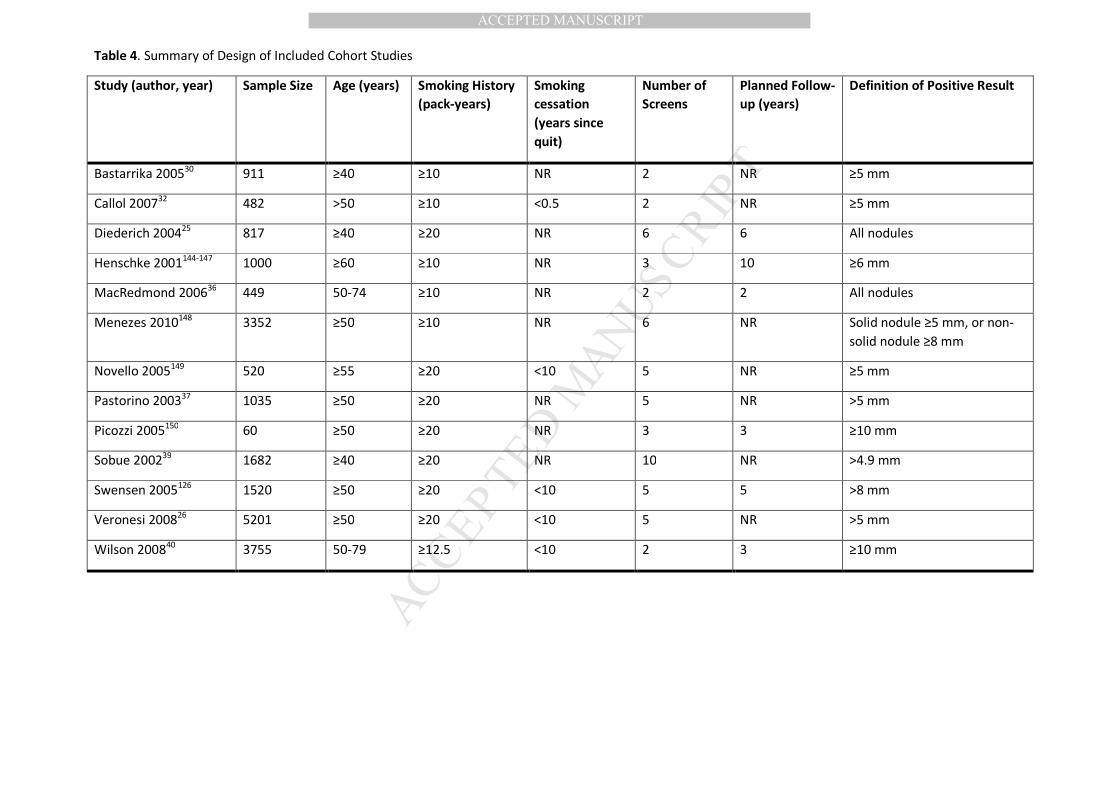
MANUSCRIP
T
ACCEPTED
ACCEPTED MANUSCRIPT
Table 4. Summary of Design of Included Cohort Studies
Study (author, year) Sample Size Age (years) Smoking History
(pack-years)
Smoking
cessation
(years since
quit)
Number of
Screens
Planned Follow-
up (years)
Definition of Positive Result
Bastarrika 200530 911 ≥40 ≥10 NR 2 NR ≥5 mm
Callol 200732 482 >50 ≥10 <0.5 2 NR ≥5 mm
Diederich 200425 817 ≥40 ≥20 NR 6 6 All nodules
Henschke 2001144-147 1000 ≥60 ≥10 NR 3 10 ≥6 mm
MacRedmond 200636 449 50-74 ≥10 NR 2 2 All nodules
Menezes 2010148 3352 ≥50 ≥10 NR 6 NR Solid nodule ≥5 mm, or non-
solid nodule ≥8 mm
Novello 2005149 520 ≥55 ≥20 <10 5 NR ≥5 mm
Pastorino 200337 1035 ≥50 ≥20 NR 5 NR >5 mm
Picozzi 2005150 60 ≥50 ≥20 NR 3 3 ≥10 mm
Sobue 200239 1682 ≥40 ≥20 NR 10 NR >4.9 mm
Swensen 2005126 1520 ≥50 ≥20 <10 5 5 >8 mm
Veronesi 200826 5201 ≥50 ≥20 <10 5 NR >5 mm
Wilson 200840 3755 50-79 ≥12.5 <10 2 3 ≥10 mm
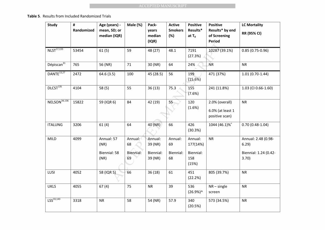
MANUSCRIP
T
ACCEPTED
ACCEPTED MANUSCRIPT
Table 5. Results from Included Randomized Trials
Study #
Randomized
Age (years) -
mean, SD; or
median (IQR)
Male (%) Pack-
years
median
(IQR)
Active
Smokers
(%)
Positive
Results*
at T0
Positive
Results* by end
of Screening
Period
LC Mortality
RR (95% CI)
NLST17,133 53454 61 (5) 59 48 (27) 48.1 7191
(27.3%)
10287 (39.1%) 0.85 (0.75-0.96)
Dépiscan31 765 56 (NR) 71 30 (NR) 64 24% NR NR
DANTE13,27 2472 64.6 (3.5) 100 45 (28.5) 56 199
(15.6%)
471 (37%) 1.01 (0.70-1.44)
DLCST135 4104 58 (5) 55 36 (13) 75.3 155
(7.6%)
241 (11.8%) 1.03 (CI 0.66-1.60)
NELSON34,136 15822 59 (IQR 6) 84 42 (19) 55 120
(1.6%)
2.0% (overall)
6.0% (at least 1
positive scan)
NR
ITALUNG 3206 61 (4) 64 40 (NR) 66 426
(30.3%)
1044 (46.1)%+ 0.70 (0.48-1.04)
MILD 4099 Annual: 57
(NR)
Biennial: 58
(NR)
Annual:
68
Biennial:
69
Annual:
39 (NR)
Biennial:
39 (NR)
Annual:
69
Biennial:
68
Annual:
177(14%)
Biennial:
158
(15%)
NR Annual: 2.48 (0.98-
6.29)
Biennial: 1.24 (0.42-
3.70)
LUSI 4052 58 (IQR 5) 66 36 (18) 61 451
(22.2%)
805 (39.7%) NR
UKLS 4055 67 (4) 75 NR 39 536
(26.9%)^
NR – single
screen
NR
LSS33,143 3318 NR 58 54 (NR) 57.9 340
(20.5%)
573 (34.5%) NR
MANUSCRIP
T
ACCEPTED
ACCEPTED MANUSCRIPT
*=Number of patients with positive results, not number of nodules; see prior table for definition of positive result in each study ^ = if include f/u imaging at 1 year (since a single screen trial) the number would be 1015 (50.9%) + = 1044 is total number of positives from T0-T4; unable to determine if this excludes positive results from the baseline (T0) screen

MANUSCRIP
T
ACCEPTED
ACCEPTED MANUSCRIPT
Table 6: Summary of Biopsies in Included Randomized Controlled Trials
Study Non-surgical
biopsy/procedure
Non-surgical biopsy/procedure
with benign result
Surgical
procedure
Surgical procedure
with benign result
Complications from
invasive procedures
Death after+
invasive procedures
NLST 993 293 673 164 (24.4%) 84a 16
Dépiscan NR NR 9 3 (33.3%) NR NR
DANTE NR NR 90 17 (18.9%) NR NR
DLCST NR NR 25 7 (28%) 4 (0.19%)b NR
NELSON NR 6 NR 61 NR NR
ITALUNG 38 1 (2.6%) 38 4 (10.5%) NR 6 (3.7%)
MILD NR NR 45 4 (8.9%) NR NR
LUSI NR NR NR NR NR NR
UKLS NR NR 39 4 (10.3%) NR NR
LSS 29 16 (55.1%) 46 18 (39.1%) NR NR
aMajor complications include: Acute respiratory failure, Anaphylaxis, Bronchopulmonary fistula, Cardiac arrest, Cerebral vascular accident (CVA)/stroke,
Congestive heart failure (CHF), Death, Hemothorax requiring tube placement, Myocardial infarction, Respiratory arrest, Bronchial stump leak requiring tube
thoracostomy or other drainage for more than 4 days, Wound dehiscence, Empyema, Injury to vital organ or vessel, Prolonged mechanical ventilation over 48
hours post-operatively, Thromboembolic complications requiring intervention, Chylous fistula, Brachial plexopathy, Lung collapse, Infarcted sigmoid colon
bMajor complications include Empyema, myocardial infarction
+ = Death after invasive procedures refers to mortality following an invasive follow-up procedure that was initiated by screening. In the NLST and ITALUNG, it is
reported as death within 60 days of invasive procedure.

MANUSCRIP
T
ACCEPTED
ACCEPTED MANUSCRIPT

MANUSCRIP
T
ACCEPTED
ACCEPTED MANUSCRIPT
Online supplements are not copyedited prior to posting and the author(s) take full responsibility for the accuracy of all data.
e-Table 1.
Screening for Lung Cancer
Recommendation or
Suggestions
Peter
Mazzone, MD, MPH,
FCCP
Gerard A
Silvestri, MD, MS,
FCCP
Sheena
Patel, MPH
Jeffrey P Kanne, MD,
FCCP
Linda S Kinsinger,
MD
Renda
Soylemez Wiener,
MD, MPH
Guy Soo Hoo, MD,
FCCP
Frank C. Detterbeck,
MD, FCCP
Financial COI
Financial COI
Financial COI
Financial COI
Financial COI
Financial COI
Financial COI
Financial COI
Description
of COI
Description
of COI
Description
of COI
Description
of COI
Description
of COI
Description
of COI
Description
of COI
Description
of COI
1. For asymptomatic smokers and former smokers age 55 to 77 who
have smoked 30 pack years or more and either continue to smoke
or have quit within the past 15
years, we suggest that annual screening with low-dose CT should
be offered. (Weak recommendation, moderate-quality evidence)
no relevant COIs
no relevant COIs
no relevant COIs
no relevant COIs
no relevant COIs
no relevant COIs
no relevant COIs
no relevant COIs
2. For asymptomatic smokers and
former smokers who do not meet the smoking and age criteria in
Recommendation #1 but are deemed to be at high risk of
having/developing lung cancer
based on clinical risk prediction calculators, we suggest that low-
dose CT screening should not be routinely performed. (Weak
recommendation, low-quality
evidence)
no relevant
COIs
no relevant
COIs
no relevant
COIs
no relevant
COIs
no relevant
COIs
no relevant
COIs
no relevant
COIs
no relevant
COIs
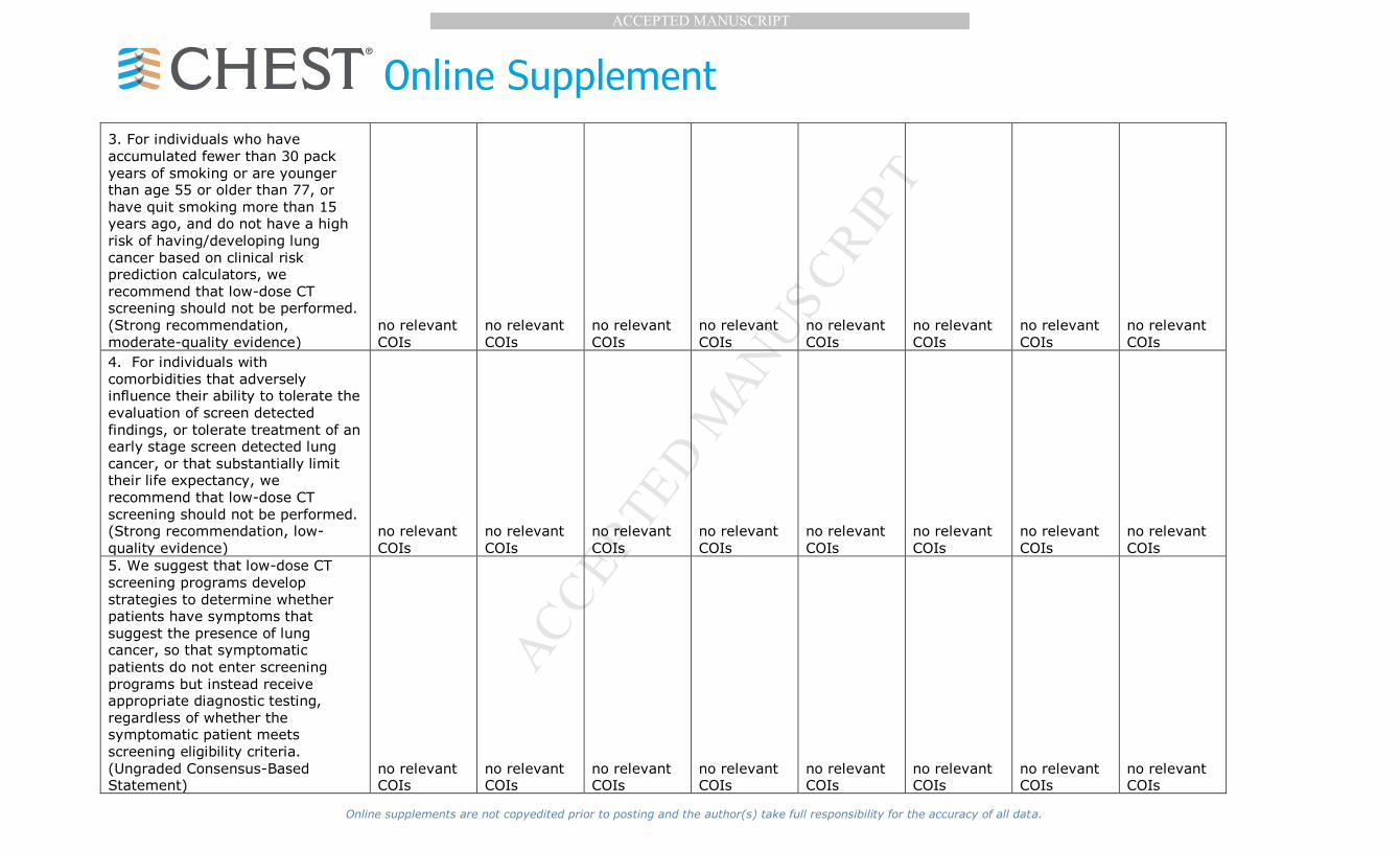
MANUSCRIP
T
ACCEPTED
ACCEPTED MANUSCRIPT
Online supplements are not copyedited prior to posting and the author(s) take full responsibility for the accuracy of all data.
3. For individuals who have
accumulated fewer than 30 pack
years of smoking or are younger than age 55 or older than 77, or
have quit smoking more than 15 years ago, and do not have a high
risk of having/developing lung
cancer based on clinical risk prediction calculators, we
recommend that low-dose CT screening should not be performed.
(Strong recommendation,
moderate-quality evidence)
no relevant
COIs
no relevant
COIs
no relevant
COIs
no relevant
COIs
no relevant
COIs
no relevant
COIs
no relevant
COIs
no relevant
COIs
4. For individuals with
comorbidities that adversely influence their ability to tolerate the
evaluation of screen detected
findings, or tolerate treatment of an early stage screen detected lung
cancer, or that substantially limit their life expectancy, we
recommend that low-dose CT
screening should not be performed. (Strong recommendation, low-
quality evidence)
no relevant
COIs
no relevant
COIs
no relevant
COIs
no relevant
COIs
no relevant
COIs
no relevant
COIs
no relevant
COIs
no relevant
COIs
5. We suggest that low-dose CT
screening programs develop
strategies to determine whether patients have symptoms that
suggest the presence of lung cancer, so that symptomatic
patients do not enter screening
programs but instead receive appropriate diagnostic testing,
regardless of whether the symptomatic patient meets
screening eligibility criteria.
(Ungraded Consensus-Based Statement)
no relevant COIs
no relevant COIs
no relevant COIs
no relevant COIs
no relevant COIs
no relevant COIs
no relevant COIs
no relevant COIs

MANUSCRIP
T
ACCEPTED
ACCEPTED MANUSCRIPT
Online supplements are not copyedited prior to posting and the author(s) take full responsibility for the accuracy of all data.
6. We suggest that screening
programs define what constitutes a
positive test on the low-dose CT based on the size of a detected
solid or part-solid lung nodule, with a threshold for a positive test that
is either 4 mm, 5 mm, or 6 mm in
diameter. (Weak recommendation, low-quality evidence)
no relevant COIs
no relevant COIs
no relevant COIs
no relevant COIs
no relevant COIs
no relevant COIs
no relevant COIs
no relevant COIs
7. We suggest that low-dose CT screening programs develop
strategies to maximize compliance
with annual screening exams. (Ungraded Consensus-Based
Statement)
no relevant
COIs
no relevant
COIs
no relevant
COIs
no relevant
COIs
no relevant
COIs
no relevant
COIs
no relevant
COIs
no relevant
COIs
8. We suggest that low-dose CT
screening programs develop a comprehensive approach to lung
nodule management, including multi-disciplinary expertise
(Pulmonary, Radiology, Thoracic
Surgery, Medical and Radiation Oncology), and algorithms for the
management of small solid nodules, larger solid nodules, and sub-solid
nodules. (Ungraded Consensus-
Based Statement)
no relevant
COIs
no relevant
COIs
no relevant
COIs
no relevant
COIs
no relevant
COIs
no relevant
COIs
no relevant
COIs
no relevant
COIs
9. We suggest that low-dose CT
screening programs develop strategies to minimize
overtreatment of potentially
indolent lung cancers. (Ungraded Consensus-Based Statement)
no relevant COIs
no relevant COIs
no relevant COIs
no relevant COIs
no relevant COIs
no relevant COIs
no relevant COIs
no relevant COIs

MANUSCRIP
T
ACCEPTED
ACCEPTED MANUSCRIPT
Online supplements are not copyedited prior to posting and the author(s) take full responsibility for the accuracy of all data.
10. For current smokers undergoing low-dose CT screening,
we recommend that screening programs provide evidence-based
tobacco treatment as recommended
by the US Public Health Service. (Strong recommendation, low-
quality evidence)
no relevant
COIs
no relevant
COIs
no relevant
COIs
no relevant
COIs
no relevant
COIs
no relevant
COIs
no relevant
COIs
no relevant
COIs
11. We suggest that low-dose CT
screening programs develop
strategies to provide effective counseling and shared decision-
making visits prior to the performance of the LDCT screening
exam.
no relevant
COIs
no relevant
COIs
no relevant
COIs
no relevant
COIs
no relevant
COIs
no relevant
COIs
no relevant
COIs
no relevant
COIs
12. We suggest that low-dose CT screening programs follow the
ACR/STR protocols for performing low radiation dose chest CT scans.
(Ungraded Consensus-Based
Statement)
no relevant
COIs
no relevant
COIs
no relevant
COIs
no relevant
COIs
no relevant
COIs
no relevant
COIs
no relevant
COIs
no relevant
COIs
13. We suggest that low-dose CT
screening programs use a
structured reporting system to report the exam results. (Ungraded
Consensus-Based Statement)
no relevant
COIs
no relevant
COIs
no relevant
COIs
no relevant
COIs
no relevant
COIs
no relevant
COIs
no relevant
COIs
no relevant
COIs
14. We suggest that low-dose CT
screening programs develop
strategies to guide the management of non-nodule
findings. (Ungraded Consensus-Based Statement)
no relevant COIs
no relevant COIs
no relevant COIs
no relevant COIs
no relevant COIs
no relevant COIs
no relevant COIs
no relevant COIs
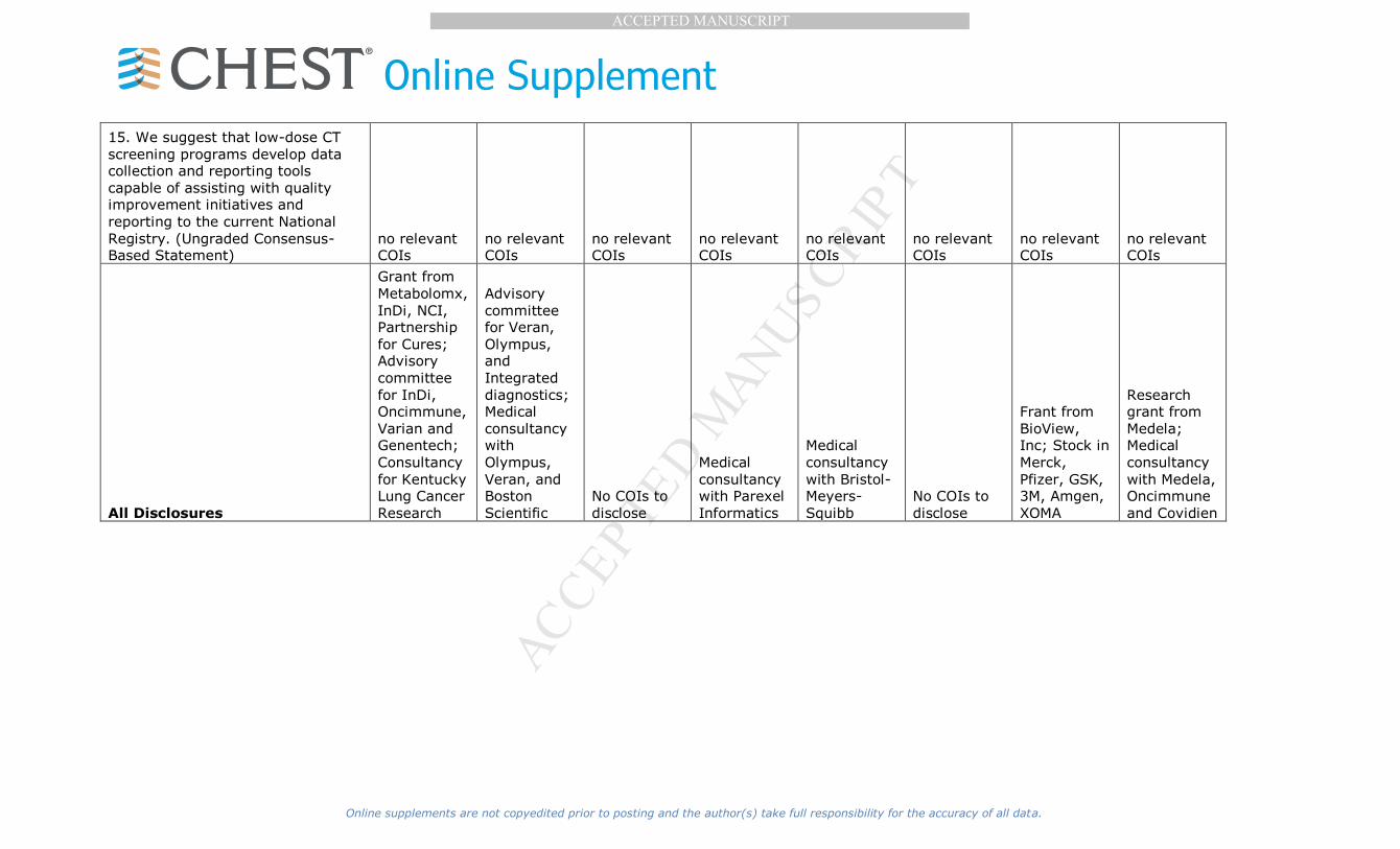
MANUSCRIP
T
ACCEPTED
ACCEPTED MANUSCRIPT
Online supplements are not copyedited prior to posting and the author(s) take full responsibility for the accuracy of all data.
15. We suggest that low-dose CT
screening programs develop data collection and reporting tools
capable of assisting with quality improvement initiatives and
reporting to the current National
Registry. (Ungraded Consensus-Based Statement)
no relevant COIs
no relevant COIs
no relevant COIs
no relevant COIs
no relevant COIs
no relevant COIs
no relevant COIs
no relevant COIs
All Disclosures
Grant from
Metabolomx,
InDi, NCI, Partnership
for Cures; Advisory
committee
for InDi, Oncimmune,
Varian and Genentech;
Consultancy
for Kentucky Lung Cancer
Research
Advisory
committee for Veran,
Olympus, and
Integrated
diagnostics; Medical
consultancy with
Olympus,
Veran, and Boston
Scientific
No COIs to
disclose
Medical
consultancy with Parexel
Informatics
Medical
consultancy
with Bristol-Meyers-
Squibb
No COIs to
disclose
Frant from
BioView, Inc; Stock in
Merck,
Pfizer, GSK, 3M, Amgen,
XOMA
Research grant from
Medela; Medical
consultancy
with Medela, Oncimmune
and Covidien

MANUSCRIP
T
ACCEPTED
ACCEPTED MANUSCRIPT
Online supplements are not copyedited prior to posting and the author(s) take full responsibility for the accuracy of all data.
e-Table 2. Implications of strong and weak recommendations for different users of
guidelines
Strong
Recommendation
Conditional (weak)
Recommendation
For patients Most individuals in this
situation would want the
recommended course of action and only a small
proportion would not.
The majority of individuals
in this situation would
want the suggested course of action, but some would
not.
For clinicians Most individuals should
receive the recommended
course of action. Adherence to this
recommendation according to the guideline could be
used as a quality criterion
or performance indicator. Formal decision aids are
not likely to be needed to help individuals make
decisions consistent with
their values and
preferences.
Recognize that different
choices will be appropriate
for different patients, and that you must help each
patient arrive at a management decision
consistent with her or his
values and preferences. Decision aids may well be
useful helping individuals making decisions
consistent with their values
and preferences. Clinicians should expect to spend
more time with patients
when working towards a
decision.
For policy makers The recommendation can be adapted as policy in
most situations including
for the use as performance
indicators.
Policy making will require substantial debates and
involvement of many
stakeholders. Policies are also more likely to vary
between regions. Performance indicators
would have to focus on the
fact that adequate deliberation about the
management options has
taken place.
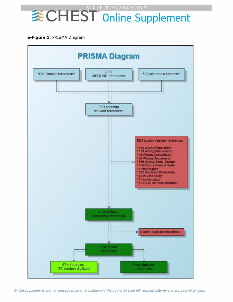
MANUSCRIP
T
ACCEPTED
ACCEPTED MANUSCRIPT
Online supplements are not copyedited prior to posting and the author(s) take full responsibility for the accuracy of all data.
e-Figure 1. PRISMA Diagram

MANUSCRIP
T
ACCEPTED
ACCEPTED MANUSCRIPT
Online supplements are not copyedited prior to posting and the author(s) take full responsibility for the accuracy of all data.
e-Table 3. GRADE Evidence Profile – Lung Cancer Mortality
Question: What is the rate of death from lung cancer among individuals at elevated risk of lung cancer who undergo screening with LDCT compared to either no
screening or screening with another modality?
Bibliography: Pinsky PF, Church TR, Izmirlian G, Kramer BS. The National Lung Screening Trial: results stratified by demographics, smoking history, and lung cancer histology. Cancer. 2013;119(22):3976-3983. Infante M, Cavuto S, Lutman FR, et al. Long-Term Follow-up Results of the DANTE Trial, a Randomized Study of Lung
Cancer Screening with Spiral Computed Tomography. American journal of respiratory and critical care medicine. 2015;191(10):1166-1175.; Saghir Z, Dirksen A, Ashraf
H, et al. CT screening for lung cancer brings forward early disease. The randomised Danish Lung Cancer Screening Trial: status after five annual screening rounds with low-dose CT. Thorax. 2012;67(4):296-301; Pastorino U, Rossi M, Rosato V, et al. Annual or biennial CT screening versus observation in heavy smokers: 5-year results
of the MILD trial. European journal of cancer prevention : the official journal of the European Cancer Prevention Organisation (ECP). 2012;21(3):308-315. Paci E, Puliti D, Lopes Pegna A, et al. Mortality, survival and incidence rates in the ITALUNG randomised lung cancer screening trial. Thorax. 2017.
Quality assessment Events/№ of patients
screened Effect Quality Importance
№ of
studies
Study
design
Risk
of
bias Inconsistency Indirectness Imprecision
Other
considerations LDCT CXR
Relative
(95% CI)
Absolute
(95% CI)
Lung Cancer Mortality - LDCT vs CXR (follow up: median 6.5 years)
1
randomised
trial
not
serious not serious not serious not serious none
469/26722
(1.8%)
552/26730
(2.1%)
RR 0.85
(0.75 to
0.96)
3 fewer per
1,000 (from 1
fewer to 5 fewer)
⨁⨁⨁⨁ HIGH CRITICAL
Lung Cancer Mortality - Annual LDCT vs usual care (follow up: range 2.8 years to 12 years)
4
randomised
trials
serious b not serious not serious serious a none
129/6119
(2.1%)
133/6554
(2.0%)
RR 1.08
(0.70 to
1.64)
2 more per 1,000
(from 6 fewer to
13 more)
⨁⨁◯◯
LOW CRITICAL
Lung Cancer Mortality - Biennial LDCT vs usual care (follow up: median 4.4 years)
1
randomised
trial
serious c not serious not serious serious a none
6/1186
(0.5%)
7/1723
(0.4%)
RR 1.25
(0.42 to
3.70)
1 more per 1,000
(from 2 fewer to
11 more)
⨁⨁◯◯
LOW CRITICAL
CI: Confidence interval; RR: Risk ratio
a. Low number of events; 95% CI crosses no effect
b. All included studies had an overall unclear risk of bias. Unclear randomization in one study and unclear allocation concealment in 3 studies. Two of the studies were rated as high risk of bias for baseline differences across groups and all studies were underpowered to detect a difference in the outcome of interest.
c. Unclear risk of bias due to unclear random sequence generation and allocation concealment. Study is also underpowered

MANUSCRIP
T
ACCEPTED
ACCEPTED MANUSCRIPT
Online supplements are not copyedited prior to posting and the author(s) take full responsibility for the accuracy of all data.
e-Figure 2a. Total number of invasive procedures – CXR
e-Figure 2b. Total number of invasive proceudres – LDCT

MANUSCRIP
T
ACCEPTED
ACCEPTED MANUSCRIPT
Online supplements are not copyedited prior to posting and the author(s) take full responsibility for the accuracy of all data.
e-Figure 3a. Death following Invasive Procedure – CXR
e-Figure 3b. Death following Invasive Procedure – LDCT

MANUSCRIP
T
ACCEPTED
ACCEPTED MANUSCRIPT
Online supplements are not copyedited prior to posting and the author(s) take full responsibility for the accuracy of all data.
e-Table 4. GRADE Evidence Profile for Death following Invasive Procedure
Question: What is the rate of death from invasive procedures among patients who undergo screening with LDCT and CXR
Bibliography: Aberle DR, Adams AM, Berg CD, et al. Reduced lung-cancer mortality with low-dose computed tomographic screening. The New England journal of
medicine. 2011;365(5):395-409; Infante M, Cavuto S, Lutman FR, et al. A randomized study of lung cancer screening with spiral computed tomography: three-year results from the DANTE trial. American journal of respiratory and critical care medicine. 2009;180(5):445-453.; Petersen RH, Hansen HJ, Dirksen A, Pedersen JH. Lung
cancer screening and video-assisted thoracic surgery. Journal of thoracic oncology : official publication of the International Association for the Study of Lung Cancer.
2012;7(6):1026-1031; Veronesi G, Bellomi M, Mulshine JL, et al. Lung cancer screening with low-dose computed tomography: a non-invasive diagnostic protocol for baseline lung nodules. Lung cancer (Amsterdam, Netherlands). 2008;61(3):340-349. Diederich S, Thomas M, Semik M, et al. Screening for early lung cancer with low-
dose spiral computed tomography: results of annual follow-up examinations in asymptomatic smokers. European radiology. 2004;14(4):691-702. Rzyman W, Jelitto-Gorska M, Dziedzic R, et al. Diagnostic work-up and surgery in participants of the Gdansk lung cancer screening programme: the incidence of surgery for non-malignant
conditions. Interactive cardiovascular and thoracic surgery. 2013;17(6):969-973. Paci E, Puliti D, Lopes Pegna A, et al. Mortality, survival and incidence rates in the
ITALUNG randomised lung cancer screening trial. Thorax. 2017.
Quality assessment № of patients Effect
Quality Importance
№ of studies Study design
Risk of bias Inconsistency Indirectness Imprecision
Other considerations
Events/Total number of
procedures (%)*
Proportion per 1,000
procedures (95% CI)#
Death due to Invasive Procedure – screening with LDCT
6 Mixed (5 RCTs, 1 observational)
not serious not serious not serious serious a none
19/ 2,405 (0.8%) 7.7 (4.2, 11.2)
⨁⨁◯◯
LOW CRITICAL
Death due to Invasive Procedure – screening with CXR
1 Randomised trial
not serious not serious not serious serious a none
10/758 (1.3%) 13.2 (5.1, 21.3)
⨁⨁⨁◯
MODERATE CRITICAL
a. Low number of events * = reflects raw percentage as described in manuscript
# = overall summary measure of effect generated using random effects model
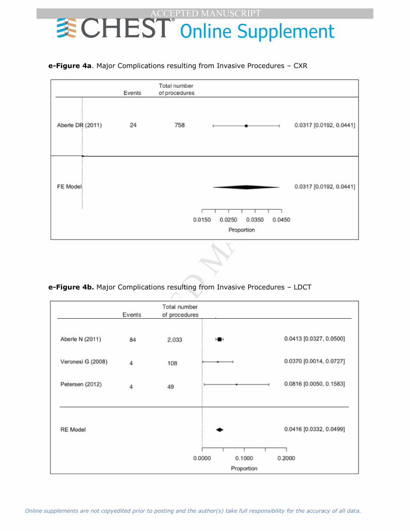
MANUSCRIP
T
ACCEPTED
ACCEPTED MANUSCRIPT
Online supplements are not copyedited prior to posting and the author(s) take full responsibility for the accuracy of all data.
e-Figure 4a. Major Complications resulting from Invasive Procedures – CXR
e-Figure 4b. Major Complications resulting from Invasive Procedures – LDCT

MANUSCRIP
T
ACCEPTED
ACCEPTED MANUSCRIPT
Online supplements are not copyedited prior to posting and the author(s) take full responsibility for the accuracy of all data.
e-Table 5. GRADE Evidence Profile for Major Complications Resulting from Invasive Procedures
Question: LDCT compared to CXR for Major complications due to invasive procedures
Bibliography: Aberle DR, Adams AM, Berg CD, et al. Reduced lung-cancer mortality with low-dose computed tomographic screening. The New England journal of
medicine. 2011;365(5):395-409.; Veronesi G, Bellomi M, Mulshine JL, et al. Lung cancer screening with low-dose computed tomography: a non-invasive diagnostic protocol for baseline lung nodules. Lung cancer (Amsterdam, Netherlands). 2008;61(3):340-349.; Petersen RH, Hansen HJ, Dirksen A, Pedersen JH. Lung cancer
screening and video-assisted thoracic surgery. Journal of thoracic oncology : official publication of the International Association for the Study of Lung Cancer.
2012;7(6):1026-1031.
Quality assessment № of patients Effect
Quality Importance
№ of studies
Study design
Risk of bias Inconsistency Indirectness Imprecision
Other considerations
Events/Total
Undergoing Procedure (%)*
Proportion per 1,000 (95% CI)#
Major Complications – screening with LDCT
3 Randomised trials
not serious not serious not serious not serious none 92/2190 (4.2%) 41.6 (33.2 – 49.9)
⨁⨁⨁⨁ HIGH CRITICAL
Major Complications – screening with CXR
1 Randomised trial
not serious not serious not serious serious a none 24/758 (3.2%) 31.7 (19.2 – 44.1)
⨁⨁⨁◯
MODERATE CRITICAL
a. Wide 95% CI * = reflects raw percentage as described in manuscript
# = overall summary measure of effect generated using random effects model
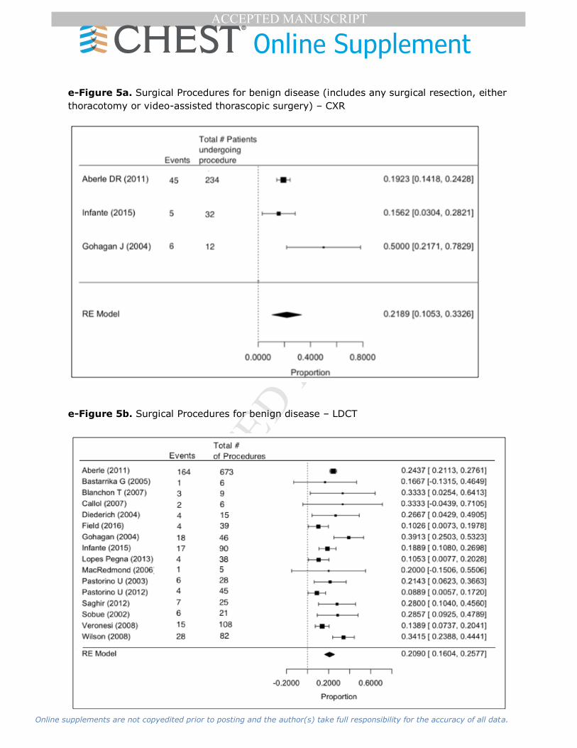
MANUSCRIP
T
ACCEPTED
ACCEPTED MANUSCRIPT
Online supplements are not copyedited prior to posting and the author(s) take full responsibility for the accuracy of all data.
e-Figure 5a. Surgical Procedures for benign disease (includes any surgical resection, either
thoracotomy or video-assisted thorascopic surgery) – CXR
e-Figure 5b. Surgical Procedures for benign disease – LDCT

MANUSCRIP
T
ACCEPTED
ACCEPTED MANUSCRIPT
Online supplements are not copyedited prior to posting and the author(s) take full responsibility for the accuracy of all data.
e-Table 6. GRADE Evidence Profile for Surgery for benign disease
Quality assessment № of patients Effect
Quality Importance
№ of studies
Study design
Risk of bias Inconsistency Indirectness Imprecision
Other considerations
Events/Total
undergoing procedure (%)*
Proportion
per 1,000 (95% CI)#
Rate of surgery for benign disease – screening with LDCT
16
Mixed (10
RCTs, 6 observational
studies)
not
serious not serious not serious not serious none 284/1,236 (22.9%)
209.0 (160.4-
257.7)
⨁⨁◯◯
LOW CRITICAL
Rate of surgery for benign disease – screening with CXR
3
randomised
trials serious not serious not serious not serious none 56/278 (20.1%)
218.9
(105.3-
332.6)
⨁⨁⨁◯
MODERATE CRITICAL
* = reflects raw percentage as described in manuscript # = overall summary measure of effect generated using random effects model

MANUSCRIP
T
ACCEPTED
ACCEPTED MANUSCRIPT
Online supplements are not copyedited prior to posting and the author(s) take full responsibility for the accuracy of all data.
e-Figure 6a. Non-surgical procedures for benign disease (includes non-surgical biopsies,
cytology, bronchoscopy) – CXR
e-Figure 6b. Non-surgical procedures for benign disease – LDCT

MANUSCRIP
T
ACCEPTED
ACCEPTED MANUSCRIPT
Online supplements are not copyedited prior to posting and the author(s) take full responsibility for the accuracy of all data.
e-Table 7. GRADE Evidence Profile for non-surgical procedures for benign disease
Quality assessment № of patients Effect
Quality Importance
№ of studies
Study design
Risk
of bias Inconsistency Indirectness Imprecision
Other considerations
Events/Total number of
procedures (%)*
Proportion
per 1,000 (95% CI)#
Rate of surgery for benign disease – screening with LDCT
6
Mixed (3
RCTs, 3 observational
studies)
not
serious not serious not serious serious none
348/1,147
(30.3%)
369.5 (198.1,
540.9)
⨁⨁◯◯
LOW CRITICAL
Rate of surgery for benign disease – screening with CXR
2
randomised
trials
not
serious not serious not serious serious a none
75/405
(18.5%)
368.4 (-
66.7,
803.6)
⨁⨁⨁◯
MODERATE CRITICAL
a. 95% CI crosses no effect; wide 95% CI * = reflects raw percentage as described in manuscript
# = overall summary measure of effect generated using random effects model

MANUSCRIP
T
ACCEPTED
ACCEPTED MANUSCRIPT
Online supplements are not copyedited prior to posting and the author(s) take full responsibility for the accuracy of all data.
e-Table 8. GRADE Evidence Profile for rate of overdiagnosis
Bibliography: Patz EF, Pinsky P, Gatsonis C, et al. Overdiagnosis in low-dose computed tomography screening for lung cancer. JAMA internal medicine. 2014;174(2):269-274.
Quality assessment Effect
Quality Importance № of studies
Study design
Risk of bias Inconsistency Indirectness Imprecision
Other considerations Overdiagnosis % (95% CI)
1 randomised
trial
not
serious
not serious not serious not serious none All Lung Cancers: 18.5% (5.4% - 30.6%) ⨁⨁⨁⨁ CRITICAL
NSCLC (including BAC and NOS) HIGH
22.5% (9.7% - 34.3%)
NSCLC (excluding BAC and including NOS)
11.7% (-3.7 – 25.6%)
BAC only
78.9% (62.2% - 93.5%)
NSCLC, non-small cell lung cancer BAC, bronchioloalveolar cell carcinoma
NOS, not otherwise specified

MANUSCRIP
T
ACCEPTED
ACCEPTED MANUSCRIPT
Online supplements are not copyedited prior to posting and the author(s) take full responsibility for the accuracy of all data.
e-Table 9. Program Personnel
Discipline Potential Roles
Primary Care Providers Identify eligible patients, order screening, SDM visit, manage
results, smoking cessation
Radiologists Imaging protocols, results reporting, data reporting, program
management, education
Pulmonary/IP Identify eligible patients, order screening, program management, SDM visit, nodule evaluation, manage results,
smoking cessation, data reporting, education
Thoracic Surgery Nodule evaluation, cancer care
Other subspecialists Manage other findings, cancer care
Advanced practice provider
SDM visit, manage results, smoking cessation
Administrator Infrastructure support
Marketing Program awareness, education
Billing Billing compliance, financial data
Scheduling Schedule coordination
EHR/IT specialist Order sets, structured reports, and registries; assist with test follow-up, quality management, and data reporting
e-Table 10. Scanner Requirements
Multidetector helical CT scanner (≥ 16 detector rows preferred)
Gantry rotation ≤ 0.5 seconds
Slice thickness ≤ 2.5 mm (≤ 1.25 mm preferred)
Scanner or viewing platform able to generate multiplanar reformations (MPRs) and
maximum intensity projections (MIPs)
Acquisition parameters:
Suspended full inspiration
Entirety of lungs covered (apices to costophrenic sulci)
100-140 kVp
Appropriate mA and use of automatic exposure control (AEC)
Thin collimation
Appropriate table increment and gantry rotation to minimize helical and motion artifacts
Image reconstruction parameters:
Slice thickness ≤ 2.5 mm (≤ 1.25 mm preferred)
Reconstruction interval ≤ slice thickness
High spatial frequency reconstruction kernel
FOV to include entirety of lungs
Sagittal and coronal reformations (recommended)
Axial 8-10 mm MIPs (recommended)

MANUSCRIP
T
ACCEPTED
ACCEPTED MANUSCRIPT
Online supplements are not copyedited prior to posting and the author(s) take full responsibility for the accuracy of all data.
Specific Incidental Findings
Several common findings deserve specific mention. Emphysema is a common co-morbidity
in patients at significant risk for lung cancer. The USPSTF has recently published a
systematic review and guideline regarding screening for COPD.1 The study concluded that
there was no data on the effect of screening for COPD on survival and no direct studies
examining the benefit of COPD screening on health outcomes. However there was a modest
benefit in terms of reduction of exacerbations and dyspnea scores with treatment in patients
with (known) moderate or severe COPD. Screening for COPD has involved questionnaires
(which exhibit moderate performance, NPV and PPV of 76-98% and 17-45%) and PFTs (with
somewhat better performance, NPV and PPV of 83-98% and 63-75%). However, no studies
have defined the correlation between a LDCT finding of emphysema or bronchial wall
thickening and moderate or severe COPD. Therefore these findings on screening LDCT
cannot be recommended as an indication for further investigation at this time. Additional
research will be helpful.
Cardiovascular disease is another frequent comorbidity in individuals at risk for lung cancer.
In fact, in the NLST slightly more patients died of cardiovascular disease than of lung
cancer.2 CT screening for CAD has been studied extensively and several validated scoring
systems exist that correlate with increasing risk of cardiovascular deaths and major events.
The main difference between LDCT for lung cancer screening and for CAD is that the latter
uses ECG synchronization to minimize motion artifact. However, several studies have found
that coronary artery calcification assessed on a non-gated or a lung cancer screening LDCT
is predictive of an increased risk of cardiovascular deaths in asymptomatic individuals and
those undergoing lung cancer screening.3-8 The various scoring methods, applied to lung
cancer screening, appear to function equally well. Two methods are particularly appealing
because of their simplicity and being based on well done studies: a simple visual
assessment (none, mild, moderate, severe)3 and a prediction algorithm using known
characteristics (age, smoking) and automated quantification of coronary and aortic
calcification.4
No formal guidelines exist regarding the management of patients based on coronary artery
calcification levels, even when CAC is found on ECG-gated CT CAC scans.8 A strong
argument can be made that reporting of coronary artery calcification levels might be useful,
given the correlation with risk of cardiac events and general recommendations for primary
and secondary preventive measures in adults at increased CV risk (e.g. blood pressure
control, aspirin, statins).8-11 There is a suggestion that coronary artery calcification in those
with an intermediate risk of a coronary event (10-20% in the next 10 years) is an added
risk factor that could alter clinical decision making. However it is unclear whether reporting
would have an impact in a lung cancer screening context; one study found that reporting
coronary artery calcification seen on a CT led to a change in aspirin or statin therapy in only
5% of patients.12 Given the lack of formal guidelines, the indirectness of the evidence and
the question about the potential impact, no definitive recommendation for or against
reporting levels of CAC in the context of lung cancer screening, although it would seem
there is little harm to reporting information that is readily available without costing extra
time.
MANUSCRIP
T
ACCEPTED
ACCEPTED MANUSCRIPT
Online supplements are not copyedited prior to posting and the author(s) take full responsibility for the accuracy of all data.
It should be noted that in the NLST there was no difference in non-lung cancer mortality
(p=0.28).2 It is unknown whether this reflects that identifying elevated CV risk during LDCT
lung cancer screening is not useful or whether the ability to determine CV risk and thus
react to it was not yet developed at the time of the NLST. As a result, it seems reasonable
to record the degree of CAC on a lung cancer screening LDCT scan. However a formal
recommendation to use this to select patients for (more intense) intervention in a lung
cancer screening program is premature, and must await evidence that it makes a difference.
It is important to note several aspects regarding aortic dimensions in an asymptomatic
screening population, summarized in a systematic review and multi-society guideline.13
First, the normal aortic diameter increases with age (at age 70 the normal ascending aorta
is 3.5 cm and the descending 2.7 cm; upper limit of normal is 4.2 and 3.2 cm,
respectively).13 Second, aortic enlargement should not be called an aneurysm until the size
is >50% larger than normal. Third, there is no evidence of benefit or recommendation for
screening individuals for thoracic aortic aneurysm unless there is a clear family history or
known genetic defect associated with aortic disease.13 It is beyond the scope of a lung
cancer screening program to obtain such information; it is the role of the primary care
physician to have identified such individuals. Fourth, it is important to measure the outside
of the aorta, and do so in a plane strictly perpendicular to the blood flow. While
management of blood pressure and lipids is recommended for individuals with an aneurysm
to decrease the rate of further expansion, the data comes primarily from patients with
familial risk.13 There is no clear data in other individuals, and presumably this is already part
of the primary care management. Therefore, there is little evidence to suggest that
reporting mild/moderate aortic dilation affects health outcomes. There are
recommendations to monitor aortic aneurysms either annually or biannually based on the
size, type and location of the aneurysm that programs should review. Finally, consideration
of surgical repair is recommended for patients with an ascending or descending aortic size
of ≥5.5 cm (unless there is a familial syndrome).13
Benign liver lesions are very common; fortunately, the vast majority are not concerning. In
low-risk patients (i.e. without cirrhosis, liver disease or a history of cancers that metastasize
to the liver) no further workup is needed for lesions <1.5 cm or with benign features
(sharply marginated, homogeneous, < 20 HU).14 In other scenarios further imaging with MR
or contrast-enhanced CT should be considered.
A taskforce of the ACR on incidental renal lesions recommends no further investigation for
renal lesions that are too small to characterize, and those that are homogeneous and either
-10 to 20HU or >70 HU. Other lesions (i.e. heterogeneous, thick/irregular wall, mural
nodule, septations, 21 to 67 HU) should undergo further imaging (preferably MR).15 This
pertains to lesions that do not contain fat and lesions that are either completely
characterized or incompletely characterized but with sufficient benign features to forgo
further evaluation. Lesions that contain fat (<-10 HU) require further investigation if they
also contain calcification, are multiple, or >4 cm; others do not require investigation.15
Several recent comprehensive guidelines for management of thyroid disease have been
published16,17 but were not written from the perspective of screening for other purposes; a
recent white paper from the incidental thyroid findings committee of the American College
of Radiology is much more specific.18 This group recommends no further investigation for
nodules detected incidentally by CT that are <1.5 cm, in patients >35 years old, and that

MANUSCRIP
T
ACCEPTED
ACCEPTED MANUSCRIPT
Online supplements are not copyedited prior to posting and the author(s) take full responsibility for the accuracy of all data.
have no suspicious CT features (no invasion of local tissues by the thyroid nodule or
abnormal lymph nodes – i.e. calcifications, cystic components, and/or increased
enhancement).18 Nodules >1.5 cm or with suspicious features should undergo ultrasound.
Ultrasound is much better at identifying features suspicious for malignancy; suspicious
nodules by US should undergo FNA, others can be followed be serial US.16,17 This approach
can markedly decrease the number of patients needing further investigation, with indirect
evidence that there is no clinically relevant effect on long term outcomes.18 Of note, the
thyroid guidelines do not recommend screening for thyroid nodules, even in patients with
familial high risk.16
An enlarged adrenal is a common incidental CT finding; a taskforce has developed
management recommendations (Table).19 Lesions that are <1 cm or have fat density (<10
HU need no further investigation. Lesions of 1-2 cm with >10 HU should be re-imaged in a
year. Larger lesions should receive dedicated imaging and possible biopsy.19 Most biliary
system findings are of no significance in asymptomatic patients; polyps ≥7 mm warrant
ultrasound, and biliary duct dilation warrants consideration of serum bilirubin and alkaline
phosphatase levels.20 The ACR incidental findings committee recommends further
investigation of all pancreatic cysts with benign features (absence of mural nodule,
thickening or ductal dilation).21 Most often this involves serial imaging, the frequency
depending on the size, age and communication with the main pancreatic duct.
Homogeneous, thin walled splenic lesions require no further investigation. 22
e-Table 11: Potential Categorization of Incidental Findings
Category Incidencea
Likely next
step Examples
Not clinically
relevant 50%
no directed investi-gation
necessary
Mild-moderate coronary artery calcification,b emphysema, bronchial wall thickening, skeletal
degenerative changes, liver cyst(s), renal cyst(s), hiatal hernia, focal atelectasis, mild mod aortic dilatSion,
pleural plaques, pulmonary fibrosis, adrenal lesions <10
HU, other diaphragmatic hernia, bronchiectasis, low risk thyroid nodulec, renal stone, gallstone, pancreatic cyst,
splenic cyst
Possibly clinically
relevant 10%
Further
investigation may be
indicated
adrenal lesions >10 HU, mediastinal adenopathy (>1
cm), compression fracture, breast nodule, suspicious thyroid nodulec, pancreatic cyst, moderate-severe
coronary artery calcification,b aortic aneurysm 4-5.5 cm
Clinically concerning <1%
Therapeutic
inter-vention
may be indicated
pneumonia, aortic aneurysm ≥5.5 cm, mass or lesion
suspicious for malignancy (e.g. bone destruction),
segmental/lobar atelectasis, large pleural effusion, large pericardial effusion
Examples are ordered according to reported frequency20,50,51,118,119,146 a estimated b although significant Coronary artery calcification is associated with increased
risk of cardiovascular events, there is insufficient evidence that investigation or intervention
is of benefit in asymptomatic patients. clow risk thyroid nodule (by CT) is defined as <1.5 cm without evidence of tissue invasion or node enlargement

MANUSCRIP
T
ACCEPTED
ACCEPTED MANUSCRIPT
Online supplements are not copyedited prior to posting and the author(s) take full responsibility for the accuracy of all data.
e-Table 12: Overview of Guidelines Related to Management of Incidental Findings
Site
(reference) Source
Level of
Evidence Populationa
No Further Investigation
Recommended for: Consider Further Investigation Recommended for:
Coronary
Artery
calcification 8 none
Aortic
enlargement 13
ACCF,
AHA Guideline
general referral population
(no high familial risk) diameter <3.5 cm
Consider annual surveillance imaging if 3.5-4.5 cm, biannual if
4.5-5.4 cm
consider therapeutic intervention if ≥5.5 cm
Liver 14 ACR
Consensus,
indirect general population >40 b
< 1.5 cm, or any size with benign features (sharply
marginated, homogeneous,
< 20 HU)
MR or CT with IV contract if ≥1.5 cm and suspicious features (ill-
defined margin)
Renal 15 ACR
Consensus,
indirect general population
Small (TSTC),
homogeneous and either -
10 to 20HU or >70 HU;
MR if 21-69 HU or heterogeneous (thickening, nodularity,
calcification, septations)
<-10 HU but solitary, no
calcification, <4 cm or if <-10 HU with calcifications, multiplicity or >4 cm
Thyroid18 ACR
Consensus,
indirect
general population of
adults >35
< 1.5 cm and no lack
suspicious features
US±FNA if >1.5 cm or suspicious (invasion of local tissues or
abnormal lymph nodes – i.e. calcifications, cystic components,
and/or increased enhancement)
Adrenal 19 ACR
Consensus,
indirect general population
<1 cm, or 1-4 cm but <10
HU, known to be stable for
≥ 1 year, CT in 1 year if 1-2 cm, >10 HU,
dedicated cm CT, MR if 2-4 cm and >10 HU
if >4 cm, consider biopsy, resection, PET
Pancreas
cyst 21 ACR
Consensus,
indirect general population none
serial imaging if benign features: every 4-24 mo depending on
size (<1.5, 1.5-2.5, >2.5 cm) and age (< or ≥65)
EUS/FNA if mural nodule, thickening, duct dilation (for any size
cyst)
more active workup (image every 4 mo or EUS/FNA) if no
communication with main pancreatic duct
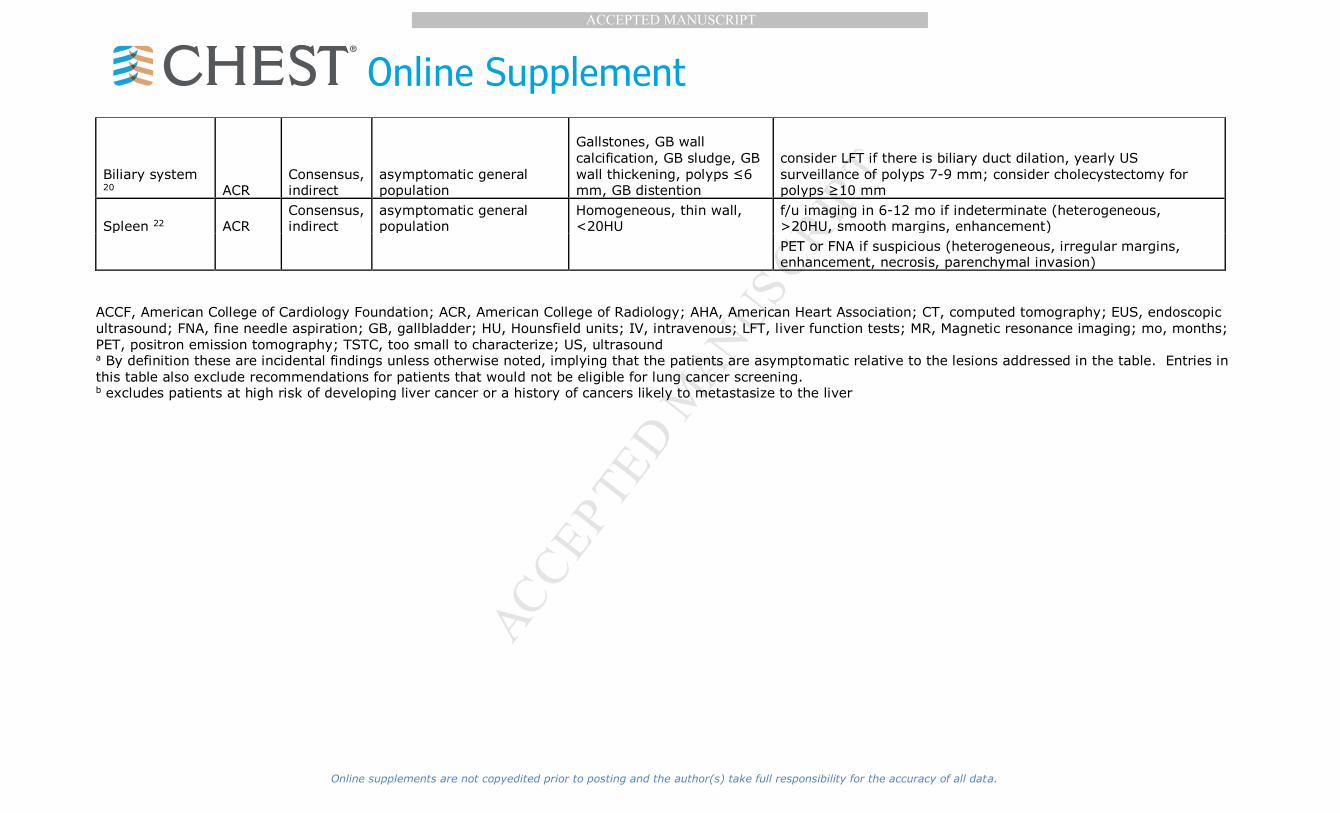
MANUSCRIP
T
ACCEPTED
ACCEPTED MANUSCRIPT
Online supplements are not copyedited prior to posting and the author(s) take full responsibility for the accuracy of all data.
Biliary system 20 ACR
Consensus, indirect
asymptomatic general population
Gallstones, GB wall
calcification, GB sludge, GB
wall thickening, polyps ≤6 mm, GB distention
consider LFT if there is biliary duct dilation, yearly US
surveillance of polyps 7-9 mm; consider cholecystectomy for polyps ≥10 mm
Spleen 22 ACR Consensus, indirect
asymptomatic general population
Homogeneous, thin wall, <20HU
f/u imaging in 6-12 mo if indeterminate (heterogeneous, >20HU, smooth margins, enhancement)
PET or FNA if suspicious (heterogeneous, irregular margins, enhancement, necrosis, parenchymal invasion)
ACCF, American College of Cardiology Foundation; ACR, American College of Radiology; AHA, American Heart Association; CT, computed tomography; EUS, endoscopic
ultrasound; FNA, fine needle aspiration; GB, gallbladder; HU, Hounsfield units; IV, intravenous; LFT, liver function tests; MR, Magnetic resonance imaging; mo, months;
PET, positron emission tomography; TSTC, too small to characterize; US, ultrasound a By definition these are incidental findings unless otherwise noted, implying that the patients are asymptomatic relative to the lesions addressed in the table. Entries in
this table also exclude recommendations for patients that would not be eligible for lung cancer screening. b excludes patients at high risk of developing liver cancer or a history of cancers likely to metastasize to the liver

MANUSCRIP
T
ACCEPTED
ACCEPTED MANUSCRIPT
Online supplements are not copyedited prior to posting and the author(s) take full responsibility for the accuracy of all data.
e-Table 13: CMS Data Requirements
Data Type Minimum Required Data Elements
Facility Identifier
Radiologist National Provider Identifier
Patient Identifier
Ordering Practitioner National Provider Identifier
CT Scanner Manufacturer, Model
Indication Lung cancer LDCT screening – absence of signs or symptoms of
lung cancer
System Lung nodule identification, classification and reporting system
Smoking history Current status; Years since quit; Pack-years; Cessation interventions
Effective radiation
dose
CT Dose Index (CTDIvol)
Screening Screen date, initial screen or subsequent screen
e-Table 14: ACR National Registry Data Elements
LCSR Data Element
Required to submit a
transaction
Transaction Header (Required)
Transaction ID Y
Transaction date time Y
Number of exam included Y
Facility ID Y
Partner ID Y
Application ID Y
Previous transaction ID N
Exam Data (Required)
Exam_Unique_ID N
Patient's first name N
Patient's middle name N
Patient's last name N
Patient ID Conditional
Refuesed to provide patient's social security number N
Patient Social Security Number Conditional
Refuesed to provide patient's medicare beneficiary ID N
Medicare Beneficiary ID Conditional
Patient's date of birth N
Patient's date of death N
How cause of death was determined N
Other method of determining cause of death, specify N
Cause of death N
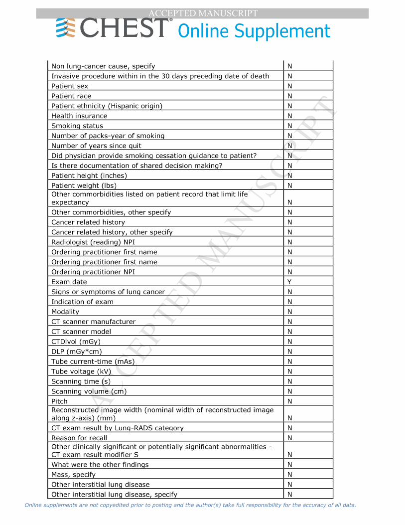
MANUSCRIP
T
ACCEPTED
ACCEPTED MANUSCRIPT
Online supplements are not copyedited prior to posting and the author(s) take full responsibility for the accuracy of all data.
Non lung-cancer cause, specify N
Invasive procedure within in the 30 days preceding date of death N
Patient sex N
Patient race N
Patient ethnicity (Hispanic origin) N
Health insurance N
Smoking status N
Number of packs-year of smoking N
Number of years since quit N
Did physician provide smoking cessation guidance to patient? N
Is there documentation of shared decision making? N
Patient height (inches) N
Patient weight (lbs) N
Other commorbidities listed on patient record that limit life
expectancy N
Other commorbidities, other specify N
Cancer related history N
Cancer related history, other specify N
Radiologist (reading) NPI N
Ordering practitioner first name N
Ordering practitioner first name N
Ordering practitioner NPI N
Exam date Y
Signs or symptoms of lung cancer N
Indication of exam N
Modality N
CT scanner manufacturer N
CT scanner model N
CTDlvol (mGy) N
DLP (mGy*cm) N
Tube current-time (mAs) N
Tube voltage (kV) N
Scanning time (s) N
Scanning volume (cm) N
Pitch N
Reconstructed image width (nominal width of reconstructed image
along z-axis) (mm) N
CT exam result by Lung-RADS category N
Reason for recall N
Other clinically significant or potentially significant abnormalities - CT exam result modifier S N
What were the other findings N
Mass, specify N
Other interstitial lung disease N
Other interstitial lung disease, specify N

MANUSCRIP
T
ACCEPTED
ACCEPTED MANUSCRIPT
Online supplements are not copyedited prior to posting and the author(s) take full responsibility for the accuracy of all data.
Prior history of lung cancer - CT exam result modifier C N
Year since prior diagnosis of lung cancer N
Education level N
Education level, other N
Radon exposure - documented high exposure levels N
Occupational exposures to carcinogens targeting the lungs N
History of cancers associated with an increased risk of developing a
new primary lung cancer N
History of cancers associated with an increased risk of developing a
new primary lung cancer - other smoking-related cancers, specify N
Lung cancer in first-degree relative N
Family history of lung cancer, other than first‐degree relative N
COPD N
Pulmonary fibrosis N
Second hand smoke exposure N
Follow-up Data
Date of follow-up Y
Follow-up diagnostic Y
Follow-up diagnostic other, specify N
Tissue diagnosis N
Tissue diagnosis method N
Location from which sample was obtained N
Location other, specify N
Histology N
Histology - Non-small cell lung cancer N
Other non-small cell lung cancer histology, specify N
Stage - Clinical or pathologic? N
Overall stage N
T status N
N status N
M status N
MANUSCRIP
T
ACCEPTED
ACCEPTED MANUSCRIPT
Online supplements are not copyedited prior to posting and the author(s) take full responsibility for the accuracy of all data.
1. Guirguis-Blake JM, Senger CA, Webber EM, Mularski RA, Whitlock EP. Screening for
Chronic Obstructive Pulmonary Disease: Evidence Report and Systematic Review for the US Preventive Services Task Force. JAMA. 2016;315(13):1378-1393.
2. Aberle D, Adams A, Berg C, et al. Reduced Lung-Cancer Mortality with Low-Dose
Computed Tomographic Screening. New England Journal of Medicine. 2011;365(5):395-409.
3. Chiles C, Duan F, Gladish GW, et al. Association of Coronary Artery Calcification and Mortality in the National Lung Screening Trial: A Comparison of Three Scoring
Methods. Radiology. 2015;276(1):82-90.
4. Mets OM, Vliegenthart R, Gondrie MJ, et al. Lung cancer screening CT-based prediction of cardiovascular events. JACC Cardiovasc Imaging. 2013;6(8):899-907.
5. Jacobs PC, Gondrie MJ, van der Graaf Y, et al. Coronary artery calcium can predict all-cause mortality and cardiovascular events on low-dose CT screening for lung
cancer. AJR Am J Roentgenol. 2012;198(3):505-511.
6. Htwe Y, Cham MD, Henschke CI, et al. Coronary artery calcification on low-dose computed tomography: comparison of Agatston and Ordinal Scores. Clin Imaging.
2015;39(5):799-802. 7. Kim YK, Sung YM, Cho SH, Park YN, Choi HY. Reliability analysis of visual ranking of
coronary artery calcification on low-dose CT of the thorax for lung cancer screening:
comparison with ECG-gated calcium scoring CT. Int J Cardiovasc Imaging. 2014;30 Suppl 2:81-87.
8. Pakdaman MN, Rozanski A, Berman DS. Incidental coronary calcifications on routine chest CT: Clinical implications. Trends Cardiovasc Med. 2017;27(7):475-480.
9. Bibbins-Domingo K. Aspirin Use for the Primary Prevention of Cardiovascular Disease
and Colorectal Cancer: U.S. Preventive Services Task Force Recommendation Statement. Ann Intern Med. 2016;164(12):836-845.
10. Miedema MD, Duprez DA, Misialek JR, et al. Use of coronary artery calcium testing to guide aspirin utilization for primary prevention: estimates from the multi-ethnic
study of atherosclerosis. Circ Cardiovasc Qual Outcomes. 2014;7(3):453-460.
11. Stone NJ, Robinson JG, Lichtenstein AH, et al. 2013 ACC/AHA guideline on the treatment of blood cholesterol to reduce atherosclerotic cardiovascular risk in adults:
a report of the American College of Cardiology/American Heart Association Task Force on Practice Guidelines. J Am Coll Cardiol. 2014;63(25 Pt B):2889-2934.
12. Uretsky S, Chokshi N, Kobrinski T, et al. The interplay of physician awareness and
reporting of incidentally found coronary artery calcium on the clinical management of patients who underwent noncontrast chest computed tomography. Am J Cardiol.
2015;115(11):1513-1517. 13. Hiratzka LF, Bakris GL, Beckman JA, et al. 2010
ACCF/AHA/AATS/ACR/ASA/SCA/SCAI/SIR/STS/SVM guidelines for the diagnosis and
management of patients with Thoracic Aortic Disease: a report of the American College of Cardiology Foundation/American Heart Association Task Force on Practice
Guidelines, American Association for Thoracic Surgery, American College of Radiology, American Stroke Association, Society of Cardiovascular Anesthesiologists,
Society for Cardiovascular Angiography and Interventions, Society of Interventional
Radiology, Society of Thoracic Surgeons, and Society for Vascular Medicine. Circulation. 2010;121(13):e266-369.
14. Berland LL, Silverman SG, Gore RM, et al. Managing incidental findings on abdominal CT: white paper of the ACR incidental findings committee. J Am Coll Radiol.
2010;7(10):754-773.
15. Herts BR, Silverman SG, Hindman NM, et al. Management of the Incidental Renal Mass on CT: A White Paper of the ACR Incidental Findings Committee. J Am Coll
Radiol. 2017. 16. Haugen BR, Alexander EK, Bible KC, et al. 2015 American Thyroid Association
Management Guidelines for Adult Patients with Thyroid Nodules and Differentiated

MANUSCRIP
T
ACCEPTED
ACCEPTED MANUSCRIPT
Online supplements are not copyedited prior to posting and the author(s) take full responsibility for the accuracy of all data.
Thyroid Cancer: The American Thyroid Association Guidelines Task Force on Thyroid
Nodules and Differentiated Thyroid Cancer. Thyroid. 2016;26(1):1-133. 17. Mitchell AL, Gandhi A, Scott-Coombes D, Perros P. Management of thyroid cancer:
United Kingdom National Multidisciplinary Guidelines. J Laryngol Otol.
2016;130(S2):S150-S160. 18. Hoang JK, Langer JE, Middleton WD, et al. Managing incidental thyroid nodules
detected on imaging: white paper of the ACR Incidental Thyroid Findings Committee. J Am Coll Radiol. 2015;12(2):143-150.
19. Mayo-Smith W, Song J, Boland G, et al. Managment of Incidental Adrenal Masses: A
White Paper of the ACR Incidental Findings Committee. J Am Coll Radiol. 2017;14(8):1038-1044.
20. Sebastian S, Araujo C, Neitlich JD, Berland LL. Managing incidental findings on abdominal and pelvic CT and MRI, Part 4: white paper of the ACR Incidental Findings
Committee II on gallbladder and biliary findings. J Am Coll Radiol. 2013;10(12):953-
956. 21. Megibow AJ, Baker ME, Morgan DE, et al. Management of Incidental Pancreatic
Cysts: A White Paper of the ACR Incidental Findings Committee. J Am Coll Radiol. 2017;14(7):911-923.
22. Heller MT, Harisinghani M, Neitlich JD, Yeghiayan P, Berland LL. Managing incidental
findings on abdominal and pelvic CT and MRI, part 3: white paper of the ACR Incidental Findings Committee II on splenic and nodal findings. J Am Coll Radiol.
2013;10(11):833-839.
172542