screening for diabetic retinopathy: new perspectives and
TRANSCRIPT

Screening for diabetic retinopathy: new perspectives and challenges
Vujosevic, S., Aldington, S. J., Silva, P., Hernández, C., Scanlon, P., Peto, T., & Simó, R. (2020). Screening fordiabetic retinopathy: new perspectives and challenges. The lancet. Diabetes & endocrinology.https://doi.org/10.1016/S2213-8587(19)30411-5
Published in:The lancet. Diabetes & endocrinology
Document Version:Peer reviewed version
Queen's University Belfast - Research Portal:Link to publication record in Queen's University Belfast Research Portal
Publisher rightsCopyright 2019 Elsevier.This manuscript is distributed under a Creative Commons Attribution-NonCommercial-NoDerivs License(https://creativecommons.org/licenses/by-nc-nd/4.0/), which permits distribution and reproduction for non-commercial purposes, provided theauthor and source are cited
General rightsCopyright for the publications made accessible via the Queen's University Belfast Research Portal is retained by the author(s) and / or othercopyright owners and it is a condition of accessing these publications that users recognise and abide by the legal requirements associatedwith these rights.
Take down policyThe Research Portal is Queen's institutional repository that provides access to Queen's research output. Every effort has been made toensure that content in the Research Portal does not infringe any person's rights, or applicable UK laws. If you discover content in theResearch Portal that you believe breaches copyright or violates any law, please contact [email protected].
Download date:03. Apr. 2022

Tiltle: SCREENING FOR DIABETIC RETINOPATHY: NEW PERSPECTIVES AND
CHALLENGES
Authors: Stela Vujosevic, MD1; Stephen J. Aldington, HND2; Paolo Silva, MD3,4; Cristina Hernández, MD5,6; Peter Scanlon, MD2; Tunde Peto,MD7; Rafael Simó, MD5,6*
1 University Hospital Maggiore della Carita, Novara, Italy. 2 Department of Ophthalmology, Gloucestershire Hospitals NHS Foundation Trust, Cheltenham, United Kingdom 3 Beetham Eye Institute, Joslin Diabetes Centre, Harvard Medical School, Boston, USA 4 Philippine Eye Research Institute. University of the Philippines, Manila, Philippines 5 Vall d’Hebron Research Institute and Autonomous University of Barcelona, Barcelona, Spain 6 Centro de Investigación Biomédica en Red de Diabetes y Enfermedades Metabólicas Asociadas (CIBERDEM), Instituto de Salud Carlos III (ICSIII), Spain 7 Queen’s University Belfast, Belfast, United Kingdom
*Corresponding author:
Rafael Simó MD, PhD Professor of Medicine & Endocrinology Head, Diabetes Research and Metabolism Unit Vall d’Hebron Research Institute Pg. Vall d’Hebron 119-129, 08035 Barcelona, Spain Phone 34 934894172 / Fax: 34 934894032 e-mail: [email protected]
ABSTRACT
Although all stages of diabetic retinopathy (DR) have been declining since 1980 in populations with improved diabetes control, the crude prevalence of visual impairment and blindness caused by DR has increased in recent years. This is mainly due to the global increase of type 2 diabetes. Screening for DR is essential to detect referable cases that need timely full ophthalmic examination and treatment in order to avoid permanent visual loss. In recent years personalized screening intervals taking into account several risk factors has been proposed with a good cost-effectiveness ratios. However, limited resources are available for nationwide screening programs. New technologies such as scanning confocal ophthalmology with ultrawide field imaging and mobile hand-held devices, tele-ophthalmology for remote grading and artificial intelligence for automated detection and classification of DR are changing screening strategies and improving its cost-effectiveness. The emergent evidence that retinal imaging is useful for identifying subjects at risk of cardiovascular disease or cognitive impairment is also a challenge that could change the concept of DR screening into a more broad examination, not limited to prevent sight-threatening disease.
Box. Searching strategy and selection criteria
We identified references for this review through searches of PubMed and Google Scholar for articles published in English up to November 7th, 2019. The key words used included “diabetic retinopathy”, “screening of diabetic retinopathy”, “retinal imaging”, “retinal neurodegeneration”, “tele-ophthalmology”, “artificial intelligence”, “diabetic complications”, “diabetic retinopathy and cardiovascular disease”, “diabetic retinopathy and dementia”. These keywords were used as single search terms or in combination. We also searched the reference list of original articles, clinical guidelines, systematic reviews and meta-analyses for further relevant material. All the authors conducted the literature search but with pre-specified assigned areas. Articles were initially selected for inclusion on the basis of the opinion of the contributing authors, with each section covered by two or three authors with particular expertise. The selected references were reviewed by all the members of the authorship when reading successive drafts.

Introduction
Diabetic Retinopathy (DR) remains as leading cause of vision loss and preventable blindness in adults aged 20-74 years, particularly in higher-income countries.1 A meta-analysis involving 35 studies conducted worldwide from 1980 to 2008, estimated an overall prevalence of 34.6% (95% CI 34.5-34.8) for any DR, 6.96% (6.87-7.04) for proliferative DR (PDR), 6.81% (6.74-6.89) for diabetic macular edema (DME), and 10.2% (10.1-10.3) for vision-threatening diabetic retinopathy (VTDR).2 Prevalence of any DR and PDR was higher in subjects with type 1 than in type 2 diabetes.2
Although the progression of DR to proliferative DR and severe visual loss has been declining since 1980 in populations with improved diabetes control,3 the crude prevalence of visual impairment and blindness caused by DR increased significantly between 1990 and 2015 according to the latest report of the Vision Loss Expert Group of the Global Burden of Disease Study.4 Thus, the number of people affected by blindness due to DR increased from 0.2 million to 0.4 million, and moderate to severe vision impairment increased from 1.4 million to 2.6 million.4 The same report indicates that low- and middle- income countries now account for >75% of global diabetes. Recently, it has been estimated that persons with diabetes affected by any diabetic eye disease in Europe will increase from 6.4 million today to 8.6 million in 2050, of whom 30% require close monitoring and/or treatment ( Li JQ, Welchowski T, Schmid M, Letow J, Wolpers C, Pascual-Camps I, Holz FG, Finger RP. Prevalence, incidence and future projection of diabetic eye disease in Europe: a systematic review and meta-analysis. Eur J Epidemiol. 2019 Sep 12. doi: 10.1007/s10654-019-00560-z. [Epub ahead of print])
Since 2000 only a few population based studies have been conducted, the incidence was higher in studies performed before 2000 compared with those published after 2000.5 However, contemporary studies including more data from developed countries are needed.
Risk factors
The most relevant risk factors for the development of DR are the duration of the disease, poor glycemic control (high HbA1c levels) and the presence of hypertension. However, the impact of blood-glucose control in the development of DR (DCCT, UKPDS) is stronger that the impact of blood-pressure control (ACCORD-EYE y ACCORDION).
DCCT, UKPDS
Chew EY, Davis MD, Danis RP, et al.; Action to Control Cardiovascular Risk in Diabetes Eye Study Research Group. The effects of medical management on the progression of diabetic retinopathy in persons with type 2 diabetes: the Action to Control Cardiovascular Risk in Diabetes (ACCORD) Eye Study. Ophthalmology 2014;121:2443–2451
Action to Control Cardiovascular Risk in Diabetes Follow-On (ACCORDION) Eye Study Group and the Action to Control Cardiovascular Risk in Diabetes Follow-On (ACCORDION) Study Group. Persistent Effects of Intensive Glycemic Control on Retinopathy in Type 2 Diabetes in the Action to Control Cardiovascular Risk in Diabetes (ACCORD) Follow-On Study. Diabetes Care. 2016 Jul;39(7):1089-100
Other risk factors for DR include dyslipidemia, a higher body mass index, puberty, pregnancy, and cataract surgery.2 Despite the influence in the development and progression of DR by the above mentioned risk factors, clinical studies on patients living with diabetes reveal a substantial variation in the onset and severity of DR that is not fully explained by the known risk factors, and all clinicians know that a subset of patients with poor control of glycemia and/or uncontrolled

blood pressure do not develop DR. On the other hand there are patients with very good control of blood glucose levels and without hypertension that develop DR. In fact, the DCCT/EDIC Research Group showed that HbA1c values explained up to 11% of the risk of DR, and that the unexplained 89% of variation in risk is due to elements of the diabetic milieu not captured by the mean HbA1c value.6 These data suggest that other factors such as glucose variability7,8 and genetic determinants (9-12) play a significant role in the development and progression of DR.
The cost-effectiveness of screening
Several reports from different countries around the world have reported on the cost-effectiveness of screening, especially when applied to detect VTDR.13-15 Cost-effectiveness of population-based screening programs is heavily dependent on the frequency of retinal examinations and retinal imaging. [16] Extending the screening interval from annual to every two-, three- or even more years in diabetes patients found to have no evidence of any DR at first eye examination has been reported to be cost-effective by several European studies. [17-20] + (Diabetic Medicine (2019:36;1199-1208). Perhaps even more significantly, differentiating patients into low- and high-risk groups has the potential to further improve cost-effectiveness. [20-22]
Screening programs for DR using 2D non-stereoscopic digital fundus photography are currently implemented at primary care level. However, interpretation of the photographs requires specialized knowledge and expertise in diabetic eye disease. Given that the number of patients with diabetes is rising rapidly, manual grading for DR in screening programs will not be sustainable. In this scenario the emerging use of validated automated grading software will undoubtedly influence the cost-effectiveness of screening.23,24 More controversial is the use of ultra-wide field (UWF) imaging systems for the evaluation of additional peripheral retinal lesions with major risk of DR progression.25,26 Regarding the screening of DME, the use of optical coherence tomography (OCT) as the first line screening tool has yet to be fully justified from the financial viewpoint13 but it has been shown to be cost effective as a second line screening tool in those with positive 2-dimensional markers at the primary screening event.27
Screening of DR: who, when and how
Current guidelines and procedures
The most recent guidelines and procedures for DR screening have been proposed by the International Council of Ophthalmology (ICO) in 2017 as part of the Guidelines for Diabetic Eye Care and by the American Diabetes Association (ADA) as part of the Position Statement for Diabetic Retinopathy in 2017. 28,29 The ADA recommends a well-defined and different timing of the first eye examination based on the type of DM supported by the level B of evidence (Table 1). To ensure appropriate referral to ophthalmologist, the minimum screening examination would need to include vision screening examination and retinal examination adequate for DR classification.29 The ICO classification for DR and DME considers 5 stages of DR (no DR, mild non proliferative (NP) DR, moderate NPDR, severe NPDR and proliferative DR) (Figure 1A), whereas DME classification has been recently updated in no DME, non-center-involving DME and center-involving DME (Figure 1B), as this classification is the most one used with the latest available treatments (intravitreal drugs).28
Retinal examination includes either retinal photography or fundus examination (ophthalmoscopy). Mydriatic and non-mydriatic (NM) color retinal photography has shown better sensitivity in DR screening than direct and indirect ophthalmoscopy.30,31 Multifield (3 fields) NM imaging has been found to have a good agreement with dilated ophthalmoscopy or 7 ETDRS fields (Lee JC, Nguyen L, Hynan LS, Blomquist PH. J Diabetes Complications. 2019 Sep 12:107441. doi: 10.1016/j.jdiacomp.2019.107441. [Epub ahead of print], but this is not the case when only one or 2 field non-mydriatic 45 degree images are used (Boucher et al. Can J

Ophthalmol 2003:557-68 + Lee JC et al. J Diabetes Complications 2019). Currently, there is still a lack of agreement regarding the number of NM fundus fields required to reliable detect and grade DR and DME in telemedicine screening programs. Although 7 standard mydriatic and stereoscopic 30º fundus fields (7-ETDRS fields) were considered (in the past) the gold standard for fundus evaluation in all clinical trials for DR, it is a time consuming and expensive procedure, thus not applicable in the screening setting. Therefore, fewer fundus fields (three, two even one central varying from 45º to 60º) have been proposed for DR and DME screening.32-35 Data in the literature report sensitivity (80% to 98%) and specificity (86% to 100%) of limited number of fundus fields (2-4) vs. 7 ETDRS fields for detecting any DR; whereas, lower sensitivity (54% to 78%) and specificity (88% to 89%) were recorded when single central field vs. 7 ETDRS fields was analysed. 33,36 The major clinical value of DR screening is to detect referable cases that need timely full ophthalmic examination and treatment in order to avoid permanent visual loss. For this reason high sensitivity values (>80%) are required for the effective screening program and, therefore, 1-NM 45-degree central-field photography is not suitable for a community-based screening program.37 Recently, the ultrawide field (UWF) systems covering up to 200° of the fundus have been used for DR screening (Figure 2). With UWF imaging system, pharmacologic mydriasis may be required for single retinal lesion evaluation to decrease the percentage of ungradable images (from 4.5% to 0%).38
A screening program must be coordinated with access to adequate and timely referral to ophthalmologist in order to be useful for patients with DR, needing further management. The timing of next screening and referral to ophthalmologist in both high income and medium/low income settings are shown in table 2.
Pre-requisites for a nationwide screening program: influence of socio-economic factors
The principles for screening for human diseases that were derived from the public health papers produced by the WHO in 196839 included the statement that ‘the cost of the case-finding programme (including early diagnosis and treatment of patients diagnosed) should be economically balanced in relation to possible expenditure on medical care as a whole’. Hence, even in resource-rich countries, the frequency of invited screening and uptake by those most in need of attending is compromised by a relative or absolute lack of resources and by a reluctance in some to recognise the importance of screening.40-45 Equally, regular annual screening of those at a very low risk of developing any serious complications has questionable value, both economically and socially.41,42 It should be recognized however that such repeated screening does reassure the committed, well-controlled but worried patient that things are still not progressing.46
Diabetic eye screening, frequently at a time when a patient attends for other investigations (i.e. opportunistic screening), has been successfully implemented in countless locations and regions; many of which have become well-established and do tremendous service. The caveat however is that these are most often only to a very defined and ‘local’ population. Systematic screening however, aiming to include the whole population at risk in its target group, is clearly far more complex to implement and to sustain and has been successfully implemented in few locations.
True nationwide population-based systematic diabetic eye screening programmes really exist only in Iceland47,48, the UK40,49 and in the Republic of Ireland.50
Many parts of mainland Europe (particularly Norway, Denmark, Sweden, The Netherlands, Czech Republic, Italy, Poland, Serbia, Hungary and Turkey) have however made significant strides towards establishing and providing regional or even supra-regional screening and treatment services.20,51-53 Parts of Asia, particularly China, Singapore, Indonesia and Bangladesh and in Africa (Botswana in particular) are also making tremendous progress although there is still a way to go.
What separates the areas with successful near-systematic implementation is clearly multi-factorial but depends essentially on having equitable and widespread access to (laser) treatment, trained staff (included administrative staff), coupled with local screening programmes expanding to regions and then to entire nations. Countries with the lowest financial resources, decentralised healthcare or mainly private, insurance-based health care, always encounter barriers to implementation.
When systematic screening for DR was introduced in the UK 4-nations, it was implemented to an extent that all eligible patients were theoretically offered screening on an annual basis until such time as they required referral to a hospital eye specialist, or they became bilaterally functionally blind or too unwell to attend.40 Since then several issues have become abundantly clear: that an ever-increasing eligible patient population (+5% per annum) in the environment of reduced budgets is not sustainable 16,18, 22,54-57; that a significant proportion of screened patients have repeated annual ‘no retinopathy detected’ results and are demonstrably at very low risk of future vision loss; 22, 37, 38 there is a frustrating ceiling of <85% take-up of annual eye screening invites in the English NHS Diabetic Eye Screening Programme ([NHS KPI data 2018-19]); that non-attendance at screening significantly increases the risk of the subsequent detection of vision-threatening retinal features; 45,58 and to have greatest effect screening has to be offered at locations and times which matches the needs of the patient, not the provider.59-61 These findings may have even greater implication in resource-poor situations and in those locations where systematic screening is being newly introduced.30
A key message however is that programmes which are being designed or initialized must learn from the mistakes made by those with already-established screening programmes. Fixed annual screening for all people with diabetes, regardless of risk of future visual loss, is demonstrably not deliverable or sustainable; centralised register(s) of eligible people are essential and need constant updates; buy-in from healthcare professionals, patients and their families is crucial; accurate data collection, review and reporting, including managing patients who fail to attend or are lost to follow-up or treatment, is critical to reduce vision loss and blindness in those at highest risk.
Retinal screening, if carried out only by a specialist ophthalmologist’s clinical examination, can rarely be shown to be cost-effective, nor does it produce an auditable result. In some circumstances however, opportunistic DR screening via direct patient examination, if supported by immediate access to treatment, education and support, is the only viable option in rural and remote communities.
In nationwide screening programmes skilled manpower, access to treatment, financial resources, appropriate healthcare professional and patient education must be in place to reduce the global DR burden. In addition, novel strategies in engaging and integrating primary and secondary healthcare providers appropriately in order to ensure timely diagnosis, referral and treatment of DR is essential to improve population level coverage and cost-effectiveness.
Strategies to Tackle the Global Burden of Diabetic Retinopathy: From Epidemiology to Artificial Intelligence. Wong TY, Sabanayagam C. Ophthalmologica. 2019 Aug 13:1-12. doi: 10.1159/000502387. [Epub ahead of print] Review. PMID:31408872
https://www.iapb.org/resources/a-global-compendium-on-good-practice-integrated-care-for-diabetes-and-eye-health/ (accessed on 10th November 2019)
However, provision of the screening episode, assuming it is based on some form of permanent retinal imaging, is only a small part of the full program for reducing the risk of visual loss associated with DR development. Eye screening as an isolated event, if divorced from diabetes
management,60 specialist ophthalmic services 40 and patient-ownership of their condition (with whatever caveats and limitations), is almost certainly bound to fail. 41,42,62
The changing landscape of screening strategies
Novel Methods of Retinal Imaging for Ocular Telehealth Programs
New technologies involved in DR screening essentially fall into the following categories: technological advances in image capture, image analysis and in risk assessment. The use of scanning (laser) confocal ophthalmoscope based cameras with UWF imaging or conventional cameras but with improvements such as mobile hand-held devices fall into novel methods of image capture; these include also advances in OCT imaging with possibility of hand-held devices and the recent introduction of OCT-angiography. The automated image analysis and the use of Artificial Intelligence (AI) can give a significant contribution in tele-ophthalmology for automated detection and classification of retinal disease (not just DR but also cardiovascular/cerebrovascular disease and neurodegenerative disease). New methods for the risk assessment include the use of alternative methods for screening (ex. tearfilm and potentially mRNA, genetic information) or visual function data. Prior to implementation however, such new technologies will need to clearly demonstrate clinical- and cost-effectiveness.
The use of scanning confocal ophthalmoscopy (using lasers or white-light LED illuminator), particularly with UWF imaging technology, can potentially improve image quality and the field of view without the need for pupillary dilation.63 In a nationwide tele-ophthalmology program for DR in the Indian health service in the USA it was demonstrated that non-mydriatic UWF imaging was able to significantly reduce the rate of ungradable images and substantially increase not only the detection of DR, but also the referable DR in comparison with non-mydriatic multi-field fundus imaging.64 In addition, the presence of predominantly peripheral lesions seen only with UWF images enabled the identification of a more severe level of DR in 7.2% of eyes (9.6% of patients), thus suggesting a higher risk of DR progression in these eyes. 25 These findings support the substantial potential advantages of UWF imaging for large DR screening tele-ophthalmology programs.
Approaches to Retinal Image Analysis and Prediction of DR Progression
The introduction of AI models for retinal image interpretation in the screening of DR is rapid evolving.65,66 Historically, AI systems have relied on ‘hard-coded’ image-processing and specific lesion detection algorithms. More recent computing advances have been capable of delivering outstanding results through Deep Learning (DL) processes that enable AI systems to self-learn and improve with increasing number of images evaluated. Leveraging current image databases that are in the tens to hundreds of thousands, DL AI based algorithms have now surpassed traditional machine learning methods. The use of DL has led to substantial improvements in DR detection with novel DR screening AI DL based algorithms achieving significantly higher sensitivity (87 to 90%) and specificity (98%).67
In traditional machine learning, features needed to be extracted manually using specific feature detection algorithms, before they are fed into the machine algorithm. In contrast, DL primary relies on large data sets to generate data representations rather than feature specific algorithms. This allows DL algorithms to program autonomously by learning from a large set of examples that demonstrate the desired behavior. This removes the need to specify rules explicitly allowing unsupervised learning as the features are automatically learned.
One of the widely-used DL models is convolutional neural networks (CNN) which can take in an input image, assign importance to various features to achieve the desired outcome and behavior.

(Figure 3) Several studies have shown the increased sensitivity and specificity of using the CNN in detecting DR. 67 Using CNN in microaneurysm detection shows a sensitivity value of 0.8 for every mean of >6 false positive per image.69 The automated analysis of retinal color images for DR has been studied extensively71-73 with systems that are in clinical use in both Europe and the US. Apart from fundus images, CNN models have been used in SD-OCT segmentation models to identify hyper-reflective foci (HRF) surpass the accuracy of traditional methods.70
Automated retinal image analysis algorithm development was initially confined to small, often start-up software houses, the potential market size and growth potential has led to a myriad of entrants including industry-leading players such as Google and IBM investing significant resources into these developments.
Currently, systems for the automated and computer-assisted detection, classification and diagnosis of DR vary greatly in design and validation, their degree of autonomy and their use in the clinical setting. A number of automated and computer assisted approaches can be used to detect DR and monitor patients who are at risk for DR progression and vision loss.
Nevertheless, while AI technologies are in the process of resolving clinical and cost-effectiveness challenges, issues around image acquisition and image quality, acceptance by both patients and healthcare professionals and unresolved medico-legal questions limit implementation in most countries at present.
An Ophthalmologist's Guide to Deciphering Studies in Artificial Intelligence. Ting DSW, Lee AY, Wong TY. Ophthalmology. 2019 Nov;126(11):1475-1479. doi: 10.1016/j.ophtha.2019.09.014. Epub 2019 Sep 21.
Predictive Risk Models for Diabetic Retinopathy Progression
Multiple models for predicting the risk of developing DR and its progression is centered on the creation of a learning system that enables the aggregation and analysis of the rich wealth of diverse patient conditions and the treatment approaches taken by eye care providers. The greater use of electronic medical records has resulted in the creation of large high resolution health information databases that can be approached using DL or AI models. This has resulted in the concept of a more personalized medicine with the goal of providing the right treatment to the right patient at the right time. With the use of the predictive modeling, a wide array of factors like clinical care with genomics, metabolomics, proteomics, imaging and other disciplines as well as a highly complex physician decision making process will be integrated. This can potentially optimize care of complex chronic diseases such as diabetes and predict the risk of developing DR in a personalized manner.
Application of predictive modelling promises several innovative approaches which utilizes a multidisciplinary approach to diabetes care leveraging the clinical expertise of eye care providers to potentially facilitate the use of the model in day-to-day clinical practice. Unique and complex clinical scenarios that would not be easily addressed or not financially viable through randomized clinical trials can be addressed through this collaborative clinician-engineer process.74,75
Retinal Imaging in Low Resource Settings – Mobile Handheld Retinal Imaging
Western Australia and Surabaya, Indonesia were the first regions to implement hand-held retinal cameras to provide community-based clinical examinations for DR in low-income settings. Ian Constable and colleagues from the Lions Eye Institute in Australia reported one of the first

projects to establish these remote DR evaluations in 2000.77 A major limitation at the time, however, was that the imaging resolution was far inferior to those currently available. The programme did however signpost the way and demonstrated the possibilities of implementing such systems to extend eye-care to low-income and otherwise hard-to-reach locations. The retinal images not only served as the patient’s individual clinical record but also significantly supported the education of patients and staff and formed the basis for initial research into such methods. Sadly, many of the aspirations of those original projects have yet to be fully realized on a wider scale. That said, recent technological developments include new portable imaging devices that are readily available (including smartphone technologies) and relatively cheap. In addition, they are easy to use and have adequate levels of validated sensitivity and specificity and, therefore, their implementation appears both feasible and realistic.78-81
Several studies show that eye diseases related to diabetes can be prevented through early detection and screening. Retinal evaluations help in the prevention of vision loss resulting from DR. However, the increasing number of patients with diabetes-related ocular complications makes retinal evaluation for DR unfeasible since this involves in-person examination of the patient.82 Hence, DR evaluation through telemedicine is a practical approach to prevent vision loss caused by diabetic retinopathy. To make the utilization of telemedicine for DR screening efficient, retinal specialists and image graders assess retinal images at reading centers and then referrals are made depending on the results of the graded images.82
Globally there are insufficient skilled human readers of retinal images to meet current, let alone future demands. To meet this need, software manufacturers have developed or modified existing AI systems to support the detection, differentiation and ultimately the classification/triaging of retinal conditions, most particularly DR. Through pattern recognition and image classification used by AI systems, assessment of retinal images for DR screening can be made possible.83
As the rates of development of information technology and digital imaging increase, we are now finally in a position where image capture, transmission and storage potentially support practical telemedicine and, ultimately, various forms of automated analysis. Telemedicine for DR has historically been hampered by insufficiencies in all these aspects and been far from cost-effective, certainly in low-resource settings, while generating only limited data of value.
A more effective utilisation of appropriate digital retinal imaging coupled with telemedicine to transmit images (and return reports) is urgently required, if the issues involved in providing equitable access in low-resource settings are to be addressed. Similarly, the application of automated analysis and AI systems will increasingly have a place in supporting developments in both low- and higher-income settings.
As the worldwide burden of diabetes continues to increase largely unabated, low-income countries are disproportionally affected. Indeed, in many such resource-poor locations, diabetic retinal complications may become the major cause of irreversible visual loss due to a majority of patients with diabetes being unable to achieve the recognised target for the control of glycaemia and hypertension.
Screening for retinal neurodegeneration
Diabetic retinopathy has historically been considered to be an exclusively microvascular condition. More recently, this concept has shifted to propose that it is a more complex complication significantly involving retinal neurodegeneration. [84-86]. The American Diabetes Association (ADA) has in fact recently redefined DR as a “ highly tissue-specific neurovascular complication”. [29]. At present there is no evidence that retinal neurodegeneration is related to the development and progression of microvascular disease. This question should be tested in future long-term clinical trials using highly sensitive new technologies combined with improved

stratification of patients. Nevertheless, since the neuron loss that occurs in the diabetic retina is related to deficient sensory capacity and vision-related quality of life, periodic assessments of neurodegeneration or neurodysfunction in the diabetic population have been recommended.85,87
There are several methods for measuring retinal neurodegeneration. Multifocal electroretinography (mfERG) and spectral domain OCT (SD-OCT) are among the current methods used for this purpose. These non-invasive technologies permit us to detect functional (i.e. delayed P1 implicit time, decreased amplitude) and morphological (i.e. thinning of inner retinal layers and the nerve fiber layer) retinal abnormalities, respectively. It should be noted that in the diabetic retina neurodysfunction precedes the morphological changes 88,89 and, therefore, test addressed to detect functional changes will identify early stages of retinal neurodegeneration which could be potentially reversible. However, mfERG is a cumbersome and time-consuming procedure requiring specialized personnel. Therefore, it is not suitable for screening of retinal neurodegeneration and is mainly reserved for clinical trials. In recent years, microperimetry has emerged as a simple test with even higher sensitivity than mfERG in detecting early functional changes of the retina and can be used for screening.90 Furthermore, a full-field flicker electroretinogram using a hand-held recording device has recently been successfully used for detecting early neurodysfunction in patients with diabetes without clinically detectable retinopathy, and has been related to HbA1c levels.91
In addition, the recent experimental findings using drugs such GLP-1 92,93 or DPP-IV inhibitors,94 which conferred both neuroprotection and prevention of vascular leakage open up a new scenario in which the screening for retinal neurodysfunction can be contemplated as critical for identifying the subset of patients in whom neuroprotective treatment might be of benefit.
The usefulness of DR screening in the context of diabetic complications: looking beyond the retina
Apart from revealed retinal neurovascular disease, the presence of DR means that microcirculation has already been damaged by the diabetic milieu and, therefore, DR can be considered a reliable biomarker of the deleterious effects of diabetes in a specific individual. Therefore, DR identifies a subset of the diabetic population at a high risk of developing not only other microangiopathic complications such as diabetic nephropathy and neuropathy (including diabetic foot) but also is a herald of macrovascular outcomes.95, 96 Interestingly, a recent population-based cohort study showed that the cumulative burden of microvascular disease impacts on the risk of future CVD among individuals with T2D. 97 However, whereas micro and macroalbuminuria are frequently included among the risk factors in the studies addressed to evaluate cardiovascular events the presence and degree DR is often missing. This is a serious error because DR is an independent risk factor for CVD and cardiovascular mortality. 95
The increasing interest in the development of automated analysis software using AI/deep neural learning for analysis of retina images makes it foreseeable that specific software will be developed to better define the cardiovascular risk of diabetic individuals based on the retinal structural and functional changes of the microvasculature. In fact, in a recent study deep-learning models trained on data obtained from retinal fundus images were shown to be able to predict CV risk factors and major cardiac events (AUR=70).98
DR as a risk factor of cognitive impairment and dementia
Patients with type 2 diabetes have a demonstrably higher risk of developing neurodegenerative conditions, particularly cognitive dysfunction such as Alzheimer’s disease. [99]. As the retina is embrionically a brain-derived tissue, the eye may truly provide an effective “window of the

brain”; supporting easy non-invasive investigation of neurodegenerative similarities between retina and brain. In fact, the measurement of the thickness of the neuroretina or retinal fiber layer by SD-OCT 100 or the assessment of retinal sensitivity 101 and gaze fixation 102 by microperimetry have been useful for identify T2D patients with mild cognitive impairment, which could be a prodromal stage of Alzheimer’s disease. This findings open up a new avenue in the strategies of the screening of DR in individuals older than 60, which would not be limited to avoid sight–threatening disease but to identify those subjects at risk of severe cognitive decline. This is an important issue because cognitive impairment can affect treatment adherence and diabetes self-management, resulting in poor glycemic control and an increased frequency of severe hypoglycemia and hospital admissions.99 The early diagnosis of cognitive impairment enable us a more personalized treatment approach taking into account the cognitive capacity of patients such as recommends the ADA.103
Concluding remarks
Healthcare affordability, quality, and accessibility for DR screening are important factors in the prevention of blindness in at risk population. The combination of automated retinal image analysis with telemedicine has the potential to substantially improve the manner by which diabetes eye care is delivered by providing automated real-time patient evaluation in a more personalized manner. In addition, the impact of new technologies on DR screening will improve its cost-effectiveness. Finally, the capacity of identifying subjects at risk of cardiovascular disease or cognitive impairment by retinal examination is a challenge that could change the concept of DR screening in a more broad examination not limited to prevent sight-threatening disease.
Contributors
All authors are fully responsible for all content, were involved at all stages of writing and development of the Review, and have approved the final version.
Declaration of interests
We declare no competing interests
REFERENCES
1. Cheung N, Mitchell P, Wong TY. Diabetic retinopathy. Lancet 2010; 376:124-36. 2. Yau JWY, Rogers SL, Kawasaki R, et al. Global prevalence and major risk factors of diabetic retinopathy. Diabetes Care 2012; 35:556–64. 3. Wong TY, Mwamburi M, Klein R, et al. Rates of progression in diabetic retinopathy during different time periods: a systematic review and meta-analysis. Diabetes Care 2009; 32:2307-13. 4. Flaxman SR, Bourne RRA, Resnikoff S, et al. Global causes of blindness and distance vision impairment 1990–2020: a systematic review and meta-analysis. Lancet Glob Health 2017; 5:e1221-e34. 5. Sabanayagam C, Banu R, Chee ML, et al. Incidence and progression of diabetic retinopathy: a systematic review. Lancet Diabetes Endocrinol 2019; 7:140-9.

6. Lachin JM, Genuth S, Nathan DM, Zinman B, Rutledge BN, DCCT/EDIC Research Group: Effect of glycemic exposure on the risk of microvascular complications in the diabetes control and complications trial-revisited. Diabetes 2008; 57:995–1001. 7. Lu J, Ma X, Zhou J, et al. Association of Time in Range, as Assessed by Continuous Glucose Monitoring, With Diabetic Retinopathy in Type 2 Diabetes. Diabetes Care 2018; 41:2370–6. 8. Zhao Q, Zhou F, Zhang Y, Zhou X, Ying C. Fasting plasma glucose variability levels and risk of adverse outcomes among patients with type 2 diabetes: A systematic review and meta-analysis. Diabetes Res Clin Pract 2019; 148:23–31. 9. Arar NH, Freedman BI, Adler SG, et al. Heritability of the severity of diabetic retinopathy: the FIND-Eye study. Invest Ophthalmol Vis Sci 2008; 49:3839–45. 10. Hietala K, Forsblom C, Summanen P, Groop P-H, FinnDiane Study Group. Heritability of proliferative diabetic retinopathy. Diabetes 2008; 57:2176–80. 11. Hallman DM, Huber JC, Gonzalez VH,et al. Familial aggregation of severity of diabetic retinopathy in Mexican Americans from Starr County, Texas. Diabetes Care 2005; 28:1163–8. 12. Monti MC, Lonsdale JT, Montomoli C, Montross R, Schlag E, Greenberg DA. Familial risk factors for microvascular complications and differential male-female risk in a large cohort of American families with type 1 diabetes. J Clin Endocrinol Metab 2007; 92:4650–5. 13. Scanlon PH. Update on Screening for Sight-Threatening Diabetic Retinopathy. Ophthalmic Res 2019; 27:1-7. 14. Nguyen HV, Tan GS, Tapp RJ, et al. Cost-effectiveness of a National Telemedicine Diabetic Retinopathy screening program in Singapore. Ophthalmology 2016; 123: 2571–80. 15. Rachapelle S, Legood R, Alavi Y, Lindfield R, Sharma T, Kuper H, et al. The cost-utility of telemedicine to screen for diabetic retinopathy in India. Ophthalmology 2013; 120: 566–73. 16. Scanlon PH. Screening Intervals for Diabetic Retinopathy and Implications for Care. Curr Diab Rep 2017; 17: 96. 17. Jones CD, Greenwood RH, Misra A, Bachmann MO. Incidence and progression of diabetic retinopathy during 17 years of a population-based screening program in England. Diabetes Care. 2012;35:592-6. 18. Agardh E, Tababat-Khani P. Adopting 3-year screening intervals for sight-threatening retinal vascular lesions in type 2 diabetic subjects without retinopathy. Diabetes Care 2011; 34:1318-9. 19. Looker HC, Nyangoma SO, et al, Scottish Diabetes Research Network (SDRN) Epidemiology Group and the Scottish Diabetic Retinopathy Collaborative. Predicted impact of extending the screening interval for diabetic retinopathy: The Scottish diabetic retinopathy screening programme. Diabetologia 2013; 56:1716-25. 20. Vujosevic S, Pucci P, Casciano M, et al. A decade-long telemedicine screening program for diabetic retinopathy in the north-east of Italy. J Diabetes Complications 2017; 31:1348-53. 21. Lund SH, Aspelund T, Kirby P, et al. Individualised risk assessment for diabetic retinopathy and optimisation of screening intervals: a scientific approach to reducing healthcare costs. Br J Ophthalmol 2016; 100: 683–7. 22. Stratton IM, Aldington SJ, Taylor DJ, Adler AI, Scanlon PH. A simple risk stratification for time to development of sight-threatening diabetic retinopathy. Diabetes Care 2013; 36:580-5. 23. Tufail A, Kapetanakis VV, Salas-Vega S, et al. An observational study to assess if automated diabetic retinopathy image assessment software can replace one or more steps of manual imaging grading and to determine their cost-effectiveness. 2016. p. 1–73. Available from: https://www.journalslibrary.nihr.ac.uk/hta/hta20920/#/full-report 24. van der Heijden AA, Abramoff MD, Verbraak F, van Hecke MV, Liem A, Nijpels G. Validation of automated screening for referable diabetic retinopathy with the IDx-DR device in the Hoorn Diabetes Care System. Acta Ophthalmol 2018; 96: 63–8. 25. Silva PS, Cavallerano JD, Haddad NM, et al. Peripheral Lesions Identified on Ultrawide Field Imaging Predict Increased Risk of Diabetic Retinopathy Progression over 4 Years. Ophthalmology 2015; 122:949-56. 26. Fenner BJ, Wong RL, Lam WC, Tan GS, Cheung GC. Advances in Retinal Imaging and Applications in Diabetic Retinopathy Screening: A Review. Ophthalmol Ther 2018; 7: 333–46. 27. Leal J, Luengo-Fernandez R, Stratton IM, Dale A, Ivanova K, Scanlon PH. Cost-effectiveness of digital surveillance clinics with optical coherence tomography versus hospital eye service follow-up for patients with screen-positive maculopathy. Eye 2019; 33:640-7. 28. Wong TY, Sun J, Kawasaki R, et al. Guidelines on Diabetic Eye Care: The International Council of Ophthalmology Recommendations for Screening, Follow-up, Referral, and Treatment Based on Resource Settings. Ophthalmology 2018; 125:1608-22. 29. Solomon SD, Chew E, Duh EJ, et al. Diabetic retinopathy: a position statement by the American Diabetes Association. Diabetes Care 2017; 40:412-8.
30. Pugh JA, Jacobson JM, van Heuven WA, et al. Screening for diabetic retinopathy. The wide-angle camera. Diabetes Care 1993; 16:889-95. 31. Harding SP, Broadbent DM, Neoh C, et al. Sensitivity and specificity of photography and direct ophthalmoscopy in screening for sight threatening eye disease: the Liverpool Diabetic Eye Study. BMJ 1995: 311:1131–5. 32. Bursell SE, Cavallaro JD, Cavallaro AA. Stereo nonmydriatic digital-video color retinal imaging compared with Early Treatment Diabetic Retinopathy Study seven standard field 35-mm stereo color photos for determining level of diabetic retinopathy. Ophthalmology 2001; 108:572-85. 33. Vujosevic S, Benetti E, Massignan F, et al. Screening for diabetic retinopathy: 1 and 3 nonmydriatic 45-degree digital fundus photographs vs 7standard early treatment diabetic retinopathy study fields. Am J Ophthalmol 2009; 148, 111-8. 34. Scanlon PH, Malhotra R, Greenwood RH, et al. Comparison of 2 reference standards in validating 2 field mydriatic digital photography as a method of screening for diabetic retinopathy. Br J Ophthalmology 2003; 87:1258-63. 35. Williams GA, Scott IU, Haller JA, et al. Single-field fundus photography for diabetic retinopathy screening: a report by the American Academy of Ophthalmology. Ophthalmology 2004; 111:1055-62. 36. Aptel F, Denis P, Rouberol F, Thivolet C. Screening of diabetic retinopathy: effect of field number and mydriasis on sensitivity and specificity of digital fundus photography. Diabetes Metab 2008; 34:290 -293. 37. Scanlon P. The English national screening programme for sight-threatening diabetic retinopathy. J Med Screen 2008; 15:1-4. 38. Silva PS, Cavallerano JD, Sun JK, Soliman AZ, Aiello LM, Aiello LP. Peripheral lesions identified by mydriatic ultrawide field imaging: distribution and potential impact on diabetic retinopathy severity. Ophthalmology 2013; 120:2587-95. 39. Wilson J, Jungner G. The principles and practice of screening for disease. Public Health Papers 34. Public Health Papers. Geneva: WHO, 1968 40. Scanlon PH. The English National Screening Programme for diabetic retinopathy 2003-2016. Acta Diabetol 2017; 54:515-25. 41. Graham-Rowe E, Lorencatto F, Lawrenson JG, et al. Barriers to and enablers of diabetic retinopathy screening attendance: a systematic review of published and grey literature. Diabet Med 2018; 35:1308-19. 42. Lawrenson JG, Graham-Rowe E, Lorencatto F, et al. What works to increase attendance for diabetic retinopathy screening? An evidence synthesis and economic analysis. Health Technol Assess 2018; 22(29). Available at: https://www.journalslibrary.nihr.ac.uk/hta/hta22290 43. Leese GP, Boyle P, Feng Z, Emslie-Smith A, Ellis JD. Screening Uptake in a Well-Established Diabetic Retinopathy Screening Program: The role of geographical access and deprivation. Diabetes Care 2008; 31: 2131-5. 44. Scanlon PH, Stratton IM, Leese GP, et al. Screening attendance, age group and diabetic retinopathy level at first screen. Diabet Med 2016; 33:904-11. 45. Forster AS, Forbes A, Dodhia H, et al. Non-attendance at diabetic eye screening and risk of sight-threatening diabetic retinopathy: a population-based cohort study. Diabetologia 2013; 56:2187-93. 46. Cavan D, Makaroff LE, da Rocha Fernandes J, et al. Global perspectives on the provision of diabetic retinopathy screening and treatment: Survey of health care professionals in 41 countries. Diabetes Research and Clinical Practice 2018; 143:170-8. 47. Danielsen R, Jonasson F, Helgason T. Prevalence of retinopathy and proteinuria in type 1 diabetics in Iceland. Acta Medica Scandinavica 1982; 212:277-80. 48. Kristinsson J K, Hauksdottir H, Stefansson E, et al. Active prevention in diabetic eye disease. A 4‐year follow‐up. Acta Ophthalmol Scand 1997; 75: 249-54. 49. Gillow, J. T. and J. A. Gray (2001). "The National Screening Committee review of diabetic retinopathy screening." Eye (Lond) 15(Pt 1): 1-2. 50. Diabetic RetinaScreen - The National Diabetic Retinal Screening Programme. http://www.diabeticretinascreen.ie/ Accessed 26 July 2019 51. Kohner EM, Porta M. Protocols for screening and treatment of diabetic retinopathy in Europe. Eur J Ophthalmol 1991; 1:45-54. 52. Conference on screening for diabetic retinopathy in Europe 2005. http://www.drsceening2005.org.uk 53. Scanlon 2011. Screening for diabetic retinopathy in Europe—strategies for overcoming hurdles to progress http://www.drscreening2005.org.uk/gdansk_2011.html. Accessed 03 Feb 2019 54. Broadbent DM, Sampson CJ, Wang A, et al. for the ISDR Study Group. Individualised screening for diabetic retinopathy: the ISDR study-rationale, design and methodology for a randomised controlled trial comparing annual and individualised risk-based variable-interval screening. BMJ Open 2019; 9: e025788. 55. Scanlon PH, Aldington SJ, Leal J, et al. Development of a cost-effectiveness model for optimisation of the screening interval in diabetic retinopathy screening. Health Technol Assess 2015; 19:1-116.

56. Leese GP, Stratton IM, Land M, et al. Progression of diabetes retinal status within community screening programs and potential implications for screening intervals. Diabetes Care 2015; 38: 488–94. 57. Taylor-Phillips S, Mistry H, Leslie R, et al. Extending the diabetic retinopathy screening interval beyond 1 year: systematic review. Br J Ophthalmol 2016; 100: 105–14. 58. Scanlon PH, Aldington SJ, Stratton IM. Delay in diabetic retinopathy screening increases the rate of detection of referable diabetic retinopathy. Diabet Med 2014; 31: 439-42. 59. Pasquel FJ, Hendrick AM, Ryan M, Cason E, Ali MK, Narayan V. Cost-effectiveness of different diabetic retinopathy screening modalities. J Diab Sci Tech 2015; 10: 301-307. 60. Orton E, Forbes-Haley A, Tunbridge L, Cohen S. Equity of uptake of a diabetic retinopathy screening programme in a geographically and socio-economically diverse population. Public Health 2013; 127: 814-21. 61. Mamtora S, Sandinha T, Carey PE, Steel DHW. Optimizing medical management in patients with sight-threatening diabetic retinopathy. Ophthalmol Ther 2017; 6: 105–14. 62. Piyasena MMPN, Murthy GVS, Yip JLY, et al. Systematic review on barriers and enablers for access to diabetic retinopathy screening services in different income settings. PLoS ONE 2019; 14: e019879. 63. Silva PS, Cavallerano JD, Sun JK, Noble J, Aiello LM, Aiello LP. Nonmydriatic ultrawide field retinal imaging compared with dilated standard 7-field 35-mm photography and retinal specialist examination for evaluation of diabetic retinopathy. Am J Ophthalmol 2012; 154:549-59. 64. Silva PS, Horton MB, Clary D, et al. Identification of Diabetic Retinopathy and Ungradable Image Rate with Ultrawide Field Imaging in a National Teleophthalmology Program. Ophthalmology 2016; 123:1360-7. 65. Cheung CY, Tang F, Ting DSW, Tan GSW, Wong TY. Artificial Intelligence in Diabetic Eye Disease Screening. Asia Pac J Ophthalmol (Phila). 2019 Apr 24. doi: 10.22608/APO.201976. [Epub ahead of print] 66. Padhy SK, Takkar B, Chawla R, Kumar A. Artificial intelligence in diabetic retinopathy: A natural step to the future. Indian J Ophthalmol 2019; 67:1004-1009. 67. Wong TY, Bressler NM. Artificial Intelligence With Deep Learning Technology LooksInto Diabetic Retinopathy Screening. JAMA 2016; 316:1-3. 68. Lundervold AS, Lundervold A. An overview of deep learning in medical imaging focusing on MRI. Zeitschrift für Medizinische Physik 2019; 29:102-27. 69. Eftekhari N, Pourreza HR, Masoudi M, Ghiasi-Shirazi K, Saeedi E. Microaneurysm detection in fundus images using a two-step convolutional neural network. Biomed Eng Online 2019; 18:67. 70. Yu C, Xie S, Niu S, et al. Hyper‐reflective Foci Segmentation in SD‐OCT Retinal Images with Diabetic Retinopathy using Deep Convolutional Neural Networks. Med Phys 2019. Jul 17. doi: 10.1002/mp.13728. [Epub ahead of print] 71. Abramoff MD, Folk JC, Han DP, et al. Automated analysis of retinal images for detection of referable diabetic retinopathy. JAMA Ophthalmol 2013; 131:351-7. 72. Ege BM, Hejlesen OK, Larsen OV, et al. Screening for diabetic retinopathy using computer based image analysis and statistical classification. Comput Methods Programs Biomed 2000; 62:165-75. 73. Trucco E, Ruggeri A, Karnowski T, et al. Validating retinal fundus image analysis algorithms: issues and a proposal. Invest Ophthalmol Vis Sci 2013; 54:3546-59. 74. Cosgriff CV, Celi LA, Ko S, et al. Developing well-calibrated illness severity scores for decision support in the critically ill. NPJ Digit Med 2019; 2:76. 75. Celi LA, Hinske LC, Alterovitz G, Szolovits P. An artificial intelligence tool to predict fluid requirement in the intensive care unit: a proof-of-concept study. Crit Care 2008; 12: R151.76. 76. Xu K, Feng D, Mi H. Deep Convolutional Neural Network-Based Early Automated Detection of Diabetic Retinopathy Using Fundus Image. Molecules 2017; 22: pii: E2054. 77. Constable IJ, Yogesan K, Eikelboom R, Barry C, Cuypers M. Fred Hollows lecture: digital screening for eye disease. Clin Experiment Ophthalmol 2000; 28:129-32. 78. Bilong Y, Katte JC, Koki G, et al. Validation of Smartphone-Based Retinal Photography for Diabetic Retinopathy Screening. Ophthalmic surgery, lasers & imaging retina 2019; 50:S18-S22. 79. Toy BC, Myung DJ, He L, et al. Smartphone-Based Dilated Fundus Photography and near Visual Acuity Testing as Inexpensive Screening Tools to Detect Referral Warranted Diabetic Eye Disease. Retina 2016; 36:1000-8. 80. Russo A, Morescalchi F, Costagliola C, Delcassi L, Semeraro F. Comparison of smartphone ophthalmoscopy with slit-lamp biomicroscopy for grading diabetic retinopathy. Am J Ophthalmol 2015; 159:360-4 e1. 81. Rajalakshmi R, Arulmalar S, Usha M, et al. Validation of smartphone based retinal photography for diabetic retinopathy screening. PLoS One 2015; 10:e0138285. 82. Salongcay R, Silva P. The Role of Teleophthalmology in the Management of Diabetic Retinopathy. The Asia-Pacific Journal of Ophthalmology 2018; 7:17-21.

83. Cheung CY, Tang F, Ting, DSW, Tan, GSW, Wong, TY. Artificial Intelligence in Diabetic Eye Disease Screening. Asia-Pacific Journal of Ophthalmology (Philadelphia) 2019; 8:158-64. 84. Simó R, Hernández C; European Consortium for the Early Treatment of Diabetic Retinopathy (EUROCONDOR) Neurodegeneration in the diabetic eye: new insights and therapeutic perspectives. Trends Endocrinol Metab 2014; 25:23–33. 85. Simó R, Stitt AW, Gardner TW. Neurodegeneration in diabetic retinopathy: does it really matter? Diabetologia 2018; 61:1902-12. 86. Sohn EH, van Dijk HW, Jiao C, et al. Retinal neurodegeneration may precede microvascular changes characteristic of diabetic retinopathy in diabetes mellitus. Proc Natl Acad Sci USA 2016; 113:E2655-2664. 87. Abramoff MD, Fort P, Han IC, Jayasundera KT, Sohn EH, Gardner TW. Approach for clinically useful comprehensive classification of vascular and neural aspects of diabetic retinal disease. Invest Ophthalmol Vis Sci 2018; 59:519-27 88. Santos AR, Ribeiro L, Bandello F, et al; European Consortium for the Early Treatment of Diabetic Retinopathy (EUROCONDOR). Functional and Structural Findings of Neurodegeneration in Early Stages of Diabetic Retinopathy: Cross-sectional Analyses of Baseline Data of the EUROCONDOR Project. Diabetes 2017; 66:2503-10. 89.Simó R, Hernández C, Porta M, et al; European Consortium for the Early Treatment of Diabetic Retinopathy (EUROCONDOR). Effects of Topically Administered Neuroprotective Drugs in Early Stages of Diabetic Retinopathy: Results of the EUROCONDOR Clinical Trial. Diabetes 2019; 68:457-463. 90. Wu Z, Ayton LN, Guymer RH, Luu CD. Comparison between multifocal electroretinography and microperimetry in age-related macular degeneration. Invest Ophthalmol Vis Sci 2014; 26:55:6431-9. 91. Zeng Y, Cao D, Yu H, et al. Early retinal neurovascular impairment in patients with diabetes without clinically detectable retinopathy. Br J Ophthalmol 2019 Jan 23. pii: bjophthalmol-2018-313582. doi: 10.1136/bjophthalmol-2018-313582. [Epub ahead of print] 92. Hernández C, Bogdanov P, Corraliza L, et al. Topical Administration of GLP-1 Receptor Agonists Prevents Retinal Neurodegeneration in Experimental Diabetes. Diabetes 2016; 65:172-87. 93. Sampedro J, Bogdanov P, Ramos H, et al. New Insights into the Mechanisms of Action of Topical Administration of GLP-1 in an Experimental Model of Diabetic Retinopathy. J Clin Med 2019; 8; pii: E339. 94. Hernández C, Bogdanov P, Solà-Adell C, et al. Topical administration of DPP-IV inhibitors prevents retinal neurodegeneration in experimental diabetes. Diabetologia 2017; 60:2285-98. 95. Pearce I, Simó R, Lövestam-Adrian M, Wong DT, Evans M. Association between diabetic eye disease and other complications of diabetes: Implications for care. A systematic review. Diabetes Obes Metab 2019; 21:467–78. 96. Rosenson RS, Fioretto P, Dodson PM. Does microvascular disease predict macrovascular events in type 2 diabetes? Atherosclerosis 2011; 218:13-8. 97. Brownrigg JR, Hughes CO, Burleigh D, et al. Microvascular disease and risk of cardiovascular events among individuals with type 2 diabetes: a population-level cohort study. Lancet Diabetes Endocrinol 2016; 4:588-97. 98. Poplin R, Varadarajan AV, Blumer K, et al. Prediction of cardiovascular risk factors from retinal fundus photographs via deep learning. Nat Biomed Eng 2018; 2:158-64. 99. Simó R, Ciudin A, Simó-Servat O, Hernández C. Cognitive impairment and dementia: a new emerging complication of type 2 diabetes-The diabetologist's perspective. Acta Diabetol 2017; 54:417-24. 100. Cheung CY, Ikram MK, Chen C, Wong TY. Imaging retina to study dementia and stroke. Prog Retin Eye Res 2017; 57:89-107. 101. Ciudin A, Simó-Servat O, Hernández C, et al. Retinal Microperimetry: A New Tool for Identifying Patients With Type 2 Diabetes at Risk for Developing Alzheimer Disease. Diabetes 2017; 66:3098-104. 102. Simó-Servat O, Ciudin A, Ortiz-Zúñiga ÁM, Hernández C, Simó R. Usefulness of Eye Fixation Assessment for Identifying Type 2 Diabetic Subjects at Risk of Dementia. J Clin Med 2019; 8: pii: E59. 103. American Diabetes Association. Clinical practice recommendations. Diabetes Care 2013; 36 (Suppl 1): S3.

Table 1. Diabetes Screening Recommendations
Type of Diabetes Mellitus Timing of Examination Procedure
Type 1 Within 5 years after onset of diabetes 1.Visual Acuity*§ 2.Dilated Ophthalmoscopy *,§or Fundus Photography*,§ Type 2 At time of diagnosis
Women with preexisting diabetes planning pregnancy or who have become pregnant
Before pregnancy or in first trimester
Gestational diabetes Not required*
*American Diabetes Association. §International Council of Ophthalmology

Table 2. Screening and Referral Recommendations based on International Classification of Diabetic Retinopathy (28, 29)
Diabetic Retinopathy Severity Next Screening
High-Resource Settings (Medium-or Low-Resource settings)
Referral to Ophthalmologist
Diabetic Retinopathy No apparent DR
1-2 yrs*,§
(1-2 years)§
Not required§ Required within 1 year*
Mild nonproliferative
6-12 months§ (1-2 years)§
1 year*
Not required§ Required within 1 year*
Moderate nonproliferative DR
3-6 months§ (6-12 months)§ 6-9 months*
Required-not urgent§ Required within 3-6 months*
Severe nonproliferative DR
<3 months§ (<3 months)§ 3-6 months*
Required-urgent*§
Proliferative DR
<1 months§ (<1 months)§
3 months*
Required-urgent*§
Diabetic Macular Edema No DME
1-2 years§
(1-2 years)§
Not required§ Required within 1 year*
Non-center involving DME
3 months§ (3 months)§ 6 months*
Required§ (Not required but recommended if laser sources available)§ Required within 3-6 months*
Center-involving DME
1 month§ (1 month)§
1-4 months*
Required-urgent*,§
*American Diabetes Association 29
§International Council of Ophthalmology28
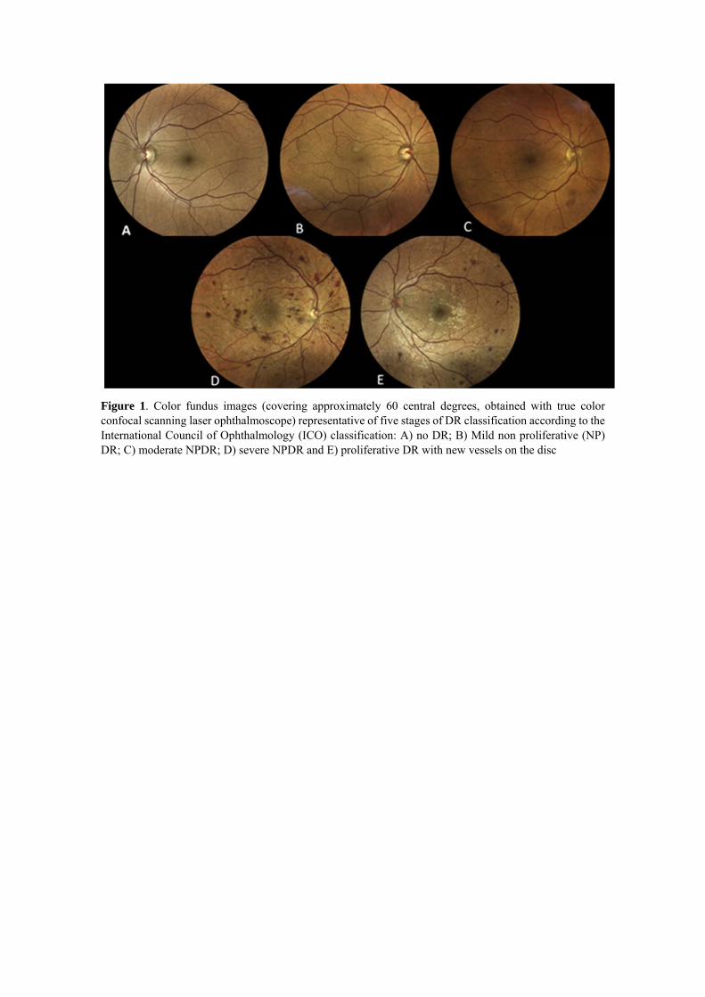
Figure 1. Color fundus images (covering approximately 60 central degrees, obtained with true color confocal scanning laser ophthalmoscope) representative of five stages of DR classification according to the International Council of Ophthalmology (ICO) classification: A) no DR; B) Mild non proliferative (NP) DR; C) moderate NPDR; D) severe NPDR and E) proliferative DR with new vessels on the disc

Figure 2. Color fundus images (on the left) and optical coherence tomography (OCT) images (on the right) representative of three stages of DME classification according to the International Council of Ophthalmology (ICO) classification: A) no DME; B) non-center-involving DME and C) center-involving DME.

Figure 3. Left: central 60° fundus field of the right eye showing center-involving DME with moderate NPDR. Right: Ultra wide-field composite image covering 150° of the left eye Left eye showing proliferative DR (with new vessels on the disc, numerous hemorrhages and venous beading) with some peripheral laser.

Figure 4. Bayesian network model predicting the 5 year risk of diabetic retinopathy progression that depict the relation between multiple variables. Potentially this will facilitate a contemporary data-driven model for diabetic retinopathy progression in various clinical scenarios, most of which have not been directly addressed by clinical trials. PDR: proliferative diabetic retinopathy, NPDR: nonproliferative diabetic retinopathy, A1c: hemoglobin A1c, BP: blood pressure, DBP: diastolic blood pressure; Max: maximum; Min: minimum; SBP: systolic blood pressure.

Figure 5. Basic structural design of a convolutional neural network (CNN) includes convolution, pooling and a fully connected layer. A CNN is a deep learning algorithm which can take in an input image, assign importance to different features in an image and is able to differentiate one from the other. This self-learning approach removes the need to explicitly identify the features in images that in the past had made retinal image analysis difficult to automate. Typical CNN contains parallel mechanisms of convolution and pooling operations alternating to one another. The output from the convolution and pooling layers represent high-level features of the input image and the purpose of the fully connected layer is to use these features for classifying the input image into various classes based on the training dataset.