scoliosis - gss · 3, then gradual reduction). seas (scientific exercises approach to scoliosis)...
TRANSCRIPT
This Provisional PDF corresponds to the article as it appeared upon acceptance. Fully formattedPDF and full text (HTML) versions will be made available soon.
"Brace technology" thematic series - The Sforzesco and Sibilla braces, and theSPoRT (Symmetric, Patient oriented, Rigid, Three-dimensional, active) concept
Scoliosis 2011, 6:8 doi:10.1186/1748-7161-6-8
Stefano Negrini ([email protected])Gianfranco Marchini ([email protected])
Fabrizio Tessadri ([email protected])
ISSN 1748-7161
Article type Methodology
Submission date 3 January 2011
Acceptance date 9 May 2011
Publication date 9 May 2011
Article URL http://www.scoliosisjournal.com/content/6/1/8
This peer-reviewed article was published immediately upon acceptance. It can be downloaded,printed and distributed freely for any purposes (see copyright notice below).
Articles in Scoliosis are listed in PubMed and archived at PubMed Central.
For information about publishing your research in Scoliosis or any BioMed Central journal, go to
http://www.scoliosisjournal.com/info/instructions/
For information about other BioMed Central publications go to
http://www.biomedcentral.com/
Scoliosis
© 2011 Negrini et al. ; licensee BioMed Central Ltd.This is an open access article distributed under the terms of the Creative Commons Attribution License (http://creativecommons.org/licenses/by/2.0),
which permits unrestricted use, distribution, and reproduction in any medium, provided the original work is properly cited.

- 1 -
Brace technology thematic series - The Sforzesco and Sibilla braces, and the SPoRT (Symmetric, Patient oriented, Rigid, Three-dimensional, active) concept
Stefano Negrini1§
, Gianfranco Marchini2, Fabrizio Tessadri
3
1ISICO (Italian Scientific Spine Institute), Milan, Italy
2Centro Ortopedico Lombardo, Milan, Italy
3Orthotecnica, Trento, Italy
§Corresponding author
Email addresses:

- 2 -
Abstract
Background
Bracing is an effective strategy for scoliosis treatment, but there is no consensus on
the best type of brace, nor on the way in which it should act on the spine to achieve
good correction. The aim of this paper is to present the family of SPoRT (Symmetric,
Patient-oriented, Rigid, Three-dimensional, active) braces: Sforzesco (the first
introduced), Sibilla and Lapadula.
Methods
The Sforzesco brace was developed following specific principles of correction. Due to
its overall symmetry, the brace provides space over pathological depressions and
pushes over elevations. Correction is reached through construction of the envelope,
pushes, escapes, stops, and drivers. The real novelty is the drivers, introduced for the
first time with the Sforzesco brace; they allow to achieve the main action of the brace:
a three-dimensional elongation pushing the spine in a down-up direction.
Brace prescription is made plane by plane: frontal (on the “slopes”, another novelty of
this concept, i.e. the laterally flexed sections of the spine), horizontal, and sagittal.
The brace is built modelling the trunk shape obtained either by a plaster cast mould or
by CAD-CAM construction. Brace checking is essential, since SPoRT braces are
adjustable and customisable according to each individual curve pattern.
Treatment time and duration is individually tailored (18-23 hours per day until Risser
3, then gradual reduction). SEAS (Scientific Exercises Approach to Scoliosis)
exercises are a key factor to achieve success.
Results
The Sforzesco brace has shown to be more effective than the Lyon brace (matched
case/control), equally effective as the Risser plaster cast (prospective cohort with

- 3 -
retrospective controls), more effective than the Risser cast + Lyon brace in treating
curves over 45 degrees Cobb (prospective cohort), and is able to improve aesthetic
appearance (prospective cohort).
Conclusions
The SPoRT concept of bracing (three-dimensional elongation pushing in a down-up
direction) is different from the other corrective systems: 3-point, traction, postural,
and movement-based. The Sforzesco brace, being comparable to casting, may be the
best brace for the worst cases.
Background Bracing is an effective strategy for scoliosis treatment, even if proof regarding its
efficacy is currently still weak [1, 2]. Nevertheless, since the efficacy of bracing
comes from both good quality construction and good compliance [3], bracing should
never be interpreted only in terms of the brace applied, but also in terms of the
management of patients [4]. In fact, compliance is a characteristic neither of the
treatment only, nor of the patient alone, but of the good interaction between these two
factors and an expert treatment team able to reduce the burden of the brace and
increase the coping abilities of the patient.
The expert members of the international Society on Scoliosis Orthopaedic and
Rehabilitation Treatment (SOSORT) have not been able to reach a consensus on an
optimal brace design, nor on the way it should act on the spine to achieve good
correction [5]; on the contrary, they have reached consensus on the proper
management of patients to achieve good results [4]. Looking at the existing studies
performed using the Scoliosis Research Society (SRS) methodological criteria, and
dividing them into two groups (one respecting also the SOSORT criteria [6, 7], and
another not doing so) it appears that the best results are obtained by the first group [8].

- 4 -
So, the currently available international knowledge seems to agree that the type of
brace used is less important than the way in which a brace is applied (SOSORT
criteria) [4].
Nevertheless, this way of thinking could drive the field to a form of nihilism, where
what you do (brace) is less important than how you do it (SOSORT criteria).
Consequently, a comparison among the different tools applied by different physicians
is mandatory, in order to understand these tools and to be able to separate their
different indications. Until now, there have been very few comparison studies on
different braces: one RCT [9], and some studies mainly with historical controls [10-
16]. A critical assessment of some of these studies is vital, since in certain cases there
has been doubt that the authors were experts in the use of the types of braces
evaluated in the study. As a consequence, a more sound understanding of the basis
behind the use of different braces is required to increase common background
knowledge and to finally be able to safely compare the different instruments.
The aim of this paper is to present in a journal article format the SPoRT braces
(Sforzesco, Fig. 1; Sibilla, Fig. 2; and Lapadula, Fig. 3), which today constitute a
family of braces constructed following a single concept of bracing (SPoRT). A
complete booklet version of this work can be freely downloaded
(http://www.isico.it/uk/sforzesco).
History The Sforzesco brace, named in honour of the Medieval Sforza family (Fig. 4), was
developed by two of the authors (SN and GM) in the autumn of 2004 while searching
for a way to avoid casting for the worst patients Subsequently, the SPoRT
(Symmetric, Patient-oriented, Rigid, Three-dimensional, active) concept of bracing

- 5 -
was developed [10, 17, 18]. which also included the previously existing Lapadula and
Sibilla brace designs [19, 20].
In the development and construction of the Sforzesco brace, it is possible to recognise
elements of various previously-developed braces: Risser cast [20-22] (Fig. 5A), Lyon
[23] (Fig. 5B), Sibilla [19, 20] (Fig. 5C), and Milwaukee [24, 25] (Fig. 5D) braces.
After the first development, “contaminations” with braces from expert builders from
all over the world (i.e. changes made looking at other concepts) was achieved,
including now elements from the Cheneau (Fig. 5E) [26-29] and Rigo Cheneau
System (RCS) (Fig. 5F) [26, 28, 30] braces.
Theoretical principles From a theoretical perspective, the authors started this research with very well-
established principles of correction that had developed over the years. These
principles are divided in terms of efficacy (type and quality of the brace) and
acceptability (compliance). The efficacy principles include [31, 32]: mechanical
efficacy, the active brace principle (http://www.youtube.com/watch?v=u87UonO-
1Yg&feature=player_embedded), versatility and adaptability, teamwork, compliance.
The acceptability principles of correction (meaning compliance as well as a human
approach to the patient) include: perfect body design and minimal visibility (Fig. 6),
maximum freedom in the Activities of Daily Life (Fig. 7), assumption of
responsibility, cognitive-behavioural approach by the entire professional team [33].
The SPoRT acronym [10, 17, 18, 31, 34], developed according to these principles,
means: Symmetric, Patient-oriented [35], Rigid, Three-dimensional, active.
The Brace Three braces follow the SPoRT concept of correction. The Sforzesco brace (Fig. 1) is
constructed with rigid polycarbonate, in two pieces, connected posteriorly at the
- 6 -
midline by a vertical aluminium bar and anteriorly by a closure that is rigid over the
breast and below is made of soft inelastic bands. While the brace appears to be in full
contact, in reality due to its symmetry and according to the theoretical body shape the
patient would have without scoliosis, it provides space over depressions and pushes
over pathological elevations.
The other two braces are made of polyethylene. In terms of construction and
correction approach, the Sibilla (Fig. 2) and Lapadula braces (Fig. 3) are completely
analogous to the Sforzesco brace, and therefore they will be considered together. The
only difference between the two is that the Lapadula brace does not have the upper
plastic part over the breast (it also addresses kyphosis in combination with scoliosis
through the use of acromion metallic pushes - Fig. 8).
The main innovation of the SPoRT braces can be found in the concept of drivers. This
was introduced for the first time in bracing with the Sforzesco brace [10, 31], and was
discovered due to the abundance of material used to guarantee the rigidity that was
necessary to emulate the strength of the Risser cast. This material no longer allowed
the trunk to escape from the pushes: the only real escape remaining to the spine as
soon as the maximum external symmetry is achieved (i.e. the drivers are reached) is in
elevation (Fig. 9).
Correction is reached through construction (shape of the envelope), pushes, drivers
(concept newly-introduced with this brace), escapes, stops.
Practical Issues
How to prescribe the brace and principles of correction
Prescribing the SPoRT braces requires a careful three-dimensional evaluation of the
characteristics of the curve of each single patient. Clinical reasoning follows a
systematic path by looking progressively at the single component of the deformity.
- 7 -
Frontal plane correction
The slopes
Correction on the frontal plane is based on the identification not of the curves (as
usual), but of the slopes, that are the most frontally flexed segments of the spine. In
fact, since the brace works by pushing the spine from behind, and due to the presence
of the drivers that avoid undesired actions, pushes are focussed on the most severely
flexed area of the spine (slopes). In a down-up direction, the following slopes can be
described:
• Low lumbar (Fig. 10A): in a lumbar curve, below the apical vertebra.
• High lumbar (Fig. 10B): in a short thoracolumbar curve, below the apical
vertebra; or in a very short lumbar curve, above the apical vertebra.
• Lumbar (Fig. 10C): in a wide thoracolumbar curve, below the apical vertebra.
• Thoracolumbar (Fig. 10D): in a lumbar curve, above the apical vertebra; or in
a low thoracic curve, below the apical vertebra.
• Thoracic (Fig. 10E): in a thoracolumbar curve, above the apical vertebra; or in
a single thoracic curve, below the apical vertebra; or in a double thoracic
curve, above the apical vertebra of the distal curve and below the apical
vertebra of the proximal one.
• Distal thoracic (Fig. 10F): specified only in Double Moe curves where three
thoracic slopes are present, below the apical vertebra of the distal curve.
• Proximal thoracic (Fig. 10G): in a thoracic curve, above the apical vertebra.
When evaluating slopes, it is important to decide which is the most important to
correct and where the CPO should focus in constructing the brace. Once the main
slopes to be corrected have been defined, the correction follows automatically as
shown in an example in Fig. 11. In Table 1 the corrections to be made according to
the identified slopes are reported.

- 8 -
At the thoracic level, the ribs to be pushed must be identified, corresponding to the
flexed vertebrae avoiding the apical vertebra.
The possible actions (not mutually exclusive) at the flanks include:
• Shift: in the case of a low lumbar slope.
• Stop: when there is a lumbar curve on the side opposite to the main slope.
• Remodelling: to improve the aesthetics of one flattened flank.
One main point to be carefully considered is the correction of high thoracic slopes
above the T5 vertebra. Over the years, many possible solutions have been tried,
including pushes on the cervical transverse processes, elevation of one shoulder, and
finally something called “Cheneauisation”, that is an inclination of the entire brace
above the apical vertebra of the thoracic curve opposite to the proximal slope,
together with an advancement of the shoulder on the same side (Fig. 12). The term
Cheneauisation was used to underline the fact that it derives from the contamination
of our own brace with one of the other most well-known braces at the international
level, the Cheneau brace. A cervical push on the transverse process (Fig. 13) can be
prescribed in many situations when it is deemed important to act on the cervico-
thoracic junction.
The drivers
On the frontal plane, the main drivers are placed laterally on the concave side, i.e. at
the level of the waist and/or the thorax. They act mainly in a down-up direction from
the apical vertebra of the curve, even if their action starts where the contra lateral push
begins. They direct the forces above.
The horizontal plane The correction on the horizontal plane is totally based on the hump characteristics
combined with the needs on the sagittal plane. In general the push is realised with a
- 9 -
plastazote addition inside the external envelope following exactly the apparent
prominence, as shown in Fig. 14.
At the lumbar level, any horizontal derotatory push on the hump corresponds to a
useful reconstruction of the lordosis usually needed in this area. There are no real
concerns of sagittal damage. As a consequence, the push is directly on the transverse
apophysis, which can potentially also add a frontal plane corrective action (Fig. 15A).
Obviously, in the rare cases of associated hyperlordosis all the brace will be built in
delordosis.
At the thoracic level, on the contrary, the derotatory push can damage the sagittal
plane, and must be carefully planned. In this respect, it is mandatory not to reach the
transverse processes, so as to allow for possible leverage by the ribs that could even
result in a kyphosing action (Fig. 15B). This leverage is at the base of the derotation
and possibly deflexion action of the push on the hump. Moreover, the push must be
below the apex of kyphosis to avoid its flattening. Above it, the push should be on the
proximal counter-rotation appearing as a consequence of the thoracic thrust on the
hump. This will allow on one hand an action to reconstruct the kyphosis, and on the
other hand will increase the direct derotating (and modelling) push on the hump, as
well as a realignment of the shoulder girdle otherwise rotated opposite to the convex
side of the curve, due to the push on the thoracic hump.
At the thoracolumbar level, the action is usually similar to that at the lumbar one. In
fact, most of the cases in this region appear with a junctional kyphosis, which is
contrasted by a posterior push on the hump. In the few cases in which a junctional
lordosis is present, the push must be present, but moderate to avoid increasing the
sagittal deformity.

- 10 -
The drivers
On the horizontal plane, the main drivers are anterior, where they avoid the anterior
escape of the trunk driving in rotation, and posterior on the opposite side of the push,
which are reached only when complete derotation is achieved and the push is driven
upward.
The sagittal plane This correction is almost completely done through the construction, since afterwards
during checks it is almost impossible to really correct this point. The sagittal shaping
of the brace during construction almost always changes according to the given
patient’s sagittal curve.
The drivers
On the sagittal plane, all the drivers previously listed for the other planes play a
crucial role in driving the forces not only upward but also slightly backward at the
thoracolumbar junction, and anteriorly over the apex of kyphosis.
How to build the brace
The SPoRT concept always requires a customised construction of the brace according
to the patient’s individual requirements. CAD-CAM technologies usually allow us to
obtain the best results, without using pre-built forms stored in databases, as is often
done by others. Orthotists must directly shape the scanned trunk according to the
patient’s requirements. Once done, a final test must be made on the patient so as to
change the first theoretical project and adapt it in the best possible way, depending on
the real interaction between the body and the brace.
The brace is built through careful modelling of the trunk shape either on the cast
mould or on the PC screen. The cast is sometimes constructed in a step by step
procedure in down-up direction already trying to achieve a good correction. At first,
maximum symmetry is searched for among the trunk volumes in three dimensions,

- 11 -
looking at circumferences (Fig. 16A) and shapes (Fig. 16B). Then, the sagittal plane
is shaped. Finally, all planes are re-checked.
When the mould is ready, the plastic TLSO is fabricated, and the patient is fit
according to his/her needs, allowing for good sitting position and total freedom of
movement. The pushes are finally added at the level of the humps according to the
desired corrections.
How to check the brace
Brace checking is a fundamental step in any brace construction [4]. This is especially
true in braces following the SPoRT concept, since they are adjustable and
customisable according to any individual curve pattern. The reaction of the body to
predisposed project of the brace should also be considered during prescription and
building. Brace checking is moreover a key psychological intervention on the patient
and family, mainly, but not only, with the first brace.
On the frontal plane, one has to search for the area in which correction is not ideal:
corrections may be applied increasing the pushes or decreasing the drivers and
counter pushes. On the sagittal plane, besides the appearance of the brace that must be
properly aligned with respect to a normal kyphosis and lordosis, it is necessary to
check inside the brace, and eventually either act on the posterior aluminium bar of the
brace, or add plastazote pushes. On the horizontal plane, the check is made without
the brace looking at the effect of the pushes on the humps. Finally, the total balance of
the braced trunk is assessed, to avoid sagittal or frontal shifts (and rarely horizontal
rotations). Other technical points to be checked include the non-overlap of pushes,
that must be done on a plane by plane basis and the balance among the pushes (in the
Sforzesco brace, pushing too much on a secondary curve has the consequence of
reducing the efficacy on the main one).

- 12 -
An “in brace” radiograph is usually done only once, almost 45 days after the initial
fitting of the first brace, but sometimes more often if there are problems.
Protocols and everyday usage
Brace treatment must almost always achieve very good aesthetic body shaping [36]. It
is intended to achieve radiographic results that are compatible with good functioning
of the spine in adulthood, while the quality-of-life impact and psychological
disturbances due to the brace must be minimised [5, 17, 37].
The type of brace is chosen according to the rigidity of the scoliosis to be treated. In
large curves (over 40°), that are always rigid, the Sforzesco brace is used. Before
puberty, in juveniles or infantile scoliosis patients, the Sibilla brace is prescribed with
the very rare exceptions of a very rigid curve; in all other clinical situations, a case by
case choice is made. The Lapadula brace is used as an alternative to the Sibilla in
lumbar and thoraco-lumbar curves.
The goal of brace treatment varies according to the degree of curvature considered,
and the forces that(in terms of rigidity of the brace and the hours of usage) are
consequently administered [31]. Treatment is tailored according to individual
preferences, anthropometric characteristics and other risk factors such as rotation,
hump, lumbar curve take-off, imbalance, etc. It usually starts at full time. Actually,
the applied full-time concept varies between 18 and 23 hours per day [3, 38] with the
goal of obtaining compliance. Treatment is carried out by wearing the brace at least
18 hours per day until the period of rapid growth is over and other adjustments due to
the pathology are not foreseen. This is usually achieved at Risser stage 3.
Weaning requires a two-hour reduction every six months. This protocol has been
developed in our Institute over many years in order to help the postural neuro-
muscular system maintain the achieved correction [31] as well as to maximize

- 13 -
compliance. In fact, while scoliosis is a bone deformity, there is also a postural
component of the curve [39] that always increases it [40] and can be the basis of its
progression [31, 41]. Moreover, while movement has been shown to be a crucial
progression factor [42, 43], it can also be reorganized to become a stability factor
[44]. Braces directly interfere with such neuromuscular functions [1, 2, 41]. Because
posture and movement require long-term adaptations [41, 45], the longer the weaning
phase, the better the neuromuscular system should adapt, hopefully maintaining the
inputs received by the brace even after complete weaning. In this respect, proper
stabilization exercises should play a major role reducing the concertina effect (Fig.
17) [31, 46]. All this should positively interfere with bone tissue formation [42, 43],
even if the postural system per se is part of the problem to be corrected [39-41].
Exercises
We apply the Scientific Exercises Approach to Scoliosis (SEAS) as developed by
ISICO in these years [31]. The main goals of exercises in brace treatment are
elimination or reduction of side effects caused by immobility (muscular hypotrophy
and joint rigidity), or the brace itself (reduction of sagittal curves, mainly kyphosis,
and breathing impairment) and accentuation of brace corrective pushes [47-49].
Moreover, exercises aim at not loosing correction while weaning the brace [46]. Such
goals are pursued through specific therapeutic modalities, subdivided into the
following three treatment phases: preparation for bracing (Fig. 18A); brace wearing
period (Fig. 18B and 18C); complete brace weaning (Fig. 18D) [31].
We have recently shown in prospective controlled studies the importance of exercises
in preparation for brace treatment so to increase its efficacy at first wearing [50], and
in retrospective studies the usefulness of SEAS exercises in order to not lose

- 14 -
correction while weaning from the brace [46]. We have also shown which exercises
are more useful in increasing the pushes of the brace [49].
Results and case reports The short term results currently available on the SPoRT concept relate to the
Sforzesco brace and are quite promising. Although the first treated patients have
already reached the end of treatment, there are not yet enough of them to be able to
perform a formal study. Nevertheless, even if we are perfectly aware that clinical case
reports (Figure 19, 20, 21, 22, and 23) are not comparable to strong scientific data
coming from other studies, those we presents here convey in our view an important
message to the reader and allow a deeper understanding of the effectiveness of this
brace.
With specific studies we have shown that the Sforzesco brace:
• is more effective than the Lyon brace after six months of treatment, with a
prospective matched case/control prospective study [10, 18] on 30 AIS patients
aged 13 years and with curves of 38° Cobb on average: in the Sforzesco group
80% of patients improved and none worsened, while the Lyon group had results of
53% and 13%, respectively.
• is equally effective as the Risser plaster cast to achieve the maximal correction
after 18 months of treatment, with a prospective cohort study with a retrospective
control group [34] on 41 AIS patients aged 14 years and with curves of 40° Cobb.
The Sforzesco brace was shown to be more effective at reducing the thoracic
curve, and its results were super imposable for the other regions. The Risser
plaster brace was shown to be more effective on the thoracic hump and in regard
to the cosmetic appearance of the flanks, but it also caused a serious reduction in
kyphosis.

- 15 -
• is more effective than the Risser cast + Lyon brace in treating curves over 45°
Cobb at the end of growth, with a prospective cohort study in patients who utterly
refused surgery [51] on 28 AIS patients aged 14 years and with curves of in a
range of from to 58° Cobb. The patients braced with the Sforzesco had better
results than those treated with the Risser cast in the thoracic curves, without any
sagittal plane worsening. For the other parameters, the results were similar.
• is able to improve aesthetics in scoliosis patients, with a prospective not-
controlled cohort study [52] on 34 consecutive AIS patients 13 years old with
curves of 32° Cobb with Aesthetic Index (AI) [36] scores of at least 5/6. At
baseline, median AI was 6 (95% IC 5-6), but the score decreased to 3 (95% IC 0-
5; p<0.05) after six months with the brace, and this value was maintained in the 29
who completed the treatment (95% IC 1-6; p<0.05 with respect to baseline).
Discussion The Sforzesco brace has been developed recently, but it is already one of the most
tested TLSOs in the very weak scientific history of bracing. We are not able to
compare it with any other that we did not use personally, but we can already state
according to our results that its efficacy is higher than that of the Lyon brace [10], and
comparable (or even higher as well) to that of the Risser cast [16]. In fact, we use to
think of the Sforzesco brace as a cast, with the great advantage on one hand that it can
be removed to greatly increase patient comfort, and on the other hand that it can be
used from the beginning to the end of treatment without problems, which cannot be
done with the Risser cast. We cannot exclude in the future the possibility that the cast
(or the Lyon brace) will still find a place in scoliosis treatment for some particular
curves or patients, but we are not able now to exactly identify these clinical situations.

- 16 -
According to the reported results, we have a strong basis for reasoning that this brace
could be more effective in the worst curves than other braces. In fact, to our
knowledge, there is only one published paper with good results on curves over 45°
Cobb, and they have been obtained either with Risser casting or with Sforzesco
bracing [51]. This conclusion needs to be supported by future evaluations and
understanding, as well as study results reported by others with other braces.
Limitations can obviously be found today in the fact that the use of this brace is
limited to Italy; we can anyway already state that the usage of the Sforzesco brace has
already spread outside the first orthotic manufacturer and the first physician and his
team. Nevertheless, we need studies from other teams, as is common with instruments
at their first stages of development. A typical disadvantage of this instrument is that it
is apparently simple. In fact, to a superficial observer it could appear as a simple full-
contact brace. In reality, there are complex mechanical concepts and understanding
that must be developed to be able to correctly apply this family of braces. Its apparent
simplicity could easily drive its spread but could also lead to misconduct in its
application. Moreover, another disadvantage is that the messages given to the patients
are vital to success as well, and must be well understood. The SPoRT concept could
also be applied to other braces beyond the ones presented here.
Conclusions Looking at the braces used around the world, most of them are based on three-points
systems, more or less three dimensional [26, 28, 53-61], but we can also recognise a
traction system [62-64], a postural one [65-67], and finally a corrective-movement
based [44, 68]. The SPoRT concept of bracing, due to its three-dimensional action of
elongation pushing the spine in a down-up direction, is different from all the other
corrective systems. The Sforzesco brace appears as the best brace for the worst curve

- 17 -
magnitudes, being comparable to casting [16, 51, 69], with the obvious advantage of
being removable and applicable for all duration of treatment.
Bracing is very hard work, in terms of conceptualisation of the practical work to be
done, and of the interaction with the whole team, starting from the MD-CPO
relationship, to the PT, the patient and the family. It is a demanding, progressive,
slow, artisanal effort in the art of patience. In this respect, it is quite the opposite of
the short, one-shot, quick, highly demanding, current surgical fusion. As we use to say
to our patients, bracing corresponds to the very slow pace of building oneself that
humans usually have to face, contrary to the fast solution that he/she may tend to
prefer and see as less demanding. Bracing in this respect also becomes a philosophy
of one’s approach to life, and this is one reason why it is difficult that the slow pace of
a good conservative physician can also be the fast speed of a good orthopaedic
surgeon, and vice versa. As well, there will always be patients who prefer bracing and
others who prefer surgery. This relates to those with high degree curves; in low
degree curves, the choice is between bracing and a “wait and see” strategy, applied in
cases in which bracing is too demanding for that particular patient. But, in our own
experience, at least in Italy, this is very rare [70], even if not avoidable.
Competing interests SN is a physician everyday prescribing braces; he also owns a stock of ISICO.
GM is an Orthotist everyday building braces and owns Centro Ortopedico Lombardo.
FT is an Orthotist everyday building braces and owns Orthotecnica.
Authors' contributions SN drafted the text and figures. GM and FT revised and accepted it, and contributed
with some figures. All authors read and accepted the final version of the manuscript.

- 18 -
Acknowledgements We wish to thank Michele Romano for drawings; all our patients, who allowed us to
increase our knowledge and skill; our collaborators who, working with us in team,
were continuously part of what we learned and were able to do.
References 1. Negrini S, Minozzi S, Bettany-Saltikov J, Zaina F, Chockalingam N, Grivas
TB, Kotwicki T, Maruyama T, Romano M, Vasiliadis ES: Braces for
idiopathic scoliosis in adolescents. Cochrane Database Syst Rev
2010(1):CD006850.
2. Negrini S, Minozzi S, Bettany-Saltikov J, Zaina F, Chockalingam N, Grivas
TB, Kotwicki T, Maruyama T, Romano M, Vasiliadis ES: Braces for
idiopathic scoliosis in adolescents. Spine (Phila Pa 1976) 2010, 35(13):1285-
1293.
3. Landauer F, Wimmer C, Behensky H: Estimating the final outcome of brace
treatment for idiopathic thoracic scoliosis at 6-month follow-up. Pediatr
Rehabil 2003, 6(3-4):201-207.
4. Negrini S, Grivas TB, Kotwicki T, Rigo M, Zaina F: Guidelines on
"Standards of management of idiopathic scoliosis with corrective braces in everyday clinics and in clinical research": SOSORT Consensus 2008.
Scoliosis 2009, 4(1):2.
5. Rigo M, Negrini S, Weiss H, Grivas T, Maruyama T, Kotwicki T: 'SOSORT
consensus paper on brace action: TLSO biomechanics of correction (investigating the rationale for force vector selection)'. Scoliosis 2006,
1:11.
6. Negrini S, Atanasio S, Fusco C, Zaina F: Effectiveness of complete
conservative treatment for adolescent idiopathic scoliosis (bracing and
exercises) based on SOSORT management criteria: results according to the SRS criteria for bracing studies - SOSORT Award 2009 Winner.
Scoliosis 2009, 4:19.
7. Aulisa AG, Guzzanti V, Galli M, Perisano C, Falciglia F, Aulisa L:
Treatment of thoraco-lumbar curves in adolescent females affected by
idiopathic scoliosis with a progressive action short brace (PASB):
assessment of results according to the SRS committee on bracing and nonoperative management standardization criteria. Scoliosis 2009, 4:21.
8. Janicki JA, Poe-Kochert C, Armstrong DG, Thompson GH: A comparison of
the thoracolumbosacral orthoses and providence orthosis in the treatment
of adolescent idiopathic scoliosis: results using the new SRS inclusion and assessment criteria for bracing studies. J Pediatr Orthop 2007, 27(4):369-
374.
- 19 -
9. Wong MS, Cheng JC, Lam TP, Ng BK, Sin SW, Lee-Shum SL, Chow DH,
Tam SY: The effect of rigid versus flexible spinal orthosis on the clinical
efficacy and acceptance of the patients with adolescent idiopathic scoliosis. Spine 2008, 33(12):1360-1365.
10. Negrini S, Marchini G: Efficacy of the symmetric, patient-oriented, rigid,
three-dimensional, active (SPoRT) concept of bracing for scoliosis: a prospective study of the Sforzesco versus Lyon brace. Eura Medicophys
2007, 43(2):171-181; discussion 183-174.
11. Weiss HR, Weiss GM: Brace treatment during pubertal growth spurt in
girls with idiopathic scoliosis (IS): a prospective trial comparing two different concepts. Pediatr Rehabil 2005, 8(3):199-206.
12. Katz DE, Richards BS, Browne RH, Herring JA: A comparison between the
Boston brace and the Charleston bending brace in adolescent idiopathic scoliosis. Spine 1997, 22(12):1302-1312.
13. Howard A, Wright JG, Hedden D: A comparative study of TLSO,
Charleston, and Milwaukee braces for idiopathic scoliosis. Spine 1998,
23(22):2404-2411.
14. Bunnell WP, MacEwen GD, Jayakumar S: The use of plastic jackets in the
non-operative treatment of idiopathic scoliosis. Preliminary report. J Bone
Joint Surg Am 1980, 62(1):31-38.
15. Climent JM, Sanchez J: Impact of the type of brace on the quality of life of
Adolescents with Spine Deformities. Spine 1999, 24(18):1903-1908.
16. Negrini S, Atanasio S, Negrini F, Zaina F, Marchini G: The Sforzesco brace
can replace cast in the correction of adolescent idiopathic scoliosis: A controlled prospective cohort study. Scoliosis 2008, 3(1):15.
17. Negrini S, Marchini G, Tomaello L: Efficacy of the Symmetric, Patient-
oriented, Rigid, Three-Dimensional (SPoRT) concept of bracing for
scoliosis: a pair-controlled retrospettive short-term study on the Sforzesco Brace. In: 3rd International SOSORT Meeting: 2006 April 07-08 2006;
Poznan, Poland: SOSORT; 2006.
18. Negrini S, Marchini G, Tomaello L: The Sforzesco brace and SPoRT
concept (Symmetric, Patient-oriented, Rigid, Three-dimensional) versus the Lyon brace and 3-point systems for bracing idiopathic scoliosis. Stud
Health Technol Inform 2006, 123:245-249.
19. Sibilla P: Trent'anni di scoliosi. Lezione "non" magistrale. In: Rachide &
Riabilitazione 2002. Edited by Negrini S, Rainero G, vol. 1. Vigevano:
Gruppo di Studio Scoliosi e patologie vertebrali; 2002: 73-92.
20. Sibilla P: Il trattamento conservativo attivo della scoliosi idiopatica in
Italia. In: Le deformità vertebrali: stato dell'arte. Edited by Negrini S, Sibilla
P, vol. 2. Vigevano: Gruppo di Studio Scoliosi e patologie vertebrali; 2001:
20-41.
21. Risser JC: Scoliosis treated by cast correction and spine fusion. Clin
Orthop Relat Res 1976(116):86-94.
22. Mammano S, Scapinelli R: Plaster casts for the correction of idiopathic
scoliosis. Acta Orthop Belg 1992, 58 Suppl 1:81-84.
23. Stagnara P: Les deformations du rachis. Paris: Expansion Scientifique
Francaise; 1976.
24. Blount WP, Schmidt A: The Milwaukee brace in the treatment of scoliosis.
J Bone Joint Surg 1957, 37:693.

- 20 -
25. Moe JH: Indications for Milwaukee brace non-operative treatment in
idiopathic scoliosis. Clin Orthop Relat Res 1973(93):38-43.
26. Weiss HR, Rigo M: The cheneau concept of bracing--actual standards.
Stud Health Technol Inform 2008, 135:291-302.
27. Kotwicki T, Cheneau J: Passive and active mechanisms of correction of
thoracic idiopathic scoliosis with a rigid brace. Stud Health Technol Inform
2008, 135:320-326.
28. Rigo M, Weiss HR: The Cheneau concept of bracing--biomechanical
aspects. Stud Health Technol Inform 2008, 135:303-319.
29. Kotwicki T, Cheneau J: Biomechanical action of a corrective brace on
thoracic idiopathic scoliosis: Cheneau 2000 orthosis. Disabil Rehabil Assist
Technol 2008, 3(3):146-153.
30. Rigo MD, Villagrasa M, Gallo D: A specific scoliosis classification
correlating with brace treatment: description and reliability. Scoliosis
2009, 5(1):1.
31. Negrini S: The Evidence-Based ISICO Approach to Spinal Deformities,
1st edition edn. Milan, Boston: ISICO; 2007.
32. Negrini S: Bracing adolescent idiopathic scoliosis today. Disabil Rehabil
Assist Technol 2008, 3(3):107-111.
33. Negrini S, Grivas TB, Kotwicki T, Rigo M, Zaina F: Guidelines on
"Standard of management of idiopathic scoliosis with corrective braces in everyday clinics and in clinical research": SOSORT Consensus 2008.
Scoliosis 2009, 4(1):2.
34. Negrini S, Zaina F, Negrini F, Marchini G, Aulisa AG: Sforzesco brace
(SPoRT Concept) versus Risser cast in adolescent idiopathic scoliosis treatment: similar efficacy, with reduced spinal side effects for the brace.
In: 4th International Conference on Conservative Management of Spinal
Deformities: 13-16 May 2007 2007; Boston: SOSORT (Society on Scoliosis
Orthopaedic and Rehabilitation Treatment); 2007.
35. Bunge EM, de Bekker-Grob EW, van Biezen FC, Essink-Bot ML, de Koning
HJ: Patients' preferences for scoliosis brace treatment: a discrete choice
experiment. Spine (Phila Pa 1976) 2010, 35(1):57-63.
36. Zaina F, Negrini S, Fusco C, Atanasio S: How to improve aesthetics in
patients with Adolescent Idiopathic Scoliosis (AIS): a SPoRT brace treatment according to SOSORT management criteria. Scoliosis 2009,
4:18.
37. Negrini S, Grivas TB, Kotwicki T, Maruyama T, Rigo M, Weiss HR: Why do
we treat adolescent idiopathic scoliosis? What we want to obtain and to avoid for our patients. SOSORT 2005 Consensus paper. Scoliosis 2006,
1:4.
38. Wiley JW, Thomson JD, Mitchell TM, Smith BG, Banta JV: Effectiveness of
the boston brace in treatment of large curves in adolescent idiopathic scoliosis. Spine 2000, 25(18):2326-2332.
39. Duval-Beaupere G, Lespargot A, Grossiord A: Flexibility of scoliosis. What
does it mean? Is this terminology appropriate? Spine 1985, 10(5):428-432.
40. Torell G, Nachemson A, Haderspeck-Grib K, Schultz A: Standing and
supine Cobb measures in girls with idiopathic scoliosis. Spine 1985,
10(5):425-427.

- 21 -
41. Smania N, Picelli A, Romano M, Negrini S: Neurophysiological basis of
rehabilitation of adolescent idiopathic scoliosis. Disabil Rehabil 2008,
30(10):763-771.
42. Stokes IA, Burwell RG, Dangerfield PH: Biomechanical spinal growth
modulation and progressive adolescent scoliosis - a test of the 'vicious
cycle' pathogenetic hypothesis: Summary of an electronic focus group debate of the IBSE. Scoliosis 2006, 1:16.
43. Burwell RG, Aujla RK, Grevitt MP, Dangerfield PH, Moulton A, Randell TL,
Anderson SI: Pathogenesis of adolescent idiopathic scoliosis in girls - a
double neuro-osseous theory involving disharmony between two nervous
systems, somatic and autonomic expressed in the spine and trunk:
possible dependency on sympathetic nervous system and hormones with implications for medical therapy. Scoliosis 2009, 4:24.
44. Coillard C, Leroux MA, Zabjek KF, Rivard CH: SpineCor--a non-rigid
brace for the treatment of idiopathic scoliosis: post-treatment results. Eur
Spine J 2003, 12(2):141-148.
45. Didier J: La plasticité de la fonction motrice, 1st edn. Paris: Springer; 2004.
46. Zaina F, Negrini S, Atanasio S, Fusco C, Romano M, Negrini A: Specific
exercises performed in the period of brace weaning can avoid loss of
correction in Adolescent Idiopathic Scoliosis (AIS) patients: Winner of SOSORT's 2008 Award for Best Clinical Paper. Scoliosis 2009, 4(1):8.
47. Nachemson AL, Peterson LE: Effectiveness of treatment with a brace in
girls who have adolescent idiopathic scoliosis. A prospective, controlled
study based on data from the Brace Study of the Scoliosis Research Society. J Bone Joint Surg Am 1995, 77(6):815-822.
48. Stagnara P, Mollon G, De Mauroy J: Reeducation des scolioses. Paris:
Expansion Scientifique Francaise; 1990.
49. Romano M, Carabalona R, Petrilli S, Sibilla P, Negrini S: Forces exerted
during exercises by patients with adolescent idiopathic scoliosis wearing fiberglass braces. Scoliosis 2006, 1:12.
50. Negrini S, Negrini A, Romano M, Verzini N, Parzini S: A controlled
prospective study on the efficacy of SEAS.02 exercises in preparation to bracing for idiopathic scoliosis. Stud Health Technol Inform 2006, 123:519-
522.
51. Negrini S, Negrini F, Fusco C, Zaina F: Idiopathic scoliosis patients with
curves over 45 Cobb degrees refusing surgery can be effectively treated through bracing with curve improvements. The Spine Journal 2011 (in
press).
52. Zaina F, Negrini S, Atanasio S: TRACE (Trunk Aesthetic Clinical
Evaluation), a routine clinical tool to evaluate aesthetics in scoliosis patients: development from the Aesthetic Index (AI) and repeatability.
Scoliosis 2009, 4(1):3.
53. Cheneau J: Corset-Cheneau. Manuel d'Orthopèdie des scolioses suivant la
technique originale, vol. 1. Paris: Frison Roche; 1994.
54. Weiss HR, Werkmann M, Stephan C: The ScoliOlogiC "Cheneau light"
brace--does the reduction of material affect the desired correction? Stud
Health Technol Inform 2006, 123:250-254.
55. Bassett GS, Bunnell WP, MacEwen GD: Treatment of idiopathic scoliosis
with the Wilmington brace. Results in patients with a twenty to thirty-nine-degree curve. J Bone Joint Surg Am 1986, 68(4):602-605.

- 22 -
56. D'Amato CR, Griggs S, McCoy B: Nighttime bracing with the Providence
brace in adolescent girls with idiopathic scoliosis. Spine 2001, 26(18):2006-
2012.
57. de Mauroy JC, Fender P, Tato B, Lusenti P, Ferracane G: Lyon brace. Stud
Health Technol Inform 2008, 135:327-340.
58. de Mauroy JC, Lecante C, Barral F, Daureu D, Gualerzi S, Gagliano R: The
Lyon brace. Disabil Rehabil Assist Technol 2008, 3(3):139-145.
59. Emans JB, Kaelin A, Bancel P, Hall JE, Miller ME: The Boston bracing
system for idiopathic scoliosis. Follow-up results in 295 patients. Spine
1986, 11(8):792-801.
60. Grivas T, Vasiliadis E, Chatziargiropoulos T, Polyzois V, Gatos K: The effect
of a modified Boston brace with anti-rotatory blades on the progression of curves in idiopathic scoliosis: aetiologic implications. Pediatric
Rehabilitation 2003, 6:237 - 242.
61. Veldhuizen AG, Cheung J, Bulthuis GJ, Nijenbanning G: A new orthotic
device in the non-operative treatment of idiopathic scoliosis. Med Eng
Phys 2002, 24(3):209-218.
62. Blount W, Moe J: The Milwaukee Brace. Baltimore: The William and
Wilkins Company; 1973.
63. Lonstein JE, Winter RB: The Milwaukee brace for the treatment of
adolescent idiopathic scoliosis. A review of one thousand and twenty patients. J Bone Joint Surg Am 1994, 76(8):1207-1221.
64. Maruyama T, Takeshita K, Kitagawa T: Milwaukee brace today. Disabil
Rehabil Assist Technol 2008, 3(3):136-138.
65. The Charleston Bending Brace. An Orthothist's Guide to Scoliosis
Management [http://www.srs.org/professionals/bracing_manuals/section7.pdf]
66. Price CT, Scott DS, Reed FR, Jr., Sproul JT, Riddick MF: Nighttime bracing
for adolescent idiopathic scoliosis with the Charleston Bending Brace: long-term follow-up. J Pediatr Orthop 1997, 17(6):703-707.
67. Grivas TB, Rodopoulos GI, Bardakos NV: Night-time braces for treatment
of adolescent idiopathic scoliosis. Disabil Rehabil Assist Technol 2008,
3(3):120-129.
68. Coillard C, Vachon V, Circo AB, Beausejour M, Rivard CH: Effectiveness of
the SpineCor brace based on the new standardized criteria proposed by the scoliosis research society for adolescent idiopathic scoliosis. J Pediatr
Orthop 2007, 27(4):375-379.
69. Negrini S, Atanasio S, Zaina F, Romano M, Parzini S, Negrini A: End-
growth results of bracing and exercises for adolescent idiopathic scoliosis. Prospective worst-case analysis. Stud Health Technol Inform 2008, 135:395-
408.
70. Negrini S, Carabalona R: Social acceptability of treatments for adolescent
idiopathic scoliosis: a cross-sectional study. Scoliosis 2006, 1:14.
71. Negrini S, Fusco C, Romano M, Zaina F, Atanasio S: Clinical and postural
behaviour of scoliosis during daily brace weaning hours. Stud Health
Technol Inform 2008, 140:303-306.

- 23 -
Figures
Figure 1 - The Sforzesco brace
The Sforzesco brace: anterior (A), posterior (B), left (C), top (D), and bottom (E)
views.
Figure 2 - The Sibilla brace
The Sibilla brace: anterior (A), posterior (B), left (C), top (D), and bottom (E) views.
Figure 3 - The Lapadula brace
The Lapadula brace: anterior (A), posterior (B), left (C), top (D), and bottom (E)
views.
Figure 4 - The Sforzesco brace owns its name to the Sforesco castles of Milan and Vigevano
The Sforzesco brace was named according to the two main cities of the experience of
the main author (SN): Vigevano and Milano, which both have castles named
Sforzesco for the Medieval Sforza family.
Figure 5 - Braces at the base of the SPoRT concept development
The concept of SPoRT bracing was developed from the following braces: Risser cast
(A), Lyon (B), Sibilla (C) and Milwaukee (D). The last changes made to the SPoRT
braces also allowed us to consider among their ancestors the last Cheneau brace (E)
and the Rigo Cheneau brace (F).
Figure 6 - Patients want correction and an invisible brace
These patients are wearing their braces in various everyday activities.
Figure 7 - The Sforzesco brace invisibility
The Sforzesco brace was developed in a town of fashion (Milan), and some patients
have stated that this is reflected in its design, that increases wearability.

- 24 -
Figure 8 - The Lapadula brace to treat scoliosis and hyperkyphosis
The Lapadula brace has much versatility and can be adapted to treat an hyperkyphosis
associated with scoliosis.
Figure 9 - The first Sforzesco brace causes a sudden lengthening of the trunk requiring correction in 2 months
Typical correction made to the first Sforzesco brace after a wearing period of 2
months in patients with important thoracic curves: it becomes too short under the
concave shoulder and must be lengthen.
Figure 10 - The slopes
The slopes. Low lumbar (A); High lumbar (B); Lumbar (C); Thoracolumbar (D);
Thoracic (E); Distal thoracic (F); Proximal thoracic (G).
Figure 11 - Example of correction on the frontal plane slopes
Example of correction on the frontal plane slopes of one patient with a thoraco-
lumbar curve of 48° (Risser 1). (A) X-ray pre-brace (48°); (B) aesthetics pre-brace;
(C) aesthetics after 4 months of bracing; (D) x-ray without the brace after 4 months of
bracing: corrected to 23° (i.e. reduction of 25°, -52%); (E) pre-brace planning with
pushes on the right thoraco-lumbar and left thoracic slopes, as well as a lateral shift to
the right of the flanks; (F) constructed brace; (G) application of the pushes in the
constructed brace; (H) in-brace x-ray with a correction to 13° (i.e. 35° of correction,
73%).
Figure 12 - The Cheneauisation of the Sforzesco brace
We used the term Cheneauisation to underline that it derives from the contamination
of our own brace with the internationally well-known Cheneau brace: in fact it aims at
posturally changing the scoliosis curve through a thrust at level of the convexity of the
apical proximal curve and an elevation/medialization of the shoulder at the concave

- 25 -
side. Due to our own SPoRT principles the Cheneuization also includes an
anteposition of this same shoulder.
First line, from left to right: (A) x-rays of the patient at start of treatment (12/07), (B)
after 6 months (6/08), (C) in-brace without Cheneuization (4/09); (D) in-brace with
Cheneuization (5/09), (E) in-brace with Cheneuization after two months of treatment
(7/09).
Second line: graph of x-rays measurements.
Third line, from left to right: (F) the first brace used; the brace trial: (G) without
Cheneuization; (H) with Cheneuization; (I) the brace brace with Cheneuization at
time of the 7/09 x-ray.
This is the first situation in which we used the Cheneuization due to the absence of
correction in a patient with an high-degree sacoliosis refusing surgery, and presenting
with a curve possibly responding to such a change. We then made two braces and
compared their results with in-brace x-rays, with favorable results for the
Chenuization (5/09), that was even increase by time and brace corrections (7/09). The
final out-of-brace progression of scoliosis (1/10) was due to a sudden decrease on
brace usage that this specific patient suffered.
Figure 13 - Cervical push on the transverse process of C7 and above
Cervical push on the transverse process of C7 and above.
Figure 14 - Identification of the prominence to localize the derotation pushes
Identification of the prominence to add plastazote pushes to the envelope. On the left,
top down direction: anterior bending to precisely identify the hump height and ribs
involved and mark them; markers on the skin in standing. On the right: marks
reported on the brace.

- 26 -
Figure 15 - Action of the derotation pushes at thoracic and lumbar levels
At the lumbar level, the push on the hump helps to reconstruct lordosis, and as a
consequence it is directly on the transverse apophysis, potentially also adding a
frontal plane corrective action (A). At the thoracic level, on the contrary, the push can
damage the sagittal plane, and must not reach the transverse processes, so to allow a
possible leverage by the rib that could even result in a kyphotisating action (B).
Figure 16 - Modelling of the trunk shape during brace building
During brace building a careful modelling of the trunk shape is made either on the
cast mould or on the PC screen. In this figure the correction of circumferences (left)
and shapes (right) of a single patient with a right thoraco-lumbar curve is reported. On
the left there are the original (red) and corrected (blue) horizontal sections of the body
at the level of the horizontal lines reported in the middle body shape, where the
original laser scan of the trunk is represented. On the right the frontal contour of the
original (red) and corrected (blue) body shapes are reported, while inside these lines,
in grey, there is the final trunk shape from which the brace is going to be built.
Figure 17 - The concertina effect of brace correction
The concertina effect [31] could explain the importance of patient’s compliance.
According to this hypothesis, each time a brace is weaned the deformity gradually
moves back from the maximal in-brace correction to the original out-of-brace
situation; this reversal is due to a postural collapse [39-41], that is correlated to the
length of brace weaning and the rigidity (flexibility) of the spine [39] (itself correlated
to the stage of growth, the bone age, the muscular endurance and the usual brace
wearing). According to the “concertina effect” hypothesis, the deformity reached at
the end of daily brace weaning gives the allowed compression of the wedged
vertebrae, and consequently the final results. In fact, the more the brace is weaned
- 27 -
daily, the worst the results. We published some preliminary proves of this hypothesis
[71].
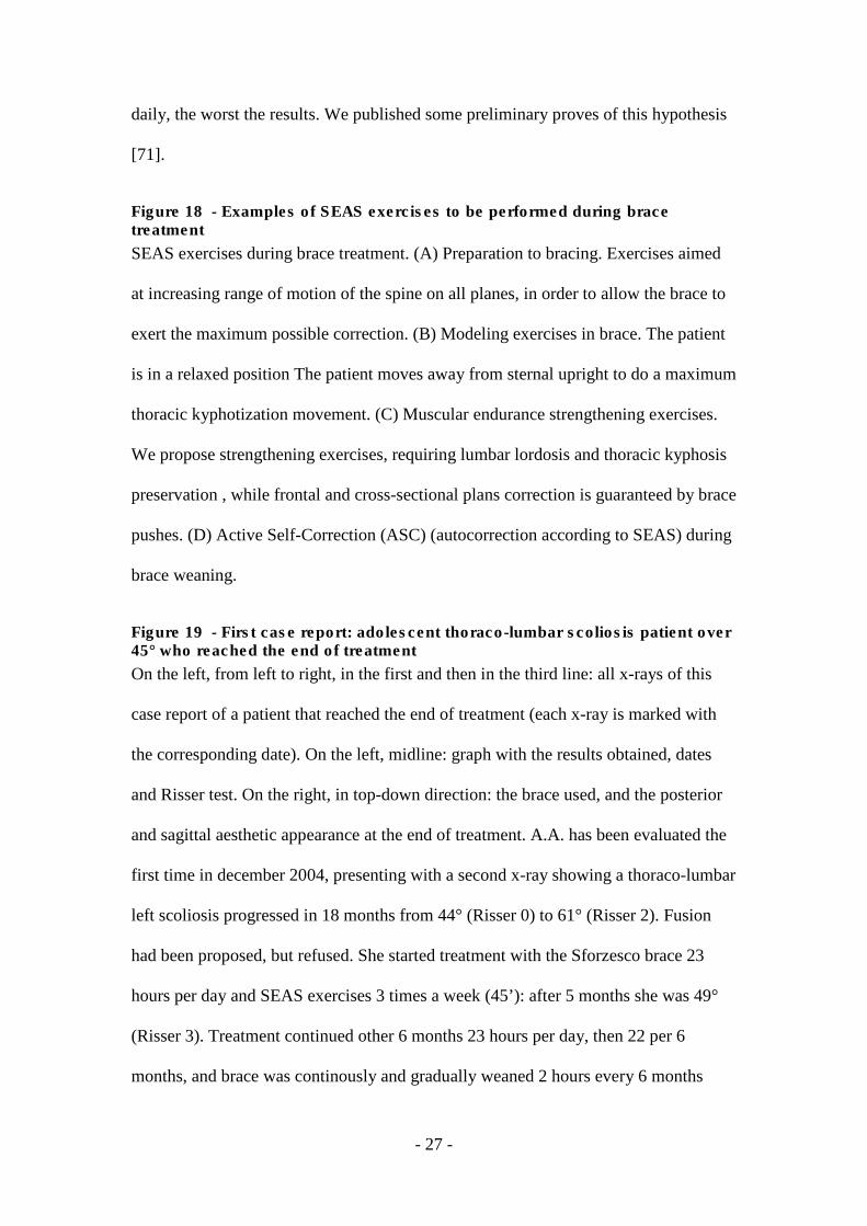
Figure 18 - Examples of SEAS exercises to be performed during brace treatment
SEAS exercises during brace treatment. (A) Preparation to bracing. Exercises aimed
at increasing range of motion of the spine on all planes, in order to allow the brace to
exert the maximum possible correction. (B) Modeling exercises in brace. The patient
is in a relaxed position The patient moves away from sternal upright to do a maximum
thoracic kyphotization movement. (C) Muscular endurance strengthening exercises.
We propose strengthening exercises, requiring lumbar lordosis and thoracic kyphosis
preservation , while frontal and cross-sectional plans correction is guaranteed by brace
pushes. (D) Active Self-Correction (ASC) (autocorrection according to SEAS) during
brace weaning.
Figure 19 - First case report: adolescent thoraco-lumbar scoliosis patient over 45° who reached the end of treatment
On the left, from left to right, in the first and then in the third line: all x-rays of this
case report of a patient that reached the end of treatment (each x-ray is marked with
the corresponding date). On the left, midline: graph with the results obtained, dates
and Risser test. On the right, in top-down direction: the brace used, and the posterior
and sagittal aesthetic appearance at the end of treatment. A.A. has been evaluated the
first time in december 2004, presenting with a second x-ray showing a thoraco-lumbar
left scoliosis progressed in 18 months from 44° (Risser 0) to 61° (Risser 2). Fusion
had been proposed, but refused. She started treatment with the Sforzesco brace 23
hours per day and SEAS exercises 3 times a week (45’): after 5 months she was 49°
(Risser 3). Treatment continued other 6 months 23 hours per day, then 22 per 6
months, and brace was continously and gradually weaned 2 hours every 6 months
- 28 -
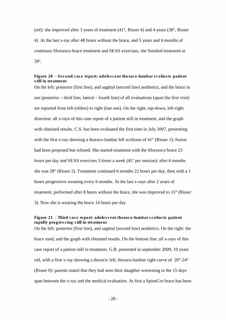
(ref): she improved after 3 years of treatment (41°, Risser 4) and 4 years (38°, Risser
4). At the last x-ray after 48 hours without the brace, and 5 years and 6 months of
continuos Sforzesco brace treatment and SEAS exercises, she finished treatment at
39°.
Figure 20 - Second case report: adolescent thoraco-lumbar scoliosis patient still in treatment
On the left: posterior (first line), and sagittal (second line) aesthetics, and the brace in
use (posterior – third line; lateral – fourth line) of all evaluations (apart the first visit)
are reported from left (oldest) to right (last one). On the right, top-down, left-right
direction: all x-rays of this case report of a patient still in treatment, and the graph
with obtained results. C.S. has been evaluated the first time in July 2007, presenting
with the first x-ray showing a thoraco-lumbar left scoliosis of 41° (Risser 1): fusion
had been proposed but refused. She started treatment with the Sforzesco brace 23
hours per day and SEAS exercises 3 times a week (45’ per session): after 6 months
she was 28° (Risser 2). Treatment continued 6 months 22 hours per day, then with a 1
hours progressive weaning every 6 months. At the last x-rays after 2 years of
treatment, performed after 8 hours without the brace, she was improved to 15° (Risser
3). Now she is wearing the brace 14 hours per day.
Figure 21 - Third case report: adolescent thoraco-lumbar scoliosis patient rapidly progressing still in treatment
On the left: posterior (first line), and sagittal (second line) aesthetics. On the right: the
brace used, and the graph with obtained results. On the bottom line: all x-rays of this
case report of a patient still in treatment. G.B. presented in september 2009, 10 years
old, with a first x-ray showing a thoracic left, thoraco-lumbar right curve of 28°-24°
(Risser 0): parents stated that they had seen their daugther worsening in the 15 days
span between the x-ray and the medical evaluation. At first a SpineCor brace has been

- 29 -
prescribed but the x-ray within brace showed such a bad situation (14°-30°) to suggest
to re-evaluate a radiograph without the brace: scoliosis was rapidly worsening (26°-
39°). We decided to move to a SPoRT brace and SEAS exercises (twice a week, 45’
per session): Sibilla 23 hours per day. In 6 months, while growing 6 cm. (from 145 to
151), she corrected to 17°-18°, and in 6 more months wearing the brace 22 hurs per
day, she arrived to 13°-14°, during an height increase of other 6 cm. (from 151 to
157). Now she continues to be Risser 0, and is wearing the brace 21 hours per day.
Figure 22 - Fourth case report: adolescent double thoracic, lumbar scoliosis patient over 45° still in treatment
On the left: posterior (first line), and sagittal (second line) aesthetics, and the brace in
use (posterior – third line; lateral – fourth line) of all evaluations (apart the first visit,
where aesthetics and first x-rays are shown) are reported from left (oldest) to right
(last one). On the right, top-down, left-right direction: all x-rays of this case report of
a patient still in treatment, and the graph with obtained results in the two curves:
upper line, thoracic curve, lower one lumbar curve. C.F. has been evaluated the first
time in September 2008, presenting with the first x-ray showing a thoracic right
lumbar left scoliosis of 46°-39° (Risser 0). She started treatment with the Sforzesco
brace 23 hours per day and SEAS exercises 2-3 times a week (45’ per session): after 6
months, while growing 5.5 cm. (from 158.5 to 164), she was 36°-31° (Risser 0).
Treatment continued 6 more months 23 hours per day, then reduced to 22: in this
year, while growing 4 cm. (to 168), she reduced her scoliosis to 29°-27°. After 6
months at 20 hours, now she is wearing the brace 18 hours per day.
Figure 23 - Fifth case report: juvenile thoracic scoliosis patient of 45° who weaned the brace but is still in treatment
First line, left to right: x-rays at brace wearing (March 2005) and at brace weaning
(October 2008), frontal and sagittal aesthetics in the last evaluations after brace
- 30 -
weaning. Second line: all frontal x-rays of this case report of a patient still in
treatment. Third line: sagittal x-rays, and the graph with obtained results. B.C.
infantile thoracic right scoliosis was discovered at the age of 13 months, and rapidly
progressed without treatment from 27° to 40° in 4 months as soon as she reached the
standing position. A Sibilla brace treatment was then started 23 hours per day per 4
months and 20 during the summer, with an immediate reduction to 16° in 8 months.
Brace was then gradually reduced 2 hours every six months while maintaining
correction, and finally weaned with the curve at 10°. At the age of 6, as soon as was
possible, everyday SEAS exercises (20’ per session) have been started and are the
only actual treatment.
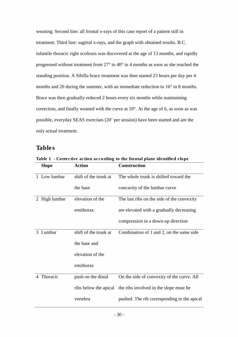
Tables
Table 1 - Corrective action according to the frontal plane identified slope
Slope Action Construction
1 Low lumbar shift of the trunk at
the base
The whole trunk is shifted toward the
concavity of the lumbar curve
2 High lumbar elevation of the
emithorax
The last ribs on the side of the convexity
are elevated with a gradually decreasing
compression in a down-up direction
3 Lumbar shift of the trunk at
the base and
elevation of the
emithorax
Combination of 1 and 2, on the same side
4 Thoracic push on the distal
ribs below the apical
vertebra
On the side of convexity of the curve. All
the ribs involved in the slope must be
pushed. The rib corresponding to the apical

- 31 -
vertebra is not involved (and sometimes
also that below the apical)
5 Distal thoracic push on the distal
ribs above the apical
vertebra
On the side of concavity of the curve
6 Proximal
thoracic
push on the distal
ribs above the apical
vertebra
On the side of concavity of the curve
7 Thoracolumbar elevation of the
emithorax and push
on the distal ribs
below the apical
vertebra
Combination of 2 and 4, on the same side

Figure 1

Figure 2

Figure 3

Figure 4

A B C
D E F
Figure 5
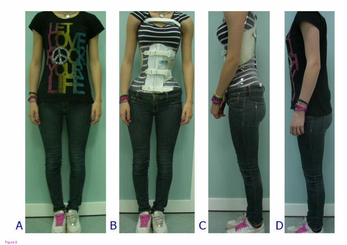
Figure 6

Figure 7
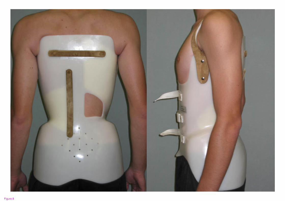
Figure 8
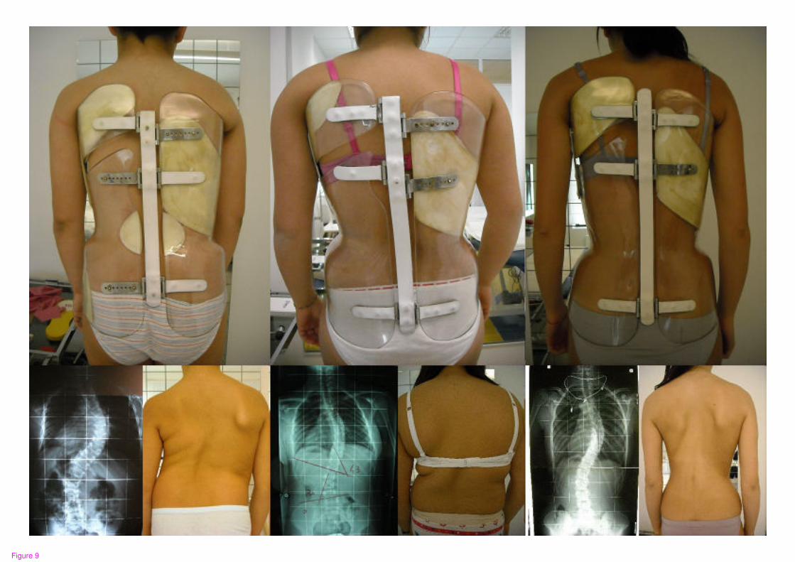
Figure 9

Figure 10

Figure 11

Figure 12

Figure 13

Figure 14

A B
Figure 15
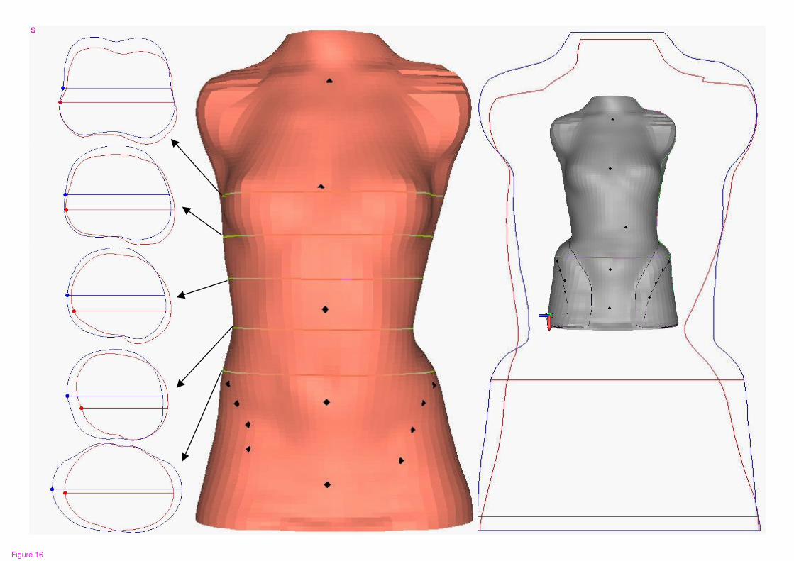
Figure 16

Case 1: compliant
10
15
20
25
30
35
40
D1 h 14:00
D1 h 15:00
D1 h 16:00
D1 h 17:00
D1 h 18:00
D1 h 19:00
D1 h 20:00
D1 h 21:00
D1 h 22:00
D2 h 14:00
D2 h 15:00
D2 h 16:00
D2 h 17:00
D2 h 18:00
D2 h 19:00
D2 h 20:00
D2 h 21:00
D2 h 22:00
Day & Hour
Co
bb
de
gre
es
Case 2: non compliant
10
15
20
25
30
35
40
D1 h 14
:00
D1 h 16
:00
D1 h 18
:00
D1 h 20
:00
D1 h 22
:00
D2 h 15
:00
D2 h 17
:00
D2 h 19
:00
D2 h 21
:00
Day & Hour
Co
bb
de
gre
es
No brace
In brace result
Real result
Treatment results
Figure 17

A
B
C
D
Figure 18

Figure 19

Figure 20

Figure 21
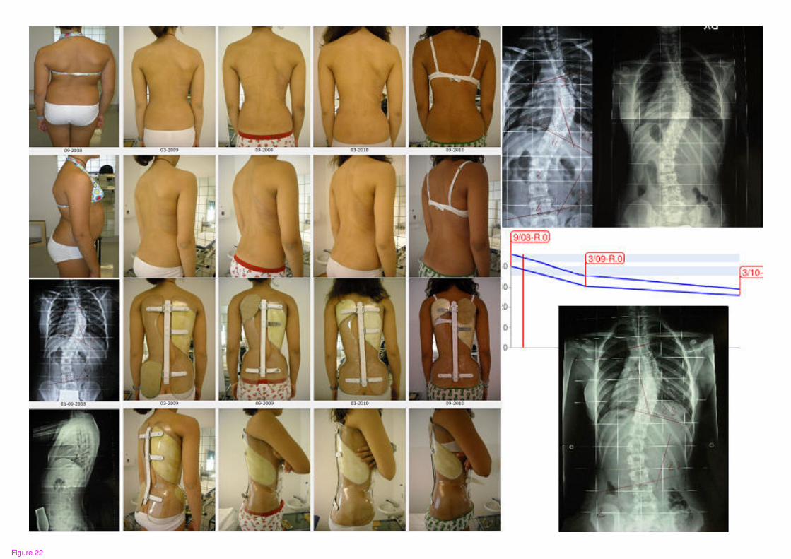
Figure 22

Figure 23