scl-mediated regulation of the cell-cycle regulator p21 is critical for
TRANSCRIPT

PLATELETS AND THROMBOPOIESIS
SCL-mediated regulation of the cell-cycle regulator p21 is critical for murinemegakaryopoiesisHedia Chagraoui,1 *Mira Kassouf,1 *Sreemoti Banerjee,1 Nicolas Goardon,1 Kevin Clark,1 Ann Atzberger,1
Andrew C. Pearce,2 Radek C. Skoda,3 David J. P. Ferguson,4 Steve P. Watson,2 Paresh Vyas,1 and Catherine Porcher1
1Medical Research Council Molecular Haematology Unit, Weatherall Institute of Molecular Medicine, John Radcliffe Hospital, University of Oxford, Oxford,United Kingdom; 2Centre for Cardiovascular Sciences, Institute for Biomedical Research, College of Medical and Dental Sciences, University of Birmingham,Birmingham, United Kingdom; 3Department of Research and Experimental Hematology, University Hospital Basel, Switzerland; and 4Nuffield Department ofClinical Laboratory Sciences, University of Oxford, John Radcliffe Hospital, Oxford, United Kingdom
Megakaryopoiesis is a complex processthat involves major cellular and nuclearchanges and relies on controlled coordi-nation of cellular proliferation and differ-entiation. These mechanisms are orches-trated in part by transcriptional regulators.The key hematopoietic transcription fac-tor stem cell leukemia (SCL)/TAL1 is re-quired in early hematopoietic progenitorsfor specification of the megakaryocyticlineage. These early functions have, sofar, prevented full investigation of its role
in megakaryocyte development in loss-of-function studies. Here, we report thatSCL critically controls terminal mega-karyocyte maturation. In vivo deletion ofScl specifically in the megakaryocytic lin-eage affects all key attributes of mega-karyocyte progenitors (MkPs), namely,proliferation, ploidization, cytoplasmicmaturation, and platelet release. Genome-wide expression analysis reveals in-creased expression of the cell-cycle regu-lator p21 in Scl-deleted MkPs. Importantly,
p21 knockdown-mediated rescue of Scl-mutant MkPs shows full restoration ofcell-cycle progression and partial rescueof the nuclear and cytoplasmic matura-tion defects. Therefore, SCL-mediatedtranscriptional control of p21 is essentialfor terminal maturation of MkPs. Our studyprovides a mechanistic link between amajor hematopoietic transcriptionalregulator, cell-cycle progression, andmegakaryocytic differentiation. (Blood.2011;118(3):723-735)
Introduction
Megakaryocytes (MKs) are specialized blood cells that releaseplatelets, the effectors of coagulation processes. During megakaryo-poiesis, megakaryocyte progenitors (MkPs) coordinately prolifer-ate and differentiate to develop into mature MKs that, ultimately,shed platelets. This complex biologic process requires profound cellularand nuclear changes (cytoplasm remodeling, cell size increase, nuclearpolyploidization, and cytoskeletal dynamics) relying on an exquisitecoordination of key cellular and molecular mechanisms.1
At an early stage in their development, MKs enter endomitosis(or abortive mitosis), during which DNA replicates without celldivision. This process results in nuclear polyploidization, increasein cell size, and is linked to platelet formation.2,3 Formation of thedemarcation membrane system (DMS, a reservoir for proplateletmembranes) and secretory granules reflects some of the specificcharacteristics of the cytoplasmic maturation of MKs.4,5
Signaling by thrombopoietin (TPO), the major megakaryocyticcytokine, induces PI3K activity, downstream of which lies themammalian target of rapamycin pathway.6 Mammalian target ofrapamycin is a kinase that controls cell size and cell-cycleprogression in mammals and Drosophila.7,8 It also regulates keyMK attributes (proliferation, cell size, cytoskeleton organization,and platelet formation) in part through control of G1/S cell-cycleprogression.6,9 This control is mediated by sequential action of thecell-cycle regulators cyclin D3 (CCND3) and P21 (cyclin-dependent cell-cycle inhibitor, CDKN1a).9 In mouse MKs, cyclinD3 expression is high during endomitosis, leading to polyploidiza-
tion10; in contrast, P21 expression is high in mature high ploidyMKs leading to G1/S progression blockade, thereby inducingcell-cycle arrest and terminal maturation.9
Numerous nuclear regulators play important roles in the tran-scriptional control of megakaryopoiesis.11,12 Several lines of evi-dence suggest that the basic helix-loop-helix protein stem cellleukemia (SCL)/TAL1, critical for hematopoietic specification13-16
and erythroid maturation17 (and references therein), is also function-ally involved in megakaryopoiesis.
Enforced expression of SCL in human hematopoietic progeni-tors or mouse bone marrow cells enhances megakaryopoiesis.18-20
Loss-of-function studies have shown a requirement for SCL inmegakaryopoiesis both during embryonic development and at theadult stage. Indeed, SCL is necessary for development of fetal livermegakaryopoiesis.21 In adult mice, analysis of short-term defects inScl-deleted bone marrow cells revealed a block in MK develop-ment and loss of MkPs in vitro.22,23 This block was in part the resultof perturbation of early committed hematopoietic progenitors andtherefore precluded full analyses of the function of SCL in MKs.Characterization of long-term defects in the same mouse modelrevealed a late defect in MK differentiation under stress conditionsleading to defective platelet production.24 In steady state, Scl-deleted mice displayed normal numbers of MKs with normalpolyploidization. This overall mild phenotype probably resultedfrom the development of compensatory mechanisms. Taken to-gether, these loss-of-function analyses strongly suggest that full
Submitted January 7, 2011; accepted April 30, 2011. Prepublished online asBlood First Edition paper, May 19, 2011; DOI 10.1182/blood-2011-01-328765.
*M.K. and S.B. contributed equally to this study.
The online version of this article contains a data supplement.
The publication costs of this article were defrayed in part by page chargepayment. Therefore, and solely to indicate this fact, this article is herebymarked ‘‘advertisement’’ in accordance with 18 USC section 1734.
© 2011 by The American Society of Hematology
723BLOOD, 21 JULY 2011 � VOLUME 118, NUMBER 3
For personal use only.on February 2, 2018. by guest www.bloodjournal.orgFrom
investigation of the functions of SCL in MK differentiationrequires a lineage-specific deletion approach.
SCL exerts its activity as part of large multiprotein complexes.Initial studies in erythroid cells have characterized its interactionwith other transcription factors (E47, its heterodimerization part-ner, the LIM-domain binding protein LDB1, and the LIM-onlydomain protein LMO2) to form the SCL “core” complex.25,26 SCLacquires activating or repressing functions on recruitment ofcofactors. ETO2 is a critical corepressor; its recruitment to the SCLcomplex determines the onset of red blood cell27 and MK terminaldifferentiation.28 SCL exerts its transcriptional control over a rangeof target genes either directly on binding to its recognition DNAmotif, an E-box (CANNTG), or independently of its DNA bindingactivity.17,29 Interaction with the hematopoietic transcription factorGATA1 leads to formation of a pentameric complex composed ofSCL, E47, LMO2, LDB1, and GATA1,30 that selectively activatestarget genes on binding to E-box/GATA composite DNA motifs inred cells.29,31,32 When acting independently of its DNA binding site,SCL is mostly recruited through GATA motifs.29,32 Similar SCL-containing multiprotein complexes are observed in MKs.25 Theirfunction and genomic targets remain unknown.
So, despite evidence supporting the function and transcriptionalactivity of Scl in megakaryopoiesis, its exact role in this lineageremained to be fully characterized. To achieve this, we selectivelyinactivated SCL in MKs by crossing Scl-floxed mice (Sclfl/fl) to arecently described MK-specific Pf4-Cre recombinase mousestrain.33-35 We show that SCL is critically required for key aspectsof megakaryocytic development. This function is mediated in partthrough the transcriptional control of p21 expression and cell-cycleprogression.
Methods
Ploidy
Bone marrow cells derived from 5-FU–treated mice were plated in TPO-containing StemPro-34 medium (Invitrogen). After 4 days, cells were stainedwith CD41-FITC or IgG1-FITC antibodies (BD Biosciences PharMingen) andfixed in 70% ethanol for 3 hours at 4°C. Cells were incubated in 500 �L of 0.1%Triton X-100 (Sigma-Aldrich) containing 50 �g/mL propidium iodide (Sigma-Aldrich) and 200 �g/mL RNAse A (Sigma-Aldrich). Ploidy of CD41� cells wasanalyzed on a Cyan analyzer (DakoCytomation).
Day 4 liquid cultures of MKs derived from sorted MkPs were stainedwith CD41-FITC or IgG1-FITC antibodies (BD Biosciences PharMingen).Cells were incubated in Iscove modified Dulbecco medium/10% FCScontaining 0.01mM Hoechst 33342 (Sigma-Aldrich) at 37°C for 2 hours.Ploidy was analyzed as described earlier in “Ploidy.”
Proliferation assay
Purified MkPs were grown in IMDM/10% FCS supplemented with TPO(1% conditioned medium), erythropoietin (2 U/mL), IL-3 (10 ng/mL),IL-11 (5 ng/mL), Kit ligand (50 ng/mL), FMS-like tyrosine kinase-3 ligand(10 ng/mL), and granulocyte macrophage colony-stimulating factor (2 ng/mL). At day 4, 10�M 5-bromo-2�-deoxyuridine (BrdU) was added for4 hours, cells harvested, stained with CD41-FITC or IgG antibodies, andcell-cycle analysis performed using BrdU Flow Kit (BD BiosciencesPharMingen).
Microarray analysis
Expression profiling was performed using Sentrix Mouse-6 ExpressionBeadChip arrays from Illumina on 3 independent Cre:Sclwt/wt and Cre;Sclfl/fl
MkP populations. Experiments were performed as described.29 Expressionarray data have been submitted to Gene Expression Omnibus database(accession number GSE24969).
Lentivirus production
Antisense shRNA oligonucleotides were designed in the coding region ofthe murine p21 gene, p21 scramble shRNA sequences were designed ascontrol (supplemental Methods, available on the Blood Web site; see the
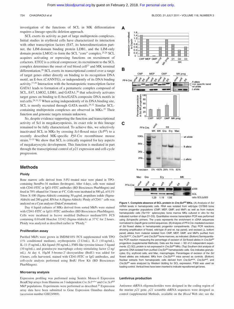
Figure 1. Complete absence of SCL protein in Cre;Sclfl/fl MKs. (A) Analysis of SclmRNA levels in hematopoietic cells. RNA was isolated from wild-type C57Bl/6 bonemarrow progenitor populations (CMP, MEP, GMP, and MkP) as well as from maturehematopoietic cells (Ter119� splenocytes; bone marrow MKs cultured in vitro for theindicated number of days D1-D3). Quantitative reverse transcription PCR was performedusing Scl-specific primers. The y-axis represents the enrichment in cDNA sequencesnormalized to Gapdh gene control sequences. (B)Analysis of PF4-Cre–mediated excisionof Scl floxed alleles in hematopoietic progenitor compartments. (Top) PCR reactionsshowing amplification of floxed, wild-type (fl and wt, top panel), and excised (�, bottompanel) alleles from material isolated from CMP, MEP, GMP, and MkPs purified fromCre;Sclwt/wt, Cre;Sclwt/fl, and Cre;Sclfl/fl bone marrows, as indicated. (Bottom) Semiquantita-tive PCR reaction measuring the percentage of excision of Scl floxed alleles in Cre;Sclfl/fl
progenitors (supplemental Methods). Data are the mean � SD of 2 independent experi-ments. (C) SCL protein is not expressed in Cre;Sclfl/fl MKs. (Top) Southern blot analysis ofgenomic DNA isolated from purified Cre;Sclfl/fl hematopoietic cells. Gra indicates granulo-cytes; Ery, erythroid cells; and Mac, macrophages. Percentages of excision of the Sclfloxed alleles are indicated. MKs from Cre;Sclwt/wt mice served as controls. (Bottom)Nuclear extracts from hematopoietic cells derived from Cre;Sclwt/wt, Cre;Sclwt/fl, andCre;Sclfl/fl were analyzed by Western blotting for SCL expression. P300 was used asloading control. Vertical lines have been inserted to indicate repositioned gel lanes.
724 CHAGRAOUI et al BLOOD, 21 JULY 2011 � VOLUME 118, NUMBER 3
For personal use only.on February 2, 2018. by guest www.bloodjournal.orgFrom
Supplemental Materials link at the top of the online article). The oligonucle-otides were introduced into pSuper plasmid (BVTech) downstream of PolIIIH1 promoter. H1-shRNA DNA fragments were transferred into thelentiviral vector pTRIP/�U3EF1�-green fluorescent protein (GFP).36,37
Lentiviral supernatant was produced as described.36,37
MkP transduction
A total of 1 to 3 � 104 purified Cre;Sclfl/fl MkPs were plated in 300 �L ofIMDM/10% FCS containing cytokines (see “Proliferation assay”). Cellswere transduced with viruses expressing p21 shRNA or control sequencesat a multiplicity of infection of 50 to 100. At 48 ours later, cells wereFACS-sorted and GFP� cells used in colony-forming cell assays, orreplated in liquid culture and assessed for ploidy and cell cycle, or cytospunfor acetylcholinesterase (AChE) staining.
ChIP assays
A total of 107 MKD1 cells (ES cell–derived MK cell line bearing floxed Sclalleles, H.C., P.V., C.P. manuscript in preparation) or 0.5 to 1 � 106 primaryMKs at day 4 of the TPO culture were used for each chromatin immunoprecipi-tation (ChIP) assay. Experimental conditions and antibodies were asdescribed.17,25,28
Luciferase assay
Luciferase expression was driven by sequences of the mouse p21 proximalpromoter and first intron (90 to 647 relative to transcriptional start site) ormutated versions of them (produced by PCR-based mutagenesis) using thepGL4 reporter plasmid (Promega). MKD1 cells were transfected withAmaxa cell line nucleofector kit V (VCA-1003, Lonza Group) followingthe manufacturer’s recommendations. After 24 hours, luciferase and -galactosidase (to normalize for transfection efficiency) activities were
measured using standard procedures. To excise Scl floxed alleles, MKD1cells were cotransfected with EF1�-GFP-Cre plasmid or EF1�-GFPplasmid (control) along with p21-736 reporter. After 24 hours, GFP� cellswere sorted and luciferase and -galactosidase assayed.
Results
In vivo generation of Scl�/� MKs
A conditional knockout approach was used to specifically inacti-vate SCL in vivo in MKs. Sequences coding for the core domain ofSCL,38 the basic helix-loop-helix region, were targeted in murineES cells by homologous recombination; a LoxP-flanked Neo-IRES-TK selection cassette was introduced into intron 6 of Scl andanother LoxP site into the 3� untranslated region sequences(supplemental Figure 1A). Neomycin-resistant ES cell clones wereidentified and characterized by Southern blotting (data not shown).Clones with Cre-mediated excision of the Neo-TK cassette (Sclwt/fl)were obtained from 2 independently targeted ES cell clones (Sclwt/fl-Neo).Two independent Sclwt/fl clones were injected into C57Bl/6 blastocysts,and mouse lines heterozygous for the Scl floxed allele were generated.Supplemental Figure 1B-C shows a representative genomic analysis ofF1 mice. Heterozygous Sclwt/fl mice were bred to homozygosity andSclfl/fl mice presented at a Mendelian ratio.
To selectively abolish the function of SCL in adult megakaryo-poiesis, Sclfl/wt mice were bred to a transgenic strain expressing Crerecombinase driven by the regulatory sequences of platelet factor 4(Pf4), a MK-specific CXC chemokine.34 Newborn mice (thereafterreferred to as Cre;Sclfl/fl, Cre;Sclwt/fl, and Cre;Sclwt,wt) were obtained
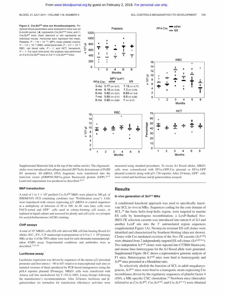
Figure 2. Cre;Sclfl/fl mice are thrombocytopenic. Pe-ripheral blood parameters were analyzed in mice over an8-month period. (�) represents Cre;Sclwt/wt mice; and U,Cre;Sclfl/fl mice. Each diamond or dot represents anindividual mouse. Horizontal bars represent the mean.Platelets, P � 1.8 � 1012; MPV, mean platelet volume,P � 1.2 � 105; WBC, white blood cells, P � 3.7 � 105;RBC, red blood cells, P � .1; and HCT, hematocrit,P � .1. For each time point, the analysis was performedon 5 to 8 Cre;Sclfl/fl mice or 3 to 11 Cre;Sclwt/wt mice.
SCL CONTROLS MEGAKARYOCYTE DEVELOPMENT 725BLOOD, 21 JULY 2011 � VOLUME 118, NUMBER 3
For personal use only.on February 2, 2018. by guest www.bloodjournal.orgFrom
at the expected Mendelian ratio. Homozygous mice lived toadulthood.
Because of Scl’s broad expression in bone marrow hematopoi-etic progenitors and differentiated cells (Scl is highly expressedin common myeloid progenitor [CMP], bipotent erythroid/megakaryocytic progenitor [MEP], MkPs, erythroid [Ter119�]cells, and in in vitro-cultured MKs [MK, D1-D3], Figure 1A), weevaluated the level of Cre-mediated excision of the Sclfl allele inCMP, MEP, granulocyte-macrophage progenitor (GMP), and MkPpopulations isolated from Cre;Sclwt/wt, Cre;Sclwt/fl, and Cre;Sclfl/fl
mice. Following the PCR strategy detailed in supplemental Figure1C (top panel), both excised (�) and floxed (fl) alleles wereamplified from CMP, MEP, and GMP derived from Cre;Sclwt/fl andCre;Sclfl/fl mice (Figure 1B top panel). The data suggested partialexcision of the Sclfl allele in these populations. Indeed, semiquanti-tative PCR analysis performed on the deleted (�) allele (Figure 1Bbottom panel) showed 34% excision in Cre;Sclfl/fl-derived CMP andMEP and 54% in Cre;Sclfl/fl-derived GMP. In contrast, full excisionwas observed in Cre;Sclfl/fl MkPs, as shown by the completeabsence of the amplified floxed allele with 100% excision (Figure1B top and bottom panels). Importantly, we did not observesignificant differences in the percentages of each progenitorcompartment in Cre;Sclfl/fl-derived bone marrow compared withcontrols (supplemental Figures 2-3).
Next, we purified mature hematopoietic cells from bone marrow(MKs and macrophages) and spleen (erythroid cells and granulo-cytes) derived from Cre;Sclfl/fl mice and assessed the level ofexcision of the Sclfl allele by Southern blot (Figure 1C top). Greaterthan 90% excision was observed in the Cre;Sclfl/fl MK sample. Thiscorrelated with a complete absence of the SCL protein (Figure 1Cbottom). Low-level excision in Cre;Sclfl/fl Ter119� erythroid cells(11%, Figure 1C top) did not affect protein levels (Figure 1Cbottom). Finally, the Scl sequences were also excised in granulo-cytes and macrophages (29% and 83%, respectively). However,because SCL is not expressed in these 2 lineages (Figure 1Cbottom), genomic excision in these cells probably does not affecthematopoiesis.
In conclusion, we successfully generated a conditional knock-out model with complete ablation of SCL expression specifically inthe megakaryocytic lineage.
Cre;Sclfl/fl mutant mice are thrombocytopenic
Hematologic parameters were determined over an 8-month period(Figure 2). Platelet counts in mutant Cre;Sclfl/fl mice were de-creased on average by 1.5- to 2-fold, with increased mean plateletvolume compared with controls, suggesting perturbed terminalmegakaryopoiesis and platelet release. Mutant mice also showed anaverage of 1.5- to 2-fold increase in white blood cell numbers. Redblood cell counts and hematocrit were similar, suggesting no grossperturbation in the mutant erythroid lineage.
FACS analysis of bone marrow hematopoietic lineages showed nosignificant difference in the numbers of Ter119�, Mac1�, and Gr1� cellsbetween Cre;Sclfl/fl and control mice (supplemental Figure 4), suggest-ing that the levels of excision observed in the CMP, MEP, and GMPcompartments (Figure 1B) did not perturb the differentiation of down-stream lineages. We did, however, observe an increase in the percentageof B220� cells in Cre;Sclfl/fl mutant mice.
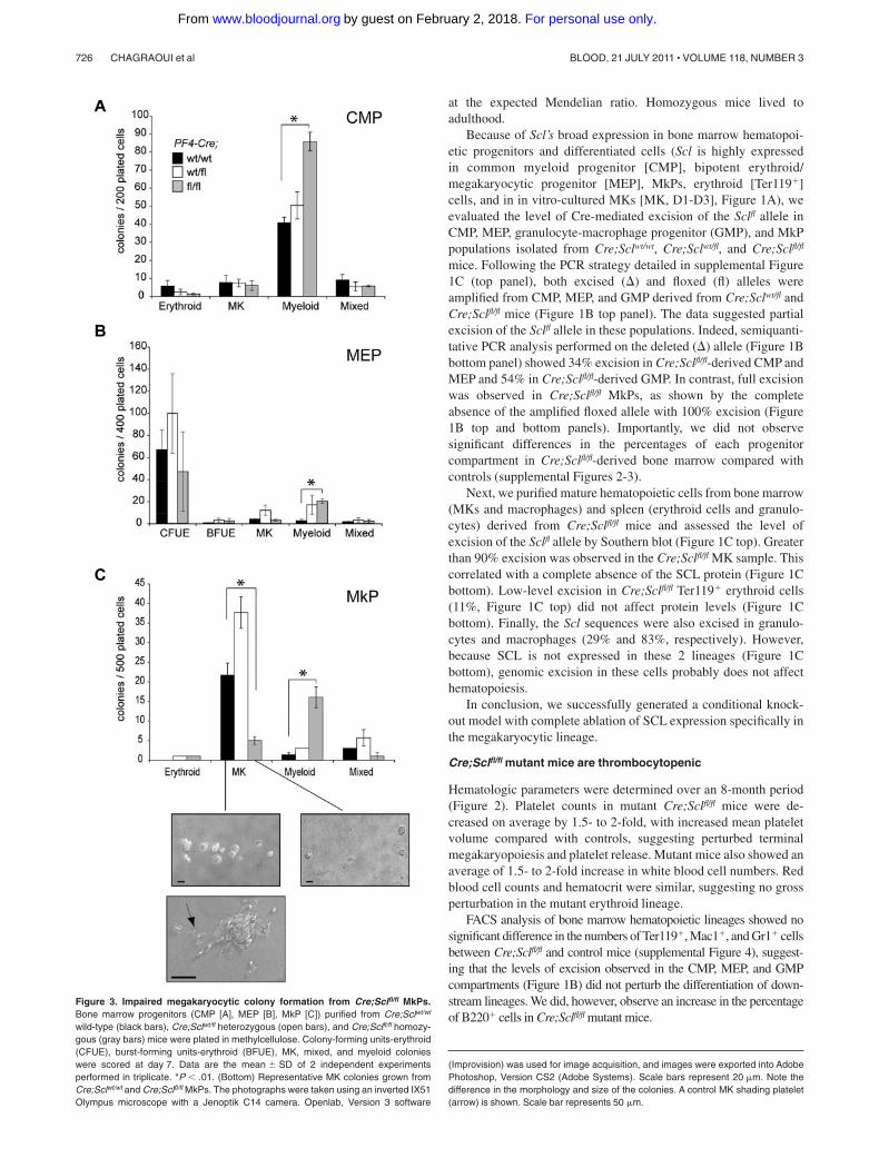
Figure 3. Impaired megakaryocytic colony formation from Cre;Sclfl/fl MkPs.Bone marrow progenitors (CMP [A], MEP [B], MkP [C]) purified from Cre;Sclwt/wt
wild-type (black bars), Cre;Sclwt/fl heterozygous (open bars), and Cre;Sclfl/fl homozy-gous (gray bars) mice were plated in methylcellulose. Colony-forming units-erythroid(CFUE), burst-forming units-erythroid (BFUE), MK, mixed, and myeloid colonieswere scored at day 7. Data are the mean � SD of 2 independent experimentsperformed in triplicate. *P .01. (Bottom) Representative MK colonies grown fromCre;Sclwt/wt and Cre;Sclfl/fl MkPs. The photographs were taken using an inverted IX51Olympus microscope with a Jenoptik C14 camera. Openlab, Version 3 software
(Improvision) was used for image acquisition, and images were exported into AdobePhotoshop, Version CS2 (Adobe Systems). Scale bars represent 20 �m. Note thedifference in the morphology and size of the colonies. A control MK shading platelet(arrow) is shown. Scale bar represents 50 �m.
726 CHAGRAOUI et al BLOOD, 21 JULY 2011 � VOLUME 118, NUMBER 3
For personal use only.on February 2, 2018. by guest www.bloodjournal.orgFrom
Abnormal in vitro growth of Scl�/� MkPs
The in vitro growth capacity of bone marrow CMP, MEP, andMkPs was analyzed in colony assays. Mutant Cre;Sclfl/fl CMPs andMEPs gave rise to numbers of erythroid, megakaryocytic, andmixed colonies similar to that generated from wild-type controls,
and to increased numbers of myeloid colonies (Figure 3A-B).Morphologically, the colonies appeared normal (not shown).
In the megakaryocytic compartment, Cre;Sclfl/fl MkPs were notsignificantly affected in the production of mixed colonies, erythroidcolonies were detected at a very low frequency, and there was an
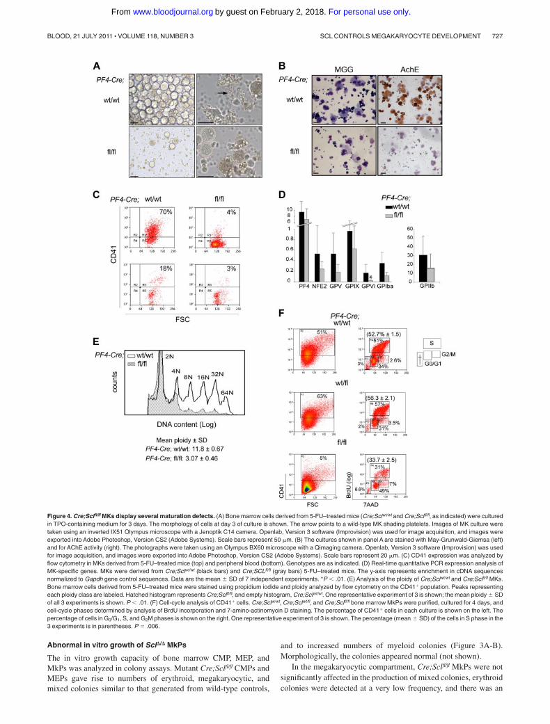
Figure 4. Cre;Sclfl/fl MKs display several maturation defects. (A) Bone marrow cells derived from 5-FU–treated mice (Cre;Sclwt/wt and Cre;Sclfl/fl, as indicated) were culturedin TPO-containing medium for 3 days. The morphology of cells at day 3 of culture is shown. The arrow points to a wild-type MK shading platelets. Images of MK culture weretaken using an inverted IX51 Olympus microscope with a Jenoptik C14 camera. Openlab, Version 3 software (Improvision) was used for image acquisition, and images wereexported into Adobe Photoshop, Version CS2 (Adobe Systems). Scale bars represent 50 �m. (B) The cultures shown in panel A are stained with May-Grunwald-Giemsa (left)and for AChE activity (right). The photographs were taken using an Olympus BX60 microscope with a Qimaging camera. Openlab, Version 3 software (Improvision) was usedfor image acquisition, and images were exported into Adobe Photoshop, Version CS2 (Adobe Systems). Scale bars represent 20 �m. (C) CD41 expression was analyzed byflow cytometry in MKs derived from 5-FU–treated mice (top) and peripheral blood (bottom). Genotypes are as indicated. (D) Real-time quantitative PCR expression analysis ofMK-specific genes. MKs were derived from Cre;Sclwt/wt (black bars) and Cre;SCLfl/fl (gray bars) 5-FU–treated mice. The y-axis represents enrichment in cDNA sequencesnormalized to Gapdh gene control sequences. Data are the mean � SD of 7 independent experiments. *P .01. (E) Analysis of the ploidy of Cre;Sclwt/wt and Cre;Sclfl/fl MKs.Bone marrow cells derived from 5-FU–treated mice were stained using propidium iodide and ploidy analyzed by flow cytometry on the CD41� population. Peaks representingeach ploidy class are labeled. Hatched histogram represents Cre;Sclfl/fl; and empty histogram, Cre;Sclwt/wt. One representative experiment of 3 is shown; the mean ploidy � SDof all 3 experiments is shown. P .01. (F) Cell-cycle analysis of CD41� cells. Cre;Sclw/wt, Cre;Sclwt/fl, and Cre;Sclfl/fl bone marrow MkPs were purified, cultured for 4 days, andcell-cycle phases determined by analysis of BrdU incorporation and 7-amino-actinomycin D staining. The percentage of CD41� cells in each culture is shown on the left. Thepercentage of cells in G0/G1, S, and G2M phases is shown on the right. One representative experiment of 3 is shown. The percentage (mean � SD) of the cells in S phase in the3 experiments is in parentheses. P � .006.
SCL CONTROLS MEGAKARYOCYTE DEVELOPMENT 727BLOOD, 21 JULY 2011 � VOLUME 118, NUMBER 3
For personal use only.on February 2, 2018. by guest www.bloodjournal.orgFrom

increase in the numbers of myeloid colonies (Figure 3C). Of note,this myeloid expansion, detected from all mutant progenitors, is inagreement with previous observations from our SCL DNA-bindingmutant mouse model17 and probably reflects the role of SCL in fatedetermination.
Specific defects in the production of MK colonies were noticed(Figure 3C). Strikingly, the number of MK colonies derived fromScl-deleted MkPs was decreased 5- to 7-fold compared withcontrols. Furthermore, the size of mutant MK colonies was reducedto 2 or 3 cells/colony compared with 10 to 15 cells/colony incontrol plates. Finally, proplatelet-forming MKs were detected incontrol samples but absent in Cre;Sclfl/fl cultures. Collectively,these data suggested impaired cell survival, proliferation, and/ormaturation of MkPs. To address this, we analyzed several aspectsof megakaryopoiesis in freshly isolated bone marrow MKs.
Scl�/� MKs display defects in proliferation, polyploidization,and cytoplasmic maturation
To generate sufficient numbers of MKs, mice were treated with5-FU and in vitro–cultured bone marrow MKs were harvested.Cells isolated from Cre;Sclfl/fl mice gave rise to sparse culturescompared with control samples that consisted of large, round cells(Figure 4A left panels). Proplatelet-forming MKs were detected incontrol samples but not in Scl-deleted samples (Figure 4A rightpanels, arrow). Several assays were used to further characterizeMK cultures. First, May-Grunwald-Giemsa staining revealed poly-lobulated nuclear morphology and abundant cytoplasm of large,mature MKs, and confirmed the low number of MKs in mutantCre;Sclfl/fl populations (Figure 4B left panels). Second, AChEactivity, specific to immature and mature MKs, was detected incontrol cultures but was absent from mutant cultures (Figure 4Bright panels), suggesting an early block in megakaryocytic differen-tiation in Scl�/� cells. Very rare AChE-positive MKs could be seenin mutant cultures derived from sorted MkPs (supplemental Figure5). Third, analysis of CD41 (MK cell surface marker) expression
showed that 70% of the cells obtained from Cre;Sclwt/wt bonemarrow were CD41� as opposed to 4% in Cre;Sclfl/fl cultures(Figure 4C top). Analysis of blood cells from control and mutantmice confirmed this difference (Figure 4C bottom panels).
Next, we examined mRNA expression of selected MK-specificmarkers by real-time PCR (Figure 4D). Of all the markers tested,only GpV1 expression was markedly down-regulated (7-foldreduction) in Scl-null MKs compared with control cells. Expressionof Pf4, Nf-e2, GpV, GpIX, GpI�, and IIb was not significantlyaltered in mutant cells.
A typical feature of MK maturation is polyploidization, whereMKs switch from a classic mitotic division process to an endomi-totic process to increase their ploidy. To assess whether this processwas affected in Scl-deleted MKs, we measured the DNA content ofbone marrow–derived CD41� cells by flow cytometry. A markedlylower ploidy level was observed from Cre;Sclfl/fl cells (mean ploidy3N) compared with control Cre;Sclwt/wt cells (mean ploidy 12N)(Figure 4E).
Next, we determined the effect of absence of SCL on cell cycle.Cre;Sclwt/wt, Cre;Sclwt/fl, and Cre;Sclfl/fl MkPs were purified and thecell cycle analyzed by BrdU incorporation. Similar to findingsobtained from cultured MKs (Figure 4C), homozygous mutantMkPs gave rise to less CD41� cells than the control cells (Figure4F left panels). Analysis of BrdU incorporation in CD41� cellsrevealed that Cre;Sclfl/fl MKs had a lower proliferation rate withonly 31% of BrdU� cells compared with 51% and 57% in SCL-expressing MKs (Cre;Sclwt/wt and Cre;Sclwt/fl, respectively), leading to anaccumulation of cells in G0/G1 (Figure 4F right panels).
Finally, ultrastructural studies showed rarefaction and disorgani-zation of the DMS accompanied by dilated rough endoplasmicreticulum in Cre;Sclfl/fl MKs, confirming a perturbed cytoplasmicmaturation (compare Figure 5A,C). In addition, unusual elongatedorganelles containing crystalline structure were noticed in themutant (Figure 5C arrow). Emperipolesis (presence of cells withinthe MKs) was markedly increased in mutant MKs (compare
Figure 5. Scl�/� MKs show ultrastructural defects. Cre;Sclwt/wt
(A-B) and Cre;Sclfl/fl (C-D) bone marrow MKs were analyzed by electronmicroscopy. Note the absence of organized DMS in mutant MKs. Note thepresence of 6 cells within the mutant MK (emperipolesis). N indicatespolylobulated nucleus; and ER, endoplasmic reticulum. Arrowheads indi-cate �-granules; and arrows, crystalline structure. (A,C) Scale barsrepresent 500 nm. (B,D) Scale bars represent 2 �m.
728 CHAGRAOUI et al BLOOD, 21 JULY 2011 � VOLUME 118, NUMBER 3
For personal use only.on February 2, 2018. by guest www.bloodjournal.orgFrom

Table 1. Differentially expressed genes in Scl�/� MkPs
Gene symbol Fold change* Accession no.
Adhesion/migrationCol18a1† 7.77 NM_009929.2Megf10 3.63 NM_001001979Emilin2 3.29 NM_145158Cd72 2.73 NM_007654.1Serpine2† 2.22 AK045954Map17 2.12 NM_026018.1Cd63† 1.77 NM_007653.1Emid1 1.75 NM_080595.1Cxcr4† 1.75 NM_009911.2Ltbp1 1.61 NM_019919.2Mfge8 1.45 NM_008594Tens1 0.66 XM_109868Elmo1 0.6 NM_080288.1Esam1 0.55 NM_027102.1Selp† 0.4 NM_011347.1
Cell cycle/apoptosisCdkn1a† 3.26 NM_007669.2Tnfrsf18 1.97 NM_009400.1Rassf2 1.64 NM_175445.3Bin1 1.62 AK041729.1Bcl2† 1.59 NM_177410.1Ndg2 1.5 NM_175329.3Tmbim4 1.5 NM_026617.1Cryab 1.37 NM_009964.1Nalp6 0.65 NM_133946.1Catnal1 0.47 NM_018761.2
TranscriptionMta3 2.2 NM_054082.1Carhsp1 2.04 NM_025821.2Lyl1 1.77 NM_008535.1Rcor1 1.66 NM_054048.1Gata2 1.57 NM_008090.3Hes6 1.52 NM_019479.2Foxp1 1.48 NM_053202.1Mta2 1.48 NM_011842.2Chd7 0.71 XM_149413.3Fhl1 0.69 NM_010211.1Ccndbp1 0.68 NM_010761.1Hoxb5 0.67 NM_008268.1Mysm1 0.65 NM_177239.1Fos 0.61 NM_010234.2Ctdspl2 0.59 XM_283758.1Tle1 0.52 NM_011599.2Ear10 0.35 NM_053112.1Ear2 0.34 NM_007895Tal1 0.2 NM_011527.1
Immune responseIgh-6 6.66 XM_354710.1Cst7 2.57 NM_009977.1Clecsf8 2.44 NM_010819.1Cd59a 0.54 NM_007652.2
Signal transductionFcer1g† 6 NM_010185.2P2ry14 2.84 NM_133200.2Wbscr5 2.51 NM_022964.2Ppp1r1c 2.13 NM_033264.1Cd69 1.98 XM_132882.1Mrvi1† 1.97 NM_194464.1Itgb3bp 1.88 NM_026348.2Il15 1.87 NM_008357.1Centd1† 1.81 XM_132099.5Rpel1 1.79 NM_198419.2Dok4 1.75 NM_053246.1Tas1r1 1.7 NM_031867.1Fgf3 1.69 NM_008007.1Irak3 1.69 NM_028679.2Dusp4 1.69 NM_176933Cysltr1 1.63 NM_021476.2Diras2 1.68 NM_001024474Lat† 1.54 NM_010689.2Plekhg5 1.5 NM_001004156Agtrap 1.48 NM_009642.3Rab27a 1.48 NM_023635.2Ly6e 1.46 NM_008529Il18r1 1.45 NM_008365.1Cd244 1.36 NM_018729.1Dusp1 0.69 NM_013642.1Grb10 0.67 AK012646Gucy1a3 0.62 NM_021896.3
Table 1. Continued
Gene symbol Fold change* Accession no.
Rab11a 0.62 NM_017382Fcgr2b 0.6 NM_010187.1Pdgfb 0.57 NM_011057.2Drctnnb1a 0.56 NM_053090Il6ra 0.48 AK020663Gnaz† 0.45 NM_010311.2Pde10a 0.37 NM_011866.1
TransportAlox5 5.53 XM_132832.3Tnni1 2.04 NM_021467Emid1 1.98 NM_080595.1Tnni3 1.97 NM_009406.2Slc6a13 1.85 NM_144512.1Hbb-b1 1.79 AK005442Tpcn1 1.73 NM_145853.2Ddx25 1.72 NM_013932.2Spns3 1.71 XM_126365.3Igf2r 1.56 NM_010515.1Slc5a9 1.56 NM_145551.2Slc14a1 0.68 NM_028122.2Abcc5 0.67 NM_176839.1Car2 0.53 NM_009801.3Hebp1 0.51 NM_013546.1
Cytoskeleton organizationMyo18b† 4.84 XM_144513.3Tpm2 2.4 NM_009416.2Cap1 2.07 NM_007598.2Myo6† 1.65 NM_008662.1Ehd2 1.53 AK029240Homer2 0.46 XM_133550.4
UbiquitinationUsp20 1.56 NM_028846.1Usp40 0.54 XM_129956.2
MetabolismChst1 4.71 NM_023850.1Alox5 3.95 XM_132832Abat 2.31 NM_172961.2Padi2 2.2 NM_008812.1Hmgcs2 1.88 NM_008256.2Acas2l 1.65 NM_080575.1Ltc4s 1.65 NM_008521.1Rsdr1-pending 1.64 AK087137Dhrs3 1.45 NM_011303.2Gchfr 0.71 NM_177157.2Chsy3 0.65 NM_178636Oxr1 0.63 AK040881Hyi 0.6 XM_489058Mtap 0.6 NM_024433.1Serpinb1a 0.56 NM_025429.1Gucy1a3 0.47 NM_021896.3Sdh1 0.45 NM_146126.1Serpina3g 0.4 XM_354694.1Serpina3b 0.38 NM_173024.1
Unidentified9830134C10Rik 3.43 AK0365632310047A01Rik 2.56 XM_484355LOC381142 2.09 XM_355058.16430559E15Rik 1.99 XM_131537.4C130080K17Rik 1.71 AK081831LOC382231 1.69 XM_356343.11110012N22Rik 1.68 XM_126634.3BC023181 1.6 XM_194180.34930427A07Rik 1.51 NM_134041.1BC057022 1.5 NM_001004180.10610037L13Rik 0.72 NM_028754.15730446C15Rik 0.69 NM_146096B930074N03Rik 0.68 AK047478BC023823 0.68 NM_153566.1C530050O22Rik 0.68 NM_172871.1AY078069 0.66 NM_172142.1D8Ertd594e 0.66 NM_133791.32900008M13Rik 0.63 XM_1101212010300G19Rik 0.61 NM_028097.2BC049975 0.59 XM_138237.29030611O19Rik 0.57 NM_027828.2
*Fold change: ratio Scl�/� MkPs/WT MkP.†Specific functions in megakaryopoiesis and/or platelet biology.
SCL CONTROLS MEGAKARYOCYTE DEVELOPMENT 729BLOOD, 21 JULY 2011 � VOLUME 118, NUMBER 3
For personal use only.on February 2, 2018. by guest www.bloodjournal.orgFrom
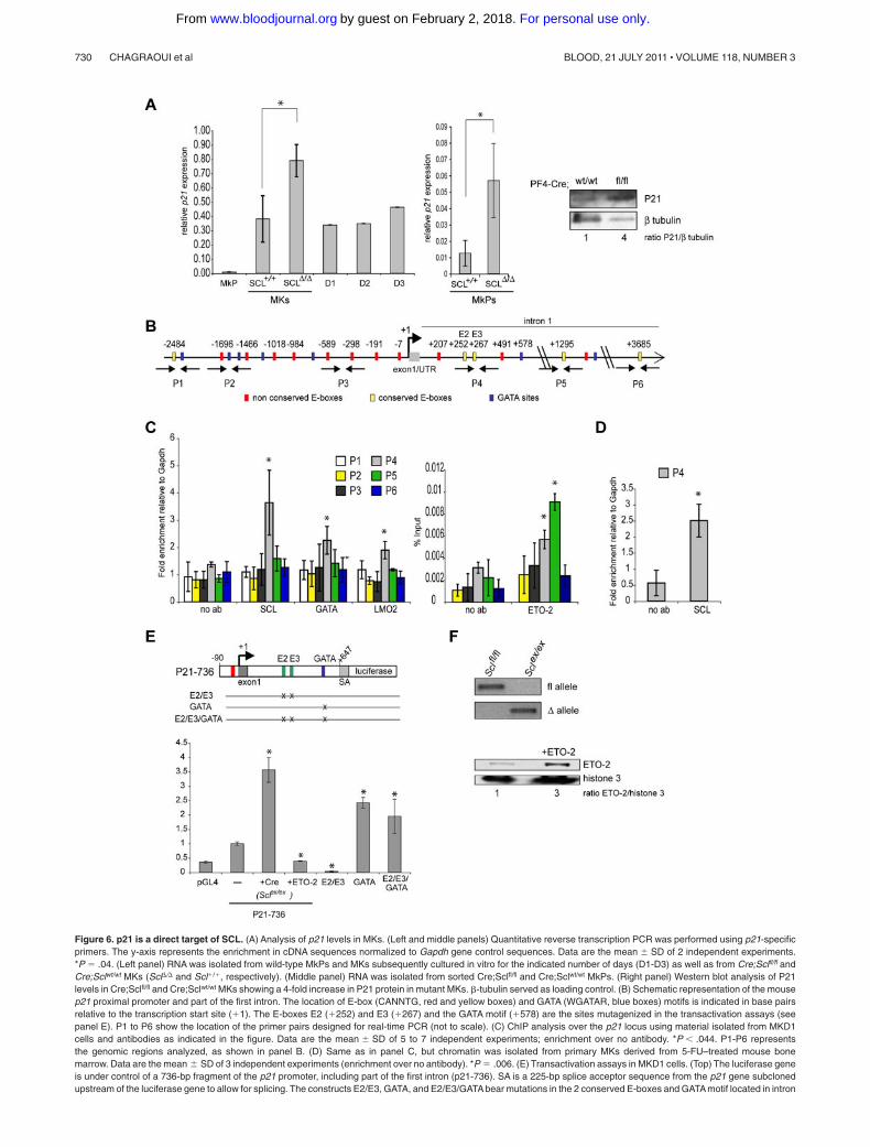
Figure 6. p21 is a direct target of SCL. (A) Analysis of p21 levels in MKs. (Left and middle panels) Quantitative reverse transcription PCR was performed using p21-specificprimers. The y-axis represents the enrichment in cDNA sequences normalized to Gapdh gene control sequences. Data are the mean � SD of 2 independent experiments.*P � .04. (Left panel) RNA was isolated from wild-type MkPs and MKs subsequently cultured in vitro for the indicated number of days (D1-D3) as well as from Cre;Sclfl/fl andCre;Sclwt/wt MKs (Scl�/� and Scl�/�, respectively). (Middle panel) RNA was isolated from sorted Cre;Sclfl/fl and Cre;Sclwt/wt MkPs. (Right panel) Western blot analysis of P21levels in Cre;Sclfl/fl and Cre;Sclwt/wt MKs showing a 4-fold increase in P21 protein in mutant MKs. -tubulin served as loading control. (B) Schematic representation of the mousep21 proximal promoter and part of the first intron. The location of E-box (CANNTG, red and yellow boxes) and GATA (WGATAR, blue boxes) motifs is indicated in base pairsrelative to the transcription start site (�1). The E-boxes E2 (�252) and E3 (�267) and the GATA motif (�578) are the sites mutagenized in the transactivation assays (seepanel E). P1 to P6 show the location of the primer pairs designed for real-time PCR (not to scale). (C) ChIP analysis over the p21 locus using material isolated from MKD1cells and antibodies as indicated in the figure. Data are the mean � SD of 5 to 7 independent experiments; enrichment over no antibody. *P .044. P1-P6 representsthe genomic regions analyzed, as shown in panel B. (D) Same as in panel C, but chromatin was isolated from primary MKs derived from 5-FU–treated mouse bonemarrow. Data are the mean � SD of 3 independent experiments (enrichment over no antibody). *P � .006. (E) Transactivation assays in MKD1 cells. (Top) The luciferase geneis under control of a 736-bp fragment of the p21 promoter, including part of the first intron (p21-736). SA is a 225-bp splice acceptor sequence from the p21 gene subclonedupstream of the luciferase gene to allow for splicing. The constructs E2/E3, GATA, and E2/E3/GATA bear mutations in the 2 conserved E-boxes and GATA motif located in intron
730 CHAGRAOUI et al BLOOD, 21 JULY 2011 � VOLUME 118, NUMBER 3
For personal use only.on February 2, 2018. by guest www.bloodjournal.orgFrom

Figure 5B,D). To assess the characteristics of platelets produced invivo from SCL-deficient MKs, platelet aggregation and densegranule secretion were monitored after stimulation with lowconcentrations of platelet agonists. These properties appearedrelatively unchanged in the absence of SCL (supplemental Figure6).
Collectively, these data indicate that SCL regulates criticalaspects of megakaryopoiesis, namely, proliferation, polyploidiza-tion, and cytoplasmic maturation.
Numerous biologic processes are affected in Scl�/� MkPs
To identify the molecular mechanisms underlying the early cellulardefects observed in Cre;Sclfl/fl MkPs, we performed whole genomeexpression analyses. A total of 145 genes grouped into 8 biologicfunctions were significantly differentially expressed in the mutantpopulations compared with controls (from 1.4- to 7-fold, Table 1).
At least 14 genes (9.7% of total genes, footnoted in Table 1)show specific functions in megakaryopoiesis and/or platelet biol-ogy. One of them is the cyclin-dependent kinase inhibitor p21,which shows a 3.26-fold increase in expression in Cre;Sclfl/fl MKs.Given the role P21 plays in megakaryopoiesis (“Introduction”), wedecided to investigate whether SCL-mediated regulation of p21expression was functionally important in megakaryopoiesis.
The cyclin-dependent kinase inhibitor p21 is a direct target ofSCL in MKs
As shown in Figure 6A, p21 is expressed at low levels in MkPs, andits expression is dramatically increased in more mature MKs(Scl�/�) and sustained at high levels in in vitro cultures (D1-D3).Confirming the microarray data, overexpression of p21 mRNA wasobserved in SCL�/� MkPs (Figure 6A middle panel, 4.5-foldincrease), as well as in SCL-depleted MKs compared with control(Figure 6A left panel, 2.5-fold increase), suggesting that thetranscriptional control exerted by SCL on p21 expression in MkPsextends to maturing MKs. Up-regulation of p21 mRNA wasreflected at the protein level in mutant MKs (Figure 6A right panel).
We asked whether p21 could be a direct target of SCL usingChIP analyses. We first searched the mouse p21 proximal promoterand first intron (2.5 kb/�5 kb) for SCL-binding motifs. Weidentified 28 E-boxes (CANNTG), 5 of them showing conservationbetween human and mouse sequences (Figure 6B). Anti-SCL ChIPassay was performed on material isolated from an immortalizedMK cell line generated from Sclfl/fl mouse ES cells and presentingfeatures of immature MkPs (thereafter referred to as MKD1 cells;H.C. and C.P., manuscript in preparation). We observed SCLbinding exclusively in the 5� region of the first intron of thegene, which contains 2 nonconserved and 2 conserved E-boxes(CAGGTG), as well as a GATA-binding site (TCTATCT)(Figure 6C left graph, region P4). Enrichment in GATA1 and, toa lesser extent, in LMO2 was also noticed in the P4 region,suggesting that the SCL multiprotein complex could be recruitedto the p21 locus. Finally, we observed binding of SCL in thesame region of the p21 gene in bone marrow-derived matureMKs, suggesting that SCL binds the p21 gene throughout MKdifferentiation (Figure 6D).
Because p21 is normally repressed by SCL in primary MkPs(Table 1), we determined whether corepressors could also bind thep21 promoter. A potential candidate was ETO2, a potent corepres-sor and known partner of SCL in MKs and erythroid cells.25,27,28
ETO2 was detected on P4 and P5 regions of p21 in MKD1 cells(Figure 6C right graph). These data suggested that SCL is recruitedto the first intron of the p21 gene, possibly as part of a repressorcomplex composed of at least GATA1, LMO2, and ETO2, eitherthrough the E-boxes or the GATA site identified in this region.
We then assayed the transcriptional activity of the p21 promoterin MKD1 cell line. These cells present 2 important advantages:(1) the experiments are performed in a “megakaryocytic” environ-ment and not in surrogate cells; and (2) the Scl locus is floxed inthese cells, thereby allowing the assessment of its excision on p21promoter activity. A 736-bp p21 promoter fragment (90 to �647)encompassing the intronic region bound by SCL was used to driveluciferase expression (construct p21-736, Figure 6E top). Thisp21-736 reporter construct led to a 2.5-fold increase in transcrip-tional activity compared with the empty vector pGL4 (Figure 6Egraph, condition p21-736). To check whether SCL was involved inthis basal activity, we excised Scl floxed alleles on Cre-recombinase expression in MKD1 cells to obtain Sclex/ex cells(Figure 6F top panel) and repeated the assay. We observed a3.5-fold increase in luciferase levels (Figure 6E graph, conditionp21-736 �Cre, Sclex/ex), suggesting that SCL represses the p21promoter. This is in agreement with our in vivo data (microarraydata, Table 1; Figure 6A). To test whether ETO2 was involved inSCL-mediated repression, we overexpressed ETO2 protein inMKD1 cells (Figure 6F bottom panel). This significantly decreasedp21 transcriptional activity by 2.5-fold (Figure 6E graph, conditionp21-736 �ETO2).
To characterize the cis-elements required for SCL-mediatedrepression, we introduced point mutations in its potential bindingsites (the conserved E2/E3 E-box motifs and GATA site; Figure 6Etop). In contrast to what was observed in Sclex/ex cells, E2/E3 E-boxmutation led to low transcriptional activity (Figure 6E graph,E2/E3), suggesting that they contribute to maintaining a basalexpression level; this activity may be SCL independent and rely onother basic helix-loop-helix proteins given the functional differ-ence observed when Scl alleles are excised. Remarkably, the GATAsite mutation led to derepression of the luciferase activity to a levelsimilar to that observed on Scl deletion (2.5- and 3.5-fold respec-tively, compared with p21-736 WT reporter gene, Figure 6E graph,compare p21-736 �Cre [Sclex/ex] with GATA condition), suggestinga functional link between the GATA motif and SCL activity.Finally, the E2/E3/GATA mutant construct exhibited luciferaselevels similar to that of the GATA mutant alone (Figure 6E graph).This indicated that the E-boxes might not be critically required inthe absence of transcriptional repression mediated through theGATA motif.
These data support a model by which p21 repression requiresbinding of SCL and possibly ETO2 through recruitment byGATA1. This activity seems dominant over the transcriptionalactivation mediated through the conserved E-boxes. However, inthe light of the results obtained with the E2/E3 mutant construct,
Figure 6. (continued). 1 as shown. The graph represents relative luciferase activity measured in MKD1 cells nucleotransfected with the wild-type reporter (P21-736, —)or mutated versions as indicated. The P21-736 construct was also assayed in MKD1 cells on Cre-mediated excision of the Scl floxed alleles (�Cre, Sclex/ex) andoverexpression of ETO2 (�ETO2). Data are mean � SD of 3 or 4 independent experiments performed in duplicate. *P .01 (vs control cells transfected with p21-736construct). (F; Top) PCR showing amplification of the floxed (fl) and excised (�) alleles in MKD1 cells (Sclfl/fl) and after Cre-mediated excision (Sclex/ex). (Bottom) Westernblot analysis of ETO2 expression in MKD1 cells and after overexpression of ETO2 (�ETO2, 3-fold increase in ratio ETO2/histone H3). Histone H3 served as a loadingcontrol.
SCL CONTROLS MEGAKARYOCYTE DEVELOPMENT 731BLOOD, 21 JULY 2011 � VOLUME 118, NUMBER 3
For personal use only.on February 2, 2018. by guest www.bloodjournal.orgFrom

we propose that these E-boxes may be important to counterbalanceSCL-mediated repression. The mechanism underlying this regula-tion remains to be determined.
P21 levels are critical in megakaryopoiesis
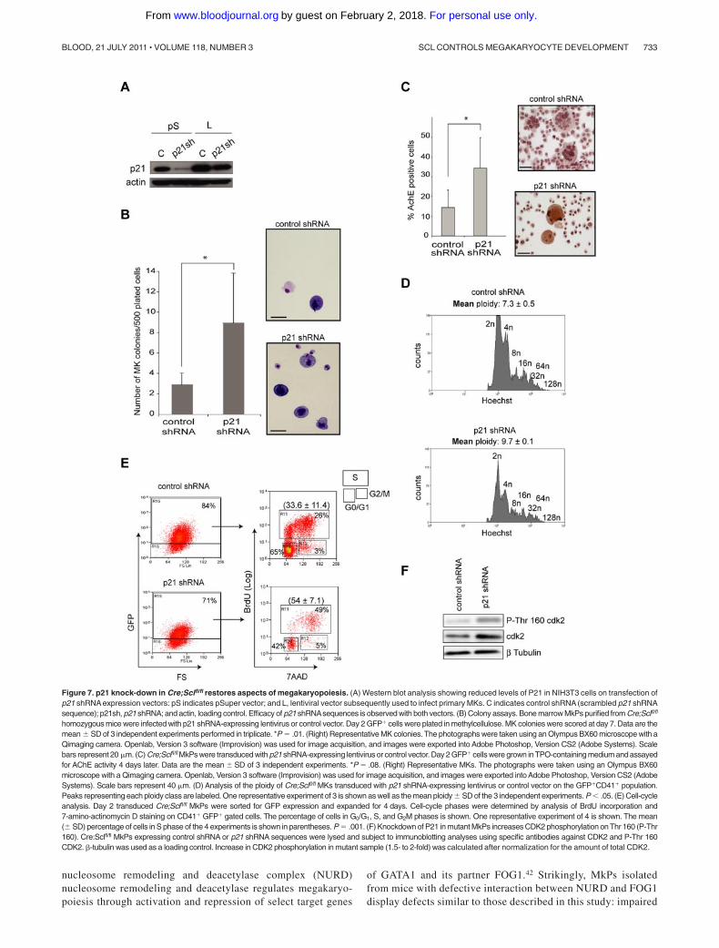
To further investigate whether SCL-mediated regulation of p21was functionally required in megakaryopoiesis, we attempted aphenotypic rescue of the defects observed in Cre;Sclfl/fl MKsthrough shRNA-mediated knockdown of P21 levels. Cre;Sclfl/fl
MkPs were transduced with a lentivirus expressing p21 shRNAsequences shown to significantly decrease P21 protein levels onexpression in 3T3 cells (Figure 7A). Transduced cells were purifiedon the basis of GFP expression and the aspects of megakaryopoi-esis perturbed in Cre;Sclfl/fl MKs reassessed.
In colony assays, GFP� Cre;Sclfl/fl MkPs transduced with p21shRNA sequences showed a 3-fold increase in the number of MKcolonies compared with cells infected with control lentivirus (p21shRNA scrambled sequences). The colonies were smaller thanthose obtained from Cre;Sclwt/wt MkPs (2-10 cells as opposed to10-15 respectively, not shown). Interestingly, we noticed thepresence of many single cells whose megakaryocytic nature wasconfirmed on May-Grunwald-Giemsa staining, in the p21 knock-down dishes but not in the control (Figure 7B). Therefore,down-regulation of P21 in Cre;Sclfl/fl MkPs partially alleviated theirblock in forming MK colonies in vitro.
Next, we examined AChE activity. GFP� Cre;Sclfl/fl MkPstransduced with p21 shRNA sequences gave rise to a significantlyhigher number of AChE� MKs compared with control cells (Figure7C), indicating that P21 levels are critical for MK cytoplasmicmaturation.
Polyploidization was analyzed next (Figure 7D). The meanploidy in Cre;Sclfl/fl GFP�CD41� MKs transduced with the p21knockdown expression vector (N � 9.7) was increased comparedwith control (N � 7.4), confirming P21 role in polyploidization.
Next, we determined whether down-regulation of p21 in mutantMkPs could rescue the cell cycle defect (Figure 7E). Remarkably,54% � 7.1% of GFP�CD41� Cre;Sclfl/fl cells transduced with thep21 shRNA expression vector were in the S phase, as opposed to33.6% � 11.4% in the control (scrambled shRNA) population.This is similar to the percentage of Cre;Sclwt/wt CD41� cells in Sphase (52.7% � 1.5%, Figure 4F), demonstrating a completerescue of the cell cycle.
Finally, we checked whether the effects of P21 down-regulationon the cell cycle could correlate with an increase in the activity ofcyclin-dependent kinase 2 (CDK2), a direct P21 downstreameffector.39 Indeed, phosphorylation of threonine-160, a mark ofCDK2 activation that is inhibited on association with P21, wasincreased in p21 knockdown/Cre;Sclfl/fl MkPs by 1.5- to 2-fold(Figure 7F).
Taken together, these results show that knockdown of P21expression in Cre;Sclfl/fl MkPs partially alleviates the cytoplasmicand nuclear maturation blocks and fully restores cell cycle progres-sion. Tight regulation of P21 levels by the SCL transcriptionalcomplex is therefore required for normal megakaryocyticdevelopment.
Discussion
Despite SCL’s established requirement in MK development, itsexact function, mechanisms of action, and target genes in thislineage remained largely unknown. Through lineage-specific dele-
tion, our study shows that SCL is instrumental for many aspects ofMK biology in a steady-state situation, provides new insights intoits role in proliferation, cellular maturation, and polyploidization,and identifies its genomic targets in MkPs.
Scl-deleted MkPs are impaired in their ability to formcolonies confirming the critical role of SCL in the early stagesof MK development.22,23,40 G1/S cell-cycle progression isblocked in Scl-null MKs, leading to reduced proliferation,polyploidization, and cytoplasmic maturation are impaired,suggesting a major regulatory role for SCL in MK biology.Finally, SCL is also involved in platelet formation, as plateletbiogenesis (but not platelet function) is affected, as previouslyreported.24,40
Gene expression analysis established a molecular link betweensome of these phenotypic observations and deregulation of p21expression. Increased p21 mRNA levels are detected in Scl�/�
MkPs as well as in maturing MKs. Phenotypic rescue experimentsclearly demonstrate that this increase affects, above all, prolifera-tion, and, to a certain extent, polyploidization and cytoplasmicmaturation. In agreement with a functional role of P21 in MKdevelopment, endomitotic cell-cycle arrest is observed on overex-pression of p21 in wild-type cultured MKs.41 Our study demon-strates that SCL is involved in the tight control of P21 levels in theearly stages of MK differentiation to allow completion of polyp-loidization before the cells withdraw from the cell cycle and engageinto terminal maturation.
Functional links between SCL and p21 have been reported inerythroid progenitors where p21 expression is repressed by theSCL core complex and corepressor ETO2 to allow cellularproliferation.27 Regulation of p21 expression in MKs by SCL isalso probably direct as (1) binding of SCL is detected in the firstintron of p21, and (2) we demonstrate that SCL exerts transcrip-tional repression on the p21 promoter. Importantly, SCL and ETO2are present in multiprotein complexes in MKs.25,28 Here, we showthat ETO2 overexpression decreases the transcriptional activity ofp21 promoter sequences in transactivation assays, suggesting thatSCL represses p21 through interaction with ETO2 in MKs.Interestingly, we observed a derepression of p21 promoter activitywhen the GATA motif was mutated, but not with mutations in theconserved E-boxes. Therefore, we propose that the SCL/ETO2complex is recruited to the GATA site via interaction with GATA1to exert its repressive activity on the p21 promoter. In agreementwith this, GATA1 is a key determinant for SCL recruitment onGATA motifs in the absence of E-boxes.32 Interestingly, MkPsisolated from mice expressing a DNA-binding mutant form ofSCL17 give rise to normal numbers of MK colonies in in vitroassays, suggesting DNA-binding independent mechanisms of ac-tion in this lineage (M.K. and C.P., unpublished observations, June2009). Altogether, these data strongly suggest that SCL-mediatedrepression of p21 is independent of its direct DNA-binding activityand mediated through interaction with GATA1. Similarly, SCL’sDNA binding activity is dispensable for binding and activatingNfe-2 promoter,24 a late target of SCL. Interestingly, it wassuggested that SCL might be recruited through a GATA motifpresent in that region.
SCL binding on the p21 promoter is detected in immature MKs(MKD1 cells) as well as in mature primary MKs and, in bothcontexts, exerts a repressive activity. Therefore, SCL may controlp21 expression throughout MK differentiation, and we antici-pate that this regulation is complex. ETO2 expression is almostundetectable in mature MKs28 and therefore cannot account forSCL-repressive activities. One possible candidate is the
732 CHAGRAOUI et al BLOOD, 21 JULY 2011 � VOLUME 118, NUMBER 3
For personal use only.on February 2, 2018. by guest www.bloodjournal.orgFrom
nucleosome remodeling and deacetylase complex (NURD)nucleosome remodeling and deacetylase regulates megakaryo-poiesis through activation and repression of select target genes
of GATA1 and its partner FOG1.42 Strikingly, MkPs isolatedfrom mice with defective interaction between NURD and FOG1display defects similar to those described in this study: impaired
Figure 7. p21 knock-down in Cre;Sclfl/fl restores aspects of megakaryopoiesis. (A) Western blot analysis showing reduced levels of P21 in NIH3T3 cells on transfection ofp21 shRNA expression vectors: pS indicates pSuper vector; and L, lentiviral vector subsequently used to infect primary MKs. C indicates control shRNA (scrambled p21 shRNAsequence); p21sh, p21 shRNA; and actin, loading control. Efficacy of p21 shRNAsequences is observed with both vectors. (B) Colony assays. Bone marrow MkPs purified from Cre;Sclfl/fl
homozygous mice were infected with p21 shRNA-expressing lentivirus or control vector. Day 2 GFP� cells were plated in methylcellulose. MK colonies were scored at day 7. Data are themean � SD of 3 independent experiments performed in triplicate. *P � .01. (Right) Representative MK colonies. The photographs were taken using an Olympus BX60 microscope with aQimaging camera. Openlab, Version 3 software (Improvision) was used for image acquisition, and images were exported into Adobe Photoshop, Version CS2 (Adobe Systems). Scalebars represent 20 �m. (C) Cre;Sclfl/fl MkPs were transduced with p21 shRNA-expressing lentivirus or control vector. Day 2 GFP� cells were grown inTPO-containing medium and assayedfor AChE activity 4 days later. Data are the mean � SD of 3 independent experiments. *P � .08. (Right) Representative MKs. The photographs were taken using an Olympus BX60microscope with a Qimaging camera. Openlab, Version 3 software (Improvision) was used for image acquisition, and images were exported into Adobe Photoshop, Version CS2 (AdobeSystems). Scale bars represent 40 �m. (D) Analysis of the ploidy of Cre;Sclfl/fl MKs transduced with p21 shRNA-expressing lentivirus or control vector on the GFP�CD41� population.Peaks representing each ploidy class are labeled. One representative experiment of 3 is shown as well as the mean ploidy � SD of the 3 independent experiments. P .05. (E) Cell-cycleanalysis. Day 2 transduced Cre;Sclfl/fl MkPs were sorted for GFP expression and expanded for 4 days. Cell-cycle phases were determined by analysis of BrdU incorporation and7-amino-actinomycin D staining on CD41� GFP� gated cells. The percentage of cells in G0/G1, S, and G2M phases is shown. One representative experiment of 4 is shown. The mean(� SD) percentage of cells in S phase of the 4 experiments is shown in parentheses. P � .001. (F) Knockdown of P21 in mutant MkPs increases CDK2 phosphorylation on Thr 160 (P-Thr160). Cre:Sclfl/fl MkPs expressing control shRNA or p21 shRNA sequences were lysed and subject to immunoblotting analyses using specific antibodies against CDK2 and P-Thr 160CDK2. -tubulin was used as a loading control. Increase in CDK2 phosphorylation in mutant sample (1.5- to 2-fold) was calculated after normalization for the amount of total CDK2.
SCL CONTROLS MEGAKARYOCYTE DEVELOPMENT 733BLOOD, 21 JULY 2011 � VOLUME 118, NUMBER 3
For personal use only.on February 2, 2018. by guest www.bloodjournal.orgFrom
in vitro MK colonies formation, no AChE activity and, similar towhat we observed in mature MKs, down-regulation of GpVIexpression.42 Whether SCL associates with the FOG1/NURDcomplex via GATA1 interaction to activate late megakaryocyticgenes, such as GpVI, and repress p21 gene in mature MKsremains to be investigated.
A link between SCL and cell proliferation/quiescence has beenrecently documented in other cellular systems,43,44 stressing thatone critical function of SCL in lineage maturation is to controlcell-cycle progression. Moreover, recent studies have shown func-tional associations between other key transcription factors (RUNX1and GATA1) and cell-cycle regulators (P19 and cyclin D1) inmegakaryopoiesis.45,46 Therefore, an exquisite control of expres-sion of cell-cycle regulatory genes by key transcription factors, toeither promote or arrest cell-cycle progression, is necessary for MKdevelopment.
Complete restoration of polyploidization and differentiationfrom Scl�/� MKs is not achieved in our rescue experiments. Thiscould be the result of incomplete down-regulation of p21 levelsand/or to the involvement of other putative targets of SCL in MKdevelopment. Interestingly, some of the genes identified in ourexpression analysis are involved in MK-specific processes, linkingSCL to maturation mechanisms. As examples, Myo18b and Myo6are involved in cytoskeleton organization and platelet function;Col18a1, Rab11a, and Gnaz have previously been implicated inMK differentiation47; Cxcr4, Ltbp1, Selp, and Cd63, involved inproplatelet formation and platelet functions, have also recentlybeen linked to nuclear maturation.3 Supporting a role for SCL in thelatest stages of MK maturation, an independent study describedNfe-2 as a target of SCL in the terminal phase of plateletproduction.24 Therefore, SCL seems to control numerous pathwaysin megakaryopoiesis, from proliferation of MkPs to terminalmaturation processes.
In conclusion, the in vivo lineage-specific gene deletion ap-proach used in this study has allowed to bypass the early defectsobserved when Scl is deleted in the whole adult hematopoieticsystem. This has revealed the key functions of SCL in megakaryo-poiesis and provided a mechanistic link between SCL, the cell-cycle regulator P21, and megakaryocytic development.
Acknowledgments
The authors thank S. Soneji for microarray data analysis, G. Jubanfor technical advice, A. Schmitt for analyzing the electronicmicroscopy images, W. Vainchenker for helpful discussion,A. Elefanty and C.G. Begley for providing the Scl floxed targetingvector (AE655), the Genomic Services Group (Wellcome TrustCenter of Human Genetics, Oxford) for Illumina expressionprofiling, and the Biomedical Services (University of Oxford) formouse blastocyst injection and chimera production.
This work was supported by the Medical Research Council. P.V.was supported by the Medical Research Council Disease TeamAward, the Medical Research Council Molecular HematologyUnit, and the Oxford Partnership Comprehensive BiomedicalResearch Center with funding from the Department of Health’sNIHR Biomedical Research Centres.
Authorship
Contribution: H.C. designed and performed the experiments,analyzed the data, and wrote the paper; M.K. and S.B. performedexperiments; N.G., K.C., and A.A. provided assistance with FACSsorting; R.C.S. provided the Pf4-Cre mouse strain; A.C.P. andS.P.W. performed the work on platelet function; D.J.P.F. performedthe electronic microscopy; P.V. analyzed the data; and C.P.designed the experiments, analyzed the data, and wrote the paper.
Conflict-of-interest disclosure: The authors declare no compet-ing financial interests.
The current affiliation for A.C.P. is Novartis Horsham ResearchCentre, Horsham, United Kingdom. The current affiliation for A.A.is Institute of Molecular Medicine, Trinity College Dublin, Dublin,Ireland.
Correspondence: Catherine Porcher, Medical Research CouncilMolecular Haematology Unit, Weatherall Institute of MolecularMedicine, John Radcliffe Hospital, Oxford, OX3 9DS, UnitedKingdom; e-mail: [email protected].
References
1. Battinelli EM, Hartwig JH, Italiano JE Jr. Deliver-ing new insight into the biology of megakaryopoi-esis and thrombopoiesis. Curr Opin Hematol.2007;14(5):419-426.
2. Cramer EM, Norol F, Guichard J, et al. Ultrastruc-ture of platelet formation by human megakaryo-cytes cultured with the Mpl ligand. Blood. 1997;89(7):2336-2346.
3. Raslova H, Kauffmann A, Sekkai D, et al. Interre-lation between polyploidization and megakaryo-cyte differentiation: a gene profiling approach.Blood. 2007;109(8):3225-3234.
4. Schulze H, Korpal M, Hurov J, et al. Character-ization of the megakaryocyte demarcation mem-brane system and its role in thrombopoiesis.Blood. 2006;107(10):3868-3875.
5. Hartwig JH, Italiano JE Jr. Cytoskeletal mecha-nisms for platelet production. Blood Cells Mol Dis.2006;36(2):99-103.
6. Drayer AL, Olthof SG, Vellenga E. Mammaliantarget of rapamycin is required for thrombopoi-etin-induced proliferation of megakaryocyte pro-genitors. Stem Cells. 2006;24(1):105-114.
7. Fingar DC, Salama S, Tsou C, Harlow E, Blenis J.Mammalian cell size is controlled by mTOR andits downstream targets S6K1 and 4EBP1/eIF4E.Genes Dev. 2002;16(12):1472-1487.
8. Zhang H, Stallock JP, Ng JC, Reinhard C,Neufeld TP. Regulation of cellular growth by theDrosophila target of rapamycin dTOR. GenesDev. 2000;14(21):2712-2724.
9. Raslova H, Baccini V, Loussaief L, et al. Mamma-lian target of rapamycin (mTOR) regulates bothproliferation of megakaryocyte progenitors andlate stages of megakaryocyte differentiation.Blood. 2006;107(6):2303-2310.
10. Zimmet JM, Ladd D, Jackson CW, Stenberg PE,Ravid K. A role for cyclin D3 in the endomitoticcell cycle. Mol Cell Biol. 1997;17(12):7248-7259.
11. Goldfarb AN. Transcriptional control of mega-karyocyte development. Oncogene. 2007;26(47):6795-6802.
12. Pang L, Weiss MJ, Poncz M. Megakaryocyte biol-ogy and related disorders. J Clin Invest. 2005;115(12):3332-3338.
13. D’Souza SL, Elefanty AG, Keller G. SCL/Tal-1 isessential for hematopoietic commitment of thehemangioblast, but not for its development.Blood. 2005;105(10):3862-3870.
14. Patterson LJ, Gering M, Patient R. Scl is requiredfor dorsal aorta as well as blood formation in ze-brafish embryos. Blood. 2005;105(9):3502-3511.
15. Robb L, Lyons I, Li R, et al. Absence of yolk sac
hematopoiesis from mice with a targeted disrup-tion of the scl gene. Proc Natl Acad Sci U S A.1995;92(15):7075-7079.
16. Shivdasani RA, Mayer EL, Orkin SH. Absence ofblood formation in mice lacking the T-cell leukae-mia oncoprotein tal-1/SCL. Nature. 1995;373(6513):432-434.
17. Kassouf MT, Chagraoui H, Vyas P, Porcher C.Differential use of SCL/TAL-1 DNA-binding do-main in developmental hematopoiesis. Blood.2008;112(4):1056-1067.
18. Calkhoven CF, Muller C, Martin R, et al. Transla-tional control of SCL-isoform expression in hema-topoietic lineage choice. Genes Dev. 2003;17(8):959-964.
19. Elwood NJ, Zogos H, Pereira DS, Dick JE,Begley CG. Enhanced megakaryocyte and ery-throid development from normal human CD34(�) cells: consequence of enforced expression ofSCL. Blood. 1998;91(10):3756-3765.
20. Valtieri M, Tocci A, Gabbianelli M, et al. EnforcedTAL-1 expression stimulates primitive, erythroidand megakaryocytic progenitors but blocks thegranulopoietic differentiation program. CancerRes. 1998;58(3):562-569.
21. Schlaeger TM, Mikkola HK, Gekas C, Helgadottir HB,
734 CHAGRAOUI et al BLOOD, 21 JULY 2011 � VOLUME 118, NUMBER 3
For personal use only.on February 2, 2018. by guest www.bloodjournal.orgFrom
Orkin SH. Tie2Cre-mediated gene ablation de-fines the stem-cell leukemia gene (SCL/tal1)-dependent window during hematopoietic stem-cell development. Blood. 2005;105(10):3871-3874.
22. Hall MA, Curtis DJ, Metcalf D, et al. The criticalregulator of embryonic hematopoiesis, SCL, isvital in the adult for megakaryopoiesis, erythro-poiesis, and lineage choice in CFU-S12. ProcNatl Acad Sci U S A. 2003;100(3):992-997.
23. Mikkola HK, Klintman J, Yang H, et al. Haemato-poietic stem cells retain long-term repopulatingactivity and multipotency in the absence of stem-cell leukaemia SCL/tal-1 gene. Nature. 2003;421(6922):547-551.
24. McCormack MP, Hall MA, Schoenwaelder SM,et al. A critical role of the transcription factor Scl inplatelet production during stress thrombopoiesis.Blood. 2006;108(7):2248-2256.
25. Schuh AH, Tipping AJ, Clark AJ, et al. ETO-2 as-sociates with SCL in erythroid cells and mega-karyocytes and provides repressor functions inerythropoiesis. Mol Cell Biol. 2005;25(23):10235-10250.
26. Valge-Archer VE, Osada H, Warren AJ, et al. TheLIM protein RBTN2 and the basic helix-loop-helixprotein TAL1 are present in a complex in erythroidcells. Proc Natl Acad Sci U S A. 1994;91(18):8617-8621.
27. Goardon N, Lambert JA, Rodriguez P, et al. ETO2coordinates cellular proliferation and differentia-tion during erythropoiesis. EMBO J. 2006;25(2):357-366.
28. Hamlett I, Draper J, Strouboulis J, Iborra F,Porcher C, Vyas P. Characterization of mega-karyocyte GATA1-interacting proteins: the core-pressor ETO2 and GATA1 interact to regulateterminal megakaryocyte maturation. Blood. 2008;112(7):2738-2749.
29. Kassouf MT, Hughes JR, Taylor S, et al. Genome-wide identification of TAL1’s functional targets:insights into its mechanisms of action in primary
erythroid cells. Genome Res. 2010;20(8):1064-1083.
30. Wadman I, Li J, Bash RO, et al. Specific in vivoassociation between the bHLH and LIM proteinsimplicated in human T cell leukemia. EMBO J.1994;13(20):4831-4839.
31. Cheng Y, Wu W, Ashok Kumar S, et al. ErythroidGATA1 function revealed by genome-wide analy-sis of transcription factor occupancy, histonemodifications, and mRNA expression. GenomeRes. 2009;19(12):2172-2184.
32. Tripic T, Deng W, Cheng Y, et al. SCL and associ-ated proteins distinguish active from repressiveGATA transcription factor complexes. Blood. 2009;113(10):2191-2201.
33. Hitchcock IS, Fox NE, Prevost N, Sear K, Shattil SJ,Kaushansky K. Roles of focal adhesion kinase (FAK)in megakaryopoiesis and platelet function: studiesusing a megakaryocyte lineage specific FAK knock-out. Blood. 2008;111(2):596-604.
34. Tiedt R, Schomber T, Hao-Shen H, Skoda RC.Pf4-Cre transgenic mice allow the generation oflineage-restricted gene knockouts for studyingmegakaryocyte and platelet function in vivo.Blood. 2007;109(4):1503-1506.
35. Wen Q, Leung C, Huang Z, et al. Survivin is notrequired for the endomitotic cell cycle of mega-karyocytes. Blood. 2009;114(1):153-156.
36. Sirven A, Ravet E, Charneau P, et al. Enhancedtransgene expression in cord blood CD34(�)-derived hematopoietic cells, including developingT cells and NOD/SCID mouse repopulating cells,following transduction with modified trip lentiviralvectors. Mol Ther. 2001;3(4):438-448.
37. Amsellem S, Ravet E, Fichelson S, Pflumio F,Dubart-Kupperschmitt A. Maximal lentivirus-mediated gene transfer and sustained transgeneexpression in human hematopoietic primitivecells and their progeny. Mol Ther. 2002;6(5):673-677.
38. Porcher C, Liao EC, Fujiwara Y, Zon LI, Orkin SH.
Specification of hematopoietic and vascular de-velopment by the bHLH transcription factor SCLwithout direct DNA binding. Development. 1999;126(20):4603-4615.
39. Abbas T, Jha S, Sherman NE, Dutta A. Autocata-lytic phosphorylation of CDK2 at the activatingThr160. Cell Cycle. 2007;6(7):843-852.
40. Gekas C, Rhodes KE, Gereige LM, et al. Mef2Cis a lineage-restricted target of Scl/Tal1 and regu-lates megakaryopoiesis and B-cell homeostasis.Blood. 2009;113(15):3461-3471.
41. Baccini V, Roy L, Vitrat N, et al. Role of p21(Cip1/Waf1) in cell-cycle exit of endomitotic megakaryo-cytes. Blood. 2001;98(12):3274-3282.
42. Miccio A, Wang Y, Hong W, et al. NuRD mediatesactivating and repressive functions of GATA-1and FOG-1 during blood development. EMBO J.2010;29(2):442-456.
43. Dey S, Curtis DJ, Jane SM, Brandt SJ. The TAL1/SCL transcription factor regulates cell cycle pro-gression and proliferation in differentiating murinebone marrow monocyte precursors. Mol Cell Biol.2010;30(9):2181-2192.
44. Lacombe J, Herblot S, Rojas-Sutterlin S, et al. Sclregulates the quiescence and the long-term com-petence of hematopoietic stem cells. Blood. 2010;115(4):792-803.
45. Gilles L, Guieze R, Bluteau D, et al. P19INK4Dlinks endomitotic arrest and megakaryocytematuration and is regulated by AML-1. Blood.2008;111(8):4081-4091.
46. Vyas P, Ault K, Jackson CW, Orkin SH,Shivdasani RA. Consequences of GATA-1deficiency in megakaryocytes and platelets.Blood. 1999;93(9):2867-2875.
47. Chen Z, Hu M, Shivdasani RA. Expression analy-sis of primary mouse megakaryocyte differentia-tion and its application in identifying stage-specific molecular markers and a noveltranscriptional target of NF-E2. Blood. 2007;109(4):1451-1459.
SCL CONTROLS MEGAKARYOCYTE DEVELOPMENT 735BLOOD, 21 JULY 2011 � VOLUME 118, NUMBER 3
For personal use only.on February 2, 2018. by guest www.bloodjournal.orgFrom

online May 19, 2011 originally publisheddoi:10.1182/blood-2011-01-328765
2011 118: 723-735
Catherine PorcherAndrew C. Pearce, Radek C. Skoda, David J. P. Ferguson, Steve P. Watson, Paresh Vyas and Hedia Chagraoui, Mira Kassouf, Sreemoti Banerjee, Nicolas Goardon, Kevin Clark, Ann Atzberger, murine megakaryopoiesis
is critical forp21SCL-mediated regulation of the cell-cycle regulator
http://www.bloodjournal.org/content/118/3/723.full.htmlUpdated information and services can be found at:
(773 articles)Platelets and Thrombopoiesis Articles on similar topics can be found in the following Blood collections
http://www.bloodjournal.org/site/misc/rights.xhtml#repub_requestsInformation about reproducing this article in parts or in its entirety may be found online at:
http://www.bloodjournal.org/site/misc/rights.xhtml#reprintsInformation about ordering reprints may be found online at:
http://www.bloodjournal.org/site/subscriptions/index.xhtmlInformation about subscriptions and ASH membership may be found online at:
Copyright 2011 by The American Society of Hematology; all rights reserved.of Hematology, 2021 L St, NW, Suite 900, Washington DC 20036.Blood (print ISSN 0006-4971, online ISSN 1528-0020), is published weekly by the American Society
For personal use only.on February 2, 2018. by guest www.bloodjournal.orgFrom