scienze radiologiche - università degli studi brescia · scienze radiologiche - università degli...
TRANSCRIPT
Il ruolo dell’imaging radiologico nel mesotelioma
A. Borghesi Scienze Radiologiche - Università degli Studi Brescia
U.O. Radiologia Diagnostica 2 - ASST Spedali Civili Brescia
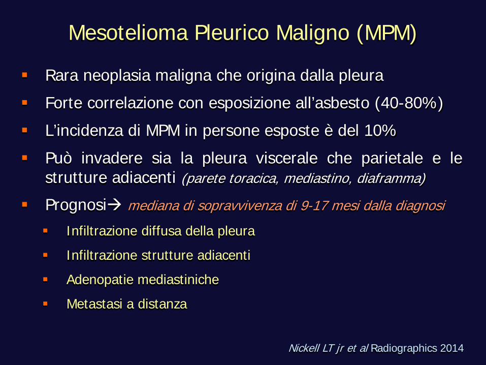
Mesotelioma Pleurico Maligno (MPM)
Rara neoplasia maligna che origina dalla pleura Forte correlazione con esposizione all’asbesto (40-80%) L’incidenza di MPM in persone esposte è del 10% Può invadere sia la pleura viscerale che parietale e le
strutture adiacenti (parete toracica, mediastino, diaframma)
Prognosi mediana di sopravvivenza di 9-17 mesi dalla diagnosi Infiltrazione diffusa della pleura Infiltrazione strutture adiacenti Adenopatie mediastiniche Metastasi a distanza
Nickell LT jr et al Radiographics 2014
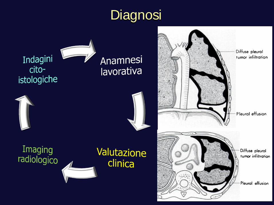
Diagnosi
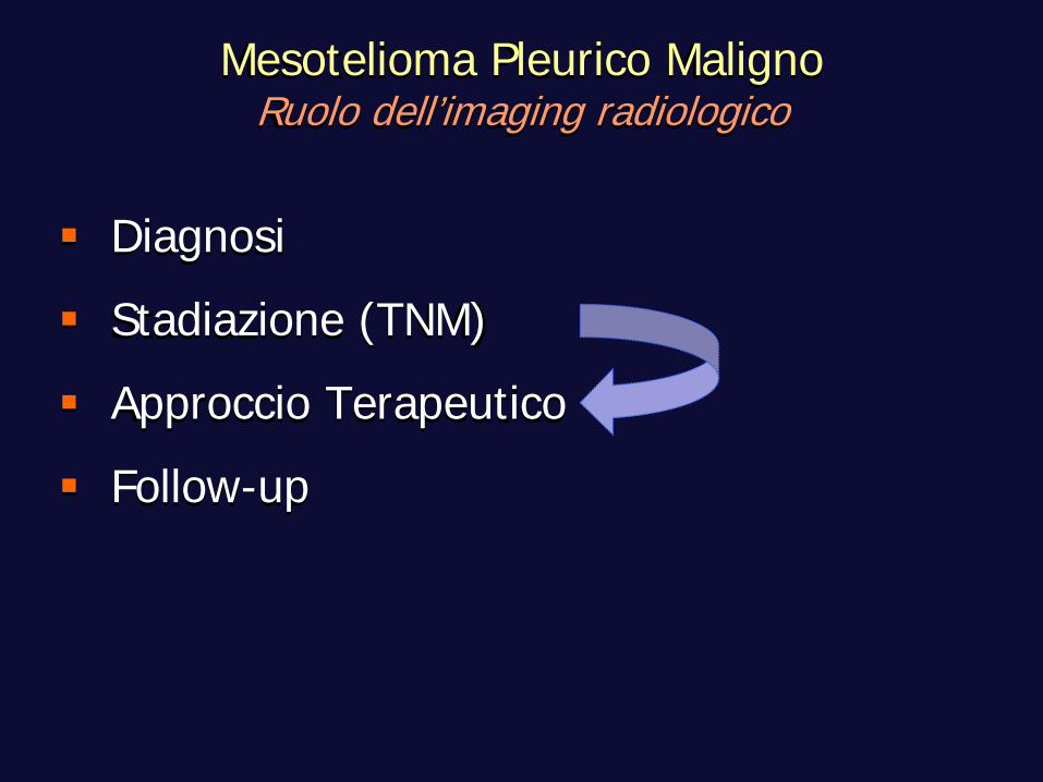
Diagnosi Stadiazione (TNM) Approccio Terapeutico Follow-up
Mesotelioma Pleurico Maligno Ruolo dell’imaging radiologico
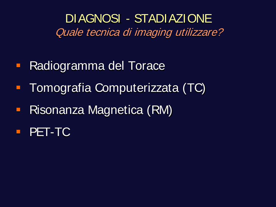
DIAGNOSI - STADIAZIONE Quale tecnica di imaging utilizzare?
Radiogramma del Torace Tomografia Computerizzata (TC) Risonanza Magnetica (RM) PET-TC
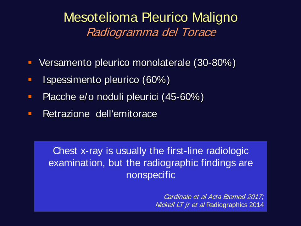
Mesotelioma Pleurico Maligno Radiogramma del Torace
Versamento pleurico monolaterale (30-80%) Ispessimento pleurico (60%) Placche e/o noduli pleurici (45-60%) Retrazione dell’emitorace
Chest x-ray is usually the first-line radiologic examination, but the radiographic findings are
nonspecific
Cardinale et al Acta Biomed 2017; Nickell LT jr et al Radiographics 2014
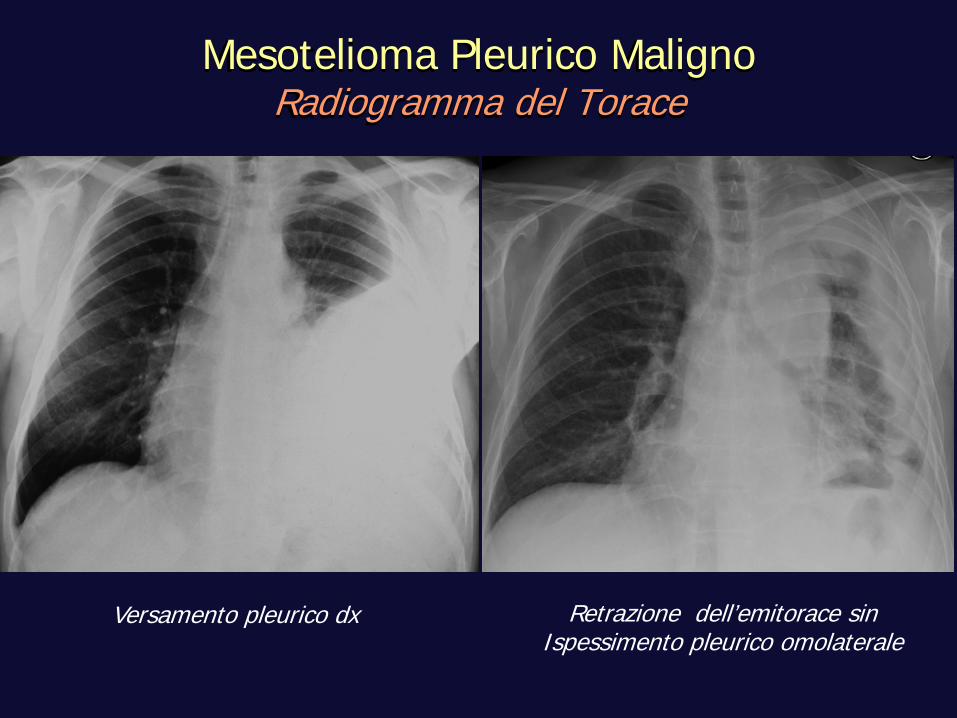
Retrazione dell’emitorace sin Ispessimento pleurico omolaterale
Mesotelioma Pleurico Maligno Radiogramma del Torace
Versamento pleurico dx
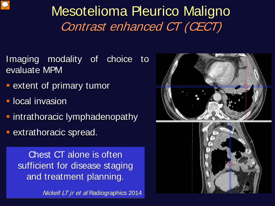
Mesotelioma Pleurico Maligno Contrast enhanced CT (CECT)
Imaging modality of choice to evaluate MPM extent of primary tumor local invasion intrathoracic lymphadenopathy extrathoracic spread.
Chest CT alone is often sufficient for disease staging
and treatment planning.
Nickell LT jr et al Radiographics 2014
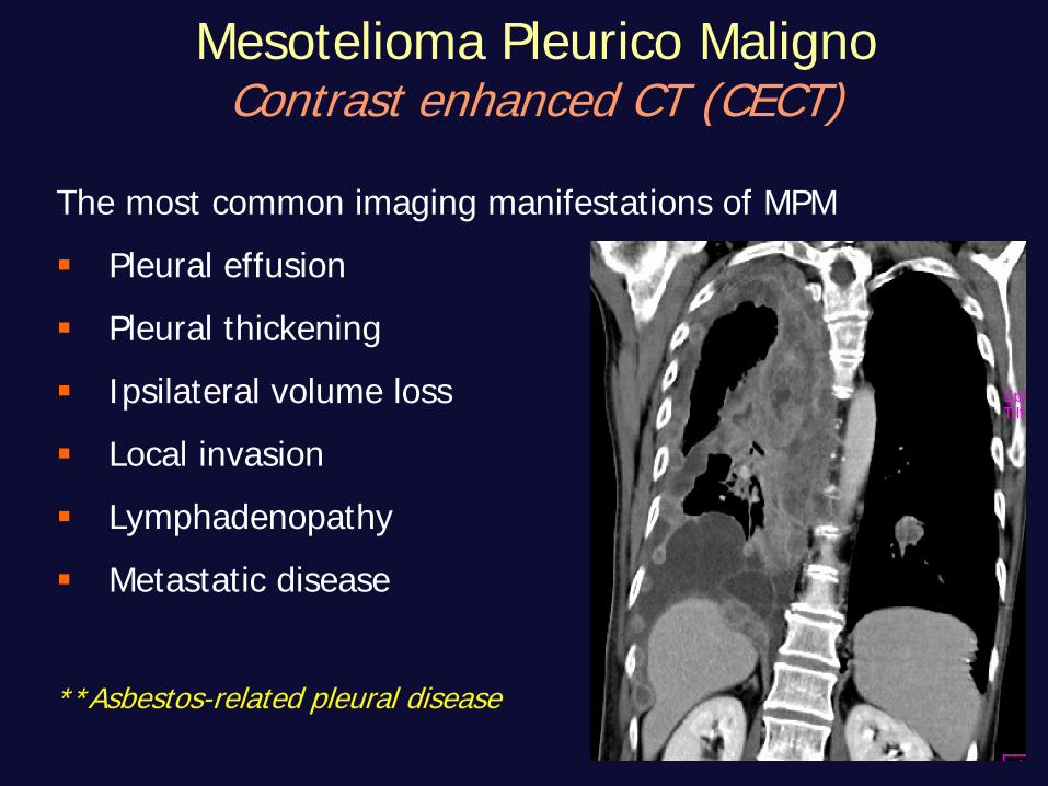
The most common imaging manifestations of MPM Pleural effusion Pleural thickening Ipsilateral volume loss Local invasion Lymphadenopathy Metastatic disease
**Asbestos-related pleural disease
Mesotelioma Pleurico Maligno Contrast enhanced CT (CECT)
Mesotelioma Pleurico Maligno Contrast enhanced CT (CECT)
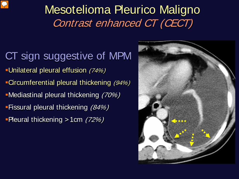
CT sign suggestive of MPM Unilateral pleural effusion (74%)
Circumferential pleural thickening (94%)
Mediastinal pleural thickening (70%) Fissural pleural thickening (84%) Pleural thickening >1cm (72%)
Mesotelioma Pleurico Maligno Contrast enhanced CT (CECT)
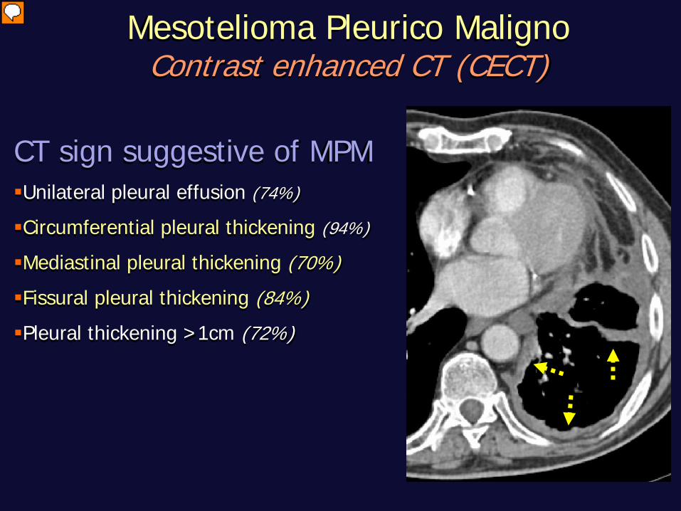
CT sign suggestive of MPM Unilateral pleural effusion (74%)
Circumferential pleural thickening (94%)
Mediastinal pleural thickening (70%) Fissural pleural thickening (84%) Pleural thickening >1cm (72%)
Mesotelioma Pleurico Maligno Contrast enhanced CT (CECT)
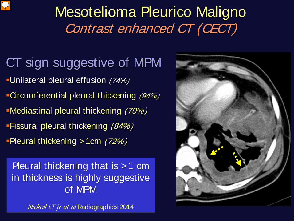
Pleural thickening that is >1 cm in thickness is highly suggestive
of MPM
Nickell LT jr et al Radiographics 2014
CT sign suggestive of MPM Unilateral pleural effusion (74%)
Circumferential pleural thickening (94%)
Mediastinal pleural thickening (70%) Fissural pleural thickening (84%) Pleural thickening >1cm (72%)
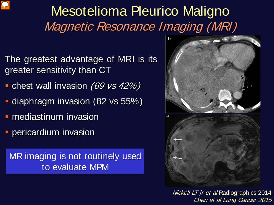
Mesotelioma Pleurico Maligno Magnetic Resonance Imaging (MRI)
The greatest advantage of MRI is its greater sensitivity than CT chest wall invasion (69 vs 42%) diaphragm invasion (82 vs 55%) mediastinum invasion pericardium invasion
MR imaging is not routinely used
to evaluate MPM
Nickell LT jr et al Radiographics 2014 Chen et al Lung Cancer 2015
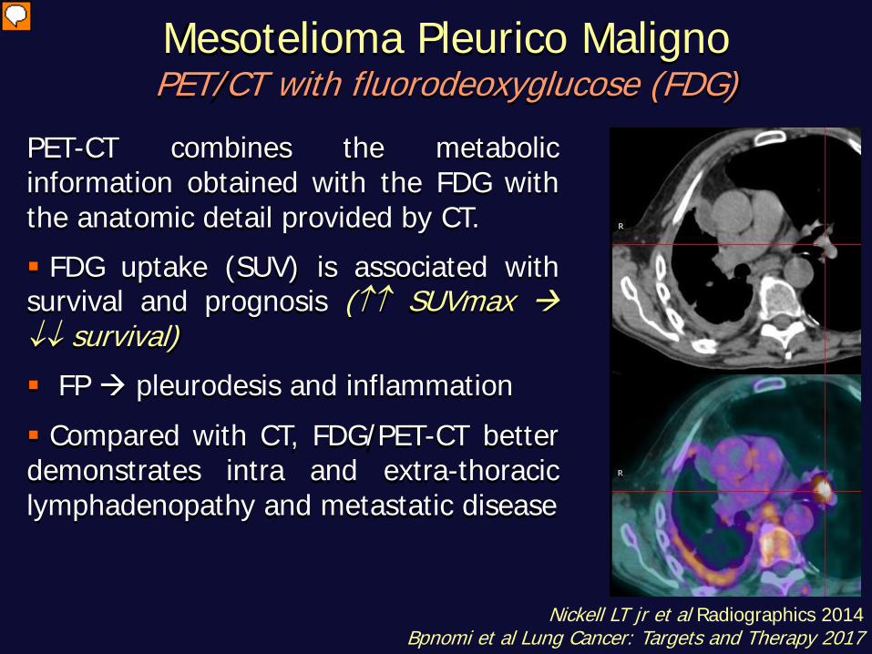
Mesotelioma Pleurico Maligno PET/CT with fluorodeoxyglucose (FDG)
PET-CT combines the metabolic information obtained with the FDG with the anatomic detail provided by CT. FDG uptake (SUV) is associated with survival and prognosis (↑↑ SUVmax ↓↓ survival) FP pleurodesis and inflammation Compared with CT, FDG/PET-CT better demonstrates intra and extra-thoracic lymphadenopathy and metastatic disease
Nickell LT jr et al Radiographics 2014 Bpnomi et al Lung Cancer: Targets and Therapy 2017
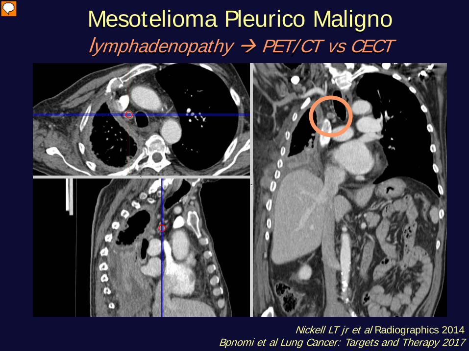
Mesotelioma Pleurico Maligno lymphadenopathy PET/CT vs CECT
Nickell LT jr et al Radiographics 2014 Bpnomi et al Lung Cancer: Targets and Therapy 2017
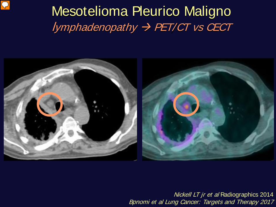
Mesotelioma Pleurico Maligno lymphadenopathy PET/CT vs CECT
Nickell LT jr et al Radiographics 2014 Bpnomi et al Lung Cancer: Targets and Therapy 2017
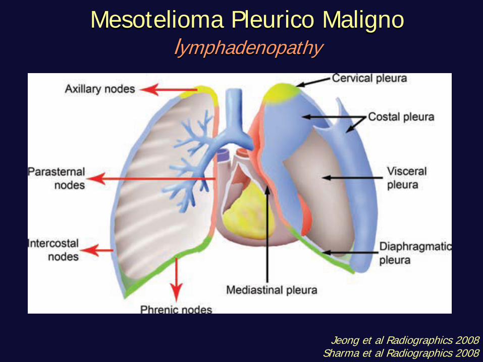
Mesotelioma Pleurico Maligno lymphadenopathy
Jeong et al Radiographics 2008 Sharma et al Radiographics 2008
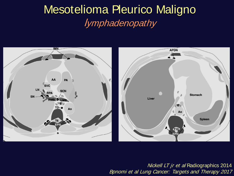
Mesotelioma Pleurico Maligno lymphadenopathy
Nickell LT jr et al Radiographics 2014 Bpnomi et al Lung Cancer: Targets and Therapy 2017
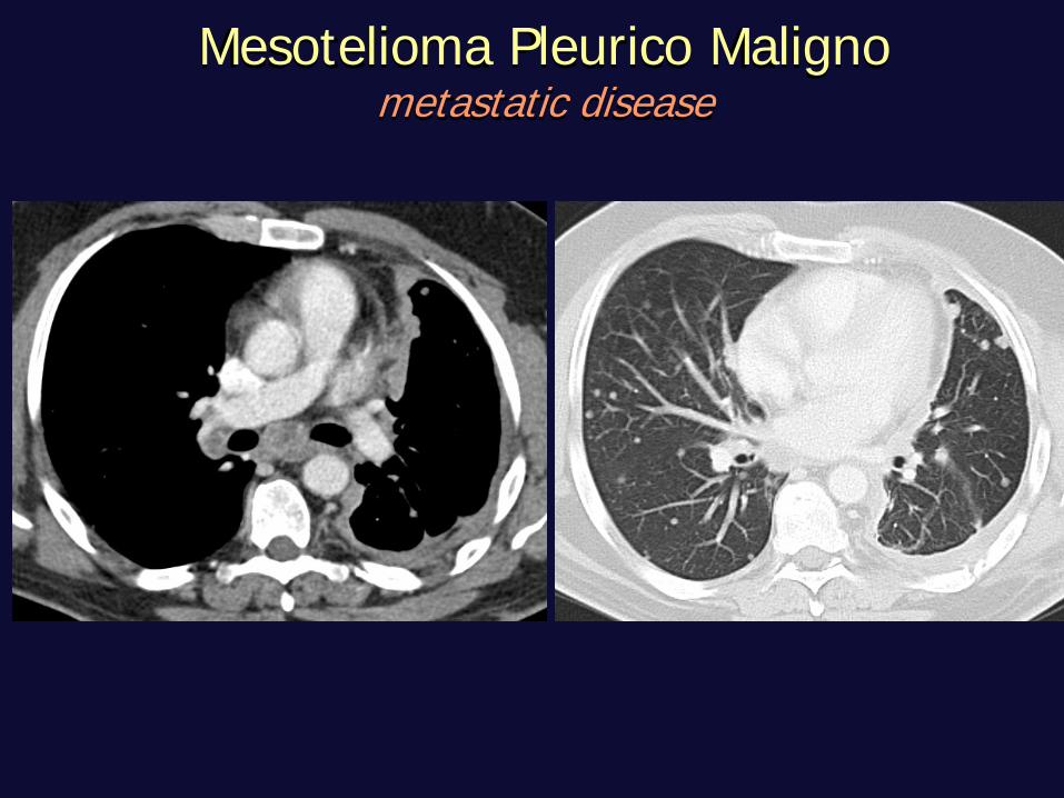
Mesotelioma Pleurico Maligno metastatic disease
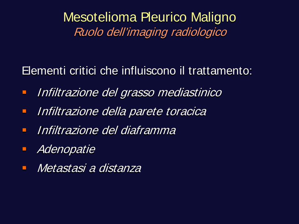
Elementi critici che influiscono il trattamento:
Infiltrazione del grasso mediastinico Infiltrazione della parete toracica Infiltrazione del diaframma Adenopatie Metastasi a distanza
Mesotelioma Pleurico Maligno Ruolo dell’imaging radiologico
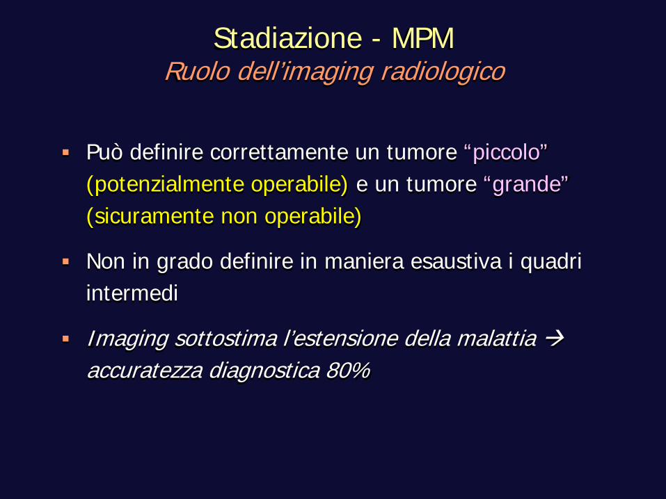
Può definire correttamente un tumore “piccolo” (potenzialmente operabile) e un tumore “grande” (sicuramente non operabile)
Non in grado definire in maniera esaustiva i quadri intermedi
Imaging sottostima l’estensione della malattia accuratezza diagnostica 80%
Stadiazione - MPM Ruolo dell’imaging radiologico
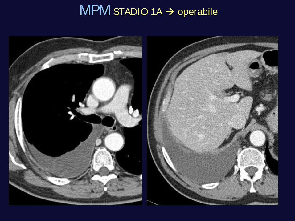
MPM STADIO 1A operabile
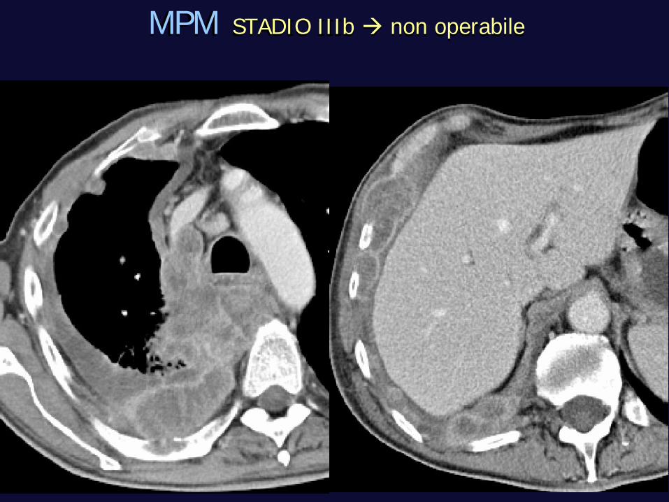
MPM STADIO IIIb non operabile
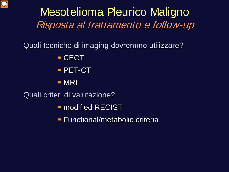
Quali tecniche di imaging dovremmo utilizzare? CECT PET-CT MRI
Quali criteri di valutazione? modified RECIST Functional/metabolic criteria
Mesotelioma Pleurico Maligno Risposta al trattamento e follow-up
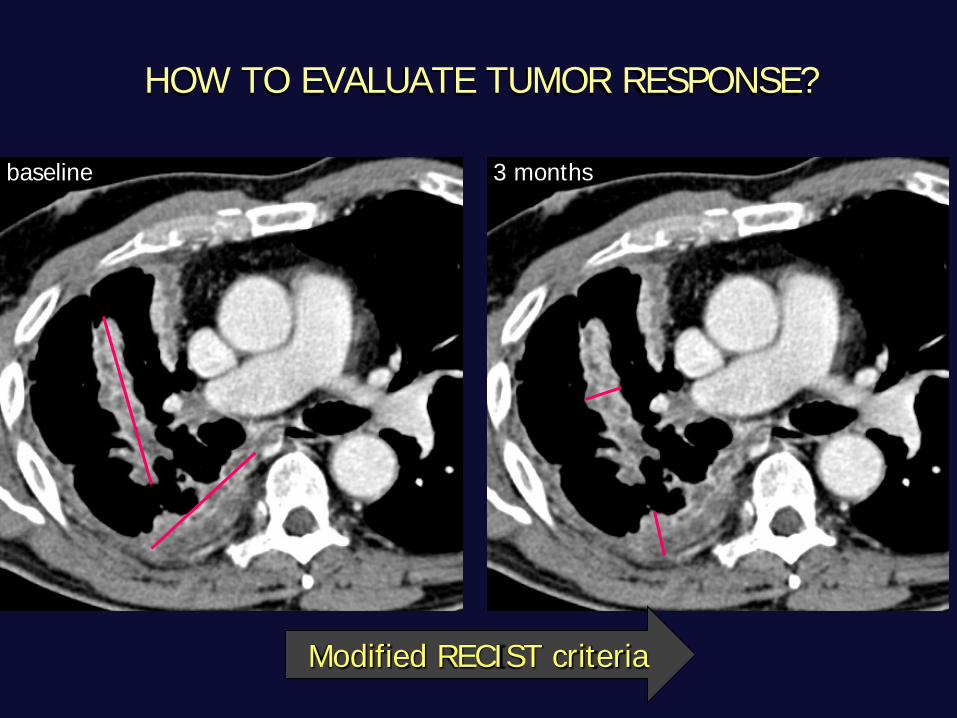
baseline 3 months
Modified RECIST criteria
HOW TO EVALUATE TUMOR RESPONSE?
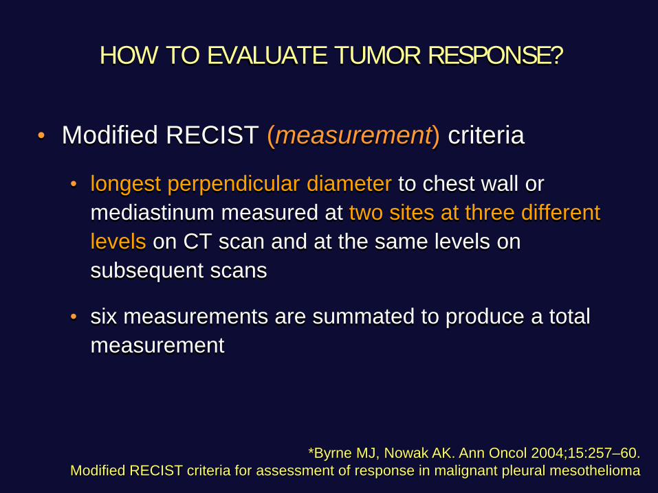
• Modified RECIST (measurement) criteria
• longest perpendicular diameter to chest wall or mediastinum measured at two sites at three different levels on CT scan and at the same levels on subsequent scans
• six measurements are summated to produce a total measurement
*Byrne MJ, Nowak AK. Ann Oncol 2004;15:257–60. Modified RECIST criteria for assessment of response in malignant pleural mesothelioma
HOW TO EVALUATE TUMOR RESPONSE?
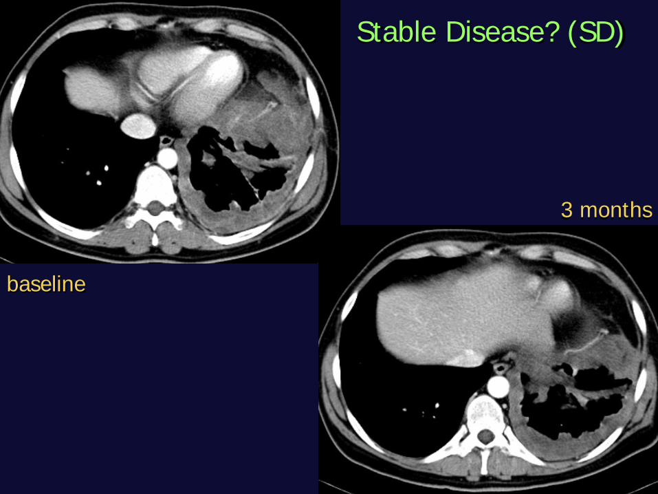
baseline
3 months
Stable Disease? (SD)
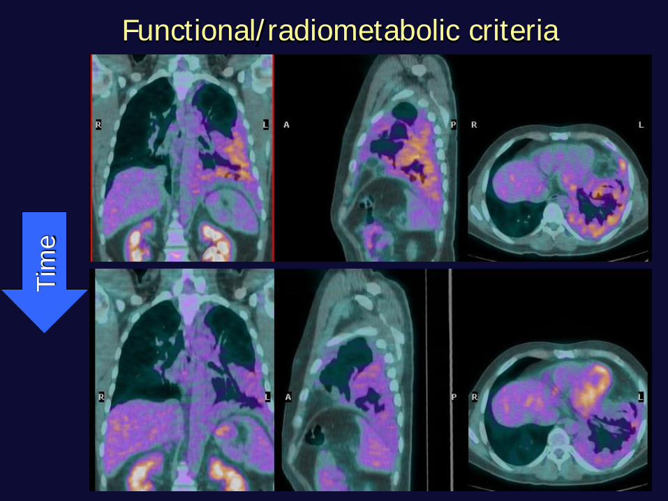
Tim
e Functional/radiometabolic criteria
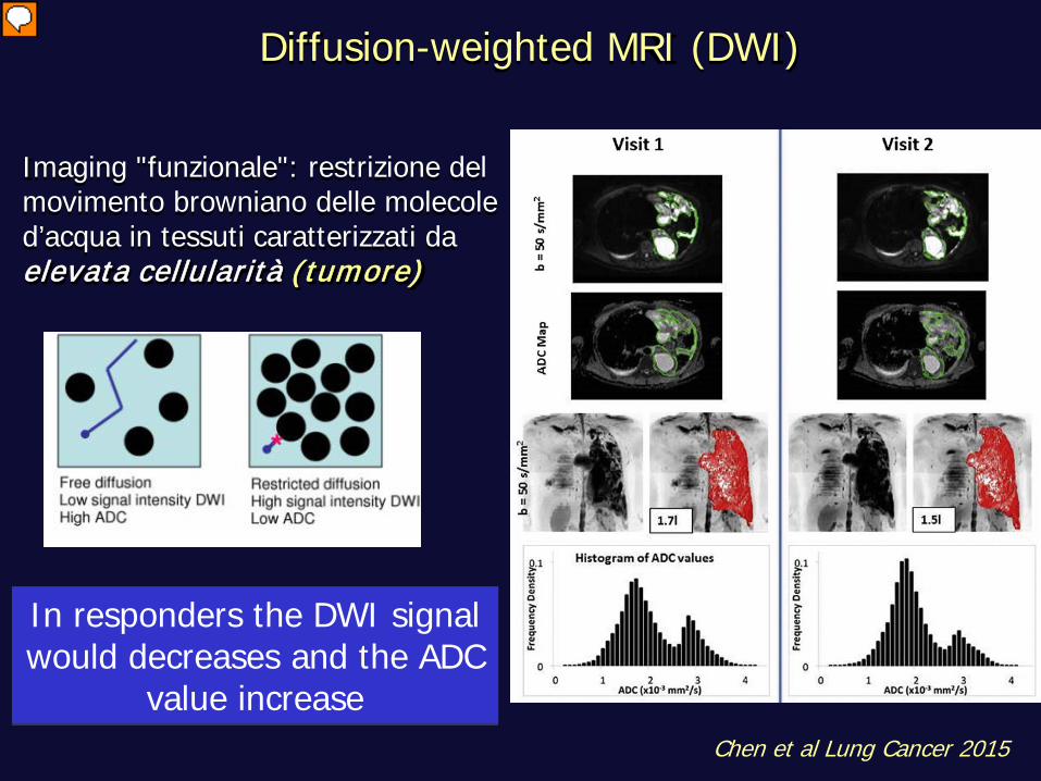
Diffusion-weighted MRI (DWI)
Imaging "funzionale": restrizione del movimento browniano delle molecole d’acqua in tessuti caratterizzati da elevata cellularità (tumore)
In responders the DWI signal would decreases and the ADC
value increase Chen et al Lung Cancer 2015
CONCLUSIONI STAGING
identificazione della malattia e giudizio di resecabilità
CECT
+
PET-CT
MRI
CONCLUSIONI FOLLOW-UP
Valutazione della risposta al trattamento
CECT
+
PET-CT
DWI (MRI)

Grazie per l’attenzione
A. Borghesi Scienze Radiologiche - Università degli Studi Brescia
U.O. Radiologia Diagnostica 2 - ASST Spedali Civili Brescia