scientific operating procedure sop-p-2 july 2015 … · sample preparation of nano-sized inorganic...
TRANSCRIPT

ERD
C/G
SL S
R-15
-1
Environmental Consequences of Nanotechnologies
Sample Preparation of Nano-sized Inorganic Materials for Scanning Electron Microscopy or Transmission Electron Microscopy Scientific Operating Procedure SOP-P-2
Geo
tech
nica
l and
Str
uctu
res
Labo
rato
ry
Charles A. Weiss, Jr., and Robert D. Moser July 2015
Approved for public release; distribution is unlimited.

The U.S. Army Engineer Research and Development Center (ERDC) solves the nation’s toughest engineering and environmental challenges. ERDC develops innovative solutions in civil and military engineering, geospatial sciences, water resources, and environmental sciences for the Army, the Department of Defense, civilian agencies, and our nation’s public good. Find out more at www.erdc.usace.army.mil.
To search for other technical reports published by ERDC, visit the ERDC online library at http://acwc.sdp.sirsi.net/client/default.

Environmental Consequences of Nanotechnologies
ERDC/GSL SR-15-1 July 2015
Sample Preparation of Nano-sized Inorganic Materials for Scanning Electron Microscopy or Transmission Electron Microscopy Scientific Operating Procedure SOP-P-2
Charles A. Weiss, Jr., and Robert D. Moser Geotechnical and Structures Laboratory U.S. Army Engineer Research and Development Center 3909 Halls Ferry Rd. Vicksburg, MS 39180-6199
Final report Approved for public release; distribution is unlimited.
Prepared for U.S. Army Corps of Engineers Washington, DC 20314-1000
Under Project Environmental Consequences of Nanotechnologies

ERDC/GSL SR-15-1 ii
Abstract
This protocol outlines how to prepare nano-sized materials for examination using electron microscopy with emphasis on particle size analysis. The type of sample preparation is dependent on the type of material. The protocol subdivides techniques for preparation of wet and dry samples, as well as conductive vs. nonconductive samples.
The preparation of nanomaterial samples for imaging can be challenging as these materials tend to agglomerate or aggregate. One way of handling this is to disperse the samples in a liquid and spray them onto a substrate. A sonic probe may also be used to break up aggregated or agglomerated samples. Another way is to extract the material from the liquid. In selected cases, imaging of the nanoparticles is aided through the removal of the solid particles from liquid matrix by using an organic solvent.
Conversely, solid inorganic nanomaterials are often poor conductors and may need to be coated to passivate the surface so that electrons from the electron beam are carried from the surface. Certain metal nanoparticles have been sputter etched to remove the oxide coating on the surface to promote better imaging. Carbon nanotubes or other organics and polymers may have a tendency to be damaged during imaging.
DISCLAIMER: The contents of this report are not to be used for advertising, publication, or promotional purposes. Citation of trade names does not constitute an official endorsement or approval of the use of such commercial products. All product names and trademarks cited are the property of their respective owners. The findings of this report are not to be construed as an official Department of the Army position unless so designated by other authorized documents. DESTROY THIS REPORT WHEN NO LONGER NEEDED. DO NOT RETURN IT TO THE ORIGINATOR.

ERDC/GSL SR-15-1 iii
Contents Abstract .......................................................................................................................................................... ii
Figures and Tables ........................................................................................................................................ iv
Preface ............................................................................................................................................................. v
1 Introduction ............................................................................................................................................ 1
2 Background ............................................................................................................................................ 2
3 Scope ....................................................................................................................................................... 4
4 Terminology ............................................................................................................................................ 6 4.1 Related documents ....................................................................................................... 6 4.2 Definitions ...................................................................................................................... 6 4.3 Acronyms ........................................................................................................................ 7
5 Materials and Apparatus ..................................................................................................................... 8 5.1 Materials ........................................................................................................................ 8 5.2 Apparatus ....................................................................................................................... 8
6 Procedure ............................................................................................................................................... 9 6.1 Specimen preparation ................................................................................................... 9
6.1.1 Extraction of nanoparticles for media........................................................................ 9 6.1.2 Specimen preparation for analysis .......................................................................... 10
6.2 Example of sample preparation .................................................................................. 14
7 Reporting .............................................................................................................................................. 16 7.1 Analysis of results ........................................................................................................ 16 7.2 Key results provided .................................................................................................... 16 7.3 QA/QC considerations ................................................................................................. 16
References ................................................................................................................................................... 17
Appendix A: Annex–Notes and Supplementary Data .......................................................................... 19
Report Documentation Page

ERDC/GSL SR-15-1 iv
Figures and Tables
Figures
Figure 1. Flow path for preparation of samples for electron microscopy. .............................................. 5 Figure A1. FEI Co. Nova NanoSEM. ............................................................................................................ 21
Tables
Table A1. Various mounting media used in SEM (University of California, Riverside, n.d.). ............... 19 Table A2. Types of backgrounds available for use in SEM (University of California, Riverside, n.d.). ............................................................................................................................................. 20

ERDC/GSL SR-15-1 v
Preface
This procedure was developed under the U.S. Army Engineer Research and Development Center (ERDC), Environmental Quality and Technology (EQT) Research Program titled “Environmental Consequences of Nanotechnologies.” Procedures link to the ERDC NanoGRID (Guidance for Risk Informed Deployment) framework for testing the exposure and hazard of nanotechnology Environmental Health and Safety (EHS). The technical monitor was Dr. Elizabeth A. Ferguson, CEERD-EZT.
The work was accomplished by the Research Group (GMR) and the Concrete and Materials Branch (GMC) of the Engineering Systems and Materials Division (GM), U.S. Army Engineer Research and Development Center, Geotechnical and Structures Laboratory (ERDC-GSL). At the time of publication, Christopher M. Moore was Chief, CEERD-GMC; Dr. Larry N. Lynch was Chief, CEERD-GM; and Dr. Elizabeth A. Ferguson, CEERD-EZT, was the Technical Director for Military Environmental Engineering and Sciences. The Acting Deputy Director of ERDC-GSL was Dr. Gordon W. McMahon, and the Acting Director was Dr. William P. Grogan.
LTC John T. Tucker III was the Acting Commander of ERDC, and Dr. Jeffery P. Holland was the Director.
ERDC/GSL SR-15-1 1
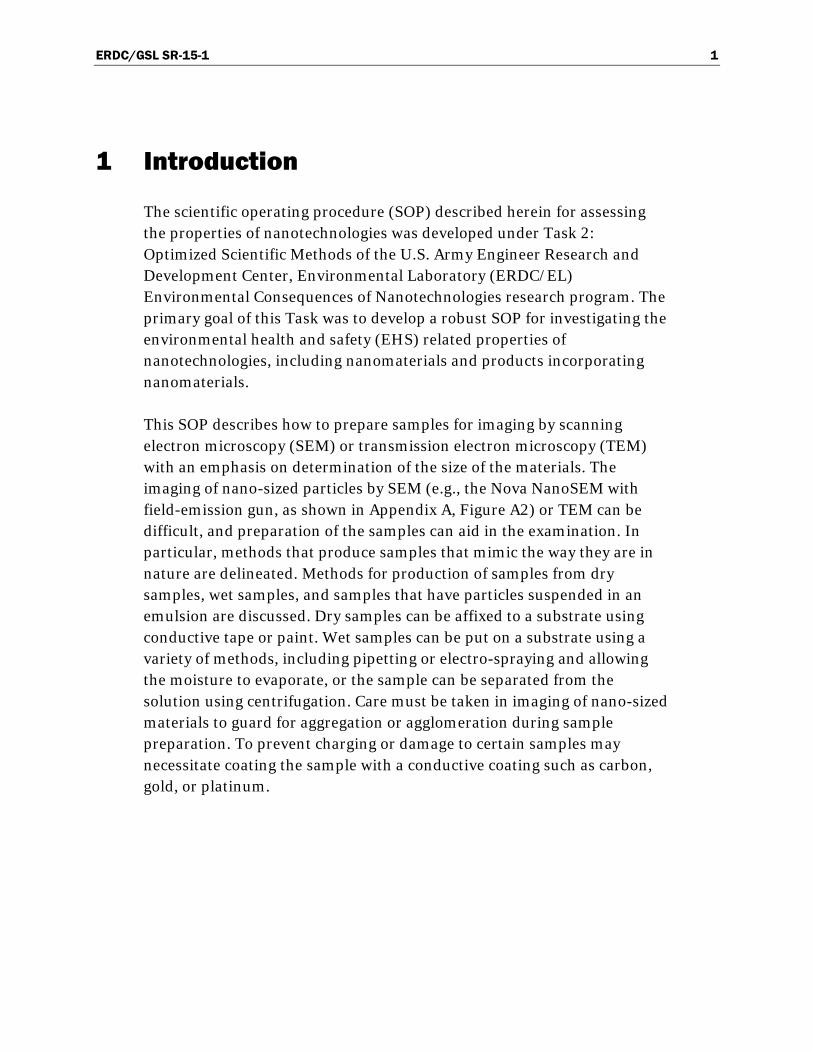
1 Introduction
The scientific operating procedure (SOP) described herein for assessing the properties of nanotechnologies was developed under Task 2: Optimized Scientific Methods of the U.S. Army Engineer Research and Development Center, Environmental Laboratory (ERDC/EL) Environmental Consequences of Nanotechnologies research program. The primary goal of this Task was to develop a robust SOP for investigating the environmental health and safety (EHS) related properties of nanotechnologies, including nanomaterials and products incorporating nanomaterials.
This SOP describes how to prepare samples for imaging by scanning electron microscopy (SEM) or transmission electron microscopy (TEM) with an emphasis on determination of the size of the materials. The imaging of nano-sized particles by SEM (e.g., the Nova NanoSEM with field-emission gun, as shown in Appendix A, Figure A2) or TEM can be difficult, and preparation of the samples can aid in the examination. In particular, methods that produce samples that mimic the way they are in nature are delineated. Methods for production of samples from dry samples, wet samples, and samples that have particles suspended in an emulsion are discussed. Dry samples can be affixed to a substrate using conductive tape or paint. Wet samples can be put on a substrate using a variety of methods, including pipetting or electro-spraying and allowing the moisture to evaporate, or the sample can be separated from the solution using centrifugation. Care must be taken in imaging of nano-sized materials to guard for aggregation or agglomeration during sample preparation. To prevent charging or damage to certain samples may necessitate coating the sample with a conductive coating such as carbon, gold, or platinum.

ERDC/GSL SR-15-1 2
2 Background
This protocol will focus on proper ways to prepare samples of nano-sized materials for imaging in an electron microscope. Echlin (2009) states, “Every specimen that goes into the SEM needs some form of sample preparation. There are no exceptions.”
Before a sample can be observed with the SEM, it is often necessary to prepare the sample with additional processing steps to allow the sample to be imaged in an SEM. This process often makes the sample conductive, more stable in a vacuum and under the electron beam, or optimizes the geometry between the sample surface, the electron, and one (or more) of the detectors attached to the SEM.
In particular, nano-sized materials have the issue that they agglomerate or aggregate, and proper imaging of these samples requires special treatment of the samples (Roselina and Azizan 2012). The type of sample preparation is dependent on the type of material. Conductive nano-sized materials such as metal oxides need little preparation, are very stable under vacuum, and often are stable under the electron beam. Sample preparation for these types of samples oftentimes is to affix them to a stub by using conductive tape. Because of their conductive nature, they often do not need to be sputter coated with a metal or carbon to aid in dissipation of charge buildup on the surface of the sample. Any charge buildup on the surface will often make it difficult to get quality images, as the buildup will affect the electron beam during imaging. Nano-sized metals provide a different problem in that they may react with oxygen when opened in air and may be a safety issue.
Nanomaterials that have liquid (water, oil, etc.) either in their structure as a major component of their composition or as a medium for the sample are divided into two groups: wet samples and liquid samples. Preparing these samples for imaging poses challenges. Echlin (2009) defines wet samples as samples that have a recognizable structure during imaging but have liquid as a major component of their structure. They do not flow under normal temperatures. Liquid samples are defined as samples that have no discernible structure during imaging and flow under normal temperatures.

ERDC/GSL SR-15-1 3
One way of handling aggregation or agglomeration is to disperse the samples in a liquid and spray them onto a substrate (Hamamatsu Nano Technology 2014; Alsyouri et al. 2013). A sonic probe may also be used to break up aggregated or agglomerated samples (Safarova et al. 2007; Taurozzi et al. 2012a, b). Another way is to extract the material from the liquid. In one example, hexane was used in a method to remove TiO2 nanoparticles from sunscreen (Nischwitz and Goenaga-Infante 2012).
Conversely, solid, inorganic nanomaterials are often poor conductors, and as such may, need to be coated to passivate the surface so that electrons from the electron beam are carried from the surface. Other materials, such as NiFe metal nanoparticles, have been sputter etched to remove the oxide coating on the surface to promote better imaging (Somwang et al. 2009). Still other materials, such as carbon nanotubes or other organics and polymers, have a tendency to be damaged by the electron beam during imaging (Suzuki 2004).
In general, agglomeration and aggregation of the nano-sized materials pose a problem for nanomaterials when imaging (Taib and Sorrell 2007, 2008). Some techniques for sample preparation of nanomaterials include dispersiing a sample onto a substrate or stub (Alsyouri et al. 2013), coating with a stabilizing solution (Yao and Kimura 2007; Bonevich and Haller 2010), removing the liquid to allow better imaging of individual particles in the suspension (Nischwitz and Goenaga-Infante 2012), and polishing of materials, especially for microchemical analysis. In addition, a metal or carbon coating may be applied to allow dissipation of electron buildup on the surface of the sample.
The preparation of samples is critical to allow good images and proper particle size analysis of the nano-sized materials. Understanding of the sample is critical to obtain accurate dimensions of nano-sized materials. Removal of an emulsion or other liquid that may hamper imaging of the material should aid in particle imaging. Centrifugation can be used to concentrate the solid particles in a solution. After removal, imaging of the native particles can be conducted. If aggregation of the particles is an issue, use of a sonic probe can aid in disaggregation of the sample. Nano-sized particles have a tendency to agglomerate during sample preparation.

ERDC/GSL SR-15-1 4
3 Scope
This SOP is used to determine the method to prepare nanomaterials for investigations in the SEM (Figure 1). It is particularly tailored for use with nano-sized materials either as discrete powders or incorporated in a liquid.

ERD
C/GSL SR
-15-1 5
Figure 1. Flow path for preparation of samples for electron microscopy.
Sample Preparation for Electron Microscopy for Materials Containing
Nano-sized particles Sample
'~ Are particles In a solution, liquid, emulsion, etc,? I
J, Yes ..v J IJ
J Dovou want to ~eep particles No Does the sample need to be No I'' tl>e sample to be smeared Preparnppropriote sUbstrote In the solution, liquid, - dlsaaare1ated uslna a sonic on a substrate? l•lumlnum stub, aloss ca.~er emulsion, etc.? techniqut?
~ Yes slip, TEMarld, silicon, etc.).
,l Yes Yes lit II
I. No Sonlcote somple. I Preporesmeeron appropriote I Will an appropriate material Will electro-spray deposition substrate. No bell5ed? ~ J, be used to affix the material
Yes (carbon paint, silver paint; 1-Didthl: material adequately No carbon tape, copper tape,
1: separate? etc.)?
Is centrifugat ion needed or ~t, Yes Yes
desired to separate tile - Is a solvent needed to remove ~ " sample? No
the emulsion? Un epproprlate odtlesive on
Yes ~ --sUbstr•te. J Yes I SeporatethesompletJSi~
centnfuaatlon. Useapproprltte solvent to
J, emlc.te nentHizedportlcles.
I I Did the material adequately ~0 ptt~ceanappropriate amount separate? of sompleon sllbstrato
I Yes (pipette, etc.).
I Doesthe sample need to be I· coated to eliminate mar1ina?
ERDC/GSL SR-15-1 6
4 Terminology
4.1 Related documents
• ASTM E1617-97, Standard Practice for Reporting Particle Size Characterization Data
4.2 Definitions
The following definitions are relevant to this SOP:
• agglomerate, n—in nanotechnology, an assembly of particles held together by relatively weak forces (e.g., Van der Waals or capillary) that may break apart into smaller particles upon processing. The International Union of Pure and Applied Chemistry (IUPAC) defines it as “a process of contact and adhesion whereby dispersed particles are held together by weak physical interactions ultimately leading to phase separation by the formation of precipitates of larger than colloidal size.”
• agglomeration (except in polymer science), n—process of contact and adhesion whereby dispersed particles are held together by weak physical interactions ultimately leading to phase separation by the formation of precipitates of larger than colloidal size. Agglomeration is a reversible process.
• aggregate, n—in nanotechnology from IUPAC (http://goldbook.iupac.org/A00184.html), certain materials used as catalysts or supports consist of spheroids smaller than 10 nanometers (nm) in diameter (diam), cemented into larger entities. A primary particle should be defined as the smallest discrete identifiable entity, and the method of identification should be mentioned (e.g., TEM, SEM). An assemblage of such primary particles exhibiting an identifiable collective behavior (e.g., chemical nature of the aggregated primary particles, texture of the aggregate, resistance to mechanical separation upon grinding) constitutes an aggregate. When describing the aggregates, the criterion of identification should be mentioned. Strongly bonded aggregates are called agglomerates.

ERDC/GSL SR-15-1 7
4.3 Acronyms
Relevant acronyms include the following:
• SEM – scanning electron microscopy • TEM – transmission electron microscopy • XRF – X-ray fluorescence • XRD – X-ray diffraction.
ERDC/GSL SR-15-1 8
5 Materials and Apparatus
5.1 Materials
• Zero background plate(s)–for nanoparticles to be dispersed onto for XRD analysis
• Ethanol (reagent grade), hexane, alcohol, acetone, methanol, and isopropanol–for cleaning of substrates
• Sample stubs – for affixing of samples • Conductive tape (carbon, copper, etc.)–for adhering powders to stubs
5.2 Apparatus
• Particle deposition system–sample preparation system for SEM/TEM for affixing of nanomaterials onto a substrate without aggregation
• Centrifuge
ERDC/GSL SR-15-1 9
6 Procedure
• Determine what is desired to be seen in the SEM photomicrograph that will be impacted by the sample preparation.
• Determine the best way to prepare a sample based on what is desired to be seen and what sample is available.
• Prepare the sample based on the nature of the sample (i.e., if it is in dry or liquid form). Also important in this is its tendency to agglomerate.
• Examine the sample by electron microscopy.
6.1 Specimen preparation
Sample cleaning and drying–Before SEM characterization, samples must be thoroughly degreased and dried to eliminate any outgassing from organic contamination and water. Samples can be cleaned ultrasonically using solvents like acetone and methanol (or alcohol). When cleaned by a volatile solvent, samples can be blown dry by using a compressed gas. After being cleaned by water, samples should be dried completely using an oven or a hot plate or be allowed to air dry. Any surface contamination can be removed by blowing a compressed gas (Vladar and Ming 2011).
6.1.1 Extraction of nanoparticles for media
When the nanoparticles are powders, no extraction is necessary. For those samples that have nano-sized materials in a solvent or liquid, sometimes it is preferable to remove the nanomaterials before sample preparation.
For samples such as sunscreens, removal of the lipid carrier of the sample with hexane followed by sonication with an aqueous extractant was found to provide stable suspensions of secondary titanium dioxide particles for a variety of analytic techniques. Further addition of a small amount of hexane to the aqueous extractant resulted in particle disaggregation and thus allowed for characterization of the primary particle (Nischwitz and Goenaga-Infante 2012).

ERDC/GSL SR-15-1 10
6.1.2 Specimen preparation for analysis
6.1.2.1 Handling and sample mounting
Handling–Grease from hands is a major contamination to the SEM system. Therefore, gloves must be worn all the time during sample preparation. Samples, sample holders, sample stubs, or sample exchange tools must not be touched with bare hands. All parts and tools should be handled on a clean paper such as Kimwipes.
Sample mounting–Normally, samples are mounted onto the holders or stubs by using double-sided conductive tapes or conductive paint. Only the vacuum-compatible carbon and copper tapes provided by the SEM lab should be used. Silver paint can also be used for sample mounting with complete drying before loading into the SEM chamber. Examples of various mounting adhesives are given in Table A1 (University of California, Riverside, n.d.).
6.1.2.2 Dispersions
Dispersions are often some of the most difficult sample types to prepare properly. Charging of the sample is often a problem that manifests itself in bright and dark areas and/or image drift. This category can be split again into two types for simplicity, wet dispersions (in suspension) or dry dispersions (powders/fibers).
6.1.2.3 Wet dispersions: Suspensions
Polymers, paint pigment, and biological cultures are examples of materials that can be prepared using this method. Variables that determine how successful this technique will be include the density of suspension and the size range of the material in the suspension. It is important that the particles be separated. Biological cultures often present other challenges, as destruction of detail structures may occur because this technique involves air drying, which may collapse cell structures.
6.1.2.4 Dilution
To ascertain the correct dilution of a suspension, many samples need to be made at several concentrations. The liquids used for suspensions are usually distilled or de-ionized water, methanol, ethanol, acetone, or isopropanol. The properties of the sample material and/or the suspension

ERDC/GSL SR-15-1 11
liquid already present determine which solution is the appropriate one. Samples should be sufficiently agitated in an ultrasonic bath for at least 10 minutes to ensure separation of particles (University of California, Riverside, n.d.). These solutions can then be pipetted onto alcohol-cleaned coverslips mounted on aluminum stubs and allowed to dry. If needed, the samples can then be coated with a conductive material such as carbon or metal (Au, Au/Pd). Researchers studying gold nanoparticles have stabilized them using carboxylates (Yao and Kimura 2007) or citrates (Bonevich and Haller 2010). A correct suspension will have neither the entire surface of the coverslips covered with particles nor agglomeration more than 2 to 3 particles high A choice of pipetting or atomizing the suspension then can be made based mainly on the size and agglomeration effects seen on the test sample. The size range for pipetting is far larger, submicron to hundreds of microns, where atomizing is more limited from submicrometer to a few tens of micrometers (University of California, Riverside, n.d.). Agglomeration of material that will be pipetted can be further agitated in the ultrasonic bath to separate particles. It should be noted that excessive ultrasonic agitation can damage samples. Atomization helps to disperse agglomerates of fine sizes, but it is always useful to ultrasonically disaggregate the sample suspension beforehand.
6.1.2.5 Pipette
In this method a suspension is drawn up in a pipette immediately after agitation, and a couple of droplets are placed onto a prepared substrate. The sample is then allowed to dry and then sputter coated. A new pipette should be used for each sample so as not to contaminate samples. In this method the particles will be thickest in the middle and thinnest at the edges.
6.1.2.6 Atomization
Atomizing pens or airbrushes can also be used to distribute various sized particles on substrates. The advantage of this technique is that it allows better control of size change at the dispersing nozzle aperture so that a particular size range of particles can be delivered. Samples should not be sprayed directly onto a substrate, as this promotes a quick buildup of particles and may increase the possibility of agglomeration. The recommended method (University of California, Riverside, n.d.) is to spray at a 45° angle into a vertical tube approximately 10 cm diameter by 30 cm long, with the sample substrate sitting at the bottom of the tube. Several

ERDC/GSL SR-15-1 12
attempts are usually needed to ascertain the appropriate length of time to spray. Thoroughly cleaning the pen spray nozzle before changing samples is critical to this process. Acetone should never be used with an atomizer, as it may dissolve some of the parts (University of California, Riverside, n.d.).
6.1.2.7 Size separation
The Cascade Impactor is a device that allows separation of particles by size. In some cases, it is desired to separate the size ranges of material suspensions before they are placed into the SEM. This device has the ability to separate a range of particle sizes, starting at the largest and ending with the smallest. In this system a vacuum is drawn through the device at the bottom, and a dispersion is sprayed out at the top; at each size position, there is an aperture backed by a collecting substrate, and the relative size particles are trapped through the aperture. As the apertures range from large to small, the largest particle size stays at the top of the device, and the smallest size stays at the last position. Any combination of progressively smaller or intermediate apertures can be added to further separate the sizes (University of California, Riverside, n.d.)
6.1.2.8 Sandwich technique
A sandwich technique can be used for those suspensions that have a tendency to charge during imaging. In this method a material is put between two conducting layers. A substrate such as a 10 mm coverslip is washed in alcohol, dried, and placed with adhesive onto an aluminum stub. This substrate is then sputter coated with a thick, uniform layer of gold. The substrate is then coated again for a shorter time but at a higher current to crack the initial coating. The resultant substrate has a layer of gold with deep cracks of very small width. The sample suspension is placed over the gold and allowed to dry. Through capillary action the particles are drawn into the cracks in the gold. To improve the conductivity, a second thinner coating of any sputtered metal can then be applied onto the sample to sandwich the particles (University of California, Riverside, n.d.).
6.1.2.9 Dry dispersions: Powders/fibers
Powder samples–Powder materials are hard to mount properly and do not become dislodged and enter the column. As such, they need special handling for sample mounting. Small amounts of powder can be dispersed

ERDC/GSL SR-15-1 13
in a volatile solvent like acetone and alcohol, and then drops of the mixture can be dripped onto a clean substrate. After drying, the powder particles should be dispersed onto the substrate surface. Carbon tape and copper tape can be used for powder mounting. A spatula is used to distribute the powder lightly and to press lightly to have the sample adhere to the tape. The sample holder is then turned upside down and tapped to remove loose material or is blown off with a compressed gas.
In the case for nonmetal powdered materials, it is imperative that some of the particles are well conducting when mounted for the SEM. Proper selection of a proper adhesive may assist in the prevention of charging and may be important if X-ray analysis will be used.
Metal powders are usually not a problem, but adhesives such as double-sided tape should be avoided, as they have been known to contain traces of zinc carbonate that can be picked up by reflection and show on the XRF (University of California, Riverside, n.d.).
Fibers can be more difficult to image due to their poor connections to each other and to the substrate. It is wise to use carbon or silver paint with these stubs to ensure good adherence (Murata et al. 2012). Selection of a suitable background may be important, as fibers of all kinds usually have a substructure. Sputter coating of organic fibers should be performed carefully because they can be easily damaged by the elevated heat during sputter coating (University of California, Riverside, n.d.).
6.1.2.10 Electro-spray deposition
Electro-spray deposition is a method that has the capability of depositing nanomaterials onto a substrate and minimizes or eliminates aggregation. It is preferable to treat nanomaterials as a nanosuspension, which is dispersed in poor solvent, because nanomaterials are hard to handle and aggregate or agglomerate easily. It is important for fundamental research and application development of nanomaterials to investigate a size, a form, and aggregation degree of respective nanomaterial in nanosuspension. SEM and TEM are suitable for nanomaterials observation. However, it is hard to observe a respective nanomaterial due to the aggregation in the vaporized process of solvent.
Less conductive samples–When samples are not very conductive, charge effect will cause image distortion or drift. Low acceleration voltage should

ERDC/GSL SR-15-1 14
be used to reduce the charge effect if samples cannot be coated with a conductive coating. To completely eliminate the charge effect, samples should be coated. An SEM lab has a sputtering coater for gold coating. The coating thickness could be several nanometers to tens of nanometers, depending on whether the coating interferes with the morphology of the sample. After coating, the sample should be mounted with a conductive conduit (e.g., carbon/copper tapes or silver paint) connected from the top surface of the sample to the sample holder.
6.2 Example of sample preparation
An example procedure in Figure 1 is given here.
1. Are the particles in a solution, liquid or emulsion? If no, go to 14. If yes, go to 2.
2. Do you want to keep particles in the solution, liquid, or emulsion? If no, go to 7. If yes, go to 3.
3. Will electro-spray deposition be used? If no, go to 4. If yes, go to 18. 4. Is centrifugation needed to separate the sample? If no, go to 7. If yes, go to 5. 5. Separate the sample using a centrifuge. 6. Did the material adequately separate? If no, go to 5. If yes, go to 18. 7. Does the sample need to be disaggregated using a sonic technique? If no,
go to 12. If yes, go to 8. 8. Use a sonic probe or sonic bath to disaggregate the sample. 9. Did the material adequately disaggregate? If no, go to 8. If yes, go to 10. 10. Is a solvent needed to remove the emulsion? If no, go to 12. If yes, go to 11. 11. Use appropriate solvent to remove emulsion. 12. Is the sample to be smeared onto a substrate? If no, go to 14. If yes, go to 13. 13. Prepare smear on appropriate substrate 14. Prepare appropriate substrate for given sample. Examples of substrates
are given in Table A2. 15. Will an appropriate material be used to affix the material to the substrate?
Examples of mounting media are given in Table A1. If no go to 14. If yes, go to 16.
16. Apply appropriate adhesive to substrate. 17. Place an appropriate amount of the sample onto the substrate. If the
sample is liquid, use a pipette, dropper, etc.; if it is solid, use a spatula. The sample may be weighed to ascertain the amount to put onto the substrate.
18. Does the sample need to be coated to eliminate charging? If no, go to 20. If yes go, to 19.

ERDC/GSL SR-15-1 15
19. Coat the sample with an appropriate conductive coating (carbon, gold, platinum, etc.) using a sputter coater.
20. Image the sample.

ERDC/GSL SR-15-1 16
7 Reporting
7.1 Analysis of results
Verification of how well the sample is prepared is determined through imaging in the SEM.
7.2 Key results provided
The result of this protocol should be a properly prepared sample for imaging.
7.3 QA/QC considerations
Based on imaging of the sample, replicates may be made to try to provide the best samples for imaging.

ERDC/GSL SR-15-1 17
References Alsyouri, H. M., M. A. Abu-daabes, A. Alassali, and J. Y. S. Lin. 2013. Ordered
mesoporous silica prepared by quiescent interfacial growth method–Effects of reaction chemistry. Nanoscale Research Letters 8:1–15.
American Society for Testing and Materials (ASTM). 1997. Standard practice for reporting particle size characterization data. Designation E1617-97. West Conshohocken, PA: American Society for Testing and Materials.
Bonevich, J. E., and W.K. Haller. 2010. NIST - NCL joint assay protocol, PCC-7 measuring the size of nanoparticles using transmission electron microscopy (TEM) 1–13.
Echlin, P. 2009. Handbook of sample preparation for scanning electron microscopy and x-ray microanalysis. New York: Springer.
Hamamatsu Nano Technology. 2014. Sample preparation system for SEM / TEM: Fixing of nanomaterials on a substrate without aggregation. 2–4. http://www.hamanano.com/e/products/c1/c1_2/index.html.
International Union of Pure and Applied Science Compendium of Chemical Terminology, 2nd ed. (the "Gold Book"). Compiled by A. D. McNaught and A. Wilkinson. Blackwell Scientific Publications, Oxford (1997). XML on-line corrected version: http://goldbook.iupac.org (2006-) created by M. Nic, J. Jirat, B. Kosata; updates compiled by A. Jenkins. ISBN 0-9678550-9-8. doi:10.1351/goldbook. Last update: 2014-02-24; version: 2.3.3. DOI of this term: doi:10.1351/goldbook.A00184.
Murata, M., Y. Hasegawa, T. Komine, and T. Kobayashi. 2012. Preparation of bismuth nanowire encased in quartz template for Hall measurements using focused ion beam processing. Nanoscale Research Letters 7:505.
Nischwitz, V., and H. Goenaga-Infante. 2012. Improved sample preparation and quality control for the characterisation of titanium dioxide nanoparticles in sunscreens using flow field flow fractionation on-line with inductively coupled plasma mass spectrometry. Journal of Analytical Atomic Spectrometry 27:1084. doi:10.1039/c2ja10387g.
Roselina N. R. Nik, and A. Azizan. 2012. Ni nanoparticles: Study of particles formation and agglomeration. Procedia Engineering 41:1620–1626. doi:10.1016/ j.proeng.2012.07.359.
Safarova, K., A. Dvorak, R. Kubinek, M. Vujtek, and A. Rek. 2007. Usage of AFM, SEM and TEM for the research of carbon nanotubes. In Modern Research and Educational Topics in Microscopy, ed. A. Méndez-Vilas and J. Díaz, 513–519.
Somwang, N., S. Porntheeraphat, P. Pengpad, A. Pankiew, and K. Bunyavani. 2009. NiFe native oxide preparation by sputter etching for nanoscale resolution in FE-SEM. Journal of Microscopy of Thailand 23:147–151.

ERDC/GSL SR-15-1 18
Suzuki, S. 2004. Low-energy irradiation damage in single-wall carbon nanotubes. In Electronic Properties of Carbon Nanotubes, ed. J. M. Marulanda, 329–352.
Taib, H., and C. C. Sorrell. 2007. Preparation of tin oxide. Journal of the Australian Ceramic Society 43:56–61.
____. 2008. Assessment of particle sizing methods applied to agglomerated nanoscale tin oxide (SnO2). Journal of the Australian Ceramic Society 44:47–51.
Taurozzi, J. S., V. A. Hackley, and M. R.Wiesner. 2012a. Preparation of nanoparticle dispersions from powdered material using ultrasonic disruption. NIST Special Publication 1200-2. Gaithersburg, MD: National Institute of Standards and Technology.
_____. 2012b. Preparation of nanoscale TiO2 dispersions in an environmental matrix for eco-toxicological assessment. NIST Special Publication 1200-1. Gaithersburg, MD: National Institute of Standards and Technology.
University of California, Riverside. n.d. Sample preparation manual. Riverside, CA: University of Riverside.
Vladar, A. E., and B. Ming. 2011. NIST - NCL joint assay protocol, PCC-15 measuring the size of colloidal gold nano-particles using high-resolution scanning electron microscopy. NIST - NCL Joint Assay Protocol, PCC-15 Version 1.1, 1–20. Gaithersburg, MD: National Institute of Standards and Technology.
Yao, H., and K. Kimura. 2007. Field emission scanning electron microscopy for structural characterization of 3D gold nanoparticle superlattices. In Modern Research and Educational Topics in Microscopy, ed. A. Méndez-Vilas and J. Díaz, 568–575.

ERDC/GSL SR-15-1 19
Appendix A: Annex–Notes and Supplementary Data
Table A1. Various mounting media used in SEM (University of California, Riverside, n.d.).
Mounting Media Types
Solvent Base Generally Used
Permanent or Temporary
Conductive or Nonconductive Common Applications
Gold paint Ketone/pantane Permanent
Conductive Small items/high resolution
Silver paint
Ketone/pantane/ amyl-acetate
Permanent
Conductive General samples/imaging modes
Copper paint Ketone/pantane Permanent
Conductive Large items/imaging modes
Carbon paint Ketone/pantane Permanent
Conductive X-ray analysis/low Z background
Colloidal carbon Water Temporary Conductive Aqueous solvent only
Carbon plast ???/semi-solid Temporary Conductive X-ray/irregular sizes and shapes
Carbon tabs/disc ???/semi-solid Temporary with bulk samples
Conductive X-ray analysis/bulk/powders
Microstick Dichloroethylene Temporary Nonconductive Micro particles/ powders/imaging
Tempfix ???/solid <40 °C Permanent <40 °C Nonconductive Nonsinking media powders/ delicate samples

ERDC/GSL SR-15-1 20
Table A2. Types of backgrounds available for use in SEM (University of California, Riverside, n.d.).
Background Material
Best Method of Application
Texture (smooth–middle–rough)
SE Contrast (light–gray–dark)
Magnification Range/ Detection Method
Aluminum stub
Polish surface to 1 um.
Low mag – circles High mag – smooth
High Kv – light Low Kv – gray
10× – 20 k× SE / BSE / CL
Glass cover slip 10 – 15 mm diam
Wash with alcohol; Au sputter coat.
Fine coat – Smooth; heavy coat - middle
High Kv – light Low Kv – light /gray
10× – 50 k× CE / CL
Glass cover slip 10 – 15 mm diam
Wash with alcohol; carbon coat.
Fine coat – very smooth
High Kv – light (Si) Low Kv – dark (C)
10× – 100 k× SE / BSE / low voltage
Silver paint Mix well; apply with stick; allow to become tacky.
Rough- flakes All Kv's – light 10× – 250 k× SE / CL
Carbon paint Mix well; apply with stick; allow to become tacky.
Middle - flakes All Kv's – dark 10× – 1000× SE / BSE / CL / EDX / IA
Carbon tabs/disc
Sprinkle or press sample onto surface.
Smooth All Kv's – dark (can charge at high Kv)
10× – 5-k× + SE /BSE /CL) /EDX /IA / low voltage
Microstick Paint onto substrate; fast evaporation.
As substrate surface As substrate or as metal coating
10× – 1000× SE / BSE / low voltage
Tempfix Dissolve at 40 °C onto substrate.
Very smooth Au coat – light C coat – dark
10× – 50k× + SE /BSE / EDX / IA / low voltage
Filter (fiber) Sample material absorbed onto surface.
Rough Uncoated – charge Au coated – gray
10× – 150× SE / CL / low voltage
Fiber (neclear) Sample material absorbed onto surface.
Middle - smooth Uncoated – charge Au coated – light C coated – gray
10× – 1000× + SE / BSE / CL/ low voltage

ERDC/GSL SR-15-1 21
Figure A1. FEI Co. Nova NanoSEM.

REPORT DOCUMENTATION PAGE Form Approved
OMB No. 0704-0188 Public reporting burden for this collection of information is estimated to average 1 hour per response, including the time for reviewing instructions, searching existing data sources, gathering and maintaining the data needed, and completing and reviewing this collection of information. Send comments regarding this burden estimate or any other aspect of this collection of information, including suggestions for reducing this burden to Department of Defense, Washington Headquarters Services, Directorate for Information Operations and Reports (0704-0188), 1215 Jefferson Davis Highway, Suite 1204, Arlington, VA 22202-4302. Respondents should be aware that notwithstanding any other provision of law, no person shall be subject to any penalty for failing to comply with a collection of information if it does not display a currently valid OMB control number. PLEASE DO NOT RETURN YOUR FORM TO THE ABOVE ADDRESS. 1. REPORT DATE (DD-MM-YYYY)
July 20152. REPORT TYPE
Special Publication3. DATES COVERED (From - To)
4. TITLE AND SUBTITLESample Preparation of Nano-Sized Inorganic Materials for Scanning Electron Microscopy or Transmission Electron Microscopy: Scientific Operating Procedure SOP-P-2
5a. CONTRACT NUMBER
5b. GRANT NUMBER
5c. PROGRAM ELEMENT NUMBER
6. AUTHOR(S)
Charles A. Weiss, Jr., Robert D. Moser
5d. PROJECT NUMBER
5e. TASK NUMBER
5f. WORK UNIT NUMBER
7. PERFORMING ORGANIZATION NAME(S) AND ADDRESS(ES) 8. PERFORMING ORGANIZATION REPORT NUMBER
Geotechnical and Structures Laboratory US Army Engineer Research and Development Center 3909 Halls Ferry Rd. Vicksburg, MS 39180
ERDC/GSL SR-15-1
9. SPONSORING / MONITORING AGENCY NAME(S) AND ADDRESS(ES) 10. SPONSOR/MONITOR’S ACRONYM(S)
11. SPONSOR/MONITOR’S REPORT NUMBER(S)
12. DISTRIBUTION / AVAILABILITY STATEMENT
Approved for public release; distribution is unlimited.
13. SUPPLEMENTARY NOTES
14. ABSTRACT
This protocol outlines how to prepare nano-sized materials for examination using electron microcopy and in particular as an aid for particle size analysis. The type of sample preparation is dependent on the type of material. The protocol subdivides techniques for preparation of wet and dry samples, as well as conductive vs. non-conductive samples.
Nanomaterial samples pose challenges when preparing samples for imaging. One challenge is that these materials tend to agglomerate or aggregate and proper imaging of these samples requires special treatment of the samples. One way of handling aggregation or agglomeration is to disperse the samples in a liquid, and spray them on a substrate. Use of a sonic probe may also be used to break up aggregated or agglomerated samples. Another way is to extract the material from the liquid. In selected cases imaging of the nanoparticles is aided through the removal of the solid particles from liquid matrix using an organic solvent.
Solid inorganic nanomaterials on the other hand are often poor conductors and as such may need to be coated to passivate the surface so that electrons from the electron beam are carried from the surface. Other materials such as certain metal nanoparticles have been sputter etched to remove the oxide coating on the surface to promote better imaging. Other materials such as carbon nanotubes or other organics and polymers may have a tendency to be damaged during imaging. 15. SUBJECT TERMSScanning Electron Microscopy Sample Preparation
Agglomeration Aggregation Nanomaterials
16. SECURITY CLASSIFICATION OF: 17. LIMITATIONOF ABSTRACT
18. NUMBEROF PAGES
19a. NAME OF RESPONSIBLE PERSON Charles Weiss
a. REPORT
Unclassified
b. ABSTRACT
Unclassified
c. THIS PAGE
Unclassified SAR 27 19b. TELEPHONE NUMBER (include area code) 601-634-3298
Standard Form 298 (Rev. 8-98) Prescribed by ANSI Std. 239.18