science photos of the year 2015
TRANSCRIPT
Science Photos Of The Year 2015
Science has a way of surprising even the most jaded observer, and that's certainly the casewith the images selected for the Wellcome Image Awards 2015. The science images of theyear feature cutting-edge technology and the hard work of researchers and theirinstitutions. A panel of judges selected 20 images from the many acquired by theWellcome Images library over the past year; the 20 will be honored at a March 18ceremony, where an overall winner will be announced.
The 20 images cover a broad range of scientific topics, imaging techniques and materials.One photo from Dave Farnham depicts 3D-printed human lungs inside the rib cage of awoman with Hodgkin's lymphoma. The 3D rendering was created using data from herCT scans.
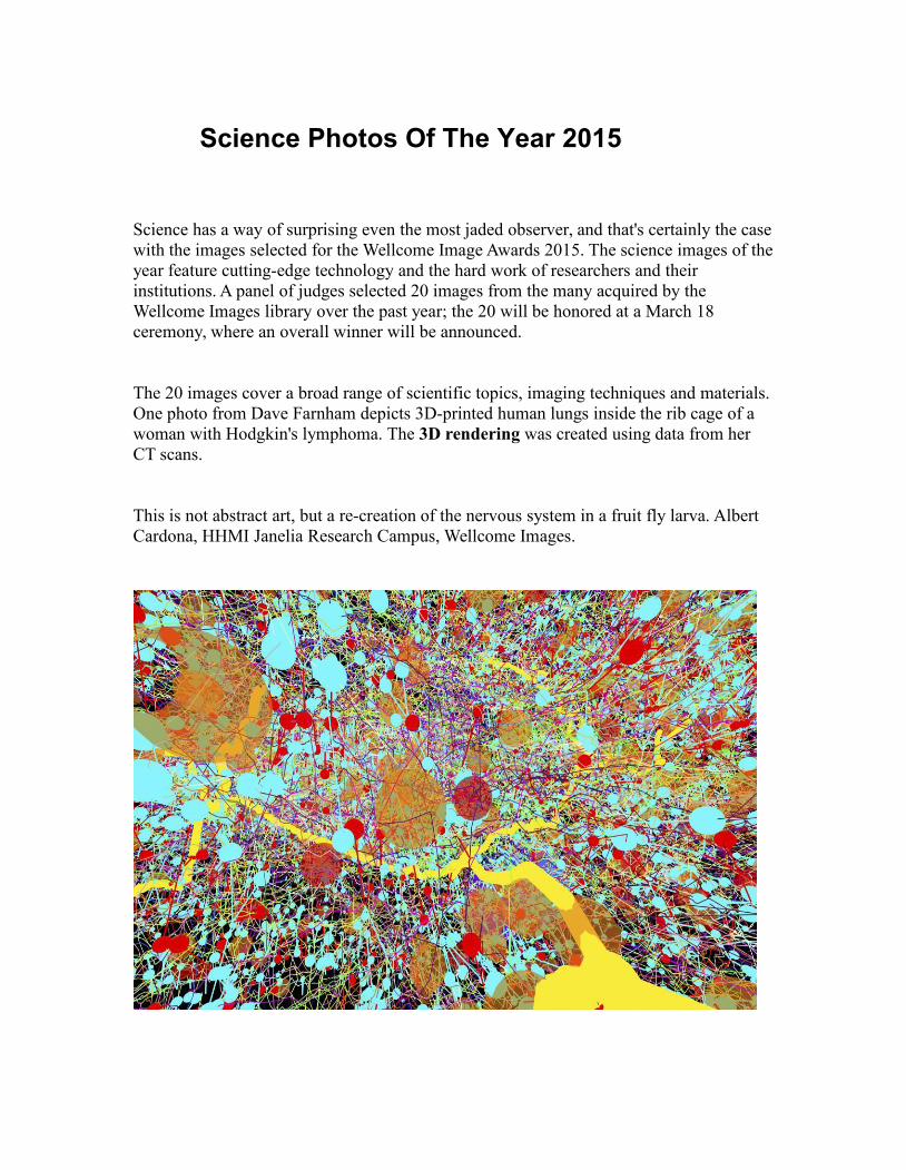
This is not abstract art, but a re-creation of the nervous system in a fruit fly larva. AlbertCardona, HHMI Janelia Research Campus, Wellcome Images.

The reticulum, or stomach chamber of a goat. The opening in the center leads to anotherstomach chamber, the omasum. Goats have four stomach chambers -- the rumen, thereticulum, the omasum and the abomasum. Courtesy of Wellcome Images.
Photograph of a mare's uterus. Courtesy of Wellcome Images.
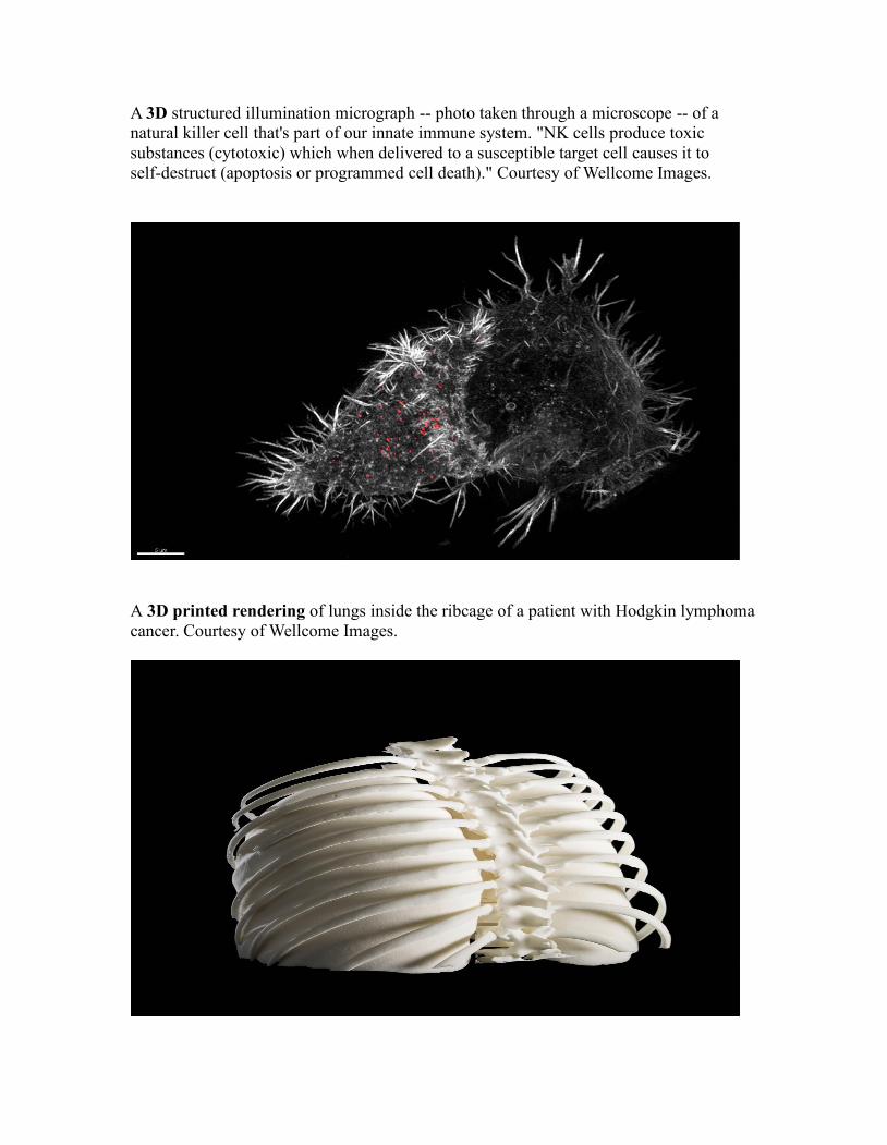
A 3D structured illumination micrograph -- photo taken through a microscope -- of anatural killer cell that's part of our innate immune system. "NK cells produce toxicsubstances (cytotoxic) which when delivered to a susceptible target cell causes it toself-destruct (apoptosis or programmed cell death)." Courtesy of Wellcome Images.
A 3D printed rendering of lungs inside the ribcage of a patient with Hodgkin lymphomacancer. Courtesy of Wellcome Images.
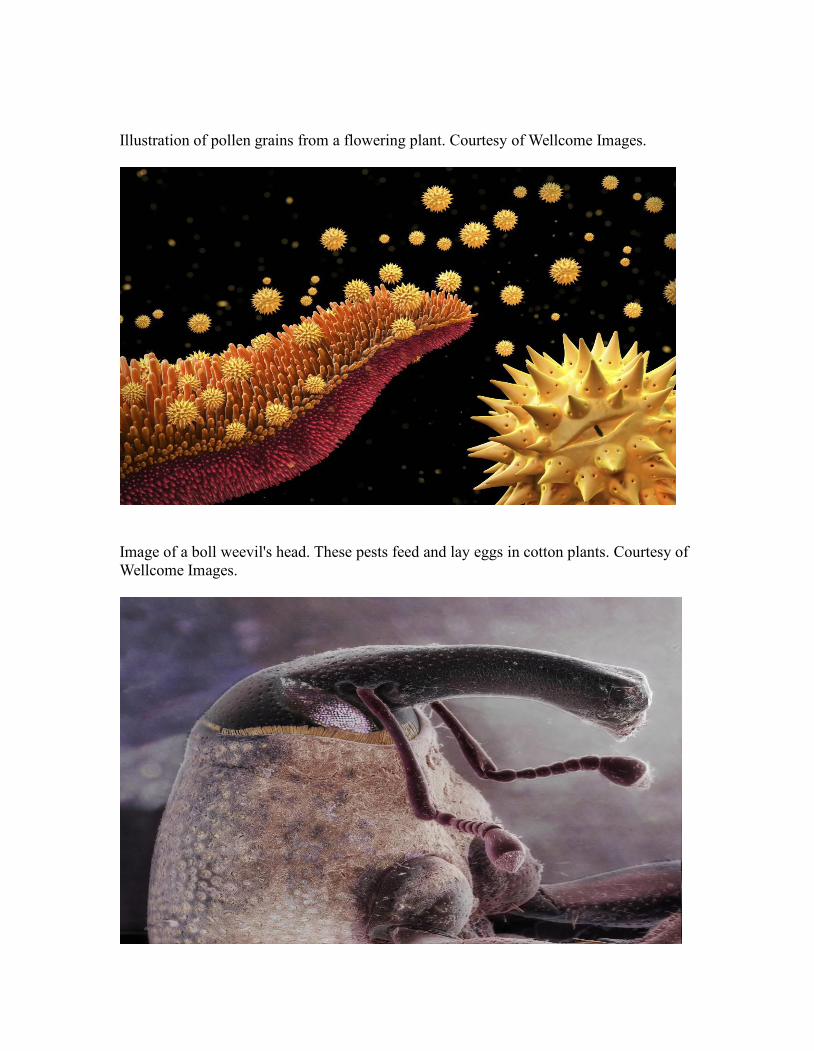
Illustration of pollen grains from a flowering plant. Courtesy of Wellcome Images.
Image of a boll weevil's head. These pests feed and lay eggs in cotton plants. Courtesy ofWellcome Images.

Cross section of a cat's tongue. Courtesy of Wellcome Images.
A reconstruction of a tuatara, a rare reptile found only in New Zealand. Courtesy ofWellcome Images.

Coronal -- or frontal view -- of a mouse brain. Neurones -- or nerve cells -- are colorcoded based on depth. At the top is red followed by orange, yellow, purple, blue andgreen. Courtesy of Wellcome Images.
Dendritic tree -- branched projections of neurons -- in the cerebellar cortex of a rat brain."The cerebellar cortex forms part of the cerebellum, the region of the brain which plays arole in controlling accuracy and coordination of movement." Courtesy of WellcomeImages.
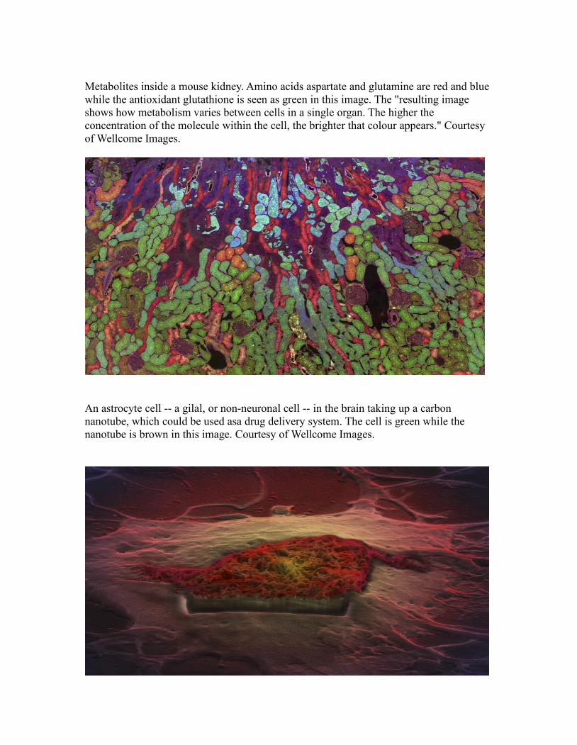
Metabolites inside a mouse kidney. Amino acids aspartate and glutamine are red and bluewhile the antioxidant glutathione is seen as green in this image. The "resulting imageshows how metabolism varies between cells in a single organ. The higher theconcentration of the molecule within the cell, the brighter that colour appears." Courtesyof Wellcome Images.
An astrocyte cell -- a gilal, or non-neuronal cell -- in the brain taking up a carbonnanotube, which could be used asa drug delivery system. The cell is green while thenanotube is brown in this image. Courtesy of Wellcome Images.

A paraitoid wasp, Wallaceaphytis kikiae, which helps out farmers around the world. "Itsclose relatives in the genus Aphytis successfully control populations of scale insects(sap-sucking agricultural pests), which attack oranges and other citrus fruits around theworld. The female wasps lay their eggs in the scale insects which are then killed by thewasp larvae." Courtesy of Wellcome Images.
A scanning electron micrograph of a greenfly (aphid) eye. Courtesy of Wellcome Images.

View of a healthy adult, living human brain. The front of head is toward the left of theimage and a MRI was used to collect information on the "pathways of white matter in thebrain." Courtesy of Wellcome Images..
Old model used in the teaching of Anatomy. Courtesy of Wellcome Images.
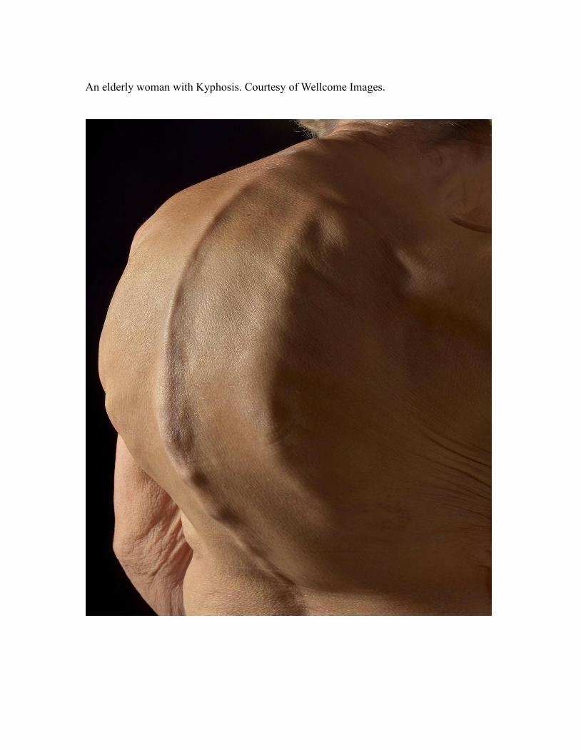
An elderly woman with Kyphosis. Courtesy of Wellcome Images.
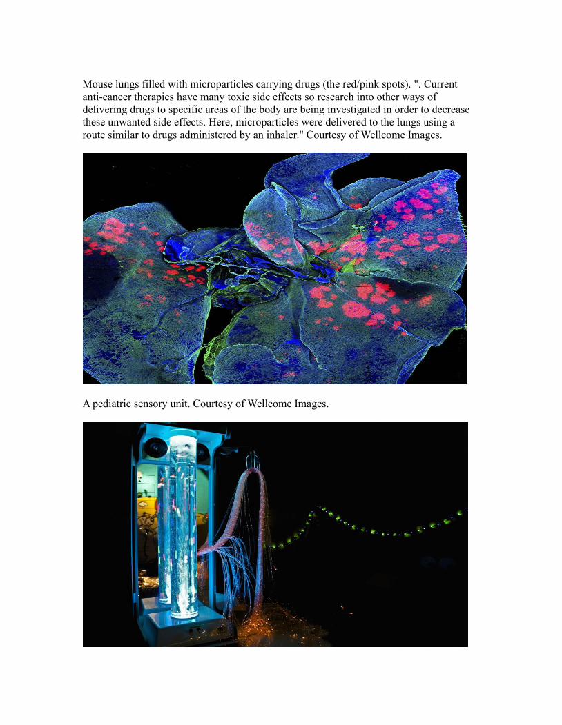
Mouse lungs filled with microparticles carrying drugs (the red/pink spots). ". Currentanti-cancer therapies have many toxic side effects so research into other ways ofdelivering drugs to specific areas of the body are being investigated in order to decreasethese unwanted side effects. Here, microparticles were delivered to the lungs using aroute similar to drugs administered by an inhaler." Courtesy of Wellcome Images.
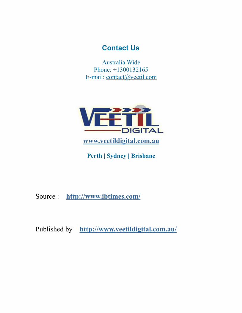
A pediatric sensory unit. Courtesy of Wellcome Images.
Contact Us
Australia WidePhone: +1300132165
E-mail: [email protected]
www.veetildigital.com.au
Perth | Sydney | Brisbane
Source : http://www.ibtimes.com/
Published by http://www.veetildigital.com.au/