school improvement plan - long county schools
TRANSCRIPT

NEUROIMAGING DATABASES AS A RESOURCE FORSCIENTIFIC DISCOVERY
John Darrell Van Horn, John Wolfe, Autumn Agnoli, Jeffrey Woodward, MichaelSchmitt, James Dobson, Sarene Schumacher, and Bennet Vance
The fMRI Data Center, Dartmouth CollegeHanover, New Hampshire 03755
I.
INTE
NEUR
DOI:
I
R
1
ntroduction
NATIONAL REVIEW OF 55OBIOLOGY, VOL. 66
Copyright 2005, Elsevier In
All rights reserve
0.1016/S0074-7742(05)66002-3 0074-7742/05 $35.0
II.
E xamining Cognitive Function with fMRIIII.
L arge-Scale Archiving of fMRI Study DataIV.
T he Emergence of ‘‘Discovery Science’’V.
D ata Sharing in NeuroscienceVI.
T he Role of Computation in NeuroscienceVII.
B rain Data Repositories as a Shared Resource for NeuroscienceVIII.
f MRI Data Archiving, Mining, and VisualizationIX.
N euroinformatics—The Nexus of Brain, Computational, and Computer SciencesX.
C urrent Challenges for Neuroscience DatabasesXI.
C onclusionR
eferencesThe field of neuroscience has an increasing need for access to primary
research data in order to more thoroughly explore fundamental neural function
beyond those examined in the original published article. For instance, functional
magnetic resonance imaging (fMRI) studies of the human brain during the
performance of cognitive tasks involve the collection of several gigabytes of image
volume time course data as well as detailed meta-data concerning subject,
experimental, and scanner protocols. Much of this data is unseen by anyone
other than the original study authors but could be used by others to gain new
insights into basic cognitive processes. We describe how several eVorts have
sought to archive the primary data from brain imaging studies and make them
available to researchers in the community. We detail several aspects of neurosci-
entific data sharing that can help promote new inquiry. Essential in this process is
the design of hierarchical frameworks for encapsulating fMRI study data for the
purposes of extensible study organization that helps to encourage data sharing
between collaborators or centralized data archives. In this chapter, we feature
our own eVort, the fMRI Data Center, as an example of large-scale archiving of
fMRI study data from the peer-reviewed literature and how this is being used to
explore data beyond the scope of the original article. Through such eVorts, brainimaging has begun following the lead of the biological sciences by leveraging its
accumulated data into new knowledge about fundamental brain processes.
c.
d.
0

56 VAN HORN et al.
I. Introduction
The ambitious attempts being made across the biological sciences to promote
the sharing of data and to facilitate meta-analysis (Becker, 2001; Mavroudis and
Jacobs, 2000; Mirnics, 2001; Nowinski et al., 2002; Reidpath and Allotey, 2001;
Richard and Williams, 2002) have now transformed the process of science in the
digital age. Large-scale scientific databases and collaborating networks of research-
ers are being developed to enhance scientific interaction and promote the sharing of
primary research information (Collins and Mansoura, 2001; Shepherd et al., 1998).
For example, scientific databases containing experimental data and results, such as
the Protein Data Bank (Berman et al., 2000), permit re-examination or comparison
of data on 3D protein structures in order to test new hypotheses and enable data
mining from across research centers to reveal trends that may give rise to new
avenues of research. Much excitement has occurred in the neurosciences about
large-scale databasing (Koslow, 2000) and the promise that they hold for under-
standing cellular properties (Martone et al., 2002), neuronal models (Marenco et al.,
1999), and underlying biochemical pathways (Karp et al., 2002).
In the 1990s, use of fMRI during performance of neurocognitive tasks
rapidly emerged, surpassing the use of positron emission tomography (PET), as
the fundamental tool for mapping brain function (D’Esposito, 2000; Detre and
Floyd, 2001; Savoy, 2001). The data collected in these studies are exceedingly
rich and hold potential for understanding the complex neurobiological mechan-
isms underlying brain systems such as memory (Cabeza et al., 2002), language
(Mechelli et al., 2003a), and motor function (Kawato et al., 2003) among many
cognitive domains. Most contemporary fMRI studies routinely include multiple
functional scan runs (i.e., on the order of five to ten minutes per run), the
collection of high-resolution anatomical images of the brain, involve multiple
levels of experimental manipulation, and increasingly involve several experimen-
tal groups. Stimulus input and behavioral output files also contribute to the body
of study meta-data, 4-dimension image volumes, and images of brain structure.
However, despite their being summarized and interpreted in the peer-reviewed
literature, the raw image data and accompanying meta-data from these studies
are often not accessible to other researchers in the neuroscientific community.
This makes it diYcult for others to rigorously inspect results, verify published
claims, or to conduct novel analyses of these rich datasets. In the field of
functional neuroimaging, despite being available for over a decade, databases
of systems-level functional brain imaging studies of cognition have begun to
receive increasing attention (Van Horn and Gazzaniga, 2002).
The field of neuroimaging has for some time recognized this need for archiv-
ing functional neuroimaging results (Fox and Lancaster, 1994; Fox et al., 1994b).
However, databasing and data sharing remain relatively unfamiliar ideas to many

NEUROIMAGING DATABASES 57
in the field of functional neuroimaging. Coupled with the fact that the size of
fMRI studies is growing rapidly, few tools exist that assist investigators in manag-
ing the data they collect and that facilitate eYcient data sharing. The needs of the
community are particularly unique and extant software models are not broadly
applicable for dealing with many of the issues a large archive like this requires as
well as helping users manage data at their own sites. Particular problems include:
Users of databases that store experimental results are often faced with entering
data retrospectively; certain models for sophisticated high-throughput data anal-
ysis (Roland et al., 2001) put certain constraints on the type and form of data that
can be stored; the management of study data is left up to the investigator until
completion (e.g., publication) of the study; and finally, the tools available from
existing databases are largely bibliographic and not entirely useful until all
information about a study has been collected. In other words, software tools that
help an investigator gather, manage, and share fMRI data that preserves that
data’s utility to the investigator beyond its being shared do not widely exist.
Simply managing these large amounts of data, however, is an increasing
challenge for many in the field. Future investigations of in vivo brain function
using fMRI can be expected to continue gaining in sophistication, as the ques-
tions being asked about the brain, and the methods themselves, become more
elaborate. With possibly hundreds or thousands of data files, each recorded under
conditions that vary over time, the amount of data may soon overwhelm the
abilities of investigators to keep track of this information from even a single
fMRI investigation. Being able to manage and organize these large and varied
collections of data is a prelude to subsequent data sharing and databasing.
In this chapter, we review the form, size, and means of processing of func-
tional neuroimaging data but highlight the potential of these data to be used
beyond the initial interpretation by the original investigators; how these data may
be used to develop and test new ideas about human brain function as well as for
developing useful computational algorithms for extracting new results; we discuss
the emerging role of high-performance and Grid computing; the use of clever
visualization techniques, and the availability of online databases of neuroscientific
data. Finally, we discuss some of the challenges that must be overcome for these
and other resources to fully leverage the wealth of neuroscientific information
into new discovery.
II. Examining Cognitive Function with fMRI
The signal of interest using positron emission tomography (PET) is based on
the fact that changes in the cellular activity of the brain of normal, awake humans
and laboratory animals are accompanied almost invariably by changes in local

58 VAN HORN et al.
blood flow (Raichle, 1975, 2001b; SokoloV, 1981). Early PET studies of the
brain’s response to cognitive tasks provided a level of precision in the measure-
ment of blood flow that opened up the modern era of functional human brain
mapping (Raichle, 2003). Functional MRI, on the other hand, distinguishes itself
from PET by capitalizing on endogenous magnetic properties of deoxygenated to
oxygenated hemoglobin in order to track regional cerebral blood flow (Hoppel
et al., 1993; Rosen et al., 1993). Using fMRI to visualize brain function in vivo,
neuroscientists have demonstrated that the mental operations carried out by the
human brain can be empirically and repeatedly measured (Bandettini and Wong,
1997; D’Esposito, 2000) and, since the early 1990s, fMRI has taken the place of
PET as the most widely used method for brain mapping and studying the neural
basis of human cognition. Though now enjoying widespread practice throughout
the world, an incomplete understanding of the physiological basis of the fMRI
signal has remained to confidently interpret the data with respect to neuronal
activity. The biological origins for these signals is an area of much interest for the
application of tools for cognitive neuroscience research and modeling (Raichle,
2001a; Woo and Hathout, 2001). Understanding the origins of the BOLD signal
is useful for informing models of the hemodynamic response function (Buxton
and Frank, 1997; Buxton et al., 1998) or to guide characterization of the neuro-
physiological processes that occur in advance of BOLD signal change as a result
of many higher-order cognitive models (Friston, 2002; Friston and Price, 2001;
Price and Friston, 2002).
New insights into higher cognitive functions, such as episodic and working
memory (Cabeza et al., 2002; Carpenter et al., 2000), linguistic processes (Binder
et al., 1997; Buchel et al., 1998; Crosson et al., 1999), and object visual processing
(Beauchamp et al., 2002) have been described. Face perception is one particular
cognitive operation to be extensively examined using fMRI (Haxby et al., 2000)
and appears to be governed principally in the ventral portion of the temporal
lobe—the ‘‘fusiform face area’’ (Kanwisher et al., 1997). Brain areas bordering
this region may be sensitive to the spatial properties of pictures of other objects,
such as chairs and houses (Ishai et al., 2000) with spatially distributed but over-
lapping portions (Haxby et al., 2001). Additional research has indicated that this
region may, in fact, be specialized for visual recognition expertise which includes
processing for faces (Gauthier and Nelson, 2001; Gauthier et al., 1999). Work has
also investigated the social context of face perception, in particular with respect to
the perception of threat (Adolphs, 2003; Haxby et al., 2002; Richeson et al., 2003),
the familiarity of faces (Leveroni et al., 2000), and the processing of faces in
diseases such as autism (Adolphs et al., 2001). Visuospatial attention has also been
explored using fMRI (Binkofski et al., 2002; Culham et al., 2001; Hamalainen
et al., 2002; Kanwisher and Wojciulik, 2000). fMRI studies have pointed toward a
network of cortical visuospatial and oculomotor control areas, specifically the
lateral occipital cortex, precentral sulcus, and intraparietal sulcus, as being active

NEUROIMAGING DATABASES 59
in covert shifts of spatial attention (Beauchamp et al., 2001). In parietal and frontal
cortical areas, BOLD activation increased with attentional load, suggesting that
these areas are directly involved in attentional processes, though this was not
evident in the fusiform gyrus (Culham et al., 2001), indicating possibly separate
but complimentary systems underlying attention to stimuli such as human faces.
A number of factors have been implicated in the origins of the BOLD response
including energetics, oxygen consumption, as well as parameters such as blood
volume and flow (Buxton et al., 1998). The question of whether the BOLD
response is the result of neuronal output or if it is due to the internal communica-
tion among localized populations of cells has also been recently addressed.
Logothetis and coworkers (2001) conducted the first simultaneous intra-cortical
recordings of neural signals and hemodynamic responses. Varying the temporal
characteristics of the stimulus, they observed a moderate to strong association
between the neural activity measured with microelectrodes and the pooled BOLD
signal from around a small area near the microelectrode tips. However, the
BOLD signal showed significantly higher variability than the neural activity,
indicating that human fMRI coupled with traditional statistical methods under-
estimates the reliability of the neuronal activity. To further characterize the relative
contribution of several types of neuronal signals to the hemodynamic response,
they compared local field potentials (LFPs), single- and multi-unit activity (MUA)
with high spatiotemporal fMRI responses recorded simultaneously in primate
visual cortex. Selecting recording sites having transient responses, only the LFP
signal showed significant correlation with the hemodynamic response and were
superior to MUA at predicting the fMRI response. Thus, BOLD signal is a
putative measure of the input and processing of neuronal information within brain
foci, not the output signal transmitted to other brain areas.
Epoch or ‘‘block’’ experimental designs have been the work horse of fMRI
experimentation and are those in which stimuli are presented for some period of
seconds (several TRs or brain volume sampling intervals) and alternated randomly
or pseudo-randomly over the course of the data acquisition period. They are the
easiest to conduct and tend to provide robust activation in most tasks but may limit
the number of stimulus types that can be presented. Conversely, event-related
experimental designs are characterized by having a baseline time course that is
punctuated with stimulus events. Event-related methods, conversely, have permit-
ted a broad array of task designs to be explored with brain imaging techniques
(Buckner, 1998; Buckner et al., 1996; Rosen et al., 1998). Individual trial events can
be presented rapidly, in randomly or intermixed order, and the hemodynamic
responses associated with each trial event type reliably estimated (Dale and
Buckner, 1997). The basis of event-related studies is that the hemodynamic
response tracks neuronal activity on the temporal scale of seconds and, in many
situations, summates over trials in amanner well predicted by a linearmodel that is
suYcient even for very briefly spaced stimuli (e.g.,�2 seconds).With this increased

60 VAN HORN et al.
interest in event-related paradigms in fMRI, there has been considerable eVort inidentifying the optimal stimulus timing, especially when the inter-stimulus interval
is varied during the imaging acquisition run (Birn et al., 2002; Dreher et al., 2002).
Experimental designs for event-related functional magnetic resonance imag-
ing can be characterized by both their detection power, a measure of the ability
to detect activation, and their estimation eYciency, a measure of the ability to
estimate the shape of the hemodynamic response. Computer simulation studies
have indicated that estimation of the hemodynamic response function is opti-
mized when stimuli are frequently alternated between task and control states,
having shorter interstimulus intervals and stimulus durations, while the overall
detection ability of activated areas is optimized when using blocked designs (Birn
et al., 2002; Mohamed et al., 2000). This suggests that event-related designs may
provide more accurate estimates of the HRF than epoch-related designs, with the
maximal response to events occurring sooner and returning to baseline later than
in a stimulus epoch (Mechelli et al., 2003b). The choice of data processing
operations, however, can aVect statistical inference in all designs and means for
optimizing data processing pipelines is an area of active research (LaConte et al.,
2003; Lukic et al., 2002; Strother et al., 2002).
Functional neuroimaging using MRI promises to continue growing as the
principal method for examining in vivo brain function. Though individual studies
using fMRI promise to reveal much about such basic brain processes, there also
exists great potential for contrasting, comparing, and combining these studies to
explore fundamental properties of cognitive function as well as the properties of
the BOLD signal itself. The large amount of information collected in an individual
study, however, and how this could be mined by others to produce novel research
is an under-appreciated aspect of this work that is worth addressing further. There
is often more information contained in a neuroimaging study that can be ade-
quately described in a single neuroimaging article. The expertise required to
extract this information, however, is often not necessarily possessed by the original
study authors. Finally, new imaging facilities are very costly and to install MRI
scanners in psychology departments across the country may not be cost eVective incontrast to providing an open archive of such data where researchers can readily
obtain the original fMRI time series and subject them to new analyses.
III. Large-Scale Archiving of fMRI Study Data
The advent and development of fMRI has resulted in a quantum leap in the
ability to visualize the brain’s capabilities. However, this has also vastly increased
the amount of information that brain researchers must manipulate, manage, and
store. fMRI study data sets are large, often exceeding several gigabytes (GB) in

NEUROIMAGING DATABASES 61
size. As advances are made in MRI scanner technology to permit the more rapid
acquisition of data, functional imaging experiments will consist of more data per
unit time over the same scan duration. As cognitive neuroscientists ask ever more
sophisticated questions about fundamental brain processes, they will undoubtedly
collect data on a greater number of subjects and more fMRI time courses per
subject. Indeed, archives equivalent in size to that of several petabytes are not out
of the question and will likely be the norm within the next decade. A number of
individual fMRI data sets already rival the full size of many extant large genetic
(Ackerman, 1999; Ackerman and Banvard, 2000) and protein (Chen and Xu,
2003; Legato et al., 2003; Noguchi and Akiyama, 2003) science data archives (see
Table II, below, for comparison). For instance, the complete study data from
Buckner et al. (2000) (fMRIDC Accession#: 2-2000-1118W) represents a study in
excess of 20GB. It can be expected that as technological advances are made in
MRI scanner technology which improve the spatiotemporal resolution of the
data obtained, the amount of brain image data collected in published articles will
routinely rival the size of the human genomic database. A challenge therefore
exists in devising eYcient means for comparing and contrasting these data on a
large-scale but within a reasonable time frame.
As fMRI use in cognitive, clinical, and social neuroscience grows and be-
comes more widespread, individual researchers must be prepared for the large
disk storage requirements that are needed to contain the data and their analyses.
A greater number of subjects, for instance, improves the inferential power of the
statistical tests performed and helps researchers to be confident in the eVects theyobserve (Van Horn et al., 1998). However, increases in sample size readily require
increased costs associated with each fMRI study, in terms of scanner time,
subject reimbursement, among other expenses. Publicly accessible archives of
these data (for example see Table I) can help spread the costs of this research over
the community, whereby the researcher may perform re-analyses on existing data
at a greatly reduced cost compared to collecting the data themselves.
With these issues in mind, the f MRI Data Center (f MRID C; http://www.
fmridc .org ) w as estab lished as a public archive for f MRI study data and the
associated experimental meta-data. The fMRIDC began receiving data from
researchers in 2000 and began making datasets publicly available in 2001. At
present, the archive contains over 100 complete data sets which researchers may
request online and have shipped to them free of charge. Authors of fMRI studies
have been asked to provide the details of their experiments across several levels:
description of the subjects taking part in the experiment (e.g., their
age, handedness, clinical diagnosis, etc.); the description of the MRI scanner
(e.g., manufacturer, model, software revision, field strength, etc.) as well as the
scanning session protocols used during the study (e.g., number of slices acquired,
echo time (TE), relaxation time (TR), etc.); and, finally, the details of the experi-
mental design (e.g., stimulus time course information, number of experimental

TABLE I
B RAIN DATABASE RESOURCES AVAILABLE ONLINE
Database
name
Principle
modality
Data sets
provided
Public
access? Species
Country and
funding
sourcea Web site URL
Allen Brain
Atlas
Anatomical
sections
Photomicrographs
of gene expression
Limited Mouse US; Private http://
www.brainatlas.org
BIRN MRI Images (MRI and cell
photomicrograph)
Limited; greater
access to
participating
BIRN centers
Human,
Mouse
US; NCRR http://nbirn.net/
BRAID MRI, f MRI Image volume data Limited Human US; N/A http://
www.rad.upenn.edu/
sbla/braid/
publications/all.shtml
Brain Gene
Expression
Map (BGEM)
Anatomical
sections
Photomicrographs
of gene expression
Open Mouse NIH/NINDS,
ALSAC
http://
www.stjudebgem.org/
web/mainPage/
mainPage.php
BrainMapDBJ PET/fMRI Results local maxima Limited;
greater access
to participating
ICBM centers
Human US; NLM http://
www.brainmapDBJ.org
BrainWeb MRI Simulated MRI image
volume data
Open; part of the
LONI/ICBM
consortium
Human Canada;
NIMH/HBP
Non-US
http://
www.bic.mni.mcgill.ca/
brainweb/
BREDE fMRI Results local maxima,
VRML, XML
Open Human Denmark;
NIMH/HBP
Non-US
http://
hendrix.imm.dtu.dk/
software/brede/
62

CoCoMac Single and
multi-unit
recordings
Neural connectivity
data
Open Non-Human
Primate
Germany;
Non-US
http://
www.mon-kunden.de/
cocomac/
EarLab Single/multi-
unit recording
Cell recording time
series
Open Non-Human US;
NIMH/HBP
http://
earlab.bu.edu/
fMRIDC fMRI, MRI Raw, processed,
results, anatomical
brain images and
study meta-data
Open Human US; NSF, Keck,
NIMH/HBP
http://
www.fmridc.org
International
Brain Volume
Database (IBDV)
MRI High resolution
structural Image
volumes
Open Human US; NIMH/HBP http://
www.cma.mgh.
harvard.edu/ibvd/
LONI/ICBM PET, MRI,
fMRI, EEG,
MEG
Image data Limited; greater
access to ICBM
centers
Human International;
NIMH/HBP
NCRR, Private
funding
http://
www.ioni.ucla.edu
Mouse Brain
Library (MBL)
Anatomical
sections
Photomicrographs Open Mouse US; NIMH/HBP http://www.mbl.org
Neurodatabase.org Single/multi-unit
recording
Cortical neuron
electrical recordings
Open Multiple US; NIMH/HBP
NINDS
http://
neurodatabase.org
Neurogenerator PET, fMRI Imaging data
submitted by users
is organized into
a database that is
returned to the user
Limited Human Sweden; The
European
Commission
http://
www.neurogenerator.org
SenseLab Single/multi-unit
recording
Cell recordings from
multiple sources
Open Non-Human
Primate
US;
NIMH/HBP
http://
www.senselab.yale.edu
Surface
Management
System (SuMS)
MRI Digitally-based cortical
surface models
Open Human,
Non-Human
Primate
US;
NIMH/HBP
http://
brainmap.wustl.edu/
sumshome/
aWhere evident from the database Web site
63

64 VAN HORN et al.
runs, etc.). Table I presents a summary of the items requested from authors which
describe their experimental data. The principle intent of obtaining this degree of
information about each study is that it should be complete such that another
researcher could take the information and the accompanying brain image data
and reconstruct the results reported in the literature by the original authors (see
Van Horn et al., 2001 for review).
IV. The Emergence of ‘‘Discovery Science’’
The collection of biological data into large databases has led to a change in
thinking about the potentially restrictive nature of strictly hypothesis-based re-
search. Increasingly, researchers are beginning to move toward a science of
discovery—examining vast and disparate collections of data and hunting for
unseen patterns that might provide clues to underlying biological mechanisms.
The mountains of data being collected in many fields provide input for pattern-
seeking and other relevant algorithms that can provide additional insights
into complex, multidimensional data (Jones and Swindells, 2002; Ma et al.,
2002; Schutte et al., 2002). These patterns can suggest mechanisms, and the
mechanisms can, in turn, suggest testable biological experiments to foster new
hypothesis-driven research. Confirmed mechanisms add to the knowledge base of
the biological sciences and provide the basis for further discoveries including
those that will improve quality of life and provide the means for attacking disease.
Such mining of the integrated resources developed and disseminated by the
NCBI, Genbank, and the Human Genome Project has led to several scientific
advances. The discovery of the genes for hereditary nonpolyposis colorectal
cancer (HNPCC) is one such example. HNPCC is thought to account for one-
sixth of all colon cancer cases (van Stolk, 2002). Although most forms of cancer
appear to be nongenetic, there are certain forms where a person has a hereditary
risk attributable to a single altered gene (Calvert and Frucht, 2002). Using the
tools developed through the Human Genome Project, notably Genbank, an
international research team tracked the gene to a specific region of chromosome
2 (Lindblom et al., 1993). Researchers then identified a second gene on chromo-
some 3 that was also associated with this form of cancer (Peltomaki, 1994).
Together, mutations within these two genes are responsible for the majority of
cases of HNPCC. Researchers have used this new knowledge to develop blood
tests to screen select individuals for these gene mutations (Ramesar et al., 2000;
Thomas, 1994). Detecting the presence of the mutated genes for HNPCC within
a family allows clinicians to target relatives most likely to benefit from treatment.
By identifying an unaVected family member at risk for HNPCC, physicians
may then more closely monitor them for signs of disease development. Family

NEUROIMAGING DATABASES 65
members determ ined to be nonc arriers no long er have to su Ver throug h exten -sive medica l examination s. Mo st import antly, patients demon stratin g early signs
of can cer and determ ined to carry a gene mutation m ay undergo promp t medical
treatm ent. Due to the role played by inform atics, when dia gnosed and trea ted
early, HNP CC is nearly 100 perc ent curab le ( Boardm an, 2002 ).
Several such larg e-scale, infr astructural, and discovery -focused database re -
searc h e Vorts that are already seein g considerable scienti fic payo V are underwa yin the biologic al and astrophysica l sciences. The successes of these m olecular
biolog ical, biomed ical, and astrophy sics infrastru ctures are wel l know n. They
have provided the m eans for expert s in co mputer science, m athematics, and
statis tics to make sig nificant contribut ions to these fields from which most of their
expert ise would have been exclu ded withou t the infr astructure. These successes
are not neces sarily uniqu e but bui lding upo n them and extendin g them to a wider
set of scien tific resear ch arenas is an ever-pr esent theme (Altma n, 2003; Brookes ,
2001; Perss on, 2000 ).
Before this proces s can begin for any par ticular field of scienc e, however, an
infras tructure must be laid down tha t w ill support these new approac hes. The
path blaze d by the molecula r biologists is, once again, illustra tive. Likening these
e Vor ts to civil engineering projects, Eric Lander, Director of the Whitehead-M IT
Cente r for Genome Researc h, has noted that progr ams to develop co mputationa l
infras tructure represe nt ‘‘ very im portant roads.’’ (Incyte Genomics i nterview
(2001) (htt p://www.incyte .com/)).
V. Data Sharing in Neuroscience
The driving force behind m any biological and ph ysical scien ce infor matics,
data minin g, and resea rch initiatives has revolve d arou nd the sharin g of prima ry
resear ch da ta (Be cker, 2001; Ilioudis and Pang alos, 2001; Rei dpath an d Allotey,
2001 ). The Nation al Institutes of Health (NIH) in the Un ited State s ha ve
recogn ized the benefit s that the sharing of prima ry resea rch data has for ad vanc-
ing science and has recently im plement ed policy re quiring data sharing for grants
in exces s of US$500K /yr in direct costs (Final NIH Data Sha ring Policy Noti ce :
http://grants.n ih.gov/g rants/gui de/notic e-files/NO T-OD-0 3-032.h tml). 1 Like-
wise, the National Science Foundation (NSF) in the US has encouraged data
sharin g for seve ral years, in the socia l and econom ic scien ces in particu lar, (http://
www.nsf.gov/sbe /ses/common/ archiv e.htm ). The Medica l Res earch Council (MRC ;
1Further information on the NIH data sharing policy may be found on the NIH Data Sharing
Web Page: http://grants.nih.gov/grants/policy/data_sharing/

66 VAN HORN et al.
http://www.mrc .ac.uk ) in Grea t Britain also str ongly encourages open scientific data
sharing.
The NIH’s position on coordinated scientific data sharing, particularly as it
relates to neuroscience, has recently been underscored in an essay by several NIH
institute directors: ‘‘EVorts driven by collaboration, coordination, and computa-
tion should yield the data, tools, and resources that neuroscientists will need in the
coming decades.’’ (Insel et al., 2003). The sharing of primary research data is
needed to provide a record of the scientific body of work, permit comparison of
various approaches to studying brain function, and enable large-scale analyses
across data sets. There are several models for the sharing of research data that form
a spectrum of complexity and detail. These include models for data archive access;
the simple model of an anonymous FTP site, where a data set is placed and openly
available but with no guarantee that the data have not been subjected to quality
control, been published in a peer-reviewed publication, or that the dataset will be
maintained; peer-to-peer models, wherein individual investigators set up and
personally maintain private data sharing relationships with colleagues and co-
investigators of both published and unpublished data; the conforming site model,
in which consortiums of several research centers agree to exchange data through
conventions established and governed by one of the consortium member centers,
but with no guarantee that nonconsortium members may have access to the data
archive or will have access to consortium-derived software tools needed for inter-
acting with the data; and the centralized repository model, in which complete
datasets are contributed, curated, and maintained in a central site by dedicated
personnel and made openly available to the entire research community.
Data sharing models also focus on the type and amount of data that should be
shared. Recent commentary has suggested that the value of shared neuroimaging
data is greatest only after processing has been applied and interpretation
provided by study authors (Fox and Lancaster, 2002). However, the information
content of the image voxel time course data remains the same or is reduced by
every step of processing (Van Horn and Gazzaniga, 2004). Therefore, it is
unclear as to the amount of added value when archiving only statistical local
maxima tables obtained after the data are heavily processed.
The model for sharing peer-reviewed study data in which the potential
benefits of a data set are likely to be greatest is when the data are curated in a
centralized location by a dedicated staV, complete study data have been indexed,
and are freely available to the entire community (Van Horn and Gazzaniga,
2004). Accompanied by detailed, ontologically-structured, study meta-data, and
a comprehensive description of data processing methods, experiment image data
may be examined by other researchers at various points in the processing chain
(raw, processed, or results) depending upon the needs and interests of colleagues
or independent researchers. Through centralized curation and open distribution,
eVorts to subject functional data to re-analysis or perform mega-analyses across

NEUROIMAGING DATABASES 67
data sets may be maxim ally successf ul and thereb y prom ote unique scienti fic
disco very and adva nce educ ation. This m odel of data sharin g helps promote the
cycle of scien ce by adding an extra com ponent to the public ation process that
may enha nce new resear ch and educ ation, foster new avenu es for resea rch, and
contribu te back into the collectiv e body o f knowl edge.
VI. The Role of Computation in Neuroscience
The NIH Roadmap (http://nihroadma p.nih.gov/ ) stresses the importa nce of
compu tational biolog y, bioinforma tics, an d the estab lishment of digital scienc e
librarie s. A re cent NSF Blue Rib bon Advi sory Report emphasiz es that compu -
ters, computer scien ce, an d technology are at the heart of the futur e of a range of
resear ch fields that have ‘‘ prof ound broader implica tions for edu cation, com -
merc e, an d social good .’’ (http://www.co mmunityte chnology.org/nsf_ ci_report/ , (Blue
Ribbon Advisory Panel On Cyberinfrastructure, 2003)). Though perhaps over-
due in fully recognizing the potential of computers and the internet, the field of
neuroscience is now growing in its dependence on high-end computational
infrastructure. Neuroscience, in particular cognitive neuroscience, has emerged
over the past decade as a cross-cutting aggregate of these key areas with an
emphasis on the human brain information mining, modeling, and visualization
(Adolphs, 2003; Casey, 2002; Corchs and Deco, 2002; Toga, 2002a). To broaden
participation in understanding brain function derived from technologies such as
brain imaging, greater reliance upon computational infrastructure to facilitate
research collaboration is required. The sharing of large data sets via the internet;
being able to collect, archive, and index these data; and subject these data to
high-throughput analysis is not an option, but a mandatory next step in the
advancement of understanding of normal brain function. Via this route, large,
culturally-, and gender-valid norms must be established, for example, forming a
benchmark against which to provide diagnosis in brain illness and disease.
Several novel concepts have been borne out of this interest in large-scale,
scientific collaborative infrastructure. Most notably is the concept of Grid com-
puting (Butler, 2003), the basis of which has existed for several years in the form
of distributed computing, and the emerging need for high-speed Internet con-
nectivity. The middleware for the development of global interconnected comput-
er systems has made great progress in the last few years (Avery, 2002). The Grid
software enables users, tools, and computer hardware to interact and share
resources over high speed connections in an Internet- and Internet standards-
compliant fashion (Foster, 2003). Users of Grid-enabled systems will be able
to write applications to these published interfaces and will expect to be able to
run on large-scale heterogeneous systems (Fig. 1). The Web, and in particular

FIG. 1. Neuroimaging data processing pipelines are ideally suited to Grid-based distributed
computing.
68 VAN HORN et al.
We b – service s, can provide a model for this larg e-scale compu tational system .
Various fields have been earlier adopters of Grid compu ting, nota bly the H igh
Energy Physics com munity. Severa l w orldwide an d multi-in stitutio nal Grid pro-
jects are underwa y to enable simu lation s on large distrib uted datase ts. The
TeraG rid (http://www.tera grid.org ) is a NSF- funded project to promot e distrib uted
scientific computing using Grid infrastructure to connect the nation’s largest
supercomputer centers. Using the Grid as a backbone, The Globus Toolkit
(http://www.globus .org) has become the de facto standard for Global Grid com -
munications. The Globus Alliance leads the development of the toolkit. The
Global Grid Forum (GGF) is currently leading the standards eVorts which
Globus implements. Shared computational infrastructure, tools, and tool devel-
opment, as well as collaborative research on archival data leading to new, testable

NEUROIMAGING DATABASES 69
hypoth eses is becoming easier each day. These techno logical advances in larg e-
scale compu ting on shared inf rastruc ture have importa nt implica tions for neu -
roscien tific researc h dealing with massiv e amou nts of da ta as is the case in
functio nal neuroima ging.
VII. Brain Data Repositories as a Shared Resource for Neuroscience
When the f MRI Data Center e V ort was initia ted, one of the ce ntral inten tswas to bui ld an Internet-a ccessibl e platform through which re searchers based at
other institu tions might acces s the growin g collection of f MRI stud y da ta to
evalua te m ethodologie s for data processin g ( LaConte et al., 2003; Lukic et al .,
2002 ); guide the des ign open-so urce software tools for data managemen t (Va n
Horn et al., 2002 ); constru ct means to summarize these larg e da ta sets to facilitat e
rapid search, visua lization , and disco very; all w ith a view towar d driving new
hypoth esis-ba sed f MRI resea rch. The f MRID C team, in particul ar, has worked
to co nstruct a shared comm unity acces s cluste r Grid syst em for the analysis of
functio nal datase ts from the f M RIDC archive. This syste m has seen increased
utilization as an analysis platform to mitigate the current CD delivery of datasets.
Accounts on the system are available to any member of the fMRI community
who wishes to perform a large analy sis of data from the f MRIDC (see http://www.
fmridc .org/grid ).
The software infrastructure provided by Grid services are needed to perform
meta-analyses on the multi-gigabyte fMRI datasets housed in the fMRIDC
archive. The fMRIDC has implemented a number of components from the
Globus Toolkit 3.0, including the Grid-FTP service. Current plans exist for the
full toolkit to be installed to enable multi-institutional scheduling of resources. The
Globus software will abstract the fMRIDC’s own Grid scheduling system and the
systems of our collaborators as well as those of other systems. With advanced
computational resources dedicated to neuroimaging and that are deployed using
Globus, new methods can be developed through testing on what is fast becoming
the world’s largest data warehouse of functional neuroimaging data.
VIII. fMRI Data Archiving, Mining, and Visualization
Like much of neuroscience, literature-driven, hypothesis-based approaches
have been the underlying approach to most cognitive neuroscientific investiga-
tions over the past 100 years. However, streamlined, computationally eYcient
approaches to examining large amounts of data are emerging as advantageous
where volumes of information from diverse sources impede a straightforward test

70 VAN HORN et al.
of experimental hypotheses (Baumgartner et al., 2000; Friman et al., 2002; Jarmasz
and Somorjai, 2002). Approaches used in the biological sciences include nonpara-
metric clustering methods (Cordes et al., 2002; Goutte et al., 2001; Salli et al., 2001),
pattern searching (Cummings et al., 2002; Jones and Swindells, 2002), as well as
novel visualization techniques (Baumgartner and Somorjai, 2001; Teo et al., 1997).
In brain imaging, similar methods might be used on a database as a precursor to
more thorough parametric hypothesis testing using more sophisticated modeling
methods (Cox, 1996; Cox and Hyde, 1997; Friston et al., 2002; Lohmann et al.,
2001) on subsets of the overall study space. Exploratory data analysis approaches
(Tukey, 1977; Velleman, 1981) often assume little about the underlying data,
which allows the data to more freely inform the investigator about itself using
more elementary statistical approaches thanmight be true if the data were assessed
using highly parameterized modeling procedures. These methods have been
successfully applied to fMRI data with promising results (Baumgartner and
Somorjai, 2001; Baumgartner et al., 2000; Friman et al., 2002; McKeown and
Sejnowski, 1998). They can often be used to highlight relationships among the
overall collection of data in ways not possible when viewing only a single data set.
These relationships may not emerge until viewed in the light of a large number of
other studies to which they may be compared. Thus, neuroscience, in particular
studies using fMRI, has a great potential to become a discovery-based science,
where exploratory analyses can lead to new ideas worth pursuing with hypotheses-
generated experimentation (Van Horn and Gazzaniga, 2002). But to put such
ideas into practice, however, requires an interdisciplinary approach, bringing
together experts in cognitive, computer, and mathematical science to work jointly
in solving the challenges inherent in large-data science.
Initiatives to archive neuroscience data form a unique collaboration between
cognitive neuroscientists, mathematicians, and computer scientists to explore the
challenges inherent in (1) data warehousing—en mass data storage; (2) data
mining—how to apply eYcient mathematical and computer algorithms to sift
through large amounts of data to extract unique and interesting features from
fMRI study data; and (3) data distribution—eVective means to permit others to
interact with the data archive. For example, the eYcient retrieval of useful
information from these large datasets poses many interesting problems for which
computer scientists play an important role in providing answers. Methods for
applying sophisticated search queries across multiple levels of neuroimaging
study data (Table II) that could be investigated include: (1) searches for key text
phrases across the published research article itself (i.e., the PDF version of the
published study) and subsequent document clustering by assessing the usage of
similar words at similar rates; (2) queries across the study ‘‘meta-data’’ composed
of scanner protocol, experimental paradigm, subject demographic, and other
information provided by the study authors; and (3) the 4D fMRI image time
course data itself by, for instance, performing image timecourse-based clustering

TABLE II
BASIC fMRI STUDY INFORMATION COLLECTED FOR THE fMRIDC ARCHIVE
MR Scanner Protocol Information
� Scanner Protocol ID� Scanner Head Coil Type� Pulse Sequence Type� Flip Angle (degrees)� TE (in milliseconds)� TR (in milliseconds)� Number of time-points� Number of acquisitions� Number of dummy scans� Number of slices� Slice thickness (in millimeters)� Slice skip (in millimeters)� Interleaved or sequential slice acquisition� Field of View (FOV)� Receiver bandwidth (MHz)� Original image acquisition matrix size� Reconstructed image acquisition matrix size� Full or partial K-space� Image Acquisition Orientation� Ramp sampling� Echo train length� Echo shift in asymmetric spin-echo� Type of reference scan for reconstruction
Subject Information
� Subject ID� Experimental group code� Gender� Age� Health Status� Assessments (e.g., handedness, etc.)� Medication status� Other (e.g., diagnostic, etc.)
Scan Session Information
� Scan Session ID� Scanner Manufacturer� Scanner Model� Scanner software revision� Magnet field strength� Scanner Gradient Slew rate� Date of scan session� Duration of scan session� Other
(Continued )
NEUROIMAGING DATABASES 71

TABLE II (Continued )
Experimental Protocol
� Experimental Protocol ID� Number of groups� Number of subjects per group� Number of functional runs� Epoch-related conditions� Event-related conditions� Experimental methods� Stimulus regressor files� Other (e.g., additional condition descriptions, associated data files, etc.)
72 VAN HORN et al.
accompanied by a posteriori probabilistic classifier algorithm to measure classifica-
tion reliability. The latter of these approaches has necessitated the application of
summarizing signal processing and information theoretic methods for rapidly
analyzing large fMRI data sets. The integration of these levels using leading-
edge, computer-based IR algorithms permit ‘‘global’’ study clustering in order to
‘‘learn’’ what is needed to identify interesting patterns within and between these
levels of data. The application of algorithms that permit fMRI data self-descrip-
tion, allowing the data to tell an investigator about itself rather than through the
fitting of statistical models, as is common in functional neuroimaging, is a
promising application of ‘‘machine learning’’ (Mitchell, 1997; Mitchell, 1999).
Another area of active interest in neuroimaging data representation lies in
identifying unique approaches to the visualization of this massive amount of
information. Traditional approaches to visualizing brain imaging study results
have relied on overlay patterns of brain activation from functional scans on top of
high-resolution structural images (Fig. 2). Popular methods of display include
representing patterns of functional activity on flattened models of the cortical
surface (Van Essen et al., 2001a; Van Essen et al., 2001b). Still other, novel
methods for displaying and interacting with more abstract representations of
large collections of information are needed that may reveal previously unseen
relationships in the data. For instance, approaches centering on nontraditional
and abstract methods of data exploration, such as taking the data out of ‘‘brain
space’’ and placing it in some alternative parameter space, and examining
patterns in the data that might have been invisible in the original anatomically-
based space (Baumgartner and Somorjai, 2001; Cordes et al., 2002). These may
also include iconic representations of data endowed with synthetic physical
properties that distort the relative coordinates of data in an abstracted space.
Such models have been successfully employed in visualizing biochemical and
metabolic pathways (Becker and Rojas, 2001; Karp and Paley, 1994; Ogata et al.,
2000). Coherent subspaces within this abstracted space can be identified and
examined as those where something of neuropsychological interest might be
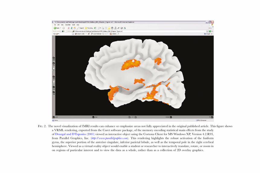
F IG. 2. The novel visualization of f MRI results can enhance or emphasize areas not fully appreciated in the original published article. This figure shows
a VRML rendering, exported from the Caret software package, of the memory encoding statistical main effects from the study
of Druzgal and D’Esposito (2001) viewed as interactive object using the Cortona Client for MS Windows XP, Version 4.2.R93,
from Parallel Graphics, Inc. ( http://www.parallelgraphics.com). This rendering highlights the robust activation of the fusiform
gyrus, the superior portion of the anterior cingulate, inferior parietal lobule, as well as the temporal pole in the right cerebral
hemisphere. Viewed as a virtual reality object would enable a student or researcher to interactively translate, rotate, or zoom in
on regions of particular interest and to view the data as a whole, rather than as a collection of 2D overlay graphics.

74 VAN HORN et al.
occurring. As fMRI archives continue to grow through active data sharing,
working with the shear amount of data, as well as its direct visualization, becomes
increasingly diYcult. These computational and visualization methods will allow
large amounts of data to be processed and visualized.
IX. Neuroinformatics—The Nexus of Brain, Computational, and Computer Sciences
Given recent success stories from the domains of genomics (Escribano and
Coca-Prados, 2002; Feolo et al., 2000; Rafalski et al., 1998) and proteomics
(Berman et al., 2000; Ezzell, 2002; Persson, 2000) for organizing large amounts
of data, cognitive neuroscientists are likewise becoming intimately familiar with
large-scale data analysis, applying high performance computing systems, and
using sophisticated computer science to extract information from large archives
of neurophysiological data. The evolution of the cognitive neuroscience field is
fast approaching the time when it forms a confluence of brain science, high-
performance computing systems, and leading edge computer science (Beltrame
and Koslow, 1999; Wong and Koslow, 2001). As such, a more thorough under-
standing of the brain and its cognitive processes will necessitate increased
computational infrastructure, novel software technology to accelerate data anal-
ysis and to mine vastly larger amounts of data, and the sharing of primary
research data. Moreover, these data must be understood on a level that permits
the representation of the dynamic examination of brain data and brain systems
required for cognitive processes such as memory function, visual abilities, and
motor skill. This eVort must reach beyond the level of the examination of
individual loci of brain activity to that of identifying patterns of activity across
individuals that speak to the dynamics and complexity of the neural processes
that are not typically reported in the scientific literature though may be worthy of
additional scrutiny and study.
In response, the field of neuroscience is rapidly moving beyond its roots as a
theoretical and experimental science toward becoming a highly computational
science ever more dependent upon lead edge technologies in computer science,
engineering, and mathematics. This is the origin of neuroinformatics, a unifying
discipline at the nexus of information technology, computer science, and the
neurosciences. It also involves the incorporation of high performance computing,
visualization, and data mining techniques with the fundamentals of experimental
design, image processing, and spatial and temporal statistics for neurophysiologi-
cal data, in particular, for functional neuroimaging (Douglas et al., 1996; Smaglik,
2000; Young and Scannell, 2000). By using computers to organize, link, analyze,
and examine large, complex sets of neuroscientific data, raw data may be con-
verted into meaningful knowledge that can be used for further experimentation

NEUROIMAGING DATABASES 75
into co gnitive funct ion and the treat ment of pa tients with neurologi cal and neu -
ropsyc hiatric dise ase (Bel trame and Koslow, 1999 ).
The Human Brain Projec t (HBP ) funds many of the current da tabase and
neuroi nformatics e Vorts . The HBP is a broad-b ased initiati ve which supports
resear ch and developmen t of adva nced techno logies and infr astructur e support
through co operative e Vorts among neur oscient ists and infor mation scienti sts
(com puter scien tists, physi cists, m athematicia ns, and engineers) (Brink ley and
Rosse, 2002; Shepherd et al ., 1998 ). The prin ciple aim of the NIM H-based
HBP is to guide the prod uction of new digital cap abilitie s that provi de Inter -
net-d riven inf ormation managemen t syst ems in the form of interop erabl e
database s and associate d neu roscience da ta managemen t too ls ( Shepherd et al .,
1998 ). Suc h softwa re tools inclu de graph ical interfaces , queryin g and mining
approa ches, inform ation retrieval, statistica l an alysis, visua lization and manipu -
lation , integratin g too ls for data analysis , biologic al m odeling and simulation , and
tools for electronic collabor ation. The e Vor t strongl y supports open da ta sharing,believ ing tha t the prima ry da ta from neuroscien ce investigation s has continued
value to the fiel d long after its initial publication (Huert a and Koslow, 1 996;
Koslo w, 2000; Kos low, 2002 ). The HBP seek s to make neuroin formatics e Vor tsfunde d un der i ts auspices interop erabl e with other database s, tools, and centers,
simila r to some genomic an d protein database s, and, there by, create the capabil i-
ty to explore bra in funct ional and struct ural intera ctions in even greate r detail.
The HBP also encou rages resea rchers to levera ge the emer ging Internet capabil -
ities for openin g novel channel s of com munication and collabo ration between
geographically distinct sites.
A number of neuroscience databases exist that provide a variety of informa-
tion and data pertaining to neural function. For neuroimaging, in addition to
the fMRIDC, two other notable eVorts exist: (1) The BrainMapDBJ database
from the University of South Texas Health Sciences Center (USTHSC), and
its curren t incarnat ion Brain MapDBJ (http ://www.brainma pdbj.org ),—pioneerin g
eVorts to provide access to the human brain-mapping literature and its results-
based data in a manner to promote quantitative meta-analysis of related studies
(Fox et al., 1994a). It is comprised of a multi-level indexing scheme describing the
study of experimental protocol as well as the derived Talairach-normalized local
maxima from brain-mapping studies. However, raw, processed, or results image
data are not provided to users of the database. Submissions to BrainMapDBJ are
voluntary but peer-reviewed by the database editorial board, independently of
the journal peer-review process, to review content suitability and correct coding.
Access to the database is open to the public but its contents are limited only to the
provided meta-data and the reported study local maxima. (2) The UCLA Labo-
ratory of Neuroimaging (LONI) structural image database (Toga, 2002b) and the
International Consortium of Brain Mapping (ICBM) Probabilistic Brain Atlas
(Mazziotta et al., 2001) have been constructed to provide a rigorous means for

76 VAN HORN et al.
data arch iving and protect ion of collabor ator-collec ted image data (http://www.
loni.ucla .edu/; http://www.loni. ucla.edu/IC BM/index. html ). Database query mechan -
isms ensure that no image data or identifying patient information is accessible to
the public or to any others without the appropriate authorization and the
expressed permission to release data from the ICBM collaborator that acquired
and provided the data. The LONI database provides an integrated access and
security mechanism such that perusal through archives is organized by author-
ized scientific groups within each ICBM laboratory. LONI maintains a large-
scale computer infrastructure to maintain this archive and for use in pipelined
processing of data (Rex et al., 2003). Each of these eVorts is a rich resource for
finding information about brain function from both published and unpublished
neuroimaging studies at multiple levels of detail. However, access to complete
study information may be limited either due to proprietary restrictions from
study investigators or through a limited scope of the data that is available.
The fMRIDC eVort has helped to catalyze the field in considering the
benefits of neuroscientific databasing and its potential for further advancing
progress in understanding cognitive function through the open sharing of these
large data sets (Van Horn, 2002; Van Horn and Gazzaniga, 2002; Van Horn
et al., 2001). The fMRIDC has committed to a policy of open science and
provides its archive contents and software to the scientific community free of
charge. The neuroscientific outcomes derived from the services oVered by the
fMRIDC via this novel computational resource are now beginning to bear fruit
in the peer-reviewed scientific literature (Table III) and add markedly to the
knowledge base of brain research.
X. Current Challenges for Neuroscience Databases
As with many neuroscience data archives, the challenge they are now pre-
sented with is how to best utilize the information contained in their database
toward novel scientific outcomes that could not have existed without that large
collection of data. Finding useful, rigorous, and timely answers to these and other
questions will serve to demonstrate the promise of large-scale databasing and
neuroinformatics methods and their utility in the study of brain function. In
coming years, it can be expected that database-driven research will, indeed, help
to supplement hypothesis-based experimentation, spur the formation of novel
lines of research, and help to educate the next generation of neuroscientists.
It should be recognized, however, that a single online resource will not be
capable of organizing and indexing all possible types of brain data. By linking
information from one online resource with that contained in another, the wealth
and richness of information provided by themboth is increased. These linkages need

TABLE III
RECENT REANALYSES OF fMRI STUDY DATA
New authors New journal Purpose of new analysis
Original dataset reference
(fMRIDC accession number)
Carlson et al. J. Cog. Neuro., (2003) Used canonical discriminant
analysis to examine object
categories
Ishai et al. (2000) JOCN, 12 Suppl 2, 35–51
Greicius and
Menon
J. Cog. Neuro., (2004) Used ICA to assess
default-mode activity in
auditory processing
Laurienti et al. (2002) JOCN, 14(3), 420–429
Greicius et al. Proc. Nat. Acad. Sci., (2004) Used ICA to assess alterations
in default-mode activity in
normal, older, and demented
subjects
Buckner et al. (2000) JOCN, 12 Suppl 2, 24–34
Liou et al. J. Cog. Neuro., (2003) Characterize the statistical
reproducibility of fMRI
block design results
Ishai et al. (2000) JOCN, 12 Suppl 2, 35–51
Lloyd J. Cog. Neuro., (2002) Data assessed for patterns
relevant to human
consciousness
Ishai et al. (2000) JOCN, 12 Suppl 2, 35–51
Hazeltine, Poldrack, and Gabrieli (2000) JOCN,
12 Suppl 2, 118–129
Postle et al. (2000) JOCN, 12 Suppl 2, 2–24
Mechelli et al. (2000) JOCN, 12 Suppl 2, 145–156
Mechelli et al. J. Cog. Neuro., (2003c) To assess functional
connectivity using dynamic
causal modeling
Ishai et al. (2000) JOCN, 12 Suppl 2, 35–51
Penny et al. Neuroimage, (2004) Data used to compare
dynamic causal models
Ishai et al. (2000) JOCN, 12 Suppl 2, 35–51

FIG. 3. (Continued )
78 VAN HORN et al.

FIG. 3. (a) The fMRI Data Center web site permits researchers to browse and request complete
fMRI data sets from peer reviewed, published journal articles; (b) The Society for Neuroscience
provides an ever increasing portal to online neuroscience data archives (http://web.sfn.org/content/
Programs/NeuroscienceDatabaseGateway/index.html), from molecular- to systems-levels, in which
workers may obtain data for novel analysis and visualization; and (c) The NIMH Human Brain
Project actively promotes the sharing of primary research data and lists the database efforts of
investigators funded under its program (http://ycmi-hbp.med.yale.edu/hbpdb/).
NEUROIMAGING DATABASES 79
not be part of a strictly federalized scheme of database participation but should span
multiple independent archiving and data sharing eVorts. In time, far-reaching
linkages between individual resources, encouraged by the governing societies and
organizations in neuroscience, will form a dynamic web of brain-related information
spanning multiple temporal and spatial scales. ‘‘ Th is n o ti on i s n o w b ei ng r e co gn iz ed
by numerous funding awarding bodies. From their point of view, the sharing of
primary data is now an integral part of science funding (for example, see Fi g. 3 ). ’’
How the larg e amou nts of da ta obtai ned in neuroscien ce experimen ts is best
organi zed is another area of active interes t. Ontologies have the advanta ge over
other database fram ewor ks tha t they have been dev eloped to handl e and sea rch
over qualitativ e info rmation—o ften genera lly referr ed to as ‘‘ knowl edge’’—
as e asily as the more traditio nal forma ts deal with quan titative infor mation
(Hend ler, 2003 ). Knowledge ba ses, which are database s organi zed accor ding to
an o ntology (Ol iver et al., 2002 ), rathe r than a strictly relation al database sch ema,
expose a midd le ground between very loose and very rigid data architec tures.
Howe ver, they must possess the structure require d for data re-use and sharing,

80 VAN HORN et al.
while maintaining the flexibility required to accommodate variations from lab to
lab, researcher to researcher, and as the field concerned evolves. This approach
makes sense in a context of pre-existing data management tools that need merely
to be interconnected.
XI. Conclusion
Neuroscience databases are a rapidly growing resource for scientific discovery
whose role in everyday neuroscience can be expected to increase in coming years.
These rich archives of physiological data, brain images, genomic information,
and behavioral assessments can be mined by students wishing to leverage existing
knowledge into new hypotheses or used by established investigators to explore
unforeseen relationships not discussed in the original published research article.
Linking these resources, thereby permitting an ever denser, more enriched
collection of scientific knowledge, will serve to promote and enhance brain
sciences by leveraging our previous understanding toward the collection of new
and exciting knowledge about brain function.
References
Ackerman, M. J. (1999). The Visible Human Project: A resource for education. Acad. Med. 74,
667–670.
Ackerman, M. J., and Banvard, R. A. (2000). Imaging outcomes from the national library of
medicine’s visible human project. Comput. Med. Imaging Graph. 24, 125–126.
Adolphs, R. (2003). Cognitive neuroscience of human social behaviour. Nat. Rev. Neurosci. 4, 165–178.
Adolphs, R., Sears, L., and Piven, J. (2001). Abnormal processing of social information from faces in
autism. J. Cogn. Neurosci. 13, 232–240.
Altman, R. B. (2003). The expanding scope of bioinformatics: Sequence analysis and beyond. Heredity
90, 345.
Avery, P. (2002). Data Grids: A new computational infrastructure for data-intensive science. Philos.
Transact Ser. A Math. Phys. Eng. Sci. 360, 1191–1209.
Bandettini, P. A., and Wong, E. C. (1997). Magnetic resonance imaging of human brain function.
Principles, practicalities, and possibilities. Neurosurg. Clin. N Am. 8, 345–371.
Baumgartner, R., Ryner, L., Richter, W., Summers, R., Jarmasz, M., and Somorjai, R. (2000).
Comparison of two exploratory data analysis methods for fMRI: Fuzzy clustering vs. principal
component analysis. Magn. Reson. Imaging 18, 89–94.
Baumgartner, R., and Somorjai, R. (2001). Graphical display of fMRI data: Visualizing
multidimensional space. Magn. Reson. Imaging 19, 283–286.
Beauchamp, M. S., Lee, K. E., Haxby, J. V., and Martin, A. (2002). Parallel visual motion processing
streams for manipulable objects and human movements. Neuron 34, 149–159.
Beauchamp, M. S., Petit, L., Ellmore, T. M., Ingeholm, J., and Haxby, J. V. (2001). A parametric
fMRI study of overt and covert shifts of visuospatial attention. Neuroimage 14, 310–321.

NEUROIMAGING DATABASES 81
Becker, K. G. (2001). The sharing of cDNA microarray data. Nat. Rev. Neurosci. 2, 438–440.
Becker, M. Y., and Rojas, I. (2001). A graph layout algorithm for drawing metabolic pathways.
Bioinformatics 17, 461–467.
Beltrame, F., and Koslow, S. H. (1999). Neuroinformatics as a megascience issue. IEEE Trans. Inf.
Technol. Biomed. 3, 239–240.
Berman, H. M., Westbrook, J., Feng, Z., Gilliand, G., Bhat, T. N., Weissig, H., Shindyalov, I. N., and
Bourne, P. E. (2000). The protein data bank. Nucleic Acids Res. 28, 235–242.
Binder, J. R., Frost, J. A., Hammeke, T. A., Cox, R. W., Rao, S. M., and Prieto, T. (1997). Human
brain language areas identified by functional magnetic resonance imaging. J. Neurosci. 17,
353–362.
Binkofski, F., Fink, G. R., Geyer, S., Buccino, G., Gruber, O., Shah, N. J., Taylor, J. G., Seitz, R. J.,
Zilles, K., and Freund, H. J. (2002). Neural activity in human primary motor cortex areas 4a and
4p is modulated diVerentially by attention to action. J. Neurophysiol. 88, 514–519.
Birn, R. M., Cox, R. W., and Bandettini, P. A. (2002). Detection versus estimation in event-related
fMRI: Choosing the optimal stimulus timing. Neuroimage 15, 252–264.
Blue Ribbon Advisory Panel On Cyberinfrastructure (2003). Revolutionizing Science and
Engineering Through Cyberinfrastructure. The National Science Foundation, Washington DC.
Boardman, L. A. (2002). Heritable colorectal cancer syndromes: Recognition and preventive
management. Gastroenterol Clin. North Am. 31, 1107–1131.
Brinkley, J. F., and Rosse, C. (2002). Imaging and the Human Brain Project: A review. Methods Inf.
Med. 41, 245–260.
Brookes, A. J. (2001). Rethinking genetic strategies to study complex diseases. Trends Mol. Med. 7,
512–516.
Buchel, C., Price, C., and Friston, K. (1998). A multimodal language region in the ventral visual
pathway. Nature 394, 274–277.
Buckner, R. L. (1998). Event-related fMRI and the hemodynamic response. Hum. Brain. Mapp. 6,
373–377.
Buckner, R. L., Bandettini, P. A., O’Craven, K. M., Savoy, R. L., Petersen, S. E., Raichle, M. E., and
Rosen, B. R. (1996). Detection of cortical activation during averaged single trials of a cognitive
task using functional magnetic resonance imaging. Proc. Natl. Acad. Sci. USA 93, 14878–14883.
Buckner, R. L., Snyder, A. Z., Sanders, A. L., Raichle, M. E., and Morris, J. C. (2000). Functional
brain imaging of young, nondemented, and demented older adults. J. Cogn. Neurosci. 12, 24–34.
Butler, D. (2003). The Grid: Tomorrow’s computing today. Nature 422, 799–800.
Buxton, R. B., and Frank, L. R. (1997). A model for the coupling between cerebral blood flow and
oxygen metabolism during neural stimulation. J. Cereb. Blood Flow Metab. 17, 64–72.
Buxton, R. B., Wong, E. C., and Frank, L. R. (1998). Dynamics of blood flow and oxygenation
changes during brain activation: The balloon model. Magn. Reson. Med. 39, 855–864.
Cabeza, R., Dolcos, F., Graham, R., and Nyberg, L. (2002). Similarities and diVerences in the neural
correlates of episodic memory retrieval and working memory. Neuroimage 16, 317–330.
Calvert, P. M., and Frucht, H. (2002). The genetics of colorectal cancer. Ann Intern Med 137, 603–612.
Carlson, T. A., Schrater, P., and He, S. (2003). Patterns of activity in the categorical representations of
objects. J. Cogn. Neurosci. 15, 704–717.
Carpenter, P. A., Just, M. A., and Reichle, E. D. (2000). Working memory and executive function:
Evidence from neuroimaging. Curr. Opin. Neurobiol. 10, 195–199.
Casey, B. J. (2002). Neuroscience. Windows into the human brain. Science 296, 1408–1409.
Chen, Y., and Xu, D. (2003). Computational analyses of high-throughput protein–protein interaction
data. Curr. Protein Pept. Sci. 4, 159–181.
Collins, F. S., and Mansoura, M. K. (2001). The human genome project. Cancer 91, 221–225.
Corchs, S., and Deco, G. (2002). Large-scale neural model for visual attention: Integration of
experimental single-cell and fMRI data. Cereb. Cortex. 12, 339–348.

82 VAN HORN et al.
Cordes, D., Haughton, V., Carew, J. D., Arfanakis, K., and Maravilla, K. (2002). Hierarchical
clustering to measure connectivity in fMRI resting-state data. Magn. Reson. Imaging. 20, 305–317.
Cox, R. W. (1996). AFNI: Software for analysis and visualization of functional magnetic resonance
neuroimages. Comput. Biomed. Res. 29, 162–173.
Cox, R. W., and Hyde, J. S. (1997). Software tools for analysis and visualization of fMRI data. NMR
Biomed. 10, 171–178.
Crosson, B., Rao, S. M., Woodley, S. J., Rosen, A. C., Bobholz, J. A., Mayer, A., Cunningham, J. M.,
Hammeke, T. A., Fuller, S. A., Binder, J. R., Cox, R. W., and Stein, E. A. (1999). Mapping of
semantic, phonological, and orthographic verbal working memory in normal adults with
functional magnetic resonance imaging. Neuropsychology 13, 171–187.
Culham, J. C., Cavanagh, P., and Kanwisher, N. G. (2001). Attention response functions:
Characterizing brain areas using fMRI activation during parametric variations of attentional
load. Neuron 32, 737–745.
Cummings, L., Riley, L., Black, L., Souvorov, A., Resenchuk, S., Dondoshansky, I., and Tatusova, T.
(2002). Genomic BLAST: Custom-defined virtual databases for complete and unfinished
genomes. FEMS Microbiol. Lett. 216, 133–138.
Dale, A. M., and Buckner, R. L. (1997). Selective averaging of rapidly presented individual trials
using fMRI. Human Brain Mapping 5, 329–340.
D’Esposito, M. (2000). Functional neuroimaging of cognition. Semin Neurol 20, 487–498.
Detre, J. A., and Floyd, T. F. (2001). Functional MRI and its applications to the clinical
neurosciences. Neuroscientist 7, 64–79.
Douglas, R., Mahowald, M., and Martin, K. (1996). Neuroinformatics as explanatory neuroscience.
Neuroimage 4, S25–S28.
Dreher, J. C., Koechlin, E., Ali, S. O., and Grafman, J. (2002). The roles of timing and task order
during task switching. Neuroimage 17, 95–109.
Escribano, J., and Coca-Prados, M. (2002). Bioinformatics and reanalysis of subtracted expressed
sequence tags from the human ciliary body: Identification of novel biological functions. Mol. Vis.
8, 315–332.
Ezzell, C. (2002). Proteins Rule. Scientific American. 286, 40–47.
Feolo, M., Helmberg, W., Sherry, S., and Maglott, D. R. (2000). NCBI genetic resources supporting
immunogenetic research. Rev. Immunogenet. 2, 461–467.
Foster, I. (2003). The grid: Computing without bounds. Sci. Am. 288, 78–85.
Fox, P., and Lancaster, J. (2002). Mapping context and content: The BrainMap model. Nature Reviews
Neuroscience 3, 319–321.
Fox, P. T., and Lancaster, J. L. (1994). Neuroscience on the net. Science 266, 994–996.
Fox, P. T., Mikiten, S., Davis, G., and Lancaster, J. (1994a). BrainMap: A database of human
function brain mapping. In ‘‘Functional Neuroimaging Technical Foundations’’ (R. W. Thatcher,
M. Hallett, T. ZeYro, E. R. John, and M. Heurta, Eds.), pp. 95–105. Academic Press, San
Diego.
Fox, P. T., Mikiten, S., Davis, G., and Lancaster, J. L. (1994b). Brain-Map: A database of human
functional brain mapping, pp. 95–105. Academic Press, San Diego.
Friman, O., Borga, M., Lundberg, P., and Knutsson, H. (2002). Exploratory fMRI analysis by
autocorrelation maximization. Neuroimage 16, 454–464.
Friston, K. (2002). Beyond phrenology: What can neuroimaging tell us about distributed circuitry?
Annu. Rev. Neurosci. 25, 221–250.
Friston, K. J., Glaser, D. E., Henson, R. N., Kiebel, S., Phillips, C., and Ashburner, J. (2002).
Classical and Bayesian inference in neuroimaging: Applications. Neuroimage 16, 484–512.
Friston, K. J., and Price, C. J. (2001). Generative models, brain function and neuroimaging. Scand.
J. Psychol. 42, 167–177.

NEUROIMAGING DATABASES 83
Gauthier, I., and Nelson, C. A. (2001). The development of face expertise. Curr. Opin. Neurobiol. 11,
219–224.
Gauthier, I., Tarr, M. J., Anderson, A. W., Skudlarski, P., and Gore, J. C. (1999). Activation of the
middle fusiform ‘face area’ increases with expertise in recognizing novel objects. Nature
Neuroscience. 2, 568–573.
Goutte, C., Hansen, L. K., Liptrot, M. G., and Rostrup, E. (2001). Feature-space clustering for fMRI
meta-analysis. Hum. Brain Mapp. 13, 165–183.
Greicius, M. D., and Menon, V. (2004). Default-mode activity during a passive sensory task:
Uncoupled from deactivation but impacting activation. J. Cogn. Neurosci. 16, 1484–1492.
Greicius, M. D., Srivastava, G., Reiss, A. L., and Menon, V. (2004). Default-mode network activity
distinguishes Alzheimer’s disease from healthy aging: Evidence from functional MRI. Proc. Natl.
Acad. Sci. USA 101, 4637–4642.
Hamalainen, H., Hiltunen, J., and Titievskaja, I. (2002). Activation of somatosensory cortical areas
varies with attentional state: An fMRI study. Behav. Brain Res. 135, 159.
Haxby, J. V., Gobbini, M. I., Furey, M. L., Ishai, A., Schouten, J. L., and Pietrini, P. (2001).
Distributed and overlapping representations of faces and objects in ventral temporal cortex.
Science 293, 2425–2430.
Haxby, J. V., HoVman, E. A., and Gobbini, M. I. (2000). The distributed human neural system for
face perception. Trends Cogn. Sci. 4, 223–233.
Haxby, J. V., HoVman, E. A., and Gobbini, M. I. (2002). Human neural systems for face recognition
and social communication. Biol. Psychiatry 51, 59–67.
Hazeltine, E., Poldrack, R., and Gabrieli, J. D. (2000). Neural activation during response
competition. J. Cogn. Neurosci. 12, 118–129.
Hendler, J. (2003). COMMUNICATION: Enhanced: Science and the Semantic Web. Science 299,
520–521.
Hoppel, B. E., WeisskoV, R. M., Thulborn, K. R., Moore, J. B., Kwong, K. K., and Rosen, B. R.
(1993). Measurement of regional blood oxygenation and cerebral hemodynamics. Magn. Reson.
Med. 30, 715–723.
Huerta, M. F., and Koslow, S. H. (1996). Neuroinformatics: Opportunities across disciplinary and
national borders. Neuroimage 4, S4–S6.
Ilioudis, C., and Pangalos, G. (2001). A framework for an institutional high level security policy for the
processing of medical data and their transmission through the Internet. J. Med. Internet Res. 3, E14.
Insel, T. R., Volkow, N. D., Li, T-K., Battey, J. F., and Landis, S. C. (2003). Neuroscience Networks:
Data-sharing in an Information Age. Public Library of Science: Biology 1, 9–11.
Ishai, A., Ungerleider, L. G., Martin, A., and Haxby, J. V. (2000). The representation of objects in the
human occipital and temporal cortex. J. Cogn. Neurosci. 12, 35–51.
Jarmasz, M., and Somorjai, R. L. (2002). Exploring regions of interest with cluster analysis (EROICA)
using a spectral peak statistic for selecting and testing the significance of fMRI activation time-
series. Artif. Intell. Med. 25, 45–67.
Jones, D. T., and Swindells, M. B. (2002). Getting the most from PSI-BLAST. Trends Biochem. Sci. 27,
161–164.
Kanwisher, N., McDermott, J., and Chun, M. M. (1997). The fusiform face area: A module in human
extrastriate cortex specialized for face perception. J. Neurosci. 17, 4302–4311.
Kanwisher, N., and Wojciulik, E. (2000). Visual attention: Insights from brain imaging. Nat. Rev.
Neurosci. 1, 91–100.
Karp, P. D., Paley, S., and Romero, P. (2002). The Pathway Tools software. Bioinformatics 18,
S225–S232.
Karp, P. D., and Paley, S. M. (1994). Representations of metabolic knowledge: Pathways. Proc. Int.
Conf. Intell. Syst. Mol. Biol. 2, 203–211.

84 VAN HORN et al.
Kawato, M., Kuroda, T., Imamizu, H., Nakano, E., Miyauchi, S., and Yoshioka, T. (2003). Internal
forward models in the cerebellum: fMRI study on grip force and load force coupling. Prog. Brain.
Res. 142, 171–188.
Koslow, S. H. (2000). Should the neuroscience community make a paradigm shift to sharing primary
data? Nat. Neurosci. 2, 863–864.
Koslow, S. H. (2002). Opinion: Sharing primary data: A threat or asset to discovery? Nat. Rev. Neurosci.
3, 311–313.
LaConte, S., Anderson, J., Muley, S., Ashe, J., Frutiger, S., Rehm, K., Hansen, L. K., Yacoub, E.,
Hu, X., Rottenberg, D., and Strother, S. (2003). The Evaluation of Preprocessing Choices in
Single-Subject BOLD fMRI Using NPAIRS Performance Metrics. Neuroimage 18, 10–27.
Laurienti, P. J., Burdette, J. H., Wallace, M. T., Yen, Y. F., Field, A. S., and Stein, B. E. (2002).
Deactivation of sensory-specific cortex by cross-modal stimuli. J. Cogn. Neurosci. 14, 420–429.
Legato, J., Knepper, M. A., Star, R. A., and Mejia, R. (2003). Database for renal collecting duct
regulatory and transporter proteins. Physiol. Genomics. 13, 179–181.
Leveroni,C. L., Seidenberg,M.,Mayer, A.R.,Mead, L. A., Binder, J. R., andRao, S.M. (2000). Neural
systems underlying the recognition of familiar and newly learned faces. J. Neurosci. 20, 878–886.
Lindblom, A., Tannergard, P., Werelius, B., and Nordenskjold, M. (1993). Genetic mapping of a
second locus predisposing to hereditary non-polyposis colon cancer. Nat. Genet. 5, 279–282.
Liou, M., Su, H-R., Lee, J-D., and Cheng, P. E. (2003). Bridging functional MR images and
scientific inference: Reproducibility maps. J. Cog. Neurosci. 15, 934–945.
Lloyd, D. (2002). Functional MRI and the study of human consciousness. J. Cogn. Neurosci. 14,
818–831.
Logothetis, N. K., Pauls, J., Augath, M., Trinath, T., and Oeltermann, A. (2001). Neurophysiological
investigation of the basis of the fMRI signal. Nature 412, 150–157.
Lohmann, G., Muller, K., Bosch, V., Mentzel, H., Hessler, S., Chen, L., Zysset, S., and von Cramon,
D. Y. (2001). LIPSIA—a new software system for the evaluation of functional magnetic
resonance images of the human brain. Comput. Med. Imaging Graph 25, 449–457.
Lukic, A. S., Wernick, M. N., and Strother, S. C. (2002). An evaluation of methods for detecting brain
activations from functional neuroimages. Artif. Intell. Med. 25, 69–88.
Ma, B., Tromp, J., and Li, M. (2002). PatternHunter: Faster and more sensitive homology search.
Bioinformatics 18, 440–445.
Marenco, L., Nadkarni, P., Skoufos, E., Shepherd, G., and Miller, P. (1999). Neuronal database
integration: The Senselab EAV data model. Proc. AMIA Symp. 10, 102–106.
Martone, M., Gupta, A., Wong, M., Qian, X., Sosinsky, G., Ludascher, B., and Ellisman, M. (2002).
A cell-centered database for electron tomographic data. J. Struct. Biol. 138, 145.
Mavroudis, C., and Jacobs, J. P. (2000). Congenital heart surgery nomenclature and database project:
Overview and minimum dataset. Ann. Thorac. Surg. 69, S2–S17.
Mazziotta, J., Toga, A. W., Evans, A., Fox, P., Lancaster, J., Ziles, K., Woods, R., Paus, T., Simpson, G.,
Pike, B., Holmes, C., Collins, L., Thompson, P., MacDonald, D., Iacoboni, M., Schormann, T.,
Amunts, K., Palomero-Gallagher, N., Geyer, S., Parsons, L., Narr, K., Kabani, N., LeGoualher, G.,
Boomsma, D., Cannon, T., Kawashima, R., and Mazoyer, B. (2001). A probabilistic atlas and
reference system for the human brain: International Consortium for Brain Mapping (ICBM). Philos.
Trans. R Soc. Lond. B Biol. Sci. 356, 1293–1322.
McKeown, M. J., and Sejnowski, T. J. (1998). Independent component analysis of fMRI data:
Examining the assumptions. Hum. Brain Mapp. 6, 368–372.
Mechelli, A., Friston, K. J., and Price, C. J. (2000). The eVects of presentation rate during word and
pseudoword reading: A comparison of PET and fMRI. J. Cogn. Neurosci. 12, 145–156.
Mechelli, A., Gorno-Tempini, M. L., and Price, C. J. (2003a). Neuroimaging studies of word and
pseudoword reading: Consistencies, inconsistencies, and limitations. J. Cogn. Neurosci. 15, 260–271.

NEUROIMAGING DATABASES 85
Mechelli, A., Henson, R. N., Price, C. J., and Friston, K. J. (2003b). Comparing event-related and
epoch analysis in blocked design fMRI. Neuroimage 18, 806–810.
Mechelli, A., Price, C., Noppeney, U., and Friston, K. (2003c). A dynamic causal modelling study on
category eVects: Bottom-up or top-down mediation? J. Cogn. Neurosci. 15, 925–934.
Mirnics, K. (2001). Microarrays in brain research: The good, the bad and the ugly. Nat. Rev. Neurosci.
2, 444–447.
Mitchell, T. (1997). ‘‘Machine Learning.’’ McGraw-Hill, New York.
Mitchell, T. (1999). ‘‘Machine Learning and Data Mining.’’ Communications of the ACM 42.
Mohamed, F. B., Tracy, J. I., Faro, S. H., Emperado, J., Koenigsberg, R., Pinus, A., and Tsai, F. Y.
(2000). Investigation of alternating and continuous experimental task designs during single finger
opposition fMRI: A comparative study. J. Comput. Assist. Tomogr. 24, 935–941.
Noguchi, T., and Akiyama, Y. (2003). PDB-REPRDB: A database of representative protein chains
from the Protein Data Bank (PDB) in 2003. Nucleic Acids Res. 31, 492–493.
Nowinski, W. L., Belov, D., and Benabid, A. L. (2002). A community-centric internet portal for
stereotactic and functional neurosurgery with a probabilistic functional atlas. Stereotact. Funct.
Neurosurg. 79, 1–12.
Ogata, H., Fujibuchi, W., Goto, S., and Kanehisa, M. (2000). A heuristic graph comparison
algorithm and its application to detect functionally related enzyme clusters. Nucleic Acids Res. 28,
4021–4028.
Oliver, D. E., Rubin, D. L., Stuart, J. M., Hewett, M., Klein, T. E., and Altman, R. B. (2002).
Ontology development for a pharmacogenetics knowledge base. Pac. Symp. Biocomput. 6, 5–76.
Peltomaki, P. T. (1994). Genetic basis of hereditary nonpolyposis colorectal carcinoma (HNPCC).
Ann. Med. 26, 215–219.
Penny, W. D., Stephan, K. E., Mechelli, A., and Friston, K. J. (2004). Comparing dynamic causal
models. Neuroimage 22, 1157–1172.
Persson, B. (2000). Bioinformatics in protein analysis. Exs 88, 215–231.
Postle, B. R., Berger, J. S., Taich, A. M., and D’Esposito, M. (2000). Activity in human frontal cortex
associated with spatial working memory and saccadic behavior. J. Cogn. Neurosci. 12, 2–14.
Price, C. J., and Friston, K. J. (2002). Degeneracy and cognitive anatomy. Trends Cogn. Sci. 6,
416–421.
Rafalski, J. A., Hanafey, M., Miao, G. H., Ching, A., Lee, J. M., Dolan, M., and Tingey, S. (1998).
New experimental and computational approaches to the analysis of gene expression. Acta Biochim.
Pol. 45, 929–934.
Raichle, M. E. (1975). Cerebral blood flow and metabolism. Ciba. Found Symp. 85–96.
Raichle, M. E. (2001a). Cognitive neuroscience. Bold insights. Nature 412, 128–130.
Raichle, M. E. (2001b). Functional Neuroimaging: A historical and physiological perspective.
In ‘‘Handbook of Functional Neuroimaging of Cognition’’ (R. Cabeza and A. Kingstone, Eds.),
pp. 3–26. MIT Press, Cambridge, MA.
Raichle, M. E. (2003). Functional brain imaging and human brain function. J. Neurosci. 23,
3959–3962.
Ramesar, R. S., Madden, M. V., Felix, R., Harocopos, C. J., Westbrook, C. A., Jones, G., Cruse, J. P.,
and Goldberg, P. A. (2000). Molecular genetics improves the management of hereditary non-
polyposis colorectal cancer. S Afr. Med. J. 90, 709–714.
Reidpath, D. D., and Allotey, P. A. (2001). Data sharing in medical research: An empirical
investigation. Bioethics 15, 125–134.
Rex, D. E., Ma, J. Q., and Toga, A. W. (2003). The LONI pipeline processing environment.
Neuroimage 19, 1033–1048.
Richard, A. M., and Williams, C. R. (2002). Distributed structure-searchable toxicity (DSSTox)
public database network: A proposal. Mutat. Res. 499, 27–52.

86 VAN HORN et al.
Richeson, J. A., Baird, A. A., Gordon, H. L., Heatherton, T. F., Wyland, C. L., Trawalter, S., and
Shelton, J. N. (2003). An fMRI investigation of the impact of interracial contact on executive
function. Nat. Neurosci. 6, 1323–1328.
Roland, P., Svensson, G., Lindeberg, T., Risch, T., Baumann, P., Dehmel, A., Frederiksson, J.,
Halldorson, H., Forsberg, L., Young, J., and Zilles, K. (2001). A database generator for human
brain imaging. Trends Neurosci. 24, 562–564.
Rosen, B. R., Aronen, H. J., Kwong, K. K., Belliveau, J. W., Hamberg, L. M., and Fordham, J. A.
(1993). Advances in clinical neuroimaging: Functional MR imaging techniques. Radiographics 13,
889–896.
Rosen, B. R., Buckner, R. L., and Dale, A. (1998). Event-related functional MRI: Past, present, and
future. Proc. Nat. Acad. Sci. USA 95, 773–780.
Salli, E., Aronen, H. J., Savolainen, S., Korvenoja, A., and Visa, A. (2001). Contextual clustering for
analysis of functional MRI data. IEEE Trans. Med. Imaging 20, 403–414.
Savoy, R. L. (2001). History and future directions of human brain mapping and functional
neuroimaging. Acta Psychologica 107, 9–42.
Schutte, B. C., Mitros, J. P., Bartlett, J. A., Walters, J. D., Jia, H. P., Welsh, M. J., Casavant, T. L., and
McCray, P. B., Jr. (2002). Discovery of five conserved beta -defensin gene clusters using a
computational search strategy. Proc. Natl. Acad. Sci. USA 99, 2129–2133.
Shepherd, G. M., Mirsky, J. S., Healy, M. D., Singer, M. S., Skoufos, E., Hines, M. S., Nadkarni,
P. M., and Miller, P. L. (1998). The Human Brain Project: Neuroinformatics tools for integrating,
searching and modeling multidisciplinary neuroscience data. Trends Neurosci. 21, 460–468.
Smaglik, P. (2000). Internet gateway planned for neuroinformatics data. Nature 405, 603.
SokoloV, L. (1981). Relationships among local functional activity, energy metabolism, and blood flow
in the central nervous system. Fed. Proc. 40, 2311–2316.
Strother, S. C., Anderson, J., Hansen, L. K., Kjems, U., Kustra, R., Sidtis, J., Frutiger, S., Muley, S.,
LaConte, S., and Rottenberg, D. (2002). The quantitative evaluation of functional neuroimaging
experiments: The NPAIRS data analysis framework. Neuroimage 15, 747–771.
Teo, P. C., Sapiro, G., and Wandell, B. A. (1997). Creating connected representations of cortical gray
matter for functional MRI visualization. IEEE Trans. Med. Imaging 16, 852–863.
Thomas, G. (1994). Advances in the genetics and molecular biology of colorectal tumors. Curr. Opin.
Oncol. 6, 406–412.
Toga, A. W. (2002a). Imaging databases and neuroscience. Neuroscientist 8, 423–436.
Toga, A. W. (2002b). The laboratory of neuro imaging: What it is, why it is, and how it came to be.
IEEE Trans. Med. Imaging 21, 1333–1343.
Tukey, J. W. (1977). Exploratory Data Analysis. Addison, Wesley.
Van Essen, D. C., Drury, H. A., Dickson, J., Harwell, J., Hanlon, D., and Anderson, C. H. (2001a).
An integrated software suite for surface-based analyses of cerebral cortex. J. Am. Med. Inform.
Assoc. 8, 443–459.
Van Essen, D. C., Lewis, J. W., Drury, H. A., Hadjikhani, N., Tootell, R. B., Bakircioglu, M., and
Miller, M. I. (2001b). Mapping visual cortex in monkeys and humans using surface-based atlases.
Vision Res. 41, 1359–1378.
Van Horn, J. D. (2002). Maturing as a science: The new perspectives in fMRI research award.
J. Cogn. Neurosci. 14, 817.
Van Horn, J. D., Ellmore, T. M., Esposito, G., and Berman, K. F. (1998). Mapping voxel-based
statistical power on parametric images. Neuroimage 7, 97–107.
Van Horn, J. D., and Gazzaniga, M. S. (2002). Databasing fMRI studies—Toward a ‘Discovery
Science’ of brain function. Nat. Rev. Neurosci. 3, 314–318.
Van Horn, J. D., and Gazzaniga, M. S. (2004). Maximizing information content in shared
neuroimaging studies of cognitive function. In ‘‘Databasing the Brain: From Data to Knowledge’’
(A. Subramanian, Ed.). John Wiley and Sons, New York.

NEUROIMAGING DATABASES 87
Van Horn, J. D., Grethe, J. S., Kostelec, P., Woodward, J. B., Aslam, J. A., Rus, D., Rockmore, D.,
and Gazzaniga, M. S. (2001). The functional magnetic resonance imaging data center
(fMRIDC): The challenges and rewards of large-scale databasing of neuroimaging studies. Philos.
Trans. R. Soc. Lond B. Biol. Sci. 356, 1323–1339.
Van Horn, J. D., Woodward, J. B., Simonds, G., Vance, B., Grethe, J. S., Montague, M., Aslam, J. A.,
Rus, D., Rockmore, D., and Gazzaniga, M. S. (2002). The fMRI data center: Software tools for
neuroimaging data management, inspection, and sharing. In ‘‘A Practical Guide to Neuroscience
Databases and Associated Tools’’ (R. Kotter, Ed.), pp. 221–235. Kluwer, Amsterdam.
van Stolk, R. U. (2002). Familial and inherited colorectal cancer: Endoscopic screening and
surveillance. Gastrointest Endosc. Clin. N. Am. 12, 111–133.
Velleman, P., and Hoaglin, D. (1981). The ABC’s of EDA: Applications, basics, and computing of
exploratory data analysis: Duxbury.
Wong, S. T., and Koslow, S. H. (2001). Human brain program research progress in bioinformatics/
neuroinformatics. J. Am. Med. Inform. Assoc. 8, 103–104.
Woo, J. H., and Hathout, G. M. (2001). Systems analysis of functional magnetic resonance imaging
data using a physiologic model of venous oxygenation. J. Cereb. Blood. Flow. Metab. 21, 517–528.
Young, M. P., and Scannell, J. W. (2000). Brain structure-function relationships: Advances from
neuroinformatics. Philos. Trans. R. Soc. Lond. B. Biol. Sci. 355, 3–6.
Further Reading
Druzgal, T. J., and D’Esposito, M. (2001). Activity in fusiform face area modulated as a function of
working memory load. Brain. Res. Cogn. Brain Res. 10, 355–364.