schistosoma mansoni and host-parasite interactions
TRANSCRIPT
Schistosoma mansoni andHost-Parasite Interactions
Saskia de Walick

ISBN 978-94-6169-659-5
© Saskia de Walick, 2015
Cover: Magali de WalickPrinted by: Optima Grafische Communicatie, Rotterdam, The Netherlands

Schistosoma mansoni andHost-Parasite Interactions
Schistosoma mansoni engastheer-parasiet interacties
Proefschrift
ter verkrijging van de graad van doctor aan deErasmus Universiteit Rotterdamop gezag van de rector magnificus
Prof.dr. H.A.P. Pols
en volgens besluit van het College voor Promoties.
De openbare verdediging zal plaatsvinden opvrijdag 22 mei 2015 om 9.30 uur
door
Saskia Marie-Claude Annick de Walick
geboren te Voorburg

Promotiecommissie
Promotoren: Prof.dr. A.G.M. TielensProf.dr. H.A. Verbrugh
Overige leden: Prof.dr.M. YazdanbakhshProf.dr. Ph.G. de GrootProf.dr. E.C.M. van Gorp
Copromotor: Dr. J.J. van Hellemond

Ce n’est pas important la guerre des moutons et des fleurs ?(Is the war between sheep and flowers not important?)
Le Petit PrinceAntoine de Saint-Exupéry


Contents
Chapter 1 General introduction 1
Chapter 2 The proteome of the insoluble Schistosoma mansoni eggshellskeleton 15
Chapter 3 Schistosoma mansoni: The egg, biosynthesis of the shell andinteraction with the host 37
Chapter 4 Binding of von Willebrand factor and plasma proteins to theeggshell of Schistosoma mansoni 51
Chapter 5 Evaluation ofmolecular and serological methods for diagnosingtwo cases of neuroschistosomiasis 67
Chapter 6 The tegumental surface membranes of Schistosoma mansoniare enriched in parasite-speci c phospholipid species 81
Chapter 7 Summarizing discussion 103
117
131
Appendix A Acknowledgements 137
Appendix B Curriculum vitae 141
Appendix C List of publications 143
Appendix D Portfolio 145
v
References
Samenvatting
fi


Chapter 1General introduction
1

2 1. General introduction
1.1 Schistosomiasis
Blood-dwelling parasitic trematodes (flatworms) of the genus Schistosoma causethe disease schistosomiasis or Bilharzia. There are 5 different Schistosoma speciesthat infect humans and many other infecting different mammals. Over 200 millionpeople worldwide are infected with schistosomes, mainly of the species S. haema-tobium, S. mansoni and S. japonicum [37, 197]. The disease is endemic in tropicalareas and endemicity is dependent on the presence of the intermediate host, anaquatic snail, in fresh surface water. There is a strong variation in epidemiologybetween regions and localities, depending on local determinants such as irrigationor draining canals and human sanitary conditions [74].
The life cycle of S. mansoni
The life cycle of S. mansoni is depicted in Fig. 1.1. Infection of the human host oc-curs upon water contact, where larvae called cercariae are released from the snailsand swim in the water in search for their mammalian host. They penetrate theskin. At this point, cercariae lose their tail and the cercarial body transforms intoa migrating juvenile worm, a schistosomulum. In the circulation, schistosomulamigrate to the lungs. In the case of S. mansoni schistosomula further migrate tothe portal vein in the liver where male and female worms pair and mature beforehoming at their final destination, the mesenteric veins surrounding the gut. Thisoccurs about six weeks after skin penetration [74, 155].
In the mesenteric veins, adult worm pairs can reside for many years [37], feed-ing on blood and meanwhile producing 300 eggs per day [32]. About half of thedeposited eggs flow with the circulation and get trapped in the liver, where granu-lomas are formed around the eggs due to a strong immune response. The othereggs extravasate the vessel wall and pass through the intestinal wall in order tobe secreted with the feces from about one week after being released by the femaleworm. It may take up to six weeks for the eggs to exit the host. However, eggs diewithin two weeks after oviposition [37], thus not all excreted eggs are viable.
Viable eggs that are released in water hatch and the larvae called miracidia cansubsequently infect the intermediate host, the aquatic snail Biomphalaria glabrataor B. pfeifferi. Within the snail, massive clonal replication of the parasite, nowsporocysts, occurs. About 5 weeks after infection, snails start to shed cercariae.Some of the cercariae will infect a human host.

1.1. Schistosomiasis 3
Figure 1.1: The life cycle of S. mansoni. (With courtesy of Dr. A.M. Polderman)
Disease symptoms
Infections with S. mansoni often occur without notice. However, symptoms maybe present in all stages of the infections [75].The penetration of the skin can cause some local irritation called swimmer’s
itch. This usually does not last longer than a few hours. When seen, this is mostoften in primary infections, mainly in migrants and tourists. It is more often seenafter infections with non-permissive schistosome species, such as Trichobilharzia[35, 75].Acute schistosomiasis, also called Katayama fever, can occur in primary in-
fections with high numbers of migrating schistosomula [35]. It results from ahypersensitivity T helper 1 T-cell (Th1) response against migrating juvenile worms.The symptoms are flu-like and may include fever, fatigue, myalgia and dry cough.At this stage, antibodies against schistosomes may be found, but stool and urinesamples are negative for schistosome eggs. There is a marked eosinophilia andpatchy infiltrates can be seen on chest radiography. Usually, symptoms disappearwithin a few weeks [35, 75].

4 1. General introduction
Abdominal symptoms caused by the migration and positioning of matureworms can occur at a later stage, when worms have matured and migrate to theirfinal destination [74].Schistosomiasis is most often apparent as a chronic disease. At this stage,
much damage can be done of which some is irreversible. The major symptoms ofdisease are caused by the eggs that elicit a strong immune response. The severityof disease symptoms is dependent on the intensity and the duration of the infec-tion as well as on host immune factors. In and around the intestine the immunereaction to eggs may lead to micro-ulcerations, pseudopolyps and microscopicbleeding causing abdominal pain, loss of appetite, diarrhea, anemia and malnu-trition. The more serious disease symptoms are caused by eggs trapped in theliver. The immune reaction causes granulomas to form around the eggs. But it canalso cause damage to the liver, leading to fibrosis of the liver, liver enlargementand portal hypertension. It is referred to as hepatosplenic schistosomiasis whenportal hypertension leads to splenomegaly, portocaval shunting and/or externalor gastrointestinal varices with the potential of fatal bleeding [74].Ectopic schistosomiasis can develop in many tissues accidentally reached by
worms and/or eggs. In advanced hepatosplenic schistosomiasis, portocaval shuntsor portopulmonary anastomoses via the azygos vein may form as a consequenceof portal hypertension. As a result, eggs may pass into the arterial circulationand get trapped in the lungs [75]. Granulomas have also been found in the skin,adrenal glands, skeletal muscle, spinal cord and in the brain [27]. The most severeform of ectopic schistosomiasis is neuroschistosomiasis, where eggs are presentin the central nervous system (CNS). Cerebral involvement can be asymptomaticin immune patients with chronic hepatosplenic schistosomiasis, when eggs arescattered around the brain. Symptomatic cerebral involvement is more often seenin S. japonicum infection than in infections with other schistosome species. Itusually starts within weeks after the infection [28] and may present with an acuteor subacute onset of headaches, seizures, altered sensorium, motor weakness,focal neurological deficit, visual impairment or nystagmus, speech disturbances,and cerebellar symptoms [27].
Acute transverse myelopathy is most often caused by S. mansoni or S. haemato-bium. It is a severe disabling condition and is underdiagnosed in endemic areas[27]. Transverse myelopathy due to schistosomiasis is more common in youngadults, teenagers and children of themale sex. This may be explained by the higherexposure of male individuals to infected water during playing and working activi-

1.1. Schistosomiasis 5
ties [63]. The transverse myelopathy develops in an early stage of the infection,just after the start of egg deposition and often in the absence of other systemicsymptoms [27]. The eggs are deposited in situ after improper migration of adultworms to blood vessels of the central nervous system, usually equal or below T6,particularly at T11-L1 [63]. Inflammatory and cellular immune reactions aroundthe eggsmechanically suppress the spinal cord and cause neurological dysfunction.The main clinical symptoms are lower limb weakness that may be severe enoughto prevent walking, lower limb pain, usually irradiating to the lower limbs withsymmetrical or asymmetrical distribution, deep tendon reflexes abnormalities(hyper, hypo, and areflexia) and bladder, intestinal and sexual dysfunction. Usu-ally, sensory symptoms and pain precede motor symptoms. The time betweenthe initial symptoms and full neurological picture is often less than two weeks[28, 63].
Diagnosis
Diagnosis of an S. mansoni infection is based on the microscopic detection of eggsin stool samples. The eggs of S. mansoni are relatively large, measuring around140× 60 µm, and have a characteristic lateral spine. The severity of an infectioncan be estimated by determination of the amount of eggs per gram feces usingthe Kato-Katz method [91], although there is a wide day to day variation in eggexcretion [7, 45]. Microscopic detection of eggs in stool samples is a simple, cheapand quick method of detection However, sensitivity is low for infections with asmall number of worms [44]. Eggs can also be detected in material obtained froma rectal biopsy. Although a higher sensitivity can be achieved this way [75], rectalbiopsies are not routinely done as this is invasive and costly.There are dipstick tests that detect circulating cathodic antigen (CCA) or cir-
culating anodic antigen (CAA) with monoclonal antibodies. The sensitivity ofthese tests is modest, but they have shown useful for screening, follow-up ofchemotherapy and re-infection, control programmes and epidemiological studies[75].
More sensitive are the serological tests that detect the presence of anti-schis-tosome antibodies in blood using ELISA or indirect hemagglutination assays (IHA).Seroconversion usually occurs 4 to 8 weeks after infection [73, 75]. Although moresensitive, a major drawback is that test results may remain positive long afterthe infection is cleared [75]. These tests are therefore very useful for detection of

6 1. General introduction
S. mansoni infections in travelers and other incidentally exposed individuals, butare much less useful in endemic areas.Schistosome DNA can be detected in stool samples. With real-time PCR, the
amount of DNA and thus the intensity of an infection can be quantified [180].However, due to costs and technical issues, PCRs are rarely performed routinely inlow-income endemic areas.
Additional tests can be performed to determine pathology. Esophagal varicescan be visualized by endoscopy or contrast radiography, while ultrasound, la-paroscopy and wedge biopsy can reveal granulomas or periportal fibrosis [75]. Inectopic schistosomiasis, imaging techniques such as CT or MRI scans may reveallesions and edema for example around the spinal cord or in the brain. Thesefindings are unspecific for schistosomiasis, but may be of additional diagnosticvalue [28, 63].
Treatment and control
Praziquantel (PZQ) is the main drug used to treat schistosomiasis. It is cheap,safe and highly effective against all schistosome species. Unfortunately, eggs andjuvenile worms are not affected by PZQ and the drug does not prevent reinfection[50, 75]. A treatment consists of one single oral dose in endemic areas wherereinfection is likely. In travellers, two treatmentswith onemonth interval are givento eradicate all worms [207]. In addition to the low costs and high effectivenessof PZQ, the side-effects of PZQ are mild and transient [50]. They include nausea,vomiting, malaise and abdominal pain. But in heavy infections, massive wormshifts and antigen release after treatment may cause acute colic with bloodydiarrhea [75]. PZQ is effective within a few hours after intake [74]. It causes tetaniccontractions of adults worms as a result of a rapid influx of calcium ions, andcauses disruption of the tegument, the worm’s surface, after which worms arecleared by the immune cells [50].
Although PZQ treatment does not prevent reinfection, it may help to developincreased immunity to reinfection. Adult worms release antigens when they die,inducing a humoral immune reaction that is partially protective against futureinfections [122]. This indicates that immunity to schistosomes can be acquired[78, 114], a finding which is further supported by the fact that the rate and intensityof infection in endemic areas generally decreases with age, i.e. children are moreoften and more heavily infected than adults. However, no successful vaccine has

1.2. Host-parasite interactions 7
been developed so far [37].For the treatment of neuroschistosomiasis, it is essential to administer corti-
costeroids in addition to PZQ [87]. This is because the immune response followingPZQ treatment can aggravate the neurological disorder. As corticosteroids areimmunosuppressing, they reduce the inflammation around eggs and dying worms,thereby resolving or reducing neurological damage.
1.2 Host-parasite interactions
Immunology
The immune response in schistosomiasis follows a characteristic pattern. In theacute phase, schistosomula migrating through tissues induce a Th1 response. Thisresponse is characterized by elevated levels of the pro-inflammatory cytokinesIFN-γ and interleukin (IL)-2 [76, 135].However, after about 8 weeks of infection, when worms have matured, have
migrated to the mesenteric veins and have started egg production, a Th2 re-sponse becomes dominant. This response is characterized by high levels of thecytokines IL-4 and IL-5 and the presence of alternatively activated macrophages,a macrophage phenotype that usually occurs in the context of a Th2 cytokine en-vironment [52]. Furthermore, the Th2 response in schistosomiasis is accompaniedby elevated levels of eosinophils and IgE. The elevation of IL-4 and IL-5 is linkedto a down-regulation of the Th1 response, which is marked by a decrease in IFN-γand IL-2 secretion [135]. The switch from Th1 to predominantly Th2 is initiatedby the deposition of eggs by mature female worms [76, 135]. IL-5 is needed forthe development of eosinophilia but has no effect on the circulating levels of IgE[167]. In contrast, IL-4 does increase IgE levels [136].
When the disease becomes chronic, which is around 12 to 16 weeks after infec-tion, the immune response gets dominated by a regulatory T-cell response causingthe immune suppression characteristic to chronic schistosomiasis [53, 137]. Thegranuloma sizes around the eggs now reduce. The induction of regulatory T-cellsseems to be an important way of controlling over-vigorous immune responses dur-ing the course of chronic schistosome infection [53]. IL-10, a regulatory cytokine,has an important role in tempering both Th1 and Th2 responses. Together withTGF-β, IL-10 redundantly reduces liver pathology by suppressing proinflammatorycytokine production [80, 84].

8 1. General introduction
Egg secretion products
As mentioned above, the majority of disease symptoms in schistosomiasis is dueto cellular immune responses towards eggs trapped in the host tissues [75, 76].Live mature eggs actively secrete proteins, some of which are heavily glycosylated.It has been shown that these glycans in SEA are responsible for the induction of aTh2 response from SEA [129]. The major egg secretion proteins, the glycoproteinsIPSE/alpha-1 and Omega-1 are both strong inducers of IL-4 production and Th2development [59, 164, 178]. IPSE/alpha-1 triggers basophils to produce IL-4 [164],whereas Omega-1 acts on dendritic cells to induce Th2 polarization [59, 178].Omega-1 can induce Th2 responses even independently of IL-4 [59].
It is this characteristic Th2 response that induces granuloma formation aroundeggs. In addition, Th2 responses are involved in the development of naturallyacquired resistance to reinfection with schistosomes [137]. A T-cell reactionagainst eggs is also of major importance for the extravasation and excretion ofeggs out of the host [49, 54, 90].
The eggshell
Not only the egg secretion products, but also the eggshell itself is immunogenic.The eggshell is the outer layer of the eggs and hence a direct site of interactionwith the immune system. It is a hardened and tanned structure made from pro-teins cross-linked by quinone tanning, rendering eggshell intractable to proteaseactivity. It is formed in the female reproductive tract, where the fertilized oocytegets surrounded by vitelline cells that contain eggshell precursor proteins thatare released upon contractions of the ootype [175]. Tyrosinase activity causescross-linking of the precursor eggshell proteins within single proteins as well asbetween neighbouring proteins. The resulting eggshell is protease resistant andvery rigid, while the eggshell is at the same time very porous, enabling passage ofegg secretion products.
The tegument
Adult worms are able to prevent an adequate immune response, allowing themto reside within the veins for many years. The outer surface of the schistosomes,which is called the tegument, is believed to play an essential role in this. Thisstructure is unique to blood-dwelling trematodes and contains unique proteins and
1.2. Host-parasite interactions 9
lipids [21, 184]. The tegument is composed of two closely apposed lipid bilayerswith different properties on a layer of fused cells, the syncytium. The syncytiumcontains inclusion bodies from which the tegument is formed and maintained.The inclusions move apically and merge with the existing plasma membrane,where they release their content, which is then incorporated into the outer bilayer[82, 83, 113].
The bilayers mainly consist of phospholipids and large amounts of cholesterol.On a molar basis they contain more cholesterol than phospholipids [2]. Thislarge amount of cholesterol together with the high content of sphingomyelinand saturated (ether-linked) phospholipid species in the tegumental membranesresults in a tight packing of the tegumental membranes, which renders themmorerigid in physical terms.The most abundant phospholipid classes in the tegumental membranes are
phosphatidylcholine (PC) and phosphatidylethanolamine (PE) [151]. This is alsotrue for most eukaryotic membranes. However, the fatty acid composition ofphospholipids in the tegumental membranes of schistosomes is different fromthat of the membranes of the blood cells of the final host [2]. Among the fattyacids of the tegumental membranes are several unconventional fatty acid speciesthat are absent in this host. As schistosomes do not synthesize fatty acids de novoand all their fatty acids originate from the host, schistosomes must be capable ofmodifying fatty acids. It has been shown that schistosomes can modify fatty acidsby chain elongation and by the introduction of desaturations [22, 118].With its surface-enlarging folds, spines, and pits, the tegument appears as
typical digestive-absorptive epithelium [70, 81]. Indeed, the tegument is essentialfor the absorption of nutrients [5, 25, 39, 153]. In addition, the tegument is neededfor the uptake of cholesterol and other lipids [120, 152]. There is a low-densitylipoprotein (LDL)-like receptor on the surface of the tegument [158], however,phospholipid or cholesterol uptake from LDL has never been demonstrated.
Since the tegument is the interface between the parasite and its host, it is likelyto play a role in the protection of the worm against the defence mechanism of thehost [111, 112]. It is possible that binding of LDL is a way to hide the worm from theimmune systemby coating it with hostmaterial and thus shielding its own antigens[205]. Other possible immune evasion properties of the tegument are proteolyticdegradation of host defence proteins, rigid membrane biophysical properties anda rapid turnover of the tegumental membrane [1]. A possible mechanical functionhas been assigned to the typical spines that are present on the surface. They were

10 1. General introduction
proposed to prevent occlusion of the veins as blood can flow through them. Inaddition, the spines could prevent damage caused by complement.
Despite the fact that the tegument contains immunogenic proteins [17, 184], itdoes not seem to elicit an immune response that lethally harms the worms. Otherblood-dwelling flukes have similar tegumental structures, while it is absent in in-testinal worms or free-living worms. This suggests that the tegument consisting ofa double membrane is specially adapted for worm survival in the blood circulationof their host [113, 199].
Hemostasis
Hemostasis is the process of stopping bleeds. It consists of both clot formationand wound healing. In the intact circulation, clotting is prevented by secretionof thrombomodulin, nitric oxide (NO) and prostaglandin by intact endothelialcells. In the case of vascular injury, blood loss is prevented by vasoconstrictionand formation of a platelet plug followed by coagulation.
Platelet plug formation – primary hemostasis
Primary hemostasis involves the formation of a platelet plug at the site of injuryin order to reduce blood loss through the injured blood vessel. It requires plateletactivation, adhesion to the vessel wall and release of platelet granule contentresulting in platelet aggregation. Platelets, by their receptor glycoprotein (GP)Ia/IIa adhere to collagen exposed through the damaged endothelium. This occurswithin a few seconds of the injury. The process is stabilized by von Willebrandfactor (VWF) which bridges GPIb and subendothelial collagen. Bound plateletsget activated and degranulate. They release adenosine diphosphate and throm-boxane A2, which promote platelet aggregation. In addition, platelets releaseserotonin and thromboplastin, which induces vasoconstriction and stimulatessecondary coagulation respectively. Platelets aggregate using fibrinogen and VWFas connecting proteins.
The structure and the role of VWF in hemostasis
VWF is a 250 kDa protein that forms largemultimers. It is made by endothelial cellsand by megakaryocytes. In megakaryocytes, VWF is stored in α-granules that arelater partitioned into platelets. In endothelial cells, VWF is stored in cytoplasmicgranules called Weibel-Palade bodies. VWF is released upon platelet activation

1.2. Host-parasite interactions 11
and endothelial damage and in response to stimuli including histamine, thrombin,fibrin, vasopressing, epinephrine, dopamine and nitric oxide (NO) [105, 159].Each VWF monomer contains a number of specific domains in the following
order: D1-D2-D’-D3-A1-A2-A3-D4-B1-B2-B3-C1-C2-CK. The A1 domain bindsGPIb, the only receptor for VWF on non-activated platelets. After binding to VWF,platelets get activated. On activated platelets, the GPIIb/IIIa (integrin αIIbβ3)becomes surface exposed. This enables the platelets to also bind the C1 domainof VWF. The A1 domain further binds heparin and collagen. The A3 domain alsobinds collagen. Hence VWF can serve as a bridging agent between the platelet plugand the extracellular tissue matrix exposed by the damaged vessel wall [105, 159].
The role of endothelium in hemostasis
Intact endothelium inhibits platelet aggregation and coagulation by release ofthrombomodulin, NO and prostaglandin I2. Upon injury, subendothelial collagengets exposed to the intravascular lumen and passing platelets bind to the collagenwith their GPIa/IIa receptor and get activated. Damaged endothelium secretesVWF, which also binds and activates platelets. Hence, when primary hemostasisis initiated, it triggers secundary hemostasis, which comprises the proteolyticclotting factor cascade and results in fibrin fibers and thrombus formation. TissueFactor (TF) is another initiater of secundary hemostasis. TF is stored in suben-dothelial cells annd released upon damage of the endothelium. Shear stress is alsoan other activator of endothelial cells and thereby an activator of clot formation[177].
Schistosomes and the hemostatic system
Although schistosomes live in the blood circulation, they rarely give hemostaticcomplications. This is remarkable for an object sizing 0.5mm in diameter and 5mmlong (sizes of an adult worm pair) in the mesenteric veins, which have a diameterof 1 to 4 mm. Such an obstruction causes turbulence and shear stress, conditionsthat normally predispose the development of thrombotic events as a result ofendothelial damage, activation of platelets and blood coagulation [103, 177]. Alsothe attachment of the adults to the vessel wall is expected to activate or damagethe endothelium [58, 130]. Furthermore, schistosomes provide a foreign surfaceto blood, an event that is also commonly associated with platelet activation andthrombus formation. However, platelets dominimally adhere to adult schistosome

12 1. General introduction
or isolated tegument [203]. In contrast, platelets adheremassively to eggshell. Theadhered platelets showed extensive spreading [203]. It is likely that the binding ofplatelets to eggshell is essential for egg extravasation from the circulation.
1.3 Thesis outline/scope
The protein composition of different developmental stages of S. mansoni hasbeen subject of investigation of many other researchers [15–17, 30, 40, 77, 110,184]. So far, detailed information on the protein composition of the eggshell wasmissing, while this structure is a long lasting site of direct interaction betweenthe parasite and the host immune system. We analyzed the protein compositionof the S. mansoni eggshell. Due to its close contact with the miracidium and hostplasma and due to the rigid structure of the eggshell, several technical challengesneeded to be dealt with in order to retrieve a clean and analytic fraction of theS. mansoni eggshell. The results are presented in chapter 2. In addition to theproteome, eggshell glycans were analyzed.
Chapter 3 describes the formation and composition of the eggshell with the newinsights acquired from the study described in chapter 2. In this chapter, possibleconsequences of the eggshell structure and composition for its interaction withthe host are discussed.
One of the interactions between the eggshell and the host is further investigatedin chapter 4. As was previously described, platelets bind to eggshell and getactivated [203]. We here analyzed the direct binding of host plasma proteins andin particular of VWF to the eggshell of S. mansoni. We demonstrated that multipleplasma proteins bind eggshell and that VWF binds eggshell directly through theA1 domain. This led to our hypothesis that binding of platelets, VWF and otherplasma proteins is essential for the adhesion of eggs to the endothelium, which isthe first step in extravasation from the circulation.
Chapter 5 reports the use of an immunologic assay to diagnose neuroschistoso-miasis. As neuroschistosomiasis can rapidly progress into full paralysis with per-manent nerve damage, it is of great importance to rapidly diagnose this condition.The therapy for neuroschistosomiasis with immunosuppressive corticosteroidscan be detrimental in case of viral or bacterial causes of transverse myelitis. There-fore, it is important to exclude any other infectious cause of myelopathy and to becertain of the diagnosis neuroschistosomiasis. The described method is based ondifferential humoral immune responses to eggs and worms in plasma and in cere-

1.3. Thesis outline/scope 13
bralspinal fluid (CSF). Several other possible molecular and immunologic methodswere also evaluated for their usefulness in the diagnosis of neuroschistosomiasis.
The phospholipid composition of the tegumental membranes were analyzedand compared to whole worm and to mammalian cellular membranes. The resultsare described in chapter 6. In addition, culture supernatants and blood plasma ofinfected hamsters were analyzed for schistosome lipid secretion products. Theseanalyses showed that tegument contain tegument-specific phospholipids of whichspecies composition is considerably different fromwhole worm or host cells. Thesetegument-specific phospholipids could not be detected in serum of infected ham-sters nor in culture supernatant. However, this does not necessarily imply thatthey are not secreted by schistosomes in situ in the host.
Chapter 7 summarises the conclusions of the work described in this thesis andpossible implications of the results are discussed.


Chapter 2The proteome of the insoluble
Schistosoma mansoni eggshell skeleton
Saskia deWalick1
Michiel L. Bexkens1
Bas W.M. van Balkom2,3
Ya-Ping Wu4
Cornelis H. Smit5
Cornelis H. Hokke5
Philip G. de Groot4
Albert J.R. Heck2,6
Aloysius G.M. Tielens1
Jaap J. van Hellemond1
International Journal for Parasitology 2011, 41:523–532.
1 Department of Medical Microbiology and Infectious Diseases, Erasmus MC, Rotterdam,The Netherlands
2 Department of Biomolecular Mass Spectrometry, Bijvoet Center for Biomolecular Research andUtrecht Institute for Pharmaceutical Sciences, Utrecht University, Utrecht, The Netherlands
3 Institute for Veterinary Research, IVW/GSAH, Utrecht, The Netherlands4 Department of Haematology, University Medical Center Utrecht, Utrecht, The Netherlands5 Department of Parasitology, Leiden University Medical Center, Leiden, The Netherlands6 Netherlands Proteomics Centre, Utrecht, The Netherlands
15
16 2. The eggshell proteome
Abstract
In schistosomiasis, the majority of symptoms of the disease is caused by theeggs that are trapped in the liver. These eggs elicit an immune reaction thatleads to the formation of granulomas. The eggshell, which is a rigid insolu-ble structure built from cross-linked proteins, is the site of direct interactionbetween the egg and the immune system. However, the exact protein com-position of the insoluble eggshell was previously unknown. To identify theproteins of the eggshell of Schistosoma mansoni we performed LC-MS/MS anal-ysis, immunostaining and amino acid analysis on eggshell fragments. For this,eggshell protein skeleton was prepared by thoroughly cleaning eggshells ina four-step stripping procedure of increasing strength including urea and SDSto remove all material that is not covalently linked to the eggshell itself, butis part of the inside of the egg, such as Reynolds’ layer, von Lichtenberg’s en-velope and the miracidium. We identified 45 proteins of which the majorityare non-structural proteins and non-specific for eggs, but are house-keepingproteins that are present in large quantities in worms and miracidia. Some ofthese proteins are known to be immunogenic, such as HSP70, GST and enolase.In addition, a number of schistosome-specific proteins with unknown functionand no homology to any known annotated protein were found to be incorpo-rated in the eggshell. Schistosome-specific glycoconjugates were also shownto be present on the eggshell protein skeleton. This study also confirmed thatthe putative eggshell protein p14 contributes largely to the eggshell. Together,these results give new insights into eggshell composition as well as eggshellformation. Those proteins that are present at the site and time of eggshellformation are incorporated in the cross-linked eggshell and this cross-linkingdoes no longer occur when the miracidium starts secreting proteins.

2.1. Introduction 17
2.1 Introduction
Schistosomiasis is a tropical disease affecting an estimated 200 million peopleworldwide, mainly in sub-saharan Africa [75]. The disease is caused by a parasiticflatworm of the Schistosoma genus which can reside in the veins of its host formany years. Apparently, the host fails to elicit an adequate immune response tothe adult worms. In contrast, the eggs provoke a strong immune response and arethe main cause of pathology in schistosomiasis. To complete the life cycle and beexcreted with the faeces, the Schistosoma mansoni eggs have to extravasate fromthe vessel and penetrate through the intestinal wall, thereby damaging the tissuesthey pass. This process is dependent on an immune reaction which is elicited bythe eggs [49]. Furthermore, the eggshells appear to be potent inducers of plateletactivation, which may facilitate extravasation [203]. Although the eggs have to beexcreted with the faeces to propagate the life cycle, many of the eggs do not reachthe intestines. Instead, they follow the blood flow and are trapped in the liver,where they are responsible for granuloma formation which can lead to fibrosis andportal hypertension with all its complications in chronic disease. The granulomasare a result of massive inflammation around the eggs, due to the strong immuneresponse that they induce. This immune response is skewed towards a T-helper 2response, which is characteristic for helminth infections.The Schistosoma eggshell is a porous protein structure with microspines on
the outer surface [123]. The proteins that form the eggshell are cross-linked byquinone tanning, making the eggshell a very rigid structure [194]. This quinonetanning is a result of tyrosinase activity, which converts tyrosine residues intoo-quinones. These o-quinones react with nucleophiles such as lysine and histidine,resulting in a series of cross-linkswithin a single protein aswell as between distinctproteins. Tyrosinase activity has been shown to be essential in eggshell formation[65].Although the eggs play an important role in development of the disease, the
protein composition of their shell, which is the site of direct interaction betweenthe eggs and the immune system, has not yet been reported, even though female-specific proteins, as determined by mRNA expression or by labeled amino aciduptake, have been postulated to be putative eggshell proteins [14, 33, 88, 144].
Recently, a study was published where several protein fractions of the Schisto-soma egg were analysed by two-dimensional (2-D) gel electrophoresis and massspectrometry (MS) [110]. These five different fractions consisted of mature and

18 2. The eggshell proteome
immature eggs, miracidia, hatch fluid and egg secreted proteins (ESPs). In all ofthe fractions except ESP, the list of identified proteins was similar, containing arange of proteins from different functional categories. The ESP fraction appearedto contain just a small and unique subset of the total egg proteome, in strongcontrast with a previous study where the list of proteins secreted by the eggs wassimilar to those from the other fractions [30]. The only fraction of the S. mansoniegg of which the proteome has not been characterised is the insoluble shell of theegg, which is the actual site of direct interaction with the host.A published proteomic analysis of eggshell of the closely related Schistosoma
japonicum identified 520 proteins. The list of proteins of S. japonicum eggshellincluded a wide range of proteins such as putative eggshell proteins, previouslycharacterised egg proteins, motor proteins, chaperones and enzymes [100]. How-ever, this list is probably too extensive as in these samples the eggshells werenot purified from all attached material from the inside of the eggs. Proteinsfrom the structures underlying the eggshell are not cross-linked and may be over-represented in the MS analysis as they are easier to detect and identify than thecross-linked eggshell proteins.Despite its importance in pathogenicity and host-parasite interactions, the
composition of the S. mansoni eggshell has not been determined. In this study weperformed LC-MS/MS on purified eggshell fragments to identify the proteins ofwhich the shell is composed. The results showed that the Schistosoma eggshellis not only made from known putative eggshell proteins, but from a range ofproteins, some of which are known to be immunogenic. Identification of theeggshell proteins showed that apparently eggshell formation is completed beforematuration of the egg.
2.2 Materials and methods
Isolation of eggs
A Puerto Rican strain of S. mansoni was maintained in Golden hamsters for whichanimal ethics was approved (licence EUR1860-11709). Animal care and mainte-nance was in accordance with institution and governmental guidelines. Eggs wereisolated by overnight digestion of livers from infected hamsters in 500 ml 1.8%NaCl with 5 ml collagenase buffer (6.8 mM NaCl, 0.7 mM KCl, 9.2 mM HEPES, 0.8mM CaCl2 and 745 U/ml collagenase Type A1 (Sigma, St Louis, MO, USA) pH 7.8)

2.2. Materials and methods 19
[166]. Subsequently, for the isolation of eggs, the digested liver suspension waspassed over sieves. Undigested liver material was removed by the first two sieves(425 and 180 µmmesh), after which the eggs were collected on the third (45 µmmesh). The eggs were then washed in Dutch spring water (Bar-le-Duc, Utrecht,The Netherlands) and allowed to hatch for at least 3 h. Eggshells were collected,frozen in liquid nitrogen and crushed in a micro-dismembrator S (Braun BiotechInt., Melsungen, Germany) by shaking at 2000 rpm for 2 min. Finally, the eggshellfragments were layered on a 60% (w/v) sucrose cushion and centrifuged at 650gfor 2 min. Subsequently, the eggshell pellets were washed three times with MilliQwater to remove remaining sucrose.
Purification of eggshells
In order to remove all attached cellular material from the eggshells, the eggshellfragments were further purified in four sequential steps. First, the eggshell frag-mentswere incubated in 2MNaCl in PBS at room temperature for 30min. Next, theeggshell fragments were incubated in 1% (v/v) Triton X-100 in PBS at room temper-ature for 30 min followed by incubation in 8 M urea with 0.6% β-mercaptoethanolin PBS at room temperature for 30 min. Finally, the eggshell fragments wereincubated in 1% (w/v) SDS at 95 °C for 30 min. Eggshell fragments were washedthree times after each of the first three purifications steps and five times after thefinal step.After each purification step, eggshell fragments were collected for protein
identification by LC-MS/MS after trypsin treatment.
Trypsin digestion
Laemmli buffer was added to the eggshell fragments and proteins were incubatedat 37 °C for 30 min before loading on a 12% SDS-PAGE. The samples were rununtil the loading front reached the stacking/running gel interface. The gel wasthen fixed in 5% acetic acid 30%methanol and subsequently proteins were stainedusing GelCode Blue reagent (Pierce, Rockford, IL, USA). After destaining in MilliQwater, a single gel piece surrounding the loaded well (containing all proteins andeggshell fragments) was excised and subjected to in-gel trypsin digestion. Forthis, the gel piece was cut into small pieces, which were rinsed with MilliQ waterand treated as described previously [184].

20 2. The eggshell proteome
HPLC and MS
Peptides generated by in-gel digestion were analysed by nanoflow liquid chro-matography using an Agilent 1100 HPLC system (Agilent Technologies, Wald-bronn, Germany) coupled on-line to a 7-tesla LTQ-FT mass spectrometer (ThermoElectron, Bremen, Germany). The system was operated in a set-up essentiallyas previously described [116]. ReproSil-Pur C18-AQ, 3 µm (Dr. Maisch, GmbH,Ammerbuch, Germany) was used as a resin for capillary reversed phase chromatog-raphy. Peptides were trapped at 5 µl/min on a 1 cm column (100-µm internaldiameter, packed in-house) and eluted to a 15 cm column (50-µm internal di-ameter, packed in-house) at 150 nl/min on a 60 min gradient from 0% to 50%acetonitrile in 0.1 M acetic acid. The eluent was sprayed via emitter tips (madein-house) butt-connected to the analytical column. Themass spectrometer was op-erated in data-dependent mode, automatically switching between MS and MS/MSacquisition. Full scan MS spectra were acquired in Fourier-transform ion cyclotronresonance (FT-ICR) MS with a resolution of 20,000 at a target value of 2,000,000.The three most intense ions were then isolated for accurate mass measurementsby a selected ion monitoring scan in FT-ICR with a resolution of 50,000 at a targetaccumulation value of 50,000. These ions were then fragmented in the linear iontrap using collision-induced dissociation at a target value of 15,000.
Database searching
TandemMSwere extracted, charge state deconvoluted and deisotoped by BioWorksversion 2.0. All MS/MS samples were analysed using Mascot (Matrix Science,London, UK; version 2.2.1). Mascot was set up to search the GeneDB_Smansoni_Proteins.v4.0h, assuming the digestion enzyme trypsin and allowing for twomissedcleavages. Mascot was searched with a fragment ion mass tolerance of 0.80 Daand a parent ion tolerance of 50 parts per million (PPM). Iodoacetamide derivativeof cysteine was specified in Mascot as a fixed modification. Oxidation of histidine,methionine and tryptophan was specified in Mascot as a variable modification.
Criteria for protein identification
Scaffold (version 3.0, Proteome Software Inc., Portland, OR, USA) was used tovalidate MS/MS based peptide and protein identifications. Peptide identificationswere accepted if they could be established at greater than 95.0% probability as

2.2. Materials and methods 21
specified by the Peptide Prophet algorithm [92]. Protein identifications werefiltered using a false discovery rate of <5% and were accepted if they could beestablished at greater than 99.0% probability and contained at least two identifiedpeptides. Protein probabilities were assigned by the Protein Prophet algorithm[124]. Proteins that contained similar peptides and could not be differentiatedbased onMS/MS analysis alone were grouped to one family to satisfy the principlesof parsimony. Proteins that were identified in at least two out of three independentexperiments were considered as true hits.
Scanning electron micrography with immunogold nanoparticle label-ing
Egg or eggshell fragment samples were fixed in 2% glutaraldehyde (Merck, Ger-many)/HEPES prior to incubation with primary antibody against mouse heat shockprotein 70 (HSP70); control samples were mock treated. Then, a second antibodygoat anti-mouse 30 nm IgG-Gold nanoparticle (DGMGL-B001, BioAssay Works,LLC, Ijamsville, MD, USA) was used for recognition of the primary antibody. Sam-ples were fixed for a second time with 3% glutaraldehyde/HEPES and subsequentlydehydrated in a 80%, 90% and 100% graded series of ethanol and 1,1,1,3,3,3-Hexamethyldisilazane (MP Biomedicals Inc., Solon, Ohio, USA). The samples weresputter-coated with a thin layer of 6.5 nm platinum (spurt density:21.45) in a sput-ter coater and viewed in a scanning electron microscope (Philips XL30, Philips,Netherlands) with backscatter electron detector and secondary electron detector,Bias (volt) -216 V, Acc V 5.00 kV, work distance 2.9 and magnification 100,000×.
Western blots
Soluble egg antigens (SEA), adult worm antigens (AWA) and approximately 1 µgof schistosome GST and rabbit muscle enolase (Sigma) were run through a 12%SDS-PAGE. SEA and AWA were prepared by homogenisation in PBS followed bycentrifugation. Purified protein standards (Fermentas, St. Leon-Rot, Germany)were loaded onto the gel. After separation, proteins were transferred to Polyvinyli-dene Fluoride (PVDF). Pooled sera from three infected hamsters were used in1:100 dilutions as the primary antibody. Horseradish peroxidase (HRP) conjugatedanti-hamster IgG (Abcam, Cambridge, UK) was used as a secondary antibody. Blotswere developed using Enhanced Chemiluminescence (ECL) detection reagents(Pierce) on hyperfilm ECL (Amersham, GE healthcare, Diegem, Belgium) according

22 2. The eggshell proteome
to the manufacturers’ instructions.Glycan-specific monoclonal antibodies (mAbs) generated from schistosome-
infected or immunized mice were obtained and characterised as previously de-scribed. The following mAbs were used to detect eggshell-associated glycans indot blots: 114-5B1 (IgG1; binds to GalNAcβ1-4(Fucα1-2Fucα1-3)GlcNAc (LDN-DF) [192]); 128-1E7-C (IgM; binds to Fucα1-3GalNAcβ1-4(Fucα1-3)GlcNAc (F-LDN-F) and Fucα1-3GalNAcβ1-4GlcNAc (F-LDN) [41, 149]); 100-4G11 (IgM; bindsto Man3GlcNAc2 [41, 191]); 120-1B10 (IgG1; binds to Circulating Anodic Anti-gen (CAA) [46]); 291-4D10 (IgM; binds to Galβ1-4(Fucα1-3)GlcNAc (Lewis X)[149, 192]).Rabbit antibodies against p14 were kindly provided by Prof. Dr. P.T. LoVerde
(University of Texas Health Science Center, San Antonio, Texas, USA). Eggshellfragments were incubated with antibodies, washed three times and subsequentlyincubated with HRP conjugated anti-mouse or anti-rabbit immunoglobulin anti-bodies (Dako, Glostrup, Denmark). After washing, the eggshell fragments wereincubated in ECL detection reagent and dotted on Whatman paper. Chemilumi-nescent signal was captured on hyperfilm ECL.
Monosaccharide composition analysis
A 35 µl aliquot of eggshell suspension was dried under a flow of nitrogen in aglass vial with insert. Dried eggshells were dissolved in 50 µl 4 M trifluoroaceticacid, hydrolysed by incubation at 100 °C for 4 h and subsequently dried under aflow of nitrogen. Fluorescent labeling of reducing monosaccharides generatedby hydrolysis was performed following the method of Ruhaak et al. (2010)[157]with some slight modifications. Briefly, a solution of 23% acetic acid in DMSO wasused to dissolve anthranilic acid (Sigma, Germany) to a concentration of 48 mg/mland subsequently 2-picoline borane complex (Sigma, Germany) was added to aconcentration of 107 mg/ml. The monosaccharides were dissolved in 10 µl of thislabeling mix and reacted at 65 °C for 2 h. Labeled monosaccharides were dilutedin 190 µl 0.6% sodium acetate.
Labeled monosaccharides were separated by reverse phase (RP)-HPLC on a Su-perspher 100 RP-18 endcapped column (250× 4 mm; Grom, Germany). Solvent Aconsisted of 0.1% butylamine, 0.5% phosphoric acid and 1% tetrahydrofuran. Sol-vent B consisted of solvent A/acetonitrile 50/50.The following gradient conditionswere applied: at time t = 0 min, 8% solvent B; at t = 5 min, 8% solvent B; at t = 30

2.3. Results 23
min, 23% solvent B; at t = 32 min, 100% solvent B; at t = 42 min, 100% solvent B; att = 43 min, 8% solvent B; at t = 60 min, 8% solvent B. The flow rate was 500 µl/min.Samples were injected in a 10× dilution with 0.6% sodium acetate. Fluorescencewas detected at 360/420 nm. Monosaccharides were identified on the basis of theirretention time compared with a reference monosaccharide mixture. The relativemonosaccharide composition was calculated on the basis of fluorescence.
Amino acid analysis of eggshell fragments
The analysis of amino acid composition of purified eggshell fragments was per-formed by Ansynth service BV, Roosendaal, the Netherlands. Prior to analysis,the purified eggshell fragments were hydrolysed overnight in 6 M HCl at 110 °C.Analyses were performed using a Biochrom amino acid analyser equipped with aNinhydrin detection system. The amounts of alanine, arginine, aspartate plus as-paragine, glutamate plus glutamine, glycine, histidine, isoleucine, leucine, lysine,phenylalanine, proline, serine, threonine, tyrosine and valine were determined.
2.3 Results
Purification of the eggshells
The Schistosoma eggshell is a porous protein structure with microspines on theouter surface [123]. The proteins that form the eggshell are cross-linked by quinonetanning, making the eggshell a very rigid structure [194]. By light microscopy,the eggshells collected directly after digestion of livers appeared clean (Fig. 2.1A).However, those were expected to contain attached material from the underlyingReynolds’ layer, von Lichtenberg’s envelope and Lehman’s lacuna [123]. To ensurethat all of this attached material would be removed, we purified the fragments ofthe eggshells in four consecutive steps with reagents of increasing strength. Aftercrushing the eggshells, the eggshell fragments were treated consecutively with (i)2 M NaCl to remove proteins attached exclusively by ionic strength, (ii) 1% (v/v)Triton X-100 to remove proteins attached by hydrophobic interactions, (iii) 8 Murea containing 0.6% β-mercaptoethanol and (iv) 1% SDS at 95 °C to remove allproteins that were linked non-covalently or by disulfide bonds. As the Schistosomaeggshell is a rigid structure of which the proteins are extensively cross-linkedby quinone tanning, the eggshells were not dissolved after the final purification

24 2. The eggshell proteome
Figure 2.1: Light microscopy of eggshells of Schistosoma mansoni. Isolated eggshells before purifi-cation (A), eggshell fragments after crushing and purification (B). Bars represent 100 µm and 200µm in A and B, respectively.
step by treatment with SDS and therefore eggshell fragments were still visible(Fig. 2.1B). We refer to these purified eggshell fragments as the protein skeleton ofS. mansoni eggshells. Electron scanning microscopy showed that the structure ofthe eggshell appeared different after each purification step (Fig. 2.2). The eggshellsurface directly after isolation is a three-dimensional (3-D) structure with spinesand pores similar to previous descriptions [140, 160]. The surface became smoothafter NaCl and Triton X-100 treatment of the eggshell fragments. After treatmentwith 8 M urea, the denatured proteins had swollen. This swelling was reducedafter SDS treatment, where the eggshell skeleton was left as a wrinkled and moreflattened structure.
Analysis of the eggshell proteome
After each purification step, trypsin digests of the eggshell fragments were used forprotein identification by LC-MS/MS. The number of identified proteins declinedfrom118 directly after eggshell isolation to a final total of 45 proteins in the proteinskeleton (Fig. 2.3). The supernatant of the final wash step, after the SDS incubation,did not contain any proteins. The extensive washes after each purification stepto remove the liberated proteins thus ensured that all of the identified proteinsoriginated from the eggshell protein skeleton itself and are therefore part of theproteome of the cross-linked eggshell.The identified eggshell proteins and the number of peptides found in each of

2.3. Results 25
Figure 2.2: Scanning electronmicroscopy of eggshells ofSchistosoma mansoni. The sur-face of eggshell fragments asvisualised by scanning electronmicroscopy directly after crushing(A), after treatment with 2 MNaCl (B), 1% (v/v) Triton X-100(C), 8 M urea containing 0.6%β-mercaptoethanol (D) and 1%SDS at 95 °C (E). Bars represent500 nm.

262.The
eggshellproteome
Table 2.1: List of identified proteins in purified eggshell fragments of Schistosoma mansoni in three independent experiments. Listed are the numberof unique peptides per protein in each of the three experiments. MWs are approximated molecular weights for complete proteins. The percentcoverage is the maximum coverage found in the three experiments.
Accession Protein Number of unique peptides Protein
number Protein name MW (kDa) Exp 1 Exp 2 Exp 3 Coverage (%)
Energy metabolismSmp_002880.1 ATP synthase alpha subunit mitochondrial 60 5 13 2 30Smp_038100 ATP synthase beta subunit 56 6 9 4 25Smp_194770 ATP:guanidino kinase (Smc74) 95 2 13 0 21Smp_024110 Enolasea 47 3 5 2 10Smp_042160.2 Fructose 1,6-bisphosphate aldolase 40 0 4 2 15Smp_056970.1 Glyceraldehyde-3-phosphate dehydrogenase
(phosphorylating)36 3 9 1 32
Smp_143840 Glycogen phosphorylase 80 2 8 0 14Smp_038950 Lactate dehydrogenaseb 36 2 7 1 23Smp_035270.2 Malate dehydrogenase 31 2 7 0 22Smp_047370 Malate dehydrogenase 36 2 7 4 23Smp_130300 Na+/k+ atpase alpha subunit 93 2 5 1 7Smp_005880 Phosphoenolpyruvate carboxykinase 70 4 13 2 30Smp_187370 Phosphoglycerate kinase 18 2 5 1 30Smp_059790.2 Transketolase 63 2 4 0 9
Protein folding and stress responseSmp_073880.1 40S ribosomal protein S3A 29 2 4 0 17Smp_054160 Glutathione S-transferase 28 kDa (GST 28) (GST
class-mu)24 2 4 0 18
Smp_072330.2 Heat shock protein 81 6 25 9 38Smp_106930.2 Heat shock protein 70 69 17 30 5 46Smp_069130.2 Heat shock protein 70 (hsp70)-4 94 2 11 1 17Smp_062420.1 Heat shock protein 70 (hsp70)-interacting protein 31 0 3 2 13Smp_008545 Heat shock protein HSP60 61 3 10 1 20Smp_049250 Major egg antigen p40c 40 4 11 4 44Smp_079770.1 Protein disulfide-isomerase er-60 precursor
(erp60)54 2 4 0 10
Smp_095980 Superoxide dismutase precursor (EC 1.15.1.1) 20 9 3 3 44Smp_059480 Thioredoxin peroxidase 21 3 4 1 25

2.3.Results
27Table 2.1 continued
Accession Protein Number of unique peptides Protein
number Protein name MW (kDa) Exp 1 Exp 2 Exp 3 Coverage (%)
CytoskeletonSmp_161920 Actin 42 5 16 5 51Smp_090120.1 Alpha tubulin 50 7 13 2 30Smp_085540.6 Myosin heavy chain 211 2 9 0 6Smp_035760 Tubulin beta chain 50 13 23 7 51
Membrane proteinsSmp_020550 Low-density lipoprotein receptor (ldl) 87 7 9 4 12Smp_159420 Low-density lipoprotein receptor (ldl) 103 4 4 0 5Smp_179370 Low-density lipoprotein receptor (ldl) 94 3 3 1 6Smp_091240.1 Voltage-dependent anion-selective channel 31 2 3 0 12
Protein synthesisSmp_099870 Elongation factor 1-alpha (ef-1-alpha) 51 13 14 2 37Smp_030690 Elongation factor 1-beta 24 2 2 0 11Smp_143140 Eukaryotic translation elongation factor 22 1 2 2 15Smp_143150 Eukaryotic translation elongation factor 61 2 7 0 17
Nuclear proteinsSmp_053290 Histone H4 11 3 8 0 52
Other and unknown functionsSmp_179260 Alpha-galactosidase/alpha-n-
acetylgalactosaminidase109 5 10 0 13
Smp_174530 Aminopeptidase PILS (M01 family) 111 0 2 2 2Smp_147890 Rootletin (Ciliary rootlet coiled-coil protein) 234 31 70 8 36Smp_005860.1 Expressed proteind 47 4 7 4 21Smp_148160 Expressed proteine 35 2 4 0 14Smp_160560 Expressed proteinf 79 8 8 2 13Smp_000270 Hypothetical proteing 26 2 0 3 21a Annotated as phosphopyruvate hydratase. e Similar to spermatogenesis-associated protein 6 precursor (SPATA6).b Annotated as malate dehydrogenase. f Similar to TyrA protein.c Annotated as heat shock protein. g Similar to FS800, a female-specific protein.d Similar to heterogenous nuclear ribonucleoprotein k.

28 2. The eggshell proteome
Figure 2.3: The number of proteins identi-fied by MS after each purification step of theeggshell of Schistosoma mansoni.
the three independent experiments are listed in Table 2.1. The molecular weightsof the identified proteins and the maximum protein coverages are also shown inthe table.Among the identified eggshell proteins, we found some known schistosome
antigens, such as major egg antigen p40 and HSP70 [38, 121, 176]. In addition,multiple structural proteins that are normally part of the cytoskeleton of a cellwere identified, such as actin and β-tubulin. Extracellular structural proteins, suchas fibrin or collagen, were not identified. Furthermore, in this MS analysis we didnot identify any of the putative eggshell proteins (see below in Sections Analysisof the eggshell amino acid composition and Discussion however).
Surprisingly enough, themajority of the identified proteinswere non-structuralproteins. We identified membrane proteins, cytosolic proteins and nuclear pro-teins. A collection of enzymes was identified, of which many were glycolytic en-zymes (Table 2.1). It is yet unknown whether the enzymes present in the eggshellprotein skeleton are still catalytically active in situ. However, residual catalyticactivity of enzymatic proteins in the eggshell is highly unlikely as these proteinsare tightly cross-linked to other proteins and incorporated into the eggshell.Protein identifications by MS were verified by localising one of the identified
eggshell proteins on purified eggshell fragments by immunoscanning electronmicroscopy. Fig. 2.4 shows HSP70 on the eggshell fragments before and afterpurification. Its presence validates the protein identifications by MS.
Analysis of eggshell glycoconjugates
Schistosome eggs are known to contain and secrete glycoproteins that carry im-munogenic glycans [85]. We investigated whether glycan antigens were still

2.3. Results 29
Figure 2.4: Immunoscanning electron microscopy for heat shock protein 70 (HSP70) on eggshellfragments of Schistosoma mansoni. HSP70 immunolabeling on unpurified (A) and purified (B)eggshell fragments. Bars represent 500 nm.
present on the eggshell protein skeleton. Blotting of eggshells with a set of schis-tosome glycan reactive mAbs showed that mAb 128-1E7-C (weak) and 291-4D10(strong) generate a positive signal, indicating that the glycan epitopes F-LDN(-F)and Lewis X are expressed on eggshell glycans, whereas the other epitopes testedare not expressed or not detectable (Fig. 2.5).
To confirm the occurrence of glycans on eggshells, a monosaccharide composi-tion analysis on hydrolysed eggshells was performed (Table 2.2). The compositionanalysis indicates the presence of a number of different constituent monosaccha-rides associated with the eggshells. The occurrence of mannose residues (threein every N-linked glycan core) suggests that N-linked glycans are present, but inaddition O-linked glycans or other glycan classes would be in agreement with thisoverall composition. We have not been able to discriminate between xylose andfucose. In an N-linked glycan from schistosome eggs, up to one xylose residue ineach N-glycan may be present. This suggests that at least fucose residues are alsopresent, which is in line with the dot blot results showing staining for fucosylatedglycan epitopes. At this stage it is not possible to derive any details about eggshellglycosylation, or which proteins are carrying the putative glycans.

30 2. The eggshell proteome
Figure 2.5: Analysis of glycoconjugates on the eggshell of Schistosoma mansoni. Dot blotsof eggshell protein skeleton were blotted for schistosome-specific glycoconjugates with mono-clonal antibodies 114-5B1 (GalNAcβ1-4(Fucα1-2Fucα1-3)GlcNAc, (LDN-DF)), 128-1E7-C (Fucα1-3GalNAcβ1-4(Fucα1-3)GlcNAc (F-LDN-F)), 100-4G11 (Man3GlcNAc2), 120-1B10 (Circulating AnodicAntigen (CAA)), 291-4D10 (Galβ1-4(Fucα1-3)GlcNAc, (Lewis X)) and binding was detected withHorseradish peroxidase (HRP) conjugated anti-hamster IgG (Abcam, Cambridge, UK) as secondaryantibody (Ab). The dotted eggshell fragments are shown in panel A, chemiluminescent signal wascaptured on hyperfilm (panel B).
Table 2.2: Monosaccharide composition analysis of purified eggshell fragments of Schistosomamansoni.
Monosaccharide Relative ratios(Man set to 3)
Man 3GlcNAc 10Gal 3GalNAc 3Fuc/Xyla 4Glc 3a Not discriminated due to overlapping peaks.
Analysis of the eggshell amino acid composition
Putative eggshell proteins were identified in the late 1980s by searching for specificcDNAs that are only expressed in mature females and not in males, immaturefemale worms, miracidia or cercariae. Examples of female-specific proteins thatfulfill these criteria are p14 [14] and p48 [33]. These putative eggshell proteinsappeared to have a characteristic amino acid composition, containing either highglycine and tyrosine levels (p14) or high lysine and tyrosine levels (p48). Althoughthese proteins are expected to be abundantly present in the protein skeletonof the eggshells, we did not identify these proteins in our proteomic analysis.
2.3. Results 31
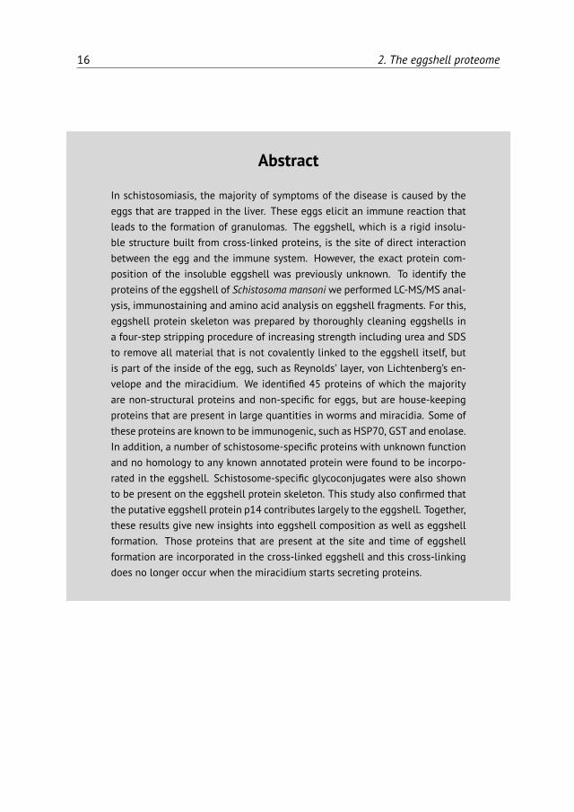
Figure 2.6: Amino acid composition of the purified eggshell skeleton of Schistosoma mansoni. Thecontribution of the various amino acids to the proteins p14 (black Bobek et al. (1988)[14]), p48 (grayChen et al. (1992)[33]), hydrolysed eggshell protein skeleton (white, this study) and the average ofthe eggshell proteins that we identified by MS (dashed bars, listed in Table 2.1).
Since p14 and p48 both contain high amounts of tyrosine residues, which aremodified and cross-linked during the process of quinone tanning, identificationof these proteins by peptide fingerprinting using MS is hampered by the post-translational modifications. Therefore, we performed amino acid analysis on thepurified eggshell fragments (Fig. 2.6) to see whether it is likely that p14 and p48were present in our sample, and hence are part of the insoluble protein skeleton ofthe eggshell. The amino acid composition of the purified eggshell skeletonwas verysimilar to previous reports [24, 150, 195]. High glycine levels (36%) suggest thatp14 is indeed present in the purified eggshell fragments and is a highly abundanteggshell protein. In female worms, p14 mRNA, which is known as the F10 gene, isthe most abundant transcript [128]. Using immunostaining we were indeed ableto confirm the presence of p14 in our samples of purified eggshells (Fig. 2.7).
Although p48 was expected to be part of the eggshell protein skeleton, ouramino acid analysis did not reveal its presence. Lysine, an abundant amino acid ofp48, was average in eggshell (6.8%) and tyrosine, another important amino acid of

32 2. The eggshell proteome
Figure 2.7: Immunoblot of eggshell of Schistosoma mansoni with anti-p14. Dot blot of eggshellprotein skeleton. The dotted eggshell fragments are shown in A, the chemiluminescent signal forp14 or from Horseradish peroxidase (HRP) conjugated anti-hamster IgG (Abcam, Cambridge, UK) assecondary antibody (Ab) only was captured on hyperfilm (B).
p48 could hardly be detected. Both of these amino acids are involved in quinonetanning and this process renders them unrecognizable as lysine and tyrosine.
Immune reactivity of the eggshell proteins
A number of the identified eggshell proteins such as GST, enolase and glyceralde-hyde-3-phosphate dehydrogenase (GAPDH), are known to elicit an antibody re-sponse upon infection [122]. Fig. 2.8 shows how GST and enolase, two of theidentified eggshell proteins, stained positive on Western blots when incubatedwith sera from infected hamsters. Sera were taken from infected hamsters at 2, 4and 6 weeks of infection. A positive stain for enolase was detected at 4 weeks ofinfection, which increased at 6 weeks of infection, when GST was also detected.In addition, antibody reactivity against SEA and AWA was tested. Like enolase,SEA started to stain positive at 4 weeks of infection and the signal increased at6 weeks of infection. Antibody reactivity against AWA started earlier, at 2 weeksof infection and also increased during the course of the infection. Note that at4 weeks of infection, worms do not produce eggs yet but enolase, a protein wedemonstrated to also be present in eggshells, is already recognised by the serum.This demonstrates that enolase is exposed or secreted by developing worms beforethis protein is presented to the host via incorporation in the Schistosoma eggshell.

2.4. Discussion 33
Figure 2.8: Antibody reactivity in hamstersduring infection with Schistosoma mansoni.Adult worm antigens (AWA), soluble egg anti-gens (SEA) or protein solutions of Schisto-soma GST (28 kDa) or rabbit enolase (47 kDa)were run through a SDS-PAGE and transferredto polyvinylidene fluoride (PVDF) membranes.Membranes were incubated with sera from in-fected hamsters at different time points (2,4 and 6 weeks) during infection as indicatedabove the blots.
2.4 Discussion
The eggshell is the site of direct interaction between the highly immunogeniceggs of S. mansoni and the immune system of its host. Eggs are known to excreteimmunogenic proteins [30][110], but it is likely that the proteins that form theeggshell also cause an immunological reaction. The exact protein composition ofthis rigid cross-linked structure was unknown. The aim of the current study wasto identify the proteins that make up the cross-linked eggshell, the outer surfaceof the egg that interacts with the immune system.
In samples of the purified protein skeleton of eggshells we identified a collec-tion of proteins, similar to those found in other proteome studies of S. mansoni[16, 30, 40, 110, 184]. The variety of proteins we detected seems rather random(Table 2.1). Included are many enzymes involved in various metabolic processes,but for instance enzymes involved in crosslinking of the eggshell were not detected.

34 2. The eggshell proteome
Surprisingly, we also could not identify by MS either one of the putative eggshellproteins p14 and p48. We have two explanations for this. First, p14 and p48 aretyrosine-rich proteins, which means that it is likely that most of the peptidesthat are obtained after trypsin digestion will contain one or more tyrosines thathave been modified to form cross-links in the quinone tanning process. Thus,tyrosines will not be detected and different side chains will add unknown andvariable masses to the peptides which then cannot be identified by peptide massfingerprinting. Second, in silico trypsin digestion of these putative eggshell pro-teins reveals that trypsin digestion leads to many very short peptides, which arevery similar to each other (not shown). This is especially the case for p48, wheretrypsin digestion results in many pentapeptides of which peptidemasses are underthe detection limit of the mass spectrometer. Even when detected, such smallpeptides will not reach a 99.0% probability score for protein identification as theyare too short to be specific and thus these peptides will not identify a protein usingour criteria for protein identification. It will therefore be impossible to identifythese putative eggshell proteins by LC-MS/MS, even if they are abundantly presentin the eggshell.Although p14 could not be identified in our MS analysis, using immunostain-
ing we demonstrated the presence of this bona fide eggshell protein in purifiedeggshell fragments (Fig. 2.7). It is not just only present, but the extremely highglycine content (36%) of purified eggshell fragments detected by amino acid anal-ysis indicates that p14 contributes largely to total eggshell. The glycine contentof eggshell, of the identified proteins and of p14 enabled us to make a generalestimation of this contribution of p14 to total eggshell. In the proteins that weidentified by MS, average glycine levels were 8.2% of the 17 analysed amino acids,ranging from 4 to 25%. In p14, 47% of the amino acids were glycine. This leadsto the following equation: [p14]× 47 + [rest]× 8.2 = 36, where [rest] = 1 - [p14],which demonstrates that p14 contributes to total eggshell for approximately 70%.
The set of proteins that contribute to the eggshell for the other 30% seemsto be rather random. However, its average glycine level is significantly higher(7.7%) than the average of all 13,529 S. mansoni proteins available on Uniprot(4.8%) (P <0.001), while tyrosine (3.1% versus 3.4%) and lysine levels (6.8% ver-sus 6.0%) are similar. Although glycine levels in identified eggshell proteins doby no means approach that of total eggshell or p14, its elevated levels in theidentified eggshell proteins are remarkable. Apparently, there is a preference forproteins with higher glycine levels during cross-linking, although glycine does

2.4. Discussion 35
not contribute to quinone tanning.While our analysis of the amino acid composition of eggshell indicated that
p14 is present in the eggshell, such an analysis could not be used to estimate thepresence of p48. Although p48 is rich in tyrosine, lysine and aspartic acid, none ofthese amino acids was abundant in our samples. As the amount of p48 in eggshellis known to be much less than p14 [33, 93, 102], tyrosine, lysine and asparticacid levels of p48 are not abundant enough to feature in a mixed sample of totaleggshell proteins. Furthermore, lysine and tyrosine that have been modulated toform cross-links by quinone tanning could not be traced back in the amino acidanalysis after hydrolysation, resulting in very low detection of these amino acids.The majority of the eggshell proteins identified by MS were non-structural
proteins. Although these proteins were unexpected, immunoscanning microscopyof HSP70 validated our results. The identified proteins are not specific for theeggshells. Many identified proteins such as actin, tubulin, p40, HSP70 and gly-colytic enzymes have also been identified by Cass et al. (2007)[30] as proteinssecreted by S. mansoni eggs. In contrast, Mathieson and Wilson (2010)[110] foundthat these proteins are not present in secretions of eggs. Secreted or not, it is notlikely that our samples were contaminated with proteins secreted by eggs becauseour eggshell fragments were cleaned extensively before proteomic analysis. Theabsence in our samples of proteins secreted by eggs is confirmed by the absenceof IPSE/alpha-1 and omega-1, the two major proteins secreted by eggs. Similarly,contamination of our purified samples with other proteins originating from VonLichtenbergs envelope, Lehman’s lacuna or Reynolds’ layer is just as unlikely dueto the extensive pre-treatment.
The proteins identified in our purified fraction (Table 2.1; Fig. 2.7) are largelycomparable with those of other egg fractions and even with those of other devel-opmental stages of the parasite [16, 40]. The best explanation for the presence ofthese abundant cellular proteins, originating from surrounding vitelline cells, isthat they happened to be around at the site and time of eggshell synthesis andwere cross-linked to the major eggshell proteins. The absence of the two mostabundant proteins secreted by eggs, IPSE/alpha-1 and omega-1, gives an indica-tion of the timing of the cross-linking process. After production of the egg furthermaturation occurs, as the egg excreted by the female worm is still undevelopedand consists of an ovum and vitelline cells surrounded by the eggshell. Our resultsthus indicate that the actual formation of the eggshell including cross-linking ofthe proteins is most likely finished before the miracidium starts secreting proteins.

36 2. The eggshell proteome
Although the observed incorporation of proteins originating from neighbour-ing vitelline cells may seem to be an unintended feature of eggshell production,the presence of these proteins may have immunological consequences. Manyof the proteins we identified as part of the eggshell protein skeleton are knownschistosome antigens such as p40, phosphoenolpyruvate carboxykinase (PEPCK)and thioredoxin peroxidase. These proteins induce cellular responses [3, 200]or antibody responses [122]. The immunogenicity of these usually intracellularproteins can now be explained as these proteins are part of the eggshell. As aconsequence of these immunogenic properties, some of the eggshell proteinsidentified, such as GST and GAPDH, were proposed as vaccine candidates. How-ever, the success of these vaccines in preventing infection was limited [133]. Thismight now be explained by the fact that the vaccines target the eggs where theproteins are exposed and not the actual infection by the adult worms. In fact,vaccination with such antigens might even induce pathology as it enhances theimmune response against the eggs, thereby enhancing granuloma formation anddisease symptoms.
Altogether, our results demonstrate that the eggshell is not only composed ofthe specific eggshell proteins p14 and p48, but also includes proteins availableat the site of eggshell production. Therefore we propose a new model of theSchistosoma eggshell. The main component of the eggshell is the putative eggshellprotein p14. It makes up the majority of the eggshell. However, at the same timeother proteins, whichever are available at the place and time of eggshell production,are also incorporated, albeit to a much lesser extent. Cross-linked together, all ofthese proteins make up a very rigid structure to protect the developing miracidium.
Acknowledgements
We thank Professor Phil LoVerde (University of Texas Health Science Center, SanAntonio, Texas, USA) for providing us with the anti-p14 antibodies and for valuablecomments on the manuscript. Niels Bohnen and Marion Schmitz are thanked fortheir contributions to experiments on the purification of eggshell.

Chapter 3Schistosoma mansoni: The egg, biosynthesis of
the shell and interaction with the host
Saskia deWalick1
Aloysius G.M. Tielens1
Jaap J. van Hellemond1
Experimental Parasitology 2012, 132:7–13.
1 Department of Medical Microbiology and Infectious Diseases, Erasmus MC, Rotterdam,The Netherlands
37

38 3. Eggshell biosynthesis and host interaction
Abstract
The schistosome eggshell is a hardened and tanned structure made from cross-linked proteins. It is synthesized within the female worm from many differentkinds of proteins and glycoproteins. Once the egg is released in the circulation,the outer surface of the eggshell is exposed and hence a direct site of inter-action between the parasite and the host. The major eggshell protein is p14,but about one third of the eggshell is made from common cellular proteins,some of which are known to be immunogenic. This has many consequencesfor parasite-host interactions. However, so far, the eggshell has gained littleattention from researchers. We will discuss the structure of the eggshell andits role in granuloma formation, host factor binding and egg excretion.

3.1. Introduction 39
3.1 Introduction
Production of the Schistosoma egg
Schistosomes are digenetic parasitic flatworms. The female Schistosoma mansoniexcretes around 350 eggs daily which equals to approximately one egg every 5 min[32]. The oocyte is produced in the ovary and released in the oviduct (Fig. 3.1).Here it is fertilized by sperm that comes from the sperm reservoir, a dilated regionin the oviduct. The fertilized oocyte moves further along the oviduct, whichjoins the vitelline duct. Here, 30–40 vitelline cells originating from the vitellinegland surround the fertilized oocyte [175]. Together, the fertilized oocyte and itssurrounding vitelline cells move to the ootype (Fig. 3.2a), thereby passing Mehlis’gland. The actual function of this structure remains elusive. Several functionsof the Mehlis’ gland have been suggested. It is proposed to lubricate the uterusfor the passage of the egg or to activate spermatozoa. Other suggested functionsof Mehlis’ gland are involved in eggshell formation: release of eggshell granules,control or initiate cross-linking of the eggshell or provide a membrane whichserves as a template on which the proteins accumulate to form the eggshell [174].Once in the ootype, contractions of the ootype make the vitelline cells releasetheir granules which contain eggshell precursor proteins (Fig. 3.2b). The eggshellformation starts here. The eggshell is shaped by the ootype and strengthenedthrough tyrosinase activity that causes cross-linking of the released eggshellprecursor proteins (Fig. 3.2c and d). The egg is now ready to pass the uterus andto be released in the circulation, where the miracidium further matures withinthe eggshell (Fig. 3.2e).
vitelliaria
ovary
sperm reservoir
oviduct
vitelline duct ootype
uterus
egg
Figure 3.1: Schematic drawing of the female reproductive tract of Schistosoma mansoni. After Gön-nert(1955) [72] and Smyth(1966) [173].

40 3. Eggshell biosynthesis and host interaction
1
a b
c d
e f
Figure 3.2: Formation of the eggshell in the ootype. Scheme of eggshell formation in Schistosomamansoni. In pink the ootype, yellow the fertilized oocyte, light blue circles are vitelline cells withvitelline droplets (orange) containing eggshell precursor proteins, green triangles are tyrosinases.In the mature egg (f) the miracidium (yellow) is surrounded by Von Lichtenberg’s envelope andReynolds’ layer (blue layers) and the eggshell (brown).

3.2. Structure of the eggshell 41
Maturation of this newly produced egg of S. mansoni, that simply consists of thecross-linked eggshell surrounding the ovum and vitelline cells, takes about a week.In this period the vitelline cells provide nutrients for the developing miracidium,which also obtains nutrients from the host [6]. Newly deposited eggs lack complexsubshell structures between the eggshell and the embryo, but these appear duringthe development of the egg [6, 123]. First, in an early stage, a few cells detach fromthe embryo and form a thin syncytial layer, known as “von Lichtenberg’s envelope”,between the eggshell and the developing miracidium [6, 123]. As developmentproceeds, this envelope becomes thicker, completely encloses the egg contents,the embryo and vitelline cells and separating them from the shell of the egg. Thisnucleated envelope contains extensive rough endoplasmic reticulum indicatingthat this is a place of active protein synthesis [6]. During the further development anew layer of extra cellular material is formed between von Lichtenberg’s envelopeand the eggshell, called Reynolds’ layer, which mainly consists of granulatedmaterial most likely originating from the envelope [6, 123]. The envelope and notthe miracidium produces the proteins secreted by the egg, including IPSE/alpha-1[109, 162].Eggs are produced one by one by female S. mansoni and the next egg will be
produced once the previous one has been released. With an egg production of350 per day, female schistosomes can be considered as true egg factories. This isreflected in the fact that about 10% of total worm mRNA encodes for one eggshellprotein, p14 [128, 170].
3.2 Structure of the eggshell
Cross-linking and tyrosinase activity
The eggshell is a hardened and tanned structure made from cross-linked pro-teins. The cross-linking process is known as quinone tanning and occurs by for-mation of quinone bonds. It is dependent on tyrosinase activity. Tyrosinasesare copper-containing glycoenzymes that can catalyze both the hydroxylation ofmono-phenols (tyrosine residues) to ortho-diphenols (L-dihydroxyphenylalanine,L-DOPA) and the subsequent oxidation of this L-DOPA to ortho-quinone (Fig. 3.3).In this way, accessible tyrosine residues of proteins are converted to o-quinones.These o-quinones are very reactive compounds and can form adducts by reactionwith nucleophilic compounds, such as free amino or sulfhydryl groups on adja-

42 3. Eggshell biosynthesis and host interaction
CH2
OH
CH
NH
C
O
CH2
OH
CH
NH
C
O
HO
CH2
O
CH
NH
C
O
O
CH2
O
CH
NH
C
O
O
CH2
O
CH
NH
C
O
N
NH
CH2
CH
C
NH
O
CH2
Tyrosine residue L-DOPA o-quinone
Tyrosinase
Schiff base
Michael-type adduct
O2
HC
HN
C
O4
4
a b c e
d
Lys
Lys
`
Tyrosinase
Figure 3.3: Quinone tanning. Tyrosinase converts tyrosine residues (a) into L-DOPA (b) and subse-quently L-DOPA residues into o-quinones (c). These very reactive o-quinones can form adducts byreaction with nucleophilic compounds, such as free amino groups of lysine residues on adjacentproteins by the formation of either N-quinonyl derivatives (Michael-type adduct) (d) or Schiff bases(e).
cent proteins. These reactions are known to occur in processes as diverse as thebrowning of foods and plant leaves, and the sclerotization and tanning of insect cu-ticles [11]. Nowadays it is known that in plants and insects the formation of thesequinone adducts occurs via two different reactions of free amino groups with theo-quinones: the formation of N-quinonyl derivatives (Michael-type adducts) andof Schiff bases (Fig. 3.3). Although it was originally suggested that in S. mansonithe cross-linking of the eggshell occurs via formation of the N-quinolyl adductsonly [174], it is in our opinion more likely that also in helminths, the eggshelltanning occurs via both processes (Fig. 3.3).This cross-linking within a single protein and between neighboring proteins,
results in an extensively cross-linked protein matrix. The appearance of theo-quinones leads to a rapid change in color and hardening of the proteins, render-ing them intractable to all protease activity and only hydrolytic treatments candissolve the cross-linked eggshell [194]. The cross-linked eggshells are highly flu-

3.2. Structure of the eggshell 43
orescent. Their excitation/emission spectra are similar to the spectra of productsformed by mushroom tyrosinase in a mixture of lysine with L-DOPA, indicatingthat in the eggshell lysine is the most probable amino acid that forms quinonebonds with tyrosine (see below) [165].Tyrosinase activity appears to be developmentally regulated with peak tran-
scription in mature female worms. Enzymatic activity is specifically located in thevitelline cells [56, 65]. Eggshell formation by tyrosinase activity is pH dependentand triggered by alkaline environment [195].
Specific eggshell proteins
Putative eggshell proteins have been identified in the late 80s by searching forspecific mRNAs that are only expressed in mature females and not in males, nor inimmature female worms, miracidia or cercariae. The corresponding genes couldeither encode eggshell precursors or proteins involved in the synthesis of the eggsand eggshell. Interestingly, tyrosinases, which are known to be developmentallyregulated and involved in eggshell synthesis, have not been identified this way.This suggests that the catalytic enzymes required for eggshell formation are notabundantly expressed when compared to structural proteins. In addition, eggshellproteins that are not exclusively produced in mature females can not be detectedthis way.The first putative eggshell protein to be described was p14 [13, 14, 93, 94,
150, 169]. There are several copies of the p14 gene in the genome of S. mansoni.They do not rearrange, and they are not sex linked [14, 169]. The p14 gene isdevelopmentally regulated and only expressed in mature female worms. The p14gene is only expressed in mature vitelline cells in the vitelline duct and within theegg enclosed by the eggshell in the ootype. Transcripts of p14 are undetectable inRNA obtained from eggs from the liver [33]. The p14 gene is the most abundantmRNA transcript in female worms and accounts for 10% of the mRNA of the entireorganism. Even in samples of mixed sex, this mRNA is by far the most abundanttranscript [128]. The p14 protein is present in all the vitelline droplets of vitellinecells in mature female worms [93]. Immunoblots confirmed the presence of p14 ineggshell [47].
Although less abundant, p48, another putative eggshell protein, shows a similarexpression pattern when compared to p14. However, the amount of p48 mRNA inworm is 10–30-fold less abundant than that of p14 [33]. In vitro translation of the
44 3. Eggshell biosynthesis and host interaction
cDNA with [3H]tyrosine produces a 48 kDa protein. A protein of the same size isalso present in translation products of mRNA from mature female worms, but notin that of male or immature female worm [88]. Antibodies raised against recombi-nant p48 proteins recognize a 48 kDa protein on Western blot in homogenates ofmature female worms, whereas no signal can be detected in homogenates of maleworms or immature female worms. In addition, antisera raised against maturefemale worm extract recognize recombinant p48 proteins, while antisera raisedagainst male or immature female worm extracts do not [33].
Reis et al. (1989)[144] described another female specific mRNA (FS800), that isonly expressed in vitelline cells of mature female worms. The mRNA sequence wasdescribed to comprise two Open Reading Frames (ORFs) in two distinct readingframes. The longest ORF with the starting codon upstream of the alternativeORF is currently annotated as the translated protein sequence in the S. mansonigenome [9]. However, to our knowledge, there are no data available yet on thetranslation of this female-specific mRNA into protein(s).Finally, one other female specific protein has been described by Menrath et
al. (1995)[117] and was described as mucin-like. Later, it was renamed p19 [119]but it has now been annotated as eggshell protein (Q26570_SCHMA) in the UniProtdatabase. However, expression of the gene can be found in the epithelium sur-rounding the female oviduct close to its entrance into the ootype, but not alongthe vitelline duct and neither in ovary, vitellarium or ootype [117]. Although thisprotein is female specific, this is apparently not an eggshell protein.
Non-specific eggshell proteins in the eggshell
Recently, the protein composition of purified eggshell fragments was investigatedby a proteomic approach [47]. This study showed that the eggshell is not onlycomposed of the above mentioned specific eggshell proteins. About one thirdof the proteins in the eggshell are common cellular proteins. Among them areknown schistosome antigens such as major egg antigen p40 and HSP 70, but alsostructural cellular proteins (actin and tubulin) and non-structural proteins ofwhich many are glycolytic enzymes [47]. Many of these non-specific proteins inthe eggshell have also been identified inside the egg [110], in adult worm tegument[17, 184] and in schistosomula and cercariae [40].The list of non-specific eggshell proteins may not be complete, as proteins
that are cross-linked are not always detectable by mass spectrometry [47]. Al-

3.2. Structure of the eggshell 45
though the incorporation of these non-specific eggshell proteins may be an un-intended feature of eggshell production, the presence of these proteins, even insmall amounts, may have immunological consequences. Many of them elicit acellular or humoral immune response in the host upon a schistosome infection.Some of the incorporated proteins, such as glutathione-S-transferase (GST) andglyceraldehyde-3-phosphate dehydrogenase (GAPDH), were proposed and testedas vaccine candidates, albeit with little success in preventing infection [133]. Vac-cination with such antigens might actually induce pathology as it enhances theimmune response against antigens in the eggshell, thereby enhancing granulomaformation and disease symptoms.
It should be noted that IPSE/alpha-1 and Omega-1, the twomajor egg secretionproteins (ESP), have not been found among the proteins of the eggshell [47]. Thisindicates that the proteins excreted by eggs are not incorporated into the shellof the egg, which suggests that cross-linking of the eggshell proteins is finishedbefore excretion of proteins by the egg begins.
Amino acid composition of the eggshell and eggshell proteins
In p14, 44% of the amino acids are glycine. This protein has some homologywith the carboxyl terminal of mouse cytoskeleton keratin and with silk mothchorion proteins [13]. The other major eggshell protein, p48, has only slightlyelevated glycine levels, but contains high levels of lysine and aspartate, and itsmost abundant amino acid is tyrosine. The other proteins we identified as partof the eggshell do not have extreme levels of glycine, although their averageglycine content is significant higher than the average glycine content of S. mansoniproteins [47].The amino acid composition of fluorescent granules in the vitelline cells is
highly similar to that of hydrolyzed eggshells, suggesting that all the reagentsfor eggshell formation are present in the vitelline cells [195]. Hydrolyzed totaleggshells revealed that 36% of the amino acids are glycine, which hereby largelyattributes to the eggshell [24, 47, 150]. The high glycine levels in the analysedeggshell hydrolysates suggest that p14 widely contributes to the eggshell. Thiscontribution was estimated to be 70%. Immunoblots with anti-p14 antibodiesshowed that p14 is indeed present in eggshell [47]. The role of the high glycinecontent of p14 and total eggshell remains elusive, but a high glycine content isassociated with coiled coils. Rodrigues et al. (1989)[150] proposed a structure

46 3. Eggshell biosynthesis and host interaction
model of p14 based on its amino acid sequence and the high glycine content.In this structure, the protein is composed of short anti-parallel beta strands inwhich glycines and other residues with small side chains lie within the strands andtyrosines are arranged at the bends. This way, tyrosine residues are still availablefor cross-linking.
As tyrosine is essential in quinone tanning, eggshells are expected to containhigh levels of this amino acid too. This is indeed the case for the twomain eggshellproteins p14 (11.3%) and p48 (26.1%). However, little tyrosine can be measuredin hydrolyzed eggshells or granules. This is probably not because of the absenceof tyrosine, but due to the changes of the tyrosine molecule upon cross-linkageduring eggshell formation, which makes them no longer recognizable as tyrosine(Fig. 3.3). Although little tyrosine could be detected in eggshell hydrolysates orfluorescent granules, radiolabeled tyrosine was efficiently incorporated in eggshellwhen injected in infected mice, indicating that eggshell does contain significantamounts of tyrosine [24].
A third amino acid that is expected to be abundant in eggshell is lysine, as thisamino acid is likely to be the residue that forms the cross-link with the o-quinones[165]. Indeed, the eggshell proteins p14 and p48 contains high levels of lysine(5.6% and 17.1%). However, no abundant levels of lysine could be detected in totaleggshell. As with tyrosine, this may be due to cross-linking of the lysine residuesto the o-quinones.
Glycans
The eggshell does not only consist of cross-linked proteins, but on these proteins,schistosome specific glycans are present. Composition analysis of glycans wassuggestive for mannose and fucose residues and the presence of N-linked glycans,but additional O-linked glycans and other glycan classes may also be present. Theglycan epitopes F-LDN(-F) and Lewis X are present on eggshell, as demonstratedby immunoblots [47]. These glycans are known to be immunogenic, but little isknown about the molecular basis of glycan-mediated host-parasite interactions[186].

3.3. Interaction with the host 47
3.3 Interaction with the host
Egg secretion proteins (ESP)
Although the eggshell is a direct site of interaction between the parasite and thehost, little research has focused on this tightly cross-linked and hard to analyzestructure. Rather, much research has focused on ESP. Secretion of ESP by themiracidia starts a few days after the eggs have left the female worm [6]. There havebeen some contradictory papers reporting that ESP contains either a wide rangeof proteins [30] or only a few [6, 110]. There is, however, consensus that the majorESP are IPSE/alpha-1 and Omega-1. The total ESP and isolated or recombinantIPSE/alpha-1 or Omega-1 have widely been investigated for their capacity to drivea strong Th2 response. This immune response protects the host from fatal disease.However, this Th2 response is a strong inducer of granuloma formation [134].Furthermore, T-cell responses are essential for excretion of eggs. Experiments inmice have shown that in the absence of T-cells, egg excretion from the body isgreatly hampered [49]. In humans, HIV patients with low CD4+ T-cell count havereduced numbers of egg excretion [90].
Granuloma formation and the eggshell
While many investigators have focused on ESP, much less research has been doneregarding the eggshell. It has recently been shown that the eggshell containsnumerous other proteins and glycoproteins besides the major eggshell proteins.Many of these other proteins and glycoproteins are known schistosome antigens,such as p40, phosphoenolpyruvate carboxykinase (PEPCK) and thioredoxin per-oxidase. These antigenic proteins induce cellular responses [4, 200] or antibodyresponses [122]. Serum of patients infected with schistosomes contains antibodiesthat recognize purified eggshells, from which all not covalently bound compoundswere removed [47] (Fig. 3.4a), whereas these were not present in control serum(Fig. 3.4b). It is thus evident that not only ESP interact with the immune system,but also the eggshell itself is recognized by antibodies. Furthermore, eggs isolatedfrom livers of infected hamsters are covered by host antibodies (Fig. 3.4c).It is known that Th2-cells play a role in granuloma formation. Likewise, anti-
bodies bound to the eggshell may enhance pathology. Since the eggshell is re-sistant to protease activity, and because the incorporated antigenic proteins inthe eggshell bind host antibodies, the eggshell surface will form an indigestible

48 3. Eggshell biosynthesis and host interaction
Figure 3.4: Antigenicity of eggshells. Purified eggshell skeletons [47] were incubated with serumfrom a human schistosomiasis patient (a) or control serum (b) and subsequently with an HRP con-jugated anti-human IgG secondary antibody. Eggs freshly isolated from livers of infected hamsterswere incubated with an HRP conjugated anti-hamster IgG antibody (c). Eggs were subsequently dot-ted on a Whatman paper (top panel) and the chemiluminescent signal of these dots was capturedon light sensitive film (bottom panel).
antigen-antibody complex. These structures are known to induce chronic inflam-matory responses that can lead to granuloma formation by a complex interactionbetween CD4+ T-cells, macrophages and cytokines [201]. Also ESP are involved ingranuloma formation around the egg [110, 134].
Activation of T-cells and binding to antibodies may also play a role in the pro-cess of extravasation through the endothelium and the gut wall. Ex vivo, antibodiesbind to ESP that exit the egg through pores in the eggshell. This is known as thecircumoval precipitin (COP) reaction. At the point of secretion, a precipitate willform. Globular blebs or septate precipitates appear on the wall of the schistosomeeggs after incubation with the serum of infected individuals [66].
How to get out of the host?
The egg is not capable of moving by itself. Thus, all movement has to be inducedby external forces. First it is released into the circulation by the female worm; nextthe circulating blood will take hold of the egg. However, the intended pathway forthe S. mansoni egg is not to follow the blood stream, but to extravasate the vesselwall and penetrate the gut wall in order to get out with the feces. This processtakes at least six days, but may also take weeks [89]. It is unlikely that the femaleworm can push such an enormous egg through the host structures. Therefore,the egg probably attracts host proteins and host cells in order to hold onto thevessel wall and help passage through the endothelium and the gut wall, allowingescaping the host and continuing the life cycle.

3.3. Interaction with the host 49
In addition to thewidely studied schistosome-specific glycoproteins IPSE/alpha-1 and Omega-1, some house-keeping enzymes have been described to be virulencefactors. Enolase and GAPDH, proteins that have also been identified as part ofthe eggshell, are such house-keeping enzymes which are also virulence factors inSchistosoma and other organisms. They both act as surface associated receptorsthat bind plasminogen, thereby capturing fibrinolytic activity of the host on theeggshell surface [206]. This activity represents a mechanism to enhance virulencepossibly required for tissue invasion and may thereby facilitate the escape of theeggs [132]. Enolase in other pathogens has also been shown to bind actin andfibronectin. Fibronectin is a main component of extracellular matrix surroundingepithelial and endothelial cells. Blocking enolase decreased adhesion of otherpathogens to endothelial cells [10, 57]. By binding to and activation of host factors,the eggshell may thus play an important role in getting out of the host. Throm-bocytopenia significantly reduces egg excretion [125]. Platelets massively adhereto eggshells of S. mansoni, resulting in activation of platelets [203]. Activatedplatelets generally activate endothelial cells and may thereby facilitate passagethrough the endothelium. The eggshell may thus play an important role in gettingout of the host by binding to and activation of host factors like plasminogen andplatelets. Additionally, escape from the host may be facilitated by proteolyticactivity of eggs and miracidia or from other (host) proteins and cells attracted bythe egg [138, 172].
Acknowledgments
Production of Fig. 3.3 by Michiel Bexkens is gratefully acknowledged. We sincerelythank the two anonymous reviewers for their helpful comments.


Chapter 4Binding of von Willebrand factor and plasma
proteins to the eggshell of Schistosoma mansoni
Saskia deWalick1
Paul J. Hensbergen2
Michiel L. Bexkens1
Christina Grosserichter-Wagener1
Cornelis H. Hokke3
André M. Deelder2
Philip G. de Groot4
Aloysius G.M. Tielens1
Jaap J. van Hellemond1
International Journal for Parasitology 2014, 44:263–268.
1 Department of Medical Microbiology and Infectious Diseases, Erasmus MC, Rotterdam,The Netherlands
2 Center for Proteomics and Metabolomics, Leiden University Medical Center, Leiden, The Netherlands3 Department of Parasitology, Center for Infectious Diseases, Leiden University Medical Center, Leiden,The Netherlands
4 Department of Clinical Chemistry and Haematology, University Medical Center Utrecht,The Netherlands
51

52 4. Plasma proteins binding to eggshell
Abstract
Schistosoma mansoni eggs have to cross the endothelium and intestinal wallto leave the host and continue the life cycle. Mechanisms involved in thisessential step are largely unknown. Here we describe direct binding to theS. mansoni eggshell of von Willebrand factor and other plasma proteins in-volved in haemostasis. Using deletion-mutants, we demonstrated that it isthe A1 domain of von Willebrand factor that binds to the eggshell. Our resultssuggest that binding of plasma proteins to the eggshell promotes binding tothe endothelium, initiating the passage of the egg through the blood-vesselwall to be excreted in the end.

53
Schistosoma spp. are helminths causing the tropical disease schistosomiasis. Anestimated 200 million people are affected, most of them living in sub-SaharanAfrica [74]. These parasites penetrate their human host through the skin afterwhich the cercariae transform into schistosomula that subsequently enter thecirculation. In the case of Schistosoma mansoni, male and female worms meet inthe portal vein, where they pair and mature before moving to the mesenteric veins,their final habitat [74]. Mature female worms excrete approximately 300 eggs perday, which is about one every 5 minutes [32]. In order to complete the schistosomelife-cycle, the eggs have to be excreted with the faeces. Therefore, eggs haveto cross the blood-vessel wall as well as the intestinal wall. Many eggs fail toextravasate and are transported by the blood flow to the liver where they get stuckand induce granuloma formation, fibrosis of the liver and portal hypertension.These non-excreted eggs are the major cause of morbidity in schistosomiasis.
Eggs have a rigid shell structure of cross-linked proteins and thus cannot propelthemselves [48]. Therefore, eggs depend on external forces in order to cross thehost tissues. Mechanisms involved in excretion of eggs are largely unknown,even though this is an essential step in the life-cycle of schistosomes. Eggs ofseveral schistosome species have a sharp spine and it could be imagined that thepresence of this spine aids in passing through the obstructing structures of thehost [89]. However, eggs of Schistosoma japonicum and Schistosoma mekongi lack asharp spine, which makes this theory unrealistic. It is known that the process ofexcretion is partly dependent on the host immune reaction directed towards eggs[49], but additional mechanisms are likely to be involved.It has been demonstrated that eggs attach to endothelium and that endothe-
lial cells actively migrate over the eggs [64]. The endothelial surface-adhesionmolecules, ICAM-1, E-selectin and VCAM-1, and surface exposed Lewis X glycanson S. mansoni eggs play an important role in egg adhesion to the vascular endothe-lium [98]. In addition, plasma factors enhance egg attachment to endothelial cellsin vitro and platelets have a role in extravasation and excretion in vivo [64, 125, 126].Furthermore, schistosome eggs were also shown to be a potent and direct activatorof platelets and platelets were shown to adhere en masse to eggs in vitro [125, 203].Activated platelets aggregate and stimulate secondary coagulation (the plasmaclotting factor cascade) which results in fibrin fibre formation to stabilise theplatelet plug. Apparently, there is an interaction of the schistosome eggshell

54 4. Plasma proteins binding to eggshell
with the host haemostatic system, but the molecular structures involved in theactivation of the haemostatic system are unknown.
We hypothesised that the egg probably attracts host proteins and host cells inorder to hold onto the vessel wall and help passage through the endothelium andthe gut wall, allowing excretion from the host and continuation of the life cycle.Therefore, we analysed the interaction of S. mansoni eggshells with vonWillebrandfactor (VWF), as this clotting factor is crucial for anchoring clotting material todamaged or activated surfaces of the blood vessel, and VWF could thus play anessential role in egg extravasation. VWF is an adhesive glycoprotein of 250 kDwhich forms multimers that can be extremely large and consist of over 80 subunits,resulting in a multimer of over 20 mega-Dalton. VWF contains many bindingdomains that can connect platelets to clotting factors and injured surfaces of theendothelium [105, 159]. VWF binds platelet glycoprotein Ib (GPIb) on plateletswhich are thereby activated and initiate clot formation.
The presence of VWF on S. mansoni eggs isolated from collagenase digestedlivers of infected hamsters was demonstrated by immunofluorescence (Fig. 4.1A).As these eggs were directly obtained from perfused livers, this demonstrates thatVWF binding occurred in vivo in the blood vessel.
Schistosome eggs were also incubated in vitro with purified human VWF. Weused both untreated eggs and stripped eggs. From stripped eggs, all non-covalentlybound proteins were removed by a procedure which includes multiple washes withhigh salt conditions, detergents, urea and even boiling in SDS, in order to beentirely sure that no host factors remained on the surface of the schistosome egg[47]. Both untreated (Fig. 4.1C) and stripped eggs (Fig. 4.1D) bound purified humanVWF in vitro, demonstrating the direct interaction between VWF and the eggshell.The binding of VWF to eggs could also be demonstrated at a more quantitativelevel using stripped eggshell fragments and enhanced chemiluminescence (ECL)measurements (Fig. 4.1H).We also incubated stripped schistosome eggs for 1 min with citrate plasma
from healthy adult human volunteers, obtained after informed consent. Bindingof VWF to eggs was then demonstrated by immune fluorescence (Fig. 4.1F and G).This result demonstrated that VWF in the presence of all other plasma proteinsstill binds to the schistosome eggs, although the punctuated fluorescence patternsuggests that other plasma proteins compete for egg binding, or that other plasmaproteins shield bound VWF prohibiting its detection by immune fluorescence.Altogether these results demonstrate that there is a direct interaction between

55
schistosome eggshells and VWF. To determine which VWF domain is involvedin eggshell binding, we incubated eggs with recombinant human VWF of whicheither the A1 [97], A2 [95] or A3 domain [96] was deleted. Reduced VWF bindingwas only observed with the A1 domain-deleted VWF, but not with VWF lackingthe A2 or A3 domain (Fig. 4.1I). This indicates that VWF binds eggshells with itsA1 domain. Domain A1 of VWF is a relatively small part of VWF as it comprisescirca 250 amino acids, which is approximately 15% of the entire VWF protein [97].In addition, deletion of the A1 domain does not affect the structure and functionof the other domains in VWF [95–97]. Hence, the dependence on a small domainof VWF for its binding to schistosome eggs suggests a specific interaction.The A1 domain of VWF also binds GPIb, a glycosylated receptor on platelets.
Since the eggshell contains glycan structures as well [47], we hypothesised thateggshell glycans may play a role in the binding of VWF. However, we could not findindications for glycan-mediated VWF binding to eggshells. Treatment of eggshellswith glycosidases did not reduce VWF binding (data not shown), but these treat-ments did not remove all glycan structures either and therefore these experi-ments were inconclusive. Blocking of glycan access with monoclonal antibodiesagainst schistosome glycans (291-2G3-A, 114-3A5, 128-4F9-A (which bind toGalβ1-4(Fucα1-3)GlcNAc (Lewis X)), 273-3F2 (which binds to GalNAcβ1-4GlcNAc(LDN)) [192] and 114-4D12 (which binds to Fucα1-2Fucα1-3 (DF) termini) [148])had no effect on the binding of VWF to eggshells (data not shown). Furthermore,treatment of eggshells with periodate, which disrupts glycan structures, did not re-duce VWF binding (data not shown). In conclusion, VWF binds directly to eggshellsthrough its A1 domain, most likely in a non-glycan-mediated manner.
When eggs are laid by the female schistosome in the circulation of the host, theyimmediately come into contact with circulating plasma proteins. We hypothesisedthat, next to VWF, other plasma proteins involved in fibrin formation, plateletactivation and aggregation, bind to eggshells. To test this, we incubated strippedeggshell fragments with plasma and subsequently washed those six times withPBS. Thereafter, bound plasma proteins were released by boiling the eggshells inLaemmli buffer. Released proteins were analysed by SDS-PAGE together with alane of diluted plasma (Fig. 4.2). A clear difference in protein band pattern wasobserved between plasma and plasma proteins bound to schistosome eggshellfragments, especially in the high molecular weight range, demonstrating selectivebinding of certain plasma proteins to eggshells. Following trypsin digestion and LC-MS/MS analysis, the identified proteins in both samples were compared (Table 4.1

56 4. Plasma proteins binding to eggshell
and Supplementary Table S1). The high molecular weight plasma protein whichspecifically bound to eggshells (gel slices 1 and 2) was identified as fibronectin.Although the pattern of protein bands between approx. 50–75 kDa in plasma wassimilar to the pattern of proteins bound to eggshells, the major proteins in thesegel slices were different. In this respect it is noteworthy that albumin, the majorplasma protein, could hardly be detected among the proteins bound to eggshells.This validates the selectivity of the binding. The plasma proteins specificallybound to eggshells in the molecular weight region of albumin consisted mainlyof fibrinogen (gel slices 5–7). Fibrinogen is a haemostatic protein and is theprecursor of fibrin. When cross-linked, fibrin networks reinforce a platelet plug.Similar to fibrin, fibronectin plays an essential role in wound healing and bloodclot formation.
Figure 4.1 (following page): vonWillebrand factor binding to Schistosoma mansoni eggshells. Eggs ofa Puerto Rican strain of S. mansoni maintained in Golden hamsters for which animal ethics was ap-proved (licence EUR1860-11709), were isolated 49 days p.i. as described previously [47]. Eggshellfragments were prepared by crushing eggshells in a micro-dismembrator S (Braun Biotech Int., Mel-sungen, Germany). “Stripped eggs” and “stripped eggshell fragments” were prepared by removal ofnon-covalently attached host material from isolated eggs or eggshell fragments by consecutivewashes with 2 M NaCl in PBS, 1% (v/v) Triton X-100 in PBS, 8 M urea with 0.6% β-mercaptoethanolin PBS, and finally with 1% (w/v) SDS at 95 °C for 30 min [47]. Untreated schistosome eggs isolatedfrom livers were incubated with polyclonal anti-von Willebrand factor rabbit antibody (Dako) (A) orPBS (B) for 1 h. Subsequently, for immune fluorescence microscopy these eggshells were washedwith PBS and stained with Alexa 633 conjugated anti-rabbit IgG antibody (Invitrogen, USA). Fluo-rescence microscope images were taken using Cell∧F software (Olympus, Germany). Untreated eggs(C) or stripped eggs (D-G) were incubated with 500 µl of PBS/BSA/Tween (E, G), or PBS/BSA/Tweenwith purified human von Willebrand factor (0.2 µg/ml) (C and D), or plasma using citrate as anti-coagulant (F). After incubation, the eggshells were washed three times with PBS and then stained forvon Willebrand factor binding as described above using a polyclonal anti-von Willebrand factor rab-bit antibody (Abcam, UK). Representative pictures from at least two independent experiments showthe immune staining for von Willebrand factor (C-G). To quantify bound von Willebrand factor (H andI), stripped eggshell fragments were incubated with or without purified human von Willebrand fac-tor (H) or with recombinant deletion mutants of von Willebrand factor (I), after which the eggshellfragments were washed with PBS, incubated with anti-von Willebrand factor antibody (Abcam) for1 h and subsequently incubated with horseradish peroxidase conjugated anti-rabbit immunoglobu-lin antibodies (Dako, Glostrup, Denmark). The eggshell fragments were then incubated in enhancedchemiluminescence detection reagents (Pierce, USA) and dotted on transparent sheets. Chemilumi-nescence was captured with Alliance 2.7 (Uvitec, UK) and intensity was determined using Image Jsoftware. The amount of eggshell material was quantified by measuring scattering at 655 nm (OD655). von Willebrand factor binding was normalised for the amount of eggshells by expression inarbitrary units ECL/OD655. The fluorescence signal in the presence of added von Willebrand factorwas set to 100%; data from 12 experiments (mean + S.D.) (H). Eggshells were also incubated withrecombinant deletion mutants of human von Willebrand factor lacking the A1, A2 or the A3 domain,respectively (I). enhanced chemiluminescence signal of full-length von Willebrand factor was setto 100%, data from three independent experiments (mean + S.D.). Magnification in A-E is identical(bar represents 100 µm) and higher than in F and G (bar represents 50 µm).

57
A
B
C
D F
G
0,0
50,0
100,0
150,0
200,0
250,0
native VWF ?A1 ?A2 ?A3 no VWF
I
VWF no VWFA1 A2 A3Δ Δ Δ
Flu
ore
scence
(%
of fu
ll-le
ngth
VW
F)
0
50
100
150
200
250
0
20
40
60
80
100
120
VWF no WVF
Flu
ore
scence
(%
)
VWF no VWF0
20
40
60
80
100
120
E
Ė B C D E F G
Eggs isolated isolated isolated stripped stripped stripped stripped
VWF - - + + - - -
Plasma - - - - - + -
Anti-VWF + - + + + + +
Anti rabbit + + + + + + +
A B C D E F G
H

58 4. Plasma proteins binding to eggshell
Table 4.1: List of main proteins in human plasma versus proteins of human plasma bound to Schis-tosoma mansoni eggshells. The numbers refer to the gel slices cut from SDS-PAGE as shown inFig. 4.2. Proteins were in-gel digested with trypsin and subsequently analysed by LC-ion trap MS/MSas described previously [208]. Spectra were searched against the human (UniProt) and schistosome(Schistosoma mansoni, release 4.0 h at www.genedb.org) database using the Mascot search algo-rithm (version 2.2.07). To examine the major differences between protein hits in correspondingbands, only proteins with at least four unique peptides with a peptide score >30 were considered.For each gel slice the most abundant proteins identified are listed, with a maximum of three, rankedby the number of peptides detected for each protein. For a comprehensive overview of the proteinidentification data, see Supplementary Table S1.
Total plasma proteins Eggshell-bound plasma proteinsGel slice Peptides Peptidesnumber Protein name detected Protein name detecteda
1 Albumin 19 Fibronectin 1 80
2 Albumin 22 Fibronectin 1 139Fibronectin 1 13 Talin 1 7Alpha-2-macroglobulin 11 -
3 Albumin 36 Fibronectin 1 34Alpha-2-macroglobulin 25 Collagen, type II, alpha 1 5Ceruloplasmin (ferroxidase) 11 -
4 - Fibronectin 1 9
5 Albumin 64 -Serotransferrin 26 -
6 Albumin 132 Fibrinogen alpha chain 57Fibrinogen alpha chain 15 Vitronectin 14Uncharacterised protein 8 Albumin 14
7 Albumin 42 Fibrinogen beta chain 30Alpha-1-antitrypsin 28 Fibrinogen alpha chain 23Ig gamma-1 chain C region 14 Uncharacterised protein 13
8 - Fibrinogen gamma chain 32- Fibrinogen alpha chain 18- Actin, beta 7
9 Albumin 19 Actin, beta 27- Apolipoprotein A-IV 12- Fibronectin 1 12
10 Albumin 25 Fibrinogen alpha chain 10Haptoglobin 16 -Apolipoprotein A-IV 4 -
11 Albumin 8 Clusterin 15- Apolipoprotein E 10- Fibrinogen alpha chain 9
12 Albumin 9 Apolipoprotein E 34Apolipoprotein E 6 Fibrinogen alpha chain 10Fibrinogen alpha chain 6 Fibrinogen gamma chain 10
13 Uncharacterised protein 12 -
14 Albumin 11 -
15 Apolipoprotein A-I 11 -a Average of two independent experiments.

59
Figure 4.2: Specific human plasma protein binding to Schistosoma mansoni eggshells. Blood wasfreshly drawn from human volunteers in containers containing citrate as anti-coagulant and spunfor 10 min at 1200g to obtain platelet-poor plasma. Stripped S. mansoni eggshell fragments wereincubated for 1 h with 1 ml of this platelet-poor plasma. After incubation, the eggshell fragmentswere washed six times with PBS, boiled in Laemmli buffer and run through 12% SDS-PAGE (laneseggs + plasma), with a control lane containing 10 µl of the used plasma diluted 1:200 in PBS (laneplasma 1:200). Molecular weight marker is shown. Slices were cut from the gel as indicated andnumbers correspond to the proteomics data provided in Table 4.1 and Supplementary Table S1.
The set of proteins bound to eggshell was also enriched in actin and talin.These cytoskeleton proteins probably originate from the remaining platelets inthe platelet-poor plasma. This is in accordance with previous reports which showthat platelets bind very well to eggshells [203]. We also observed an enrichmentof apolipoproteins E and A-IV in the set of proteins that bound to eggshells (Table1). In plasma, Apo-IV is present on chylomicrons, whereas Apo E is present onchylomicrons and on intermediate-density lipoproteins (IDLs). Their presence inthe set of proteins bound to eggshells could be a result of binding of lipoproteinsto eggshells. It is known that adult worms have a low-density lipoprotein (LDL)-receptor-like protein on their tegument [158] and that the worms bind LDL [34].The binding of LDL probably helps the adult worms to mask their antigens andhence hide from the immune system. However, on eggshells no LDL-receptor-likeprotein is known to be present.

60 4. Plasma proteins binding to eggshell
In contrast to the immune fluorescence experiments, the proteome analysis ofbound plasma proteins did not reveal VWF as a protein bound to schistosome eggs.Fluorescence is a highly sensitive detection method compared with our proteomicworkflow. Mass spectrometry detects the most abundant peptides of a trypsindigest of a protein sample, and thus this technique will only show the proteinsmostly present in the analysed material. Therefore, absence of detection by thismethod cannot be interpreted as an indication that the protein in question is notpresent. Of note, the plasma concentration of VWF (approximately 10 µg/ml) isvery low compared with that of fibrinogen and fibronectin (2–4 mg/ml, 0.3–0.4mg/ml, respectively). These plasma components each bound to the eggshell andwere extremely enriched from plasma compared with albumin and other plasmaproteins, but the unfavorable concentration ratio of VWF to fibrinogen and fi-bronectin on the eggshell seems to have left VWF undetectable by proteomics. Thenear absence of albumin in the proteins bound to eggshells further demonstratesthe specificity of the binding of other plasma proteins to S. mansoni eggshells.Moreover, the binding efficiencies and kinetics of VWF compared with the otherpossibly competing eggshell binding proteins identified in our proteomics experi-ments may also have played a role.
To check whether all eggshell bound proteins were from the host and not fromthe parasite itself, the mass spectrometric data were also searched against theschistosome database. The only schistosome protein found in this analysis wasactin, but all identified peptides showed complete homology to human actin. Asno schistosome-specific peptides derived from actin were observed, it is likelythat the human plasma and not the schistosome itself is the source of the actinfound. Altogether, the set of eggshell-binding plasma proteins is indicative offibrin formation and platelet binding to eggshell.Our results demonstrate that the schistosome eggshell binds the plasma pro-
teins, VWF, fibrinogen and fibronectin, which play an essential role in clot forma-tion. The main function of VWF is binding to other proteins by protein-proteininteraction, for which VWF comprises many binding domains. Upon injury VWFplays an important role in platelet-vessel wall adhesion by binding both platelets(via GPIb and GPIIb-IIIa) and the extracellular matrix of the damaged endotheliumexposing collagen and fibrin. Binding of VWF to the GPIIb-IIIa complex, which isexposed on activated platelets only, also plays a role in platelet cross-linking andplatelet plug expansion [105]. Direct binding of VWF to the eggshell by the A1domain may thus have two effects. VWF may be directly involved in the binding

61
of eggs to the endothelium by forming a bridge between the eggshell and theextracellular matrix. Indirectly, binding of VWF in combination with the bindingof platelets [203] may induce platelet adhesion, platelet activation and secondaryhaemostasis, allowing the formation of a stable clot. In turn, this should facilitatea firm binding of the egg to the endothelium. Next to induction of clot formation,the schistosome egg also contains fibrinolytic capacity, as it has been shown thatsoluble extracts of eggs contain a plasmin-like fibrinolytic enzyme activity thatcould counteract uncontrolled thrombogenesis [51].Coagulation normally occurs in the case of bleeding, when endothelium is
damaged and extracellular matrix is exposed to which VWF can bind. Damageto the endothelium is in itself an activator of platelets and blood coagulation, asare shear stress and turbulence [177]. In case of schistosomiasis, shear stress canbe caused by the presence of the relatively large worms and eggs. Attachmentof adult worms to the endothelium may cause endothelial damage that leads toactivation of endothelial cells [168]. As coagulation is initiated on eggshells inthe near presence of damaged and activated endothelium, VWF bound to the eggmay anchor the eggshell to the endothelium, after which platelet aggregation andfibrin clot formation can stabilise the binding.
Extravasation is the first step of excretion and is likely to occur rapidly, beforeeggs have fully matured and start secreting enzymes and other material. In vitroadhesion of eggs to endothelium takes place within 2 h [98, 126]. Within 4 h afteradhesion to the vascular endothelium, endothelial cells actively migrate over theeggs [64]. This way the eggs are removed from the blood vessel. The binding siteby which VWF binds to S. mansoni eggs is not easy to resolve experimentally, asthis target cannot be isolated because the eggshell is composed of a heavily cross-linked protein structure [48]. Furthermore, VWF is a very large protein that has nocatalytic activity and its primary function is binding to other proteins via protein-protein interactions. These interactions do not resemble classical receptor-ligandinteractions, but are rather complex protein-protein interactions with inducedconformational changes. In addition, VWF comprises many different domains forprotein-protein interactions and even domain A1, which is crucial for binding toschistosome eggs, binds to a large variety of structures, such as glycoprotein Ib(GPIb), Staphylococcus protein A, Snake Venom Metalloproteinase, collagen andheparin [156]. For these reasons, the molecular mechanisms by which VWF bindsto most other well-known interacting partners has not yet been resolved, and thisprohibits the identification of potential VWF binding sites in the schistosome

62 4. Plasma proteins binding to eggshell
eggshell on the basis of structural homology to a known target.In conclusion, our data demonstrated that eggshells of S.mansoni bind a specific
set of plasma proteins, including VWF. We suggest that binding of plasma proteinsto eggshells is physiologically relevant and a crucial step in extravasation as itpromotes binding to damaged or activated endothelium. It thereby prevents eggsbeing swept into the circulation and it facilitates egg extravasation from the bloodvessel, which is the first step in excretion of the egg.
Acknowledgements
We thank IrinaDragan (Center for Proteomics andMetabolomics, LeidenUniversityMedical Center, Leiden, The Netherlands) for expert technical support.

Supplementary data 63
Supplementary data

644.Plasm
aproteins
bindingtoeggshell
Table S1: List of proteins identified in human plasma versus proteins of human plasma bound to Schistosoma mansoni eggshells. The band numbers(BN) refer to the gel slices cut from SDS-PAGE as shown in Fig. 4.2. For each gel slice all protein identifications with at least four unique peptidesare listed. PC, protein coverage; TP, total number of peptides; UP, number of unique peptides.
Plasma Eggs1 Eggs2BN Protein name PC TP UP Protein name PC TP UP Protein name PC TP UP
1 Albumin 21.8 19 14 Fibronectin 1 20.5 47 29 Fibronectin 1 32.0 112 561 Inter-alpha (Globulin)
inhibitor H24.3 5 4 Fibrinogen alpha chain 6.9 6 6
1 Collagen, type II, alpha 1 4.8 6 5
2 Serum albumin 25.3 22 14 Fibronectin 1 38.4 160 66 Fibronectin 1 30.9 117 512 Alpha-2-macroglobulin 8.0 11 11 Talin 1 5.5 9 8 Talin 1 3.4 5 52 Fibronectin 1 7.1 13 11
3 Serum albumin 36.5 36 21 Fibronectin 1 11.8 26 20 Fibronectin 1 15.2 42 303 Alpha-2-macroglobulin 15.8 25 22 Collagen, type II, alpha 1 2.9 6 4 Collagen, type II, alpha 1 2.9 4 43 Ceruloplasmin (ferroxidase) 11.5 11 10 Talin 1 1.5 4 43 Pregnancy zone protein 8.4 6 63 Complement C3 3.7 5 4
4 Fibronectin 1 2.8 7 5 Fibronectin 1 4.9 11 84 Fibrinogen alpha chain 4.7 4 4
5 Serum albumin 49.6 64 295 Serotransferrin 33.4 26 215 Ig mu chain C region 18.1 8 75 Complement C3 4.9 5 5
6 Serum albumin 75.9 132 46 Fibrinogen alpha chain 30.6 49 28 Serum albumin 32.8 21 166 Fibrinogen alpha chain 22.5 15 14 Albumin (Fragment) 22.7 6 5 Fibrinogen alpha chain 30.9 65 316 Uncharacterized protein 12.8 8 7 Vitronectin 19.7 14 9 Vitronectin 16.7 14 86 Fibronectin 1 3.9 5 5 Fibronectin 1 4.9 9 86 Collagen, type II, alpha 1 3.5 5 4
7 Alpha-1-antitrypsin 51.2 28 17 Fibrinogen beta chain 41.1 33 22 Fibrinogen beta chain 35.6 27 197 Serum albumin 40.1 42 21 Fibrinogen gamma chain 30.2 17 12 Uncharacterized protein 19.5 8 77 Ig gamma-1 chain C region 36.1 14 10 Fibrinogen alpha chain 27.0 27 22 Fibrinogen alpha chain 19.2 19 167 Fibrinogen beta chain 22.8 14 10 Albumin 20.7 14 9 Fibrinogen gamma chain 16.5 8 67 Ig alpha-1 chain C region 15.6 6 4 Vitronectin 16.7 7 6 Fibronectin 1 5.1 6 67 Fibronectin 1 5.8 9 9

Supplementary
data65
Table S1 continued
Plasma Eggs1 Eggs2BN Protein name PC TP UP Protein name PC TP UP Protein name PC TP UP
8 Fibrinogen gamma chain 42.7 34 17 Fibrinogen gamma chain 34.7 30 138 Fibrinogen alpha chain 23.2 20 16 Actin, beta 19.1 7 78 Actin, beta 19.1 6 6 Fibrinogen alpha chain 16.7 16 128 Fibrinogen beta chain 13.2 4 4 Fibrinogen beta chain 16.5 6 68 Albumin 12.5 9 7 Uncharacterized protein 10.0 7 68 Fibronectin 1 9.7 18 16
9 Serum albumin 25.1 19 13 Actin, beta 37.0 31 19 Fibronectin 1 7.0 12 99 Apolipoprotein A-IV 21.7 10 8 Actin, beta 32.2 22 179 Fibrinogen alpha chain 21.7 14 12 Apolipoprotein A-IV 22.7 13 99 Fibronectin 1 6.5 12 10 Fibrinogen alpha chain 9.8 5 59 Fibronectin 1 2.9 7 6
10 Serum albumin 26.6 25 14 Fibrinogen alpha chain 4.2 8 4 Fibrinogen gamma chain 10.2 11 610 Haptoglobin 19.4 16 8 Clusterin 9.8 13 710 Apolipoprotein A-IV 10.1 4 4 Fibrinogen alpha chain 8.5 11 610 Fibrinogen alpha chain 5.5 7 5 Fibronectin 1 2.8 6 610 Complement C3 2.3 4 4
11 Albumin 10.2 8 5 Apolipoprotein E 24.3 10 7 Apolipoprotein E 26.8 9 811 Clusterin 15.4 17 8 Clusterin 16.7 12 911 Fibrinogen alpha chain 9.7 10 7 Fibrinogen alpha chain 11.0 8 611 Fibronectin 1 3.0 7 6 Fibrinogen gamma chain 10.2 8 5
12 Apolipoprotein E 19.9 6 6 Apolipoprotein E 42.3 36 15 Apolipoprotein E 34.7 31 1312 Serum albumin 18.7 9 8 Fibrinogen alpha chain 11.9 7 5 Fibrinogen alpha chain 10.7 12 812 Fibrinogen alpha chain 6.1 6 4 Fibrinogen gamma chain 10.2 9 6 Fibrinogen gamma chain 10.6 10 612 Fibronectin 1 3.4 12 8
13 Uncharacterized protein 17.2 12 8 Fibronectin 1 3.0 5 5
14 Albumin 19.9 11 10 Actin, beta 12.3 4 4 Tyrosine 3-monooxyge-nase/tryptophan5-monooxygenaseactivation protein, zetapolypeptide (Fragment)
22.8 5 4
15 Apolipoprotein A-I 24.1 11 4


Chapter 5Evaluation of molecular and serologicalmethods for diagnosing two cases of
neuroschistosomiasis
Saskia deWalick1
Rob Koelewijn2
Aloysius G.M. Tielens1
Jaap J. van Hellemond1,2
Manuscript in preparation
1 Department of Medical Microbiology and Infectious Diseases, Erasmus MC, Rotterdam,The Netherlands
2 Laboratory of Parasitology, Harbour Hospital and Institute for Tropical Diseases, Rotterdam,The Netherlands
67

68 5. Diagnosing neuroschistosomiasis
Abstract
Neuroschistosomiasis is the most severe ectopic form of schistosomiasis. Toprevent severe neurological damage and permanent disability, rapid diagno-sis and treatment are crucial. However, diagnostic tools for rapid diagnosisof neuroschistosomiasis are currently lacking. Here we present two cases ofconfirmed schistosome myelopathy and compared molecular as well as dis-tinct serological methods for their usefulness to diagnose neuroschistosomia-sis. Real-time quantitative PCR and Western blots did not provide positive testresults for our patients, but quantification of specific antibody levels againstadult worm antigens and egg antigens in serum and cerebrospinal fluid was in-dicative for intrathecal antibody production directed against schistosome eggs.Hence the proposed egg/worm antibody index can be a valuable tool to distin-guish between on the one hand patients with true neuroschistosomiasis andon the other hand patients that have systemic schistosomiasis but no eggs inthe spinal cord, where the transverse myelitis is caused by a different disorder.

5.1. Introduction 69
5.1 Introduction
Schistosomiasis is a tropical disease affecting an estimated 200 million peopleworldwide, mainly in sub-Saharan Africa [74]. The disease is caused by a para-sitic flatworm of the Schistosoma genus. Eggs laid by the female parasite thatare not excreted accumulate in host tissue and cause the vast majority of thedisease symptoms. Adult worms usually reside in the mesenteric veins or in theblood vessels surrounding the urine bladder. Sometimes, however, worm pairsreside in ectopic locations. The most severe ectopic form of schistosomiasis istransverse myelitis. Transverse myelitis is an inflammation of the spinal cordcausing neurological dysfunction such as muscle weakness, sensory loss, bladderdysfunction and finally paraplegia. In case of neuroschistosomiasis, it is caused bythe inflammatory response of the host to the schistosome eggs that are trappedin the spinal cord. The eggs are deposited in situ after aberrant migration ofadult worms to the central nervous system (CNS), or by egg dissemination via thebloodstream through retrograde venous flow into the Batson vertebral epiduralplexus, which connects the deep iliac veins and inferior vena cava with veins ofthe spinal cord [26, 27, 154]. The inflammatory lesions around the eggs suppressthe spinal cord causing neurological dysfunction. Neuroschistosomiasis has anacute or subacute onset and may then progress to full paralysis within 15 daysif untreated [63]. Diagnosis of neuroschistosomiasis is difficult and the disorderis often not recognized and therefore probably underdiagnosed [62]. True preva-lence is unknown, but a study in Brazil showed that 5.6% of non-traumatic andnon-tumoural myelopathies were caused by neuroschistosomiasis [29].Normally, the cerebrospinal fluid (CSF) contains little protein and few cells.
During inflammatory diseases of the CNS, the blood-brain-barrier is usually dam-aged at least to some extent. Therefore, leakage of antibodies from blood plasmainto the CSF is likely to occur. Hence, the presence of anti-schistosome antibodiesin the CSF could either be the result of intrathecal antibody production due toneuroschistosomiasis or it could be the result of leakage of antibodies from theplasma into the CSF due to damage to the blood-brain-barrier caused by a distinctneurological disorder. Leakage of plasma antibodies into the CSF is a-specificand the profile of the antibodies leaking will resemble that of plasma antibodies.Intrathecal antibody production on the other hand is specific and the antibodiesproduced are directed against the antigens of the pathogen in the central nervoussystem. As the number of (migrated) B-cells in the CSF is limited, the different

70 5. Diagnosing neuroschistosomiasis
antibodies that are produced intrathecally are also limited. In case of neuroschis-tosomiasis, where the immune response is directed against the schistosome eggsthat have crossed the blood brain barrier, intrathecal antibody production is ex-pected to be differentially directed to eggs more than to worms. However, it shouldbe realized that the egg and the adult worm share many of their antigenic proteins[40].As therapeutic delay may quickly lead to severe neurological damage and
permanent disability, rapid diagnostic tools are needed [28]. Diagnosis of spinalcord neuroschistosomiasis usually relies on the clinical diagnosis of transversemyelitis by imaging techniques (such as MRI) to show edema and swelling of thespinal cord, in combination with diagnosis of an active infection with Schistosomaspp. and exclusion of other causes of myelitis. Current methods cannot properlydiscriminate between neuroschistosomiasis as the cause of the transverse myelitisand transversemyelitis with other causes in patients with systemic schistosomiasis.The gold standard for diagnosis of neuroschistosomiasis is retrieval of eggs fromthe spinal cord [28]. However, this invasive method is rarely used, because ofthe risk for serious complications. Therefore, all current methods of diagnosisare non-specific for neuroschistosomiasis and are based on exclusion of othercauses of the myelopathy. It is important to exclude any other infectious causeof myelopathy and to be certain of the diagnosis neuroschistosomiasis becausethe therapy for neuroschistosomiasis includes a high dose of corticosteroids thatwill have adverse effects in patients with transverse myelitis caused by viral orbacterial pathogens [183].Here we report two cases of schistosome myelopathy and examined the use-
fulness of three methods to diagnose neuroschistosomiasis. These methods haveproven to be useful in the diagnosis of other infections in the CNS such as neu-rosyphilis, toxoplasmosis, and varicella zoster virus infection. The followingtests were examined: (1) detection of schistosome DNA in CSF by real-time semi-quantitative PCR in order to demonstrate the presence of the parasite in the CNS,(2) Western blot analysis to demonstrate a qualitatively different anti-schistosomeantibody composition in CSF compared to serum, and (3) indirect hemagglu-tination assay (IHA) and ELISA to demonstrate a quantitatively different anti-schistosome antibody composition in CSF compared to serum, amethod previouslydescribed [42].

5.2. Materials and Methods 71
5.2 Materials and Methods
Case descriptions
Two patients (A and B) presented at the Erasmus University Medical Center withthe clinical symptoms of progressive transverse myelitis. Paired serum and CSFsamples from each patient were taken at presentation and once thereafter.
Patient A was a 35 year-old male who in May 2008 presented to the emergencydepartment with progressive pain and muscle weakness in the upper legs [42].Two months prior to presentation, he suffered from diarrhea during a visit toBrazil. The pain in his legs started four days prior to hospital presentation and wasaccompanied by dysuria and a feeling of incomplete voiding. During the next fewdays, muscle weakness, sensory disturbances and bladder dysfunction increased.An MRI scan showed swelling and edema of the low-thoracic and lumbar spinalcord without abnormalities in the brain. Antibodies against schistosome antigenswere found in serum and CSF and levels were increased after 11 days (Table 5.1).In a rectal biopsy one dead S. mansoni egg embedded in a granuloma was found.Based on these results and the negative test results for other infectious causes oftransverse myelitis, the diagnosis neuroschistosomiasis was made and treatmentwas started. The patient received praziquantel (2 daily doses of 30mg/kg for 3 days)and high dose of intravenous (i.v.) methylprednisolone (2 daily doses of 500 mg for5 days) followed by oral prednisone (3 daily doses of 0.5 mg/kg) tapered graduallyover the next several months. Within a week the patient’s condition improveddramatically and he was discharged from the hospital. During the next months,muscle strength and sensibility improved, and bladder function normalized.Patient B was a 59-year old male who presented in September 2010 at our
neurology department. The patient was born in Suriname, a country to which hetraveled regularly and his last visit was in 2009. In January 2010 he developedprogressive lumbar pain irradiating to the lower limbs, with asymmetric loss ofsensibility and progressive muscle weakness in the legs. The patient was able towalk for 15 to 30 minutes, but fell regularly. He had problems walking stairs andneeded to pull himself on the handrail. The patient was incontinent for urine whenin high need. Otherwise miction and defecation were not affected. MRI showeda lesion in the spinal cord at the low-thoracic and lumbar typical for myelitistransversa. The protein level (1.12 g/L) and the numbers of leukocytes (19×106/L)and monocytes (13×106/L) were elevated in CSF. Furthermore, antibodies against

72 5. Diagnosing neuroschistosomiasis
schistosomes were found in paired serum and CSF samples, collected at presenta-tion and 8 days later (Table 5.1). No eggs of schistosomes could be demonstratedby microscopic methods in feces or urine. Since no other cause for the transversemyelitis was found, the patient was treated with praziquantel (2 daily doses of30 mg/kg for 3 days) and methylprednisolone (once daily 500 mg i.v. for 5 days)followed by oral prednisone (2 daily doses of 0.5 mg/kg tapered gradually over thenext several months). The physical condition of this chronic patient improvedsignificantly within a week, but the patient did not fully recover probably due toirreversible nerve damage caused in preceding nine months of disease.
DNA isolation and real-time PCR
A real-time PCR was performed on DNA extracted from 0.2 ml CSF using easyMag(bioMérieux, France) according to themanufacturer’s instructions. PhocineHerpesVirus (PhHV) was added to the lysis buffer as an internal control. The used primersand probes have been designed based on sequences from S. haematobium (GenBankaccession DQ677661), S. mansoni (AF503487) and S. intercalatum (U22166) [127].We used the following PCR cycle conditions: a denaturation step of 15 min at95 °C, followed by 50 cycles of 15s denaturation at 95 °C, 30s annealing at 60 °Cand 30s elongation at 72 °C. Fluorescence was measured at each annealing step.
Western blot analysis
Worms and eggs were retrieved from experimentally infected hamsters. Wormsand eggs were homogenized by sonification for 3× 30 sec in phosphate bufferedsaline (PBS). The homogenate was spun down for 10 min at 18000 g. The obtainedsupernatant was used to load SDS-PAGE gels (5 µg adult worm protein or 10 µgegg protein, as measured by a Lowry protein assay using bovine serum albuminas a standard). Gels were blotted on PVDF membranes, blocked for 1 h in low fatmilk powder in TBS-tween and subsequently incubated with either serum (1:1000)or CSF (1:100). The secondary antibody was HRP conjugated anti-human IgG(1:10000) (Dako, Glostrup, Denmark). Bands were visualized using ECL (Pierce,Rockford, IL, USA) on Hyperfilm (Amersham, GE healthcare, Diegem, Belgium).

5.2. Materials and Methods 73
Serological methods
Total albumin and total IgG were determined in serum and CSF by routine clinicalchemistry methods. Antibody titers to schistosome worm-antigens were deter-mined using a commercial IHA (Fumouze Diagnostics, Levallois-Perret, France)and agglutination at serum titers of 1:80 and over are positive for schistosomiasis[189]. Schistosome egg-specific antibodies were determined by ELISA of solubleworm antigen (SEA) as described before [189]. Anti-egg antibody levels were ex-pressed as Arbitrary Units (AU) calculated as Optical Density (OD)× dilution / 10and AU values >88 in serum are positive test results for schistosomiasis.
Theory and calculation of serological indices
The albumin index is a measure for the integrity of the blood-brain barrier andvalues ≥0.0090 indicate an impaired function of the blood-brain barrier [181].
Albumin index =ACAS
AC = albumin concentration in CSF (g/L)AS = albumin concentration in serum (g/L)
The total IgG index is a measure for increased concentrations of total IgG in CSFand values≥0.70 are already suggestive for intrathecal antibody production, sincealbumin leaks more easily through the blood-brain barrier when compared toantibodies [60, 104, 142].
Total IgG index =IC/ACIS/AS
IC = total IgG CSF (g/L)IS = total IgG serum (g/L)
Intrathecal antibody production will occur upon any inflammatory response inthe CNS, and therefore, the total IgG index is not specific for schistosomiasis.Demonstration of schistosome-specific antibodies in CSF is thus more informativeand this has indeed been shown to be an indication for neuroschistosomiasis[61, 131]. However, the presence of schistosomes-specific antibodies in CSF doesnot discriminate intrathecal production of these antibodies from antibody leakagefrom the plasma into the CSF. Two methods have been developed to correct forleakage of antibodies from plasma into the CSF. Antibody titers can be corrected

74 5. Diagnosing neuroschistosomiasis
for albumin concentrations in CSF, which is not commonly used, or corrected fortotal IgG in CSF, which is known as the specific antibody index. The cut-off valuefor specific antibody indices is 1.5, with higher levels indicative for intrathecalproduction of specific antibodies [143].
Schistosome-speci f ic antibody index =EC/ICES/IS
EC = anti-egg IgG in CSF (OD× dilution)ES = anti-egg IgG in serum (OD× dilution)
Transverse myelitis in neuroschistosomiasis is caused by the immune response di-rected to schistosome eggs. To differentiate between leakage of plasma antibodiesand intrathecal antibody production, we also used an index which calculateswhether the anti-schistosome IgG response in CSF is more egg-directed thanin serum. We used the egg–worm antibody index as a measure for the relativeenrichment of anti-egg IgG over anti-worm IgG in CSF when compared to serum.
Egg -worm antibody index =EC/WCES/WS
WC = anti-worm IgG in CSF (titer)WS = anti-worm IgG in serum (titer)
This is themost specific antibody indexwhichwill only show increased valueswhenantibodies directed against eggs of schistosomes are produced intrathecally, whichonly occurs when neuroschistosomiasis is the cause of the transverse myelitis.Following Luger et al. (2000)[104], we suggest a cut-off value of 2 for this egg–worm antibody index, representing a two-fold increase in egg-specific antibodiesin CSF. This means that values ≥2 indicate intrathecal production of IgG specificfor schistosome eggs, and thus indicate neuroschistosomiasis.
5.3 Results
Real-time semi-quantitative PCR
Real-time semi-quantitative PCR is a very sensitive method to detect specific DNAand it has been used to detect schistosome infections in blood [36, 198, 204]. Wehypothesized that if the transverse myelitis was caused by eggs of Schistosomaspecies, some of their DNA might leak into the CSF and this schistosome DNA

5.3. Results 75
Table 5.1: Serological data of paired serum-CSF samples.The results are shown of serological anal-ysis of the two paired serum and CSF samples collected from two patients (A and B) at presentation(day 0) and at day 11 and 8 after presentation respectively. Total albumin and total IgG were de-termined by routine clinical chemistry methods. Antibody titers to schistosome worm antigen weredetermined by a commercial indirect hemagglutination assay (IHA) and antibody levels of schis-tosome egg-specific antibodies were determined by ELISA and expressed as Arbitrary Units (AU)calculated as Optical Density (OD)× dilution / 10.
Albumin Total IgG Worm IHA SEA ELISASample Type (g/L) (g/L) titer AU
A0 serum 43.3 12.4 2560 229.6CSF 0.859 0.128 16 3.95
A11 serum 41.2 9.8 5120 286.8CSF 0.771 0.228 32 7.73
B0 serum 44.0 13.3 5120 136.0CSF 0.803 0.169 4 1.88
B8 serum 46.7 13.7 5120 185.2CSF 0.438 0.070 4 1.63
can then be detected by a sensitive PCR method. Hence, for our two patient caseswe examined the usefulness of real-time semi-quantitative PCR on CSF for thediagnosis of neuroschistosomiasis. The used PCR method targets the internal-transcribed-spacer-2 (ITS2), a multi-copy sequence in schistosomes. This assayhad a detection limit of 0.1 pg of schistosome DNA per assay as determined inour laboratory by the use of dilution series of purified chromosomal DNA of adultschistosomes (not shown). Thus, the detection limit is less than the amount ofDNA present in one single schistosome cell. Despite the high sensitivity of thePCR, schistosome DNA could not be amplified in any of the CSF samples of ourtwo neuroschistosomiasis patients while internal controls were amplified properly.Therefore, detection of schistosomal DNA by a sensitive PCR method does notseem to be useful for diagnosing neuroschistosomiasis.
Serology
When intrathecal antibody production against schistosome antigens can be demon-strated, neuroschistosomiasis is confirmed. Therefore, it is important to distin-guish between leakage of antibodies from blood plasma into the CSF and intrathe-cal antibody production. In the case of leakage of antibodies against schistosomes

76 5. Diagnosing neuroschistosomiasis
from the plasma into the CSF, the cause of the transverse myelitis is not nec-essarily neuroschistosomiasis but could be anything that causes damage to theblood-brain-barrier in patients that are coincidently infected with Schistosomaspecies. Intrathecal antibody production is characterized by a raised total IgGindex and/or oligoclonal bands in the CSF. Serological diagnosis of neuroschis-tosomiasis can only be made when intrathecal production of specific antibodiesagainst schistosomes can be demonstrated. We performed Western blots, IHA andELISA to qualify and quantify the anti-schistosome response in both serum andCSF of two patients with neuroschistosomiasis to examine their usefulness forthe diagnosis of neuroschistosomiasis. Western blots using worm and egg frac-tions were performed with both serum and CSF to detect intrathecal productionof schistosome-specific antibody production as prove for neuroschistosomiasis(Fig. 5.1). Many protein bands were detected by Western blot using serum andCSF of neuroschistosomiasis patients (Fig. 5.1A and B). However, protein bandswere also detected when blots were incubated with serum and CSF from patientsinfected with other pathogens (Fig. 5.1C and 1D), demonstrating that not all react-ing protein bands are the result of schistosomes-specific antibodies. Furthermore,the banding pattern to worm and egg fractions was similar in CSF and serum fromthe same neuroschistosomiasis patient (Fig. 5.1A and B). In CSF of patient B theWestern blot with egg antigens, shows a band at 35 kD that seems to be absentin serum, but interpretation is difficult due to the reduced staining reactivity ofthe serum blot compared to the CSF blot (Fig. 5.1B). Therefore, the Western blotscould not reveal an obvious differential schistosome-specific antibody response inCSF versus serum in our neuroschistosomiasis patients. Hence, no schistosome-specific intrathecal antibody production could be demonstrated. These results areconsistent with the findings of Pammenter et al. (1991)[131], and therefore, ourresults support their conclusion that there is no additional diagnostic value forWestern blot over conventional ELISA in the diagnosis of neuroschistosomiasis.We used an anti-egg ELISA and a worm IHA to quantify the antibody responses inboth CSF and serum to native schistosome antigens. Albumin and total IgG werealso measured (Table 5.1). With these measurements we were able to calculatedifferent indices as mentioned above which are informative about the amount ofdamage to the blood-brain barrier, the amount of intrathecal antibody productionand whether the intrathecal IgG response directed against schistosomes is egg-skewed or adult worm-skewed. In all our samples the albumin index was elevated,indicating a damaged blood brain barrier (Table 5.2). This further supports the

5.3. Results 77
Figure 5.1: Western blots on worm and egg antigen of paired serum and CSF samples. Solubleworm and egg fractions were blotted on PVDF membranes and incubated with serum (1:1000) orCSF (1:100) of patient A (A) and patient B (B), a patient with bacterial meningitis (C) and a patientwith cerebral toxoplasmosis (D).
clinical picture of transverse myelitis but does not give any clues towards the causeof the transverse myelitis. Where the albumin index is a measure for blood-braindamage, the total IgG index can indicate intrathecal antibody production. Anelevated total IgG index in schistosome-infected patients is indicative for neu-roschistosomiasis [60], but is not specific for neuroschistosomiasis and may occurin any infectious cause of transverse myelopathy. Only one of each of our pairedserum-CSF samples of the two patients had an increased total IgG index indicatingan infectious cause of the transverse myelitis (Table 5.2). Hence, this index doesnot seem to be a sensitive method for the diagnosis of transverse myelitis causedby infectious agents.
Pammenter et al. (1991)[131] and Ferrari et al. (1995)[61] showed that the pres-ence of schistosome-specific IgG in CSF is indicative for neuroschistosomiasis.However, this test has a low negative predictive value [131] or lacks sensitivity atthe suggested cut off of 1.4 µg/ml [61]. Furthermore, this test does not discriminatebetween antibody leakage form plasma and intrathecal antibody production. Aschistosome-specific antibody index or the egg–worm antibody index can confirmintrathecal egg-specific antibody production more specifically than a total IgGindex or the absolute levels of anti-worm or anti-egg IgG in the CSF. Magalhães-Santos et al. (2003)[107] noted that the IgG1 isotype levels were higher in theCSF than in serum of patients with neuroschistosomiasis. This suggests local

78 5. Diagnosing neuroschistosomiasis
Table 5.2: Calculated indices of paired serum-CSF samples. The four calculated indices for the pairedserum-CSF samples. The albumin index [albumin CSF (g/L) / albumin blood (g/L)] as a measure forthe integrity of the blood-brain barrier, the total IgG index [(total IgG CSF (mg/L) / albumin CSF(g/L)) / (total IgG serum (mg/L) / albumin serum (g/L))] as a measure for concentrations of total IgGin CSF, the schistosome-specific antibody index [(anti-egg IgG in CSF (OD× dilution) / total IgG inCSF) / (anti-egg IgG in serum (OD× dilution) / total IgG in serum)] as a measure for enrichment ofegg-specific IgG compared to total IgG and the egg–worm antibody index [(anti-egg IgG in CSF(OD× dilution) / anti-worm IgG in CSF (titer)) / (anti-egg IgG in serum (OD× dilution) / anti-wormIgG in serum (titer))] as a measure for the relative enrichment of anti-egg IgG over anti-worm IgGin CSF when compared to serum.
SchistosomeSample Albumin Total IgG specific Egg–worm
index index antibody index antibody index
A0 0.020 0.52 1.7 2.75A11 0.019 1.24 1.2 4.31B0 0.018 0.70 1.1 17.65B8 0.009 0.55 1.7 11.27
synthesis of IgG1. However, they did not calculate the specific IgG1 antibodyindex to test for specific intrathecal antibody production. Two of our four samplestested had a positive schistosome-specific antibody index - one from each patient- showing that this antibody index is more sensitive than the total IgG index, butstill not sensitive enough for the diagnosis of neuroschistosomiasis. Thus, in ourpatients, the schistosome-specific antibody index (not divided by subclasses) wasnot sensitive enough to diagnose neuroschistosomiasis as the cause of transversemyelitis. However, the egg–worm antibody index did indicate intrathecal anti-body production in all paired serum-CSF samples (Table 5.2), thereby confirmingneuroschistosomiasis. Both our patients had increased indices. Patient A had aslightly positive index for the first paired serum-CSF sample which had increasedto 4.31 after 11 days. Patient B had highly elevated indices at both time points.
5.4 Discussion
Although PCR detection of viral and bacterial pathogens can be used to diagnoseseveral infectious diseases in the CNS, this technique was not capable of detectingschistosome DNA in the CSF of confirmed neuroschistosomiasis patients. Ap-parently, insufficient amounts of DNA of the eggs are present in the CSF. Viraland bacterial pathogens infecting the CNS replicate and in general disseminate,

5.4. Discussion 79
thereby multiplying their amount of DNA in spreading areas of the CNS. Theseliving micro-organisms or DNA fragments of them can thus be retrieved in theCSF sample collected by puncture. However, schistosomes nor their eggs multiplywithin the host. Thus, the amount of schistosome DNA from the eggs in the CSFis relatively restricted. Furthermore, the eggs will be encapsulated in granulo-mas formed by the inflammatory immune response that is elicited upon proteinexcretion by the developing miracidium inside the egg, prohibiting extensive dif-fusion of DNA into the CSF. We conclude that real-time semi-quantitative PCRhas no additional value in the diagnosis of neuroschistosomiasis, although PCRon stool samples can be useful for confirmation of an active schistosome infectionin addition to serology and traditional microscopy [180].In addition to DNA amplification methods, we examined various serological
methods for the usefulness to diagnose neuroschistosomiasis. Intrathecal anti-body production against schistosomal eggs could not be demonstrated by Westernblot techniques, but the schistosome-specific antibody index and especially theegg–worm antibody index were indicative for neuroschistosomiasis in our patientcases. A further advantage of the egg–worm antibody index is that the outcomeis independent of the diagnostic method used. It can be calculated with the re-sults of any two serological tests for the diagnosis of schistosomiasis based onegg and worm antigens. Therefore, this concept could also be useful to diagnoseneuroschistosomiasis caused by S. japonicum and S. mekongi, the schistosomespecies present in Asia, by using serological methods detecting egg and wormantigens of these species [99, 101]. Every laboratory can implement a ratio withthe diagnostic methods that are currently in use in that laboratory. In addition,the test is not biased by the presence of cross reacting antibodies in serum fromprevious infections, as a positive egg–worm antibody index can only be achievedby increased levels of antibodies in CSF due to intrathecal production. On theother hand, all four test results need to be positive in order to calculate the index,which might limit the sensitivity of this test in endemic areas as antibody titersagainst schistosomes tend to decrease in time during chronic schistosomiasisdue to immune suppression induced by the schistosomes [193]. In conclusion,quantification of antibody levels against worm and egg antigens in CSF and serumto determine the egg–worm antibody index is a simple and relatively rapid methodthat seems to be useful in the diagnosis of neuroschistosomiasis. However, confir-mation on a larger set of neuroschistosomiasis patients is required for the ultimatevalidation of this method.


Chapter 6The tegumental surface membranes ofSchistosoma mansoni are enriched inparasite-specific phospholipid species
Kim Retra1,2*
Saskia deWalick3*
Marion Schmitz1
Maria Yazdanbakhsh2
Aloysius G.M. Tielens1,3
Jos F.H.M. Brouwers1
Jaap J. van Hellemond3
*These authors contributed equally to this work
International Journal for Parasitology 2015.Accepted for publication.
1 Department of Biochemistry and Cell Biology, Faculty of Veterinary Medicine, Utrecht University,Utrecht, The Netherlands
2 Department of Parasitology, Leiden University Medical Center, Leiden, The Netherlands3 Department of Medical Microbiology and Infectious Diseases, Erasmus MC, Rotterdam,The Netherlands
81
82 6. Schistosome-specific lipids in tegument
Abstract
The complex surface structure of adult Schistosoma mansoni, the tegument, isessential for survival of the parasite and consists of a syncytium of fused cellscovered by two closely-apposed lipid bilayers that form the interactive surfacewith the host. In order to identify parasite-specific phospholipids present in thetegument, the species compositions of the major glycerophospholipid classes,phosphatidylcholine (PC), phosphatidylserine (PS), phosphatidylethanolamine(PE) and phosphatidylinositol (PI), including lysophospholipid species wereanalysed in adult S. mansoni worms, in isolated tegumental membranes andin hamster blood cells. It was shown that there are large differences in speciescomposition in all four phospholipid classes between themembranes of S. man-soni and those of the host blood cells. The species compositions of PS and PCwere strikingly different in the tegument compared to the whole worm. Thetegumental membranes are especially enriched in lysophospholipids, predom-inantly eicosenoic acid (20:1)-containing lyso-PS and lyso-PE species. Further-more, the tegument was strongly enriched in PC that contained 5-octadecenoicacid, an unusual fatty acid that is not present in the host. As we showed earlierthat lysophospholipids from schistosomes affect the host-parasite interaction,excretion of these tegument-specific phospholipid species was examined invitro and in vivo. Our experiments demonstrated that these lysophospholipidsare not significantly secreted during in vitro incubations and are not detectablein peripheral blood of infected hosts. However, these analyses demonstrateda substantial decrease in PI content in blood plasma of schistosome infectedhamsters, which might indicate that schistosomes influence exosome forma-tion by the host.
6.1. Introduction 83
6.1 Introduction
Schistosoma mansoni is a parasitic worm that causes schistosomiasis, a diseasethat afflicts over 200 million people and numerous animals in many rural areas intropical countries [74]. Adult S. mansoni worms are able to maintain themselvesfor many years in the blood vessels of their mammalian hosts [75]. Despite theirrelatively large size (1 cm long with a diameter of 0.5 mm) compared to veins, andtheir abundant exposure to immune cells present in the blood of the host, theparasite apparently prevents clot formation and an adequate immune response ofits host [108, 115, 137]. Although the underlying molecular mechanisms involvedin long-term parasite survival are not yet completely understood, the tegumentsurface structure of schistosomes is of crucial importance for parasite survival andmodulation of the host response [79, 171].The tegument of flatworms is a unique structure in nature that forms the
interactive surface with the host. The tegument of schistosomes consists of asyncytium of fused cells covered by two closely-apposed lipid bilayers, constitutingan inner plasma membrane and an outer membranocalyx [23, 113, 171]. Multi-ple studies characterized the protein composition of these tegumental surfacemembranes, which demonstrated that these membranes contain a specific set ofproteins comprising nutrient transporters, structural membrane proteins, severalproteins derived from the host and many proteins with a yet unknown function[15, 31, 184]. The tegument of S. mansoni is also enriched in proteins that shareno sequence similarity to any sequence present in databases of species other thanschistosomes, demonstrating that the unique tegumental structures comprisemultiple unique proteins that are likely to fulfil schistosome-specific functionsinvolved in parasite survival [15, 171, 184].
Next to proteins, the membranes of the tegument contain many distinct lipidsand it was shown that eicosenoic acid (20:1), a rare species in the mammalianhost, is a major component of the phospholipids of the tegumental membrane[2]. The few early studies on the lipid compositions of the tegumental mem-branes provided little information on species compositions and were restrictedby the limited technical possibilities of those days [2, 151]. More recent studieson tegumental membranes of schistosomes using MS techniques showed thatthe species composition of the phospholipids in the tegumental membranes dif-fer not only substantially from those of blood cells of the host but also fromthose of whole adult worms [21, 190]. So far, the diacyl-phospholipid species

84 6. Schistosome-specific lipids in tegument
composition of the tegument has been characterized of the two most abundantphospholipid classes in membranes of schistosomes, phosphatidylcholine (PC)and phosphatidylethanolamine (PE) [21]. However, the species compositions ofthe less abundant phospholipid classes, such as phosphatidylserine (PS) and phos-phatidylinositol (PI) have not yet been characterized in the tegument. Thesephospholipids are of interest as they are known to function as precursors for theproduction of several potent signalling molecules. Furthermore, schistosome lyso-PS (monoacylphosphatidylserine) species have been shown to activate Toll-LikeReceptor 2 (TLR2) and affect dendritic cells of the host in such a manner thatmature dendritic cells induce the development of IL-10 producing regulatory Tcells, a process known to result in down regulation of the immune response [187].The present study provides a comprehensive analysis of the species compositionsof all major glycerophospholipid classes, including PS, PI and lysophospholipidsin isolated membranes of whole adult S. mansoni worms, in isolated tegumentalmembranes, and in membranes of host blood cells. In order to identify parasite-specific lipids the phospholipid species compositions in whole adult worms werecompared to those of host blood cells. Furthermore, to identify phospholipidspecies enriched in the tegumental membranes, the phospholipid species compo-sitions of tegumental membranes were compared to those of whole adult worms.Finally, the possible excretion into the environment of schistosome-specific phos-pholipids enriched in the tegumental membranes was investigated both in vitroand in infected hamsters.
6.2 Materials and Methods
Chemicals and materials
All solvents used were from Lab-Scan (Dublin, Ireland) and were of HPLC grade.Ammonium acetate, serine and Silica-60 for column chromatography were pur-chased from Merck (Darmstadt, Germany). Hydrochloric acid was purchased fromBaker (St. Louis, MO, USA) and CM-52 was obtained from Serva (Heidelberg,Germany).

6.2. Materials and Methods 85
Isolation of tegument, membranes of host blood cells and lipids in hostblood
Adult S. mansoni parasites were obtained by perfusion via the heart with 0.9%[w/v] NaCl of ether- or isofluorane-anesthetized hamsters at 45–49 days afterinfection. Isolated adult S. mansoni worms were washed in Hanks Balanced SaltSolution (HBSS) [161] before tegumental membranes were isolated by a freeze-thaw method according to Roberts et al. (1983)[147] as described by Brouwers etal. (1999)[19]. Briefly, adult worms in HBSS were plunged drop by drop into liquidnitrogen. After thawing on ice, worms were extensively washed with ice-cold Tris-buffered saline (20 mM Tris-HCl, pH 7.4, 0.9% [w/v] NaCl, 1 mM of the proteaseinhibitor phenylmethylsulfonyl fluoride). The tegument membrane complex wasremoved from the worms by applying 10 vortex pulses of 1 sec. The supernatant,containing the tegumental membranes, was passed over a fine stainless-steelmesh and the filtrate was centrifuged at 5000g for 30 min at 4 °C. The isolatedtegument membrane pellets were frozen at -20 °C until further use.Blood was drawn from anesthetized hamsters by heart punctures. To deter-
mine the phospholipid composition of host blood cells, blood from non-infectedhamsters was diluted (1:5, v/v) in water in order to lyse the red blood cells byhypotonic shock, shortly before lipid extraction. To analyse the phospholipidspossibly excreted by schistosomes while living in the blood of the host, bloodplasma from infected hamsters was prepared by centrifugation for 10 min at 1200gof citrated blood collected 45–49 days after infection with S. mansoni. As a control,plasma of non-infected hamsters was obtained by the same procedure.
Incubation of adult worms and lipid extraction from culture super-natant and from hamster blood
Adult S. mansoni worms were isolated by perfusion with warm (37 °C) 0.9% [w/v]NaCl and were then incubated for 2 h in S100 medium, which contained 20 mMHepes (pH 7.4), 85 mM NaCl, 5.4 mM KCl, 0.7 Na2HPO4, 1 mM MgSO4, 1.5 mMCaCl2, 25 mM NaHCO3, 100mM glucose and 20% heat-inactivated human serum.After incubation the adult worms were removed from the medium and ice-coldtri-chloric acid (TCA) was added to the culture supernatant after which the pro-teins were allowed to precipitate on ice for 30 min. The culture supernatant wassubsequently spun down at 18000g for 10 min. Lipids were extracted according to

86 6. Schistosome-specific lipids in tegument
the method of Bligh and Dyer (1959) [12].
Lipid extraction
Lipids were extracted from the biological samples according to themethod of Blighand Dyer (1959) [12], with the minor modification that 6M HCl (0.1% v/v) wasadded at the second chloroform wash to increase recovery of acidic phospholipids.Neutral lipids were removed from the extracted lipids by fractionation on a 3ml silica column prepared from 0.063–0.200 mm Silica 60. Lipid extracts weredissolved in chloroform and loaded on top of the silica column. Subsequently,neutral lipids were eluted with acetone (4 volumes) after which phospholipidsand free fatty acids were eluted with methanol (4 volumes). This latter fractionwas dried under nitrogen and stored at -20 °C until HPLC-MS analysis of thephospholipids.
Quantification and identification of phospholipid species composition
Isolated phospholipid samples were analyzed by an HPLC-MS method as previ-ously described [145]. In short, phospholipids were separated by HPLC, using aSynergi 4 µmMAX-RP 18A column (250× 3 mm) (Phenomenex, CA, USA). Elutionwas performed within 55 minutes with a decreasing linear gradient of water inmethanol/acetonitrile, with serine and ammonium acetate used as additives. Massspectrometry was performed using electrospray ionization on a Sciex 4000QTRAP(Applied Biosystems, Nieuwerkerk aan de IJssel, the Netherlands). Samples wereanalyzed in a multiple reaction mode (MRM) or by neutral loss scanning or pre-cursor scanning. Data analysis was performed according to the manufacturers’protocols with Markerview version 1.0 software (Sciex, Toronto, Canada).
6.3 Results
This study aimed to provide a comprehensive characterization of the glycero-phospholipid and lysophospholipid species compositions of the tegumental mem-branes of adult S. mansoni worms, in order to identify parasite and tegument-specific phospholipids that might be involved in host-parasite interactions. Weused a universal HPLC-mass spectrometry method for the identification of themolecular species of the four most abundant phospholipid classes (PC, PE, PS and

6.3. Results 87
PI) in homogenates of whole adult schistosomes, in isolated tegumental mem-branes of adult schistosomes and in blood cells of the host. The applied methodseparates isobaric species of all phospholipid classes prior to identification andquantification by mass spectrometry and therefore, allows detection and relativequantification of phospholipid species including lysophospholipids [145].
Tegumentalmembraneswere isolated by awell-established freeze-thawmethodoriginally developed by Roberts et al. (1983) [147]. A modified version of thismethod has been used in our laboratory to characterize the proteome and thespecies compositions of the phospholipids PE and PC in the tegumental mem-branes of adult schistosomes [18, 19, 22, 184].Results of the current analysis of the species compositions of the analysed
phospholipids in membranes of whole schistosomes (Table 6.1 and Tables S1-S3),are in general in agreement with our earlier study and the most abundant speciesof PS, PE, PC and PI in schistosomes were again found to be PS 18:0/22:4, PE18:0/22:4, PC 16:0/20:1 and PI 18:0/22:4 [145]. The present study did not focus onether-linked PE species, because the positive ion mode now used is less suitablefor detection of these PE phospholipids [23]. Therefore, this study could notconfirm the previously observed abundant presence of ether-linked PE species inschistosome membranes nor the specific enrichment of ether-linked PE species integumental membranes, such as 16:0/20:1 plasmalogen-PE [21].
PS species analysis
The HPLC-elution pattern of PS species derived from tegumental membranesdiffered drastically from that of membranes of whole worms and from that ofmembranes from blood cells, as the tegumental membranes clearly contain muchmore lyso-PS and etherlipid-PS (Fig. 6.1). Identification and quantification of thePS species demonstrated that schistosome membranes comprise many distinct PSspecies hardly present in the membranes of blood cells, such as PS 18:0/22:4, PS18:0/20:1 and several etherlipids (Table 6.1). PS species containing a fatty acid of 22or more carbon atoms were abundantly present in schistosomal membranes whencompared to membranes of blood cells, 57% versus 9% respectively (Table 6.1).On the other hand, species with 22 or more carbon atoms are much less present inthe tegument when compared to membranes of whole worms and comprise only28% of the PS species in the tegument (Table 6.1).
In order to identify the PS species most different in abundance between mem-

88 6. Schistosome-specific lipids in tegument
branes of adult worms, the tegument and blood cells, a discriminant analysis wasperformed. This statistical method identifies the components that differ mostbetween groups of samples and thus contribute most to the distinction betweenthese samples. This discriminant analysis identified lyso-PS 20:1 (m/z = 550),lyso-PS 22:4 (m/z = 572) and the PS species with an m/z ratio of 772, 800, 804,830 and 832 as the PS species most abundant in the tegumental membranes(Fig. S1). At least 4 of th ese 5 PS species contain ether-linked acyl chains (Ta-ble 6.1). Therefore, the PS species composition in the tegumental membranes isspecifically enriched in ether-linked PS species (Fig. 6.1), which is similar to thealready reported enrichment of ether-linked PE species in the tegument [21].
PE species analysis
Analysis of the species compositions of PE showed striking differences between PEin blood cells and in lipids of schistosomes (Table S1). The three most abundantspecies in PE of hamster blood cells (18:1/20:4, 18:1/18:2 and 16:0/18:1) are noteven among the top 10 of the most abundant PE species in worms. The reverse isalso true: the most abundant PE species in schistosomes (18:0/22:4, 18%) ranksas number 15 in PE species of hamster blood cells and is only present in traceamounts (1.2%).In contrast to the large difference in PS species composition between whole
worms and the tegument, there were no significant differences observed in PEspecies of the tegument compared to the whole worm (Table S1).
PC species analysis
The species composition of PC in schistosomes was clearly different from that ofhamster blood cells (Table S2). For example, the number one in PC of the wormswith 14% was 16:0/20:1, while that species represented less than 1% of the totalPC in hamster blood cells. The most abundant PC species in hamster blood cellswas 16:0/18:2 (20%), while this species ranked 5th and represented only 5% of thetotal PC species in the worms.
Striking, however, are also the differences in PC species composition betweenworms and their tegumental membranes. The tegument is twofold enriched in16:0/16:0 diacyl PC, the most abundant PC species in these membranes (TableS2). The most remarkable difference between tegument and worms is the grossenrichment in 16:0/18:1 PC, where the 18:1 fatty acid is very unusual as we showed

6.3. Results 89
Figure 6.1: Analysis of the phosphatidylserine (PS) species composition by HPLC-mass spectrometry.Molecular species were determined in lipid extracts of homogenates of whole S. mansoni worms(panel A), of isolated tegument membranes from adult S. mansoni worms (panel B) and of hamsterblood cells (panel C). Phosphatidylserine (PS) species were detected and quantified by multiplereaction monitoring using [M-H]- for MS1 and [M-H-87]- for MS2. Shown is the total ion intensityin arbitrary units during elution. Peak labels correspond to those in Table 6.1

90 6. Schistosome-specific lipids in tegument
Table 6.1: Molecular species of diacylphosphatidylserine (PS). Molecular species were determinedin homogenates of whole adult S. mansoni worms, in isolated tegument membranes from adult S.mansoni worms and in hamster blood cells. PS species were quantified by MRM with [M-H]- for MS1and [M-H-87]- for MS2. Percentages shown are the average percentages of the total PS species ±standard deviation, of species comprising more than 1% of the total PS. The three most abundantspecies in each membrane preparation are printed bold and species that comprise more than 2% ofthe total PS and are enriched at least twofold in tegument compared to lipids in whole worms areunderlined in red. Species marked by an * comprise less than 1% of the total PS in that membranepreparation. Rt, retention time in minutes; #, not identified; $, tentative identification based on m/z,retention time and head group fragmentation as full product spectra could not be obtained.
Whole worm Tegument Hamster blood cells(n=5) (n=3) (n=5)
m/z Rt Acyl chains Component % ± S.D. % ± S.D. % ± S.D.
760 24.7 16:0 / 18:1 diacyl PS * * 1.0 ± 0.1772 29.7 16:0 / 20:1 plasmalogen PS 1.9 ± 0.2 5.2 ± 1.4 *782 21.7 16:0 / 20:4 diacyl PS * * 1.8 ± 0.2784 22.7 18:1 / 18:2 diacyl PS * * 1.3 ± 0.1786 25.1 18:0 / 18:2 diacyl PS 2.6 ± 0.2 1.5 ± 0.1 2.5 ± 0.1788 28.5 18:0 / 18:1 diacyl PS 5.3 ± 0.5 3.2 ± 0.5 2.4 ± 0.3796 22.8 17:0 / 20:4 diacyl PS * * 1.9 ± 0.1798 31.4 # 1.6 ± 0.2 3.8 ± 1.2 *800 35.8 18:0 / 20:1 plasmalogen PS 3.6 ± 0.6 10.1 ± 2.9 *802 27.6 # * 2.0 ± 0.7 *804 29.6 38:0 $ alkyl,acyl PS * 7.1 ± 0.7 *806 19.9 18:2 / 20:4 diacyl PS * * 2.4 ± 0.2808 22.0 18:1 / 20:4 diacyl PS * * 14.8 ± 0.6810 24.1 18:0 / 20:4 diacyl PS 5.4 ± 0.3 2.3 ± 0.3 36.0 ± 2.7812 25.6 18:0 / 20:3 diacyl PS 2.0 ± 0.1 * 4.8 ± 0.3814 28.1 18:0 / 20:2 diacyl PS 1.9 ± 0.1 1.3 ± 0.2 *816 32.1 18:0 / 20:1 diacyl PS 3.5 ± 0.4 5.4 ± 0.3 *818 32.1 18:0 / 20:0 diacyl PS * 1.8 ± 0.1 *822 29.0 # 1.3 ± 0.2 1.4 ± 0.2 *824 25.3 19:0 / 20:4 diacyl PS * * 1.1 ± 0.1830 19.2 20:4 / 20:4 diacyl PS * * 4.4 ± 0.5830 29.8 # * 4.1 ± 0.5 *832 22.0 40 : 7 diacyl PS * * 3.0 ± 0.1832 35.0 40 : 0 alkyl,acyl PS * 9.7 ± 1.4 *834 22.0 18:1 / 20:5 diacyl PS * * 1.9 ± 0.1834 23.2 18:0 / 22:6 diacyl PS 7.6 ± 1.3 4.2 ± 0.2 3.5 ± 0.3836 24.1 18:1 / 22:4 diacyl PS * * 1.9 ± 0.1836 25.2 18:0 / 22:5 diacyl PS 5.2 ± 0.5 3.0 ± 0.9 1.2 ± 0.1838 26.1 18:0 / 22:4 diacyl PS 34.3 ± 1.8 16.5 ± 2.2 2.8 ± 0.2840 28.9 # 5.3 ± 0.2 2.6 ± 0.4 *854 27.9 # * 1.6 ± 0.5 *864 27.0 # 1.7 ± 0.2 * *866 29.4 20:0 / 22:4 diacyl PS 1.1 ± 0.1 * *866 30.0 18:0 / 24:4 diacyl PS 1.4 ± 0.1 * *

6.3. Results 91
earlier that it contains the double bond at C5, instead of the common C9 position[22]. In tegumental membranes this 16:0/18:1Δ5 PC represented 12% of the totalPC, while worm PC contained only 2%. This is in agreement with our earlier studywhere we showed that this rare PC species is not detectable in blood of the hostand is, apart from dipalmitoyl PC, the most abundant PC species in the tegumentof schistosomes [22].
PI species analysis
Analysis of the species composition of PI revealed that also large differences existedbetween the PI species composition of blood cells and that of the schistosomemembranes (Table S3). Blood cells contain predominantly PI 18:0/20:4 and PI18:0/18:2, representing together half of the PI species in hamster blood cells. Onthe other hand, membranes of whole worms as well as tegumental membranescontained in addition to PI 18:0/20:4, a large amount of PI 18:0/18:1 (21 and 27%in worms and tegument respectively), which represents in hamster blood cellsonly 2% of the total PI species (Table S3).When compared to the differences in species compositions of PC and PS, the
differences we observed in PI species composition are remarkably small betweenwhole worms and tegumental membranes. PI species are well known to generatepotent signaling molecules, but the absence of specific enrichment of certainPI species in the tegument, suggests that specific PI species are not involved integument-specific functions.
Analysis of lysophospholipids
Next to analysis of the diacyl-phospholipid species, we also investigated thepresence of monoacyl-phospholipid (lyso-PL) species of the four above men-tioned phospholipid classes, as it had been reported that lyso-PC as well aslyso-PS species are important in host-parasite interactions in schistosomiasis[69, 106, 187]. Lysophospholipids and diacyl-phospholipids do not ionize withthe same efficiency, and therefore, these phospholipids are not detected with thesame efficiency by mass spectrometry. For this reason, the molar ratio betweenlysophospholipids and diacyl-phospholipids cannot be estimated by mass spec-trometry unless careful calibration is performed with a large array of standards,which are not commercially available. However, in order to determine whether oneof the three investigatedmembrane fractions contains a relatively large proportion

92 6. Schistosome-specific lipids in tegument
Table 6.2: Ratio of lysophospholipids over diacylphospholipids. Molecular species were determinedand quantified as described in thematerials andmethods in homogenates of adult S. mansoniworms,in isolated tegument membranes from adult S. mansoni worms and in hamster blood cells. Ratiosshown are the average ratios of all detected lysophospholipid species over that of all diacylphos-pholipids species ± standard deviation of each phospholipid class in arbitrary units where the ratioin whole worms was set at 1.0.
Whole worms Tegument Hamster blood cellsratio ± S.D. ratio ± S.D. ratio ± S.D.
(n=5) (n=4) (n=4)
lyso-PS / diacyl-PS 1.0 ± 0.2 15.8 ± 4.0 2.3 ± 2.9lyso-PE / diacyl-PE 1.0 ± 0.3 7.2 ± 7.1 1.3 ± 0.4lyso-PC / diacyl-PC 1.0 ± 0.4 1.6 ± 0.9 1.4 ± 0.5lyso-PI / diacyl-PI 1.0 ± 0.1 3.4 ± 0.2 0.2 ± 0.1
of lysophospholipid species, the ratio of the detected signals of lysophospholipidspecies over that of diacyl-phospholipid species can be compared between thesefractions. Table 6.2 shows the ratio of detected signals derived from lysophospho-lipid species over that of diacyl-phospholipids for each phospholipid class in thethree analyzed membrane preparations. These results are in line with earlier ob-servations that the schistosomula of S. mansoni can produce lyso-PC [71], and wenow show that in adult schistosomes the tegumental membranes, when comparedto whole worms, are enriched in lyso-phospholipid species, especially lyso-PS andlyso-PE.Analysis of the lysophospholipid species of the distinct phospholipid classes
demonstrated that in blood cells the total acyl-chain length and number of de-saturations of the minor amounts of lyso-PC, lyso-PE and lyso-PS were verysimilar to each other, because over 75% of the lysophospholipid species in theseclasses comprised acyl chains with 16 or 18 carbon atoms and none or a singledesaturation (not shown). The lyso-PI species composition in blood cells alsocontained a large amount of lyso-PI 20:4 in addition to the lyso-PI species withacyl chains of 16 and 18 carbon atoms (not shown). On the other hand, mem-branes of adult S. mansoni worms had a lyso-phospholipid species compositionwith species containing unsaturated acyl chains of 16 or 18 carbon atoms andsubstantial amounts (over 20%) of lyso-PS 20:1 and lyso-PE 20:1 (Table 6.3). In-terestingly, these two lysophospholipid species were drastically enriched in thetegumental membranes when compared to whole worms, and these species wereby far the most abundant lyso-PS and lyso-PE species present in the tegumentalmembranes (Table 6.3). Eicosenoic acid (20:1) is synthesized by the parasite by

6.3. Results 93
chain elongation of oleic acid (18:1) and it is an abundantly present fatty acid inschistosomes while virtually absent in the host [18, 20, 68]. The enrichment inthe tegument of eicosenoic acid containing lyso-PS and lyso-PE is not observedin lyso-PC and lyso-PI, and the species composition of these lysophospholipidclasses is dominated by species containing unsaturated acyl chains of 16 or 18carbon atoms in lyso-PC and lyso-PI, respectively (Table 6.3). Therefore, no ma-jor differences exist in the lysophopholipid species composition of lyso-PC andlyso-PI between tegumental membranes and those whole worms, whereas thetegumental membranes are drastically enriched in lyso-PE and lyso-PS speciescontaining eicosenoic acid (20:1).
Analysis of possible excretion of lysophospholipids by schistosomes
Because of the increased watersolubility of lysophospholidpids compared to diacylphospholipids, the specific enrichment of lysophospholipids in the tegumentalmembranes that form the outer-surface of the parasite, and because of the sug-gested function of lysophospholipids in host-parasite interactions [106, 146, 187],we investigated whether these lysophospholipids are excreted into their envi-ronment. Lysophospholipid secretion by schistosomes was examined in vitro bytotal phospholipid analysis of medium incubated with or without adult schisto-somes. None of the schistosome-specific lysophospholipid or phospholipid speciesappeared to be present in the medium incubated with schistosomes, and no sig-nificant differences in species composition of PC, PE, PS and PI were detectedbetween medium incubated with schistosomes and control medium (not shown).
The possible excretion of tegument specific lysophospholipids was also investi-gated in vivo by analysis of total phospholipids present in blood plasma of infectedand non-infected hamsters. Again none of the schistosome-specific or tegument-specific lysophospholipids or diacylphospholipid species was increased in plasmaderived from schistosome infected hamsters when compared to non-infected ham-sters. These results demonstrate that the tegument-specific lyso-phospholipidsare not significantly secreted into aqueous solutions with albumin and do notcirculate in blood of infected hosts. However, a substantial decrease in total PIcontent was detected in plasma from infected hamsters compared to non-infectedhamsters (Table 6.4). All these 5 PI species are common and abundant diacyl PIspecies in host blood cells (Table S3).

946.Schistosom
e-specificlipids
integum
ent
Table 6.3: Molecular species of lyso-phospholipids. Molecular species were determined in lipid extracts of homogenates of whole S. mansoni wormsand isolated tegument membranes from S. mansoni worms. Lyso-PS species were quantified by MRM with [M-H]- for MS1 and [M-H-87]- for MS2.Lyso-PE species are quantified byMRMwith [M+H]+ for MS1 and [M+H-141]+ for MS2. Lyso-PC species are quantified byMRMwith [M+H]+ for MS1 andm/z 184 for MS2. Lyso-PI species are quantified by MRM with [M-H]- for MS1 and m/z 241 for MS2. Percentages shown are the average percentagesof the total detection of lysophospholipid species ± standard deviation, of species comprising more than 1% of the total lysophospholipids. Speciesmarked by an * comprise less than 1.0% of the total lysophospholipids in that membrane preparation. Species containing an ether-linkage areindicated by the addition alk and the most abundant lysophospholipid species in each fraction is printed bold. Species that are enriched at leasttwofold in tegument compared to whole worms are underlined in red.
Lyso PS (% ± S.D.) Lyso PE (% ± S.D.) Lyso PC (% ± S.D.) Lyso PI (% ± S.D.)Worms Tegument Worms Tegument Worms Tegument Worms Tegument
Fatty acyl m/z (n=5) (n=5) m/z (n=5) (n=5) m/z (n=5) (n=5) m/z (n=5) (n=5)
16:0 496 1.5 ± 0.3 2.5 ± 1.1 454 7.1 ± 4.7 7.7 ± 3.3 496 38.4 ± 4.0 47.8 ± 3.1 571 1.8 ± 0.9 5.6 ± 1.118:1 alk 508 7.6 ± 2.3 4.1 ± 3.8 466 * * 508 * * 583 * *18:0 alk 510 2.1 ± 0.6 * 468 * * 510 * * 585 5.3 ± 0.6 2.6 ± 1.118:2 520 * * 478 * * 520 2.4 ± 1.0 2.6 ± 1.8 595 * 4.5 ± 0.818:1 522 1.3 ± 0.7 1.5 ± 0.4 480 8.3 ± 4.2 11.5 ± 2.2 522 9.1 ± 1.7 11.4 ± 3.1 597 3.2 ± 2.0 11.7 ± 3.618:0 524 46.2 ± 4.4 9.3 ± 4.2 482 63.1 ± 8.5 19.8 ± 9.5 524 27.5 ± 2.2 23.9 ± 5.9 599 80.2 ± 4.3 62.8 ± 4.820:4 544 * * 502 * * 544 2.6 ± 0.3 * 619 1.3 ± 0.4 3.1 ± 1.020:2 548 * * 506 * * 548 3.6 ± 1.2 3.1 ± 1.3 623 * 1.5 ± 0.920:1 550 24.0 ± 4.5 62.6 ± 7.2 508 21.6 ± 10.4 57.1 ± 8.1 550 9.0 ± 1.1 8.0 ± 3.6 625 * 2.0 ± 0.920:0 552 2.1 ± 0.4 2.6 ± 0.4 510 * 1.4 ± 2.2 552 2.3 ± 0.3 * 627 * *22:4 572 8.1 ± 1.4 10.1 ± 0.7 530 * * 572 1.3 ± 0.7 * 647 * *22:1 578 * 1.3 ± 0.7 536 * * 578 * * 653 * *24:1 606 1.3 ± 0.2 * 564 * * 606 * * 681 5.2 ± 1.4 4.4 ± 1.2

6.4. Discussion 95
Table 6.4: Phospholipid species with a significantly different abundance in blood plasma of Schis-tosoma mansoni infected versus non-infected control hamsters. Molecular species of PC, PE, PS andPI were determined in blood plasma of hamsters infected with S. mansoni or non-infected controlhamsters. Species were quantified by MRM as described in material and methods and the legends ofTable 6.1 and Tables S1-S3.The five phospholipid species are shown that differ most significantly, atleast twofold in abundance in blood plasma of infected hamsters compared to non-infected controlhamsters and represent at least 0.4% of the total phospholipid signal.
m/z PL class Acyl chains Ratio infected/control P-value
833 PI 34:2 0.35 1.17 10-7
861 PI 36:2 0.37 5.46 10-6
857 PI 36:4 0.38 1.21 10-6
887 PI 38:3 0.40 4.96 10-5
885 PI 38:4 0.42 2.61 10-6
6.4 Discussion
The tegumental surface membranes of adult schistosomes, a unique biologicalstructure which forms the site of interaction with the host [113], comprises manyschistosome-specific and tegument-specific lipids. In addition to the alreadyreported tegument-specific enrichment of several PC and ether-linked PE speciescompared to whole worms [18, 22], our results showed that alsomultiple PS speciesare specifically enriched in the tegument. On the other hand, no tegument-specificPI species could be detected. The species composition of the most abundantphospholipid classes (PC and PS) in tegumentalmembranes differs drastically fromthat of whole worms. However, this phenomenon is not true for all phospholipidclasses, as the species compositions of PI and PE in tegumental membranes did notdiffer from those of whole worms. Furthermore, the enrichment in the tegumentof eicosenoic acid (20:1) containing lysophospholipids was only observed for PSand PE and not for PC and PI. These results, therefore, suggest that the tegumentcomprises mechanisms that facilitate the enrichment of certain phospholipids inthe membranes of the tegument. The function of most of the identified tegument-specific phospholipids and lysophospholipids is not yet known, but the lyso-PSand lyso-PC species of schistosomes have been shown to activate TLR-2 on hostimmune cells, resulting in a down-regulation of the host immune response andeosinophil activation and recruitment, respectively [106, 146, 187].Interestingly, the various phospholipid species enriched in the tegument of
the distinct phospholipids classes do not contain a similar set of acyl chains,as different species for PC and PS are enriched in the tegument (Table 6.1 and

96 6. Schistosome-specific lipids in tegument
supplementary data). Hence, the tegumental membranes are enriched in specificphospholipid species that differ between the distinct phospholipid classes.Earlier observations on a different life cycle stage, schistosomula, clearly
demonstrated that this stage exports home made lyso-phospholipids [69]. It isunknown yet whether adult schistosomes excrete the schistosome-specific and/ortegument-specific phospholipids. Detection of the possible excretion of theselipids is hampered by the biophysical properties of the lipids in question, as theycontain relatively hydrophobic long-chain fatty acids and lysophospholipids thatcan be expected to partition quickly into nearby phospholipid membranes. Invivo, the tegument-specific lysophospholipids might spread quickly to nearbymembrane structures, which would then allow receptor activation of host immunecells as reported in in vitro studies [106, 146, 187]. Knowing this, we neverthelesssearched specifically for excretions of those schistosome-specific lipids by adultschistosomes. We could, however, not detect any significant secretion of theselipids by schistosomes, not during our in vitro incubations, nor in the blood ofinfected hamsters. Therefore, the in vivomechanism by which these lysophospho-lipids might fulfil the in vitro observed effects on host immune cells remains to beresolved and probably requires sophisticated co-culture studies that are beyondthe scope of this study.
However, analysis of the phospholipid composition in blood plasma of infectedand non-infected hamsters demonstrated another interesting difference, as asubstantial decrease in PI content was observed in blood plasma of schistosome in-fected hamsters. As phospholipids are not bound to albumin, because this proteinbinds predominantly fatty acids, these phospholipids probably constitute smallvesicular structures, such as exosomes. The substantial decrease in PI is peculiaras this phospholipid is well known for its precursor function in signal transduc-tion. Therefore, these results might indicate that the schistosome infection altersexosome formation by endothelial cells or by circulating immune cells, which isunder current investigation.

Supplementary data 97
Supplementary data
Figure S1 (following page): Discriminant analysis of the phosphatidylserine (PS) species composition.Molecular species were determined in lipid extracts of homogenates of whole S. mansoni worms,of isolated tegumental membranes from S. mansoni worms and of hamster blood cells. Panel Ashows the score plot of the discriminant analysis, in which the differences in PS species compositionbetween hamster blood cells and schistosomemembranes (from both whole worms and tegumentalmembranes) become visible on the X-axis (discriminant 1, D1), whereas the differences in PS speciescomposition in tegumental membranes compared to those of whole worms are visible on the Y-axis(D2, discriminant 2). Panel B shows the loading plot, in which the detected PS species are plottedat the location corresponding with their value in discriminant 1 and 2. The PS species that differmost between the three membrane preparations are labeled with their m/z ratio @ retention time,as listed in Table 6.1.This discriminant analysis revealed that the differences among the independent replicates of thethree investigated membrane preparations (whole worms, tegument and host blood cells), weremuch smaller than the differences between the three distinct membrane preparations (Fig. S1A).Discriminant 1 (represented by the X-axis in panel A) indicates the differences in PS species betweenblood cells and those of schistosomes, whereas discriminant 2 (represented by the Y-axis in panelA) indicates the differences in PS between tegumental membranes and those of whole adult worms.In the corresponding loading plot (panel B) all PS species are represented by a dot and those PSspecies that do not vary in abundance between the distinct membrane preparations are plottedclose to the origin of the X-axis and Y-axis, as they hardly contribute to the distinction between thethree membrane preparations. On the other hand, the PS species that vary in abundance betweenblood cells and those of schistosomes are plotted far from the origin of the X-axis. For instance, PS18:0/20:4 and PS 18:1/20:4 with a m/z ratio of 810 and 808, respectively, are the two most abundantPS species in blood cells and present in very low amounts in both schistosome samples (Table 6.1).Hence, these species are major discriminating PS species between blood cells and schistosomesamples, and therefore, these PS species are located in the loading plot of the discriminant analysisat a position with a high loading on discriminant 1 (Fig. S1B, on the X-axis, far right in bottomcorner). Similarly, PS 18:0/22:4 (m/z = 838), which is abundantly present in lipid samples of wholeadult schistosomes, less present in tegumental membranes and far less present in blood cells (Table6.1), is plotted at with a high loading on both discriminant 1, representing the differences betweenblood cells and schistosome lipids (Fig. S1B, X-axis, far left), as well as with a high loading ondiscriminant 2, representing the differences between the PS species in tegumental membranes andthose of whole adult worms (Fig. S1B, Y-axis, far to the bottom).
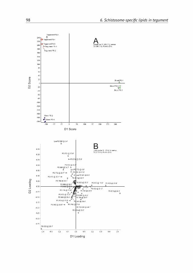
98 6. Schistosome-specific lipids in tegument

Supplementary data 99
Table S1: Molecular species of phosphatidylethanolamine (PE).Molecular species were determinedin homogenates of adult S. mansoniworms, in isolated tegumental membranes from adult S. mansoniworms and in hamster blood cells. Species are quantified by MRM with [M+H]+ for MS1 and [M+H-141]+ for MS2. Percentages shown are the average percentages of the total PE species ± standarddeviation. Species are shown if these were among the 10 most abundant species in one of the threeexamined membrane preparations. The three most abundant species in each fraction are printed inbold and species marked by an * comprise less than 1% of the total PE of that membrane preparation.
Whole worm Tegument Hamster blood cells(n=5) (n=3) (n=5)
m/z Acyls % ± S.D. % ± S.D. % ± S.D.
718 16:0 / 18:1 2.8 ± 0.3 5.3 ± 0.5 7.5 ± 0.5740 16:0 / 20:4 1.2 ± 0.2 1.5 ± 0.2 6.4 ± 0.2742 18:1 / 18:2 2.1 ± 0.3 2.5 ± 0.2 7.4 ± 0.4744 18:1 / 18:1 7.1 ± 0.6 8.4 ± 0.3 6.6 ± 0.2746 18:0 / 18:1 8.2 ± 0.3 9.5 ± 1.0 3.9 ± 0.2764 18:2 / 20:4 1.2 ± 0.1 1.1 ± 0.2 6.4 ± 0.1766 18:1 / 20:4 2.8 ± 0.1 2.9 ± 0.2 10.1 ± 0.1768 16:0 / 22:4 4.6 ± 0.2 6.1 ± 0.8 3.3 ± 0.1768 18:0 / 20:4 4.1 ± 0.2 2.3 ± 0.3 4.8 ± 0.1770 18:2 / 20:1 5.5 ± 0.1 6.4 ± 0.4 1.8 ± 0.1772 18:1 / 20:1 3.9 ± 0.1 5.5 ± 0.4 *788 20:4 / 20:4? * * 5.1 ± 0.3790 18:1 / 22:6? 2.7 ± 0.1 2.0 ± 0.0 6.6 ± 0.2792 18:1 / 22:4 4.8 ± 0.4 3.1 ± 0.5 4.0 ± 0.1794 18:0 / 22:5 6.3 ± 0.5 5.5 ± 0.8 3.8 ± 0.3796 18:0 / 22:4 17.9 ± 1.0 15.0 ± 2.1 1.2 ± 0.1798 18:0 / 20:3 4.3 ± 0.4 4.7 ± 0.3 *818 20:1 / 22:6 4.5 ± 0.5 3.2 ± 0.2 *

100 6. Schistosome-specific lipids in tegument
Table S2: Molecular species of phosphatidylcholine (PC). Molecular species were determined inhomogenates of adult S. mansoni worms, in isolated tegumental membranes from adult S. mansoniworms and in hamster blood cells. Species are quantified by MRM with [M+H]+ for MS1 and m/z184 for MS2. Percentages shown are the average percentages of the total PC species ± standarddeviation. Species are shown if these were among the 10 most abundant species in one of the threeexamined membrane preparations. The three most abundant species in each fraction are printedin bold and species that are enriched at least twofold in tegument compared to lipids in wholeworms are underlined in red. Species marked by an * comprise less than 1.0% of the total PC in thatsample. Of unidentified species the attached acyl-chains are given in total number of carbon atomsand unsaturations. Species containing an ether-linkage are indicated by the addition alk.
Whole worm Tegument Hamster blood cells(n=5) (n=3) (n=5)
m/z Acyls % ± S.D. % ± S.D. % ± S.D.
720 16:0 / 16:0 alk 1.5 ± 0.1 5.8 ± 1.3 *734 16:0 / 16:0 11.5 ± 0.7 23.1 ± 2.2 6.3 ± 0.2746 34:1 alk 1.8 ± 0.1 2.2 ± 0.3 *758 16:0 / 18:2 4.8 ± 0.4 5.1 ± 0.2 19.8 ± 1.1760 16:0 / 18:1 Δ9 11.7 ± 0.6 8.2 ± 0.2 14.4 ± 0.69760 16:0 / 18:1 Δ5 2.2 ± 0.1 12.4 ± 0.3 *762 16:0 / 18:0 4.6 ± 0.2 3.9 ± 0.1 2.5 ± 0.1782 16:0 / 20:4 3.5 ± 0.4 2.2 ± 0.7 6.6 ± 0.3784 18:1 / 18:2 2.4 ± 0.1 2.3 ± 0.1 3.8 ± 0.2786 18:0 / 18:2 10.1 ± 0.3 7.0 ± 0.5 11.9 ± 0.6788 16:0 / 20:1 13.8 ± 0.4 5.7 ± 0.5 *788 18:0 / 18:1 * * 3.7 ± 0.3806 16:0 / 22:6 2.0 ± 0.2 1.4 ± 0.3 6.9 ± 0.6808 18:1 / 20:4 1.5 ± 0.1 * 3.0 ± 0.2810 18:0 / 20:4 2.4 ± 0.2 1.3 ± 0.2 4.1 ± 0.3

Supplementary data 101
Table S3: Molecular species of diacylphosphatidylinositol (PI). Molecular species were determinedin homogenates of adult S. mansoniworms, in isolated tegumental membranes from adult S. mansoniworms and in hamster blood cells. Species are quantified by MRM with [M-H]- for MS1 and m/z241 for MS2. Percentages shown are the average percentages of the total PI species ± standarddeviation. Species are shown if these were among the 10 most abundant species in one of the threeexamined membrane preparations. The three most abundant species in each sample are shown inbold and species that are enriched at least twofold in tegument compared to lipids in whole wormsare underlined in red. Species marked by an * comprise less than 1.0% of the total PI. Of unidentifiedspecies the attached acyl-chains are given in total number of carbon atoms and unsaturations.
Whole worm Tegument Hamster blood cells(n=5) (n=3) (n=5)
m/z Acyls % ± S.D. % ± S.D. % ± S.D.
833 16:0 / 18:2 4.0 ± 1.6 2.2 ± 0.2 7.1 ± 0.7835 16:0 / 18:1 3.1 ± 0.6 3.7 ± 0.6 2.4 ± 0.4837 16:0 / 18:0 2.3 ± 0.6 6.3 ± 0.9 *857 16:0 / 20:4 1.1 ± 0.1 * 6.6 ± 0.6859 18:1 / 18:2 1.3 ± 0.0 1.2 ± 0.2 3.4 ± 0.2861 18:0 / 18:2 15.7 ± 1.3 13.2 ± 1.3 13.0 ± 0.7863 18:0 / 18:1 21.0 ± 2.5 27.3 ± 1.9 2.3 ± 0.1865 18:0 / 18:0 2.3 ± 0.3 2.7 ± 0.1 *883 18:1 / 20:4 * * 5.2 ± 0.1885 18:0 / 20:4 25.0 ± 1.9 9.9 ± 0.8 36.8 ± 2.1887 18:0 / 20:3 3.4 ± 0.4 3.9 ± 0.6 5.7 ± 0.3889 38:2 2.2 ± 0.2 4.3 ± 0.6 1.2 ± 0.2891 18:0 / 20:1 1.4 ± 0.1 3.3 ± 0.5 *911 40:5 * * 2.8 ± 0.3945 42:2 * 2.3 ± 0.2 *


Chapter 7Summarizing discussion
103
104 7. Summarizing discussion
7.1 Development of parasitism
Parasitism is a relationship between two organisms in which one of them (theparasite) lives in or on the other (the host) and lives at the expense of that host.Life as a parasite offers advantages that surpass its inconveniences. The mainadvantage is the excess of nutrients. In addition, parasitism offers the possibilityto lose genes encoding proteins that are essential under free-living conditions,but of which the function is taken over by the host. A parasite also benefits fromthe homeostasis of the host, giving the parasite a very constant environment tolive in. However, living in or on a host organism also means living in constantthreat of the host immune system. Another disadvantage is the more challengingway of reproduction, which is reflected in the numerous complex life cycles ofparasites.Parasitism has evolved independently many times during the history of life.
Almost all free-living organisms are host to various parasitic taxa [139]. There areparasites in nearly all ecosystems and there is substantial evidence that parasitesshape host population dynamics, alter interspecific competition, influence energyflow and appear to be important drivers of biodiversity [86].
A parasite’s way of living inevitably causes damage to its host, if not physical,than by taking up nutrients that would otherwise be for the host itself. Parasitesgenerally die when their host does. Hence, parasites have evolved in such a waythat the damage they cause will not kill their host, at least not before the parasiteshave reproduced. The harm caused by schistosomes is usually mild. They cancause anemia because they feed on blood, but also as a result of blood loss throughthe wounds made by the eggs when they pass the endothelium and gut wall.Schistosome infections can also lead to malnutrition. The eggs that do not exitthe body, elicit an immune response that initially is not very harmful to the hostbut can be detrimental when it is at a critical location, e.g. the central nervoussystem (CNS), or after long lasting accumulation causing portal hypertension andwhich may lead to liver failure. Also, prolonged inflammation may contribute tothe development of malignancies.Parasites adapted to nearly every kind of tissue of the (human) body exist.
Each parasite has evolved for optimal fitness within its niche. Schistosomes areblood-dwelling parasites. The worms have to overcome many challenges in orderto get into the circulation, maintain themselves, reproduce and to excrete the eggsfor survival of their species. They have developed a large armoury of interventions

7.2. Parasitic helminths 105
with their host to protect themselves and sometimes even to turn potential harmfulhost reactions in highly useful assistance for example in excretion of the eggs.
7.2 Parasitic helminths
Infection and migration
Penetration of the skin is a fairly common mode of entrance for parasitic worms.This can be achieved either by a vector injecting worm larvae or by skin penetrationof the larval parasites themselves. Filaria worms are injected through the skinby flies or mosquitoes. Hookworm and Strongyloides larvae penetrate the skin bythemselves, as do schistosome cercariae.Tissue migration is a common feature in the life cycles of parasitic worms.
Similar to schistosomula, both hookworm and Strongyloides larvae migrate to thelungs. However, while schistosomula further migrate via the liver to the portalvein to mate and mature, hookworm and Strongyloides larvae enter the alveoli,are coughed up and subsequently swallowed to reach their final location, which isthe intestinal lumen where they mature and reproduce. And even Ascaris larvae,which enter the human host when eggs are ingested, need to penetrate the mucosaof the gut and migrate through the body in order to fully mature before homingin the intestinal lumen – where they have been before as eggs. Thus, althoughtissue invasion and migration appears as a redundantly complicated step in thelife cycle of parasitic worms, it is so widespread that it seems to provide essentialdevelopmental needs.Tissue migration is referred to as larva migrans when it causes disease symp-
toms. As can be deduced from the life cycles of the helminths that cause larvamigrans, symptoms are temporally and disappear once the adult worms havereached their niche. Only upon recurrent reinfection or in the case of Strongy-loides, where auto-infection can occur, larva migrans can last for longer periods.Tissue migration of schistosomes doesn’t usually cause symptoms, even though itelicits a remarkable immune response.Although very common, not all parasitic worms migrate through tissues. En-
terobius and Trichuris are both ingested as eggs and hatch and develop to adultworms within the gut. And also for the flatworms Opisthorchis viverrini, Clonorchissinensis and Dicrocoelium dendriticum the only organ system they encounter fromtheir host, is the intestinal tract and the bile duct.

106 7. Summarizing discussion
On the other hand, not all ingested parasitic worms home in the intestinaltract. Some are ingested as eggs or as larvae andmigrate to other tissues to matureand reproduce. Dracunculus are ingested with their intermediate hosts, copepods,in drinking water. Larvae, released from the copepods which die in the stomach,penetrate the host stomach and intestinal wall to mature and copulate in theabdominal cavity and retroperitoneal space. The female worms then migrateto the subcutaneous tissues towards the skin surface where larvae are releasedthrough a skin blister. Also Trichinella and Echinococcus are parasitic worms thatinfect humans through ingestion. They then migrate throughout the body wherecysts are formed, which only infect the next host through carnivorism, wheningested with the current host as a meal for the new host.
Exit of progeny
In order to replicate, the progeny of parasitic worms needs a way to exit the hostand infect another host. The majority of human parasitic worms spreads througheggs or larval stages that leave their host. For intestinal parasites, the route of exitis obvious. No complex mechanisms need to be adopted as eggs or larvae, whichdo not attach to the gut wall, will automatically be excreted following peristalticbowel movements and defecation. Schistosoma mansoni also uses this mechanism,but first has to overcome some barriers to get their progeny into the gut. Asoviposition occurs in the veins, eggs have to pass the endothelial wall and thegut wall. As for intestinal worms, the exit of eggs is with the feces. Miracidia,schistosome larvae, hatch from the eggs immediately when they come into freshwater. Time to infect the intermediate host, an aquatic snail, is then limited. Someparasitic worms, such as Fasciola, Ascaris and Echinococcus have very resistanteggs or cysts which can stay viable in the environment for a long time. Hatchingoccurs when favourable conditions are met, followed by infection of the next(intermediate) host.
Free-living stages of parasitic worms are usually non-feeding and thus areeither encysted such as the metacercariae of Fasciola spp., or have a restrictedtime frame to infect the next host. The latter is the case for schistosomes, whereboth the miracidia infecting the snail and the cercariae infecting the mammalianhost have a glycogen reserve for about 12–24 hours [89, 182, 202]. Hookworm andStrongyloides have free-living stages that feed on soil bacteria (rhabditiform larvae).When transformed into non-feeding, infective filariform larvae they encounter the

7.2. Parasitic helminths 107
same time challenge of finding a suitable host as do schistosomes. Strongyloideseggs hatch within the uterus of the female worm. As transformation into infectiousfilariform larvae takes less then two days, this can occur within the host, beforethe larvae are excreted. The larvae can reinfect the host, which is referred to asauto-infection. In severely immunocompromized hosts, aberrant auto-infectionmay lead to hyperinfection syndrome. Strongyloides also have a free-living cycle,where rhabditiform larvae mature into reproductive male and female worms.
As mentioned above, intestinal parasites have an easy exit. Tissue-dwellingparasitic worms on the other hand need to be more inventive to exit and spread.Carnivorism is a well proven concept for life cycle continuation, used for exampleby some tapeworms (Taenia solium, Echinococcus) and Trichinella. Filaria larvae,microfilaria, circulate within the bloodstream and are aspirated and further spreadto their new host by the fly or mosquito vector upon feeding. Microfilariae fromtissue-dwelling filariae generally hatch in the uterus of female worms, while forblood-dwelling species microfilariae remain sheathed in the envelope of the eggand will exsheath within the intermediate host. Dracunculus worms do not layeggs, but produce live larvae. These leave the host through a sore blister in theskin, usually the foot, which is immersed in water to ease the pain.Toxocara is an intestinal parasitic worm in dogs and cats. Similar to many
intestinal parasites, eggs leave with the feces. Worms canmature and reproduce inyoung dogs. However, in infected adult dogs, Toxocara development is hampered.But the infection can be transferred transplacental, which means that an infectedpregnant dog can infect her unborn pups in utero. In addition, an infected bitchcan infect her newborns though milk. Toxocara development is then resumed inyoung dogs.
Feeding habits
Most parasitic worms that infect humans feed on tissue, blood or other body fluids,even when residing in the intestinal tract. In that respect, schistosome wormshave chosen a very pleasant location where food literally drifts by. All they needto do in order to get fed, is sit and open their mouth or absorb through theirtegument. And that is exactly what they do. Female schistosome worms feedlargely on circulating red blood cells. Males mainly absorb nutrients through theirtegument. Tissue-dwelling worms generally feed on the tissue and body fluids theylive in. Some of them eventually encyst and go into dormant stages and spread

108 7. Summarizing discussion
through carnivorism. Hookworms and Strongyloides, both intestinal worms, hookthemselves to the mucosa to feed on blood and epithelial cells. Where hookwormsjust hook themselves in the mucosa, Trichuris worms dig in half their body, whilethe other half of their body hangs freely in the gut lumen.
Some intestinal worms do not feed on human tissue but feed on the predigestedintestinal liquid content. Among them are Ascaris, Toxocara and tapeworms. Thelatter have no mouth and absorb all the nutrients through their tegument, whichis covered by microvilli to increase surface area.
Whichever locations parasitic worms chose, they generally have plenty of foodsurrounding them as they typically feed on the host. This allows them to use aninefficient energy metabolism in which substrates are not fully oxidized.
Host defence
The components of the immune system of the host are a continuous threat tothe parasites living in the host. In that respect, the choice for the circulation asa habitat of schistosomes is remarkable, as this is the body compartment whereall immune components are constantly present: immune cells, including T-cells,eosinophils andmacrophages and other immune components such as complement,cytokines and chemokines. The presence of the immune effectors is much lowerin the tissues and even lower in the gut. However, as parasitic worms are largecompared to the immune cells, there is not much these immune cells can do tothem even with help of other components of the immune system. Besides, manytissue-dwelling parasitic worms have developed effective immune evading strate-gies. Sometimes the immune activation results in encapsulation of the parasite(larvae), as is the case with larvae of species that infect through carnivorism suchas the cyst stage of Echinococcus and Taenia solium and with schistosome eggs thatare trapped in the body.
7.3 On schistosome eggs and worms
Eggs andworms of schistosomes have opposite interests, namely get out of the hostversus stay in the host. Thus egg-host interactions and worm-host interactionshave to be different.
Schistosome worms have evolved to produce a large progeny in order to maxi-mize the probability of species survival. The adult worms do not look after their

7.3. On schistosome eggs and worms 109
offspring once the eggs are laid. Eggs have to get out of the host on their own,even though they are incapable of moving by themselves and the way out is adifficult one. Eggs have to penetrate tissues in order to get to the gut lumen (urinebladder in the case of S. haematobium) where they are subsequently released inthe environment together with the feces (or urine).
Egg-host interactions
An egg is defined as an animal reproductive body consisting of an ovum/femalegamete together with its nutrient material and a protective covering and, whenfertilized, having the capacity to develop into a new individual capable of inde-pendent existence.
Schistosome eggs are build of a fertilized oocyte surrounded by vitelline cellscontaining nutrients andmaterial for the eggshell [175]. By the time the egg leavesthe mammalian host, the fertilized oocyte has developed into a fully maturedmiracidium covered by a tanned protein eggshell. Once the egg is released inthe water, osmotic changes result in swelling of the internal material therebyincreasing the internal pressure on the eggshell. This leads to hatching of themiracidium from the egg.
Formation of the eggshell
A major function of the eggshell is to restrict the permeability of the egg andmaintain a certain environment within the egg for embryonic development andsurvival [196]. Eggshells of trematodes are made of proteins which are cross-linked by tyrosinase activity forming quinone bonds. This eggshell of tannedproteins has underlying layers and envelopes. The subshell cellular layer ofschistosome eggs is named von Lichtenberg’s envelope. A non-cellular layer,Reynolds’ layer, is situated between von Lichtenberg’s envelope and the eggshell[6, 123]. Schistosome eggshell formation starts in the ootype within the female,where ootype contraction leads to release of eggshell precursor proteins fromthe vitelline droplets. These form the eggshell upon quinone tanning. Specificeggshell precursor proteins have first been described in the late 80s and early 90s[13, 14, 33, 93, 94, 150, 169]. As the eggshell is a direct site of interaction betweenthe egg and the host, its composition is of major importance in host-parasiteinteraction. Results of mass spectrometry, immunohistochemical and amino acidanalysis of the protein composition of the eggshell are described in chapter 2.

110 7. Summarizing discussion
The experiments revealed that the major component of the eggshell is the putativeeggshell protein p14. This protein has characteristic short repeat sequences ofwhich nearly half of the amino acid residues are glycines. High glycine contentis a general feature of trematode eggshell proteins and has also been describedfor Fasciola hepatica, Opisthorchis viverrini and Paragonimus westermani. Massspectrometry analysis did not reveal the presence of p14 in eggshell, probably dueto its high tyrosine content transformed in quinone bonds and due to high lysinelevels leading to very short peptides after trypsin digestion. However, the presenceof p14 was shown by immunoblot. In addition, amino acid analysis showed aglycine content of 36% in total hydrolyzed eggshell. This high glycine content isreadily explained by a high contribution of p14 to total eggshell.A wide range of schistosome proteins was detected by mass spectrometry in
purified eggshell samples. This indicates that a variety of non-eggshell specificproteins is also incorporated in the eggshell. The majority of these proteins aregenerally abundant cellular proteins such as the major egg antigen p40, HSP70and many glycolytic enzymes. These proteins are suggested to originate fromsurrounding vitelline cells and happened to be around at the site and time ofeggshell synthesis. Hence, these non-eggshell specific proteins were coincidentlycross-linked to themajor eggshell proteins. The absence of themajor egg secretionproteins IPSE/alpha-1 and omega-1 indicates that cross-linking is finished beforethe egg has fullymatured and starts secreting. The absense of host plasma proteinsin eggshell indicates that eggshell formation is finished even before oviposition.
Eggshell contribution to the journey of the S. mansoni egg
Most eggs of parasitic helminths are deposited in the gut. Their way out is straight-forward and does not need exploited assistance of the host. In contrast, schisto-some eggs exit the female body in the mesenteric capillary venules, close to thebowel. It takes at least a few days for a schistosome egg to exit the host, but it maytake up to several weeks. The eggs need to get into the gut in order to get out of thehost. However, blood flowing through the venules can easily carry the egg along,away from the bowel and heading towards the liver. To prevent this, eggs have toquickly adhere to the vessel wall. Previous experiments reported vast adhesionof platelets to eggshell [203]. In addition, the experiments in chapter 4 showedthat VWF also adheres to eggshell directly. Unfolded VWF can connect plateletsto clotting factors and injured surfaces of the endothelium through many bindingdomains [105, 159] and plays an important role in platelet-vessel wall adhesion by

7.3. On schistosome eggs and worms 111
binding both platelets and the extracellular matrix of the vessel wall containingcollagen and fibrin. Furthermore, VWF plays a role in platelet cross-linking andplatelet plug expansion [105]. By bridging between the eggshell and the extracellu-lar matrix, VWFmay attach the eggshell to the endothelium. In addition, VWF cancontribute to eggshell adhesion to endothelium indirectly as VWF in combinationwith platelets can induce platelet adhesion, platelet activation and secondaryhemostasis, allowing the formation of a stable clot. This is in accordance with thefindings of Ngaiza and Doenhoff (1990) [125] who previously demonstrated thatplatelets play a role in egg extravasation.
Binding of fibrinogen and fibronectin to eggshell was also demonstrated. Wherefibrin, the cleaved product of fibrinogen, is the main component of a blood clot,fibronectin plays an essential role in cell adhesion and wound healing. Thus, bothcan play an important role in egg adhesion to the endothelium. Enolase waspreviously identified in eggshell. Its presence there seems accidental. Enolase hasbeen described to bind host fibronectin in bacterial pathogens [57]. This could alsobe the case in S. mansoni eggshell, where enolase-binding fibronectin can furtherenhance endothelium binding of eggshell. Chapter 4 reports that fibronectin wasone of the main plasma proteins that bound to eggshell and the major one inthe high molecular weight range. No tests were performed to determine whetherenolase was the major fibronectin-binding eggshell protein.
Next to fibronectin, enolase can bind plasminogen [8, 43, 141, 206]. Plasmino-gen is the precursor of plasmin, a serine protease that cleaves fibrin and VWF.Small amounts of plasminogen may facilitate fibrinolysis of the clot surroundingthe eggs after extravasation. However, in the mass spectrometry experiments plas-minogen was not elevated in plasma proteins that bound to eggshell compared tofull plasma. This does not exclude that plasminogen is bound to eggshell. In fact,small amounts of plasmin are probably needed to prevent the excessive expansionof the clot leading to occlusion of the vessel.
Platelet activation and blood coagulation around schistosome eggs can furtherbe enhanced by shear stress and turbulence within the vessel [177]. Both can becaused by mechanical obstruction by worms and eggs. By their presence and as aresult of their adherence to the vessel wall, schistosome worms also cause damageto the endothelium, which in itself is another strong activator of platelets andblood coagulation and of endothelial cells [168]. Eggs of both S. mansoni and S.haematobium can induce endothelial cell proliferation in vitro [55, 67]. In vitro,endothelial cells overgrow schistosome eggs within a few hours after oviposition
112 7. Summarizing discussion
[64]. Thus, eggs can passively cross the endothelium by the non-specific responseof endothelial cells. File (1995) [64] also showed that eggs deposited directly byadult worms elicited a more rapid and complete response than embryonated eggsisolated from the liver tissues.In short, schistosome eggs can probably passively pass the endothelium af-
ter binding it. Binding of the eggshell is facilitated by the composition of theeggshell (chapter 2 and 3) which binds platelets and plasma proteins involved incoagulation (chapter 4).The next step is to cross the tissue of the gut wall and get excreted with the
feces. Themechanism of this process are largely unknown, but it is likely to dependon the host immune system (see below).
Eggshell interaction with the immune system
Most eggs of parasitic helminths are deposited in the gut and have little interactionwith the immune system. In contrast, schistosome eggs are in constant interactionwith the host from themoment of oviposition until they have reached the intestinallumen. Half of the eggs does not succeed to exit the blood vessel. They are draggedalong with the blood stream and get stuck in small vessels. Eggs elicit a strongimmune response and those stuck in the host tissues hence induce granulomaformation.While schistosome adult worms must prevent an effective immune response,
eggs need the immune system in order to be excreted [49]. The schistosomeeggshell and egg secretions contain many immunogenic proteins and glycans [30,40, 110, 149] (chapter 2). These immunogenic components of eggshell and eggsecretionsmay be ofmajor interest in the scope of immunemediated egg excretion.Eggshells and egg secretions induce a Th2 response via IL-4 activatedmacrophagesand CD4+ T-lymphocytes. Fecal excretion of eggs is immune mediated and eggexcretion rate and CD4+ T-cell percentage are positively correlated [49, 90]. Bothmajor egg secretion products IPSE/alpha-1 and Omega-1 alone are capable of Th2skewing [59, 163, 164, 178], but secretion of these glycoproteins only starts a fewdays after oviposition when the egg has matured. Because maturation takes abouta week, T-cells are likely to play a role during penetration through the gut wall,when the eggs have already adhered to and passed through the endothelium.
The schistosome eggshell contains more than specific eggshell proteins alone.The observed incorporation of these other proteins as reported in chapter 2, prob-ably originate from neighbouring vitelline cells. This may appear as an unspecific

7.3. On schistosome eggs and worms 113
feature of eggshell production. But many of the proteins identified to be part ofthe eggshell are known schistosome antigens, such as p40, phosphoenolpyruvatecarboxykinase (PEPCK) and thioredoxin peroxidase. These proteins induce cellularimmune responses [3, 200] or antibody responses [122]. They may hence primethe immune system and contribute to the penetration of the eggs through hosttissues.However useful the immune response may be for extravasation, immune ac-
tivation has a hindside. The main cause of pathology in schistosomiasis are thenon-excreted eggswhich are trapped in the host tissue, generally the liver. Trappedeggs are enveloped in granulomas which are composed of eosinophils, CD4+ T-lymphocytes, macrophages and collagen fibres. It is the Th2 response generatedby the host against antigens secreted by the parasite eggs that is responsible forthe granuloma formation [137]. In absence of the Th2 response, granulomas arenot formed and continuous production of egg secretion products leads to severeimmunopathology and hepatotoxic liver damage. In experimentally infected mice,this rapidly leads to death of themice. Granulomas isolate the eggs and prevent thefurther spread of egg secretions. As the eggs die, the granulomas resolve, leavingfibrotic plaques. Accumulation of eggs and hence granulomas can eventually leadto permanent fibrotic changes in the liver, liver damage and portal hypertension.
In addition to the findings that eggshell contains a range of proteins known tobe immunogenic, chapter 3 shows that immune sera contain antibodies whichreact with the eggshell. The formed antigen-antibody complex can induce chronicinflammatory responseswhere the interaction betweenCD4+T-cells, macrophagesand cytokines can cause granulomas to form [201].Next to proteins, eggshell contains glycans. Schistosome glycans are the ma-
jor inducers of the immune response [188] and the synthetic glycans GalNAcβ-4GlcNAc (LacdiNAc, LDN) and Galβ1-4GlcNAc (LacNAc, LN) alone can inducegranulomas [185].
Both eggshells and egg secretions induce antibody responses, as do adult worms.There is some overlap between worm and egg antigens, but there are enoughdifferences to employ them usefully for diagnostics. That is described in chapter5, where the differences in antibody responses to eggs and worms are used todifferentiate betweenneuroschistosomiasis and other causes of transversemyelitis.It is essential to be certain of the diagnosis neuroschistosomiasis, before the startof therapy that includes immunosuppressive corticosteroids. These are contra-indicated in case of viral or bacterial causes of transverse myelitis. The key feature
114 7. Summarizing discussion
of the proposed test is to discriminate between antibodies in the cerebrospinalfluid (CSF) produced in situ and leakage of antibodies from blood plasma into theCSF due to damage to the blood-brain-barrier. The former indicates the presenceof schistosome antigens in the CNS, while the latter points to another cause ofthe transverse myelitis. As neuroschistosomiasis is a result of nerve compressiondue to inflammation around schistosome eggs deposited near the nerves, thehypothesis is that anti-egg IgG, when normalized to anti-worm IgG, would beenriched in the CSF compared to the blood. Two schistosome-positive patientswith transverse myelitis with an infectious cause for which no other pathogen wasidentified, had a positive test result, i.e. had a ratio >2. The egg-worm antibodyindex appears to be a valuable tool in the confirmation of neuroschistosomiasis.Further validation of this ratio is required in a larger group of patients, bothtravelers and patients in schistosome-endemic areas. It is important to distinguishthese two groups of patients, as in endemic regions antibody levels may drop belowcut-off values in chronic or recurrent infections.
Worm-host interactions
Many parasitic worms travel through host tissues at some point in their life cycle.They are then in constant interaction with the host immune system and have toprevent a damaging immune response. The outer surface of the worms is a majorsite of host-parasite interaction. It protects the worms and has other functionsinvolved in immune invasion, excretion of proteins and uptake of nutrients.The surface of blood-dwelling trematodes is the tegument. It consists of two
lipid bilayers that overlay the syncytium. It forms a large surface made of lipids,glycans and proteins which can be detected by the immune system. In addition,(immunomodulatory) schistosome proteins are secreted through the tegument.
The protein composition of the tegument has been studied extensively [15–17, 184]. These tegumental proteins consist of membrane proteins, cytoskeletonproteins, proteins and secreted proteins. There is also a number of proteins uniqueto schistosomes that were specifically detected in the tegument [184].Lipids form a major component in tegument, which contains high amounts
of cholesterol, sphingomyelin and saturated (ether-linked) phospholipid species[2, 151]. The most abundant phospholipid classes in membranes of schistosomesare phosphatidylcholine (PC) and phosphatidylethanolamine (PE). The diacyl-phospholipid species composition has been characterized for these phospholipid

7.3. On schistosome eggs and worms 115
classes and this revealed that the species composition of these phospholipids istegument-specific and differs from that of whole worm [21]. In chapter 6 an analy-sis of the species composition of all major phospholipid classes is given, includingphosphatidylinositol (PI), phosphatidylserine (PS) and lysophosphopolipids inisolated tegumental membranes. For most phospholipid classes, the species com-position is substantally different in the tegument compared to whole worms. Thisis true for the PC and PS species. In contrast, diacyl-phospholipid species compo-sition is strikingly similar for PI species. Furthermore, the tegument membraneswere specifically enriched in lysophospholipids, especially lysophosphatidylserine(lyso-PS) and lysophosphatidylethanolamine (lyso-PE). In addition, the tegumentwas enriched in eicosaenoic acid (20:1) containing lysophospholipids in the phos-pholipid classes PS and PE, but not in PC and PI.Lysophospholipids are minor compounds in membranes, but they play an
important role in signal transduction. They also play a role in host-parasite inter-actions. It has been described that schistosome lysophosphatidylcholine (lyso-PC)can lead to lysis of red blood cells and cause immobilization of membrane compo-nents of red blood cells [71]. Lyso-PS species have been shown to activate TLR2on host immune cells and induce the development of IL-10 producing regulatoryT- cells [187]. Hence schistosome lyso-PS from the tegument may down regulatethe immune response. Althought tegument appeared to be specifically enrichedin lyso-PS, no lyso-PS could be detected as a secreted phospholipid in incubationmedium or in host plasma (chapter 6). Circulating amounts are thus below detec-tion level. This may be because lysophospholipids do not bind to albumin. Due totheir amphipathic nature, lysophospholipids readily incorporate into membranes.This is especially true for schistosome lysophospholipids with long chain fattyacids that are relatively hydrophobic. In vitro, no acceptor membrane is availablefor excreted lysophospholipids. Hence, lysophospholipids possibly reincorporateinto the tegumental membranes as the only available phospholipid membranes.In vivo, the acceptor membranes could be those of endothelial cells or those fromcirculating cells. While no free circulating (tegument specific) lyso-PS could be de-tected in plasma, it may still be excreted and immediately come in contact with theeffector cells of the immune system and activate them to induce a regulatory im-mune response. Furthermore, incorporation of schistosome (lyso)phospholipidsinto membranes of host immune effector cells or endothelial cells may alter thebiophysical properties of the membranes because of the structure of these unusuallong and unsaturated fatty acids.

116 7. Summarizing discussion
Phospholipid analysis in blood plasma revealed an interesting difference be-tween phospholipid composition in blood plasma of infected hamsters and that ofnon-infected hamsters. Blood plasma of hamsters infected with schistosomes hadsubstantially lower PI content. These host phospholipids have precursor functionsin lipid signalling, cell signalling and membrane trafficking. Reduction of PI byschistosomes seems peculiar and there are no mechanisms known to contributeto this.
Unlike fatty acids, phospholipids do not bind albumin. PI in blood plasma arelikely to constitute exosomes or microvesicles. Exosomes are released by activatedcells of the immune system and are involved in intercellular communication tomediate the activation of the immune response [179]. Reduction in PImay indicatethat schistosomes alter the release of exosomes, which may affect the immuneresponse.
7.4 Concluding remarks/future directives
Both schistosome worms and schistosome eggs are in constant interaction withtheir host. Worms and eggs have defined interactive surfaces which are highly un-usual and specific in nature and composition. Further analysis of these structurescould provide valuable information about the biology of schistosomes. Becausethese structures are so specific, they could serve as targets for vaccine and anti-schistosomal drug development.

References
1. Abath FG and Werkhauser RC. The tegument of Schistosoma mansoni: functional and immuno-logical features. Parasite Immunol, 1996. 18:15–20.
2. Allan D, Payares G and Evans WH. The phospholipid and fatty acid composition of Schistosomamansoni and of its purified tegumental membranes. Mol Biochem Parasitol, 1987. 23:123–128.
3. Asahi H, Hernandez HJ and Stadecker MJ. A novel 62-kilodalton egg antigen from Schistosomamansoni induces a potent CD4(+) T helper cell response in the C57BL/6 mouse. Infect Immun,1999. 67:1729–1735.
4. Asahi H, Osman A, Cook RM, LoVerde PT and Stadecker MJ. Schistosoma mansoni phospho-enolpyruvate carboxykinase, a novel egg antigen: immunological properties of the recombinantprotein and identification of a T-cell epitope. Infect Immun, 2000. 68:3385–3393.
5. Asch HL and Read CP. Transtegumental absorption of amino acids by male Schistosoma mansoni.J Parasitol, 1975. 61:378–379.
6. Ashton PD, Harrop R, Shah B and Wilson RA. The schistosome egg: development and secretions.Parasitology, 2001. 122:329–338.
7. Barreto ML, Silva JT, Mott KE and Lehman JS. Stability of faecal egg excretion in Schistosomamansoni infection. Trans R Soc Trop Med Hyg, 1978. 72:181–187.
8. Bernal D, de la Rubia JE, Carrasco-Abad AM, Toledo R, Mas-Coma S and Marcilla A. Identificationof enolase as a plasminogen-binding protein in excretory-secretory products of Fasciola hepatica.FEBS Lett, 2004. 563:203–206.
9. Berriman M, Haas BJ, LoVerde PT, Wilson RA, Dillon GP, Cerqueira GC, Mashiyama ST, Al-Lazikani B, Andrade LF, Ashton PD, Aslett MA, Bartholomeu DC, Blandin G, Caffrey CR, CoghlanA, Coulson R, Day TA, Delcher A, DeMarco R, Djikeng A, Eyre T, Gamble JA, Ghedin E, Gu Y,Hertz-Fowler C, Hirai H, Hirai Y, Houston R, Ivens A, Johnston DA, Lacerda D, Macedo CD,McVeigh P, Ning Z, Oliveira G, Overington JP, Parkhill J, Pertea M, Pierce RJ, Protasio AV, QuailMA, Rajandream MA, Rogers J, Sajid M, Salzberg SL, Stanke M, Tivey AR, White O, Williams DL,Wortman J, WuW, Zamanian M, Zerlotini A, Fraser-Liggett CM, Barrell BG and El-Sayed NM. Thegenome of the blood fluke Schistosoma mansoni. Nature, 2009. 460:352–358.
10. Bhowmick IP, Kumar N, Sharma S, Coppens I and Jarori GK. Plasmodium falciparum enolase:stage-specific expression and sub-cellular localization. Malar J, 2009. 8:179.
117

118 References
11. Bittner S. When quinones meet amino acids: chemical, physical and biological consequences.Amino Acids, 2006. 30:205–224.
12. Bligh EG and Dyer WJ. A rapid method of total lipid extraction and purification. Can J BiochemPhysiol, 1959. 37:911–917.
13. Bobek L, Rekosh DM, van Keulen H and LoVerde PT. Characterization of a female-specific cDNAderived from a developmentally regulated mRNA in the human blood fluke Schistosoma mansoni.Proc Natl Acad Sci U S A, 1986. 83:5544–5548.
14. Bobek LA, Rekosh DM and LoVerde PT. Small gene family encoding an eggshell (chorion) proteinof the human parasite Schistosoma mansoni. Mol Cell Biol, 1988. 8:3008–3016.
15. Braschi S, Borges WC and Wilson RA. Proteomic analysis of the schistosome tegument and itssurface membranes. Mem Inst Oswaldo Cruz, 2006. 101 Suppl 1:205–212.
16. Braschi S, Curwen RS, Ashton PD, Verjovski-Almeida S andWilson A. The tegument surface mem-branes of the human blood parasite Schistosoma mansoni: a proteomic analysis after differentialextraction. Proteomics, 2006. 6:1471–1482.
17. Braschi S and Wilson RA. Proteins exposed at the adult schistosome surface revealed by biotiny-lation. Mol Cell Proteomics, 2006. 5:347–356.
18. Brouwers JFHM, Gadella BM, van Golde LM and Tielens AGM. Quantitative analysis of phos-phatidylcholine molecular species using HPLC and light scattering detection. J Lipid Res, 1998.39:344–353.
19. Brouwers JFHM, Skelly PJ, Van Golde LM and Tielens AGM. Studies on phospholipid turnoverargue against sloughing of tegumental membranes in adult Schistosoma mansoni. Parasitology,1999. 119:287–294.
20. Brouwers JFHM, Smeenk IM, vanGolde LM and Tielens AGM. The incorporation, modification andturnover of fatty acids in adult Schistosoma mansoni. Mol Biochem Parasitol, 1997. 88:175–185.
21. Brouwers JFHM, van Hellemond JJ, van Golde LM and Tielens AGM. Ether lipids and their possiblephysiological function in adult Schistosoma mansoni. Mol Biochem Parasitol, 1998. 96:49–58.
22. Brouwers JFHM, Versluis C, van Golde LM and Tielens AGM. 5-octadecenoic acid: evidence for anovel type of fatty acid modification in schistosomes. Biochem J, 1998. 334:315–319.
23. Brügger B, Erben G, Sandhoff R, Wieland FT and Lehmann WD. Quantitative analysis of biolog-ical membrane lipids at the low picomole level by nano-electrospray ionization tandem massspectrometry. Proc Natl Acad Sci U S A, 1997. 94:2339–2344.
24. Byram JE and Senft AW. Structure of the schistosome eggshell: amino acid analysis and incorpo-ration of labelled amino acids. Am J Trop Med Hyg, 1979. 28:539–547.
25. Camacho M and Agnew A. Schistosoma: rate of glucose import is altered by acetylcholineinteraction with tegumental acetylcholine receptors and acetylcholinesterase. Exp Parasitol,1995. 81:584–591.
26. Carod-Artal FJ. Neurological complications of Schistosoma infection. Trans R Soc Trop Med Hyg,2008. 102:107–116.
27. Carod-Artal FJ. Neuroschistosomiasis. Expert Rev Anti Infect Ther, 2010. 8:1307–1318.

References 119
28. Carod-Artal FJ. Cerebral and spinal schistosomiasis. Curr Neurol Neurosci Rep, 2012. 12:666–674.
29. Carod-Artal FJ, Vargas AP, Horan TA, Marinho PB and Coelho Costa PH. Schistosoma mansonimyelopathy: clinical and pathologic findings. Neurology, 2004. 63:388–391.
30. Cass CL, Johnson JR, Califf LL, Xu T, Hernandez HJ, Stadecker MJ, Yates JR and Williams DL.Proteomic analysis of Schistosoma mansoni egg secretions. Mol Biochem Parasitol, 2007. 155:84–93.
31. Castro-Borges W, Simpson DM, Dowle A, Curwen RS, Thomas-Oates J, Beynon RJ and WilsonRA. Abundance of tegument surface proteins in the human blood fluke Schistosoma mansonidetermined by QconCAT proteomics. J Proteomics, 2011. 74:1519–1533.
32. Cheever AW, Macedonia JG, Mosimann JE and Cheever EA. Kinetics of egg production and eggexcretion by Schistosoma mansoni and S. japonicum in mice infected with a single pair of worms.Am J Trop Med Hyg, 1994. 50:281–295.
33. Chen LL, Rekosh DM and LoVerde PT. Schistosoma mansoni p48 eggshell protein gene: character-ization, developmentally regulated expression and comparison to the p14 eggshell protein gene.Mol Biochem Parasitol, 1992. 52:39–52.
34. Chiang CP and Caulfield JP. The binding of human low-density lipoproteins to the surface ofschistosomula of Schistosoma mansoni is inhibited by polyanions and reduces the binding ofanti-schistosomal antibodies. Am J Pathol, 1989. 134:1007–1018.
35. Clerinx J and Gompel AV. Schistosomiasis in travellers and migrants. Travel Med Infect Dis, 2011.9:6–24.
36. Cnops L, Soentjens P, Clerinx J and Esbroeck MV. A Schistosoma haematobium-specific real-timePCR for diagnosis of urogenital schistosomiasis in serum samples of international travelers andmigrants. PLoS Negl Trop Dis, 2013. 7:e2413.
37. ColleyDG, BustinduyAL, SecorWEandKingCH. Human schistosomiasis. Lancet, 2014. 383:2253–2264.
38. Cordingley JS, Taylor DW, Dunne DW and Butterworth AE. Clone banks of cDNA from the parasiteSchistosoma mansoni: isolation of clones containing a potentially immunodiagnostic antigengene. Gene, 1983. 26:25–39.
39. Cornford EM. Schistosoma mansoni, S. japonicum, and S. haematobium: permeability to acidicamino acids and effect of separated and unseparated adults. Exp Parasitol, 1985. 59:355–363.
40. Curwen RS, Ashton PD, Johnston DA and Wilson RA. The Schistosoma mansoni soluble proteome:a comparison across four life-cycle stages. Mol Biochem Parasitol, 2004. 138:57–66.
41. de Boer AR, Hokke CH, Deelder AM and Wuhrer M. General microarray technique for immobi-lization and screening of natural glycans. Anal Chem, 2007. 79:8107–8113.
42. de Jongste AH, Tilanus AM, Bax H, Willems MH, van der Feltz M and van Hellemond JJ. Newinsights in diagnosing Schistosomamyelopathy. J Infect, 2010. 60:244–247.
43. de la Torre-Escudero E, Manzano-Román R, Pérez-Sánchez R, Siles-Lucas M and Oleaga A.Cloning and characterization of a plasminogen-binding surface-associated enolase from Schisto-soma bovis. Vet Parasitol, 2010. 173:76–84.

120 References
44. de Vlas SJ and Gryseels B. Underestimation of Schistosoma mansoni prevalences. Parasitol Today,1992. 8:274–277.
45. de Vlas SJ, Gryseels B, Oortmarssen GJV, Polderman AM and Habbema JD. A model for variationsin single and repeated egg counts in Schistosomamansoni infections. Parasitology, 1992. 104:451–460.
46. Deelder AM, van Dam GJ, Kornelis D, Fillié YE and van Zeyl RJ. Schistosoma: analysis of mono-clonal antibodies reactive with the circulating antigens CAA and CCA. Parasitology, 1996. 112:21–35.
47. deWalick S, Bexkens ML, van Balkom BWM, Wu YP, Smit CH, Hokke CH, de Groot PG, Heck AJR,Tielens AGM and van Hellemond JJ. The proteome of the insoluble Schistosoma mansoni eggshellskeleton. Int J Parasitol, 2011. 41:523–532.
48. deWalick S, Tielens AGM and van Hellemond JJ. Schistosoma mansoni: the egg, biosynthesis ofthe shell and interaction with the host. Exp Parasitol, 2012. 132:7–13.
49. Doenhoff M, Musallam R, Bain J and McGregor A. Studies on the host-parasite relationship inSchistosoma mansoni-infected mice: the immunological dependence of parasite egg excretion.Immunology, 1978. 35:771–778.
50. Doenhoff MJ, Cioli D and Utzinger J. Praziquantel: mechanisms of action, resistance and newderivatives for schistosomiasis. Curr Opin Infect Dis, 2008. 21:659–667.
51. Doenhoff MJ, Stanley RG, Pryce D, Curtis RCH, Parry H, Griffiths K and Jackson CL. Identificationof a fibrinolytic enzyme in Schistosomamansoni eggs andmodulated blood fibrinogenmetabolismin S. mansoni-infected mice. Parasitology, 2003. 126:231–234.
52. Donnelly S, Stack CM, O’Neill SM, Sayed AA, Williams DL and Dalton JP. Helminth 2-Cysperoxiredoxin drives Th2 responses through a mechanism involving alternatively activatedmacrophages. FASEB J, 2008. 22:4022–4032.
53. Dunne DW and Cooke A. A worm’s eye view of the immune system: consequences for evolutionof human autoimmune disease. Nat Rev Immunol, 2005. 5:420–426.
54. Dunne DW, Hassounah O, Musallam R, Lucas S, Pepys MB, Baltz M and Doenhoff M. Mechanismsof Schistosoma mansoni egg excretion: parasitological observations in immunosuppressed micereconstituted with immune serum. Parasite Immunol, 1983. 5:47–60.
55. El-Awady MK, Gad YZ, Wen Y, Eassawi M, Effat L, Amr KS, Ismail S and Christ GJ. Schistosomahematobium soluble egg antigens induce proliferation of urothelial and endothelial cells. World JUrol, 2001. 19:263–266.
56. Erasmus DA. The subcellular localization of labelled tyrosine in the vitelline cells of Schistosomamansoni. Z Parasitenkd, 1975. 46:75–81.
57. EsgleasM, Li Y,HancockMA,Harel J, Dubreuil JD andGottschalkM. Isolation and characterizationof alpha-enolase, a novel fibronectin-binding protein from Streptococcus suis. Microbiology, 2008.154:2668–2679.
58. Esterre P, Raobelison A, Ramarokoto CE, Ravaoalimalala VE, Boisier P and Roux J. Serumconcentrations of sICAM-1, sE-, sP- and sL-selectins in patients with Schistosoma mansoniinfection and association with disease severity. Parasite Immunol, 1998. 20:369–376.

References 121
59. Everts B, Perona-Wright G, Smits HH, Hokke CH, van der Ham AJ, Fitzsimmons CM, DoenhoffMJ, van der Bosch J, Mohrs K, Haas H, Mohrs M, Yazdanbakhsh M and Schramm G. Omega-1,a glycoprotein secreted by Schistosoma mansoni eggs, drives Th2 responses. J Exp Med, 2009.206:1673–1680.
60. Ferrari TC, Correa-Oliveira R, Xavier MA, Gazzinelli G and Cunha AS. Estimation of the localsynthesis of immunoglobulin G (IgG) in the central nervous system of patients with spinal cordschistosomiasis by the IgG index. Trans R Soc Trop Med Hyg, 1999. 93:558–559.
61. Ferrari TC, Moreira PR, Oliveira RC, Ferrari ML, Gazzinelli G and Cunha AS. The value of anenzyme-linked immunosorbent assay for the diagnosis of schistosomiasis mansoni myeloradicu-lopathy. Trans R Soc Trop Med Hyg, 1995. 89:496–500.
62. Ferrari TCA andMoreira PRR. Neuroschistosomiasis: clinical symptoms and pathogenesis. LancetNeurol, 2011. 10:853–864.
63. Ferrari TCA, Moreira PRR and Cunha AS. Clinical characterization of neuroschistosomiasis dueto Schistosoma mansoni and its treatment. Acta Trop, 2008. 108:89–97.
64. File S. Interaction of schistosome eggs with vascular endothelium. J Parasitol, 1995. 81:234–238.
65. Fitzpatrick JM, Hirai Y, Hirai H and Hoffmann KF. Schistosome egg production is dependentupon the activities of two developmentally regulated tyrosinases. FASEB J, 2007. 21:823–835.
66. Ford JW, Hillyer GV and Connelly TG. The circumoval precipitin reaction on Schistosoma mansonieggs as seen by scanning electron microscopy. Am J Trop Med Hyg, 1980. 29:1246–1248.
67. Freedman DO and Ottesen EA. Eggs of Schistosoma mansoni stimulate endothelial cell prolifera-tion in vitro. J Infect Dis, 1988. 158:556–562.
68. Fripp PJ, Williams G and Crawford MA. The differences between the long chain polyenoic acids ofadult Schistosoma mansoni and the serum of its host. Comp Biochem Physiol B, 1976. 53:505–507.
69. Furlong ST and Caulfield JP. Schistosoma mansoni: synthesis and release of phospholipids,lysophospholipids, and neutral lipids by schistosomula. Exp Parasitol, 1989. 69:65–77.
70. Gobert GN, Stenzel DJ, McManus DP and Jones MK. The ultrastructural architecture of the adultSchistosoma japonicum tegument. Int J Parasitol, 2003. 33:1561–1575.
71. Golan DE, Brown CS, Cianci CM, Furlong ST and Caulfield JP. Schistosomula of Schistosomamansoni use lysophosphatidylcholine to lyse adherent human red blood cells and immobilize redcell membrane components. J Cell Biol, 1986. 103:819–828.
72. Gönnert R. [Schistosomiasis studies. II. Oogenesis of Schistosoma mansoni and the developmentof the eggs in the host organism]. Z Tropenmed Parasitol, 1955. 6:33–52.
73. Grandière-Pérez L, Ansart S, Paris L, Faussart A, Jauréguiberry S, Grivois JP, Klement E, BricaireF, Danis M and Caumes E. Efficacy of praziquantel during the incubation and invasive phase ofSchistosoma haematobium schistosomiasis in 18 travelers. Am J Trop Med Hyg, 2006. 74:814–818.
74. Gryseels B. Schistosomiasis. Infect Dis Clin North Am, 2012. 26:383–397.
75. Gryseels B, Polman K, Clerinx J and Kestens L. Human schistosomiasis. Lancet, 2006. 368:1106–1118.

122 References
76. Grzych JM, Pearce E, Cheever A, Caulada ZA, Caspar P, Heiny S, Lewis F and Sher A. Egg depositionis the major stimulus for the production of Th2 cytokines in murine schistosomiasis mansoni. JImmunol, 1991. 146:1322–1327.
77. Guillou F, Roger E, Moné Y, Rognon A, Grunau C, Théron A, Mitta G, Coustau C and GourbalBEF. Excretory-secretory proteome of larval Schistosoma mansoni and Echinostoma caproni, twoparasites of Biomphalaria glabrata. Mol Biochem Parasitol, 2007. 155:45–56.
78. Hagan P. Reinfection, exposure and immunity in human schistosomiasis. Parasitol Today, 1992.8:12–16.
79. Han ZG, Brindley PJ, Wang SY and Chen Z. Schistosoma genomics: new perspectives on schisto-some biology and host-parasite interaction. Annu Rev Genomics Hum Genet, 2009. 10:211–240.
80. Herbert DR, Orekov T, Perkins C and Finkelman FD. IL-10 and TGF-beta redundantly protectagainst severe liver injury andmortality during acute schistosomiasis. J Immunol, 2008. 181:7214–7220.
81. Hockley DJ. Ultrastructure of the tegument of Schistosoma. Adv Parasitol, 1973. 11:233–305.
82. Hockley DJ and McLaren DJ. Schistosoma mansoni: changes in the outer membrane of thetegument during development from cercaria to adult worm. Int J Parasitol, 1973. 3:13–25.
83. Hockley DJ, McLaren DJ, Ward BJ and Nermut MV. A freeze-fracture study of the tegumentalmembrane of Schistosoma mansoni (platyhelminthes:trematoda). Tissue Cell, 1975. 7:485–496.
84. HoffmannKF, Cheever AWandWynnTA. IL-10 and the dangers of immune polarization: excessivetype 1 and type 2 cytokine responses induce distinct forms of lethal immunopathology in murineschistosomiasis. J Immunol, 2000. 164:6406–6416.
85. Hokke CH, Deelder AM,HoffmannKF andWuhrerM. Glycomics-driven discoveries in schistosomeresearch. Exp Parasitol, 2007. 117:275–283.
86. Hudson PJ, Dobson AP and Lafferty KD. Is a healthy ecosystem one that is rich in parasites?Trends Ecol Evol, 2006. 21:381–385.
87. Jauréguiberry S, Ansart S, Perez L, Danis M, Bricaire F and Caumes E. Acute neuroschistosomiasis:two cases associated with cerebral vasculitis. Am J Trop Med Hyg, 2007. 76:964–966.
88. Johnson KS, Taylor DW and Cordingley JS. Possible eggshell protein gene from Schistosomamansoni. Mol Biochem Parasitol, 1987. 22:89–100.
89. Jourdane J and Théron A. Larval development: eggs to cercariae. In: Rollinson D and SimpsonAJG, eds., The biology of schistosomes, from genes to latrines, 83–113. Academic Press, London,1987.
90. Karanja DM, Colley DG, Nahlen BL, Ouma JH and SecorWE. Studies on schistosomiasis in westernKenya: I. evidence for immune-facilitated excretion of schistosome eggs from patients withSchistosoma mansoni and human immunodeficiency virus coinfections. Am J Trop Med Hyg, 1997.56:515–521.
91. Katz N, Chaves A and Pellegrino J. A simple device for quantitative stool thick-smear techniquein Schistosomiasis mansoni. Rev Inst Med Trop Sao Paulo, 1972. 14:397–400.
92. Keller A, Nesvizhskii AI, Kolker E and Aebersold R. Empirical statistical model to estimate

References 123
the accuracy of peptide identifications made by ms/ms and database search. Anal Chem, 2002.74:5383–5392.
93. Köster B, Dargatz H, Schröder J, Hirzmann J, Haarmann C, Symmons P and KunzW. Identificationand localisation of the products of a putative eggshell precursor gene in the vitellarium ofSchistosoma mansoni. Mol Biochem Parasitol, 1988. 31:183–198.
94. Kunz W, Opatz K, Finken M and Symmons P. Sequences of two genomic fragments containing anidentical coding region for a putative egg-shell precursor protein of Schistosoma mansoni. NucleicAcids Res, 1987. 15:5894.
95. Lankhof H, Damas C, Schiphorst ME, IJsseldijk MJ, Bracke M, Furlan M, Tsai HM, de Groot PG,Sixma JJ and Vink T. von Willebrand factor without the A2 domain is resistant to proteolysis.Thromb Haemost, 1997. 77:1008–1013.
96. Lankhof H, van Hoeij M, Schiphorst ME, Bracke M, Wu YP, IJsseldijk MJ, Vink T, de Groot PG andSixma JJ. A3 domain is essential for interaction of von Willebrand factor with collagen type III.Thromb Haemost, 1996. 75:950–958.
97. Lankhof H, Wu YP, Vink T, Schiphorst ME, Zerwes HG, de Groot PG and Sixma JJ. Role ofthe glycoprotein Ib-binding A1 repeat and the RGD sequence in platelet adhesion to humanrecombinant von Willebrand factor. Blood, 1995. 86:1035–1042.
98. Lejoly-Boisseau H, Appriou M, Seigneur M, Pruvost A, Tribouley-Duret J and Tribouley J. Schis-tosoma mansoni: in vitro adhesion of parasite eggs to the vascular endothelium. subsequentinhibition by a monoclonal antibody directed to a carbohydrate epitope. Exp Parasitol, 1999.91:20–29.
99. Li Y, Yu DB, Li YS, Luo XS, Liang YS, Bartley PB and McManus DP. Antibody isotype responses toSchistosoma japonicum antigens in subjects from a schistosomiasis area with repeated praziquan-tel chemotherapy compared with a new endemic zone in Hunan Province, P.R. China. Trans RSoc Trop Med Hyg, 2002. 96:210–215.
100. Liu F, Lu J, Hu W, Wang SY, Cui SJ, Chi M, Yan Q, Wang XR, Song HD, Xu XN, Wang JJ, Zhang XL,Zhang X, Wang ZQ, Xue CL, Brindley PJ, McManus DP, Yang PY, Feng Z, Chen Z and Han ZG. Newperspectives on host-parasite interplay by comparative transcriptomic and proteomic analysesof Schistosoma japonicum. PLoS Pathog, 2006. 2:e29.
101. Long GW, Yogore MG, Lewert RM, Blas BL and Pelley RP. Efficacy of purified Schistosoma japon-icum egg antigens for ELISA serodiagnosis of human schistosomiasis japonica: specificity andsensitivity. Am J Trop Med Hyg, 1982. 31:1006–1014.
102. Loverde PT and Chen L. Schistosome female reproductive development. Parasitol Today, 1991.7:303–308.
103. Lowe GDO. Virchow’s triad revisited: abnormal flow. Pathophysiol Haemost Thromb, 2003.33:455–457.
104. Luger AF, Schmidt BL and Kaulich M. Significance of laboratory findings for the diagnosis ofneurosyphilis. Int J STD AIDS, 2000. 11:224–234.
105. Luo GP, Ni B, Yang X and Wu YZ. von Willebrand factor: more than a regulator of hemostasisand thrombosis. Acta Haematol, 2012. 128:158–169.

124 References
106. Magalhães KG, Almeida PE, Atella GC, Maya-Monteiro CM, Castro-Faria-Neto HC, Pelajo-Machado M, Lenzi HL, Bozza MT and Bozza PT. Schistosomal-derived lysophosphatidylcholineare involved in eosinophil activation and recruitment through Toll-like receptor-2-dependentmechanisms. J Infect Dis, 2010. 202:1369–1379.
107. Magalhães-Santos IF, Lemaire DC, Andrade-Filho AS, Queiroz AC, Carvalho OM, Carmo TMA,Siqueira IC, Andrade DM, Rego MF, Guedes APT and Reis MG. Antibodies to Schistosoma mansoniin human cerebrospinal fluid. Am J Trop Med Hyg, 2003. 68:294–298.
108. Maizels RM. Infections and allergy - helminths, hygiene and host immune regulation. Curr OpinImmunol, 2005. 17:656–661.
109. Mathieson W, Castro-Borges W and Wilson RA. The proteasome-ubiquitin pathway in theSchistosomamansoni egg has development- andmorphology-specific characteristics. Mol BiochemParasitol, 2011. 175:118–125.
110. Mathieson W and Wilson RA. A comparative proteomic study of the undeveloped and developedSchistosoma mansoni egg and its contents: the miracidium, hatch fluid and secretions. Int JParasitol, 2010. 40:617–628.
111. McLaren DJ. Disguise as an evasive stratagem of parasitic organisms. Parasitology, 1984. 88:597–611.
112. McLaren DJ, Clegg JA and Smithers SR. Acquisition of host antigens by young Schistosomamansoni in mice: correlation with failure to bind antibody in vitro. Parasitology, 1975. 70:67–75.
113. McLaren DJ and Hockley DJ. Blood flukes have a double outer membrane. Nature, 1977. 269:147–149.
114. McManus DP and Loukas A. Current status of vaccines for schistosomiasis. Clin Microbiol Rev,2008. 21:225–242.
115. Mebius MM, van Genderen PJJ, Urbanus RT, Tielens AGM, de Groot PG and van Hellemond JJ.Interference with the host haemostatic system by schistosomes. PLoS Pathog, 2013. 9:e1003781.
116. Meiring HD, van der Heeft E, ten Hove GJ and de Jong APJM. Nanoscale LC-MS(n): technicaldesign and applications to peptide and protein analysis. Journal of Separation Science, 2002.25:557–568.
117. Menrath M, Michel A and Kunz W. A female-specific cDNA sequence of Schistosoma mansoniencoding a mucin-like protein that is expressed in the epithelial cells of the reproductive duct.Parasitology, 1995. 111:477–483.
118. Meyer F, Meyer H and Bueding E. Lipid metabolism in the parasitic and free-living flatworms,Schistosoma mansoni and Dugesia dorotocephala. Biochim Biophys Acta, 1970. 210:257–266.
119. Michel A, Knobloch J and Kunz W. P19: a female and tissue specifically expressed gene inSchistosoma mansoni, regulated by pairing with the male. Parasitology, 2003. 127:519–524.
120. Moffat D and Kusel JR. Fluorescent lipid uptake and transport in adult Schistosoma mansoni.Parasitology, 1992. 105:81–89.
121. Moser D, Doumbo O and Klinkert MQ. The humoral response to heat shock protein 70 in humanand murine schistosomiasis mansoni. Parasite Immunol, 1990. 12:341–352.

References 125
122. Mutapi F, Burchmore R, Mduluza T, Foucher A, Harcus Y, Nicoll G, Midzi N, Turner CM andMaizels RM. Praziquantel treatment of individuals exposed to Schistosoma haematobium enhancesserological recognition of defined parasite antigens. J Infect Dis, 2005. 192:1108–1118.
123. Neill PJ, Smith JH, Doughty BL and Kemp M. The ultrastructure of the Schistosoma mansoni egg.Am J Trop Med Hyg, 1988. 39:52–65.
124. Nesvizhskii AI, Keller A, Kolker E and Aebersold R. A statistical model for identifying proteins bytandem mass spectrometry. Anal Chem, 2003. 75:4646–4658.
125. Ngaiza JR and Doenhoff MJ. Blood platelets and schistosome egg excretion. Proc Soc Exp BiolMed, 1990. 193:73–79.
126. Ngaiza JR, Doenhoff MJ and Jaffe EA. Schistosoma mansoni egg attachment to cultured humanumbilical vein endothelial cells: an in vitromodel of an early step of parasite egg excretion. JInfect Dis, 1993. 168:1576–1580.
127. Obeng BB, Aryeetey YA, de Dood CJ, Amoah AS, Larbi IA, Deelder AM, Yazdanbakhsh M, HartgersFC, Boakye DA, Verweij JJ, van Dam GJ and van Lieshout L. Application of a circulating-cathodic-antigen (CCA) strip test and real-time PCR, in comparison with microscopy, for the detection ofSchistosoma haematobium in urine samples from Ghana. Ann Trop Med Parasitol, 2008. 102:625–633.
128. Ojopi EPB, Oliveira PSL, Nunes DN, Paquola A, DeMarco R, Gregório SP, Aires KA, Menck CFM,Leite LCC, Verjovski-Almeida S and Dias-Neto E. A quantitative view of the transcriptome ofSchistosoma mansoni adult-worms using SAGE. BMC Genomics, 2007. 8:186.
129. Okano M, Satoskar AR, Nishizaki K, Abe M and Harn DA. Induction of Th2 responses and IgE islargely due to carbohydrates functioning as adjuvants on Schistosoma mansoni egg antigens. JImmunol, 1999. 163:6712–6717.
130. Oliveira SDS, Quintas LEM, Amaral LS, Noël F, Farsky SH and Silva CLM. Increased endothelialcell-leukocyte interaction in murine schistosomiasis: possible priming of endothelial cells bythe disease. PLoS One, 2011. 6:e23547.
131. Pammenter MD, Haribhai HC, Epstein SR, Rossouw EJ, Bhigjee AI and Bill PL. The value ofimmunological approaches to the diagnosis of schistosomal myelopathy. Am J Trop Med Hyg,1991. 44:329–335.
132. Pancholi V and Chhatwal GS. Housekeeping enzymes as virulence factors for pathogens. Int JMed Microbiol, 2003. 293:391–401.
133. Pearce EJ. Progress towards a vaccine for schistosomiasis. Acta Trop, 2003. 86:309–313.
134. Pearce EJ. Priming of the immune response by schistosome eggs. Parasite Immunol, 2005.27:265–270.
135. Pearce EJ, Caspar P, Grzych JM, Lewis FA and Sher A. Downregulation of Th1 cytokine productionaccompanies induction of Th2 responses by a parasitic helminth, Schistosoma mansoni. J ExpMed, 1991. 173:159–166.
136. Pearce EJ, Cheever A, Leonard S, Covalesky M, Fernandez-Botran R, Kohler G and Kopf M. Schis-tosoma mansoni in IL-4-deficient mice. Int Immunol, 1996. 8:435–444.

126 References
137. Pearce EJ and MacDonald AS. The immunobiology of schistosomiasis. Nat Rev Immunol, 2002.2:499–511.
138. Pino-Heiss S, Brown M and McKerrow JH. Schistosoma mansoni: degradation of host extracellularmatrix by eggs and miracidia. Exp Parasitol, 1985. 59:217–221.
139. Poulin R. The many roads to parasitism: a tale of convergence. Adv Parasitol, 2011. 74:1–40.
140. Race GJ, Martin JH, Moore DV and Larsh JE. Scanning and transmission electronmicroscopy ofSchistosoma mansoni eggs, cercariae, and adults. Am J Trop Med Hyg, 1971. 20:914–924.
141. Ramajo-Hernández A, Pérez-Sánchez R, Ramajo-Martín V and Oleaga A. Schistosoma bovis:plasminogen binding in adults and the identification of plasminogen-binding proteins from theworm tegument. Exp Parasitol, 2007. 115:83–91.
142. Reiber H. The discrimination between different blood-CSF barrier dysfunctions and inflammatoryreactions of the CNS by a recent evaluation graph for the protein profile of cerebrospinal fluid. JNeurol, 1980. 224:89–99.
143. Reiber H and Lange P. Quantification of virus-specific antibodies in cerebrospinal fluid and serum:sensitive and specific detection of antibody synthesis in brain. Clin Chem, 1991. 37:1153–1160.
144. Reis MG, Kuhns J, Blanton R and Davis AH. Localization and pattern of expression of a femalespecific mRNA in Schistosoma mansoni. Mol Biochem Parasitol, 1989. 32:113–119.
145. Retra K, Bleijerveld OB, van Gestel RA, Tielens AGM, van Hellemond JJ and Brouwers JFHM. Asimple and universal method for the separation and identification of phospholipid molecularspecies. Rapid Commun Mass Spectrom, 2008. 22:1853–1862.
146. Retra K, van Riet E, Adegnika AA, Everts B, van Geest S, Kremsner PG, van Hellemond JJ, van derKleij D, Tielens AGM and Yazdanbakhsh M. Immunologic activity of schistosomal and bacterialTLR2 ligands in Gabonese children. Parasite Immunol, 2008. 30:39–46.
147. Roberts SM, Aitken R, Vojvodic M, Wells E and Wilson RA. Identification of exposed componentson the surface of adult Schistosomamansoni by lactoperoxidase-catalysed iodination.Mol BiochemParasitol, 1983. 9:129–143.
148. Robijn MLM, Koeleman CAM, Wuhrer M, Royle L, Geyer R, Dwek RA, Rudd PM, Deelder AMand Hokke CH. Targeted identification of a unique glycan epitope of Schistosoma mansoni eggantigens using a diagnostic antibody. Mol Biochem Parasitol, 2007. 151:148–161.
149. Robijn MLM, Wuhrer M, Kornelis D, Deelder AM, Geyer R and Hokke CH. Mapping fucosylatedepitopes on glycoproteins and glycolipids of Schistosoma mansoni cercariae, adult worms andeggs. Parasitology, 2005. 130:67–77.
150. Rodrigues V, Chaudhri M, Knight M, Meadows H, Chambers AE, Taylor WR, Kelly C and SimpsonAJ. Predicted structure of a major Schistosoma mansoni eggshell protein. Mol Biochem Parasitol,1989. 32:7–13.
151. RogersMV andMcLaren DJ. Analysis of total and surfacemembrane lipids of Schistosomamansoni.Mol Biochem Parasitol, 1987. 22:273–288.
152. Rogers RA, Jack RMand Furlong ST. Lipid andmembrane protein transfer fromhumanneutrophilsto schistosomes is mediated by ligand binding. J Cell Sci, 1993. 106:485–491.

References 127
153. Rogers SH and Bueding E. Anatomical localization of glucose uptake by Schistosoma mansoniadults. Int J Parasitol, 1975. 5:369–371.
154. Ross AG, McManus DP, Farrar J, Hunstman RJ, Gray DJ and Li YS. Neuroschistosomiasis. J Neurol,2012. 259:22–32.
155. Ross AGP, Bartley PB, Sleigh AC, Olds GR, Li Y, Williams GM and McManus DP. Schistosomiasis.N Engl J Med, 2002. 346:1212–1220.
156. Ruggeri ZM. Von Willebrand factor. Curr Opin Hematol, 2003. 10:142–149.
157. Ruhaak LR, Steenvoorden E, Koeleman CAM, Deelder AM and Wuhrer M. 2-picoline-borane: anon-toxic reducing agent for oligosaccharide labeling by reductive amination. Proteomics, 2010.10:2330–2336.
158. Rumjanek FD, Campos EG and Afonso LC. Evidence for the occurrence of LDL receptors inextracts of schistosomula of Schistosoma mansoni. Mol Biochem Parasitol, 1988. 28:145–152.
159. Sadler JE. Biochemistry and genetics of von Willebrand factor. Annu Rev Biochem, 1998. 67:395–424.
160. Sakamoto K and Ishii Y. Fine structure of schistosome eggs as seen through the scanning electronmicroscope. Am J Trop Med Hyg, 1976. 25:841–844.
161. Sambrook J, Fritsch E and Maniatis T. Molecular Cloning, a laboratory manual, 2nd ed. Cold SpringHarbour Laboratory Press, Cold Spring Harbour, 1989.
162. Schramm G, Gronow A, Knobloch J, Wippersteg V, Grevelding CG, Galle J, Fuller H, Stanley RG,Chiodini PL, Haas H and Doenhoff MJ. Ipse/alpha-1: a major immunogenic component secretedfrom Schistosoma mansoni eggs. Mol Biochem Parasitol, 2006. 147:9–19.
163. SchrammG,Hamilton JV, Balog CIA,WuhrerM, GronowA, Beckmann S,Wippersteg V, GreveldingCG, Goldmann T, Weber E, Brattig NW, Deelder AM, Dunne DW, Hokke CH, Haas H and DoenhoffMJ. Molecular characterisation of kappa-5, a major antigenic glycoprotein from Schistosomamansoni eggs. Mol Biochem Parasitol, 2009. 166:4–14.
164. Schramm G, Mohrs K, Wodrich M, Doenhoff MJ, Pearce EJ, Haas H and Mohrs M. Cutting edge:Ipse/alpha-1, a glycoprotein from Schistosoma mansoni eggs, induces IgE-dependent, antigen-independent IL-4 production by murine basophils in vivo. J Immunol, 2007. 178:6023–6027.
165. Seed JL and Bennett JL. Schistosoma mansoni: phenol oxidase’s role in eggshell formation. ExpParasitol, 1980. 49:430–441.
166. Seglen PO. Preparation of isolated rat liver cells. Methods Cell Biol, 1976. 13:29–83.
167. Sher A, Coffman RL, Hieny S, Scott P and Cheever AW. Interleukin 5 is required for the blood andtissue eosinophilia but not granuloma formation induced by infection with Schistosoma mansoni.Proc Natl Acad Sci U S A, 1990. 87:61–65.
168. Silva CL, Morel N and Noël F. Portal veins of mice infected with Schistosoma mansoni exhibit anincreased reactivity to 5-hydroxytryptamine. Mem Inst Oswaldo Cruz, 1998. 93 Suppl 1:153–155.
169. Simpson AJ, Chaudri M, Knight M, Kelly C, Rumjanek F, Martin S and Smithers SR. Characterisa-tion of the structure and expression of the gene encoding a major female specific polypeptide ofSchistosoma mansoni. Mol Biochem Parasitol, 1987. 22:169–176.

128 References
170. Simpson AJ and Knight M. Cloning of a major developmentally regulated gene expressed inmature females of Schistosoma mansoni. Mol Biochem Parasitol, 1986. 18:25–35.
171. Skelly PJ and Wilson RA. Making sense of the schistosome surface. Adv Parasitol, 2006. 63:185–284.
172. SmithMA. Radioassays for the proteolytic enzyme secreted by living eggs of Schistosomamansoni.Int J Parasitol, 1974. 4:681–683.
173. Smyth D. The Physiology of Trematodes, first ed. Oliver and Boyd, Edinburgh and London, 1966.
174. Smyth D and Halton D. The Physiology of Trematodes, second ed. Cambridge University Press,Cambridge, 1983.
175. Smyth JD and Clegg JA. Egg-shell formation in trematodes and cestodes. Exp Parasitol, 1959.8:286–323.
176. Stadecker MJ and Hernandez HJ. The immune response and immunopathology in infectionwith Schistosoma mansoni: a key role of major egg antigen sm-p40. Parasite Immunol, 1998.20:217–221.
177. Stein PD and Sabbah HN. Measured turbulence and its effect on thrombus formation. Circ Res,1974. 35:608–614.
178. Steinfelder S, Andersen JF, Cannons JL, Feng CG, Joshi M, Dwyer D, Caspar P, Schwartzberg PL,Sher A and Jankovic D. The major component in schistosome eggs responsible for conditioningdendritic cells for Th2 polarization is a T2 ribonuclease (omega-1). J Exp Med, 2009. 206:1681–1690.
179. Taylor DD and Gercel-Taylor C. Exosomes/microvesicles: mediators of cancer-associated im-munosuppressive microenvironments. Semin Immunopathol, 2011. 33:441–454.
180. ten Hove RJ, Verweij JJ, Vereecken K, Polman K, Dieye L and van Lieshout L. Multiplex real-timePCR for the detection and quantification of Schistosoma mansoni and S. haematobium infectionin stool samples collected in northern Senegal. Trans R Soc Trop Med Hyg, 2008. 102:179–185.
181. Tibbling G, Link H and Öhman S. Principles of albumin and IgG analyses in neurological disorders.I. Establishment of reference values. Scand J Clin Lab Invest, 1977. 37:385–390.
182. Tielens AGM, van de Pas FA, van den Heuvel JM and van den Bergh SG. The aerobic energymetabolism of Schistosoma mansonimiracidia. Mol Biochem Parasitol, 1991. 46:181–184.
183. Vale TC, de Sousa-Pereira SR, Ribas JGR and Lambertucci JR. Neuroschistosomiasis mansoni:Literature review and guidelines. Neurologist, 2012. 18:333–342.
184. van Balkom BWM, van Gestel RA, Brouwers JFHM, Krijgsveld J, Tielens AGM, Heck AJR and vanHellemond JJ. Mass spectrometric analysis of the Schistosoma mansoni tegumental sub-proteome.J Proteome Res, 2005. 4:958–966.
185. van de Vijver KK, Deelder AM, Jacobs W, Marck EAV and Hokke CH. LacdiNAc- and LacNAc-containing glycans induce granulomas in an in vivomodel for schistosome egg-induced hepaticgranuloma formation. Glycobiology, 2006. 16:237–243.
186. van de Vijver KK, Hokke CH, van Remoortere A, Jacobs W, Deelder AM and Marck EAV. Glycansof Schistosoma mansoni and keyhole limpet haemocyanin induce hepatic granulomas in vivo. IntJ Parasitol, 2004. 34:951–961.

References 129
187. van der Kleij D, Latz E, Brouwers JFHM, Kruize YCM, SchmitzM, Kurt-Jones EA, Espevik T, de JongEC, Kapsenberg ML, Golenbock DT, Tielens AGM and Yazdanbakhsh M. A novel host-parasitelipid cross-talk. Schistosomal lyso-phosphatidylserine activates toll-like receptor 2 and affectsimmune polarization. J Biol Chem, 2002. 277:48122–48129.
188. van Die I, van Liempt E, Bank CMC and Schiphorst WECM. Interaction of schistosome glycanswith the host immune system. Adv Exp Med Biol, 2005. 564:9–19.
189. van Gool T, Vetter H, Vervoort T, Doenhoff MJ, Wetsteyn J and Overbosch D. Serodiagnosis ofimported schistosomiasis by a combination of a commercial indirect hemagglutination test withSchistosoma mansoni adult worm antigens and an enzyme-linked immunosorbent assay with S.mansoni egg antigens. J Clin Microbiol, 2002. 40:3432–3437.
190. van Hellemond JJ, Retra K, Brouwers JFHM, van Balkom BWM, Yazdanbakhsh M, Shoemaker CBand Tielens AGM. Functions of the tegument of schistosomes: clues from the proteome andlipidome. Int J Parasitol, 2006. 36:691–699.
191. van Remoortere A, Bank CMC, Nyame AK, Cummings RD, Deelder AM and van Die I. Schistosomamansoni-infected mice produce antibodies that cross-react with plant, insect, and mammalianglycoproteins and recognize the truncated biantennary N-glycan Man3GlcNAc2-R. Glycobiology,2003. 13:217–225.
192. van Remoortere A, Hokke CH, van Dam GJ, van Die I, Deelder AM and van den Eijnden DH.Various stages of schistosoma express Lewis(x), LacdiNAc, GalNAcbeta1-4 (Fucalpha1-3)GlcNAcand GalNAcbeta1-4(Fucalpha1-2Fucalpha1-3)GlcNAc carbohydrate epitopes: detection withmonoclonal antibodies that are characterized by enzymatically synthesized neoglycoproteins.Glycobiology, 2000. 10:601–609.
193. van Riet E, Hartgers FC and Yazdanbakhsh M. Chronic helminth infections induce immuno-modulation: consequences and mechanisms. Immunobiology, 2007. 212:475–490.
194. Waite JH. Precursors of quinone tanning: dopa-containing proteins. Methods Enzymol, 1995.258:1–20.
195. Wells KE and Cordingley JS. Schistosoma mansoni: eggshell formation is regulated by pH andcalcium. Exp Parasitol, 1991. 73:295–310.
196. Wharton DA. The production and functional morphology of helminth egg-shells. Parasitology,1983. 86:85–97.
197. WHO Expert Committee. Prevention and control of schistosomiasis and soil-transmittedhelminthiasis. World Health Organ Tech Rep Ser, 2002. 912:i–vi, 1–57.
198. Wichmann D, Panning M, Quack T, Kramme S, Burchard GD, Grevelding C and Drosten C. Diag-nosing schistosomiasis by detection of cell-free parasite DNA in human plasma. PLoS Negl TropDis, 2009. 3:e422.
199. Wiest PM, Tartakoff AM,AikawaMandMahmoudAA. Inhibition of surfacemembranematurationin schistosomula of Schistosoma mansoni. Proc Natl Acad Sci U S A, 1988. 85:3825–3829.
200. Williams DL, Asahi H, Botkin DJ and Stadecker MJ. Schistosome infection stimulates host CD4(+)T helper cell and B-cell responses against a novel egg antigen, thioredoxin peroxidase. InfectImmun, 2001. 69:1134–1141.
130 References
201. Wilson MS, Mentink-Kane MM, Pesce JT, Ramalingam TR, Thompson R and Wynn TA. Im-munopathology of schistosomiasis. Immunol Cell Biol, 2007. 85:148–154.
202. Wilson RA. Cercariae to liver worms: development and migration in the mammalian host. In:Rollinson D and Simpson AJG, eds., The biology of schistosomes, from genes to latrines, 115–146.Academic Press, London, 1987.
203. Wu YP, Lenting PJ, Tielens AGM, de Groot PG and van Hellemond JJ. Differential platelet adhesionto distinct life-cycle stages of the parasitic helminth Schistosoma mansoni. J Thromb Haemost,2007. 5:2146–2148.
204. Xu J, Liu AP, Guo JJ, Wang B, Qiu SJ, Sun H, Guan W, Zhu XQ, Xia CM and Wu ZD. The sourcesand metabolic dynamics of Schistosoma japonicum DNA in serum of the host. Parasitol Res, 2013.112:129–133.
205. Xu X and Caulfield JP. Characterization of human low density lipoprotein binding proteins onthe surface of schistosomula of Schistosoma mansoni. Eur J Cell Biol, 1992. 57:229–235.
206. Yang J, Qiu C, Xia Y, Yao L, Fu Z, Yuan C, Feng X and Lin J. Molecular cloning and functional char-acterization of Schistosoma japonicum enolase which is highly expressed at the schistosomulumstage. Parasitol Res, 2010. 107:667–677.
207. Yong MK, Beckett CL, Leder K, Biggs BA, Torresi J and O’Brien DP. Long-term follow-up ofschistosomiasis serology post-treatment in Australian travelers and immigrants. J Travel Med,2010. 17:89–93.
208. Zauner G, HoffmannM, Rapp E, KoelemanCAM,Dragan I, Deelder AM,WuhrerM andHensbergenPJ. Glycoproteomic analysis of human fibrinogen reveals novel regions of O-glycosylation. JProteome Res, 2012. 11:5804–5814.

Samenvatting
Schistosomen zijn parasitaire wormen die leven in bloedvaten van zoogdieren.Wereldwijd zijn meer dan 200 miljoen mensen geïnfecteerd met een van de 5schistosomensoorten die infectieus zijn voor de mens. Bovendien zijn er nog velesoorten die dieren (waaronder vee) infecteren. Naast medische problemen leideninfecties bij mens en dier ook tot sociale en economische schade.
Dit proefschrift gaat over Schistosoma mansoni. Besmetting van de mens vindtplaats bij zoetwatercontact. Schistosoma larven (cercariae) die in het water zwem-men dringen de huid binnen. Via de bloedbaan vinden de parasieten hun weg naarde lever, waar de inmiddels volwassen geworden wormen paren en van waaruit zijnaar de bloedvaten rond de darm migreren. In de bloedvaten leven mannelijkeen vrouwelijke wormen gepaard samen. Zij kunnen daar meerdere jaren en somsdecennia lang overleven. Intussen legt het vrouwtje zo’n 300 eieren per dag. Eendeel van de eieren passeert de bloedvatwand en de darmwand en wordt met de ont-lasting uitgescheiden. De overige eieren worden door de bloedstroommeegevoerden komen uiteindelijk in de lever vast te zitten. Wanneer uitgescheiden eieren inhet water belanden en uitkomen, kunnen de larven (miracidia) de tussengastheer,een zoetwaterslak, infecteren. In de slakken vermenigvuldigen de larven zich enontwikkelen ze zich tot voor de mens infectieuze cercariae. De zoetwaterslakkendie gevoelig zijn voor Schistosoma infecties komen alleen voor in tropische wateren.Vandaar dat schistosomiasis, de naam van de ziekte veroorzaakt door Schistosomawormen, een tropische ziekte is.
Schistosoma eieren zijn erg immunogeen, hetgeen betekent dat ze een sterke af-weerreactie uitlokken. Deze afweerreactie kan ernstige schade aan de omliggendeweefsels veroorzaken. De eischalen, de buitenkant van de eieren, zijn continu ininteractie met de componenten van het afweersysteem in de bloedstroom en in deweefsels. Daarom is het van belang de samenstelling van deze eischaal goed inkaart te brengen. De eischaal bestaat uit gecrosslinkte eiwitten. Deze vormen eenijzersterke constructie die door geen enkele protease kan worden afgebroken. Decrosslinking die hiervoor zorgt heet “quinone tanning” en vindt plaats bij tyrosine-
131

132 Samenvatting
residuen in eiwitten. Onder de eischaal bevinden zich nog enkele cellulaire ennon-cellulaire lagen. Midden in het ei zit het miracidium.In hoofdstuk 2 staan de resultaten beschreven van de eiwit-analyse van de
eischalen. Er moest een zuiver product verkregen worden alvorens de eischalengeanalyseerd konden worden. De eischalen zijn in 4 stappen van oplopende agres-siviteit behandeld om ze vrij te maken van aanhangende eiwitten afkomstig vanandere ei-componenten of van de gastheer. Het resulterende materiaal bestonduitsluitend uit gecrosslinkte eiwitten en kreeg de naam gezuiverde eischaalfrag-menten.
Na elke zuiveringsstap zijn eischalen geanalyseerd met de massaspectrometer(MS). Het aantal geïdentificeerde eiwitten loopt af van 118 eiwitten direct naei-isolatie uit levers tot 45 eiwitten in de gezuiverde eischaalfragmenten. De lijstvan 45 eiwitten bestaat grotendeels uit niet-structurele eiwitten, waaronder eenaantal bekende Schistosoma antigenen.
In de jaren ’80 en ’90 zijn vrouwspecifieke eiwitten geïdentificeerd. Zij kregende naam “putative eggshell proteins” p14 en p48. Deze eiwitten zijn echter nietteruggevonden bij de MS-analyse van de gezuiverde eischaalfragmenten. Ze bevat-ten veel tyrosines en zijn daardoor waarschijnlijk sterk gecrosslinkt en als gevolgdaarvan onherkenbaar veranderd. Opvallend is het hoge percentage glycine in p14:44%. Aminozuur-analyse van de gezuiverde eischaalfragmenten toonde aan datdeze voor 36% uit glycine bestaan. Dit is een sterke aanwijzing dat p14 inderdaadin de eischalen zit en daarmee ook de grootste component van de eischaal vormt.Door middel van antilichamen tegen p14 is dit eiwit daadwerkelijk aangetoond inde gezuiverde eischaalfragmenten.Al met al tonen deze resultaten aan dat eischalen weliswaar grotendeels be-
staan uit eischaal-specifieke eiwitten, maar ook allerlei andere eiwitten bevatten.Waarschijnlijk waren die andere eiwittenmin of meer toevallig ter plekke aanwezigten tijde van eischaalformatie.
Hoofdstuk 3 beschrijft de structuur van de eischalen. Wanneer een ei door eenvrouwtje wordt uitgescheiden, bestaat dit slechts uit een eischaal, een embryo eneen klompje vitellinecellen. Gedurende tenminste een week ontwikkelen zich hetmiracidium en de cellulaire en acellulaire lagen die het miracidium omringen. Eendeel van de eiwitten die in hoofdstuk 2 zijn geïndentificeerd als onderdeel van deeischaal zijn bekende Schistosoma antigenen. In dit hoofdstuk wordt aangetoonddat eischalen zelf immunogeen zijn en zowel in vitro als in vivo door antilicha-men herkend worden. De aanwezigheid van niet-afbreekbare antigen-antilichaam

Samenvatting 133
complexen leidt tot chronische ontstekingen en zorgt voor het ontstaan van gra-nulomen.Verder is bekend dat het afweersysteem bijdraagt aan de uitscheiding van
eieren in de ontlasting. Het is waarschijnlijk dat naast de door het ei uitgescheideneiwitten, de eischaal zelf hierin een rol speelt door antilichamen te binden enT-cellen te activeren.
Het is bekend dat naast antilichamen en afweercellen ook bloedplaatjes aaneischalen binden. In hoofdstuk 4 wordt aangetoond dat von Willebrand factor(VWF), een belangrijke factor voor stolling en een activator van bloedplaatjes,rechtstreeks bindt aan eischalen. Ook fibronectine en fibrinogeen, andere factorendie betrokken zijn bij de stolling, blijken aan eischalen te binden. Stolling iseen proces dat snel wordt bewerkstelligd. De stollingsfactoren die aan eischalenbinden kunnen een brug vormen tussen de eischaal en de bloedvatwand. Zondereen dergelijke, snel aangelegde, verbinding zou het ei nadat het door het vrouwtjegelegd is snel door het bloedvat stromen en geen gelegenheid hebben door dewand van het bloedvat te dringen en zo de bloedsomloop te verlaten.
Wanneer eieren op een afwijkende plaats in het lichaam terecht komen, sprekenwe van ectopische schistosomiasis. Dit kan tot klachten leiden door de afweerre-actie die rond de eieren ontstaat waardoor omliggende structuren samengedruktof beschadigd worden. Een van de ernstigste vormen van ectopische schistoso-miasis is neuroschistosomiasis, waar de aanwezigheid van eieren in het centralezenuwstelsel binnen enkele weken kan leiden tot ernstige, soms onomkeerbareneurologische schade. Snelle diagnostiek is daarom van groot belang. Naastneuroschistosomiasis zijn er nog tal van mogelijke (infectieuze) oorzaken vanneurologische uitval. Omdat de behandeling van neuroschistosomiasis onderandere bestaat uit een hoge dosis corticosteroïden, die een afweeronderdrukkendewerking hebben, is het belangrijk neuroschistosomiasismet zekerheid aan te tonenof eventuele andere infectieuze oorzaken van de neuologische uitval uit te sluiten.De gouden standaard voor diagnose van neuroschistosomiasis is het aantonenvan eieren in het centrale zenuwstelsel. Dit is een risicovolle procedure en wordtdaarom zelden toegepast.
Bij schade aan de bloed-hersenbarriëre zullen eiwitten vanuit het bloed in deliquor (het hersenvocht) lekken. De liquor zal dan meer eiwitten bevatten dangewoonlijk. Als de neurologische aandoening veroorzaakt wordt door een infec-tieuze verwekker in het centrale zenuwstelsel, dan zullen bovendien binnen inhet centrale zenuwstelsel (intrathecaal) antilichamen worden aangemaakt tegen

134 Samenvatting
de betreffende verwekker. Wanneer alle plasma-eiwitten in ongeveer dezelfdeverhoudingen aanwezig zijn in de liquor als in het plasma, is intrathecale antili-chaamproductie niet waarschijnlijk. Maar als in de liquor antilichamen sterkervertegenwoordigd zijn dan andere plasma-eiwitten wijst dit op een infectieuzeoorzaak van de neurologische aandoening. Bij neuroschistosomiasis zijn de eierende veroorzakers van de schade en niet de wormen. In dat geval is het te verwach-ten dat er relatief meer anti-ei-antilichamen dan anti-worm-antilichamen in deliquor. Een index waarbij deze ei-worm antilichamenratio genormaliseerd werdmet de verhouding anti-ei/anti-worm antilichamen in het serum, leverde sterkeaanwijzingen voor intrathecale antilichaamproductie tegen Schistosoma eieren inpatiënten met neuroschistosomiasis. Dit staat beschreven in hoofdstuk 5 van ditproefschrift.
Niet alleen de eieren bevinden zich in constante interactie met de gastheer. Devolwassen wormen die in de bloedvaten leven worden ook continu blootgesteldaan het afweersysteem. Toch weten de wormen een ernstige afweerreactie te on-derdrukken of te vermijden. De buitenkant van de wormen, het tegument, bestaatuit een laagje gefuseerde cellen bedekt door een dubbele bilaag van fosfolipiden.In hoofdstuk 6 wordt een analyse gegeven van de fosfolipidensamenstelling vangeïsoleerd tegument. Deze wordt vergeleken met de fosfolipidensamestelling vande gehele wormen en die van de membranen van bloedcellen van de gastheer.De fosfolipidensamenstelling van schistosomen blijkt sterk af te wijken van dievan membranen van de gastheercellen. Daarbij valt op dat het tegument extraverrijkt is aan bepaalde parasiet-specifieke fosfolipiden ten opzichte van de geheleworm. Hieronder vallen fosfolipiden met langere vetzuurstaarten en met meerdubbele bindingen. Daarnaast zijn ook de lysofosfolipiden sterk verrijkt in hettegument. Dit geldt met name voor lysofosfatidylserine (lysoPS) en lysofosfati-dylethanolamine (lysoPE) met een vetzuurstaart van eicosaanzuur (20:1). Dezelysofosfolipiden werden niet meetbaar uitgescheiden in in vitro incubaties en zijnook niet gevonden in bloed plasma van Schistosoma-geïnfecteerde hamsters.Zowel Schistosoma eieren als Schistosoma wormen zijn continu in interactie
met de gastheer. De eischaal en het tegument zijn hele specifieke structurenwat betreft bouw en samenstelling. Verdere analyse van deze structuren kanwaardevolle informatie verschaffen over de biologie van schistosomen. Bovendienzijn deze structuren, vanwege hun bijzondere samenstellingen, voor de handliggende aangrijpingspunten voor de ontwikkeling van vaccins of nieuwe anti-Schistosoma therapieën.

Acknowledgments
Curriculum Vitae
List of publications
Portfolio
135
Appendices


AppendixAAcknowledgements
137

138 Acknowledgements
A larger image of this figure is available onhttp://saskia.mired.nl/thesis/acknowledgments.html

Acknowledgements 139
voor
jou
vena
puct
ions
teac
hing
supp
ort
stud
ent
snai
l car
esc
ient
ific s
taff
scie
ntifi
c for
min
g
room
ypr
omot
or
paran
ymph
pCR
orders
medium preparation
lunch
layout
hamster care
glassware
family
EDC facility
cover design
collaborator
co-promotor
co-author
co-PhD candidate
child careblood donor
administrative support
Ya-Ping Wu
Willem
ien
Willem
W
Wendy S
Wendy Ka
Suzanne
Simone
Sanne
Ron Hokke
Rogier
Rob Koelewijn
René
Paul Hensbergen
PatriciaPa
pa
Nie
ls S
mit
Nic
ole
Drie
ssen
Nel
iann
e
Mic
hiel
Meh
ri
Mat
thijs
Mar
tijn
Mar
it
Mar
ion
Schm
itz
Mar
ian
K
Mar
ian H
Maria
Yazd
anba
khshMarg
rietMarc
elMaman
Magali
Luciën van der VlugtLodewijk Tielens
LeonieLaurentLUMC hematologie
Kim Retra
KimJulie
Jos Brouwers
JeroenJasper
Janine
Jaap van Hellemond
Irma Bakker-Woudenberg
Ilona
Hermelijn Smits
Henri Verbrugh
Gerjo
Flip de Groot
Eline
Edwin
Ed
Dieter
DianaDennis M
Dennis ADeborahCorné
ChristinaCarla
BibiBas van Balkom
AstridAndré D
eelderAlex van Belkum
Albert Heck
Hoewel mijn naam op de voorkant van dit proefschrift staat, is het mede tot stand gekomen doorbijdragen van velen. Dank aan allen.


AppendixBCurriculum vitae
Saskia Marie-Claude Annick de Walick was born in 1981 in Voorburg, the Nether-lands. After secondary school, she started her study medicine at the LeidenUniversity. As an undergraduate student she did research at the Departmentof Parasitology of the Leiden University Medical Center (LUMC) under supervisionof dr. L. van Lieshout and dr. J.J. Verweij. This project comprised epidemiology ofdiagnosed malaria cases in the LUMC as well as laboratory work on the develop-ment of a real-time PCR assay for the diagnosis of malaria and differentiation ofPlasmodium spp. Still during her studies, she did an epidemiologic study on therelation between body height reduction as a measure of osteoporosis and bonefracture occurence in elderly people. This study was performed on the Leiden85-plus Study data of prof.dr. R.G.J. Westendorp under his supervision (LUMC).For her master’s thesis she went to the Immunology and Infection laboratory ofdr. C.R. Engwerda at the Queensland Institute of Medical Research in Brisbane,Australia. Here, she investigated the role of dendritic cells in the development ofexperimental cerebral malaria. After acquiring her master’s degree she started herPhD in 2008 at the Department of Medical Microbiology and Infectious Diseasesof the Erasmus Medical Center in Rotterdam under supervision of prof.dr. A.G.M.Tielens and dr. J.J. van Hellemond. The results of her work on Schistosoma mansoniand the interactions with the host are presented in this thesis. She currently workson clinical trials at the Department of Hematology of the LUMC.
141


AppendixCList of publications
deWalick S, Amante FH, McSweeney KA, Randall LM, Stanley AC, Haque A, Kuns RD,MacDonald KP, Hill GR, Engwerda CR. Cutting edge: conventional dendritic cells are thecritical APC required for the induction of experimental cerebral malaria. J Immunol, 2007.May 15;178(10):6033–7.
deWalick S, Bexkens ML, van Balkom BW, Wu YP, Smit CH, Hokke CH, de Groot PG, HeckAJ, Tielens AGM, van Hellemond JJ. The proteome of the insoluble Schistosoma mansonieggshell skeleton. Int J Parasitol, 2011. Apr;41(5):523–32.
deWalick S, Tielens AGM, van Hellemond JJ. Schistosoma mansoni: the egg, biosynthesisof the shell and interaction with the host. Exp Parasitol, 2012. Sep;132(1):7–13.
deWalick S, Hensbergen PJ, BexkensML, Grosserichter-Wagener C, Hokke CH, Deelder AM,de Groot PG, Tielens AGM, van Hellemond JJ. Binding of von Willebrand factor and plasmaproteins to the eggshell of Schistosoma mansoni. Int J Parasitol, 2014. Apr;44(5):263–8.
Retra K, deWalick S, Schmitz M, Yazdanbakhsh M, Tielens AGM, Brouwers JFHM, vanHellemond JJ. The tegumental surface membranes of Schistosoma mansoni are enriched inparasite-specific phospholipid species. Int J Parasitol, 2015. Accepted for publication.
Hempel HL, van Hellemond JJ, Meeuwis CA, Biermann K, deWalick S, ten Hove I. Humandirofilariasis due to Dirofilaria repens in the parotid gland. A Dutch case report. Submittedfor publication.
143

AppendixDPortfolio
PhD portfolio drs. S.M.A. de Walick
PhD period: 2008-2012ErasmusMC Department: Medical Microbiology and Infectious DiseasesResearch school: Erasmus Postgraduate School Molecular MedicinePromotor: Prof.dr. A.G.M. Tielens and Prof.dr. H.A. VerbrughCopromotor: Dr. J.J. van Hellemond
1. PhD training
General courses
Biomedical research techniques VII 2008
Biomedical English Writing and Communication 2009
Specific courses
The 2nd symposium and workshops on molecular microbiologyof infectious diseases
2008
Basic course on R 2010
Workshop on Adobe Photoshop and Illustrator CS4 2010
145

146 Portfolio
Seminars and workshops
Department journal club 2008-2012
Department seminars 2008-2012
(Inter)national conferences
Molecular and Cellular Biology of Helminth Parasites V, Hydra,Greece (poster presentation)
2008
Spring Meeting of the Dutch Society for Medical Microbiology,Papendal, The Netherlands (oral presentation)
2009
Spring Meeting of the Netherlands Society for Parasitology,Kerkrade, The Netherlands (oral presentation)
2009
11th International symposium on flatworm biology, Hasselt,Belgium (oral presentation)
2010
British Society of Parasitology spring Meeting, Cardiff, Wales,UK (oral presentation)
2010
Molecular and Cellular Biology of Helminth Parasites VI, Hydra,Greece (poster presentation)
2010
British Society of Parasitology spring Meeting, Nottingham, UK(oral presentation)
2011
2. Teaching
Supervising bachelor’s and master’s theses 2009-2012
Teaching assistant in course for medical students"VO infectieziekten"
2009-2012

