scanning probe microscopy for medical applications -...
TRANSCRIPT

Scanning Probe Microscopyfor Medical Applications
Helen A. McNally, PhDSchool of Engineering Technology
Birck Nanotechnology CenterBindley Biosciences Center
Purdue University
IEEE Central Indiana SectionEnCON
11 November 2017
Birck Nanotechnology Center

Outline
Basics of Scanning Probe Microscopy
SPM Integration and Optimization for Medical ApplicationsNeuroscience and other applicationsForce Microscopy – Modified LateralHyperbaric AFM DevelopmentMagnetic and Electric Force Microscopy
Summary with Discussion

Scanning Probe Microscopy (SPM)•Scanning Tunneling Microscopy – Rohrer and Binnig 1982•Atomic Force Microscopy (AFM/SFM) – Binnig et al 1986Resolution:Optical – 200nmAFM – atomic resolution possible
– tip dimension, detection system,operating conditions & controls
Measurement Capabilities:Topography andMaterial Characteristics
mechanicalelectricalchemical…..
Operating Conditions:Vacuum, air (gas), liquid,
and now in hyperbaric conditions
D'Agostino, D.*, McNally, H.A., & Dean, J.B., (2012). Hyperbaric atomic force microscopy. Journal of Microscopy, 246, (2), 129-142.
Principle of Operation, Binnig, G., Quate, C.F., & Gerber, Ch., Atomic Force Microscopy, Physics Review Letts, 1986, Vol. 56, No. 9, pp. 930-933.
Bruker, Dimension 3100, BNC 1039

Other Established Types of Scanning Probe MicroscopyCFM, chemical force microscopyC-AFM, conductive atomic force microscopyECSTM electrochemical scanning tunneling microscopeEFM, electrostatic force microscopyFMM, force modulation microscopyFOSPM, feature-oriented scanning probe microscopyKPFM, kelvin probe force microscopyMFM, magnetic force microscopyMRFM, magnetic resonance force microscopyNSOM, near-field scanning optical microscopy (or SNOM, scanning near-field optical microscopy)PFM, Piezoresponse Force MicroscopySCM, scanning capacitance microscopySECM, scanning electrochemical microscopySHPM, scanning Hall probe microscopySICM, scanning ion-conductance microscopySPSM spin polarized scanning tunneling microscopySSM, scanning SQUID microscopySSRM, scanning spreading resistance microscopySThM, scanning thermal microscopySTP, scanning tunneling potentiometrySVM, scanning voltage microscopy

Cell Body Parameters10X20 µm in diameter 1-4µm high
Height Mode Image3-D Reconstruction
H.McNally and R.Borgens, Journal of Neurocytology, V.33, I.2, (2004) pp. 251-258.

Growing Process Parameters
Neurite: 6.03 ± 2.1 µm wide and 385.1 ± 192.7 nmhigh with vertical projections of 94.87 ± 70.2nm, n=15
Growth Cone: 10.3 ± 2.89 µm wide and 260.4 ± 176 nm high with vertical projections averaging 258.5 ± 148.6 nm, n=15
Axonal Spines: ranged in caliber from 100nm to 1µm and 1 – 2 µm in length, n=5
Height Mode Image
3-D Reconstruction
H.McNally and R.Borgens, Journal of Neurocytology, V.33, I.2, (2004) pp. 251-258.

AFM Compared to Confocal Microscopy
H.McNally, B. Rajwa, J. Sturgis, and J.P. Robinson, “Living Neuron Morphology Imaged with Atomic Force Microscopy and Confocal Microscopy” Journal of Neuroscience Methods, V. 142 (2005) pp.177-184.

C
A’
CBA
CB
N
time
0
20
40
60
80
100
120
140
160
180
200
cell bodycytoplasmtotal volume
2 min 5 min5 min Time
Volu
me
(µ3 )
Change in Volume with Time
C’A’
1200 nm
600 nm
0 nm
Cell Death by AFM Probe

Prior to acrolein T+35minT+15min T+55min
T+1hr,15min T+1hr,25min
6.0µm
Effects of Endotoxin - Acrolein
110
1001000
10000100000
100000010000000
100000000
0 30 60 90 120 150 180 210 240Youn
gs M
odul
us (k
Pa)
Time (min)
Young's Modulus after Exposure to Acrolein
cellbody
P. Liu-Snyder, H. McNally, R. Shi, R. Borgens, “Acrolein-Mediated Mechanisms of Neuronal Death”, Journal of Neuroscience Research, V.84, pp.209-218, (2006)

Force Measurements – Membrane Elasticity
* Cell Body
* Growth Cone
* Poly/Lam
0 nm
1500 nm
8µm
Average ElasticityCell Body: 60KPaGrowth Cone: unknown
M. Mustata, K. Ritchie, H. McNally, Journal of Neuroscience Methods, V. 186, pp.35-41., January 2010

Force Volume of a 16X16 pixel surface (simulation) each pixel representing the cantilever deflection versus Z piezo position. Brighter spots correspond to higher stiffness points (actin filaments underlining the membrane)
10 µm
10 µm
Probing Cellular Membrane Structure and Elasticity with Atomic Force MicroscopeMirela Mustata, Helen McNally, Ken Ritchie
The goal of this project is to probe the cellular membrane and underling cytoskeleton using AFM to understand the mechanical properties of the cell and especially the structure of the ultrafine compartments of cytoskeleton underlining the cellular membrane. Previous research on neurons show that the composition and biochemical properties of the cytoskeleton differ in the neuron cell body from the properties of the cytoskeleton associated with the axon.
SINGLE PARTICLE TRACKINGdouble compartments in NRK cells. (DOPE) undergoes diffusion inside a 230 nm compartment for 11 ms and then hops to an adjacent compartment. In the 750 nm compartments the residence time is much larger (of about 330 ms)
Fujiwara, et al. (2002) Journal of Cell Biology, 157, No. 6, 1071-1081
M. Mustata, K. Ritchie, H. McNally, Journal of Neuroscience Methods, V. 186, pp.35-41., January 2010
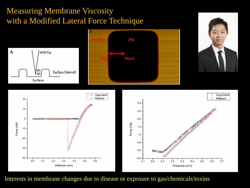
Measuring Membrane Viscosity with a Modified Lateral Force Technique
Interests in membrane changes due to disease or exposure to gas/chemicals/toxins

Vertically Directed Growth of Neurons
McNally, H.A., & Abeygunasekara, W.L. An atomic force microscopy system to investigate the effects of external electric fields on neuronal Z-projections. Journal of Neuroscience Methods, in review.
Spine like structures found in primary neurons growing vertically, appear and disappear randomly with time, present in cell body and growth cone
Is it possible to induce and direct the growth of z-projections?
Test System 2, ITO as stationary electrode, external bias circuit appliedisolated biology and electronics,

Development and testing of hyperbaric atomic forcemicroscopy (AFM) for biological applications
Dominic D'Agostino & Jay Dean, University of South Florida, Molecular Pharmacology and
PhysiologyHelen McNally Purdue University, Electrical and Computer
Engineering Technology
http://hscweb3.hsc.usf.edu/health/now/?p=96

68
72
76
64
100
80
60
40
20
0
0 30 60 90 120 150 180 210 240
Tem
pera
ture
(ºF)
Pres
sure
(psi
)
Time (minutes)
15
30
45
60
82
Decompression1 psi/min
73.9
72
Pressurization with Helium
200 nm deep pit
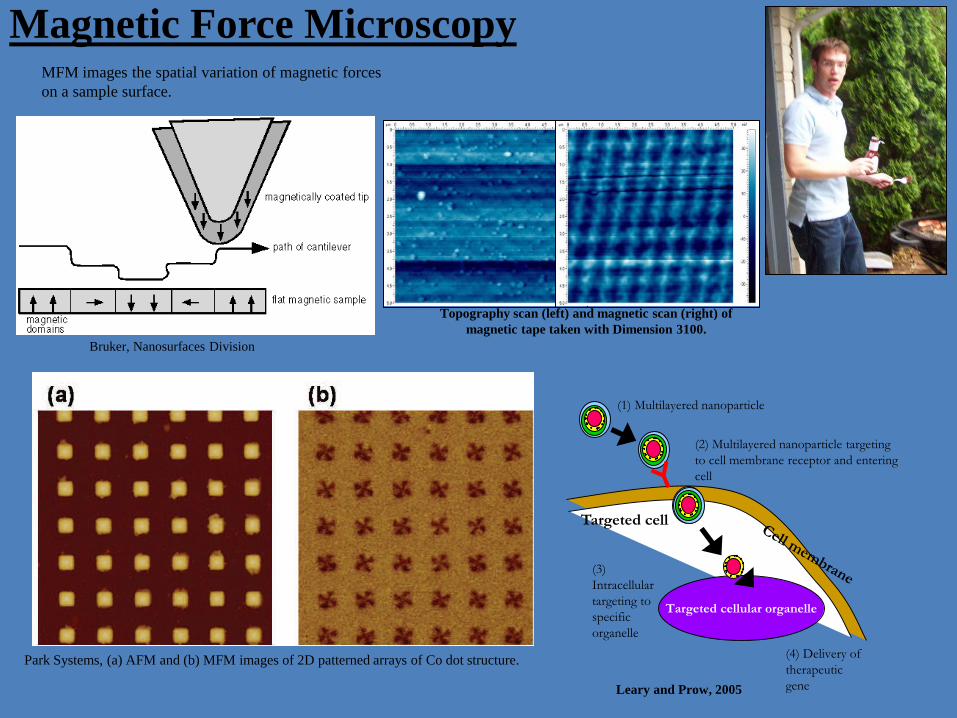
Magnetic Force Microscopy
Topography scan (left) and magnetic scan (right) of magnetic tape taken with Dimension 3100.
MFM images the spatial variation of magnetic forces on a sample surface.
Bruker, Nanosurfaces Division
(4) Delivery of therapeutic gene
Targeted cellular organelle
Targeted cell
(2) Multilayered nanoparticle targeting to cell membrane receptor and entering cell
(3) Intracellular targeting to specific organelle
(1) Multilayered nanoparticle
Leary and Prow, 2005
Park Systems, (a) AFM and (b) MFM images of 2D patterned arrays of Co dot structure.

Specific Challenges to the Electro/Magnetic Force Microscopy
• Electric/Magnetic Calibration• Resolution• EFM/MFM in fluid
magnetic tips with low spring constantsmagnetic characteristics of the solution
• External Magnetic Field required for particle magnetism• Sensitivity (detect magnetic nanoparticles inside cells)
Additional Interest in EFM/MFM● Technique Optimization, resolution and quantification● Cell-to-Cell Communications● Cell Signaling ● Neuronal magnetic aspects● Conducting polymers, 3D scaffolding

Acknowledgements
Current Students:GraduateThomas FischerTi’Air RigginsMengying Wang
UndergraduatesNeal Mahajan
Former Students:Dr. Mirela Mustata – Simmons College Waranatha Abeygunasekara – Univ. of Peradeniya, Sri LankaTejasvi Parapudi – CoE PurdueEric Milligan – Bruker, Santa Barbara, CAYen Hseu – Osha Liang, Houston, TX
Collaborators:Dr. James Curtain and Dr. Brenda Brankin, DITDr Jay Dean & Dr. Dominic D’Agostino,
University of South Florida
Funding: Morton Cure for Paralysis FundNational Science FoundationOffice of Naval ResearchPurdue Research FoundationColleges of Technology and EngineeringBirck Nanotechnology Center

Thanks a Million!
Helen A. McNallyAssociate ProfessorSchool of Engineering TechnologyPurdue University
Birck Nanotechnology Center, Room 10171205 West State StreetWest Lafayette, IN 47907-2057Office (765) 494-7491Fax (765) 496-1354
Email [email protected]
We are Purdue.What we make moves the world forward.