scanning electron microscopy of acid detergent fiber digestion by rumen microorganisms
TRANSCRIPT

J. Agric. Food Chem. 1981, 29, 899-903 899
ARTICLES
Scanning Electron Microscopy of Acid Detergent Fiber Digestion by Rumen Microorganisms
Franklin E. Barton, II,* Danny E. Akin, and William R. Windham
Leaf sections of four grasses were extracted with acid detergent reagent and/or incubated with rumen microorganisms in vitro. The samples obtained from these treatments and controls were prepared for observation with a scanning electron microscope. The response of each grass to the acid detergent reagent varied. In all cases the combination of acid detergent extraction and in vitro digestibility caused greater destruction than either treatment alone. Treatment order also had an effect. Although tissue destruction was extensive in samples extracted with the acid detergent and followed with digestion by rumen microorganisms, the extent of disruption of lignified tissues was less severe than that in the reverse order of treatments. Gravimetric analysis of ground forage confirmed that the combination of treatments removed about 6-24% more dry matter than either treatment alone and showed that the difference in order of combined treatments was 5 1 2 % units, depending on the grass species.
Recent studies by Akin et al. (1975) and Barton and Akin (1977) have identified specific fibrous tissues that make up the acid detergent fiber (ADF) and neutral de- tergent fiber (NDF) and have shown that delignification of the fibrous tissues of plant cell walls affects their sub- sequent degradation by rumen microorganisms. The values from gravimetric analyses of ADF have been cor- related with in vitro digestibility (Van Soest, 1965), and values of ADF and NDF with in vitro dry matter disap- pearance (IVDMD) (Barton et al., 1976) and intake (Van Soest and Mertens, 1977). These two fibrous residues, ADF and NDF, have been proposed as the basis for pre- dicting relative feeding value in a hay grading system (Rohweder et al., 1977) which has been applied to tem- perate forages. All of these studies suggest the use of percentage ADF for predicting forage digestibility. The studies of Akin et al. (1975) and Barton and Akin (1977) have shown that the fibrous fraction ADF is not the same for warm- and cool-season grasses. These differences could have a bearing on the use of ADF for predicting forage quality.
The ADF fraction is thought to be the most highly lig- nified and least digestible portion of the plant cell wall (Van Soest, 1963). The decomposition of lignocellulose is cited as one of the most important barriers to utilization of cellulose by microorganisms (Bryant et al., 1977; Bel- lamy, 1976). The use of ADF values to denote the po- tential extent of digestion has also been proposed (Barnes and Marten, 1979).
Both the chemical analysis, ADF, and the bioassay, IVDMD, are gravimetric measurements. The dried residue after extraction (ADF) or fermentation (IVDMD) is weighed, and the numerical weight becomes the percentage ADF or IVDMD. Neither of these procedures considers the structure of the plant as an influence on the deter- mination.
Field Crops Utilization and Marketing Research Labo- ratory, Richard B. Russell Agricultural Research Center, Agricultural Research, Science and Education Adminis- tration, US. Department of Agriculture, Athens, Georgia 30613.
In this study scanning electron microscopy (SEM) was used to observe the microbial digestion of ADF isolated from Bermuda grass, orchard grass, and fescue. The ob- jectives were to identify the tissues that make up ADF and nondigestible residue from microbial digestion and to compare the degradation of forage by the chemical and microbiological methods using microscopy and gravimetric analysis. Because we imposed two treatments on the forage (in vitro rumen microbial fermentation and acid detergent extraction), it was also necessary to determine the effect of the order of treatments. EXPERIMENTAL PROCEDURES
Preparation of Grass Samples. Coastal (CBG) and Coastcross-1 (CX-1) Bermuda grass [ Cynodon dactylon (L.) Pers.], Kentucky-31 (Ky-31) tall fescue (Festuca arundinacea Schreb.) and its ryegrass hybrid Kenhy (KHY), and Boone orchard grass (Dactylis glomerata L.) (OG) were harvested after 4 weeks of summer regrowth. These samples came from experimental plots in subse- quent years and received the same fertilization as those previously reported (Barton et al., 1976). The grass sam- ples were prepared for ADF extraction as described by Akin et al. (1975).
ADF Dry Matter Determinations and Isolation. The ADF determinations were made according to the procedures of Van Soest as modified by Barton et al. (1976). The procedure is, essentially, the extraction of a 1.0-g dried, ground grass sample with boiling 2% hexa- decyltrimethylammonium bromide in 1.0 N sulfuric acid. The isolation of ADF was also analogous to the procedures of Barton et al. (1976) except that an extra washing step was included. After each sample was washed with acetone, it was resuspended in boiling water and washed with boiling water until no detergent was visible (i.e., no foam could be seen at the base of the sintered glass disk). The sample was then washed with 3 volumes of acetone and dried at 65 "C in a forced draft oven.
Preparation of the Acid Detergent Extracted Leaf Sections. Leaf sections extracted with acid detergent were prepared for SEM in two ways. The first method was identical with that of Akin et al. (1975) in which there was no stirring of the sections in the heated flask. The second
This article not subject to U.S. Copyright. Published 1981 by the American Chemical Society

900 J. A&. FoodChem., Vol. 29, NO. 5, 1981
procedure was analogous to the analytical procedure of Barton et al. (1976) in which the samples were stirred and filtered through a sintered glass crucible. The extracted leaf sections were prepared for viewing by SEM as pre- viously reported (Barton and Akin, 1977), with the ex- ception that they were not critical point dried (Anderson, 1951). The extracted leaf sections were fixed in buffered (cacodylate, pH 7.2) glutaraldehyde, postfixed with buff- ered osmium tetraoxide mounted, and coated with a gold/paladium (6040) alloy. With tissues this fragile, the wash steps and the pressure within the dryer could disrupt or destroy the samples if care was not used in their prep- aration. Critical point drying (Anderson, 1951) involves the exchanging of water with ethanol by steps and then exchanging the ethanol with liquid carbon dioxide (CO,). This is done in a pressure vessel. After several exchanges, the temperature of the liquid CO, is raised above 88 O C ,
and the liquid and gas phases of C02 are in equilibrium. The pressure is slowly released and the dried sample re- moved.
Digestion Procedures. In vitro dry matter disap- pearance (IVDMD) was determined for samples (grasses and isolated ADF) ground through a Wiley mill to pass a 20-mesh screen by the Tilley and Terry (1963) two-stage procedure as reported by Barton et al. (1976) and Barton and Akin (1977). This procedure uses McDougall's buffer and strained rumen fluid (21) to digest 0.4 g of a sample incubated at 39 "C for 48 h. The digestion study was repeated 4 times. The ADF extraction of digested grass residue and the digestion, in vitro, of the leaf sections previously extracted with acid detergent were conducted as described above and constituted the combined treat- ments. RESULTS AND DISCUSSION
Leaf sections from these seven treatments were evalu- ated by SEM: (1) leaf section controls, (2) leaf sections in buffer (McDougall's) controls, (3) acid detergent leaf sections, (4) acid detergent leaf sections in buffer controls, (5) leaf sections digested for 48 h, (6) acid detergent leaf sections digested for 48 h, and (7) leaf sections digested for 48 h and then extracted with acid detergent. Previous histochemical and SEM studies of leaf blades had iden- tified the lignified tissues, which generally are not degraded by rumen microorganisms. Specifically, sclerenchyma cells gave a positive reaction for lignin with the chlorine-sulfite stain, whereas the xylem cells, including the inner bundle sheath of the vascular bundles, were positive for lignin when stained with acid phloroglucinol (Akin and Burdick, 1975). The terms digestion, fermentation, extraction, and degradation have slightly different meanings. However, the SEM cannot distinguish these differences nor quan- titatively measure them but only reveal the presence or absence of a particular tissue type. Composite images can be made showing differential degradation.
Leaf Section Controls. Leaf blade sections of the five grasses served as the basic control. For all the grasses, the leaf blades showed normal intact plant tissues as previously observed (Akin and Burdick, 1975).
Leaf Sections i n Buffer Control. In all five grasses the mesophyll was disturbed and slightly collapsed (Figure 1, M). For Ky-31 (Figure lb , E) the epidermis showed some distortion, and in OG the parenchyma bundle sheath (Figure Id, B) was partially collapsed in some cases. In CX-1, (Figure IC, P) the phloem was removed to some extent, but infrequently. These samples were not critical point dried during preparation for SEM. While critical point drying will preserve the intact sections, detrimental effects were noted with many of the treated samples and
Figure 1. SEM of control leaf sections incubated in buffer. (a) is labeled to show the tissue found in the blades of all of these grasses. Tissues present are epidermis (E), parenchyma bundle sheath (B), vascular bundle including the lignified inner bundle sheath and xylem cells (L) and the unlignified phloem (PI. the lignified sclerenchyma cells (C), and mesophyll (M). (a) Coastal Bermuda grass. Tissues are intact hut with collapsed mesophyll (M) cells. X240. (b) Ky-31 tall fescue. Tissues are intact hut with collapad mesophyll (M) and distorted epidermis (E). X153.6. (c) CX-I Bermuda grass. Mesophyll (M) is somewhat distorted and the phloem (P) removed from some sections. X153.6. (d) Orchard grass. Mesophyll (MI tissues are distorted and par- enchyma bundle sheath (B) partially collapsed. X153.6.
in particular with the acid detergent extracted leaf blades. These effects included disruption due to agitation during washing and dehydration and explosion of sections if the pressure of the critical point dryer was released too quickly. In some cases, the control leaf section in buffer for 48 h showed a loss in rigidity (although tissues were not re- moved) in many of the tissues when compared with the leaf section control where all tissues, except mesophyll, maintained structural integrity.
Acid Detergent Extracted Leaf Sections. Generally, the cell walls of the mesophyll, phloem, and other fragile and readily hydrolyzable cells were removed leaving a residue of lignified cells, i.e., sclerenchyma and lignified vascular bundles (Figure 2). Usually, the parenchyma bundle sheath was removed in the cool-season grasses but often remained in the warm-season grasses (Figure 2c, arrows). This residual parenchyma bundle sheath tissue (arrow) was generally found associated withthe small vascular bundles. In earlier studies (Akin et al., 1975) with no stirring and washing and without vacuum filtration of samples, considerably more parenchyma bundle sheath tissue was seen associated with the large vascular bundles. In the present study, physical agitation incurred hy stirring the sample during ADF extraction probably contributed to excessive tissue destruction and apparent removal. This physical treatment is, however, a part of the analytical procedure used on ground grass samples, and, therefore, physical disruption of tissue could occur in the normal preparation of ADF. In all the samples the sclerenchyma was separated from the vascular tissue and, in many cases, separated into individual fiber cells (Figure 2b, C) . At low magnification, a rigid structure resembling a leaf still was observed in all grasses except Ky-31. Ky-31 ADF was shattered, showing extensive destruction and tissue re- moval and a residue of loose fiber with no resemblance to a leaf section.
Figure 3 shows that the effect of buffer on the ADF residue was to further remove tissue in the acid detergent

Microscopy of AcM Detergent Fiber Digestion
Table I. Percentage Composition and IVDMDa of Grasses
J. A g k FwdCI". , Vol. 29. No. 5. 1981 801
g r w NDF ADF PML CP NDMD NDR Coastal Bermuda mass 72.66 34.90 4.34 8.54 57.41 42.59
~
Coastcross-1 Bermuda grass 74.04 36.67 4.65 9.85 61.17 38.83 Ky-31 tall fescue 47.10 25.85 3.52 13.08 64.54 35.46 Kenhy fescue 47.37 26.42 3.07 13.51 66.87 33.13 orchard grass 53.64 30.78 3.61 11.84 64.53 35.47
a NDF = neutral detergent fiber, ADF = acid detergent fiber, PML = permanganate lignin, CP = crude protein, IVDMD = in vitro dry matter disappearance, and NDR = nondigestible residue (100 - IVDMD).
Figure 2. SEM of acid detergent extracted leaf blade sections. Generally, the more fragile tissues such as mesophyll and phloem have been removed (cf. Figure la) and the lignified tissues make up the residue. Tissues me extensively distorted and disrupted. (a) Coastal Bermuda grm. The residue consists of sclerenchyma cells (C) and lignified vascular tissue (L). X480. (h) Ky-31 tall fescue. The sclerenchyma has been separated into individual cells (C), hut the lignified vascular tissue (L) is intact. Note that the parenchyma bundle sheath (arrow) oeeassiody resists extraction. X153.6. (c) Coastcross-1 Bermuda grass. In addition to scler- enchyma cells (C) and lignified vascular tissues (L), some par- enchyma bundle sheath cells remain (arrow). X153.6. (d) Kenhy tall fescue. Sclerenchyma cells (C) and lignified vascular tissue (L) remain. X153.6.
extracted samples of OG. All samples showed some ad- ditional washout of tissues, with Ky-31 exhibiting the most tissue removal. Orchard grass ADF which was incubated in buffer consisted of essentially lignified vascular bundles with phloem and patches of sclerenchyma held together by cuticle. Acid detergent leaf sections of OG control (Figure 3a) and blades incubated in buffer (Figure 3b) were essentially identical, with a little more distortion of the blade and removal of sclerenchyma in sections of ADF in buffer. The CBG sample of ADF in buffer did not have any parenchyma bundle sheath associated with the large vascular bundle.
Leaf Sections Digested for 48 Hours. All untreated grasses exhibited extensive digestion by rumen microor- ganisms. In CBG, however, some parenchyma bundle sheath remained attached to the lignified vascular bundle (Figure 4a, arrow). The sclerenchyma was difficult to distinguish from pieces of fiber which had broken loose but was obviously present in the mat of fiber obtained in Ky-31. The CX-1 and OG (parts b and d of Figure 4) samples were virtually identical, the only tissue that re- mained was lignified vascular bundles held together by the cuticle. However, all of the samples retained a rigid enough structure to resemble a leaf. These leaf section samples compared favorably with the samples from earlier studies
Figure 3. SEM of acid detergent extracted leaf sections of orchard grass. (a) Control (Le.) sample has not been incubated in buffer. Sclerenchyma is being separated into cells (C) hut the lignified vascular tissue (L) is generally intact. X240. (h) Incu- bated in buffer for 48 h. Blades lack the rigidity of controls and sclerenchyma (C) and lignified vascular tissues (L) show extreme disruption and distortion (arrows). X240.
Figure 4. SEM of leaf blades incubated with rumen microor- ganisms for 48 h. Generally, only sclerenchyma cells (C) and lignified vascular tissue (L) resist microbial digestion. (a) Coastal Bermuda grass. Note the remnants of parenchyma bundle sheaths (arrows) that resist digestion. X153.6. (b) Coastcrwa-1 Bermuda grass. X240. (c) Kenhy tall fescue. X153.6. (d) Orchard grass. X153.6.
(Akin et al., 1975), even though the IVDMD of the present grass samples was lower [Table I vs. Barton et al. (1976)l. In Ky-31 there was some evidence of breakdown of the outer portion of the inner bundle sheath cells.
Acid Detergent Extracted Leaf Sections, Digested in Vitro for 48 Hours. The acid detergent extracted residue from in vitro digestion from all the grasses showed degradation in excess of the acid detergent extracted leaf sections incubated in buffer controls, but the response was variable (Figure 5) for different species. In CBG and CX-1 some of the vascular bundles were broken, and the scler- enchyma was separated into individual cells and, for the most part, were not observed. Figure 5a,b shows the iso- lation of lignified vascular bundles of CX-1. Some par- enchyma bundle sheath tissue was infrequently attached to lignified portions of large vascular bundles in CBG, but
902 J. Agric. FoOdChe”. VoI. 29. No. 5. 1981 Barton. Akin. and Winham
.. . .
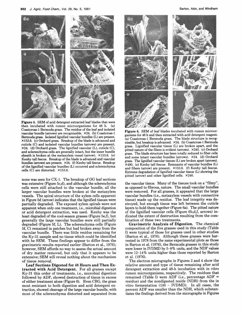
Figure 5. SEM of acid detergent extracted leaf blades that were then incubated with rumen microorganisms for 48 h. (a) Coastcroas-1 Bermuda grass. The residue of the leaf and isolated vascular bundle (arrows) are recognizable. X24. (b) Coastcross-1 Bermuda grw. Jdated lignified vascular bundles (L) are present. X153.6. (c) Orchard grass. Breakup of the hlade is advanced and cuticle (C) and isolated vascular bundles (arrows) are present. X24. (d) Orchard grass. The lignified vascular (L), cuticle (C), and sclerenchyma cells are generally intact, but the inner bundle sheath is broken at the metaxylem vessel (arrow). X153.6. (e) Kenhy tall fescue. Breakup of the blade is advanced and vascular bundles (arrows) are present. X24. (0 Kenby tall fescue. Breakup of the lignified vascular bundles (L) occurred and sclerenchyma cells (C) are distorted. X153.6.
none was seen for CX-1. The breakup of OG leaf sections was extensive (Figure 5c,d), and although the sclerenchyma cells were still attached to the vascular bundle, all the larger vascular bundles were broken at the metaxylem vessels. The spiral structure of the xylem cells apparent in Figure 5d (arrow) indicates that the lignified tissues were partially degraded. The exposed xylem spirals were not apparent when only one treatment, i.e., microbial digestion or acid detergent extraction, was used. Kenhy was the least degraded of the cool-season grasses (Figure 5e,fi, hut generally the large vascular bundles were still partially degraded (Figure 5f, L). The sclerenchyma cells (Figure 5f, C) remained in patches hut had broken away from the vascular bundle. There was little residue remaining for the Ky-31 sample and no tissue which could be identified with he SEM. These findings appear to differ from the gravimetric results reported earlier (Barton et al., 1976); however, SEM affords no way to assess the actual amount of dry matter removed, hut only that it appears to be extensive; SEM will reveal nothing ahout the mechanism of tissue removal.
Leaf Sections Digested for 48 Hours and Then Ex- t racted wi th Acid Detergent. For all grasses except Ky-31 this order of treatments, i.e., microbial digestion followed by ADF, showed destruction of tissue in excess of either treatment alone (Figure 6). Even CBG, the grass most resistant to both digestion and acid detergent ex- traction, showed cleavage of the large vascular bundle, with most of the sclerenchyma distorted and separated from
Figure 6. SEM of leaf blades incubated with rumen microor- ganisms for 48 h and then extracted with acid detergent reagent. (a) Coastcross-l Bermuda grass. The hlade structure is recog- nizable, but breakup is advanced. X24. (b) Coastcross1 Bermuda grass. Lignified vascular tissue (L) are broken apart, and the pitted nature of the fibers is evident (arrows). X240. (c) Orchard grass. The blade strzlcture bas been totally reduced to fiber cells and some intact vascular bundles (arrows). X24. (d) Orchard grw. The lignified vascular tissues (L) are broken apart (arrows). X480. (e) Kenhy tall fescue. Remnant8 of vascular bundles (L) and fibers (arrow) are present. X153.6. (0 Kenby tall fescue. Extreme degradation of lignified vascular tissue (L) showing the pitted (arrow) and other lignified cells. X240.
the vascular tissue. Many of the tissues took on a “ f h y ” , as opposed to fibrous, nature. The small vascular bundles were removed. For all grasses, it appeared that the large vascular bundles (i.e., metaxylem vessels with connective tissue) made up the residue. The leaf integrity was de- stroyed, hut enough tissue was left between the cuticle layers to hold them together (Figure 68). The pitted nature of the lignified vascular cells (Figure 6h,d,f, arrows) in- dicated the extent of destruction resulting from the com- bination of these two treatments.
Gravimetric Analysis of Digestion. The chemical composition of the five grasses used in this study (Table I) were typical of those for grasses used in other studies (Barton et al., 1976). Although these grasses were har- vested in 1978 from the same experimental plots as those in Barton et al. (1976), the Bermuda grasses in this study were lower in IVDMD hy 5 8 % units, and the NDF values were 12-14% units higher than those reported by Barton et al. (1976).
The electron micrographs in Figures 2 and 4 show the relative amount and type of tissue remaining after acid detergent extraction and 48-h incubation with in Vitro rumen microorganisms, respectively. The residues that remained (Table I) were ADF (i.e., percentage ADF = residue) and the nondigested reside (NDR) from the in vitro fermentation (100 - IVDMD). In all cases, the percent ADF was smaller than the NDR, which suhstan- tiates the fmdinge derived from the micrographs in Figures

Microscopy of Acid Detergent Fiber Digestion J, Agric. Food Chem., Vol. 29, No. 5, 1981
Table 11. Percentage Digestibility of the Five Grasses, ADF’s,= and the Combined Dry Matter Disappearance (CDMD)b
903
grass %IVDMD %IADFD %ADER CDMD-1 CDMD-2 Coastal Bermuda grass 57.41 5.66 46.56 77.24 67.08 Coastcross-1 Bermuda grass 61.17 5.91 41.17 77.16 68.53 Ky-31 tall fescue 64.54 5.76 44.73 80.40 74.93 Kenhy tall fescue 66.87 7.18 47.15 82.49 75.50 orchard grass 64.53 6.98 53.86 83.63 71.35
Lsolated ADF disappearance (IADFD) is the dry matter removed from isolated ADF by rumen microorganisms; acid de- tergent extracted residue (ADER) is the percentage dry matter removed by the extraction with acid detergent reagent of the in vitro residue. CDMD-l= (NDR x ADER/100) t IVDMD; CDMD-2 = (% ADF X IADFD/100) t (100 - ADF).
2 and 4 that the acid detergent extraction removed more dry matter than did the rumen microorganisms. However, there were certain discrepancies between the treatments in tissue types remaining. For CBG, some digestible tissue (parenchyma bundle sheath) resisted the ADF treatment but was removed by the rumen microorganisms in vitro. For Ky-31 the amount of tissue removed (hydrolyzed) by the ADF reagents far exceeded the dry matter removed by rumen microorganisms. The results of Barton et al. (1976) showed a larger difference than found in this study between the in vitro digestibility of isolated ADF from warm- and cool-season grasses. However, the warm-season grasses in that study were of higher quality (66.1 vs. 57.4% IVDMD) than those in this study, and the ADF might be expected to also have a higher digestibility.
The process of isolating the ADF could modify the tis- sues to the extent that they would be subject to increased tissue removal by a subsequent treatment, i.e., the action of rumen microorganisms during the determination of IVDMD. It is also possible that the preparation of ADF leaves traces of hexadecyltrimethylammonium bromide in the fiber that can inhibit digestion (Cross et al., 1974). Reversal of these treatments, i.e., in vitro digestion followed by treatment with acid detergent reagents, would remove more tissue than either alone. The electron micrographs in Figure 3, 5, and 6 show the effect on the grasses of combined (i.e., digestion and acid detergent extraction) treatments. The gravimetric results of those combined dry matter disappearance (CDMD) values are given in Table 11. In all cases the effect of combined treatments (Table II; Figures 3,5, and 6) was that more tissue was removed than was removed by either treatment alone. The order of the treatment also was important. More tissue was removed by treating digested leaf sections with acid de- tergent reagent than the reverse (Figures 5f and 6e).
Gravimetrically, the difference between the CDMD values was 5-12% units, depending on the grass (Table 11). The methods of calculating CDMD reflected the treatment order (CDMD-1 and -2), Le., for the effect of acid detergent reagents on the NDR (CDMD-1) and the effect of digestion (in vitro rumen microorganism fer- mentation) on isolated acid detergent extracted leaf sec- tions (CDMD-2). These values (CDMD-1 and -2) are, essentially, material balances for the combined treatments.
These results support the conclusions of previous studies and show that the tissues in the leaf sections of the grass species examined, representing temperate and tropical
grasses, respond differently to acid detergent extraction (Akin et al., 1975), digestion (Akin and Burdick, 19751, and digestion of ADF (Barton et al., 1976). Further, the com- bination and digestion and ADF treatment removes more tissues, both qualitatively and quantitavely, than either treatment alone. Also, the order of treatments is impor- tant, for more tissue was removed by treatment of in vitro residues with acid detergent reagent than were removed by the in vitro digestion of acid detergent extracted leaf sections. Finally, Figures 2 and 4 show that ADF ap- proximates the extent of digestion in vitro. The differences noted by Akin et al. (1975) and those reported herein for warm- and cool-season grasses should be used to interpret the use of ADF values in a quantitative prediction equa- tion. The ADF value for warm- and cool-season grasses would overestimate, but to a lesser extent with the warm- season grasses. Therefore, ADF, as a predictor of the extent of digestion, is not adequate when used for both warm- and cool-season grasses. However, the use of qualitative data on the structure of the forage cell wall and the response of its respective tissues to extraction with ADF reagent can add additional information on the factors influencing forage digestion. LITERATURE CITED Akin, D. E.; Barton, F. E., 11; Burdick, D. J. Agric. Food Chem.
Akin, D. E.; Burdick, D. Crop Sci. 1975, 15, 661. Anderson, T. F. Trans. N.Y. Acad. Sci. 1951, 13, 130. Barnes, R. F.; Marten, G. C. J. Anim. Sci. 1979,48, 1554. Barton, F. E., II; Akin, D. E. J. Agric. Food Chem. 1977,25,1299. Barton, F. E., II; Amos, H. E.; Burdick, D.; Wilson R. L. J. Anim.
1975,23, 924.
Sci. 1976, 43, 504. Bellamy, W. D. World Anim. Reu. 1976, 18, 39. Bryant, M. P.; Varel, V. H.; Frobish, R. A.; Isaacson, H. R. Microb.
Cross, H. H.; Smith, L. W.; DeBarth, J. V. J. Anim. Sci. 1974, Energy Convers., Proc. Semin., 1976 1977, 347.
39, 808. Rohweder, D. A.; Barnes, R. F.; Jorgenson, N. Int. Symp.: Feed
Compos., Anim. Nutr. Requir., Comput. Diets, [Proc.], l s t , 1976 1977, 249.
Tilley, J. M. A.; Terry, R. A. J. Br. Grassl. SOC. 1963, 18, 104. Van Soest, P. J. J. Assoc. Off. Anal. Chem. 1963, 46, 825. Van Soest, P. J. J. Anim. Sci. 1965, 24, 834. Van Soest, P. J.; Mertens, D. G. Proc. Znt. Meet. Anim. Prod.
Temperate Grassl. 1977, 50.
Received for review September 15, 1980. Revised manuscript received May 4, 1981. Accepted May 23, 1981.