scanning electrochemical microscopy: an analytical · pdf filealso in electroanalytical...
TRANSCRIPT

REVIEW
Scanning electrochemical microscopy: an analytical perspective
Javier Izquierdo1 & Peter Knittel1,2 & Christine Kranz1
Received: 8 September 2017 /Revised: 16 October 2017 /Accepted: 2 November 2017 /Published online: 6 December 2017# Springer-Verlag GmbH Germany, part of Springer Nature 2017
Abstract Scanning electrochemical microscopy (SECM) hasevolved fromanelectrochemical specialist tool to abroadly usedelectroanalytical surface technique, which has experienced ex-citing developments for nanoscale electrochemical studies in re-cent years. Several companies now offer commercial instru-ments, andSECMhasbeenused inabroad rangeofapplications.SECM research is frequently interdisciplinary, bridging areasranging from electrochemistry, nanotechnology, and materialsscience to biomedical research. Although SECM is consideredamodernelectroanalytical technique, itappears that lessattentionis paid to so-called analytical figures ofmerit,which are essentialalso in electroanalytical chemistry. Besides instrumental devel-opments, this review focuses on aspects such as reliability, re-peatability, and reproducibility of SECM data. The review isintended to spark discussionwithin the community on this topic,but also to raise awareness of the challenges faced during theevaluation of quantitative SECMdata.
Keywords Scanning electrochemical microscopy .
Nanoelectrodes . Analytical figures of merit . Methodvalidation
Introduction
Electroanalytical chemistry is a vital and relevant area of an-alytical chemistry, and has experienced a multitude of novel
developments leading to new applications and advancementsin electroanalysis based on advanced electrode materials, cou-pling of analytical techniques with electrochemical methods,and use of microelectrodes and nanoelectrodes. Thus, mea-surements in extremely small volumes and confined spaceswith high temporal and spatial resolution are made possiblealong with the opportunity to perform high-resolution electro-chemical mapping (i.e., electrochemical imaging) experi-ments. In particular, electrochemical imaging has significantlyadvanced in recent years, and is now applied in multidisciplin-ary research areas ranging from biomedical research to mate-rials science, corrosion, catalysis, and energy-related topicssuch as fuel cells and battery research. Scanning electrochem-ical microscopy (SECM) [1, 2] and derived Bhyphenated^electrochemical scanning probe techniques [3–8] now allowelectrochemical/(electro)analytical measurements withnanoscopic and microscopic electrodes or nanopipettes.Positioning such probes in close proximity to the investigatedsample surface allows high-resolution information on electro-chemical processes or ion fluxes occurring at the solid–liquidand liquid–liquid interface to be obtained. Besides the combi-nation with other scanning probe microscopy (SPM) tech-niques, SECM has also been coupled with spectroscopic tech-niques (i.e., surface plasmon resonance, Raman, and IR)[9–11], mass spectrometry [12], or an electrochemical quartzmicrobalance [13] to list just a few examples. The researchactivities in SECM are well documented by the breadth oforiginal contributions and a steadily increasing number ofreview articles ranging from comprehensive overviews ofthe current state of the art to focused articles on specific ap-plication areas such as bioapplications, corrosion research,and energy-related applications [14–25]. SECM is consideredan electroanalytical technique, as most classical electroanalyt-ical techniques, including stripping voltammetry at mercury-coated microe lec t rodes [26–28] , potent iometr ic
* Christine [email protected]
1 Institute of Analytical and Bioanalytical Chemistry, Ulm University,Albert-Einstein-Allee 11, 89081 Ulm, Germany
2 Fraunhofer Institute for Applied Solid State Physics, Tullastraße 72,79108 Freiburg, Germany
Anal Bioanal Chem (2018) 410:307–324https://doi.org/10.1007/s00216-017-0742-7

measurements [29–33], square wave voltammetry [34, 35],and electrochemical impedance measurements [13, 36, 37],have been demonstrated in SECM studies. Consequently, thistechnique is extremely useful in modern electroanalysis forobtaining quantitative data on specific analytes or processes.
Given the number of reviews already published on SECM,the present review is intended to give a critical perspective onSECM in terms of analytical figures of merit in the field of(electro)analytical chemistry highlighting in particular re-search from recent years. The International Union of Pureand Applied Chemistry (IUPAC), Analytical ChemistryDivision (V) has given the following definition: BAnalyticalchemistry (which includes electroanalytical chemistry) is ascientific discipline that develops and applies methods, instru-ments, and strategies to obtain information on the compositionand nature of matter in space and time, as well as on the valueof these measurements, i.e., their uncertainty, validation, and/or traceability to fundamental standards.^
The first part of the definition is clearly covered within thefield of SECM.Over the years a significant number of originalcontributions have been published on instrumental develop-ments and improvements. Such developments can be catego-rized into (1) developments regarding improved hardware,positioning of the SECM probe, and imaging modalities, (2)controlling physical parameters such as temperature and sur-rounding atmosphere, which was initially shown for biologi-cal investigations, where fixed CO2 content or oxygen-reduced atmospheres [38] play an important role, and (3) de-velopments targeting reproducible fabrication schemes andcharacterization routines for SECM nanoelectrode probesand pipette-based electrodes, which certainly have a huge im-pact on the analytical figures of merit. Figure 1 highlightsseveral examples of such recent improvements. Figure 1a il-lustrates several examples of nano-sized probes. Laser-assisted methods have been exploited for the fabrication ofnano disk electrodes (Fig. 1a, panel a). Alternatively, insteadof the use of solid metal or carbon, microelecrodes ornanoelectrodes can be based on micropipettes or nanopipettesknown from scanning ion conductance microscopy (SICM)[46]. Novel concepts for performing electrochemical scanningprobe experiments have been introduced; for example, smallelectrodes were also implemented into atomic force microsco-py (AFM) probes [5, 6, 47, 48]. Exemplary nanopipette-basedprobes are illustrated in Fig. 1a, panels b and c. Among othergroups [3, 49–51] and collaborative efforts between thegroups of Matsue, Korchev [4], and Unwin [52], Unwin andcoworkers made a significant contribution called Bpipette-based^ electrochemical SPM [42, 53–55], introducing imag-ingmethods such as the scanningmicropipette contact method[56], whereby a localized electrochemical cell is formed be-cause of attractive capillary forces between the meniscus andthe sample surface. Subsequently, they introduced a techniquetermed Bscanning electrochemical cell microscopy^
(SECCM) [57, 58] using double-barrel pipettes (i.e., thetacapillaries). Both barrels are filled with electrolyte solution,and each barrel contains a reference/counter electrode (i.e.,quasi-reference counter electrode). Again the meniscus formsa localized droplet cell when the pipette is near the samplesurface. Ions migrate across the meniscus between the twobarrels if a potential is applied between the electrodes. Byaddition of an oscillation of the pipette similar to the operationmode in SICM, an alternating current component of the ioniccurrent is periodically altered in strong dependence on themeniscus height, which is used for distance control. Becauseof sophisticated hardware (i.e., electronics) and alternativescanning approaches such as spiral scanning, SECCM hasbeen developed into a multifunctional electrochemical scan-ning probe technique, which even allows video-rate electro-chemical imaging with high lateral resolution [41].
Beyond efforts toward nanoscale probes, imaging modesallowing improved positioning of the SECM probe such asshear-force mode, alternating current mode [59], and intermit-tent contact mode [60] have been introduced. Panels a–c inFig. 1b illustrate these modes. Briefly, they involve the lateraloscillation of the SECM probe at an amplitude dependent onthe proximity of the surface (Fig. 1b, panel a), the applicationof alternate potential perturbation to the probe at varying fre-quency so that the acquired alternating current provides infor-mation on different surface features (Fig. 1b, panel b), and thevertical oscillation of the probe, sensing the presence of thesurface, which allows a constant probe–sample distance to bemaintained by use of the signal as input for a feedback loop(Fig. 1b, panel c). Likewise, imaging modalities providingenhanced information, including surface interrogation mode[surface interrogation SECM (SI-SECM)] [61] (seen in Fig.1b, panel d), and redox-competition mode (redox-competitionSECM (RC-SECM)] [62] (depicted in Fig. 1b, panel e), havebeen developed, further improved, and applied to various
�Fig. 1 Recent developments in scanning electrochemical microscopy(SECM). a Nanoelectrodes: pulled quartz pipette nanoelectrodes (a),dual carbon nanoelectrodes (b; the inset shows a magnification of theelectrode tip and a schematic drawing), and a platinum-filled nanopipetteprobe (c; the inset shows a quad-barrel probe with two carbon-filledbarrels). b Imaging modes. Modes for probe positioning: shear-forceSECM (a), alternating current SECM (b), and intermittent contactSECM (c). Imaging modalities: surface interrogation SECM (d) andredox-competition SECM (e). c Instrumentation: isothermal chamberusing vacuum insulated panels (white parts) and extruded aluminum heatsinks (black) for effective drift compensation (a), photoelectrochemicalshear-force-based SECM setup with illumination from the bottom (b),and SECM head (positioning system, tip holder, and electrochemical cell)placed under a custom-made Plexiglas bell, which is in an argon-filledglove box along with the controller (temperature is measured with athermometer inside the chamber) (c). (a Reprinted with permission from[39] (a), [40] copyright 2016 American Chemical Society (b), and [41]copyright 2016 American Chemical Society and [42] copyright 2015American Chemical Society (c). c Reprinted with permission from [43]copyright 2012 American Chemical Society (a), [44] (b), and [45] (c))
308 Izquierdo J. et al.

emerging analytical problems. RC-SECM evaluates the extentto which the probe and substrate compete for the electrochem-ical conversion of the same redox molecule. SI-SECM in-volves the surface modification (e.g., by UV illumination)inducing adsorbed species, which are next electrochemically
interrogated with appropriate reactants to evaluate theresulting changes in surface chemical composition.Capillary-based reference electrodes have been demonstratedfor detection of local current densities based on an ohmicmeasurement principle termed Bscanning ohmic microscopy^
Scanning electrochemical microscopy: an analytical perspective 309

[63, 64] providing information on adsorption and intercalationreactions with micrometer resolution; these are difficult tomap via conventional SECM.
Whereas during the first decades of SECM, with few excep-tions [65], SECM investigations focused on studying processesinvolvingmacroscopic andmicroscopic samples using solidmi-croelectrodes, a remarkable trend is currently being experiencedtoward nanoscale studies. In recent years, studies on theelectroactive properties of nano-objects such as nanoparticlesdown to the single nanoparticle level have emerged, which isclearly correlatedwith instrumental improvements and advance-ments in probe fabrication at nanoscopic dimensions. Given thefact that in SECM experiments the dimensions of the probe arestrongly related to the achievable spatial resolution, such interestin nano-objectsmust be accompanied by appropriate fabricationprocedures.Hence, reliable fabrication schemesallowingamorereproducible production of nanoelectrodes with well-definedshape and size, alongwith establishment of characterization rou-tines for nano-sized electrodes [66–68], have been importantsteps toward improving the achievable spatial resolution downto the single particle level. Besides etching procedures and laser-pipette-puller-based fabrication of nanoelectrodes, pulling glasscapillarieswithorificesdown toadiameter of 50nmfollowedbypostmodification steps for implementation of an electrode eitheras a ring (i.e., by sputtering a metal layer) or as a second barrelfilled with carbonized material has been introduced. In terms ofcharacterization, high-resolution scanning electron microscopyand in particular transmission electron microscopy [66] haveimproved the determination of the shape and size of thenanoelectrodes obtained. Surface modification of suchnanoelectrodes by electrochemical deposition processes (e.g.,platinum deposition) or modification with a mercury film [69]is required to increase the electron transfer kinetics if speciesbeyond ferrocenemethanol should be detected. In addition, onlyfewmeasurementswith nano-sized probes have been performedin ^constant height mode^ [70–73]; that is, where the probe isscanned at a fixed distance across the sample surface.
Hence, a distance control providing independent electro-chemical signals was a major step toward deconvoluting elec-trochemical and topographical information, as the samplemorphology can be recorded independently but simultaneous-ly with the electrochemical information. Shear-force-basedSECM was introduced fairly early as a current independentmode, and it has developed into a routine distance-controlledimaging mode applied by many researchers in the field.BHyphenated^ scanning probe techniques such as SICM–SECM and AFM–SECM have gained popularity, as SICM–SECM probes can be fabricated fairly easily in any laboratoryenvironment. In respect to AFM–SECM, SECM is now avail-able as a commercial add-on to AFM instrumentation [74].
In terms of instrumental developments and improve-ments, recent efforts were directed to ensure stable environ-mental conditions, which may have a significant impact on
the establishment of SECM as an analytically validatedmethod. For instance, surrounding temperature control isachieved by placing the SECM system in an isothermalchamber, avoiding thermal drifts of the positioning system,which is crucial for distance control for nanometer probes[43]. The presence of vacuum insulated panels and extrudedaluminum heat sinks (white and black as seen in Fig. 1c,panel a) has been implemented into the SECM setup toensure high thermal stability in the system. In addition, con-trolling or varying the sample temperature (e.g., by aPeltier-heated sample stage [75]) may enhance the electro-chemical response. A well-controlled surrounding atmo-sphere is not only crucial in biomedical research, but is aprerequisite, for example, in battery research, where SECMexperiments typically have to be performed in a glove box[76, 77]. Also, adding controlled illumination of the samplewithin the SECM setup has gained attention, in particularfor the study of photocatalytic materials and their perfor-mance in operando [44, 78, 79]. One achieves this eitherby illuminating the sample, supported in transparent mate-rials, from below (as seen in Fig. 1c, panel b) [44, 78] or byguiding the excitation light through the glass material of theultramicroelectrode (UME) itself [79]. Recent efforts to-ward atmospheric stabili ty in battery research byWittstock’s group [45] have resulted in the custom-designed Ar-filled setup shown in Fig. 1c, panel c, includingports for the necessary electrical connections at the rear ofthe setup, and Ar inlet and outlet at the sides (see Fig. 1c,panel c). Recently, Bard and coworkers [73] published im-provements in SECM instrumentation suitable specificallyfor nanoscale studies.
In 1992, Bard [80] described the emerging challenges inelectroanalysis and electrochemistry that will overcome someof the existing limits such as time, space, potential range, tem-perature, and pressure. Twenty-five years later, some of theselimitations have been clearly pushed beyondwhatwas feasibleat that timeby the introductionof nanoelectrodes, sophisticatedimaging modalities, and improved instrumentation. Despitethese advances, the remaining challenges are predominantlyrelated to the reliability and achievable analytical figures ofmerit (Fig. 2) of SECM-based electroanalyticalmeasurements.In the following, we focus on challenges related to uncertainty,validation, and/or traceability in SECMstudies, anddiscuss thechallenges along with a few examples highlighting some ofthese aspects, selected for their illustrative ability.
Method validation
Electroanalytical methods require addressing and reporting ofthe analytical figures of merit schematically shown in Fig. 2.In general, maximum comprehensiveness of validation in-cludes parameters such as trueness, precision, reproducibility,
310 Izquierdo J. et al.

linearity, recovery rate, selectivity, robustness, limit of detec-tion, and limit of quantification. However, what is actuallyneeded for qualifying a measurement is strongly dependenton the purpose of the analysis. As those are general require-ments for an analytical method to be validated, they eventuallyhave to be standardized if SECM is to be considered in routineanalysis. In the current section, the present state of and futureprospects for the implementation of such figures of merits inSECM studies will be discussed according to the scope andlimitations of the technique and its applications.
Challenges in method validation for SECMmeasurements
Validation of a given analytical method is built around thefigures of merits displayed in Fig. 2. Moreover, in routineanalysis, measurements and procedures are defined by nation-al and international regulations, quality control procedures,proficiency tests, and standardization by defined internationalstandards such as ISO/IEC 17025 [81]. In SPM such as near-field scanning optical microscopy, scanning tunneling micros-copy, or AFM, ISO norms have been established for calibrat-ing the probe and the Bstandardized^ use of the technique[82]; interestingly, no standards have been conceived forSECM to date. Protocols for the analysis of surface geometricquantities [83] or experimental uncertainties such as drift [84]have been standardized for the SPM techniques mentionedabove, but they have not yet been extended to SECM. Thisis of particular relevance, as SECM is likewise capable ofdetermining geometric features, which are eventually affectedby drift. ISO norms in SPM regulate the calibration and use ofSPM in nanotechnology, whereas SECM was probably notincluded at that time, achieving mostly spatial resolutions in
the micrometer range. Nowadays, SECM achieves resolutioncomparable to, for example, that achieved by near-field scan-ning optical microscopy using nano-sized electrodes andnanopipettes. Even SPM techniques mapping local electriccurrents have been validated and standardized in terms ofusage and lateral resolution [82, 85]; however, again this pro-cedure has not been extended to local electrochemical mea-surements yet. Given the broad range of applications and thedifferent modes of operation using individually fabricatedSECM probes, it is evident that these requirements are essen-tial for a more widespread routine use of SECM.
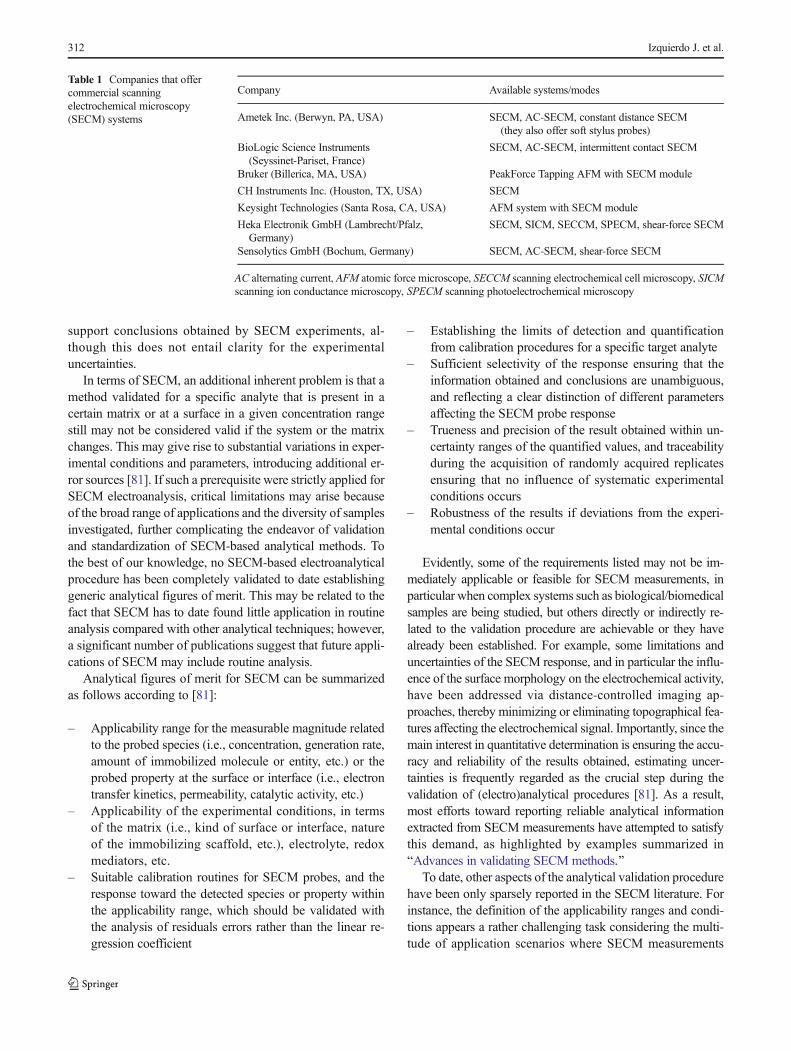
Concomitantly, the number of companies now offeringcommercial SECM equipment, given in Table 1, has increasedin the last few decades along with the widespread develop-ment of custom-built systems, including hybrid microscopes.It seems clear that the establishment of standardized equip-ment would be an important step toward the development ofvalidated protocols.
The establishment of validated analytical methods for thecalibration of the SECM response and its operation in quanti-tative analysis would be a prerequisite for the technique to bestandardized. Analytical validation includes the entire analyt-ical procedure from sampling to publication of the results, aswell as reporting the associated uncertainties. For convention-al (electro)analytical methods, such as those aiming to quan-tify a given analyte in a liquid sample or a certain solid mate-rial, validation is well established, and the certainty of theresults must be clearly reported for the results to be accepted.However, for a wide range of feasible surface and interfaceanalyses using SECM, no unified procedure has been con-ceived for data evaluation within uncertainty intervals. Inmany cases, simulations have been invoked to effectively
Fig. 2 Representation of the analytical figures of merit according to IUPAC [81]. Information, whether these figures of merits are addressed or pendingfor scanning electrochemical microscopy (SECM) is included. LOD limit of detection, LOQ limit of quantification, SD standard deviation
Scanning electrochemical microscopy: an analytical perspective 311
support conclusions obtained by SECM experiments, al-though this does not entail clarity for the experimentaluncertainties.
In terms of SECM, an additional inherent problem is that amethod validated for a specific analyte that is present in acertain matrix or at a surface in a given concentration rangestill may not be considered valid if the system or the matrixchanges. This may give rise to substantial variations in exper-imental conditions and parameters, introducing additional er-ror sources [81]. If such a prerequisite were strictly applied forSECM electroanalysis, critical limitations may arise becauseof the broad range of applications and the diversity of samplesinvestigated, further complicating the endeavor of validationand standardization of SECM-based analytical methods. Tothe best of our knowledge, no SECM-based electroanalyticalprocedure has been completely validated to date establishinggeneric analytical figures of merit. This may be related to thefact that SECM has to date found little application in routineanalysis compared with other analytical techniques; however,a significant number of publications suggest that future appli-cations of SECM may include routine analysis.
Analytical figures of merit for SECM can be summarizedas follows according to [81]:
– Applicability range for the measurable magnitude relatedto the probed species (i.e., concentration, generation rate,amount of immobilized molecule or entity, etc.) or theprobed property at the surface or interface (i.e., electrontransfer kinetics, permeability, catalytic activity, etc.)
– Applicability of the experimental conditions, in termsof the matrix (i.e., kind of surface or interface, natureof the immobilizing scaffold, etc.), electrolyte, redoxmediators, etc.
– Suitable calibration routines for SECM probes, and theresponse toward the detected species or property withinthe applicability range, which should be validated withthe analysis of residuals errors rather than the linear re-gression coefficient
– Establishing the limits of detection and quantificationfrom calibration procedures for a specific target analyte
– Sufficient selectivity of the response ensuring that theinformation obtained and conclusions are unambiguous,and reflecting a clear distinction of different parametersaffecting the SECM probe response
– Trueness and precision of the result obtained within un-certainty ranges of the quantified values, and traceabilityduring the acquisition of randomly acquired replicatesensuring that no influence of systematic experimentalconditions occurs
– Robustness of the results if deviations from the experi-mental conditions occur
Evidently, some of the requirements listed may not be im-mediately applicable or feasible for SECM measurements, inparticular when complex systems such as biological/biomedicalsamples are being studied, but others directly or indirectly re-lated to the validation procedure are achievable or they havealready been established. For example, some limitations anduncertainties of the SECM response, and in particular the influ-ence of the surface morphology on the electrochemical activity,have been addressed via distance-controlled imaging ap-proaches, thereby minimizing or eliminating topographical fea-tures affecting the electrochemical signal. Importantly, since themain interest in quantitative determination is ensuring the accu-racy and reliability of the results obtained, estimating uncer-tainties is frequently regarded as the crucial step during thevalidation of (electro)analytical procedures [81]. As a result,most efforts toward reporting reliable analytical informationextracted from SECM measurements have attempted to satisfythis demand, as highlighted by examples summarized inBAdvances in validating SECM methods.^
To date, other aspects of the analytical validation procedurehave been only sparsely reported in the SECM literature. Forinstance, the definition of the applicability ranges and condi-tions appears a rather challenging task considering the multi-tude of application scenarios where SECM measurements
Table 1 Companies that offercommercial scanningelectrochemical microscopy(SECM) systems
Company Available systems/modes
Ametek Inc. (Berwyn, PA, USA) SECM, AC-SECM, constant distance SECM(they also offer soft stylus probes)
BioLogic Science Instruments(Seyssinet-Pariset, France)
SECM, AC-SECM, intermittent contact SECM
Bruker (Billerica, MA, USA) PeakForce Tapping AFM with SECM module
CH Instruments Inc. (Houston, TX, USA) SECM
Keysight Technologies (Santa Rosa, CA, USA) AFM system with SECM module
Heka Electronik GmbH (Lambrecht/Pfalz,Germany)
SECM, SICM, SECCM, SPECM, shear-force SECM
Sensolytics GmbH (Bochum, Germany) SECM, AC-SECM, shear-force SECM
AC alternating current, AFM atomic force microscope, SECCM scanning electrochemical cell microscopy, SICMscanning ion conductance microscopy, SPECM scanning photoelectrochemical microscopy
312 Izquierdo J. et al.
have been performed. In addition, analytical reports rigorouslyproviding calibration parameters, including uncertainties andanalysis of residual errors, are, to the best of our knowledge,not commonly found in the SECM literature. Besides, veryfew reports provide accurate information on the limits of de-tection and quantification achieved. Ultimately, given the abil-ity of SECM to characterize physicochemical surface phe-nomena, it may be accepted that calibration routines for thesystem response are not always required. Instead, physico-chemical models have been typically applied to evaluate theprobe response (e.g., the determination of electron transferkinetic constants using data from amperometric probe ap-proach curves). These parameters may in fact be consideredanalytically acceptable providing that the extracted informa-tion can be traced back to the uncertainty of the response.
TheassessmentoftherobustnessisprobablythemostchallengingtaskduringthevalidationofSECMmethods.Ingeneral,toaddressthisissueforagiven(electro)analyticalmethod,aminimumnumberoflaboratoriesarerequiredtoperformthesamemeasurementsunderequivalentconditionsandcomparetheirresults(i.e.,comparabletointernationalcollaborativeringtrialsorroundrobintests).Suchtestmeasurementswould require that themeasurements be executedusingcomparableequipment(e.g.,theachievableaccuracyandstepsizeofpositionersmaydiffersignificantly),bydifferentstaffmem-bers, and indifferent laboratoryenvironments.Most commercial-izedor standardized analytical techniques fulfill this requirement,whereastherobustnessofSECM-basedmethodsmaybesignificant-lyunderestimated. InSECM,it iscommonpractice tousecustom-builtSECMsystemsandSECMprobes/electrodes,whichmaydif-fer becauseofdifferent fabricationandpolishingprocedures [86].Recently,severalSECMreportsontheuseofcarbon-basedmaterialssuch as graphene [87, 88] or nanoparticles [89, 90] appeared thatspikedsignificant interest, andmayserveas suitable examples forvalidatingtherobustnessofSECMifseveralgroupswouldpartici-pateinsuchinterlaboratorycomparisonstudies.
A significant challenge during interlaboratory validation ofSECMmethods is the diversity of systems responding to similarphysicochemical principles. This is illustrated by the determina-tion of apparent electron transfer kinetic constants usingfeedback-mode SECM. To extract this information from so-called approach curves (i.e., current–distance curves), theresulting SECM current response is modeled with respect to thekinetic constant of the electron transfer between the sample andthe redoxmediator with respect to the probe/electrode geometry[91]. These genericmodels have been exploited in several fields,arising as powerful analytical procedures to determine electro-chemical surface properties within their uncertainty ranges; forexample, for self-assembled monolayers (SAMs) [92–94], liq-uid–liquid interfaces [95, 96], conductive materials [97], andphotoinduced electron transfer phenomena [96, 98–100].
Similarly, electroanalytical methods to determine the per-meability of membranes and films via their influence on thefeedback response during SECM measurements have been
conceived for surfaces of different origin ranging from depos-ited films on conductive materials [101, 102] to lipid layersand cell membranes [103, 104]. Elegant studies have recentlybeen reported that generically model and simulate the tip-current response for such complex porous electroactive sys-tems [104, 105], thereby facilitating the future establishmentof validated methods.
An alternative strategy versus interlaboratory validationmaybe the establishment of certified reference materials (CRMs),which is well established for the general validation of analyticalmethods. To date, there are no CRMs existing or reported forSECM. Thus, only materials or electroactive species (i.e., typi-cally, outer sphere redox species) for which the SECM responseis well Baccepted^ yet with an associated uncertainty have beenused as a quasi-reference for estimating the reliability in SECM.For example, the characterization of SECM probes/electrodesusing cyclic voltammetry in solution containing a known con-centration of an outer sphere redoxmediator of known diffusioncoefficient is performed in a given electrolyte, which is a well-established procedure to determine the quality and dimension ofan SECM probe [106]. Thereby, ideal and clean electrode sur-faces are usually assumed, which may sometimes be erroneous[107]. The electrochemical response of SECM probes on anelectroactive material behaving in a well-characterized fashion(e.g., platinum electrodes when cycled in sulfuric acid solution)maybecomplementarilyconsideredforprobecharacterizationintheabsenceof appropriate redoxspecies [108].Also, thepositiveand negative feedback behavior at well characterized solid–liq-uid interfaces may be used to derive the probe geometry anddetermine the effectiveRGvalue (i.e., the ratio between the radi-us of a conductive micro disk electrode and the radius of thesurrounding insulationmaterial) [109].
Similarly, reference materials that concomitantly consumethe analyte probed at the UME would facilitate the validationof SECM methods based on RC-SECM as long as variationsin the environment and matrix do not significantly alter theprobe response. This is particularly challenging during theinvestigation of, for example, living specimens, whose respi-ratory activity has been analytically and statistically investi-gated by quantification of the depletion of the oxygen reduc-tion reaction current at a UME tip, as oxygen is consumed inthe respiration cycle. Despite the demonstrated potential andinterest in studying the life cycle of cells [110–112] and em-bryos [113–115], no suitable CRM emulating such systemshas been reported to date. Indeed, it is difficult for most bio-logical samples to be mimicked by or be studied with use ofCRMs, for example, for the analysis of species released fromstimulated cells. Such analyses can be performed only bycomparing the tip-current response toward the redox species,which are expected to be released, first in bulk solution, andthen near the biological target [116, 117].
It is accepted that the robustness of an analytical methodmay be alternatively addressed by Bspiking–recovery^
Scanning electrochemical microscopy: an analytical perspective 313

strategies based on the addition of a known amount of well-defined tracer analyte to the matrix at an early stage of theanalytical process [118]. This strategy is particularly suitablefor the determination of analytes in complex bulk samples;however, a similar strategy for surface analysis does not ap-pear straightforward. Nonetheless, it may be conceived prac-tical if, for example, known amounts of an analyte of interestare immobilized on a surface—with or without controlled re-lease function—when the surface electrochemical response isbeing mapped. Indeed, this immobilization analysis procedurehas been demonstrated to be suitable for the determination ofenzyme activities; for example, β-galactosidase present inbiofilms [119] or cytochrome c immobilized on SAMs[120]. Even biomolecules without native redox activity maybe investigated via labeling with redox-activemoieties such asp-aminophenol [121–123] or benzoquinone [124, 125]. Theresulting redox-active molecules are electrochemically ad-dressed at the probe, and the current determined is translatedinto a final concentration of the analyte with the correspond-ing uncertainties.
Hence, although it may be stated that SECM allows theacquisition of qualitative and eventually quantitative informa-tion, the latter may not be considered analytically validated.Therefore, mainly general trends on the behavior of the sam-ple are extracted when the experimental conditions are varied.This appears to sufficiently satisfy the demands of most ana-lytical questions addressed via SECM. To the best of ourknowledge, in most contributions where quantitative datahave been reported, including the corresponding uncertainties,the SECM measurements were conducted with the UMEplaced stationary close to the sample surface or by recordingapproach curves. This strategy significantly reduces the num-ber of parameters one may statistically evaluate (e.g., maxi-mum current, applied potential, and electron transfer kineticconstant.) in comparison with imaging experiments, wheremorphology and distribution of the electroactive sites at thesample surface play a major role during the localized re-sponse. Indeed, analytical evaluation of data obtained via 2-D SECM measurements would generally require tests of sig-nificance. Such statistical tests are scarcely reported in theSECM literature; for example, during the investigation ofthe morphology of living cells [126], the correlation of mor-phology and surface activity of CoCrMo alloys [127], thecorrelation of intracellular and respiratory activity of cells[112], or the distinction between cancerous and healthy cells[128]. Yet, a universally accepted method is still missing forthe investigation of local activity, in particular investigationsinvolving dynamic phenomena, where evolution is monitoredby SECM. Conversely, it is more frequent that several maxi-mum values acquired during 2-D scans are averaged and eval-uated (i.e., discarding the spatial resolution of the informationobtained); for example, during the analysis of the maximumperformance of photocatalysts [129–131]. This is particularly
remarkable since the ability to collect information from a rep-resentative cross-section or area via 2-D electrochemical anal-ysis is regarded a major and nearly exclusive advantage ofSECM. In comparison, AFM data derived from 2-D scanscan be evaluated in terms of roughness parameters, and histo-grams can be obtained from height distributions or featuredistributions (e.g., particle sizes) as valid information afterappropriate statistical analysis. It appears only logical that afuture focus in SECM should include efforts toward statisti-cally validated and sound electrochemical information, whichin turn may further facilitate the application of SECM-basedmethods as a routine electroanalytical technique.
Again, we would like to point out that the difficulty inapplying the analytical figures of merits for SECM is alsobecause they may not be universally applicable as for otherelectroanalytical techniques. SECM studies dealing, for exam-ple, with approach curves or SI-SECM share the problemswith interfacial analysis but not with imaging techniques.Hence, it is particularly difficult to come up with universallyaccepted strategies for handling and quantifying uncertaintiesin SECM measurements. This problem may be furtherconcealed by the fact that SECMmeasurements can be relatedto continuum simulation of reactivity-transport problems.Comparison with such simulation is important to establishimaging modes (trueness) but has little value in dealing withuncertainties as is common in concentration determination.
Advances in validating SECM methods
Although to date no fully validated SECMmethods accordingto the criteria defined by IUPAC have been reported [81], thissection reviews SECM research where at least certain aspects,such as reliability and repeatability, were investigated, provid-ing, for example, appropriate uncertainty intervals. The acqui-sition of repetitive measurements and the clarification of thenumber of replicate measurements performed is a prerequisitewhen precision and reliability data are being reported. Thereare indeed a significant number of SECM articles with report-ed uncertainty intervals but without information on howmanyreplicates were made. Hence, the traceability of the results iscompromised by incomplete statistical information provided.In addition, one may quantitatively estimate analytical param-eters from 2-D scans by averaging all the data points acquiredacross a defined region of interest provided that the surfaceheterogeneity is of minor influence and the system investigat-ed does not significantly change with time during the scanacquisition. The traceability of the results is still limited, sincethis ultimately reflects a single measurement.
A different situation may be conceived when one is ana-lyzing SECM data obtained from stochastically behaving sys-tems. Since the random nature of such phenomena demandsstatistical analysis of the data obtained even within a singleexperiment be performed, the resulting conclusions may be
314 Izquierdo J. et al.

accepted as analytically valid in spite of the single measure-ment. Exemplarily, the characterization of the frequency andmean parameters of the arising events has permitted the anal-ysis of membrane porosity data when membranes are blockedby individual particles, thereby reducing the current at theSECM probe sensing species diffusing through the pores[132]. Similarly, investigation of the diffusion-controlledevents resulting from collisions of metal oxide nanoparticlesin the small gap between the SECM probe and a conductive orinsulating surface [133] and investigation of the behavior andcollisions of metal nanoparticles in small cell volumes bySECCM [134] have been demonstrated.
For analytical methods providing quantitative information,calibration routines or the application of theoretical models isrequired. In the simplest case, the distinction of the registeredSECM current response obtained at two well-defined states ofa system may readily report the desired answer as a Byes/no^decision. An example is the distinction between healthy andtumorous cells, which is usually accompanied by a statisticalanalysis of significance (e.g., Student’s t test) [128]. Herein,we focus on examples where the SECM response has beenconverted into quantitative data, or at least discussed in quan-titative terms. Notwithstanding, it should be noted that thereare a breadth of SECM studies and measurements that providevaluable information without using statistics, which is outsidethe scope of the present review.
Given the direct correlation of themeasured probe current tothe redox-active species at a surface or in solution and its masstransfer in SECM experiments, the data collected can be usedfor the electroanalytical determination and quantification ofconcentration or diffusion coefficients. This can be readily ap-plied inRC-SECMorgeneration–collection (GC) SECM, pro-vided that no additional feedback response occurs. Indeed, dif-fusion coefficients in severalmedia have been determinedwithUMEs with the probe placed a few micrometers from the sub-strate behaving as the source of the redox species [135, 136].
RC-SECM or GC-SECM experiments have also been usedfor the determination of concentrations to derive a rate or flux ofredox-active molecules of (bio)analytical interest. For example,the catalase activity of Vibrio fischeri (Gammaprotoebacteria,Vibrionaceae) was estimated via hydrogen peroxide concentra-tion profiles [119]. For biomolecules that do not exhibit reactionpaths involving redox-active species, SECM characterization iscommonly conductedwith an immobilization and labeling strate-gy. Typically, the sample investigated is labeled with enzymescatalyzing a reaction forming a redox-active molecule, whichcan then be detected at the SECM probe. Commonly appliedlabeling enzymes providing analytically reliable data include ß-galactosidase catalyzing the conversion of p-aminophenyl-ß-D-galactopyranoside to p-aminophenol [121–123], and horseradishperoxidasecatalyzing theoxidationofhydroquinone tobenzoqui-none [124, 125]. The quantitative estimation of the fluxes of thegeneratedspeciesallowsthequalitativeevaluationofcells in terms
of senescence (i.e., aging), which was statistically evaluated by aStudent’s t test in terms of the level of significance [123]. Yet, themajor interest in suchmeasurements is related to the actual quan-tification of the amount of immobilized biomolecules [122, 124,125].Fromananalytical perspective, it is of interest to take advan-tageof the linear relationbetween themaximumprobecurrentandthe amount of immobilized biomolecules, thus allowing calibra-tion procedures including the linear range of themethod, the limitof detection, and the limit of quantification [124, 125].
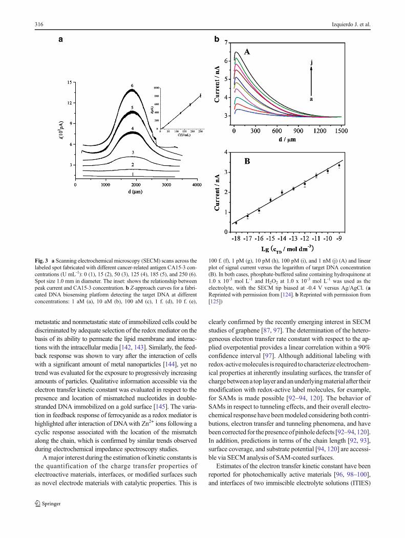
For example, Fig. 3a shows the linear correlation betweenthe amount of an immobilized cancer-related antigen labeledwith horseradish peroxidase and the maximum SECM currentresponse recorded via linear scans [124]. Fig. 3b provides theassociated calibration function for similarly labeledimmobilized DNA obtained by correlation of the maximumcurrent recorded during probe approach [125].
RC-SECM has been applied to determine the oxygen con-tent and concentration gradients in oxygen-consuming sam-ples inmaterials science. For instance, SECMwas used for therapid optimization of O2-electroreduction catalysts assembledas surface spots of different composition [137], and for mon-itoring the spontaneous formation of metal oxide films byquantitative analysis of the oxygen depletion with respect toa fully aerated electrolyte [138]. Estimation of the oxygenconsumption of a living specimen is another relevant applica-tion area of SECM. These studies have allowed the character-ization of the respiratory activity of living cells [110–112,139–141] and embryos [113–115]. Possibly because of thevariability that such living entities commonly exhibit, partic-ular attention has been paid to the reliability of the results viastatistical analysis, including the reporting of uncertainty in-tervals. The most common noninvasive strategy involves theestimation of the vertical profile of the oxygen reduction re-action current proportional to the oxygen concentration be-tween bulk solution and a sample surface. From the data, theconcentration gradient and the respiratory activity can be es-timated according to spherical diffusion theories. For suchstudies, usually repetitive measurements for the estimationof the uncertainty intervals are reported and statistical analy-sis, including, for example, t tests quantifying the significanceof the data obtained, is performed.
As an example, Sugimura et al. [115] investigated the re-spiratory activity of in vitro fertilized porcine and somatic cellnuclear transfer embryos via the annotated analysis of vari-ance, and tests of significance (p < 0.05) during data evalua-tion. This resulted in the box-and-whisker diagramreproduced in Fig. 4 for the oxygen consumption at differentgrowth stages of the embryos after implantation.
The current response during approach can be evaluated byappropriate models to obtain apparent heterogeneous electrontransfer constants for a given redox mediator. Such informationwas used to distinguish cell states from biopsy specimens, butwas also applied for other biomolecules. For example, the
Scanning electrochemical microscopy: an analytical perspective 315
metastatic and nonmetastatic state of immobilized cells could bediscriminated by adequate selection of the redoxmediator on thebasis of its ability to permeate the lipid membrane and interac-tions with the intracellular media [142, 143]. Similarly, the feed-back response was shown to vary after the interaction of cellswith a significant amount of metal nanoparticles [144], yet notrend was evaluated for the exposure to progressively increasingamounts of particles. Qualitative information accessible via theelectron transfer kinetic constant was evaluated in respect to thepresence and location of mismatched nucleotides in double-stranded DNA immobilized on a gold surface [145]. The varia-tion in feedback response of ferrocyanide as a redox mediator ishighlighted after interaction of DNAwith Zn2+ ions following acyclic response associated with the location of the mismatchalong the chain, which is confirmed by similar trends observedduring electrochemical impedance spectroscopy studies.
Amajor interest during the estimation of kinetic constants isthe quantification of the charge transfer properties ofelectroactive materials, interfaces, or modified surfaces suchas novel electrode materials with catalytic properties. This is
clearly confirmed by the recently emerging interest in SECMstudies of graphene [87, 97]. The determination of the hetero-geneous electron transfer rate constant with respect to the ap-plied overpotential provides a linear correlation within a 90%confidence interval [97]. Although additional labeling withredox-activemolecules is required tocharacterize electrochem-ical properties at inherently insulating surfaces, the transfer ofchargebetweena top layerandanunderlyingmaterialafter theirmodification with redox-active label molecules, for example,for SAMs is made possible [92–94, 120]. The behavior ofSAMs in respect to tunneling effects, and their overall electro-chemical response have beenmodeled considering both contri-butions, electron transfer and tunneling phenomena, and havebeencorrected for thepresenceofpinholedefects [92–94, 120].In addition, predictions in terms of the chain length [92, 93],surface coverage, and substrate potential [94, 120] are accessi-ble via SECM analysis of SAM-coated surfaces.
Estimates of the electron transfer kinetic constant have beenreported for photochemically active materials [96, 98–100],and interfaces of two immiscible electrolyte solutions (ITIES)
Fig. 3 a Scanning electrochemical microscopy (SECM) scans across thelabeled spot fabricated with different cancer-related antigen CA15-3 con-centrations (U mL-1): 0 (1), 15 (2), 50 (3), 125 (4), 185 (5), and 250 (6).Spot size 1.0 mm in diameter. The inset: shows the relationship betweenpeak current and CA15-3 concentration. b Z-approach curves for a fabri-cated DNA biosensing platform detecting the target DNA at differentconcentrations: 1 aM (a), 10 aM (b), 100 aM (c), 1 f. (d), 10 f. (e),
100 f. (f), 1 pM (g), 10 pM (h), 100 pM (i), and 1 nM (j) (A) and linearplot of signal current versus the logarithm of target DNA concentration(B). In both cases, phosphate-buffered saline containing hydroquinone at1.0 x 10-3 mol L-1 and H2O2 at 1.0 x 10⁻³ mol L-1 was used as theelectrolyte, with the SECM tip biased at -0.4 V versus Ag/AgCl. (aReprinted with permission from [124]. b Reprinted with permission from[125])
316 Izquierdo J. et al.

[95, 96]. The activity of photocatalysts evaluated via electrontransfer under UV–vis illumination is based on the excitation ofelectrons, and the subsequent formation of electron–hole pairs,which react with the reducible or oxidizable redox mediatorprobed at the SECM electrode [98–100]. From the extractedkinetic data, it is possible to establish the optimum compositionand photochemical conversion conditions, including suitablewavelength or light intensity. In respect to ITIES, the kineticsof the electron exchange between two species—each of themdissolved in one of the immiscible liquid phases—permits theevaluation of the charge transfer across the interface, and thequantification of its eventual inhibition by the formation ofblocking layers [95]. Moreover, combining both types of anal-ysis for the electroanalytical determination of the electron trans-fer abilities at ITIES under illumination was demonstrated byLi and Unwin [96]. In this contribution, the photoinduced ex-citation of tris(bipyridine)ruthenium(II) in the aqueous phasecould be determined at a platinum UME placed in the organicphase following the electron exchange of the ruthenium com-plex with 7,7,8,8-tetracyanoquinodimethane (TCNQ) servingas a redox mediator (Fig. 5a). SECM data fitted with simula-tions, allowing a quantitative determination and linear correla-tion of the fluxes of TCNQ with respect to the light intensity
(Fig. 5b) and derivation of the concentration of redox species inthe organic phase (Fig. 5d) along with the dependence of therate constant on the driving force (Fig. 5c).
An enhanced current response when feedbackmode is usedmay also result from an incremental change in charge or masstransfer processes different from the electron transfer process.Such systems do not follow the same physicochemical modelsestablished for electron transfer yet result in a similarfeedback-type response. Hence, alternative models are neededthat consider these phenomena instead of electron transferprocesses, and that convert the current data into relevant in-formation on the actual surface properties. For instance, thekinetic constants of the enhancing charge transfer phenomena,for example, facilitated ion transfer via ITIES, may be deter-mined with SECM. The corresponding Tafel plot then allowsthe associated transfer coefficient to be estimated [146].
In addition, SI-SECMhas emerged in the last decade, provid-ing an enhancement of the faradaic probe current determined as aresult of the electrochemical conversion of a redoxmediator sim-ilar to feedback-modeSECM.However, thismethod isdevoted tothe interrogationof theextentofasurfacemodificationresulting ingeneratedspecies,whichinfact remainadsorbed.Thus,SI-SECMallows the detection of surface modifications such as the
Fig. 4 Oxygen consumption in the preimplantation in vitro fertilized(IVF) and somatic cell nuclear transfer (SCNT) embryos. The box plotgraphs represent oxygen consumption in the preimplantation IVF (A, C)and SCNT (B, C) embryos. The box indicates two quartiles, namely, the25th and 75th percentiles, and the line indicates the median. The whiskersindicate the maximum and minimum values within the acceptable rangethat is defined by the two quartiles. The circles denote the outliers. Labelson the x-axis in B and D refer to the two-cell stage (2C), four-cell stage
(4C); morula stage (MO), and day 5 (D5BL), day 6 (D6BL), and day 7(D7BL) in the blastocyst stage after in vitro culture. The data on oxygenconsumption per cell at D5BL, D6BL, and D7BL are presented in C andD. The number of evaluated embryos in the respective stages is indicatedin parentheses. Different letters (a, b, c) indicate significant differenceswithin each panel (p < 0.05). Asterisks indicate a significant differencewith respect to oxygen consumption between the IVF and SCNTembryos(p < 0.05). (Reprinted with permission from [115])
Scanning electrochemical microscopy: an analytical perspective 317

photochemical generation of adsorbed OH• radicals, which ex-change electrons with an appropriately selected redox mediator,
thereby enhancing the current response [78]. The linear trend ofthe interrogatedchargewith the timedelaybetween thegeneration
Fig. 5 a Scheme of the scanning electrochemical microscopy/photoelectrochemical setup used in the study of photoinduced electrontransfer processes between tris(bipyridine)ruthenium(II) (Ru(bipy)3
2+)and 7,7,8,8-tetracyanoquinodimethane (TCNQ) at the interfaces of twoimmiscible electrolyte solutions (ITIES). b, c Linear correlation of theflux of TCNQ with light intensity (b) and the concentration of TCNQ(c).d Dependence of the photoinduced electron transfer kinetic constantwith the driving force, given by the sum of the difference in the formal
potentials of the TCNQ0/· and Ru(bipy)32+*/3+ couples (ΔE0’) and the
Galvani potential difference across the ITIES (Δ0w ϕ). 1,2
Dichloromethane (DCE), L/L liquid–liquid, reference electrode (RE),tetrabutylammonium (TBA), tetrakis(4-chlorophenyl) borate (TPBCI),UME ultramicroelectrode. (Reprinted with permission from [96] copy-right 2015 American Chemical Society)
Fig. 6 aReciprocal plot of the interrogation charges of OH• as a functionof delay time. b Plot of the interrogation charges of OH• as a function ofdelay time in the presence of excess MeOH (2 M) in solution. Theinterrogation charge was calculated as the integral of the interrogationcurrent versus time curves acquired after irradiation in 1 mM K2IrCl6
and 0.1 M Na2SO4 aqueous solution. The trend lines (dashed lines) and95% confidence intervals (dotted lines) are drawn to show the slope andy-intercept deviations. (Reprinted with permission from [78] copyright2013 American Chemical Society)
318 Izquierdo J. et al.

of the OH• radicals and the surface interrogation of a bismuthvanadate semiconductor photocatalyst is significantly differentin theabsenceandpresenceofa radical scavenger (e.g.,methanol)within a 95% confidence interval, as illustrated in Fig. 6. This SI-SECM study therefore allowed the determination of the rate con-stant of the reaction between a scavenger and a photochemicallygenerated radical at a photocatalyst surface [78].
Finally, theenhancedmasstransferoccurringatfilmsandmem-branes because of their porosity may be analyzed to obtain quan-titativedata, for example,onpermeability,whichwill significantlyalter the feedback current response [101, 102, 104, 147, 148].SeveralapproachesconceivedforSECMdatatreatmenthavebeenproposed for estimating permeability and porosity values. For in-stance, permeability values were determined by comparing theSECMcurrent response obtained above a sample surface of inter-estwith that obtained under the same conditions above an insulat-ing substrate (i.e., inducing purely negative feedback), which isapplicable, for example, to the study of thin film properties [101,102]. In another example, porous membranes were investigatedaccording to the diffusion across the porous material, and havebeen simulated by numerical approaches. Last but not least,stress-induced permeability of living cells when exposed to cyto-toxic agents was monitored. It was confirmed that such agentsresult in an increase in the permeability of the cell membrane[104, 148]. The extractedvertical profiles of the tip current obtain-edinfeedbackmodewithuseofavarietyofredoxmediatorsreflectthe trends of the cell membrane vulnerability to increasing expo-sure to toxic agents, as deduced from the permeability values ob-tained. Only very recently, a unified theoretical model has beenproposed describing the influence of surface porosity on the feed-back response, allowing the evaluation of all relevant parameterscharacterizing conductive porous layers [105].
Conclusions and perspective
Recent developments toward nanoscale electrochemical imagingin particular using nanopipettes and nanoelectrodes evidentlyrender SECM-based analytical techniques competitive methodsamong the family of SPMs. Nowadays, SECM provides compa-rable temporal and spatial resolution along with the advantage ofmolecular selectivity because of the (electro)chemical informa-tion that can be obtained within a remarkably broad area ofapplications. Although Bconventional^ SECM using microelec-trodes is a valuable technique in electrochemical surface science,it is evident that when quantitative data are reported, relevantanalytical figures of merit are frequently only partially addressed.SECM and Bhyphenated^ SECM techniques have evolved to-ward studying complex problems and samples in at least close toreal world scenarios, as demonstrated by the multitude of biolog-ical systems analyzed by SECM-based techniques. However, toobtain quantitative and traceable data, and for SECM to enter thedomain of routine analysis, parameters such as the reliability and
reproducibility underlined by appropriate analytical statisticshave shifted into the focus of attention. With this contribution,the authors intended to critically review the lack of attention toanalytical figures of merit in the field of SECM, and hope tospark not only interest and discussions, for example, at futureSECM workshops, but also strategies toward improving the re-liability of SECM data, potentially by the introduction of stan-dardized reference samples and methods.
Compliance with ethical standards
Conflict of interest The authors declare that they have no competinginterests.
References
1. Bard AJ, Fu-Ren FF, Kwak J, Lev O. Scanning electrochemicalmicroscopy. Introduction and principles. Anal Chem. 1989;61:132–8. https://doi.org/10.1021/ac00177a011.
2. Bard AJ, Fan FR, Pierce DT, Unwin PR, Wipf DO, Zhou F.Chemical imaging of surfaces with the scanning electrochemicalmicroscope. Science. 1991;254:68–74. https://doi.org/10.1126/science.254.5028.68.
3. Comstock DJ, Elam JW, Pellin MJ, Hersam MC. Integratedultramicroelectrode−nanopipet probe for concurrent scanning elec-trochemical microscopy and scanning ion conductancemicroscopy.Anal Chem. 2010;82:1270–6. https://doi.org/10.1021/ac902224q.
4. Takahashi Y, Shevchuk AI, Novak P, Murakami Y, Shiku H,Korchev YE, et al. Simultaneous noncontact topography and elec-trochemical imaging by SECM/SICM featuring ion current feed-back regulation. J Am Chem Soc. 2010;132:10118–26. https://doi.org/10.1021/ja1029478.
5. Macpherson JV, Unwin PR. Combined scanning electrochemical–atomic force microscopy. Anal Chem. 2000;72:276–85. https://doi.org/10.1021/ac990921w.
6. Kranz C, Friedbacher G, Mizaikofft B, Lugstein A, Smoliner J,Bertagnolli E. Integrating an ultramicroelectrode in an AFM can-tilever: combined technology for enhanced information. AnalChem. 2001;73:2491–500. https://doi.org/10.1021/ac001099v.
7. Treutler TH, Wittstock G. Combination of an electrochemicaltunneling microscope (ECSTM) and a scanning electrochemicalmicroscope (SECM): application for tip-induced modification ofself-assembled monolayers. Electrochim Acta. 2003;48:2923–32.https://doi.org/10.1016/S0013-4686(03)00357-8.
8. Ueda A, Niwa O, Maruyama K, Shindo Y, Oka K, Suzuki K.Neurite imaging of living PC12 cells with scanningelectrochemical/near-field optical/atomic force microscopy.Angew Chem Int Ed. 2007;46:8238–41. https://doi.org/10.1002/anie.200702617.
9. Wang L, Kranz C, Mizaikoff B. Monitoring scanning electro-chemical microscopy approach curves with mid-infrared spectros-copy : toward a novel current-independent positioningmode. AnalChem. 2010;82:3132–8. https://doi.org/10.1021/ac9027802.(2).
10. Wang L, Kowalik J, Mizaikoff B, Kranz C. Combining scanningelectrochemical microscopy with infrared attenuated total reflectionspectroscopy for in situ studies of electrochemically induced processes.Anal Chem. 2010;82:3139–45. https://doi.org/10.1021/ac9027802.
11. Etienne M, Dossot M, Grausem J, Herzog G. Combined Ramanmicrospectrometer and shearforce regulated SECM for corrosionand self-healing analysis. Anal Chem. 2014;86:11203–10. https://doi.org/10.1021/ac502670t.
Scanning electrochemical microscopy: an analytical perspective 319

12. Momotenko D, Qiao L, Cortés-Salazar F, Lesch A, Wittstock G,Girault HH. Electrochemical push−pull scanner with mass spec-trometry detection. Anal Chem. 2012;84:6630–7. https://doi.org/10.1021/ac300999v.
13. Keddam M, Portail N, Trinh D, Vivier V. Progress in scanningelectrochemical microscopy by coupling with electrochemical im-pedance and quartz crystal microbalance. ChemPhysChem.2009;10:3175–82. https://doi.org/10.1002/cphc.200900506.
14. MirkinMV,HorrocksBR.Electroanalyticalmeasurements using thescanning electrochemical microscope. Anal Chim Acta. 2000;406:119–46. https://doi.org/10.1016/S0003-2670(99)00630-3.
15. Amemiya S, Guo J, Xiong H. Biological applications of scanningelectrochemical microscopy: chemical imaging of single livingcells and beyond. Anal Bioanal Chem. 2006;386:458–71. https://doi.org/10.1007/s00216-006-0510-6.
16. Sun P, Laforge FO,MirkinMV. Scanning electrochemical micros-copy in the 21st century. Phys Chem Chem Phys. 2007;9:802–23.https://doi.org/10.1039/b612259k.
17. Amemiya S, Bard AJ, Fan F-RF, Mirkin MV, Unwin PR.Scanning electrochemical microscopy. Annu Rev Anal Chem.2008;1:95–131. https://doi.org/10.1146/annurev.anchem.1.031207.112938.
18. Bergner S, Vatsyayan P, Matysik F-M. Recent advances in highresolution scanning electrochemical microscopy of living cells – areview. Anal Chim Acta. 2013;775:1–13. https://doi.org/10.1016/j.aca.2012.12.042.
19. Kranz C. Recent advancements in nanoelectrodes andnanopipettes used in combined scanning electrochemical micros-copy techniques. Analyst. 2014;139:336–52. https://doi.org/10.1039/c3an01651j.
20. Ventosa E, Schuhmann W. Scanning electrochemical microscopyof Li-ion batteries. Phys Chem Chem Phys. 2015;17:28441–50.https://doi.org/10.1039/C5CP02268A.
21. Li Y, Ning X, Ma Q, Qin D, Lu X. Recent advances in electro-chemistry by scanning electrochemical microscopy. Trends AnalChem. 2016;80:242–54. https://doi.org/10.1016/j.trac.2016.02.002.
22. Zoski CG. Review—advances in scanning electrochemical mi-croscopy (SECM). J Electrochem Soc. 2016;163:H3088–100.https://doi.org/10.1149/2.0141604jes.
23. Polcari D, Dauphin-Ducharme P, Mauzeroll J. Scanning electro-chemical microscopy: a comprehensive review of experimentalparameters from 1989 to 2015. Chem Rev. 2016;116:13234–78.https://doi.org/10.1021/acs.chemrev.6b00067.
24. Takahashi Y, Kumatani A, Shiku H, Matsue T. Scanning probemicroscopy for nanoscale electrochemical imaging. Anal Chem.2017;89:342–57. https://doi.org/10.1021/acs.analchem.6b04355.
25. Clausmeyer J, Schuhmann W, Plumeré N. Electrochemical pat-terning as a tool for fabricating biomolecule microarrays. TrendsAnal Chem. 2014;58:23–30. https://doi.org/10.1016/j.trac.2014.03.004.
26. Barton ZJ, Rodríguez-López J. Lithium ion quantification usingmercury amalgams as in situ electrochemical probes in nonaque-ous media. Anal Chem. 2014;86:10660–7. https://doi.org/10.1021/ac502517b.
27. Barton ZJ, Rodríguez-López J. Fabrication and demonstration ofmercury disc-well probes for stripping-based cyclic voltammetryscanning electrochemical microscopy. Anal Chem. 2017;89:2716–23. https://doi.org/10.1021/acs.analchem.6b04022.
28. Souto RM, González-García Y, Battistel D, Daniele S. In situscanning electrochemical microscopy (SECM) detection of metaldissolution during zinc corrosion by means of mercury sphere-capmicroelectrode tips. Chem Eur J. 2012;18:230–6. https://doi.org/10.1002/chem.201102325.
29. Wei C, Bard AJ, Nagy G, Toth K. Scanning electrochemical mi-croscopy. 28. Ion-selective neutral carrier-based microelectrode
potentiometry. Anal Chem. 1995;67:1346–56. https://doi.org/10.1021/ac00104a008.
30. Etienne M, Schulte A, Mann S, Jordan G, Dietzel ID, SchuhmannW. Constant-distance mode scanning potentiometry. 1.Visualization of calcium carbonate dissolution in aqueous solu-tion. Anal Chem. 2004;76:3682–8. https://doi.org/10.1021/ac0349227.
31. Ummadi JG, Downs CJ, Joshi VS, Ferracane JL, Koley D.Carbon-based solid-state calcium ion-selective microelectrodeand scanning electrochemical microscopy: a quantitative studyof pH-dependent release of calcium ions from bioactive glass.Anal Chem. 2016;88:3218–26. https://doi.org/10.1021/acs.analchem.5b04614.
32. Wipf DO, Ge F, Spaine TW, Baur JE. Microscopic measurementof pH with iridium oxide microelectrodes. Anal Chem. 2000;72:4921–7. https://doi.org/10.1021/ac000383j.
33. Filotás D, Fernández-Pérez BM, Izquierdo J, Nagy L, Nagy G,Souto RM. Combined amperometric/potentiometric probes forimproved chemical imaging of corroding surfaces using scanningelectrochemical microscopy. Electrochim Acta. 2016;221:48–55.https://doi.org/10.1016/j.electacta.2016.10.142.
34. Janotta M, Rudolph D, Kueng A, Kranz C, Voraberger H-S,Waldhauser W, Mizaikoff B. Analysis of corrosion processes atthe surface of diamond-like carbon protected zinc selenide wave-guides. Langmuir. 2004;20:8634–40. https://doi.org/10.1021/la049042h.
35. Rudolph D, Bates D, DiChristina TJ, Mizaikoff B, Kranz C.Detection of metal-reducing enzyme complexes by scanning elec-trochemical microscopy. Electroanalysis. 2016;28:2459–65.https://doi.org/10.1002/elan.201600333.
36. Bandarenka AS, Maljusch A, Kuznetsov V, Eckhard K,Schuhmann W. Localized impedance measurements for electro-chemical surface science. J Phys Chem C. 2014;118:8952–9.https://doi.org/10.1021/jp412505p.
37. Ballesteros B, Schulte A, Calvo EJ, Koudelka-Hep M,SchuhmannW. Localised electrochemical impedance spectrosco-py with high lateral resolution by means of alternating currentscanning electrochemical microscopy. Electrochem Commun.2002;4:134–8. https://doi.org/10.1016/S1388-2481(01)00294-6.
38. Nogala W, Szot K, Burchardt M, Roelfs F, Rogalski J, Opallo M,Wittstock G. Feedbackmode SECM study of laccase and bilirubinoxidase immobilised in a sol-gel processed silicate film. Analyst.2010;135:2051–8. https://doi.org/10.1039/c0an00068j.
39. Katemann BB, SchuhmannW. Fabrication and characterization ofneedle-type. Electroanalysis. 2002;14:22–8. https://doi.org/10.1002/1521-4109(200201)14:1<22::AID-ELAN22>3.0.CO;2-F.
40. Zhang Y, Clausmeyer J, Babakinejad B, López Córdoba A, Ali T,Shevchuk A, et al. Spearhead nanometric field-effect transistorsensors for single-cell analysis. ACS Nano. 2016;10:3214–21.https://doi.org/10.1021/acsnano.5b05211.
41. KangM,Momotenko D, Page A, Perry D, Unwin PR. Frontiers innanoscale electrochemical imaging: faster, multifunctional, andultrasensitive. Langmuir. 2016;32:7993–8008. https://doi.org/10.1021/acs.langmuir.6b01932.
42. Nadappuram PB, McKelvey K, Byers JC, Güell AG, ColburnAW, Lazenby RA, Unwin, PR. Quad-barrel multifunctional elec-trochemical and ion conductance probe for voltammetric analysisand imaging. Anal Chem. 2015;87:3566–73. https://doi.org/10.1021/acs.analchem.5b00379.
43. Kim J, Shen M, Nioradze N, Amemiya S. Stabilizing nanometerscale tip-to-substrate gaps in scanning electrochemical microscopyusing an isothermal chamber for thermal drift suppression. AnalChem. 2012;84:3489–92. https://doi.org/10.1021/ac300564g.
44. Schmidt I, Plettenberg I, Kimmich D, Ellis H, Witt J, Dosche C,Wittstock, G. Spatially resolved analysis of screen printedphotoanodes of dye-sensitized solar cells by scanning
320 Izquierdo J. et al.
electrochemical microscopy. Electrochim Acta. 2016;222:735–46. https://doi.org/10.1016/j.electacta.2016.11.030 .
45. Bülter H, Peters F, Schwenzel J, Wittstock G. Comparison ofelectron transfer properties of the SEI on graphite composite andmetallic lithium electrodes by SECM at OCP. J Electrochem Soc.2015;162:A7024–36. https://doi.org/10.1149/2.0031513jes.
46. Chen C-C, Zhou Y, Baker LA. Scanning ion conductance micros-copy. Annu Rev Anal Chem. 2012;5:207–28. https://doi.org/10.1146/annurev-anchem-062011-143203.
47. Abbou J, Demaille C, Druet M, Moiroux J. Fabrication ofsubmicrometer-sized gold electrodes of controlled geometry forscanning electrochemical-atomic force microscopy. Anal Chem.2002;74:1627–14. https://doi.org/10.1021/ac020385z.
48. Knittel P, Mizaikoff B, Kranz C. Simultaneous nanomechanicaland electrochemical mapping: combining peak force tappingatomic force microscopy with scanning electrochemical micros-copy. Anal Chem. 2016;88:6174–8. https://doi.org/10.1021/acs.analchem.6b01086.
49. Şen M, Takahashi Y, Matsumae Y, Horiguchi Y, Kumatani A, InoK, et al. Improving the electrochemical imaging sensitivity ofscanning electrochemical microscopy-scanning ion conductancemicroscopy by using electrochemical Pt deposition. Anal Chem.2015;87:3484–9. https://doi.org/10.1021/acs.analchem.5b00027.
50. Morris CA, Chen C-C, Baker LA. Transport of redox probesthrough single pores measured by scanning electrochemical-scanning ion conductance microscopy (SECM-SICM). Analyst.2012;137:2933–8. https://doi.org/10.1039/c2an16178h.
51. O’Connell MA, Wain AJ. Mapping electroactivity at individualcatalytic nanostructures using high-resolution scanningelectrochemical-scanning ion conductance microcopy. AnalChem. 2014;86:12100–7. https://doi.org/10.1021/ac502946q.
52. Takahashi Y, Shevchuk AI, Novak P, Zhang Y, Ebejer N,MacPherson JV, et al. Multifunctional nanoprobes for nanoscalechemical imaging and localized chemical delivery at surfaces andinterfaces. Angew Chem Int Ed. 2011;50:9638–42. https://doi.org/10.1002/anie.201102796.
53. Perry D, Al Botros R, Momotenko D, Kinnear SL, Unwin PR.Simultaneous nanoscale surface charge and topographical map-ping. ACS Nano. 2015;9:7266–76. https://doi.org/10.1021/acsnano.5b02095.
54. Kleijn SEF, Lai SCS, Miller TS, Yanson AI, Koper MTM, UnwinPR. Landing and catalytic characterization of individual nanopar-ticles on electrode surfaces. J AmChem Soc. 2012;134:18558–61.https://doi.org/10.1021/ja309220m.
55. Kinnear SL, McKelvey K, Snowden ME, Peruffo M, ColburnAW, Unwin PR. Dual-barrel conductance micropipet as a newapproach to the study of ionic crystal dissolution kinetics.Langmuir. 2013;29:15565–72. https://doi.org/10.1021/la403630u.
56. Williams CG, Edwards MA, Colley AL, Macpherson JV, UnwinPR. Scanning micropipet contact method for high-resolution im-aging of electrode surface redox activity. Anal Chem. 2009;81:2486–95. https://doi.org/10.1021/ac802114r.
57. Ebejer N, Schnippering M, Colburn AW, Edwards MA, UnwinPR. Localized high resolution electrochemistry and multifunction-al imaging: scanning electrochemical cell microscopy. AnalChem. 2010;82:9141–5. https://doi.org/10.1021/ac102191u.
58. Ebejer N, Güell AG, Lai SCS, McKelvey K, Snowden ME,Unwin PR. Scanning electrochemical cell microscopy: a versatiletechnique for nanoscale electrochemistry and functional imaging.Annu Rev Anal Chem. 2013;6:329–51. https://doi.org/10.1146/annurev-anchem-062012-092650.
59. EckhardK, SchuhmannW.Alternating current techniques in scan-ning electrochemical microscopy (AC-SECM). Analyst.2008;133:1486–97. https://doi.org/10.1039/b806721j.
60. McKelvey K, Snowden ME, Peruffo M, Unwin PR. Quantitativevisualization of molecular transport through porous membranes:enhanced resolution and contrast using intermittent contact-scanning electrochemical microscopy. Anal Chem. 2011;83:6447–54. https://doi.org/10.1021/ac201489c.
61. Wang Q, Rodríguez-López J, Bard AJ. Reaction of Br2 withadsorbed CO on Pt, studied by the surface interrogation mode ofscanning electrochemical microscopy. J Am Chem Soc.2009;131:17046–7. https://doi.org/10.1021/ja907626t.
62. Eckhard K, Chen X, Turcu F, Schuhmann W. Redox competitionmode of scanning electrochemical microscopy (RC-SECM) forvisualisation of local catalytic activity. Phys Chem Chem Phys.2006;8:5359–65. https://doi.org/10.1039/b609511a.
63. Plettenberg I,WittstockG. Combined detection of electrochemicalreactions and topographical effects - imaging with scanning ohmicmicroscopy. Electrochim Acta. 2016;197:318–29. https://doi.org/10.1016/j.electacta.2015.12.033.
64. Plettenberg I, Wittstock G. Direct local mapping of ion transferreactions by scanning ohmic microscopy. Energy Technol.2016;4:1495–501. https://doi.org/10.1002/ente.201600166.
65. Bard AJ, Fu-Ren FF. Electrochemical detection of single mole-cules. Acc Chem Res. 1996;29:572–8. https://doi.org/10.1021/ar9502442.
66. Perry D, Momotenko D, Lazenby RA, Kang M, Unwin PR.Characterization of nanopipettes. Anal Chem. 2016;88:5523–30.https://doi.org/10.1021/acs.analchem.6b01095.
67. Nioradze N, Chen R, Kim J, Shen M, Santhosh P, Amemiya S.Origins of nanoscale damage to glass-sealed platinum electrodeswith submicrometer and nanometer size. Anal Chem. 2013;85:6198–202. https://doi.org/10.1021/ac401316n.
68. Yu Y, Noël JM, Mirkin MV, Gao Y, Mashtalir O, Friedman G,et al. Carbon pipette-based electrochemical nanosampler. AnalChem. 2014;86:3365–72. https://doi.org/10.1021/ac403547b.
69. Mauzeroll J, Hueske EA, Bard AJ. Scanning electrochemical mi-croscopy. 48. Hg/Pt hemispherical ultramicroelectrodes: fabrica-tion and characterization. Anal Chem. 2003;75:3880–9. https://doi.org/10.1021/ac034088l.
70. Mirkin MV, Sun T, Yu Y, Zhou M. Electrochemistry at one nano-particle. Acc Chem Res. 2016;49:2328–35. https://doi.org/10.1021/acs.accounts.6b00294.
71. Kim J, Renault C, Nioradze N, Arroyo-Currás N, Leonard KC,Bard AJ. Electrocatalytic activity of individual Pt nanoparticlesstudied by nanoscale scanning electrochemical microscopy. JAm Chem Soc. 2016;138:8560–8. https://doi.org/10.1021/jacs.6b03980.
72. Sun T, Yu Y, Zacher BJ, Mirkin MV. Scanning electrochemicalmicroscopy of individual catalytic nanoparticles. AngewChem IntEd. 2014;53:14120–3. https://doi.org/10.1002/anie.201408408.
73. Kim J, Renault C, Nioradze N, Arroyo-Currás N, Leonard KC,Bard AJ. Nanometer scale scanning electrochemical microscopyinstrumentation. Anal Chem. 2016;88:10284–9. https://doi.org/10.1021/acs.analchem.6b03024.
74. Keysight Technologies. Combining atomic force microscopy withscanning electrochemical microscopy. Application note. 2015.http://literature.cdn.keysight.com/litweb/pdf/5991-4600EN.pdf?id=2468595.
75. Schäfer D, Puschhof A, SchuhmannW. Scanning electrochemicalmicroscopy at variable temperatures. Phys Chem Chem Phys.2013;15:5215–23. https://doi.org/10.1039/c3cp43520b.
76. Bülter H, Peters F, Wittstock G. Scanning electrochemical micros-copy for the in situ characterization of solid-electrolyte inter-phases: highly oriented pyrolytic graphite versus graphite compos-ite. Energy Technol. 2016;4:1486–94. https://doi.org/10.1002/ente.201600071.
77. Zampardi G, Ventosa E, La Mantia F, Schuhmann W. In situvisualization of Li-ion intercalation and formation of the solid
Scanning electrochemical microscopy: an analytical perspective 321

electrolyte interphase on TiO2 based paste electrodes using scan-ning electrochemical microscopy. Chem Commun. 2013;49:9347–9. https://doi.org/10.1039/c3cc44576c.
78. Park HS, Leonard KC, Bard AJ. Surface interrogation scanninge l e c t r o c h e m i c a l m i c r o s c o p y ( S I - S E CM ) o fphotoelectrochemistry at a W/Mo-BiVO4 semiconductor elec-trode: quantification of hydroxyl radicals during water oxidation.J Phys Chem C. 2013;117:12093–102. https://doi.org/10.1021/jp400478z.
79. Conzuelo F, Sliozberg K, Gutkowski R, Grü S, Nebel M,SchuhmannW. High-resolution analysis of photoanodes for watersplitting by means of scanning photoelectrochemical microscopy.Anal Chem. 2017;89:1222–8. https://doi.org/10.1021/acs.analchem.6b03706.
80. Bard AJ. New challenges in electrochemistry and electroanalysis.Pure Appl Chem. 1992;64:185–92. doi:https://doi.org/10.1351/pac199264020185.
81. Thompson M, Ellison SLR, Wood R. Harmonized guidelines forsingle-laboratory validation of methods of analysis (IUPAC tech-nical report). Pure Appl Chem. 2002;74:835–55. https://doi.org/10.1351/pac200274050835.
82. ISO/TC 201/SC 9 scanning probe microscopy.83. ISO 11952:2014 surface chemical analysis – scanning-probe mi-
croscopy – determination of geometric quantities using SPM: cal-ibration of measuring systems.
84. ISO 11039:2012 surface chemical analysis – scanning-probe mi-croscopy – measurement of drift rate.
85. ISO 13083:2015 surface chemical analysis – scanning probe mi-croscopy – standards on the definition and calibration of spatialresolution of electrical scanning probe microscopes (ESPMs) suchas SSRM and SCM for 2D-dopant imaging and other purposes.
86. Yu Y, Sun T, Mirkin MV. Toward more reliable measurements ofelectron-transfer kinetics at nanoelectrodes: next approximation.Anal Chem. 2016;88:11758–66. https://doi.org/10.1021/acs.analchem.6b03392.
87. Molina J, Fernández J, Cases F. Scanning electrochemical micros-copy for the analysis and patterning of graphene materials: a re-view. Synth Met. 2016;222:145–61. https://doi.org/10.1016/j.synthmet.2016.10.019.
88. Güell AG, Ebejer N, Snowden ME, Macpherson JV, Unwin PR.Structural correlations in heterogeneous electron transfer at mono-layer and multilayer graphene electrodes. J Am Chem Soc.2012;134:7258–61. https://doi.org/10.1021/ja3014902 .
89. Yu Y, Sun T,MirkinMV. Scanning electrochemical microscopy ofsingle spherical nanoparticles: theory and particle size evaluation.Anal Chem. 2015;87:7446–53. https://doi.org/10.1021/acs.analchem.5b01690.
90. Ma W, Hu K, Chen Q, Zhou M, Mirkin MV, Bard AJ.Electrochemical size measurement and characterization of elec-trodeposited platinum nanoparticles at nanometer resolution withscanning electrochemical microscopy. Nano Lett. 2017;17:4354–8. https://doi.org/10.1021/acs.nanolett.7b01437.
91. Lefrou C, Cornut R. Analytical expressions for quantitative scan-ning electrochemical microscopy (SECM). ChemPhysChem.2010;11:547–56. https://doi.org/10.1002/cphc.200900600.
92. Peterson RR, Cliffel DE. Scanning electrochemical microscopydetermination of organic soluble MPC electron-transfer rates.Langmuir. 2006;22:10307–14. https://doi.org/10.1021/la061183r.
93. Kiani A, Alpuche-Aviles MA, Eggers PK, Jones M, Gooding JJ,Paddon-Row MN, et al. Scanning electrochemical microscopy.59. Effect of defects and structure on electron transfer throughself-assembled monolayers. Langmuir. 2008;24:2841–9. https://doi.org/10.1021/la702811t.
94. Salamifar SE, Mehrgardi MA, Kazemi SH, Mousavi MF. Cyclicvoltammetry and scanning electrochemical microscopy studies ofmethylene blue immobilized on the self-assembled monolayer of
n-dodecanethiol. Electrochim Acta. 2010;56:896–904. https://doi.org/10.1016/j.electacta.2010.08.068.
95. Delville M-H, Tsionsky M, Bard AJ. Scanning electrochemicalmicroscopy studies of electron transfer through monolayers con-taining conjugated species at the liquid−liquid interface.Langmuir. 1998;14:2774–9. https://doi.org/10.1021/la971247l.
96. Li F, Unwin PR. Scanning electrochemical microscopy (SECM)of photoinduced electron transfer kinetics at liquid/liquid inter-faces. J Phys Chem C. 2015;119:4031–43. https://doi.org/10.1021/jp510333d.
97. Ritzert NL, Rodríguez-López J, Tan C, Abruña HD. Kinetics ofinterfacial electron transfer at single-layer graphene electrodes inaqueous and nonaqueous solutions. Langmuir. 2013;29:1683–94.https://doi.org/10.1021/la3042549.
98. Lu X, Hu Y,WangW, Du J, He H, Ai R, et al. A novel platform tostudy the photoinduced electron transfer at a dye-sensitized solid/liquid interface. Colloids Surf B. 2013;103:608–14. https://doi.org/10.1016/j.colsurfb.2012.11.013.
99. Jiang Y, Qin D-D, Fan Y-R, Guo H-X, Wang S-X, Ning X-M,et al. Investigation of photoinduced electron transfer on TiO2
nanowire arrays/porphyrin composite via scanning electrochemi-cal microscopy. RSC Adv. 2015;5:56697–703. https://doi.org/10.1039/C5RA08485G.
100. Ning X, Ma L, Zhang S, Qin D, Shan D, Hu Y, et al. Constructionof a porphyrin-based nanohybrid as an analogue of chlorophyllprotein complexes and its light-harvesting behavior research. JPhys Chem C. 2016;120:919–26. https://doi.org/10.1021/acs.jpcc.5b11246.
101. Williams ME, Stevenson KJ, Massari AM, Hupp JT. Imagingsize-selective permeation through micropatterned thin films usingscanning electrochemical microscopy. Anal Chem. 2000;72:3122–8. https://doi.org/10.1021/ac9914622.
102. Williams ME, Hupp JT. Scanning electrochemical microscopyassessment of rates of molecular transport through mesoporousthin-films of porphyrinic Bmolecular squares.^. J Phys Chem B.2001;105:8944–50. https://doi.org/10.1021/jp010881b.
103. Zocher F, Van Der Spoel D, Pohl P, Hub JS. Local partition coef-ficients govern solute permeability of cholesterol-containingmembranes. Biophys J. 2013;105:2760–70. https://doi.org/10.1016/j.bpj.2013.11.003.
104. Li MSM, Filice FP, Henderson JD, Ding Z. Probing Cd2+-stressedlive cell membrane permeability with various redox mediators inscanning electrochemical microscopy. J Phys Chem C. 2016;120:6094–103. https://doi.org/10.1021/acs.jpcc.6b00453.
105. Kuss C, Payne NA, Mauzeroll J. Probing passivating porous filmsby scanning electrochemical microscopy. J Electrochem Soc.2016;163:H3066–71. https://doi.org/10.1149/2.0131604jes.
106. Compton RG, Banks CE. Voltammetry at microelectrodes. In:Compton RG, Banks CE, editors. Understanding voltammetry.Singapore: World Scientific. 2007;p. 151–92.
107. Sun T, Blanchard PY, Mirkin MV. Cleaning nanoelectrodes withair plasma. Anal Chem. 2015;87:4092–5. https://doi.org/10.1021/acs.analchem.5b00488.
108. Jacobse L, Raaijman SJ, Koper MTM. The reactivity of platinummicroelectrodes. Phys Chem Chem Phys. 2016;18:28451–7.https://doi.org/10.1039/C6CP05361K.
109. Cornut R, Lefrou C. New analytical approximation of feedbackapproach curves with a microdisk SECM tip and irreversible ki-netic reaction at the substrate. J Electroanal Chem. 2008;621:178–84. https://doi.org/10.1016/j.jelechem.2007.09.021.
110. Torisawa Y, Kaya T, Takii Y, Oyamatsu D, Nishizawa M, MatsueT. Scanning electrochemical microscopy-based drug sensitivitytest for a cell culture integrated in silicon microstructures. AnalChem. 2003;75:2154–8. https://doi.org/10.1021/ac026317u.
111. Sugimura S, Matoba S, Hashiyada Y, Aikawa Y, Ohtake M,Matsuda H, et al. Oxidative phosphorylation-linked respiration
322 Izquierdo J. et al.
in individual bovine oocytes. J Reprod Dev. 2012;58:636–41.https://doi.org/10.1262/jrd.2012-082.
112. Vasanthan KS, Sethuraman S, Parthasarathy M. Electrochemicalevidence for asialoglycoprotein receptor - mediated hepatocyteadhesion and proliferation in three dimensional tissue engineeringscaffolds. Anal Chim Acta. 2015;890:83–90. https://doi.org/10.1016/j.aca.2015.06.057.
113. Shiku H, Shiraishi T, Ohya H, Matsue T, Abe H, Hoshi H, et al.Oxygen consumption of single bovine embryos probed by scan-ning electrochemical microscopy. Anal Chem. 2001;73:3751–8.https://doi.org/10.1021/ac010339j.
114. Shiku H, TorisawaYS, Takagi A, Aoyagi S, Abe H, Hoshi H, et al.Metabolic and enzymatic activities of individual cells, spheroidsand embryos as a function of the sample size. Sens Actuators B.2005;108:597–602. https://doi.org/10.1016/j.snb.2004.12.030.
115. Sugimura S, Yokoo M, Yamanaka K, Kawahara M, Moriyasu S,Wakai T, et al. Anomalous oxygen consumption in porcine somat-ic cell nuclear transfer embryos. Cell Reprogram. 2010;12:463–74. https://doi.org/10.1089/cell.2009.0111.
116. Amatore C, Arbault S, Bouton C, Coffi K, Drapier JC, GhandourH, et al. Monitoring in real time with a microelectrode the releaseof reactive oxygen and nitrogen species by a single macrophagestimulated by its membrane mechanical depolarization.ChemBioChem. 2006;7:653–61. https://doi.org/10.1002/cbic.200500359.
117. Borgmann S, Radtke I, Erichsen T, Blöchl A, Heumann R,Schuhmann W. Electrochemical high-content screening of nitricoxide release from endothelial cells. ChemBioChem. 2006;7:662–8. https://doi.org/10.1002/cbic.200500399.
118. Thompson M, Ellison SLR, Fajgelj A, Willetts P, Wood R.Harmonized guidelines for the use of recovery information inanalytical measurement. Pure Appl Chem. 1999;71:337–48.https://doi.org/10.1351/pac199971020337.
119. Abucayon E, Ke N, Cornut R, Patelunas A, Miller D, NishiguchiMK, et al. Investigating catalase activity through hydrogen perox-ide decomposition by bacteria biofilms in real time using scanningelectrochemical microscopy. Anal Chem. 2014;86:498–505.https://doi.org/10.1021/ac402475m.
120. Holt KB. Using scanning electrochemical microscopy (SECM) tomeasure the electron-transfer kinetics of cytochrome cimmobilized on a COOH-terminated alkanethiol monolayer on agold electrode. Langmuir. 2006;22:4298–304. https://doi.org/10.1021/la0529916.
121. Zhao C, Sinha JK, Wijayawardhana CA, Wittstock G. Monitoringβ-galactosidase activity by means of scanning electrochemicalmicroscopy. J Electroanal Chem. 2004;561:83–91. https://doi.org/10.1016/j.jelechem.2003.07.004.
122. Kirchner CN, Szunerits S, Wittstock G. Scanning electrochemicalmicroscopy (SECM) based detection of oligonucleotide hybridi-zation and simultaneous determination of the surface concentra-tion of immobilized oligonucleotides on gold. Electroanalysis.2007;19:1258–67. https://doi.org/10.1002/elan.200703862.
123. Zhou Y, Ino K, Shiku H, Matsue T. Evaluation of senescence inindividual MCF-7 spheroids based on electrochemical measure-ment of senescence-associated β-galactosidase activity.Electrochim Acta. 2015;186:449–54. https://doi.org/10.1016/j.electacta.2015.10.115.
124. Zhang X, Peng X, Jin W. Scanning electrochemical microscopywith enzyme immunoassay of the cancer-related antigen CA15-3.Anal Chim Acta. 2006;558:110–4. https://doi.org/10.1016/j.aca.2005.11.032.
125. Chen B, Hu Q, Xiong Q, Zhang F, He P. An ultrasensitive scan-ning electrochemical microscopy (SECM)-based DNA biosensingplatform amplified with the long self-assembled DNAconcatemers. Electrochim Acta. 2016;192:127–32. https://doi.org/10.1016/j.electacta.2015.12.102.
126. Razzaghi F, Seguin J, Amar A, Griveau S, Bedioui F. Biologicalcell morphology studies by scanning electrochemical microscopyimagery at constant height: contrast enhancement using biocom-patible conductive substrates. Electrochim Acta. 2015;157:95–100. https://doi.org/10.1016/j.electacta.2015.01.033.
127. Meyer JN, Mathew MT, Wimmer MA, Lesuer RJ. Effect oftribolayer formation on corrosion of CoCrMo alloys investigatedusing scanning electrochemical microscopy. Anal Chem.2013;85:7159–66. https://doi.org/10.1021/ac400823q.
128. Rapino S, Marcu R, Bigi A, Soldà A, Marcaccio M, Paolucci F,et al. Scanning electro-chemical microscopy reveals cancer cellredox state. Electrochim Acta. 2015;179:65–73. https://doi.org/10.1016/j.electacta.2015.04.053.
129. Holmes JL, Davis F, Collyer SD, Higson SPJ. A new applicationof scanning electrochemical microscopy for the label-free interro-gation of antibody-antigen interactions: Part 2. Anal Chim Acta.2012;741:1–8. https://doi.org/10.1016/j.aca.2012.07.004 .
130. Weng Y-C, Chang H. Screening and characterization for the opti-mization of CdS-based photocatalysts. RSC Adv. 2016;6:41376–84. https://doi.org/10.1039/c6ra05245b.
131. Weng Y-C, Chou Y-D. Discovery and optimization ofZn0.3Cd0.7S-based photocatalysts by scanning electrochemicalmicroscopy and characterization of potential photocatalysts.Electrochim Acta. 2015;153:416–25. https://doi.org/10.1016/j.electacta.2014.12.015 .
132. Lee S, Zhang Y, White HS, Harrell CC, Martin CR.Electrophoretic capture and detection of nanoparticles at theopening of a membrane pore using scanning electrochemical mi-croscopy. Anal Chem. 2004;76:6108–15. https://doi.org/10.1021/ac049147p.
133. Kwon SJ, Bard AJ. Analysis of diffusion-controlled stochasticevents of iridium oxide single nanoparticle collisions by scanningelectrochemical microscopy. J Am Chem Soc. 2012;134:7102–8.https://doi.org/10.1021/ja300894f.
134. Chen CH, Ravenhill ER, Momotenko D, Kim YR, Lai SCS,Unwin PR. Impact of surface chemistry on nanoparticle-electrode interactions in the electrochemical detection of nanopar-ticle collisions. Langmuir. 2015;31:11932–42. https://doi.org/10.1021/acs.langmuir.5b03033.
135. Pyo M, Bard AJ. Scanning electrochemical microscopy. 35.Determination of diffusion coefficients and concentrations ofRu(NH3)6
3+ and methylene blue in polyacrylamide films bychronoamperometry at ultramicrodisk electrodes. Electrochim Acta.1997;42:3077–83. https://doi.org/10.1016/s0013-4686(97)00155-2.
136. Csóka B, Nagy G. Determination of diffusion coefficient in geland in aqueous solutions using scanning electrochemical micros-copy. J Biochem Biophys Methods. 2004;61:57–67. https://doi.org/10.1016/j.jbbm.2004.03.001.
137. Fernández JL, Mano N, Heller A, Bard AJ. Optimization ofBwired^ enzyme O2-electroreduction catalyst compositions byscanning electrochemical microscopy. Angew Chem Int Ed.2004;43:6355–7. https://doi.org/10.1002/anie.200461528.
138. Gilbert JL, Zarka L, Chang E, Thomas CH. The reduction half cellin biomaterials corrosion: Oxygen diffusion profiles near and cellresponse to polarized titanium surfaces. J Biomed Mater Res.1998;42:321–30. https://doi.org/10.1002/(SICI)1097-4636(199811)42:2<321::AID-JBM18>3.0.CO;2-L.
139. Kaya T, Torisawa YS, Oyamatsu D, Nishizawa M, Matsue T.Monitoring the cellular activity of a cultured single cell by scan-ning electrochemical microscopy (SECM). A comparison withfluorescence viability monitoring. Biosens Bioelectron. 2003;18:1379–83. https://doi.org/10.1016/S0956-5663(03)00083-6.
140. Torisawa YS, Shiku H, Kasai S, Nishizawa M, Matsue T.Proliferation assay on a silicon chip applicable for tumors extir-pated from mammalians. Int J Cancer. 2004;109:302–8. https://doi.org/10.1002/ijc.11693.
Scanning electrochemical microscopy: an analytical perspective 323

141. Torisawa Y, Nashimoto Y, Yasukawa T, Shiku H, Matsue T.Regulation and characterization of the polarity of cells embeddedin a reconstructed basement matrix using a three-dimensional mi-cro-culture system. Biotechnol Bioeng. 2007;97:615–21. https://doi.org/10.1002/bit.21274.
142. Liu B, Rotenberg SA, Mirkin MV. Scanning electrochemical mi-croscopy of living cells: Different redox activities of nonmetastaticand metastatic human breast cells. Proc Natl Acad Sci U S A.2000;97:9855–60. https://doi.org/10.1073/pnas.97.18.9855.
143. Rotenberg SA, Mirkin M V. Scanning electrochemical microsco-py: detection of human breast cancer cells by redox environment. JMammary Gland Biol Neoplasia. 2004;9:375–82. doi:https://doi.org/10.1007/s10911-004-1407-7.
144. Chen Z, Xie S, Shen L, DuY, He S, Li Q, et al. Investigation of theinteractions between silver nanoparticles and Hela cells by scan-ning electrochemical microscopy. Analyst. 2008;133:1221–8.https://doi.org/10.1039/b807057a.
145. Alam MN, Shamsi MH, Kraatz H-B. Scanning positional varia-tions in single-nucleotide polymorphism of DNA: an electrochem-ical study. Analyst. 2012;137:4220–5. https://doi.org/10.1039/c2an35420a.
146. Li F, Chen Y, Sun P, ZhangM, Gao Z, Zhan D, et al. Investigationof facilitated ion-transfer reactions at high driving force by scan-ning electrochemical microscopy. J Phys Chem B. 2004;108:3295–302. https://doi.org/10.1021/jp037498m.
147. Bath BD, Lee RD, White HS, Scott ER. Corporation A, E CAN,Park SL. Imaging molecular transport in porous membranes.Observation and analysis of electroosmotic flow in individualpores using the scanning electrochemical microscope. AnalChem. 1998;70:1047–58. https://doi.org/10.1021/ac971213i.
148. Henderson JD, Filice FP, Li MSM, Ding Z. Tracking live-cellresponse to hexavalent chromium toxicity by using scanning elec-trochemical microscopy. ChemElectroChem. 2017;4:856–63.https://doi.org/10.1002/celc.201600783.
324 Izquierdo J. et al.