sbi4u - durham district school · pdf filenelson biology 12: nelson education ... sbi4u grade...
TRANSCRIPT
SBI4U
Grade 12, University Preparation Biology
Version A
SBI4U – Biology Introduction
Course Description Welcome to SBI4U. This course provides students with the opportunity for in depth study of the concepts and processes that occur in biological systems. Students will study theory and conduct investigations in the areas of biochemistry, metabolic processes, molecular genetics, homeostasis, and population dynamics. Emphasis will be placed on the achievement of detailed knowledge and the refinement of skills needed for further study in various branches of the life sciences and related fields. Materials SBI4U consists of five lesson books, divided by area of study. This course is self-contained; you do not need a textbook. You may find it helpful, however, to consult reference books from time to time. The textbook currently used in Ontario schools is: Nelson Biology 12: Nelson Education Ltd. 2003 This textbook should be available in public libraries. There are, of course, many other books that could be of great benefit to you. As you work through the course, you will become aware of reference books you may want to consult. You will be required to do some research in this course and Internet access is required. You also will be required to do some experiments in this course. You will find some of the materials you will need for the experiments in your home or in your local pharmacy, grocery or hardware stores. Expectations The overall expectations you will cover in the unit are listed on the first page of each unit. Specific expectations are listed at the beginning of each lesson under the heading “What You Will Learn”. They are evaluated in the Key Questions.
How to Work Through This Course Each of the units is made up of four lessons. Each lesson has a series of assignments to be completed. In this course you must complete ALL assignments. Be sure to read through all the material presented in each lesson before trying to complete the assignments.
Important Symbols
Questions with this symbol are Key Questions. They give you an opportunity to show your understanding of the course content. Ensure that
you complete these thoroughly as they will be evaluated.
Copyright © 2009, Durham Continuing Education Page 2 of 69
SBI4U – Biology Introduction
Questions with this symbol are Support Questions. They do not need to be submitted to the marker, but they will help you understand the courmaterial more fully. Answers for support questions are included at the end of
each unit. Refer to these for suggestions of how to properly structure the answers to
se
uestions.
ents. Make sure that your
assignments are submitted in the proper order.
Midterm Examination and Final Examination
it of your course, you should make arrangements to omplete your final examination.
hat You Must Do To Get a Credit
order to be granted a credit in this course, you must
evaluation within the required time frame. This course is made up of 5 units.
Complete the mid-term exam after Unit 3.
Complete and pass a final examination.
way! Do not wait until you receive your evaluated assignments from the marker.
our Final Mark
Midterm Test 30%
• Final Examination 30%
rriculum policy document The Ontario urriculum Grades 11 and 12, Science, 2008.
q Remember, you must complete the KEY QUESTIONS successfully in order to achieve the credit in this course. Remember to write the unit number, lessonnumber and key question number on all assignm
Every credit course has a Midterm as well as a Final Test. You should complete the midterm test after you have completed twelve lessons in the course. After you have successfully completed the last unc W In
Successfully complete the Key Questions for each unit and submit them for
After you submit lessons for evaluation, begin work on your next lesson(s) right a Y
• Each Unit has 4 lessons each worth 2% (10% per Unit x 4 Units) 40% Term•
SBI4U is based on the Ministry of Education cuC
Copyright © 2009, Durham Continuing Education Page 3 of 69

SBI4U – Biology Introduction
Table of Contents Unit 1: Biochemistry Lesson 1 Introduction to Biochemistry Lesson 2 Cellular Biology Lesson 3 The Chemicals of Life Lesson 4 Enzymes Unit 2: Metabolic Processes Lesson 5 Thermodynamics Lesson 6 Photosynthesis Lesson 7 Cellular Respiration Lesson 8 Cellular Energy Unit 3: Molecular Genetics Lesson 9 Introduction to DNA Lesson 10 Protein Synthesis Lesson 11 Biotechnology and Gene Modification Lesson 12 Social, Ethical and Legal Implications of Biotechnology Unit 4: Homeostasis Lesson 13 Maintaining Balance Lesson 14 The Endocrine System Lesson 15 The Nervous System Lesson 16 The Immune System Unit 5: Population Dynamics Lesson 17 Community Interactions Lesson 18 Population Ecology Lesson 19 Population Growth Lesson 20 Human Population Growth and Ecology
Copyright © 2009, Durham Continuing Education Page 4 of 69
SBI4U
Grade 12, University Preparation Biology
SBI4U – Biology Unit 1 – Introduction
Unit 1: Biochemistry Biochemistry is the chemistry of life. In order to understand the metabolic processes that occur in our bodies, we must have a strong understanding of Biochemistry. Biochemists study the elements, compounds and chemical reactions that are controlled by enzymes and take place in all living organisms. It is focused on the structure and function of cellular components, such as carbohydrates, lipids, proteins nucleic acids, and other biomolecules. Biochemistry also focuses on the chemistry of enzyme-mediated reactions, and on the properties of proteins. In this unit, you will learn the basics of chemistry as it relates to biology. You will then look at cells and cell structure an then some more complex molecules and reactions that are specifically associated with living things. Overall Expectations:
analyse technological applications of enzymes in some industrial processes, and evaluate technological advances in the field of cellular biology;
investigate the chemical structures, functions, and chemical properties of biological molecules involved in some common cellular processes and biochemical reactions
demonstrate an understanding of the structures and functions of biological molecules, and the biochemical reactions required to maintain normal cellular function, the factors that affect the growth of various populations of species
Copyright © 2009, Durham Continuing Education Page 6 of 69
SBI4U
Grade 12, University Preparation Biology
Lesson 1 – An Introduction to Biochemistry
SBI4U – Biology Lesson 1
Lesson One: An Introduction to Biochemistry As you have learned in previous biology classes, all living things are composed of matter, and all matter is made up of atoms. Sometimes it is difficult to realize that life has a chemical basis. However, the structure and function of all living things are dependent upon chemicals. Therefore, it is important to study chemical principles as an introduction to the study of life. In this lesson, you will look at basic chemistry as it relates to living things. What You Will Learn
use appropriate terminology related to biochemistry, including: atoms, valence electrons, covalent, ionic and hydrogen bonding, electronegativity, polarity, and buffers
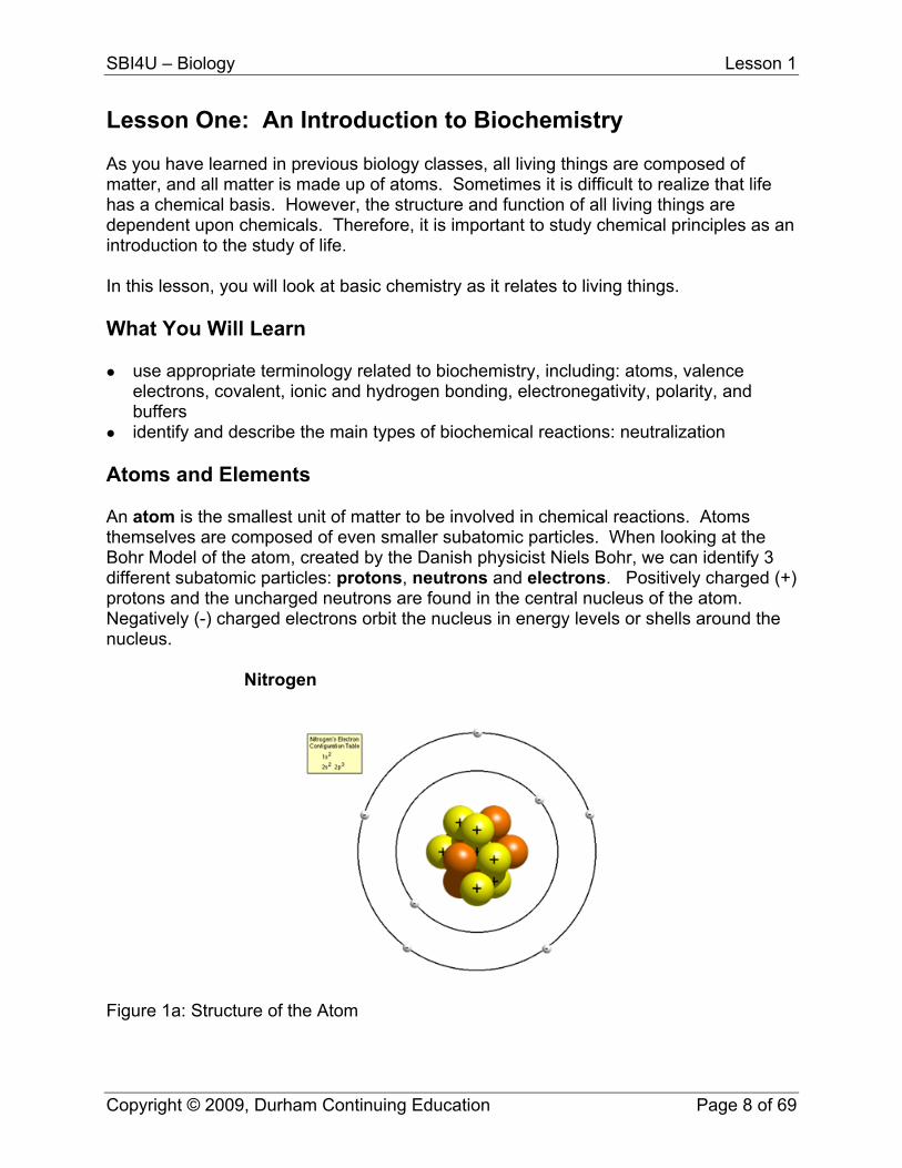
identify and describe the main types of biochemical reactions: neutralization Atoms and Elements An atom is the smallest unit of matter to be involved in chemical reactions. Atoms themselves are composed of even smaller subatomic particles. When looking at the Bohr Model of the atom, created by the Danish physicist Niels Bohr, we can identify 3 different subatomic particles: protons, neutrons and electrons. Positively charged (+) protons and the uncharged neutrons are found in the central nucleus of the atom. Negatively (-) charged electrons orbit the nucleus in energy levels or shells around the nucleus. Nitrogen
Figure 1a: Structure of the Atom
Copyright © 2009, Durham Continuing Education Page 8 of 69

SBI4U – Biology Lesson 1
Figure 1b: Structure of the Atom An element is a pure substance consisting of one type of atom and cannot be broken down into simpler substances by chemical means. The most common elements that are found in living things are: Hydrogen (H) 59%, Oxygen (O) 24%, Carbon (C), 11%, Nitrogen (N) 4%, Others such as phosphorus (P) and sulphur (S) 2% combined. Some atoms occur as single atoms (e.g. helium), some are diatomic which means they consist of 2 atoms (e.g. oxygen), and others are made up of more than two atoms (e.g. phosphorus, sulphur) An element is distinguished by its atomic number, which is the number of protons in its nucleus. In a neutral atom, the number of protons equals the number of electrons.
Atomic Number = # of Protons = # of Electrons Since electrons weigh very little, the atomic mass of an element is calculating the number of protons and the number of neutrons.
Atomic Mass = # of Protons + # of Neutrons
Tip: You can access a periodic table from; http://periodic.lanl.gov/default.htm Atoms of the same element that contain a different number of neutrons are called isotopes. Some isotopes are stable, while others are unstable and will decay or
Copyright © 2009, Durham Continuing Education Page 9 of 69
SBI4U – Biology Lesson 1
breakdown. The unstable isotopes are called radioactive isotopes. In nuclear medicine, radioactive compounds are injected into the body so that images of cells can be scanned to diagnose and treat medical conditions such as cancer and heart disease. Radioisotopes may now be used so routinely and effectively that we have come to rely on them despite concerns about production safety. Many radioactive isotopes decay at a known rate and can be used scientifically. For example, in a process called carbon dating, the decay of Carbon-14 can help archaeologists date objects up to 50,000 years old.
Support Questions
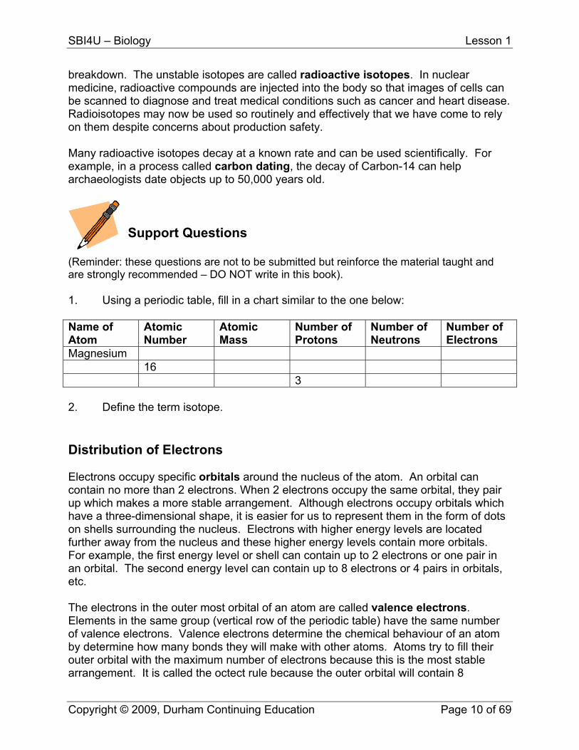
(Reminder: these questions are not to be submitted but reinforce the material taught and are strongly recommended – DO NOT write in this book). 1. Using a periodic table, fill in a chart similar to the one below: Name of Atom
Atomic Number
Atomic Mass
Number of Protons
Number of Neutrons
Number of Electrons
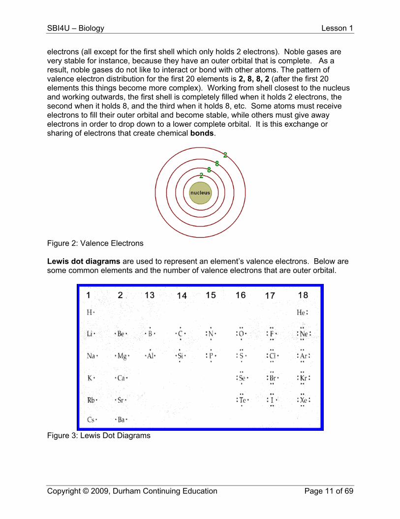
Magnesium 16 3 2. Define the term isotope. Distribution of Electrons Electrons occupy specific orbitals around the nucleus of the atom. An orbital can contain no more than 2 electrons. When 2 electrons occupy the same orbital, they pair up which makes a more stable arrangement. Although electrons occupy orbitals which have a three-dimensional shape, it is easier for us to represent them in the form of dots on shells surrounding the nucleus. Electrons with higher energy levels are located further away from the nucleus and these higher energy levels contain more orbitals. For example, the first energy level or shell can contain up to 2 electrons or one pair in an orbital. The second energy level can contain up to 8 electrons or 4 pairs in orbitals, etc. The electrons in the outer most orbital of an atom are called valence electrons. Elements in the same group (vertical row of the periodic table) have the same number of valence electrons. Valence electrons determine the chemical behaviour of an atom by determine how many bonds they will make with other atoms. Atoms try to fill their outer orbital with the maximum number of electrons because this is the most stable arrangement. It is called the octect rule because the outer orbital will contain 8
Copyright © 2009, Durham Continuing Education Page 10 of 69
SBI4U – Biology Lesson 1
electrons (all except for the first shell which only holds 2 electrons). Noble gases are very stable for instance, because they have an outer orbital that is complete. As a result, noble gases do not like to interact or bond with other atoms. The pattern of valence electron distribution for the first 20 elements is 2, 8, 8, 2 (after the first 20 elements this things become more complex). Working from shell closest to the nucleus and working outwards, the first shell is completely filled when it holds 2 electrons, the second when it holds 8, and the third when it holds 8, etc. Some atoms must receive electrons to fill their outer orbital and become stable, while others must give away electrons in order to drop down to a lower complete orbital. It is this exchange or sharing of electrons that create chemical bonds.
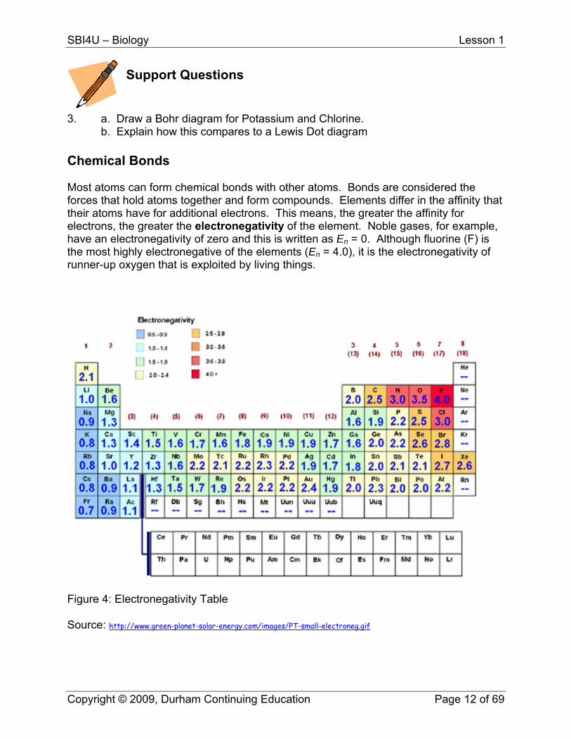
Figure 2: Valence Electrons Lewis dot diagrams are used to represent an element’s valence electrons. Below are some common elements and the number of valence electrons that are outer orbital.
Figure 3: Lewis Dot Diagrams
Copyright © 2009, Durham Continuing Education Page 11 of 69
SBI4U – Biology Lesson 1
Support Questions
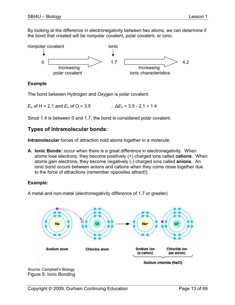
3. a. Draw a Bohr diagram for Potassium and Chlorine. b. Explain how this compares to a Lewis Dot diagram Chemical Bonds Most atoms can form chemical bonds with other atoms. Bonds are considered the forces that hold atoms together and form compounds. Elements differ in the affinity that their atoms have for additional electrons. This means, the greater the affinity for electrons, the greater the electronegativity of the element. Noble gases, for example, have an electronegativity of zero and this is written as En = 0. Although fluorine (F) is the most highly electronegative of the elements (En = 4.0), it is the electronegativity of runner-up oxygen that is exploited by living things.
Figure 4: Electronegativity Table Source: http://www.green-planet-solar-energy.com/images/PT-small-electroneg.gif
Copyright © 2009, Durham Continuing Education Page 12 of 69
SBI4U – Biology Lesson 1
By looking at the difference in electronegativity between two atoms, we can determine if the bond that created will be nonpolar covalent, polar covalent, or ionic. nonpolar covalent ionic
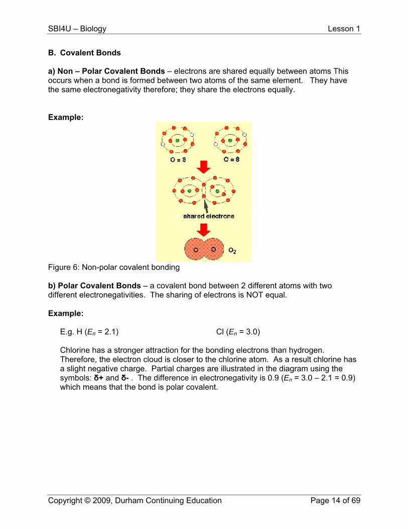
0 1.7 4.2 Increasing Increasing polar covalent ionic characteristics Example The bond between Hydrogen and Oxygen is polar covalent. En of H = 2.1 and En of O = 3.5 ∴ ΔEn = 3.5 - 2.1 = 1.4 Since 1.4 is between 0 and 1.7, the bond is considered polar covalent. Types of Intramolecular bonds: Intramolecular forces of attraction hold atoms together in a molecule. A. Ionic Bonds: occur when there is a great difference in electronegativity. When
atoms lose electrons, they become positively (+) charged ions called cations. When atoms gain electrons, they become negatively (-) charged ions called anions. An ionic bond occurs between anions and cations when they come close together due to the force of attractions (remember opposites attract!).
Example:
A metal and non-metal (electronegativity difference of 1.7 or greater)
SoFig
urce: Campbell’s Biology ure 5: Ionic Bonding
Copyright © 2009, Durham Continuing Education Page 13 of 69
SBI4U – Biology Lesson 1
B. Covalent Bonds a) Non – Polar Covalent Bonds – electrons are shared equally between atoms This occurs when a bond is formed between two atoms of the same element. They have the same electronegativity therefore; they share the electrons equally. Example:
Figure 6: Non-polar covalent bonding b) Polar Covalent Bonds – a covalent bond between 2 different atoms with two different electronegativities. The sharing of electrons is NOT equal. Example:
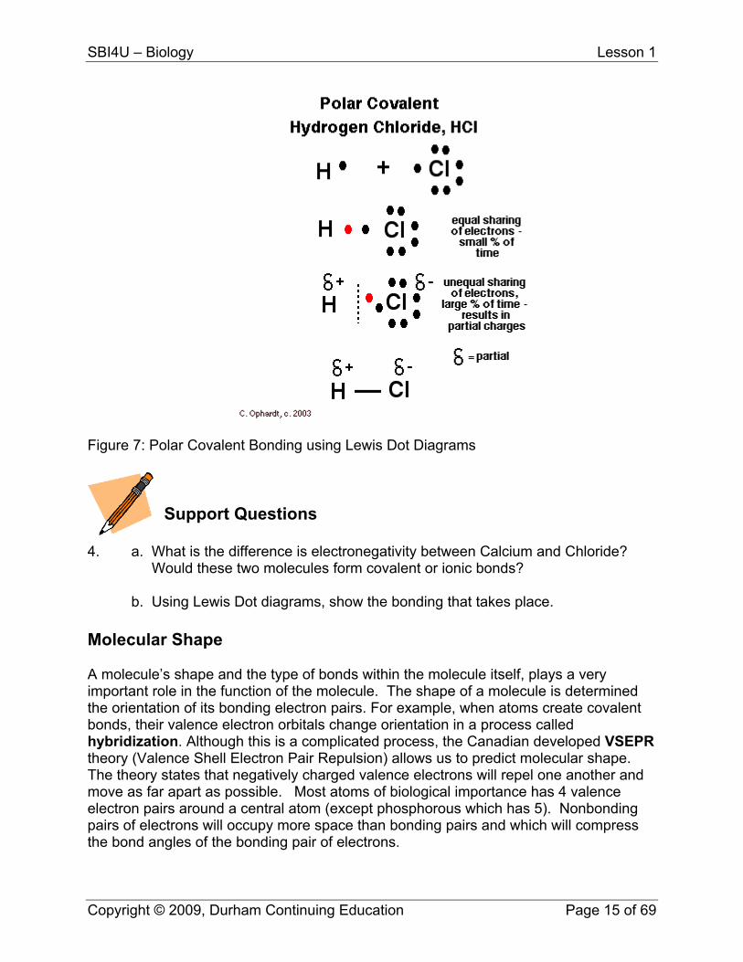
E.g. H (En = 2.1) Cl (En = 3.0) Chlorine has a stronger attraction for the bonding electrons than hydrogen. Therefore, the electron cloud is closer to the chlorine atom. As a result chlorine has a slight negative charge. Partial charges are illustrated in the diagram using the symbols: δ+ and δ- . The difference in electronegativity is 0.9 (En = 3.0 – 2.1 = 0.9) which means that the bond is polar covalent.
Copyright © 2009, Durham Continuing Education Page 14 of 69
SBI4U – Biology Lesson 1
Figure 7: Polar Covalent Bonding using Lewis Dot Diagrams
Support Questions
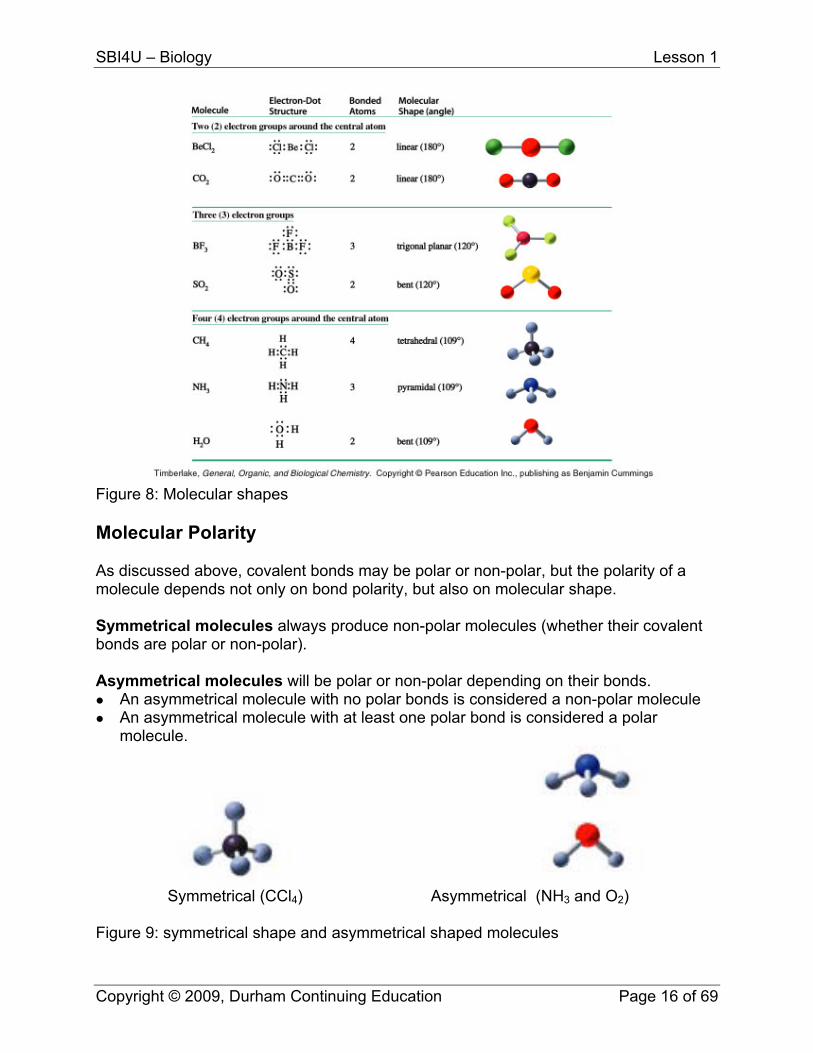
4. a. What is the difference is electronegativity between Calcium and Chloride? Would these two molecules form covalent or ionic bonds? b. Using Lewis Dot diagrams, show the bonding that takes place. Molecular Shape A molecule’s shape and the type of bonds within the molecule itself, plays a very important role in the function of the molecule. The shape of a molecule is determined the orientation of its bonding electron pairs. For example, when atoms create covalent bonds, their valence electron orbitals change orientation in a process called hybridization. Although this is a complicated process, the Canadian developed VSEPR theory (Valence Shell Electron Pair Repulsion) allows us to predict molecular shape. The theory states that negatively charged valence electrons will repel one another and move as far apart as possible. Most atoms of biological importance has 4 valence electron pairs around a central atom (except phosphorous which has 5). Nonbonding pairs of electrons will occupy more space than bonding pairs and which will compress the bond angles of the bonding pair of electrons.
Copyright © 2009, Durham Continuing Education Page 15 of 69
SBI4U – Biology Lesson 1
Figure 8: Molecular shapes Molecular Polarity As discussed above, covalent bonds may be polar or non-polar, but the polarity of a molecule depends not only on bond polarity, but also on molecular shape. Symmetrical molecules always produce non-polar molecules (whether their covalent bonds are polar or non-polar). Asymmetrical molecules will be polar or non-polar depending on their bonds.
An asymmetrical molecule with no polar bonds is considered a non-polar molecule An asymmetrical molecule with at least one polar bond is considered a polar
molecule.
Symmetrical (CCl4) Asymmetrical (NH3 and O2)
Figure 9: symmetrical shape and asymmetrical shaped molecules
Copyright © 2009, Durham Continuing Education Page 16 of 69

SBI4U – Biology Lesson 1
Intermolecular Bonds (van der Waals Forces) Intermolecular bonds or van der Waals forces are bonds between molecules and are much weaker than intramolecular bonds. Intermolecular bonds determine the physical state of substances at a given temperature ad pressure. They are broken when solids melt into liquids or when liquids evaporate in to gases. There are three types of intermolecular bonds: A. London Forces: are formed between noble gas atoms and nonpolar molecules. They are a temporary random and unequal distribution of electrons around the nuclei of atoms. These unequal electron clouds allow the electrons of a neutral atom to attract the nucleus of other nearby atoms.
Figure 10: London Forces B. Dipole-dipole Forces: are stronger than London forces and hold polar molecules to one another. For example, these forces occur when the partially negative side of a polar molecule is attracted to the partially positive side of an adjacent molecule.
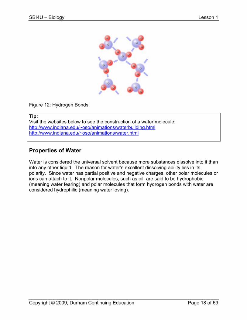
Figure 11: Dipole-Dipole Forces C. Hydrogen Bonds: are weak attractions between polar molecules (1/20th as strong as covalent) that contain hydrogen atoms bonded to the more electronegative atoms of oxygen or nitrogen. Although weak, they are the strongest type of intermolecular forces. Hydrogen bonds are the force that holds water molecules together, and what gives water its unique properties.
Copyright © 2009, Durham Continuing Education Page 17 of 69
SBI4U – Biology Lesson 1
Figure 12: Hydrogen Bonds Tip: Visit the websites below to see the construction of a water molecule: http://www.indiana.edu/~oso/animations/waterbuilding.htmlhttp://www.indiana.edu/~oso/animations/water.html Properties of Water Water is considered the universal solvent because more substances dissolve into it than into any other liquid. The reason for water’s excellent dissolving ability lies in its polarity. Since water has partial positive and negative charges, other polar molecules or ions can attach to it. Nonpolar molecules, such as oil, are said to be hydrophobic (meaning water fearing) and polar molecules that form hydrogen bonds with water are considered hydrophilic (meaning water loving).
Copyright © 2009, Durham Continuing Education Page 18 of 69
SBI4U – Biology Lesson 1
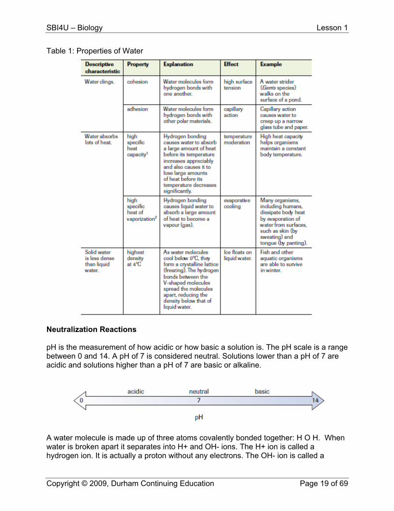
Table 1: Properties of Water
Neutralization Reactions pH is the measurement of how acidic or how basic a solution is. The pH scale is a range between 0 and 14. A pH of 7 is considered neutral. Solutions lower than a pH of 7 are acidic and solutions higher than a pH of 7 are basic or alkaline.
A water molecule is made up of three atoms covalently bonded together: H O H. When water is broken apart it separates into H+ and OH- ions. The H+ ion is called a hydrogen ion. It is actually a proton without any electrons. The OH- ion is called a
Copyright © 2009, Durham Continuing Education Page 19 of 69
SBI4U – Biology Lesson 1
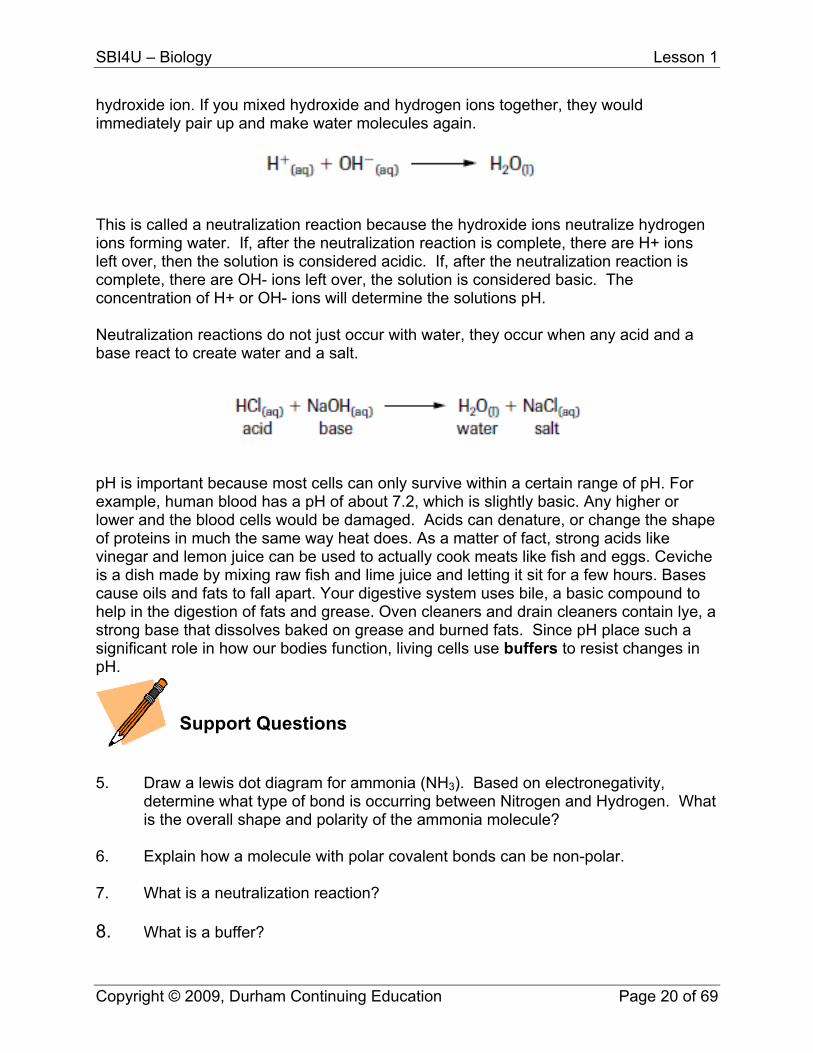
hydroxide ion. If you mixed hydroxide and hydrogen ions together, they would immediately pair up and make water molecules again.
This is called a neutralization reaction because the hydroxide ions neutralize hydrogen ions forming water. If, after the neutralization reaction is complete, there are H+ ions left over, then the solution is considered acidic. If, after the neutralization reaction is complete, there are OH- ions left over, the solution is considered basic. The concentration of H+ or OH- ions will determine the solutions pH. Neutralization reactions do not just occur with water, they occur when any acid and a base react to create water and a salt.
pH is important because most cells can only survive within a certain range of pH. For example, human blood has a pH of about 7.2, which is slightly basic. Any higher or lower and the blood cells would be damaged. Acids can denature, or change the shape of proteins in much the same way heat does. As a matter of fact, strong acids like vinegar and lemon juice can be used to actually cook meats like fish and eggs. Ceviche is a dish made by mixing raw fish and lime juice and letting it sit for a few hours. Bases cause oils and fats to fall apart. Your digestive system uses bile, a basic compound to help in the digestion of fats and grease. Oven cleaners and drain cleaners contain lye, a strong base that dissolves baked on grease and burned fats. Since pH place such a significant role in how our bodies function, living cells use buffers to resist changes in pH.
Support Questions
5. Draw a lewis dot diagram for ammonia (NH3). Based on electronegativity,
determine what type of bond is occurring between Nitrogen and Hydrogen. What is the overall shape and polarity of the ammonia molecule?
6. Explain how a molecule with polar covalent bonds can be non-polar. 7. What is a neutralization reaction? 8. What is a buffer?
Copyright © 2009, Durham Continuing Education Page 20 of 69
SBI4U – Biology Lesson 1
Key Question #1
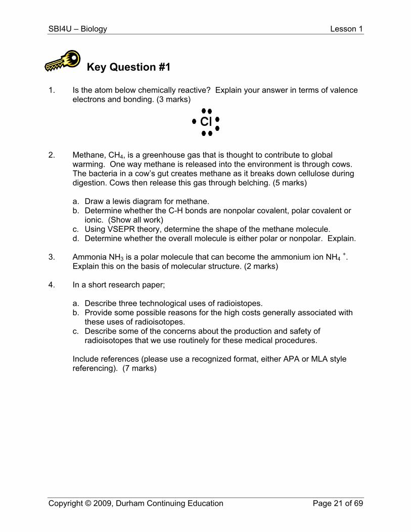
1. Is the atom below chemically reactive? Explain your answer in terms of valence electrons and bonding. (3 marks)
2. Methane, CH4, is a greenhouse gas that is thought to contribute to global warming. One way methane is released into the environment is through cows. The bacteria in a cow’s gut creates methane as it breaks down cellulose during digestion. Cows then release this gas through belching. (5 marks)
a. Draw a lewis diagram for methane. b. Determine whether the C-H bonds are nonpolar covalent, polar covalent or
ionic. (Show all work) c. Using VSEPR theory, determine the shape of the methane molecule. d. Determine whether the overall molecule is either polar or nonpolar. Explain.
3. Ammonia NH3 is a polar molecule that can become the ammonium ion NH4 +.
Explain this on the basis of molecular structure. (2 marks) 4. In a short research paper;
a. Describe three technological uses of radioistopes. b. Provide some possible reasons for the high costs generally associated with
these uses of radioisotopes. c. Describe some of the concerns about the production and safety of
radioisotopes that we use routinely for these medical procedures.
Include references (please use a recognized format, either APA or MLA style referencing). (7 marks)
Copyright © 2009, Durham Continuing Education Page 21 of 69
SBI4U – Biology Lesson 1
5. Investigate a way of creating a pH indicator out of red cabbage. Choose 3 everyday acids and 3 bases to test and complete a table similar to the one shown below. (6 marks) Please note: Use separate containers for each household solution. Do not mix chemicals.
Substance Tested Colour Indicated by
Cabbage Test Acid or Base?
1. 2.
a. In chemical terms, explain why the indicator turned different colours. (2 marks)
b. Research in more detail ph and how buffers work and explain their importance to living systems. (3 marks)
Copyright © 2009, Durham Continuing Education Page 22 of 69

SBI4U
Grade 12, University Preparation Biology
Lesson 2 – Cellular Biology
SBI4U – Biology Lesson 2
Lesson Two: Cellular Biology Cellular and Molecular Biology are fields of biology that focus on understanding living processes at a molecular level. Many of the most exciting biological discoveries in the past twenty years have occurred in these fields. These discoveries have identified some of the genes responsible for cancer, the events regulating how a cell divides, and how organisms develop from a single cell. However, to understand some of these ground breaking discoveries, one must have a basic understanding of the cell. In this lesson, you will examine the structure and function of cells, focusing in detail on the differences between prokaryotic and eukaryotic cells. You will also study the plasma membrane and its role in transporting molecules in and out of the cell. What You Will Learn
evaluate technological advances in the field of cellular biology explain the roles of various organelles, such as lysosomes, vacuoles, mitochondria,
internal cell membranes, ribosomes, smooth and rough endoplasmic reticulum, and Golgi bodies, in cellular processes
use appropriate terminology related to biochemistry, including: active and passive transport
describe the structure of cell membranes according to the fluid mosaic model, and explain the dynamics of passive transport, facilitated diffusion, and the movement of large particles across the cell membrane by the processes of endocytosis and exocytosis
plan and conduct an investigation to demonstrate the movement of substances across a membrane
Introduction to the Cell All the organisms we see around us are made up of cells. The atoms and molecules studied in Lesson One are not alive, but the cell is alive. In fact, a cell is the smallest unit of living matter. According to the cell theory, cells come only from other pre-existing cells and cells are capable of reproducing. Cell Structure and Function A cell carries out all the functions we associate with living things, such as those functions needed for growth and reproduction. We also know that particular functions are carried out by certain parts of a cell. Improved microscopy has vastly improved our ability to view the internal structures of cells, and biochemical techniques have allowed us to determine the function of these cell parts. All cells are surrounded by a plasma membrane that separates the internal and external environment. Some cells, such as plant cells, are strengthened by the addition of a cell wall that protects the plasma membrane.
Copyright © 2009, Durham Continuing Education Page 24 of 69
SBI4U – Biology Lesson 2
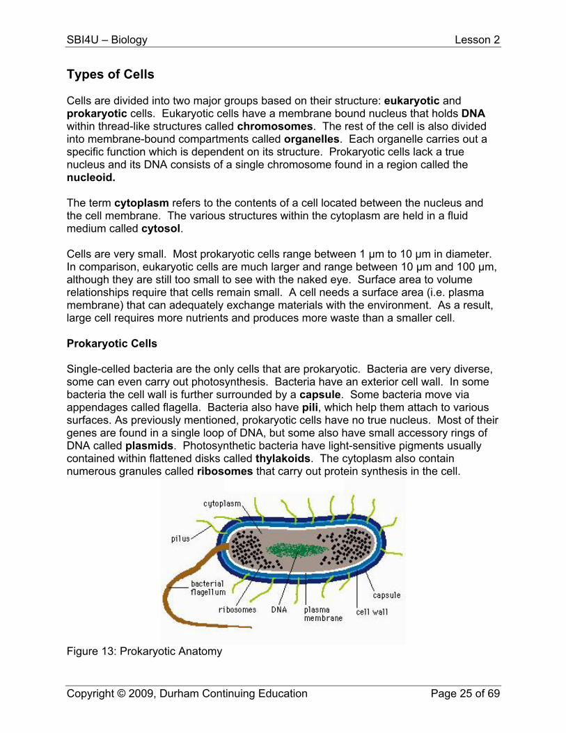
Types of Cells Cells are divided into two major groups based on their structure: eukaryotic and prokaryotic cells. Eukaryotic cells have a membrane bound nucleus that holds DNA within thread-like structures called chromosomes. The rest of the cell is also divided into membrane-bound compartments called organelles. Each organelle carries out a specific function which is dependent on its structure. Prokaryotic cells lack a true nucleus and its DNA consists of a single chromosome found in a region called the nucleoid. The term cytoplasm refers to the contents of a cell located between the nucleus and the cell membrane. The various structures within the cytoplasm are held in a fluid medium called cytosol. Cells are very small. Most prokaryotic cells range between 1 μm to 10 μm in diameter. In comparison, eukaryotic cells are much larger and range between 10 μm and 100 μm, although they are still too small to see with the naked eye. Surface area to volume relationships require that cells remain small. A cell needs a surface area (i.e. plasma membrane) that can adequately exchange materials with the environment. As a result, large cell requires more nutrients and produces more waste than a smaller cell. Prokaryotic Cells Single-celled bacteria are the only cells that are prokaryotic. Bacteria are very diverse, some can even carry out photosynthesis. Bacteria have an exterior cell wall. In some bacteria the cell wall is further surrounded by a capsule. Some bacteria move via appendages called flagella. Bacteria also have pili, which help them attach to various surfaces. As previously mentioned, prokaryotic cells have no true nucleus. Most of their genes are found in a single loop of DNA, but some also have small accessory rings of DNA called plasmids. Photosynthetic bacteria have light-sensitive pigments usually contained within flattened disks called thylakoids. The cytoplasm also contain numerous granules called ribosomes that carry out protein synthesis in the cell.
Figure 13: Prokaryotic Anatomy
Copyright © 2009, Durham Continuing Education Page 25 of 69

SBI4U – Biology Lesson 2
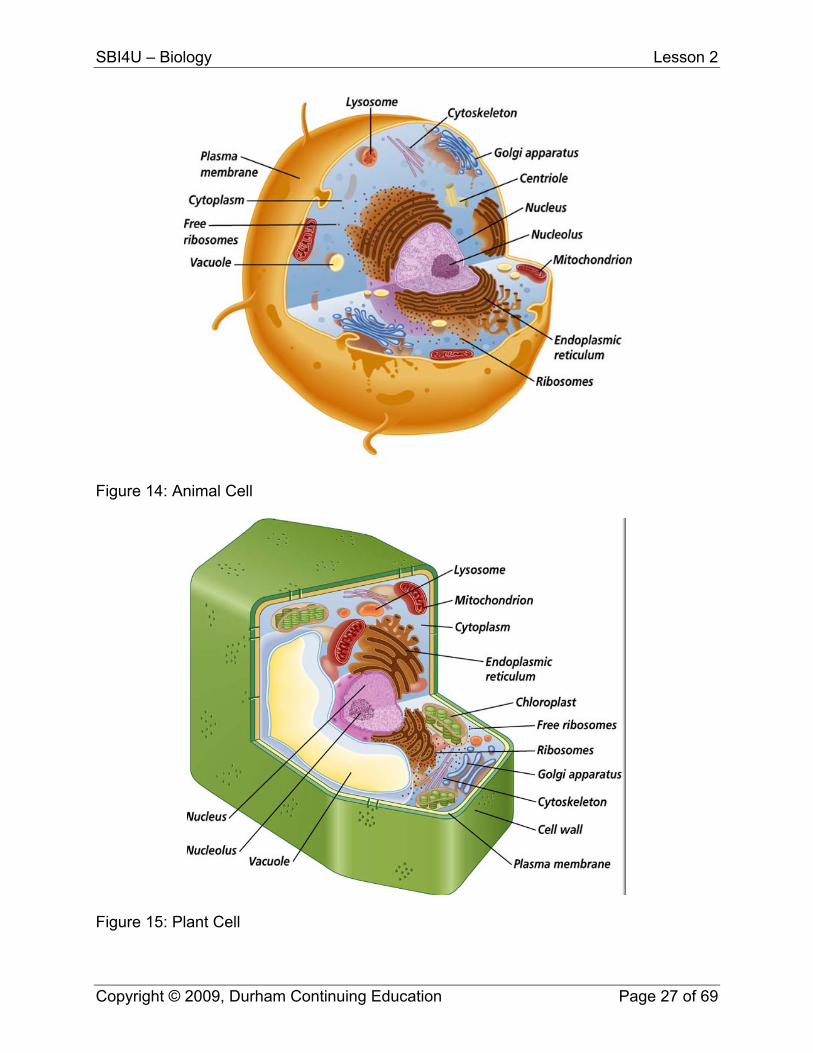
Eukaryotic Cells All other cells are considered eukaryotic. Eukaryotic organisms include algae, protozoa, fungi, plants and animals. As previously mentioned, eukaryotic cells have a nucleus an numerous organelles with specific functions. Many of these you have studied in previous Biology courses.
Eukaryotic Cell Structures Name Composition Function Cell wall Cellulose fibrils in plant cells Support and protection Plasma membrane Phospholipid bilayer with
embedded protein Passage of molecules in and out of cell
Nucleus Nuclear envelope surrounding the nucleoplasm, chromosomes, and nucleoli
Cellular reproduction and control or protein synthesis
Nucleolus Concentrated area of chromatin, RNA, and proteins
Ribosome formation
Ribosomes Protein and RNA Protein synthesis Smooth endoplasmic reticulum
Membranous flattened channels and tubular canals without ribosomes
Various; transport and/or modification of proteins and other substances, transport by vesicle formation; lipid synthesis in some cells
Rough endoplasmic reticulum
Membranous flattened channels and tubular canals studded with ribosomes
transport and/or modification of proteins and other substances, transport by vesicle formation; protein synthesis
Golgi Apparatus Stack of membranous sacs in animal cells
Processing and packaging of molecules
Vacuoles/Vesicles Membranous sacs in animal cells Storage Lysosome Membranous vesicle containing
digestive enzymes Intracellular digestion
Microbodies Membranous vesicle containing specific enzymes
Various metabolic tasks
Mitochondrion Double membrane layer Cellular respiration Chloroplast Double membrane layer in plant
cells Photosynthesis
Cytoskeleton Microtubules and microfilaments Shape of cell; movement of its parts
Cilia and flagella Microtubules in animal cells Movement of cell Centriole Microtubules in animal cells Forms basal bodies that
produces microtubules
Copyright © 2009, Durham Continuing Education Page 26 of 69
SBI4U – Biology Lesson 2
Figure 14: Animal Cell
Figure 15: Plant Cell
Copyright © 2009, Durham Continuing Education Page 27 of 69
SBI4U – Biology Lesson 2
Support Questions
1. What similar features do both prokaryotic and eukaryotic cells have? What is
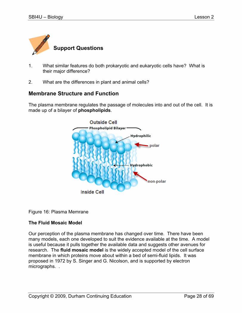
their major difference? 2. What are the differences in plant and animal cells? Membrane Structure and Function The plasma membrane regulates the passage of molecules into and out of the cell. It is made up of a bilayer of phospholipids.
Figure 16: Plasma Memrane The Fluid Mosaic Model Our perception of the plasma membrane has changed over time. There have been many models, each one developed to suit the evidence available at the time. A model is useful because it pulls together the available data and suggests other avenues for research. The fluid mosaic model is the widely accepted model of the cell surface membrane in which proteins move about within a bed of semi-fluid lipids. It was proposed in 1972 by S. Singer and G. Nicolson, and is supported by electron micrographs. .
Copyright © 2009, Durham Continuing Education Page 28 of 69
SBI4U – Biology Lesson 2
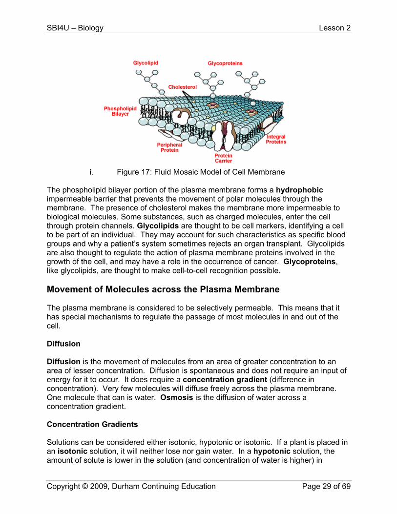
i. Figure 17: Fluid Mosaic Model of Cell Membrane
The phospholipid bilayer portion of the plasma membrane forms a hydrophobic impermeable barrier that prevents the movement of polar molecules through the membrane. The presence of cholesterol makes the membrane more impermeable to biological molecules. Some substances, such as charged molecules, enter the cell through protein channels. Glycolipids are thought to be cell markers, identifying a cell to be part of an individual. They may account for such characteristics as specific blood groups and why a patient’s system sometimes rejects an organ transplant. Glycolipids are also thought to regulate the action of plasma membrane proteins involved in the growth of the cell, and may have a role in the occurrence of cancer. Glycoproteins, like glycolipids, are thought to make cell-to-cell recognition possible. Movement of Molecules across the Plasma Membrane The plasma membrane is considered to be selectively permeable. This means that it has special mechanisms to regulate the passage of most molecules in and out of the cell. Diffusion Diffusion is the movement of molecules from an area of greater concentration to an area of lesser concentration. Diffusion is spontaneous and does not require an input of energy for it to occur. It does require a concentration gradient (difference in concentration). Very few molecules will diffuse freely across the plasma membrane. One molecule that can is water. Osmosis is the diffusion of water across a concentration gradient. Concentration Gradients Solutions can be considered either isotonic, hypotonic or isotonic. If a plant is placed in an isotonic solution, it will neither lose nor gain water. In a hypotonic solution, the amount of solute is lower in the solution (and concentration of water is higher) in
Copyright © 2009, Durham Continuing Education Page 29 of 69
SBI4U – Biology Lesson 2
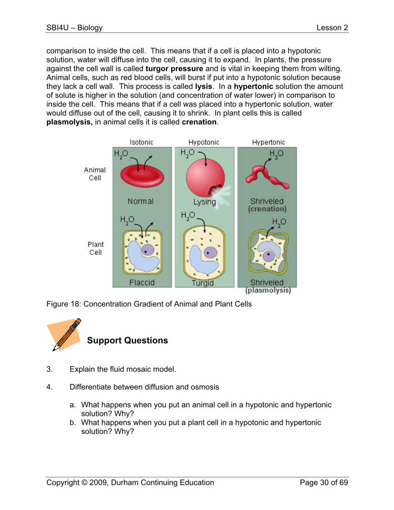
comparison to inside the cell. This means that if a cell is placed into a hypotonic solution, water will diffuse into the cell, causing it to expand. In plants, the pressure against the cell wall is called turgor pressure and is vital in keeping them from wilting. Animal cells, such as red blood cells, will burst if put into a hypotonic solution because they lack a cell wall. This process is called lysis. In a hypertonic solution the amount of solute is higher in the solution (and concentration of water lower) in comparison to inside the cell. This means that if a cell was placed into a hypertonic solution, water would diffuse out of the cell, causing it to shrink. In plant cells this is called plasmolysis, in animal cells it is called crenation.
Figure 18: Concentration Gradient of Animal and Plant Cells
Support Questions
3. Explain the fluid mosaic model. 4. Differentiate between diffusion and osmosis
a. What happens when you put an animal cell in a hypotonic and hypertonic solution? Why?
b. What happens when you put a plant cell in a hypotonic and hypertonic solution? Why?
Copyright © 2009, Durham Continuing Education Page 30 of 69
SBI4U – Biology Lesson 2
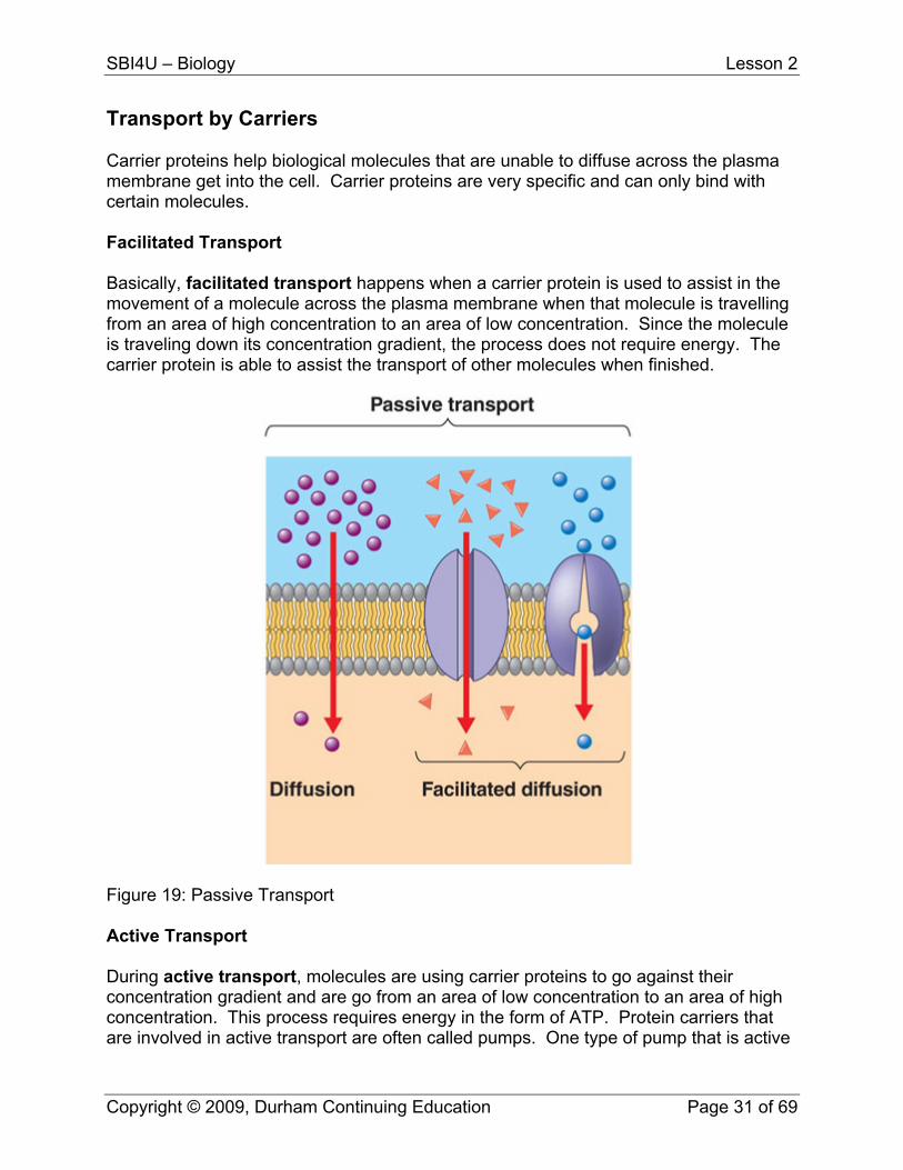
Transport by Carriers Carrier proteins help biological molecules that are unable to diffuse across the plasma membrane get into the cell. Carrier proteins are very specific and can only bind with certain molecules. Facilitated Transport Basically, facilitated transport happens when a carrier protein is used to assist in the movement of a molecule across the plasma membrane when that molecule is travelling from an area of high concentration to an area of low concentration. Since the molecule is traveling down its concentration gradient, the process does not require energy. The carrier protein is able to assist the transport of other molecules when finished.
Figure 19: Passive Transport Active Transport During active transport, molecules are using carrier proteins to go against their concentration gradient and are go from an area of low concentration to an area of high concentration. This process requires energy in the form of ATP. Protein carriers that are involved in active transport are often called pumps. One type of pump that is active
Copyright © 2009, Durham Continuing Education Page 31 of 69
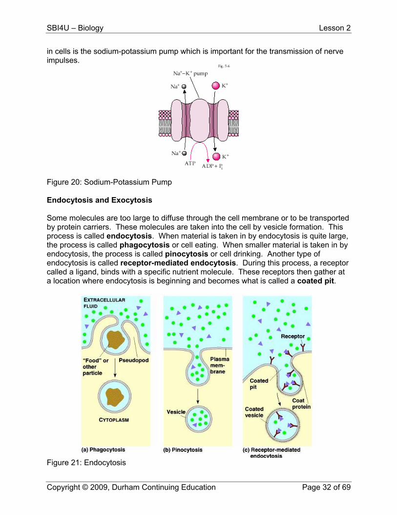
SBI4U – Biology Lesson 2
in cells is the sodium-potassium pump which is important for the transmission of nerve impulses.
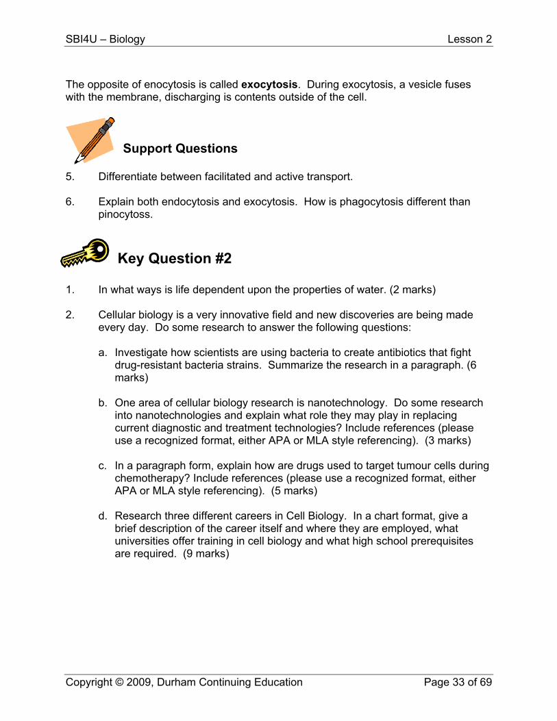
Figure 20: Sodium-Potassium Pump Endocytosis and Exocytosis Some molecules are too large to diffuse through the cell membrane or to be transported by protein carriers. These molecules are taken into the cell by vesicle formation. This process is called endocytosis. When material is taken in by endocytosis is quite large, the process is called phagocytosis or cell eating. When smaller material is taken in by endocytosis, the process is called pinocytosis or cell drinking. Another type of endocytosis is called receptor-mediated endocytosis. During this process, a receptor called a ligand, binds with a specific nutrient molecule. These receptors then gather at a location where endocytosis is beginning and becomes what is called a coated pit.
Figure 21: Endocytosis
Copyright © 2009, Durham Continuing Education Page 32 of 69
SBI4U – Biology Lesson 2
The opposite of enocytosis is called exocytosis. During exocytosis, a vesicle fuses with the membrane, discharging is contents outside of the cell.
Support Questions
5. Differentiate between facilitated and active transport. 6. Explain both endocytosis and exocytosis. How is phagocytosis different than
pinocytoss. Key Question #2
1. In what ways is life dependent upon the properties of water. (2 marks) 2. Cellular biology is a very innovative field and new discoveries are being made
every day. Do some research to answer the following questions:
a. Investigate how scientists are using bacteria to create antibiotics that fight drug-resistant bacteria strains. Summarize the research in a paragraph. (6 marks)
b. One area of cellular biology research is nanotechnology. Do some research
into nanotechnologies and explain what role they may play in replacing current diagnostic and treatment technologies? Include references (please use a recognized format, either APA or MLA style referencing). (3 marks)
c. In a paragraph form, explain how are drugs used to target tumour cells during
chemotherapy? Include references (please use a recognized format, either APA or MLA style referencing). (5 marks)
d. Research three different careers in Cell Biology. In a chart format, give a
brief description of the career itself and where they are employed, what universities offer training in cell biology and what high school prerequisites are required. (9 marks)
Copyright © 2009, Durham Continuing Education Page 33 of 69
SBI4U
Grade 12, University Preparation Biology
Lesson 3 – The Chemicals of Life
SBI4U – Biology Lesson 3
Lesson Three: The Chemicals of Life With the exception of water, almost all the chemicals of life are carbon based. Compounds that contain carbon are called organic compounds. The chemistry of carbon-based compounds is called organic chemistry. A basic understanding of organic chemistry and how macromolecules are created and broken down is important to understanding our cells’ metabolism. In this lesson, you will study macromolecule examples of carbohydrates, fats and lipids, proteins and nucleic acid. You will examine functional groups for each and learn how macromolecules are created and broken down into monomers through condensation and hydrolysis reactions. What You Will Learn
describe the structure of important biochemical compounds, including carbohydrates, proteins, lipids, and nucleic acids, and explain their function within cells
identify common functional groups within biological molecules (e.g., hydroxyl, carbonyl, carboxyl, amino, phosphate), and explain how they contribute to the function of each molecule
identify and describe the main types of biochemical reactions: oxidation-reduction [redox], hydrolysis, and condensation
draw three-dimensional molecular models of important biochemical compounds, including carbohydrates, proteins, lipids, and nucleic acids
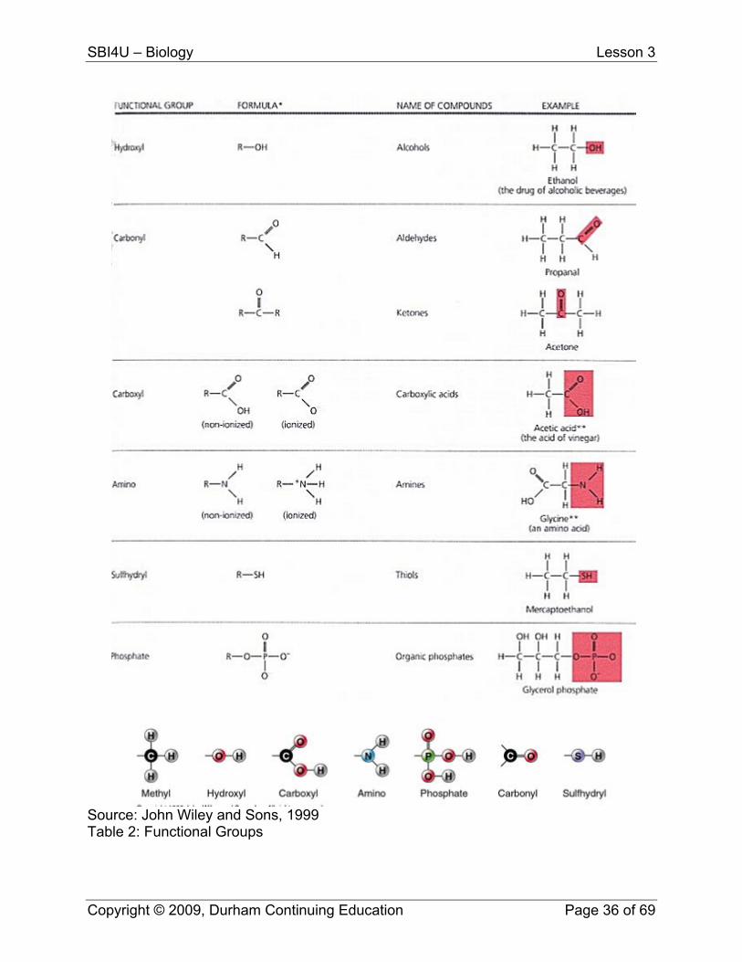
Functional Groups Functional groups are reactive clusters where much of the bonding takes place in biological molecules. Common functional groups in biomolecules are listed on the following page.
Copyright © 2009, Durham Continuing Education Page 35 of 69
SBI4U – Biology Lesson 3
Source: John Wiley and Sons, 1999 Table 2: Functional Groups
Copyright © 2009, Durham Continuing Education Page 36 of 69
SBI4U – Biology Lesson 3
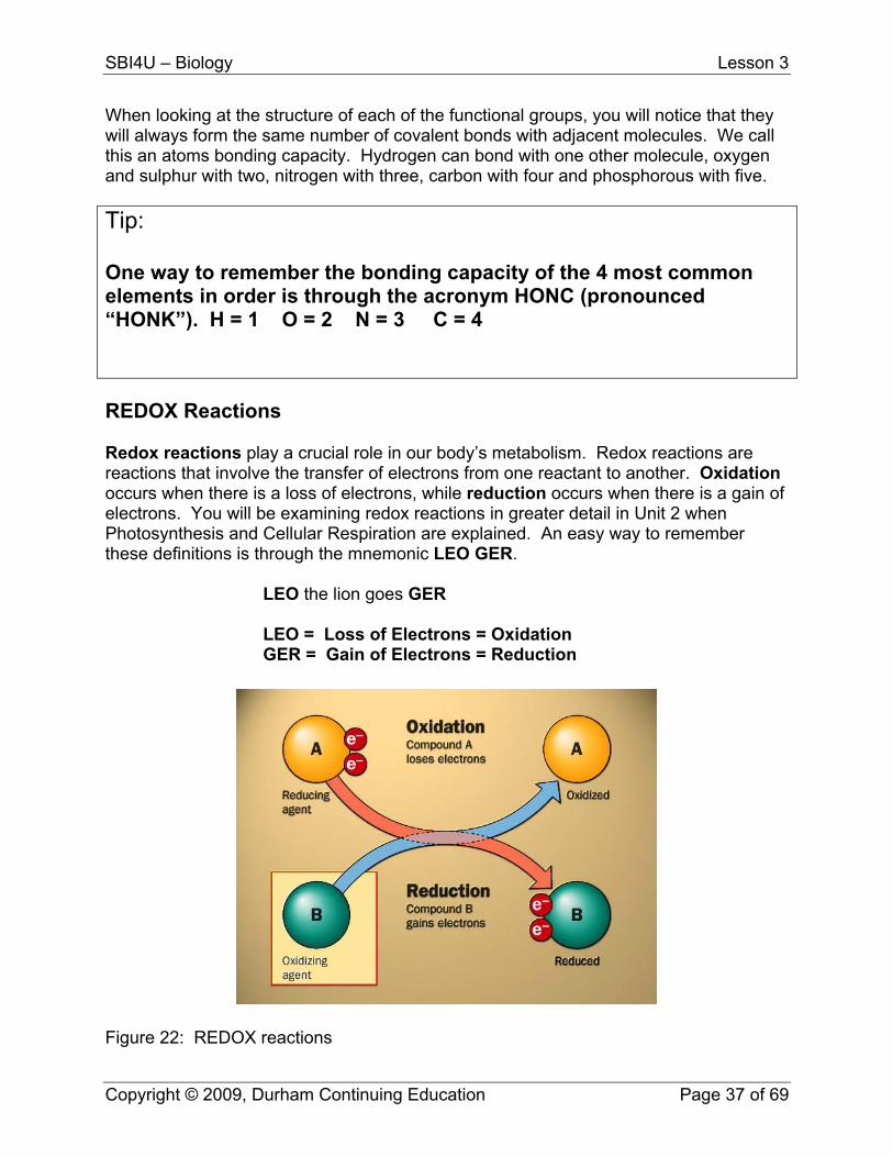
When looking at the structure of each of the functional groups, you will notice that they will always form the same number of covalent bonds with adjacent molecules. We call this an atoms bonding capacity. Hydrogen can bond with one other molecule, oxygen and sulphur with two, nitrogen with three, carbon with four and phosphorous with five. Tip: One way to remember the bonding capacity of the 4 most common elements in order is through the acronym HONC (pronounced “HONK”). H = 1 O = 2 N = 3 C = 4 REDOX Reactions Redox reactions play a crucial role in our body’s metabolism. Redox reactions are reactions that involve the transfer of electrons from one reactant to another. Oxidation occurs when there is a loss of electrons, while reduction occurs when there is a gain of electrons. You will be examining redox reactions in greater detail in Unit 2 when Photosynthesis and Cellular Respiration are explained. An easy way to remember these definitions is through the mnemonic LEO GER.
LEO the lion goes GER
LEO = Loss of Electrons = Oxidation GER = Gain of Electrons = Reduction
Figure 22: REDOX reactions
Copyright © 2009, Durham Continuing Education Page 37 of 69
SBI4U – Biology Lesson 3
Biological Macromolecules Many biologically important molecules are macromolecules, or large molecules which are sometimes composed of many repeating subunits. Listed in the chart below are the four main classes of macromolecules found in living things. Class/Group Macromolecule
(polymer) Specific Examples
Subunit (Monomer)
Examples
Carbohydrate Oligosaccharide, Polysaccharide
Complex carbohydrates (Starch, cellulose)
monosaccharide Simple sugar (glucose, fructose)
Fats/Lipids Di or Triglyceride Phospholipid Glycerol and Fatty Acids
Glycerol; Oleic Acid (in vegetable oils)
Protein
Protein Enzymes Amino Acid 20 different ones
Nucleic Acid DNA, RNA DNA mRNA tRNA
Nucleotide Phosphate Group + sugar + one of 5 nitrogenous bases
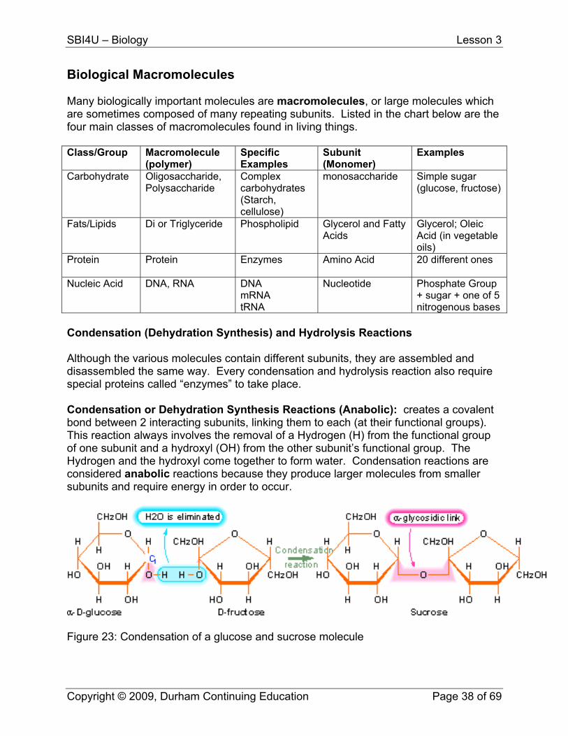
Condensation (Dehydration Synthesis) and Hydrolysis Reactions Although the various molecules contain different subunits, they are assembled and disassembled the same way. Every condensation and hydrolysis reaction also require special proteins called “enzymes” to take place. Condensation or Dehydration Synthesis Reactions (Anabolic): creates a covalent bond between 2 interacting subunits, linking them to each (at their functional groups). This reaction always involves the removal of a Hydrogen (H) from the functional group of one subunit and a hydroxyl (OH) from the other subunit’s functional group. The Hydrogen and the hydroxyl come together to form water. Condensation reactions are considered anabolic reactions because they produce larger molecules from smaller subunits and require energy in order to occur.
Figure 23: Condensation of a glucose and sucrose molecule
Copyright © 2009, Durham Continuing Education Page 38 of 69
SBI4U – Biology Lesson 3
Hydrolysis Reactions (Catabolic): break molecules into their subunits. oIt is the reverse of condensation reactions. The word “hydro” means water and “lysis” is to break, so therefore, a water molecule is used to break a covalent bond. The water provides an “H” to one subunit and an “OH” to the other. Hydrolytic reactions are considered catabolic because break down molecules and release energy.
TIP: Sometimes it is difficult to grasp the concept of hydrolysis and condensation reactions. Understanding is often enhanced by viewing animations and small video clips of some of these reactions. In order to find some of these resources online, try using the term “animation” in your search. For example, try “hydrolysis animation” to search for animated representations of the reaction. You may also wish to view the reaction demonstrated on this site: http://www.tvdsb.on.ca/westmin/science/sbioac/biochem/condense.htm
Support Questions
1. Differentiate between:
a. oxidation and reduction b. catabolic and anabolic c. hydrolysis and condensation reactions
Carbohydrates Carbohydrates contain carbon, hydrogen and oxygen in a 1:2:1 ratio, i.e. (CH2O)n and are the most common organic materials on Earth. Millions of tons of carbohydrates are produced by plants and algae each year through photosynthesis. Carbohydrates are used by organisms for energy, for building materials within the cell, and for cell-to-cell identification during metabolic processes. Carbohydrates are classified into 3 groups: monosaccharide, oligosaccharides and polysaccharides. Monosaccharides Monosaccharides are simple sugars. The word monosaccharide comes from the lat word “mono” meaning “single” and “saccharide” from “saccharum” meaning sugar. Monosaccharides consist of a single chain of carbon atoms with an “OH” or hydroxyl group attached. They are distinguished by their carbonyl group, either an aldehyde (functional group CH=O) or ketone (functional group C=O) and the number of carbons
Copyright © 2009, Durham Continuing Education Page 39 of 69
SBI4U – Biology Lesson 3
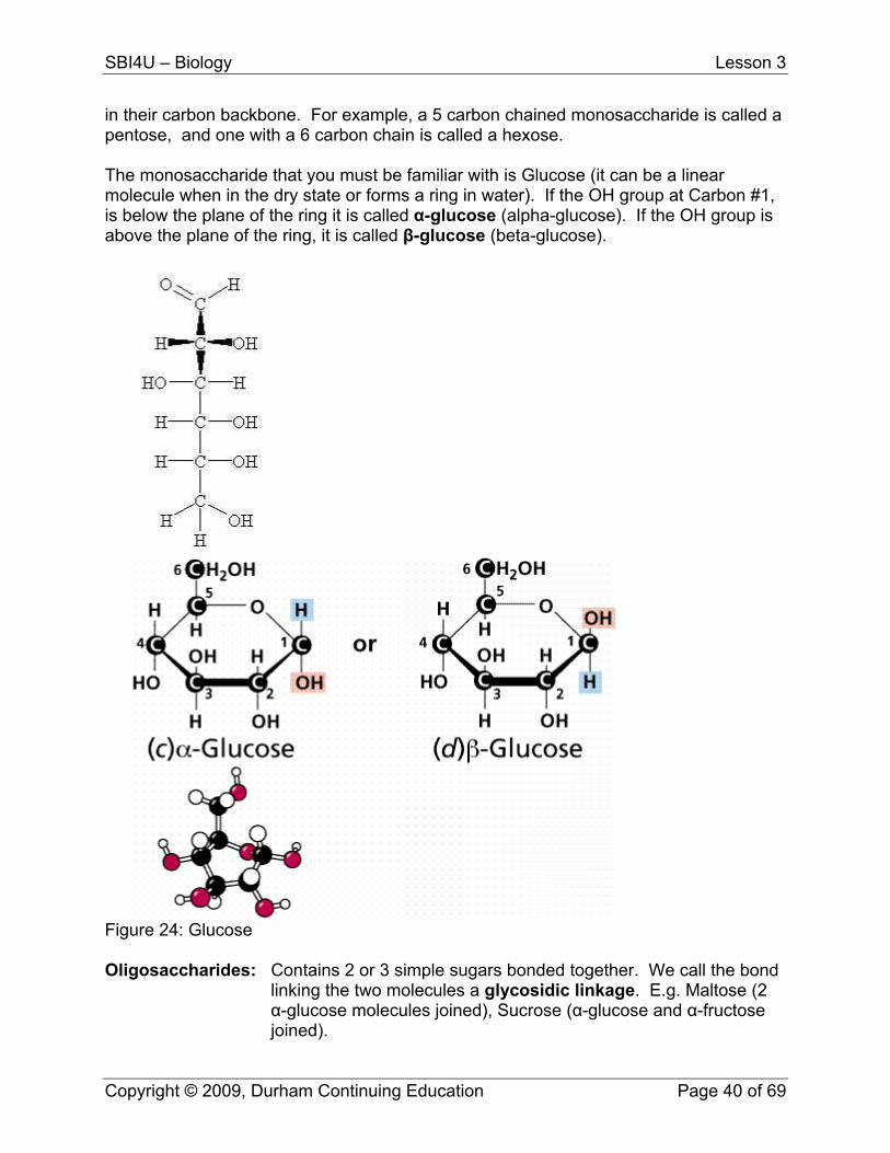
in their carbon backbone. For example, a 5 carbon chained monosaccharide is called a pentose, and one with a 6 carbon chain is called a hexose. The monosaccharide that you must be familiar with is Glucose (it can be a linear molecule when in the dry state or forms a ring in water). If the OH group at Carbon #1, is below the plane of the ring it is called α-glucose (alpha-glucose). If the OH group is above the plane of the ring, it is called β-glucose (beta-glucose).
Figure 24: Glucose Oligosaccharides: Contains 2 or 3 simple sugars bonded together. We call the bond
linking the two molecules a glycosidic linkage. E.g. Maltose (2 α-glucose molecules joined), Sucrose (α-glucose and α-fructose joined).
Copyright © 2009, Durham Continuing Education Page 40 of 69
SBI4U – Biology Lesson 3
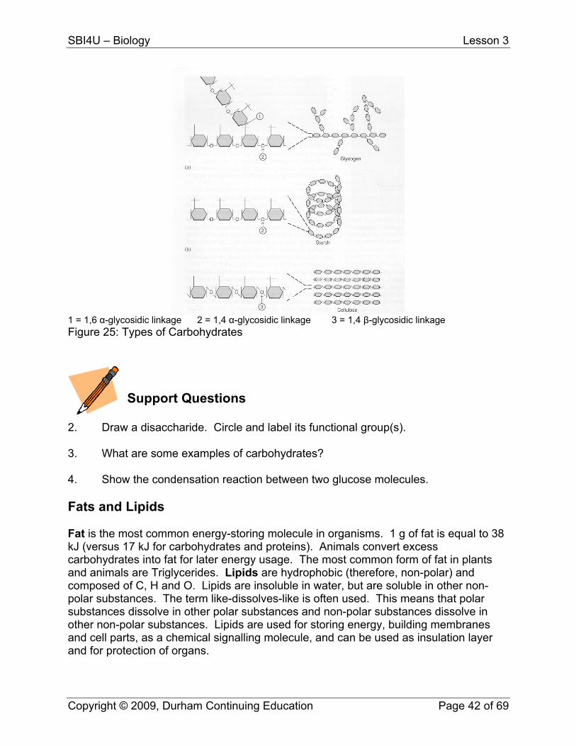
Polysaccharides: It is a complex carbohydrate composed of hundred to several
thousand monosaccharide subunits joined by glycosidic linkages. E.g. Starch and Glycogen used for energy and Cellulose and Chitin used for structural support.
Glycogen: It is stored in small amounts by humans and other animals in
muscle and liver cells. Enzymes can “hydrolyze” glycogen into glucose for energy during physical activity.
Cellulose: It is the primary structural unit of plants (major component of cell
walls). Plants produce 200 billion kg./year making it the most abundant organic substance on earth. Cellulose consists of a straight chain polymer of β-glucose molecules. Hydrogen bonding occurs between several straight chains to produce tight bundles called microfibrils. These intertwine to form tough, insoluble cellulose fibres. Humans cannot digest glycosidic linkages between β-glucose molecules in cellulose (we can digest starch which are made of α-glucose subunits), therefore, cellulose is not used as an energy source for humans, but it is still a part of a healthy diet in humans. The cellulose provides fibre which passes through our digestive system and scrapes the walls of the large intestine, stimulating the secretion of mucus and aiding in the elimination of waste. Some animals such as cows, rabbits, sheep, etc. have bacteria and protists in their guts that produce enzymes that break β-glucose linkages, allowing them to digest the cellulose for energy.
Chitin: It makes up the exoskeleton of insects and crustaceans. It is the
second most abundant organic material on earth. Chitin can also be used in contact lenses and biodegradable stitches.
Copyright © 2009, Durham Continuing Education Page 41 of 69
SBI4U – Biology Lesson 3
1 = 1,6 α-glycosidic linkage 2 = 1,4 α-glycosidic linkage 3 = 1,4 β-glycosidic linkage Figure 25: Types of Carbohydrates
Support Questions
2. Draw a disaccharide. Circle and label its functional group(s). 3. What are some examples of carbohydrates? 4. Show the condensation reaction between two glucose molecules. Fats and Lipids Fat is the most common energy-storing molecule in organisms. 1 g of fat is equal to 38 kJ (versus 17 kJ for carbohydrates and proteins). Animals convert excess carbohydrates into fat for later energy usage. The most common form of fat in plants and animals are Triglycerides. Lipids are hydrophobic (therefore, non-polar) and composed of C, H and O. Lipids are insoluble in water, but are soluble in other non-polar substances. The term like-dissolves-like is often used. This means that polar substances dissolve in other polar substances and non-polar substances dissolve in other non-polar substances. Lipids are used for storing energy, building membranes and cell parts, as a chemical signalling molecule, and can be used as insulation layer and for protection of organs.
Copyright © 2009, Durham Continuing Education Page 42 of 69
SBI4U – Biology Lesson 3
Lipids fit into 4 categories: fats, phospholipids, steroids and waxes. Glycerol: It is a 3 carbon alcohol containing a hydroxyl (OH) group attached to
each carbon. Glycerol forms the backbone of triglyceride molecules. Fatty Acids: They are long hydrocarbon chains with a single carboxyl group (COOH)
at one end. Fatty acids can be: Saturated, monounsaturated or polyunsaturated. Saturated Fatty acids have no double bonds between the carbon atoms in the chain, mono-unsaturated have one double bond and poly-unsaturated fatty acids have 2 or more double bonds. Animal fat, such as butter and lard, are composed of triglycerides with mostly saturated fatty acid chains and are solid at room temperature. Plant oils contain mono and polyunsaturated fatty acids and are liquid at room temperature.
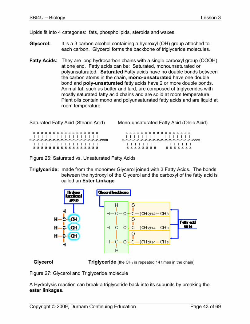
Saturated Fatty Acid (Stearic Acid) Mono-unsaturated Fatty Acid (Oleic Acid) H H H H H H H H H H H H H H H H H H H H H H H H H H H H H H H H H H | | | | | | | | | | | | | | | | | | | | | | | | | | | | | | | | | | H-C-C-C-C-C-C-C-C-C-C-C-C-C-C-C-C-C-COOH H-C-C-C-C-C-C-C-C-C=C-C-C-C-C-C-C-C-COOH | | | | | | | | | | | | | | | | | | | | | | | | | | | | | | | | H H H H H H H H H H H H H H H H H H H H H H H H H H H H H H H H
Figure 26: Saturated vs. Unsaturated Fatty Acids Triglyceride: made from the monomer Glycerol joined with 3 Fatty Acids. The bonds
between the hydroxyl of the Glycerol and the carboxyl of the fatty acid is called an Ester Linkage
Glycerol Triglyceride (the CH2 is repeated 14 times in the chain) Figure 27: Glycerol and Triglyceride molecule A Hydrolysis reaction can break a triglyceride back into its subunits by breaking the ester linkages.
Copyright © 2009, Durham Continuing Education Page 43 of 69
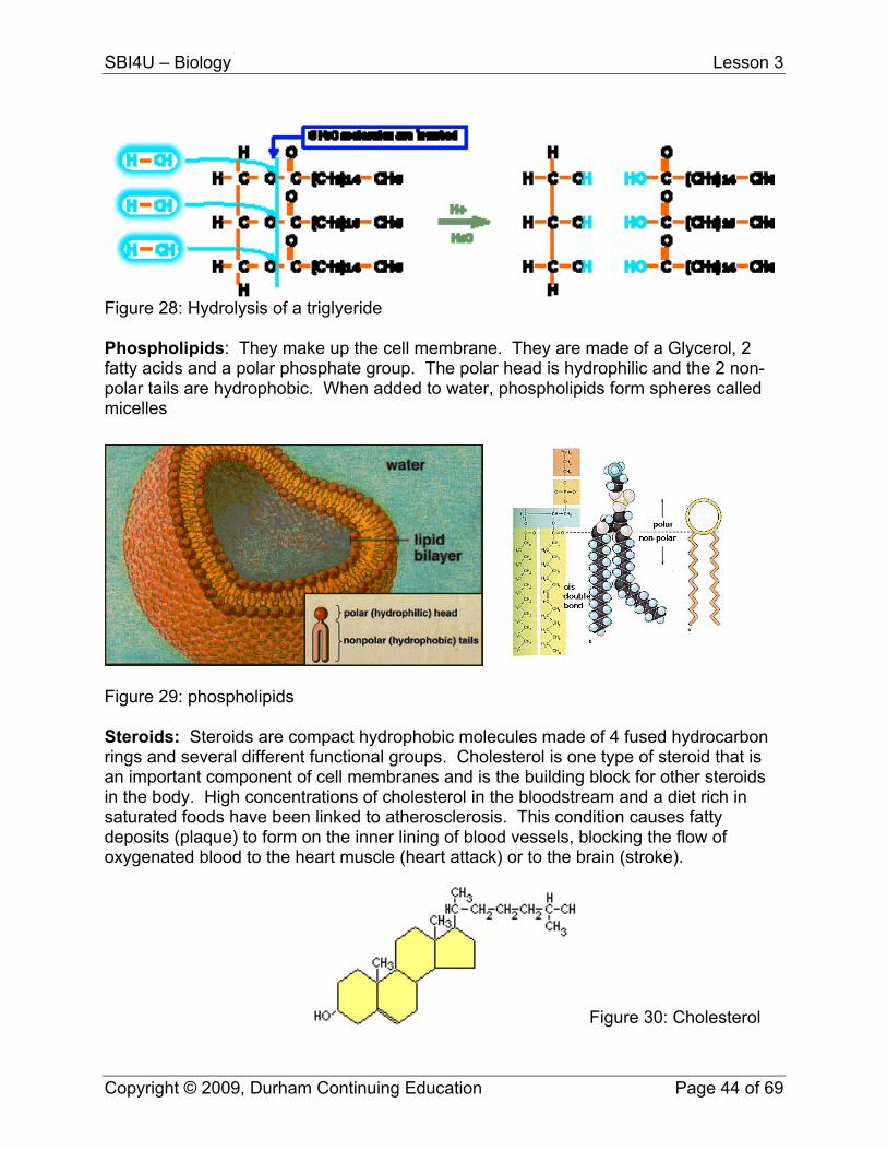
SBI4U – Biology Lesson 3
Figure 28: Hydrolysis of a triglyeride Phospholipids: They make up the cell membrane. They are made of a Glycerol, 2 fatty acids and a polar phosphate group. The polar head is hydrophilic and the 2 non-polar tails are hydrophobic. When added to water, phospholipids form spheres called micelles
Figure 29: phospholipids Steroids: Steroids are compact hydrophobic molecules made of 4 fused hydrocarbon rings and several different functional groups. Cholesterol is one type of steroid that is an important component of cell membranes and is the building block for other steroids in the body. High concentrations of cholesterol in the bloodstream and a diet rich in saturated foods have been linked to atherosclerosis. This condition causes fatty deposits (plaque) to form on the inner lining of blood vessels, blocking the flow of oxygenated blood to the heart muscle (heart attack) or to the brain (stroke).
Figure 30: Cholesterol
Copyright © 2009, Durham Continuing Education Page 44 of 69
SBI4U – Biology Lesson 3
Waxes: Waxes are lipids containing long fatty acids linked to alcohols. They are hydrophobic, firm and pliable and are used by plants (cutin) and animals for water-proofing.
Support Questions
(Reminder: these questions are not to be submitted but reinforce the material taught and are strongly recommended – DO NOT write in this book). 5. Draw a glycerol molecule. Circle and label its functional group(s). 6. Draw a 5 carbon saturated fatty acid. Circle and label its functional group(s). 7. What is a phospholipid? Why is it important to the cell? 8. What is the role of steroids in the cell? Proteins Proteins are the most diverse molecules in living organisms and among the most important. They act as structural building blocks, as functional molecules, and are involved in almost everything that a cell does. Cells contain thousands of different proteins each performing a specific task. In fact, more than 50% of the dry mass of cells is made up of proteins. All enzymes (biological catalysts) are proteins. Protein Shape
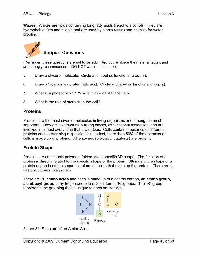
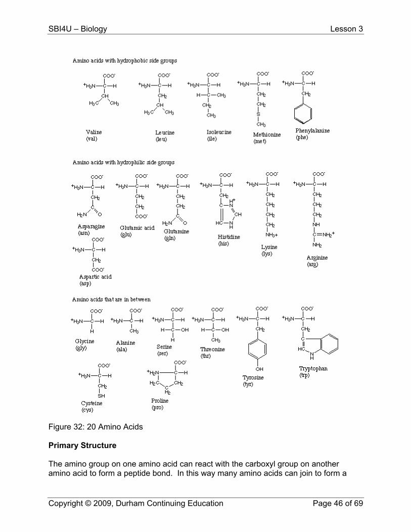
Proteins are amino acid polymers folded into a specific 3D shape. The function of a protein is directly related to the specific shape of the protein. Ultimately, the shape of a protein depends on the sequence of amino acids that make up the protein. There are 4 basic structures to a protein. There are 20 amino acids and each is made up of a central carbon, an amino group, a carboxyl group, a hydrogen and one of 20 different “R” groups. The “R” group represents the grouping that is unique to each amino acid.
Figure 31: Structure of an Amino Acid
Copyright © 2009, Durham Continuing Education Page 45 of 69
SBI4U – Biology Lesson 3
Figure 32: 20 Amino Acids Primary Structure The amino group on one amino acid can react with the carboxyl group on another amino acid to form a peptide bond. In this way many amino acids can join to form a
Copyright © 2009, Durham Continuing Education Page 46 of 69
SBI4U – Biology Lesson 3
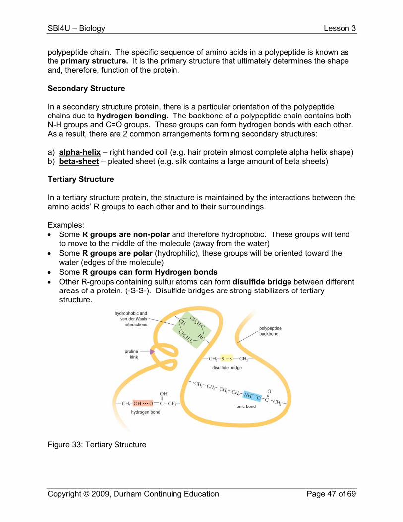
polypeptide chain. The specific sequence of amino acids in a polypeptide is known as the primary structure. It is the primary structure that ultimately determines the shape and, therefore, function of the protein. Secondary Structure In a secondary structure protein, there is a particular orientation of the polypeptide chains due to hydrogen bonding. The backbone of a polypeptide chain contains both N-H groups and C=O groups. These groups can form hydrogen bonds with each other. As a result, there are 2 common arrangements forming secondary structures: a) alpha-helix – right handed coil (e.g. hair protein almost complete alpha helix shape) b) beta-sheet – pleated sheet (e.g. silk contains a large amount of beta sheets) Tertiary Structure In a tertiary structure protein, the structure is maintained by the interactions between the amino acids’ R groups to each other and to their surroundings. Examples: • Some R groups are non-polar and therefore hydrophobic. These groups will tend
to move to the middle of the molecule (away from the water) • Some R groups are polar (hydrophilic), these groups will be oriented toward the
water (edges of the molecule) • Some R groups can form Hydrogen bonds • Other R-groups containing sulfur atoms can form disulfide bridge between different
areas of a protein. (-S-S-). Disulfide bridges are strong stabilizers of tertiary structure.
Figure 33: Tertiary Structure
Copyright © 2009, Durham Continuing Education Page 47 of 69
SBI4U – Biology Lesson 3
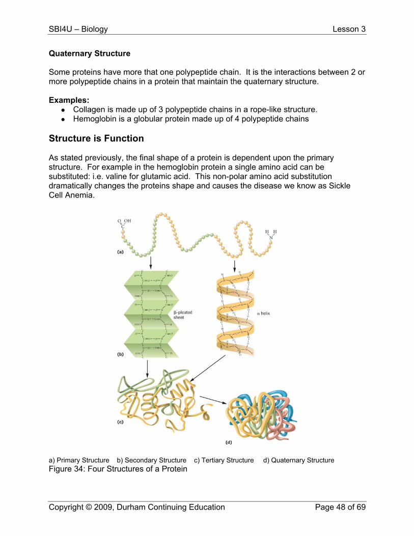
Quaternary Structure Some proteins have more that one polypeptide chain. It is the interactions between 2 or more polypeptide chains in a protein that maintain the quaternary structure. Examples:
Collagen is made up of 3 polypeptide chains in a rope-like structure. Hemoglobin is a globular protein made up of 4 polypeptide chains
Structure is Function As stated previously, the final shape of a protein is dependent upon the primary structure. For example in the hemoglobin protein a single amino acid can be substituted: i.e. valine for glutamic acid. This non-polar amino acid substitution dramatically changes the proteins shape and causes the disease we know as Sickle Cell Anemia.
a) Primary Structure b) Secondary Structure c) Tertiary Structure d) Quaternary Structure Figure 34: Four Structures of a Protein
Copyright © 2009, Durham Continuing Education Page 48 of 69
SBI4U – Biology Lesson 3
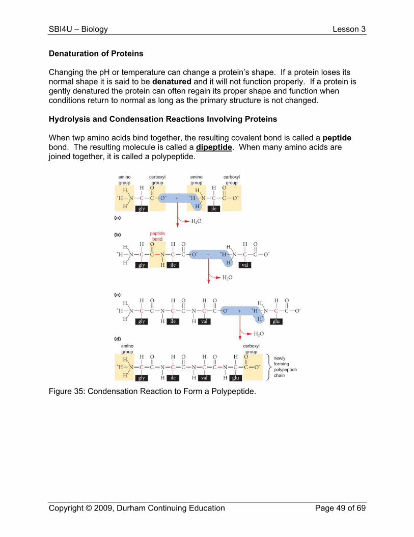
Denaturation of Proteins Changing the pH or temperature can change a protein’s shape. If a protein loses its normal shape it is said to be denatured and it will not function properly. If a protein is gently denatured the protein can often regain its proper shape and function when conditions return to normal as long as the primary structure is not changed. Hydrolysis and Condensation Reactions Involving Proteins When twp amino acids bind together, the resulting covalent bond is called a peptide bond. The resulting molecule is called a dipeptide. When many amino acids are joined together, it is called a polypeptide.
Figure 35: Condensation Reaction to Form a Polypeptide.
Copyright © 2009, Durham Continuing Education Page 49 of 69
SBI4U – Biology Lesson 3
Support Questions
9. What are the 4 structures of a protein. 10. Give 2 examples of a secondary structure protein. 11. What does denaturation mean? 12. Draw an amino acid. Circle and label its functional group(s). 13. How many different amino acids are there? What do all amino acids have in
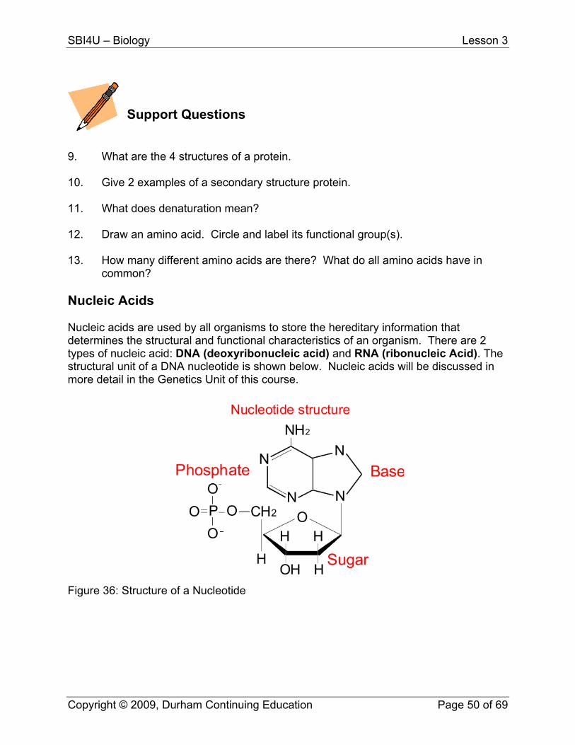
common? Nucleic Acids Nucleic acids are used by all organisms to store the hereditary information that determines the structural and functional characteristics of an organism. There are 2 types of nucleic acid: DNA (deoxyribonucleic acid) and RNA (ribonucleic Acid). The structural unit of a DNA nucleotide is shown below. Nucleic acids will be discussed in more detail in the Genetics Unit of this course.
Figure 36: Structure of a Nucleotide
Copyright © 2009, Durham Continuing Education Page 50 of 69
SBI4U – Biology Lesson 3
Key Question #3
1. Show the complete condensation reaction to form maltose. Label all molecules. (5 marks)
2. Show the complete hydrolysis of a dipeptide. Label all molecules. (5 marks) 3. Discuss the concept that life has a chemical basis. (3 marks) 4. Explain what REDOX reactions are and their importance to the cell (2 marks) 5. Although steroids in our body are necessary for biological functions, anabolic
steroid use by athletes can be quite dangerous. In a paragraph, outline why athletes take steroids and using specific examples of steroids, describe some of the hazardous side-effects. Include references (please use a recognized format, either APA or MLA style referencing). (6 marks)
6. It is mandatory for the food industry to label the nutrient content of their food.
Look at a nutrient in your home. (4 marks)
a. What types of things must be listed? b. One of the items that must be listed are transfats. What are transfats and
why should you limit them in your diet?
Copyright © 2009, Durham Continuing Education Page 51 of 69

SBI4U
Grade 12, University Preparation Biology
Lesson 4 – Enzymes
SBI4U – Biology Lesson 4
Lesson Four: Enzymes For every reaction that occurs in our body, an enzyme is involved in the process. Enzymes are highly specific. Only one enzyme will be involved in each stage of the metabolic pathway. In order to have a better understanding of metabolism, enzyme structure and function must be examined in more detail. In this lesson, you will examine enzymatic pathways, focusing on how they can activate or inhibit a reaction. Uses of enzymes will also be explored. What You Will Learn
use appropriate terminology related to biochemistry, including, but not limited to: allosteric site, substrate, substrate-enzyme complex, and inhibition
describe the chemical structures and mechanisms of various enzymes analyse technological applications related to enzyme activity in the food and
pharmaceutical industries Enzymes Enzymes are proteins that catalyze reaction and are required for all metabolic processes (anabolic or catabolic). A catalyst speeds up a chemical reaction, which means that the reactants are converted into products faster than if there were no catalyst involved. The catalyst itself is not consumed in the process. This means that a catalyst can be recycled to aid in other reactions. Most enzymes end with the letters “ase”, such as in lactase or protease. For all chemical reactions to occur, whether they are endergonic or exergonic (these will be further discussed in Unit 2), an activation energy (EA) barrier must be overcome. Heat generally provides the activation energy for many reactions. Although an increase in temperature increases the rate of most reactions, as mentioned previously, proteins are denatured at high temperatures and lose their function. This could be devastating for the cell. This means that living cells cannon rely solely on heat as a means of overcoming a reaction’s activation energy. Catalysts are the mechanism by which cells can have reactions by reducing the activation energy (EA) barrier and allowing reactions to occur at lower temperatures.
Copyright © 2009, Durham Continuing Education Page 53 of 69
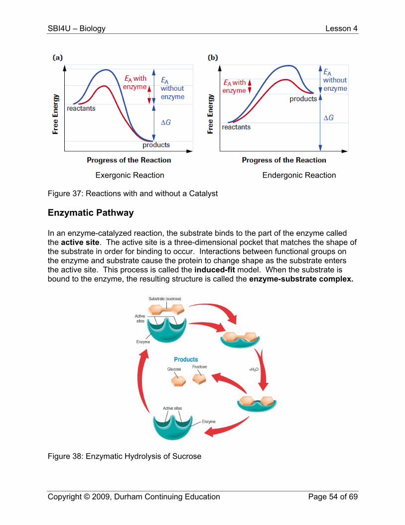
SBI4U – Biology Lesson 4
Exergonic Reaction Endergonic Reaction Figure 37: Reactions with and without a Catalyst Enzymatic Pathway In an enzyme-catalyzed reaction, the substrate binds to the part of the enzyme called the active site. The active site is a three-dimensional pocket that matches the shape of the substrate in order for binding to occur. Interactions between functional groups on the enzyme and substrate cause the protein to change shape as the substrate enters the active site. This process is called the induced-fit model. When the substrate is bound to the enzyme, the resulting structure is called the enzyme-substrate complex.
Figure 38: Enzymatic Hydrolysis of Sucrose
Copyright © 2009, Durham Continuing Education Page 54 of 69
SBI4U – Biology Lesson 4
Support Questions
1. What an activation energy barrier and why must it be overcome? 2. Why are catalysts so important to cell function? 3. Sketch and label an enzyme and its substrate. Factors Affecting Enzyme Activity There are four major factories that affect enzyme activity. These include: temperature, pH, concentration of substrate and concentration of the enzyme itself. Temperature One factor that affects enzyme activity is temperature. As the temperature rises, reacting molecules gain more and more kinetic energy. This increases the chances of a successful collision, which causes the rate of enzymatic reaction to increase as well. Every enzyme has a specific or optimal temperature where its activity is the greatest. For example, enzymes within the human body have an optimal temperature of about 37.5 °C. If the temperature increases above the optimal temperature, the enzyme structure begins to break down, or denature. This is due to the fact that at higher temperatures intra- and intermolecular bonds are broken as the enzyme molecules gain even more kinetic energy. pH Each enzyme works within quite a small pH range. There is a pH at which its activity is greatest. This is called the optimal pH. This is because changes in pH can make and break intra- and intermolecular bonds, changing the shape of the enzyme and, therefore, its effectiveness.
Copyright © 2009, Durham Continuing Education Page 55 of 69
SBI4U – Biology Lesson 4
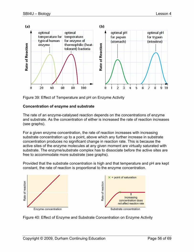
Figure 39: Effect of Temperature and pH on Enzyme Activity Concentration of enzyme and substrate The rate of an enzyme-catalysed reaction depends on the concentrations of enzyme and substrate. As the concentration of either is increased the rate of reaction increases (see graphs). For a given enzyme concentration, the rate of reaction increases with increasing substrate concentration up to a point, above which any further increase in substrate concentration produces no significant change in reaction rate. This is because the active sites of the enzyme molecules at any given moment are virtually saturated with substrate. The enzyme/substrate complex has to dissociate before the active sites are free to accommodate more substrate (see graphs). Provided that the substrate concentration is high and that temperature and pH are kept constant, the rate of reaction is proportional to the enzyme concentration.
Figure 40: Effect of Enzyme and Substrate Concentration on Enzyme Activity
Copyright © 2009, Durham Continuing Education Page 56 of 69
SBI4U – Biology Lesson 4
Enzyme inhibitors are molecules that bind to enzymes and decrease their activity. Since blocking an enzyme's activity can kill a pathogen or correct a metabolic imbalance, many drugs are enzyme inhibitors. They are also used as herbicides and pesticides. Not all molecules that bind to enzymes are inhibitors; enzyme activators bind to enzymes and increase their enzymatic activity.
Support Questions
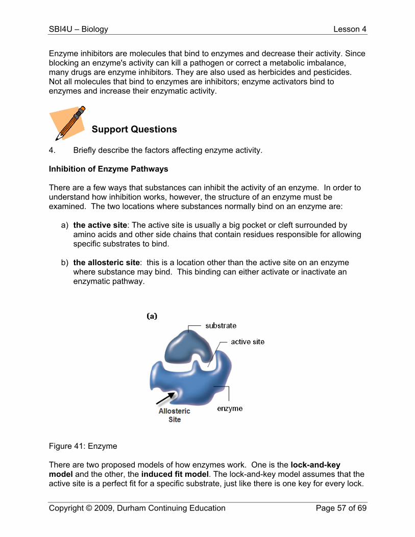
4. Briefly describe the factors affecting enzyme activity. Inhibition of Enzyme Pathways There are a few ways that substances can inhibit the activity of an enzyme. In order to understand how inhibition works, however, the structure of an enzyme must be examined. The two locations where substances normally bind on an enzyme are:
a) the active site: The active site is usually a big pocket or cleft surrounded by amino acids and other side chains that contain residues responsible for allowing specific substrates to bind.
b) the allosteric site: this is a location other than the active site on an enzyme
where substance may bind. This binding can either activate or inactivate an enzymatic pathway.
Figure 41: Enzyme There are two proposed models of how enzymes work. One is the lock-and-key model and the other, the induced fit model. The lock-and-key model assumes that the active site is a perfect fit for a specific substrate, just like there is one key for every lock.
Copyright © 2009, Durham Continuing Education Page 57 of 69
SBI4U – Biology Lesson 4
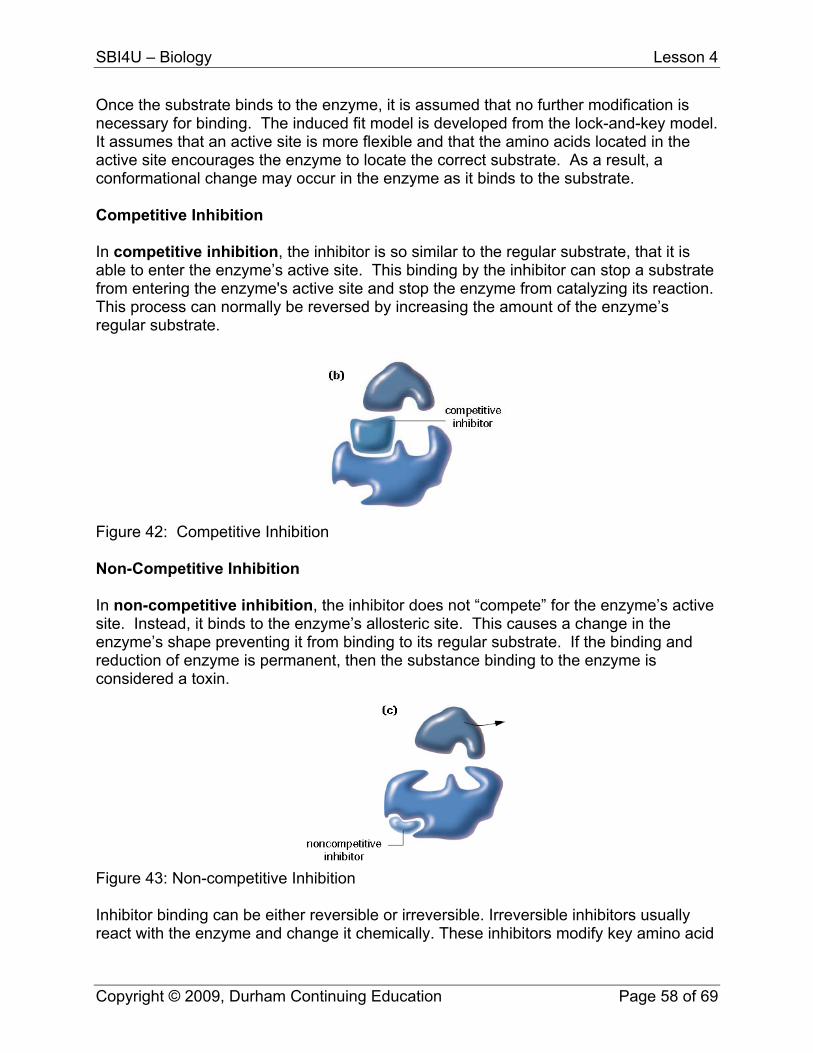
Once the substrate binds to the enzyme, it is assumed that no further modification is necessary for binding. The induced fit model is developed from the lock-and-key model. It assumes that an active site is more flexible and that the amino acids located in the active site encourages the enzyme to locate the correct substrate. As a result, a conformational change may occur in the enzyme as it binds to the substrate. Competitive Inhibition In competitive inhibition, the inhibitor is so similar to the regular substrate, that it is able to enter the enzyme’s active site. This binding by the inhibitor can stop a substrate from entering the enzyme's active site and stop the enzyme from catalyzing its reaction. This process can normally be reversed by increasing the amount of the enzyme’s regular substrate.
Figure 42: Competitive Inhibition Non-Competitive Inhibition In non-competitive inhibition, the inhibitor does not “compete” for the enzyme’s active site. Instead, it binds to the enzyme’s allosteric site. This causes a change in the enzyme’s shape preventing it from binding to its regular substrate. If the binding and reduction of enzyme is permanent, then the substance binding to the enzyme is considered a toxin.
Figure 43: Non-competitive Inhibition Inhibitor binding can be either reversible or irreversible. Irreversible inhibitors usually react with the enzyme and change it chemically. These inhibitors modify key amino acid
Copyright © 2009, Durham Continuing Education Page 58 of 69
SBI4U – Biology Lesson 4
residues needed for enzymatic activity. In contrast, reversible inhibitors bind non-covalently and different types of inhibition are produced depending on whether these inhibitors bind the enzyme, the enzyme-substrate complex, or both.
Support Questions
5. Differentiate between competitive and non-competitive inhibition. 6. What happens if the secondary substrate is not released during competitive or
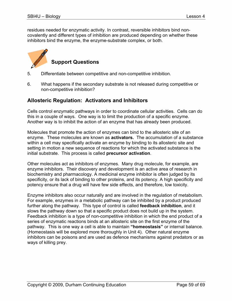
non-competitive inhibition? Allosteric Regulation: Activators and Inhibitors Cells control enzymatic pathways in order to coordinate cellular activities. Cells can do this in a couple of ways. One way is to limit the production of a specific enzyme. Another way is to inhibit the action of an enzyme that has already been produced. Molecules that promote the action of enzymes can bind to the allosteric site of an enzyme. These molecules are known as activators. The accumulation of a substance within a cell may specifically activate an enzyme by binding to its allosteric site and setting in motion a new sequence of reactions for which the activated substance is the initial substrate. This process is called precursor activation. Other molecules act as inhibitors of enzymes. Many drug molecule, for example, are enzyme inhibitors. Their discovery and development is an active area of research in biochemistry and pharmacology. A medicinal enzyme inhibitor is often judged by its specificity, or its lack of binding to other proteins, and its potency. A high specificity and potency ensure that a drug will have few side effects, and therefore, low toxicity. Enzyme inhibitors also occur naturally and are involved in the regulation of metabolism. For example, enzymes in a metabolic pathway can be inhibited by a product produced further along the pathway. This type of control is called feedback inhibition, and it slows the pathway down so that a specific product does not build up in the system. Feedback inhibition is a type of non-competitive inhibition in which the end product of a series of enzymatic reactions binds at an allosteric site on the first enzyme of the pathway. This is one way a cell is able to maintain “homeostasis” or internal balance. (Homeostasis will be explored more thoroughly in Unit 4). Other natural enzyme inhibitors can be poisons and are used as defence mechanisms against predators or as ways of killing prey.
Copyright © 2009, Durham Continuing Education Page 59 of 69
SBI4U – Biology Lesson 4
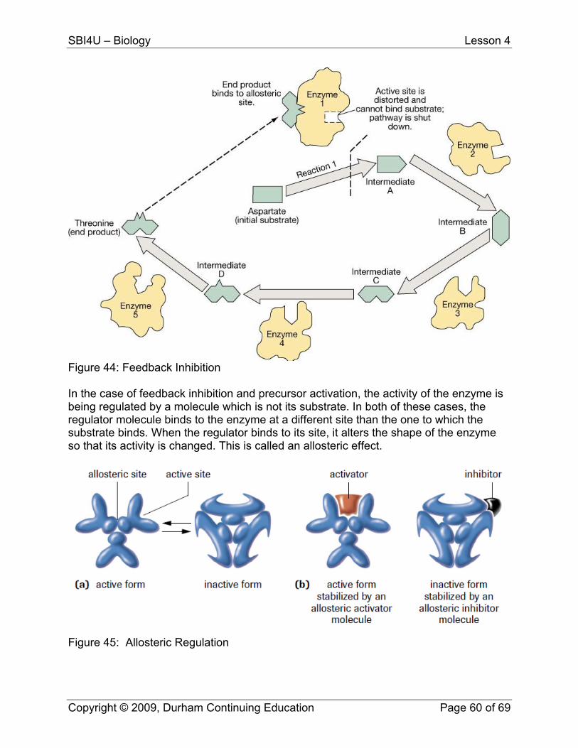
Figure 44: Feedback Inhibition In the case of feedback inhibition and precursor activation, the activity of the enzyme is being regulated by a molecule which is not its substrate. In both of these cases, the regulator molecule binds to the enzyme at a different site than the one to which the substrate binds. When the regulator binds to its site, it alters the shape of the enzyme so that its activity is changed. This is called an allosteric effect.
Figure 45: Allosteric Regulation
Copyright © 2009, Durham Continuing Education Page 60 of 69
SBI4U – Biology Lesson 4
Support Questions
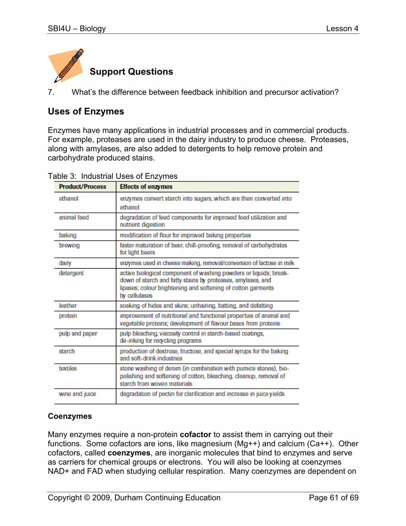
7. What’s the difference between feedback inhibition and precursor activation? Uses of Enzymes Enzymes have many applications in industrial processes and in commercial products. For example, proteases are used in the dairy industry to produce cheese. Proteases, along with amylases, are also added to detergents to help remove protein and carbohydrate produced stains. Table 3: Industrial Uses of Enzymes
Coenzymes Many enzymes require a non-protein cofactor to assist them in carrying out their functions. Some cofactors are ions, like magnesium (Mg++) and calcium (Ca++). Other cofactors, called coenzymes, are inorganic molecules that bind to enzymes and serve as carriers for chemical groups or electrons. You will also be looking at coenzymes NAD+ and FAD when studying cellular respiration. Many coenzymes are dependent on
Copyright © 2009, Durham Continuing Education Page 61 of 69
SBI4U – Biology Lesson 4
our intake of vitamins. For example, NAD+ is dependent on niacin and FAD on riboflavin. A deficiency in one of these vitamins may result in a lack of a coenzyme, and therefore a decrease in certain enzymatic reactions.
Key Question #4
1. Explain the statement “structure is function; change the structure, you change the function” as it relates to enzymes. (2 marks)
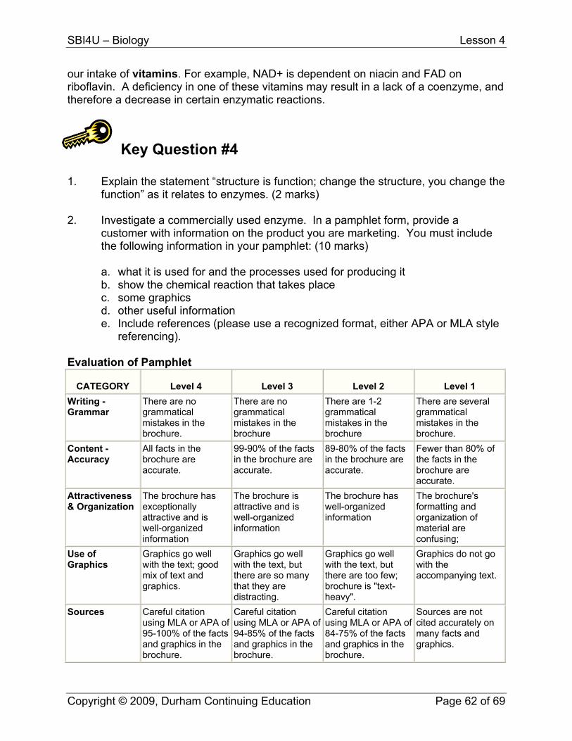
2. Investigate a commercially used enzyme. In a pamphlet form, provide a
customer with information on the product you are marketing. You must include the following information in your pamphlet: (10 marks)
a. what it is used for and the processes used for producing it b. show the chemical reaction that takes place c. some graphics d. other useful information e. Include references (please use a recognized format, either APA or MLA style
referencing). Evaluation of Pamphlet
CATEGORY Level 4 Level 3 Level 2 Level 1 Writing - Grammar
There are no grammatical mistakes in the brochure.
There are no grammatical mistakes in the brochure
There are 1-2 grammatical mistakes in the brochure
There are several grammatical mistakes in the brochure.
Content - Accuracy
All facts in the brochure are accurate.
99-90% of the facts in the brochure are accurate.
89-80% of the facts in the brochure are accurate.
Fewer than 80% of the facts in the brochure are accurate.
Attractiveness & Organization
The brochure has exceptionally attractive and is well-organized information
The brochure is attractive and is well-organized information
The brochure has well-organized information
The brochure's formatting and organization of material are confusing;
Use of Graphics
Graphics go well with the text; good mix of text and graphics.
Graphics go well with the text, but there are so many that they are distracting.
Graphics go well with the text, but there are too few; brochure is "text-heavy".
Graphics do not go with the accompanying text.
Sources Careful citation using MLA or APA of 95-100% of the facts and graphics in the brochure.
Careful citation using MLA or APA of 94-85% of the facts and graphics in the brochure.
Careful citation using MLA or APA of 84-75% of the facts and graphics in the brochure.
Sources are not cited accurately on many facts and graphics.
Copyright © 2009, Durham Continuing Education Page 62 of 69
SBI4U – Biology Lesson 4
3. Investigate 3 examples of how enzymes are used to treat digestive system
disorders such as lactose intolerance. Name the enzyme and how it works. (6 marks)
4. Find an example of a feedback inhibition pathway in the human body. Explain
how it works (in relation to enzymes). (2 marks) 5. What types of food production processes use enzymes to improve production
yields? How do they do so? (3 marks) 6. Research why there are so many different varieties of cheese when the
production process is basically the same for all cheeses. (2 marks)
Copyright © 2009, Durham Continuing Education Page 63 of 69
SBI4U
Grade 12, University Preparation Biology
Support Question Answers
SBI4U – Biology Support Question Answers
Lesson 1 1. Name of Atom
Atomic Number
Atomic Mass
Number of Protons
Number of Neutrons
Number of Electrons
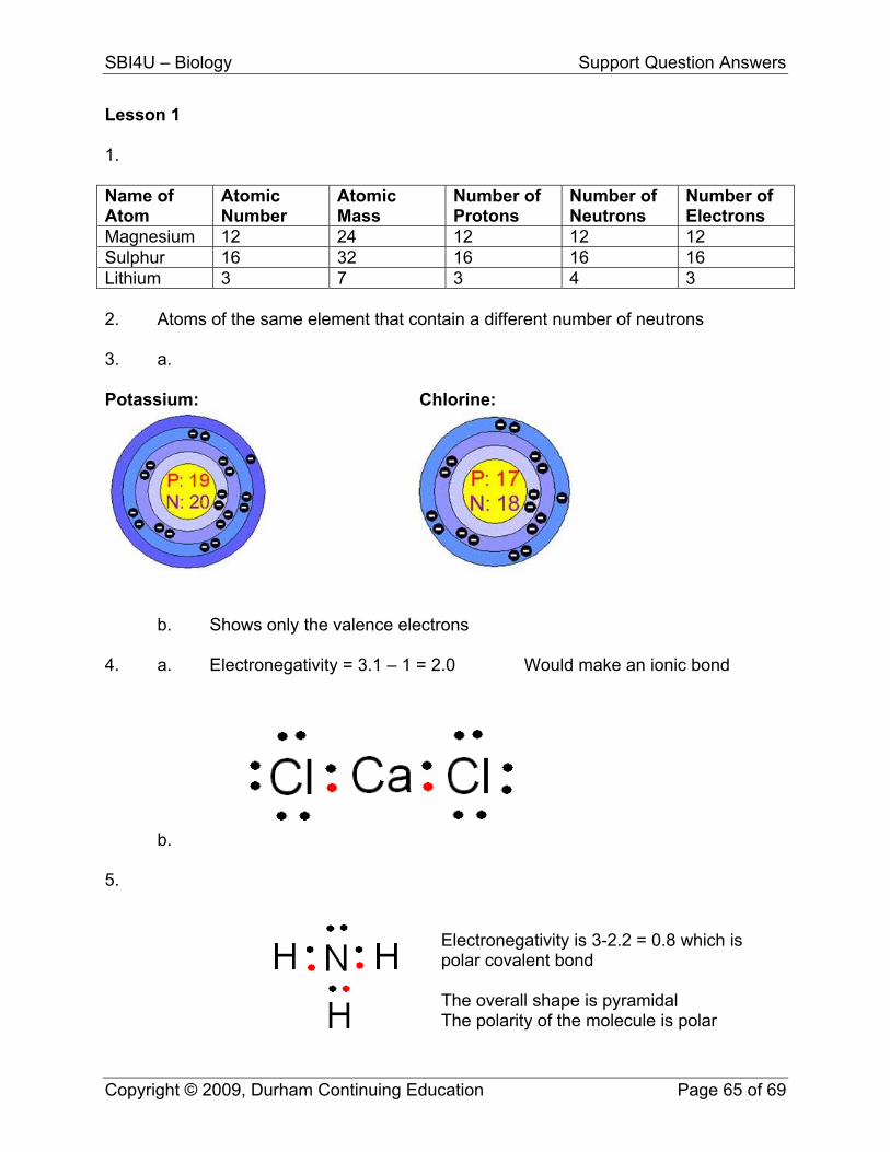
Magnesium 12 24 12 12 12 Sulphur 16 32 16 16 16 Lithium 3 7 3 4 3 2. Atoms of the same element that contain a different number of neutrons 3. a. Potassium: Chlorine:
b. Shows only the valence electrons 4. a. Electronegativity = 3.1 – 1 = 2.0 Would make an ionic bond
b. 5.
Electronegativity is 3-2.2 = 0.8 which is polar covalent bond The overall shape is pyramidal The polarity of the molecule is polar
Copyright © 2009, Durham Continuing Education Page 65 of 69
SBI4U – Biology Support Question Answers
6. Can be non-polar if the overall shape of the molecule is symmetrical. 7. It occurs when any acid and a base react to create water and a salt. 8. Buffers resist a change in pH Lesson 2 1. Both have DNA, and a cell membrane, and some other similar organelles. Main
difference is that eukaryotic cells have a membrane bound nucleus that holds DNA within thread-like structures called chromosomes and prokaryotic cells do not
2. Some different organelles: plants have cell wall, vacuoles, and chloroplasts
while animals cells do not; animal cells have golgi apparatus and microtubules. 3. It states that the cell surface membrane has proteins that move about within a
bed of semi-fluid lipids. 4. Diffusion is the movement of molecules from an area of greater concentration to
an area of lesser concentration. Osmosis is the diffusion of water across a concentration gradient.
a. In a Hypotonic solution, water will diffuse into the cell, causing it to expand
and burst. Called lysis. In a Hypertonic, water would diffuse out of the cell, causing it to shrink. Called crenation
b. In a Hypotonic solution, water will diffuse into the cell, causing it to
expand, but not burst due to the cell wall. Called turgor pressure. In a Hypertonic, water would diffuse out of the cell, causing it to shrink. Called plasmolysis.
5. Facilitated transport happens when a carrier protein is used to assist in the
movement of a molecule across the plasma membrane when that molecule is travelling from an area of high concentration to an area of low concentration. During active transport, molecules are using carrier proteins to go against their concentration gradient and are go from an area of low concentration to an area of high concentration. This process requires energy in the form of ATP.
6. Some molecules are too large to diffuse through the cell membrane or to be
transported by protein carriers. These molecules are taken into the cell by vesicle formation. This process is called endocytosis. When molecules are removed from the cell through the use of vesicles, it is called exocytosis.
Copyright © 2009, Durham Continuing Education Page 66 of 69
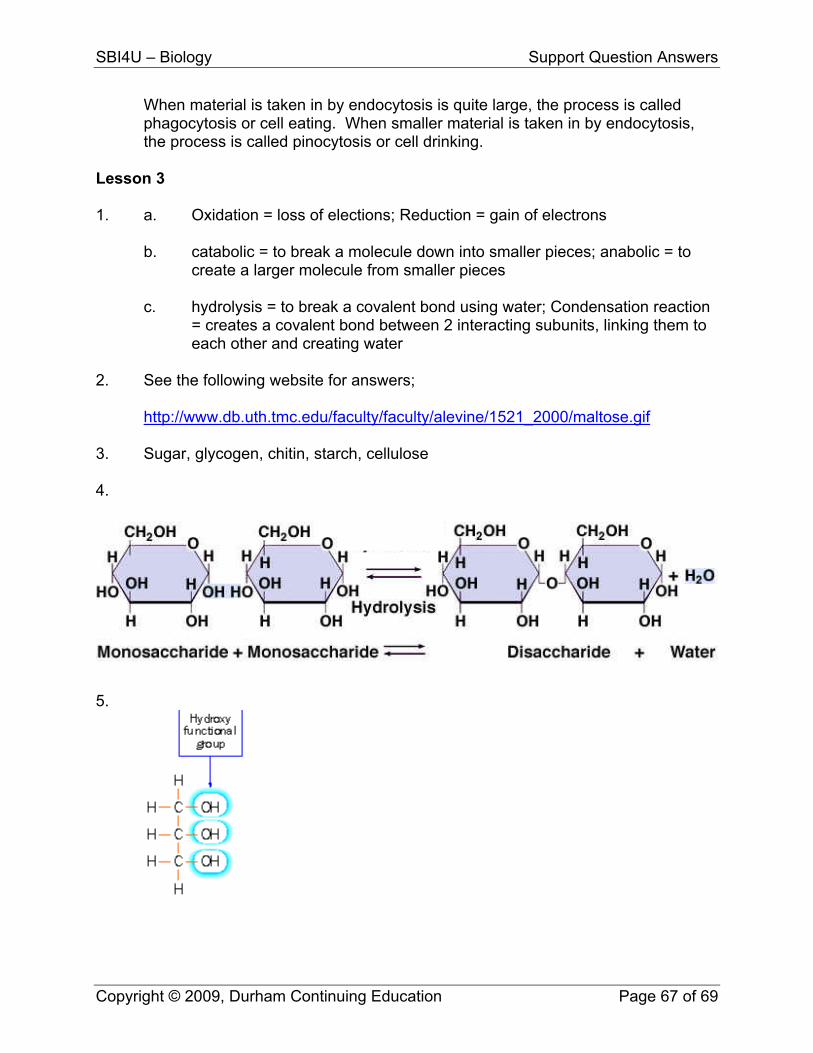
SBI4U – Biology Support Question Answers
When material is taken in by endocytosis is quite large, the process is called phagocytosis or cell eating. When smaller material is taken in by endocytosis, the process is called pinocytosis or cell drinking.
Lesson 3 1. a. Oxidation = loss of elections; Reduction = gain of electrons
b. catabolic = to break a molecule down into smaller pieces; anabolic = to create a larger molecule from smaller pieces c. hydrolysis = to break a covalent bond using water; Condensation reaction = creates a covalent bond between 2 interacting subunits, linking them to each other and creating water
2. See the following website for answers; http://www.db.uth.tmc.edu/faculty/faculty/alevine/1521_2000/maltose.gif 3. Sugar, glycogen, chitin, starch, cellulose 4.
5.
Copyright © 2009, Durham Continuing Education Page 67 of 69
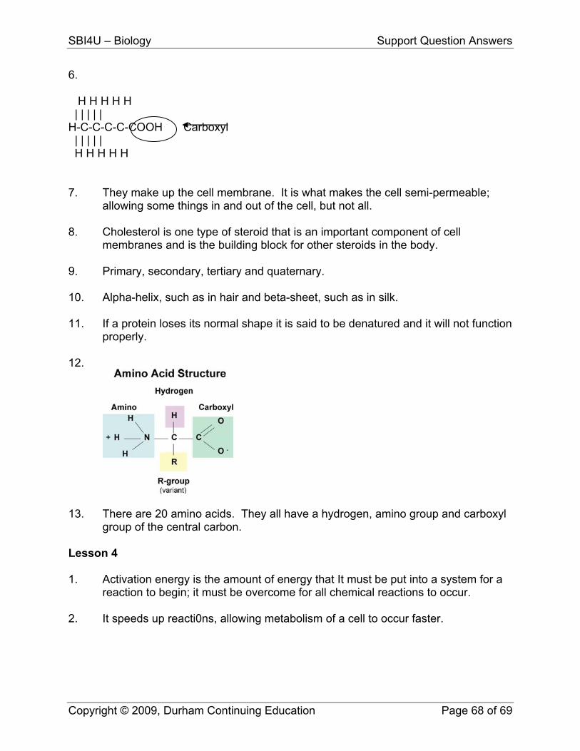
SBI4U – Biology Support Question Answers
6. H H H H H | | | | | H-C-C-C-C-COOH Carboxyl | | | | | H H H H H 7. They make up the cell membrane. It is what makes the cell semi-permeable;
allowing some things in and out of the cell, but not all. 8. Cholesterol is one type of steroid that is an important component of cell
membranes and is the building block for other steroids in the body. 9. Primary, secondary, tertiary and quaternary. 10. Alpha-helix, such as in hair and beta-sheet, such as in silk. 11. If a protein loses its normal shape it is said to be denatured and it will not function
properly. 12.
13. There are 20 amino acids. They all have a hydrogen, amino group and carboxyl
group of the central carbon. Lesson 4 1. Activation energy is the amount of energy that It must be put into a system for a
reaction to begin; it must be overcome for all chemical reactions to occur. 2. It speeds up reacti0ns, allowing metabolism of a cell to occur faster.
Copyright © 2009, Durham Continuing Education Page 68 of 69
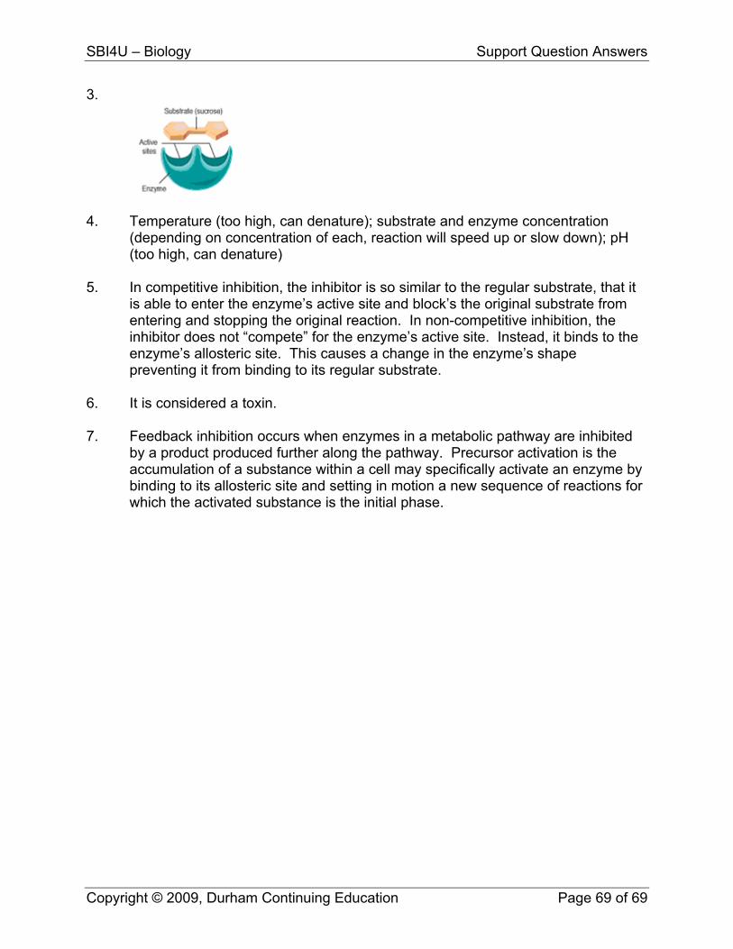
SBI4U – Biology Support Question Answers
3.
4. Temperature (too high, can denature); substrate and enzyme concentration
(depending on concentration of each, reaction will speed up or slow down); pH (too high, can denature)
5. In competitive inhibition, the inhibitor is so similar to the regular substrate, that it
is able to enter the enzyme’s active site and block’s the original substrate from entering and stopping the original reaction. In non-competitive inhibition, the inhibitor does not “compete” for the enzyme’s active site. Instead, it binds to the enzyme’s allosteric site. This causes a change in the enzyme’s shape preventing it from binding to its regular substrate.
6. It is considered a toxin. 7. Feedback inhibition occurs when enzymes in a metabolic pathway are inhibited
by a product produced further along the pathway. Precursor activation is the accumulation of a substance within a cell may specifically activate an enzyme by binding to its allosteric site and setting in motion a new sequence of reactions for which the activated substance is the initial phase.
Copyright © 2009, Durham Continuing Education Page 69 of 69