save pdf - plant methods
TRANSCRIPT

METHODOLOGY Open Access
Comparative evaluation of extraction methods forapoplastic proteins from maize leavesKatja Witzel1,3, Muhammad Shahzad1, Andrea Matros2, Hans-Peter Mock2 and Karl H Mühling1*
Abstract
Proteins in the plant apoplast are essential for many physiological processes. We have analysed and compared sixdifferent infiltration solutions for proteins contained in the apoplast to recognize the most suitable method forleaves and to establish proteome maps for each extraction. The efficiency of protocols was evaluated bycomparing the protein patterns resolved by 1-DE and 2-DE, and revealed distinct characteristics for each infiltrationsolution. Nano-LC-ESI-Q-TOF MS analysis of all fractions was applied to cover all proteins differentially extracted byinfiltration solutions and led to the identification of 328 proteins in total in apoplast preparations. The predictedsubcellular protein localisation distinguished the examined infiltration solutions in those with high or low amountsof intracellular protein contaminations, and with high or low quantities of secreted proteins. All tested infiltrationsolution extracted different subsets of proteins, and those implications on apoplast-specific studies are discussed.
Keywords: Apoplast, liquid chromatography mass spectrometry, maize, proteome analysis, two-dimensional gelelectrophoresis
BackgroundThe plant apoplast comprises the cell wall matrix andthe intercellular spaces, and plays a major role in a widerange of physiological processes, including water andnutrient transport [1], plant-pathogen interactions, andperception and transduction of environmental signals[2,3]. Proteins present in the plant apoplast reflect thisbroad functional diversity. Studies on the dynamicchange of apoplast protein composition revealed newinsights into plant responses to abiotic stress [4-7],nutrient supply [8-10], wounding [11], water deficiency[12,13], pathogen response [14-16] and xylem composi-tion [17,18]. The selection of a suitable extraction proto-col is a crucial step in proteomics surveys as proteinsreveal a high degree of biochemical heterogeneity andinvestigated plant materials can be characterized by thepresence of non-protein components interfering withsubsequent analytical techniques, e.g. two-dimensionalgel electrophoresis (2-DE) or liquid chromatography-mass spectrometry (LC-MS). These biological realitiesled to the establishment of sample preparation methods
for numerous plant species and tissues, such as Arabi-dopsis leaves [19], papaya leaves [20], sunflower leaves[21], cotton seedlings [22], apple and strawberry fruit[23], potato tuber [24], grapevine leaves and roots [25],grape berry cell wall [26], rubber latex [27], cotton fibers[28], banana meristem [29] and chloroplast [30], amongothers. Despite their biological significance, investiga-tions on apoplastic proteins are hampered due to theirlow abundance compared to intracellular protein con-centrations. The extraction of proteins from the leaf androot apoplast is mainly based on the principle ofvacuum infiltration with an extraction solution, followedby a mild centrifugation step to collect the apoplasticwashing fluid. The composition of the infiltration solu-tion is essential as it has to fulfil certain prerequisites,such as maintenance of osmotic pressure to prevent col-lapsing of plasma membrane and stringency for extract-ing cell wall-bound proteins. Borderies et al. [31]compared different solutions to extract loosely boundcell wall proteins of Arabidopsis cell suspension culturesand showed that the composition of extraction solutiondetermines the efficiency of preparation. Similarly, Bou-dart et al. [32] investigated weakly cell wall-bound pro-teins in rosettes of Arabidopsis. Here, we comparedprotein extracts obtained by six different infiltration
* Correspondence: [email protected] of Plant Nutrition and Soil Science, Christian Albrechts University,Hermann-Rodewald-Strasse 2, 24118 Kiel, GermanyFull list of author information is available at the end of the article
Witzel et al. Plant Methods 2011, 7:48http://www.plantmethods.com/content/7/1/48
PLANT METHODS
© 2011 Witzel et al; licensee BioMed Central Ltd. This is an Open Access article distributed under the terms of the Creative CommonsAttribution License (http://creativecommons.org/licenses/by/2.0), which permits unrestricted use, distribution, and reproduction inany medium, provided the original work is properly cited.

solutions already described for apoplastic proteins fromdifferent plant species. We aimed at identifying a proto-col most suitable for the extraction of leaf apoplast pro-teins of maize, a crop of high economic importance. Weevaluated the protein patterns as resolved by 1-DE or 2-DE, identified the proteins using LC-MS and locatedthem to cellular compartments.
Results and discussionIn this study, six different solutions were tested for theability to extract proteins from the maize leaf apoplast:water [8], 20 mM ascorbic acid/20 mM CaCl2 [6], 100mM sorbitol [4], 25 mM Tris-HCl [9], 100 mM sodiumphosphate buffer [16] and 50 mM NaCl [33] (Figure 1).In most cases, the infiltration solutions were applied forwheat leaves and no comparison of the efficiency of pro-tein extraction for each method was performed. Thus,this study focussed on identifying the optimal methodfor extracting apoplastic proteins from maize leaves.Proteins from the leaf apoplast and symplast extracted
with the six infiltration solutions were compared on 1-DE (Figure 2A, Additional file 1). A sharp band patternwas obtained from all apoplast extracts with a highnumber of protein bands in each extract. While theyield of protein extraction was similar, the protein pro-files showed distinct differences. A prominent band of
about 20 kDa was present in extracts of 100 mMsodium phosphate buffer, 25 mM Tris-HCl, 20 mMascorbic acid/20 mM CaCl2 and 50 mM NaCl, but notin water or 100 mM sorbitol. One protein band of highmolecular weight (approximately 100-130 kDa) wasapparent in extracts of water, 100 mM sodium phos-phate buffer and 100 mM sorbitol, but not in 25 mMTris-HCl, 20 mM ascorbic acid/20 mM CaCl2 or 50mM NaCl. While there were similarities, each extractrevealed specific protein bands indicating that differentsubsets of proteins were isolated by the six infiltrationsolutions. Proteins with a molecular weight < 15 kDawere underrepresented in all extracts and this corre-sponds to previous proteomic reports on some of theinfiltration solutions [4,16]. The observed selective pro-tein patterns generated by the individual infiltrationsolutions emphasize the necessity of careful selection ofisolation method [34]. Band patterns from symplast pre-parations did not reveal significant differences amongthe infiltrates and the overall band patterns were morecomplex as from apoplastic preparations. This demon-strates an apparent subfractionation of the cellularcompartments.Equal amounts of apoplast proteins were separated by 2-
DE to assess the protein patterns in more detail (Figure2B). We found areas of good and poor resolved proteins
Figure 1 Schematic representation of protein extraction from maize leaf apoplast. Different infiltration solutions were analyzed for theirspecificity by proteome profiling using gel-based and gel-free approaches.
Witzel et al. Plant Methods 2011, 7:48http://www.plantmethods.com/content/7/1/48
Page 2 of 11

spots on all 2-D gels. Proteins in the acidic gel region ofpH 4-6 showed horizontal streaking. Although all sampleswere precipitated, dissolved in urea-containing buffer sys-tem and dialyzed prior to 2-DE to avoid the contaminationwith nucleic acids or other interfering substances, thesepoorly separated spots were observed. Contrary to this,proteins in the basic region of 2-D gels near the pH 6-10interval showed a superior resolution with minimal streak-ing. The spot patterns resembled the band patterns to acertain extent, e.g. as observed for the 20 kDa band thatwas prominent also on 2-D gels of the respective apoplas-tic extracts. The best resolution of proteins in 25-45 kDaintervals was achieved on extracts of 20 mM ascorbicacid/20 mM CaCl2 infiltration solution, while high
molecular weight proteins separated best in extracts of100 mM sodium phosphate buffer infiltration solution.The latter was applied with success to extract proteinsfrom the leaf apoplast of lupin and resulted in the genera-tion of well resolved protein maps containing about 50spots to evaluate the effect of water and boron deficiency[9]. Our results showed that this separation was notreached, probably due to substances present in the maizeapoplast interfering with isoelectric focusing. As 2-DE didnot result in a comprehensible evaluation of the employedinfiltration solutions, we used nano-LC-ESI-Q-TOF MSfor proteomic analysis of all extracts.In order to obtain an overview of all proteins present
in the six different extracts, we aimed at establishing
Figure 2 Profiles of maize leaf protein extracts as resolved by 1-DE (A) and 2-DE (B). A: SDS-PAGE of apoplastic and symplastic proteinsextracted with water (i), 100 mM sodium phosphate buffer (ii), 25 mM Tris-HCl (iii), 100 mM sorbitol (iv), 20 mM ascorbic acid/20 mM CaCl2 (v)or 50 mM NaCl (vi). A total of 10 μg protein per lane was loaded. B: 2-DE profiles of protein extracts from the maize leaf apoplast isoelectricfocussed on IPG 3-10 and visualized by Coomassie staining. A total of 25 μg protein per gel was loaded.
Witzel et al. Plant Methods 2011, 7:48http://www.plantmethods.com/content/7/1/48
Page 3 of 11

qualitative protein profiles by LC-MS analysis. An auto-matic data directed analysis mode was applied asdescribed in materials and methods section. Resultsexceeding the PLGS score of 12 for protein identifica-tion and probability score of 50% for de novo sequen-cing of peptides were accepted.A total of 328 proteins were identified from all
extracts. Additional file 2 shows the identities of thoseproteins, along with the predicted subcellular localiza-tion and detection in the six apoplastic extracts. Addi-tional file 3 provides the respective identifier, PLGSscore, number of peptides, protein coverage, peptidesequences and peptide sequence probability score for allidentified proteins. In order to visualize and identifyinfiltration solutions with similar protein abundance pat-terns, a hierarchical clustering method was applied. Twomain clusters were found, with the first represented bythe 100 mM sodium phosphate buffer and the secondcontaining all other infiltration solutions indicating theisolation of a rather different set of proteins by the firstone than compared to all other solutions under exami-nation (Figure 3). The most similar abundance patternsderived from leaf infiltration with 25 mM Tris-HCl and50 mM NaCl reflecting a comparable degree of proteinextraction efficiency.The highest number of proteins was found in apoplasticextracts using water as infiltration solution. Here, 171proteins were detected. Extracts of 25 mM Tris-HCl,100 mM sorbitol and 20 mM ascorbic acid/20 mMCaCl2 yielded in the identification of a similar numberof 131, 133 and 133 proteins, respectively. We found114 proteins in extracts of 50 mM NaCl solution and107 proteins in those of 100 mM sodium phosphatebuffer. Out of all 328 proteins, only 28 proteins werecommon across all six extracts (Additional file 4). Asimilar observation was made for Arabidopsis cell wallproteins when extracted by different solutions; here,only 11 out of 96 proteins were found to be common inall extracts [31]. Exhydrolase II [UniProt: Q9XE93] wasfound in all extracts and its identification is illustratedin Additional file 5 as an example. Here, the amino acidsequence is shown and the 12 detected peptides aremarked within, resulting in protein sequence coverageof 28.7%.The quality of apoplastic protein preparations is esti-
mated in many cases by enzymatic measurements ofspecific proteins such as malate dehydrogenase [5,9] andglucose-6-phosphate dehydrogenase [6]. However, it isknown that the activity of those enzymes is detectablein respective cellular compartment as well [35]. Toassess the amount of symplast contaminations in oursamples, we used topology prediction tools. The identi-fied proteins were classified for their subcellular localiza-tion as deduced by Expasy tools Target P and WoLF
PSORT (Figure 4). A number of proteins in this studywere allocated to other cellular compartments then theapoplast, suggesting considerable amounts of intracellu-lar protein contaminations. However, previous reportsusing different plant species and extraction methodsdescribed the detection of cytosolic, mitochondrial orvacuolar proteins in cell wall or apoplast preparations[31,36-38]. These consistent findings point to the occur-rence of non-classical secretory pathways for proteinslacking signal sequences [39,40] and therefore, differen-tiation between yet unknown apoplastic proteins andones resident in other organelles remains difficult.Water-infiltrated leaves revealed 23 protein identifica-tions localized to the apoplast, while a high number ofintracellular proteins were detected from the vacuole(19), cytosol (46) and chloroplast (23). This observationis indicative for the disrupture of plasma membraneduring the infiltration process. Also, apoplastic extractswith 100 mM sorbitol as infiltration solution containeda superior proportion of chloroplast (26) and cytosolic(41) proteins with only 15 predicted apoplastic proteins.This result was unexpected as the sugar alcohol sorbitolwas applied to maintain the osmotic cell pressure. Simi-lar numbers of proteins in infiltrates with 25 mM Tris-HCl, 20 mM ascorbic acid/20 mM CaCl2 and 50 mMNaCl were assigned to the chloroplast (14, 17, 14), thecytosol (36, 34, 27), and the apoplast (25, 31, 25). Of alltested infiltration solutions, 100 mM sodium phosphatebuffer contained the lowest number of proteins assignedto intracellular compartments (chloroplast: 10, cytosol:16) and the highest number of proteins targeted to theextracellular apoplast with 34 identified proteins.Table 1 presents the 67 proteins allocated to the apo-
plast of maize leaves and grouped according to theirfunction into 7 classes. The largest class consisted of 39proteins related to cell structural processes, includingcarbohydrate metabolism (e.g.: lichenase 2, alpha N-ara-binofuranosidase, beta galactosidase, exoglucanase,exhydrolase II) and cell wall modification (e.g.; pectines-terase, xyloglucan endotransglycosylase hydrolase, per-oxidases). Synthesis and integration of polysaccharidesinto the cell wall and extension of this network duringplant growth are the major biological functions of pro-teins present in the apoplast [41] and our findingsreflect this reality. Fifteen proteins were involved in dis-ease and defense reactions, the second prime functionof the apoplast [42]. The third class was related to pro-teins with transporting function and here, 7 proteinswere identified. Further classes were related to cellgrowth/division, protein destination/storage, secondarymetabolism and signal transduction.The number of proteins identified exclusively in any
of the extracts was compared and revealed that 16 outof 34 apoplastic proteins were found only in extracts of
Witzel et al. Plant Methods 2011, 7:48http://www.plantmethods.com/content/7/1/48
Page 4 of 11

100 mM sodium phosphate buffer, representing thehighest number of unique proteins in all tested infiltra-tion solutions (Table 1). Usage of this infiltration solu-tion appears to prevent damaging the plasma membraneand enables extraction of proteins adhesive to the cellwall. Most polypeptides found in the analysis wereannotated as hypothetical based on an in silico match toa genome sequence, or putative due to a homology to a
protein with known function (Figure 4). The identifica-tion of these proteins in apoplastic preparations revealsthe potential inherited in proteomic surveys for estab-lishing comprehensive maps of all translated polypep-tides present in a subcellular compartment. A numberof 12 proteins with unknown function were exclusivelyidentified using the 100 mM sodium phosphate bufferinfiltration solution (see Additional file 2). As this
Figure 3 Hierarchical clustering analysis of protein abundance patterns. Columns represent LC-MS experiments on protein extracts ofindicated infiltration solutions. Rows display the presence (yellow) or absence (black) of proteins in the respective extracts. Additional file 2provides protein identifications and their detection in the respective apoplast extracts.
Witzel et al. Plant Methods 2011, 7:48http://www.plantmethods.com/content/7/1/48
Page 5 of 11

protein fraction performed best regarding contamina-tions from other cellular compartments and containedmost of the apoplastic proteins, we assume that theseyet unknown proteins are involved in physiological pro-cesses of the apoplast.
ConclusionsThe plant apoplast is a dynamic compartment with abroad range of physiological functions. To study pro-teins involved in nutrition, growth, signaling or trans-port processes, it is crucial to apply extraction methodsselective for apoplastic proteins. In this study, we com-pared six different infiltration solutions already reportedfor the isolation of this protein subset. The protein pat-terns resolved by 1-DE revealed clear differencesbetween apoplast and symplast preparations. We foundthe lowest number of intracellular protein contaminantswith the highest number of extracted proteins presentin the apoplastic fluid obtained with 100 mM sodiumphosphate buffer. Also, the number of secreted proteins
exclusively found in a single fraction was highest forthat buffer. Those findings are now employed in com-parative proteomic studies aiming at identifying proteinsinvolved in abiotic stress responses.
Materials and methodsPlant cultivationMaize grains cv. Lector (LG Seeds, http://www.lgseeds.com) were imbibed overnight in aerated 1 mM CaSO4
solution and germinated at 28°C in the dark betweentwo layers of filter paper moistened with 0.5 mMCaSO4. After 4 days, seedlings were transferred to lightin constantly aerated plastic pots containing one-fourthconcentrated nutrient solution. The concentration ofnutrient solution was increased to half and full strengthafter 2 and 4 days of cultivation, respectively. The fullstrength nutrient solution had the following concentra-tions: 2.0 mM Ca(NO3)2, 1.0 mM K2SO4, 0.2 mMKH2PO4, 0.5 mM MgSO4, 2.0 mM CaCl2, 5.0 μMH3BO3, 2.0 μM MnSO4, 0.5 μM ZnSO4, 0.3 μM CuSO4,
Figure 4 Predicted subcellular distribution of identified proteins from the maize leaf apoplast extracted by different infiltrationsolutions. Topology prediction was performed with Expasy tools Target P (http://www.cbs.dtu.dk/services/TargetP/) and WoLF PSORT (http://wolfpsort.org/).
Witzel et al. Plant Methods 2011, 7:48http://www.plantmethods.com/content/7/1/48
Page 6 of 11
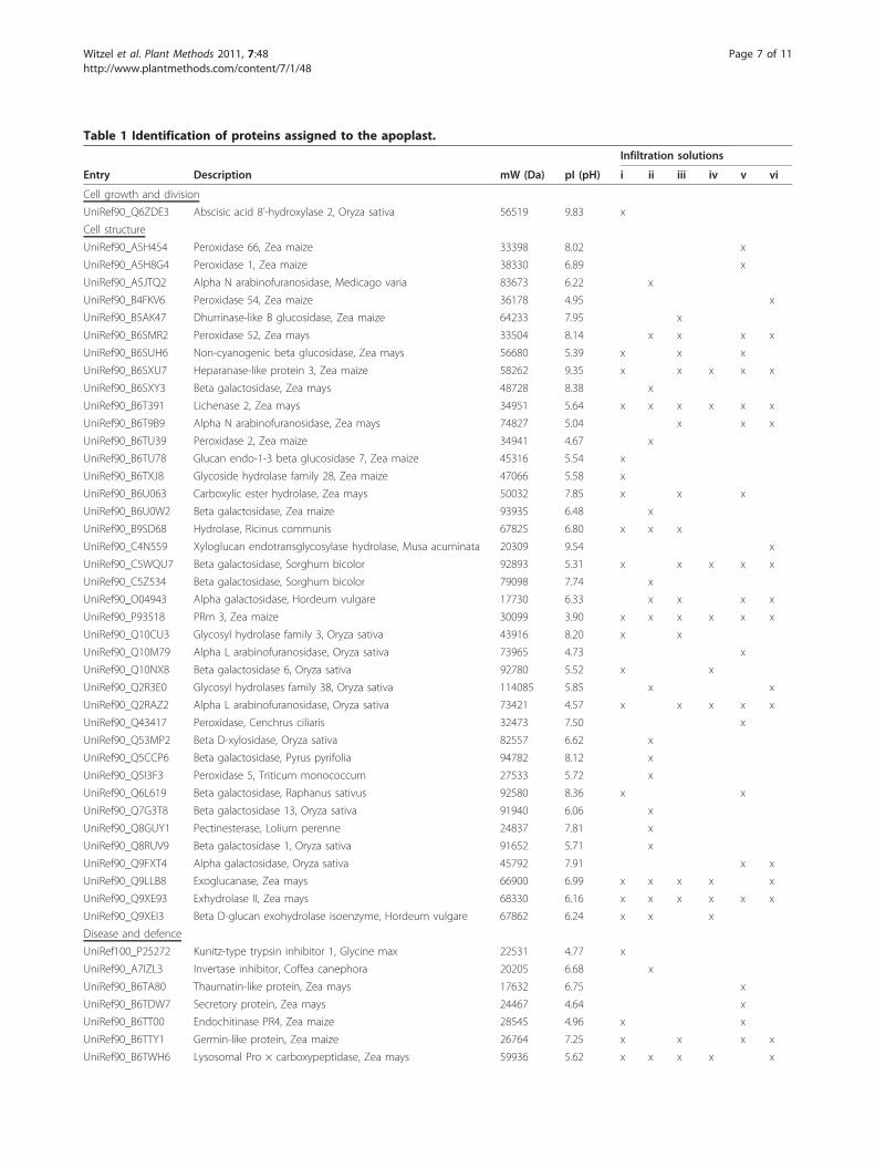
Table 1 Identification of proteins assigned to the apoplast.
Infiltration solutions
Entry Description mW (Da) pI (pH) i ii iii iv v vi
Cell growth and division
UniRef90_Q6ZDE3 Abscisic acid 8’-hydroxylase 2, Oryza sativa 56519 9.83 x
Cell structure
UniRef90_A5H454 Peroxidase 66, Zea maize 33398 8.02 x
UniRef90_A5H8G4 Peroxidase 1, Zea maize 38330 6.89 x
UniRef90_A5JTQ2 Alpha N arabinofuranosidase, Medicago varia 83673 6.22 x
UniRef90_B4FKV6 Peroxidase 54, Zea maize 36178 4.95 x
UniRef90_B5AK47 Dhurrinase-like B glucosidase, Zea maize 64233 7.95 x
UniRef90_B6SMR2 Peroxidase 52, Zea mays 33504 8.14 x x x x
UniRef90_B6SUH6 Non-cyanogenic beta glucosidase, Zea mays 56680 5.39 x x x
UniRef90_B6SXU7 Heparanase-like protein 3, Zea maize 58262 9.35 x x x x x
UniRef90_B6SXY3 Beta galactosidase, Zea mays 48728 8.38 x
UniRef90_B6T391 Lichenase 2, Zea mays 34951 5.64 x x x x x x
UniRef90_B6T9B9 Alpha N arabinofuranosidase, Zea mays 74827 5.04 x x x
UniRef90_B6TU39 Peroxidase 2, Zea maize 34941 4.67 x
UniRef90_B6TU78 Glucan endo-1-3 beta glucosidase 7, Zea maize 45316 5.54 x
UniRef90_B6TXJ8 Glycoside hydrolase family 28, Zea maize 47066 5.58 x
UniRef90_B6U063 Carboxylic ester hydrolase, Zea mays 50032 7.85 x x x
UniRef90_B6U0W2 Beta galactosidase, Zea maize 93935 6.48 x
UniRef90_B9SD68 Hydrolase, Ricinus communis 67825 6.80 x x x
UniRef90_C4N559 Xyloglucan endotransglycosylase hydrolase, Musa acuminata 20309 9.54 x
UniRef90_C5WQU7 Beta galactosidase, Sorghum bicolor 92893 5.31 x x x x x
UniRef90_C5Z534 Beta galactosidase, Sorghum bicolor 79098 7.74 x
UniRef90_O04943 Alpha galactosidase, Hordeum vulgare 17730 6.33 x x x x
UniRef90_P93518 PRm 3, Zea maize 30099 3.90 x x x x x x
UniRef90_Q10CU3 Glycosyl hydrolase family 3, Oryza sativa 43916 8.20 x x
UniRef90_Q10M79 Alpha L arabinofuranosidase, Oryza sativa 73965 4.73 x
UniRef90_Q10NX8 Beta galactosidase 6, Oryza sativa 92780 5.52 x x
UniRef90_Q2R3E0 Glycosyl hydrolases family 38, Oryza sativa 114085 5.85 x x
UniRef90_Q2RAZ2 Alpha L arabinofuranosidase, Oryza sativa 73421 4.57 x x x x x
UniRef90_Q43417 Peroxidase, Cenchrus ciliaris 32473 7.50 x
UniRef90_Q53MP2 Beta D-xylosidase, Oryza sativa 82557 6.62 x
UniRef90_Q5CCP6 Beta galactosidase, Pyrus pyrifolia 94782 8.12 x
UniRef90_Q5I3F3 Peroxidase 5, Triticum monococcum 27533 5.72 x
UniRef90_Q6L619 Beta galactosidase, Raphanus sativus 92580 8.36 x x
UniRef90_Q7G3T8 Beta galactosidase 13, Oryza sativa 91940 6.06 x
UniRef90_Q8GUY1 Pectinesterase, Lolium perenne 24837 7.81 x
UniRef90_Q8RUV9 Beta galactosidase 1, Oryza sativa 91652 5.71 x
UniRef90_Q9FXT4 Alpha galactosidase, Oryza sativa 45792 7.91 x x
UniRef90_Q9LLB8 Exoglucanase, Zea mays 66900 6.99 x x x x x
UniRef90_Q9XE93 Exhydrolase II, Zea mays 68330 6.16 x x x x x x
UniRef90_Q9XEI3 Beta D-glucan exohydrolase isoenzyme, Hordeum vulgare 67862 6.24 x x x
Disease and defence
UniRef100_P25272 Kunitz-type trypsin inhibitor 1, Glycine max 22531 4.77 x
UniRef90_A7IZL3 Invertase inhibitor, Coffea canephora 20205 6.68 x
UniRef90_B6TA80 Thaumatin-like protein, Zea mays 17632 6.75 x
UniRef90_B6TDW7 Secretory protein, Zea mays 24467 4.64 x
UniRef90_B6TT00 Endochitinase PR4, Zea maize 28545 4.96 x x
UniRef90_B6TTY1 Germin-like protein, Zea maize 26764 7.25 x x x x
UniRef90_B6TWH6 Lysosomal Pro × carboxypeptidase, Zea mays 59936 5.62 x x x x x
Witzel et al. Plant Methods 2011, 7:48http://www.plantmethods.com/content/7/1/48
Page 7 of 11

0.01 μM (NH4)6Mo7O24, 200 μM Fe-EDTA. Nutrientsolution was changed twice a week to avoid nutrientdeficiencies. The experiments were carried out undergreenhouse conditions with an average day/night tem-perature of 28/18°C and a photoperiod of 14 h for 5weeks with relative humidity about 70% ± 5%. The fifthand sixth leaf from medium part of the stem was har-vested 16 d after reaching the full nutrient solution forcollection of apoplast proteins.
Extraction of apoplastic and symplastic proteinsApoplastic proteins were collected using the infiltration-centrifugation technique [43] with minor modifications.Leaves were cut into segments of about 5.5 cm andwashed with deionised water. For infiltration, leaf seg-ments were placed in plastic syringes (60 ml) filled with40 ml of the respective infiltrating medium and wereinfiltrated by pulling the plunger, producing a reducedpressure of estimated about 20 kPa. Leaves were infil-trated either with water, 20 mM ascorbic acid/20 mMCaCl2, 100 mM sorbitol, 0.1 M sodium phosphate buffer(pH 6.5), 25 mM Tris-HCl (pH 8.0) or 50 mM NaCl(Figure 1). Thereafter, intact leave segments were care-fully blotted dry, and then placed in a 10 ml plastic ves-sel and centrifuged immediately at 400 g for 5 min at 5°
C. The clear infiltrate, now referred to as apoplast frac-tion, was collected at the bottom of the tube.After the extraction of the apoplastic fraction, the resi-
dual leaf tissue was shock frozen in liquid nitrogen,thawed, and centrifuged at 715 g for 5 min for cell sapextraction, now referred to as symplast fraction. Fourpools of extracts from five plants each were combinedfor subsequent analyses. Extracts were stored at -80°Cuntil analysis.
Gel electrophoretic protein separationProteins contained in the different extracts were precipi-tated by chloroform/methanol method [44]: 200 μl ofsample was mixed with 800 μl MeOH, 400 μl chloro-form and 600 μl deionized water. The incubation at 4°Cfor 5 min was followed by a centrifugation step (9,000 g,2 min, 4°C). The upper phase was removed and 600 μlMeOH was added to the lower and interphase. Afurther centrifugation sedimented the proteins, thesupernatant was removed and the pellet was dried in avacuum centrifuge.For one-dimensional separation of proteins, the pellets
were dissolved in 10% glycerol, 2.3% SDS, 5% b-mercap-toethanol, 0.25% bromphenol blue, 63 mM Tris-HCl(pH 6.8). The 2-D Quant Kit (GE Healthcare, http://
Table 1 Identification of proteins assigned to the apoplast. (Continued)
UniRef90_B6UB57 Lysosomal protective protein, Zea maize 53540 5.87 x x x x x
UniRef90_O24007 Chitinase, Oryza sativa 18956 4.83 x x x x x
UniRef90_P01063 Bowman-Birk-type proteinase inhibitor C II, Glycine max 9194 4.38 x
UniRef90_P29022 Endochitinase A, Zea maize 29105 7.85 x
UniRef90_Q5U1S9 Class III peroxidase 14, Oryza sativa 37174 5.77 x
UniRef90_Q6EUS1 Class III peroxidase 27, Oryza sativa 33300 8.09 x x
UniRef90_Q6TM44 Germin-like protein, Zea mays 21873 6.04 x x x x x x
UniRef90_Q7M1R1 Chitinase, Gladiolus × gandavensis 30695 5.77 x x x x x
Protein destination and storage
UniRef90_B6TG95 Vignain, Zea mays 38823 4.68 x x x x x x
UniRef90_B6TYX7 Polygalacturonase inhibitor 1, Zea mays 30011 8.08 x x x
Secondary metabolism
UniRef90_O64411 Polyamine oxidase, Zea mays 56308 5.63 x
Signal transduction
UniRef90_B6TWC3 Rhicadhesin receptor, Zea mays 23726 9.20 x x
UniRef90_B9MZ47 Fasciclin-like AGP 14 4 protein, Populus trichocarpa 24786 8.63 x
Transporters
UniRef90_B4FB54 Non-specific lipid transfer protein, Zea mays 12084 9.60 x
UniRef90_B6SP11 Non-specific lipid transfer protein, Zea mays 9802 8.73 x
UniRef90_B6SY96 Non-specific lipid transfer protein, Zea mays 12011 9.29 x
UniRef90_B6TRB2 Copper ion binding protein, Zea maize 17057 9.78 x
UniRef90_P05046 Lectin, Glycine max 30908 5.60 x x
UniRef90_P19656 Non-specific lipid transfer protein, Zea mays 11697 8.74 x x
UniRef90_Q04672 Sucrose-binding protein, Glycine max 60484 6.42 x
UniProt database identifiers, along with molecular weight (mW) and isoelectric point (pI) are shown. The identification of the respective proteins using differentextraction solutions is indicated (i: water, ii: 100 mM sodium phosphate buffer, iii: 25 mM Tris-HCl, iv: 100 mM sorbitol, v: 20 mM ascorbic acid/20 mM CaCl2, vi:50 mM NaCl).
Witzel et al. Plant Methods 2011, 7:48http://www.plantmethods.com/content/7/1/48
Page 8 of 11

www.gehealthcare.com) was used for determining theprotein concentration. A sample of 10 μg was separatedby SDS-PAGE according to Laemmli [45]. The two-dimensional separation of proteins was accomplished asdescribed in Zörb et al. [46] with the following modifi-cations. Protein pellets were first dissolved in 8 M urea,2 M thiourea, 0.5% IPG (immobilized pH gradient) buf-fer, 4% w/v CHAPS, 30 mM DTT, 20 mM Tris andthen dialyzed using 3.5 kDa cut-off membrane (Zellu-Trans, Carl Roth, http://www.carlroth.com) against thesame buffer. The protein concentration was determinedwith the 2-D Quant Kit (GE Healthcare) and 25 μg ofprotein were separated on IPG strips of 7 cm in lengthwith pH gradient of 3-10. Protein gels were stainedaccording to the hot-staining protocol with CoomassieR350 tablets (PlusOne Coomassie tablets PhastGel BlueR-350, GE Healthcare) [47] and digitized with an EpsonPerfection V700 Photo scanner (Epson, http://www.epson.com).
LC-MS-based protein identificationDialyzed protein extracts were precipitated by chloro-form/methanol method and about 30 μg of protein wereresolubilized in 50 μl 0.1% Rapigest (Waters Corpora-tion, http://www.waters.com) in 50 mM ammoniumbicarbonate. Protein concentrations were determinedusing the Bradford method [48] and bovine serum albu-min as standard protein. Five μg of protein werereduced, alkylated and digested with trypsin over nightat 37°C as described earlier [49]. The enzymatic reactionwas stopped with 1N HCl and peptide solutions wereadjusted to 0.1 μg/μl final concentration.Three μl of protein digest were used for LC-separation
on a nanoAcquity UPLC system (Waters) followed bymass spectrometry analysis on a Q-TOF Premier MSinstrument (Waters) in a data directed analysis (DDA)mode, as described in Agrawal et al. [50].Peptide separation was performed on a 180 μm × 20
mm Symmetry (5 μm) C18 precolumn (Waters) coupledto a 150 mm × 75 μm BEH130 (1.7 μm) C18 column(Waters), with a gradient of 3-40% actonitrile over 90min. The MS operated in a positive ion mode with asource temperature of 80°C, a cone gas flow of 50 l/h,and a capillary voltage of approximately 3 kV. Massspectra were acquired in a continuum V-mode andspectra integrated over 1 s intervals using MassLynx 4.1software (Waters). The instrument was calibrated usingselected fragment ions of the CID (collision-induced dis-sociation) of Glu-Fibrinopeptide B (SIGMA-ALDRICH,http://www.sigmaaldrich.com). Automatic data directedanalysis (DDA) was employed for MS/MS analysis ondoubly and triply charged precursor ions. The MS
spectra were collected from m/z 400 to m/z 1600, andproduct ion MS/MS spectra were collected from m/z 50to m/z 1600. Lock mass correction of the precursor andproduct ions was conducted with 500 fmol/μl Glu-Fibri-nopeptide B in 0.1% formic acid in AcN/water (50:50, v/v) respectively, and introduced via the reference sprayerof the NanoLockSpray interface. ProteinLynx Global-SERVER v2.3 software was used as a software platformfor data processing, deconvolution, de novo sequenceannotation of the spectra, and database search. A 10ppm peptide, 0.1 Da fragment tolerance, one missedcleavage, and variable oxidation (Met) and carbamido-methylation (Cys) were used as the search parameters.The resulting mass spectra were searched against the
protein index of the UniProt viridiplantae database(release: July 2010 with 722.718 protein sequences) forprotein identification applying the algorithm implemen-ted in the ProteinLynxGlobalServer software (PLGS,Waters Cooperation). All samples were run as technicaltriplicates. Protein identifications consistent in two outof three LC-MS runs were considered as present in thatsample. The false discovery rate was set to 4% of pro-teins included in the database.Hierarchical clustering of protein abundances was per-
formed using Gene Expression Similarity InvestigationSuite Genesis v1.7.6 [51]. Average linkage clustering wasapplied for LC-MS experiments and protein abundances.
Additional material
Additional file 1: Biological reproducibility of protein profiles fromthe maize leaf apoplast as resolved by 1-DE. Apoplastic proteins wereextracted with water (i) or 100 mM sodium phosphate buffer (ii). Twoindependent experiments were performed to assure consistent proteinpatterns.
Additional file 2: Identification of proteins from the apoplast ofmaize leaves. UniProt database identifiers, along with molecular weight(mW) and isoelectric point (pI) are shown. The cellular localisation wasassigned using Expasy tools Target P (http://www.cbs.dtu.dk/services/TargetP/) and WoLF PSORT (http://wolfpsort.org/). The identification ofthe respective proteins using different extraction solutions is indicated (i:water, ii: 100 mM sodium phosphate buffer, iii: 25 mM Tris-HCl, iv: 100mM sorbitol, v: 20 mM ascorbic acid/20 mM CaCl2, vi: 50 mM NaCl).
Additional file 3: Identification of proteins from the apoplast ofmaize leaves. Provided are the UniProt database identifiers, the PLGSscore, probability score for identification, number of identified peptides,protein coverage and the peptide sequence.
Additional file 4: Proteins identified in apoplast extracts of all sixinfiltration solutions.
Additional file 5: Example of protein identification from apoplasticextracts using nanoLC-ESI-Q-TOF MS. The database search against theprotein index of UniProt led to the identification of exhydrolase II[Q9XE93]. The amino acid sequence of the corresponding protein isshown on top with the detected peptides underlined. The de novosequence of a selected peptide with precursor mass m/z 859.4698(charge 3) is shown. This peptide is marked in bold within the proteinsequence.
Witzel et al. Plant Methods 2011, 7:48http://www.plantmethods.com/content/7/1/48
Page 9 of 11
AbbreviationsLC-MS/MS: liquid chromatography tandem mass spectrometry; 1-DE: one-dimensional gel electrophoresis; 2-DE: two-dimensional gel electrophoresis
AcknowledgementsWe thank Stephanie thor Straten and Annegret Thießen for excellenttechnical assistance. MS acknowledges the award of a DAAD scholarshipand KW the support by COST Action ‘Plant Proteomics in Europe’ (FA0603).
Author details1Institute of Plant Nutrition and Soil Science, Christian Albrechts University,Hermann-Rodewald-Strasse 2, 24118 Kiel, Germany. 2Leibniz Institute of PlantGenetics and Crop Plant Research, Corrensstrasse 3, 06466 Gatersleben,Germany. 3Leibniz Institute of Vegetable and Ornamental Crops, Theodor-Echtermeyer-Weg 1, 14979 Großbeeren, Germany.
Authors’ contributionsKW carried out protein extractions, protein separations, data evaluation andmanuscript preparation. MS performed plant cultivation, apoplasticpreparations and contributed to protein separations. AM conceived massspectrometry analyses and contributed to manuscript writing. HPMparticipated in discussions during experimental work and manuscriptpreparation. KHM conceived the project and worked on manuscriptpreparation. All authors read and approved the final manuscript.
Competing interestsThe authors declare that they have no competing interests.
Received: 1 November 2011 Accepted: 22 December 2011Published: 22 December 2011
References1. Sattelmacher B, Mühling KH, Pennewiss K: The apoplast - its significance
for the nutrition of higher plants. Zeitschrift für Pflanzenernährung undBodenkunde 1998, 161:485-498.
2. Hoson T: Apoplast as the site of response to environmental signals. JPlant Res 1998, 111:167-177.
3. Dietz K-J: The extracellular matrix of the plant cell: Location of signalperception, transduction and response. In Prog Bot. Edited by: Esser K,Lüttge U, Kadereit J, Beyschlag W. Berlin Heidelberg New York: Springer;2000:215-237.
4. Mühling KH, Läuchli A: Interaction of NaCl and Cd stress oncompartmentation pattern of cations, antioxidant enzymes and proteinsin leaves of two wheat genotypes differing in salt tolerance. Plant Soil2003, 253:219-231.
5. Dani V, Simon W, Duranti M, Croy R: Changes in the tobacco leaf apoplastproteome in response to salt stress. Proteomics 2005, 5:737-745.
6. Tasgin E, Atici O, Nalbantoglu B, Popova LP: Effects of salicylic acid andcold treatments on protein levels and on the activities of antioxidantenzymes in the apoplast of winter wheat leaves. Phytochemistry 2006,67:710-715.
7. Ramanjulu S, Kaiser W, Dietz KJ: Salt and drought stress differentiallyaffect the accumulation of extracellular proteins in barley. Zeitschrift FurNaturforschung C-a Journal of Biosciences 1999, 54:337-347.
8. Wimmer MA, Mühling KH, Läuchli A, Brown PH, Goldbach HE: Theinteraction between salinity and boron toxicity affects the subcellulardistribution of ions and proteins in wheat leaves. Plant Cell Environ 2003,26:1267-1274.
9. Alves M, Francisco R, Martins I, Ricardo CPP: Analysis of Lupinus albus leafapoplastic proteins in response to boron deficiency. Plant Soil 2006,279:1-11.
10. Fecht-Christoffers MM, Braun HP, Lemaitre-Guillier C, VanDorsselaer A,Horst WJ: Effect of Manganese toxicity on the proteome of the leafapoplast in cowpea. Plant Physiology 2003, 133:1935-1946.
11. Soares NC, Francisco R, Vielba JM, Ricardo CP, Jackson PA: Associatingwound-related changes in the apoplast proteome of Medicago withearly steps in the ROS signal-transduction pathway. J Proteome Res 2009,8:2298-2309.
12. Bhushan D, Pandey A, Choudhary MK, Datta A, Chakraborty S,Chakraborty N: Comparative proteomics analysis of differentially
expressed proteins in chickpea extracellular matrix during dehydrationstress. Mol Cell Proteomics 2007, 6:1868-1884.
13. Pandey A, Rajamani U, Verma J, Subba P, Chakraborty N, Datta A, et al:Identification of Extracellular Matrix Proteins of Rice (Oryza sativa L.)Involved in Dehydration-Responsive Network: A Proteomic Approach. JProteome Res 2010, 9:3443-3464.
14. Floerl S, Druebert C, Majcherczyk A, Karlovsky P, Kues U, Polle A: Defencereactions in the apoplastic proteome of oilseed rape (Brassica napus var.napus) attenuate Verticillium longisporum growth but not diseasesymptoms. BMC Plant Biol 2008, 8:15.
15. Goulet C, Goulet MC, Michaud D: 2-DE proteome maps for the leafapoplast of Nicotiana benthamiana. Proteomics 2010, 10:2536-2544.
16. Anand A, Lei ZT, Sumner LW, Mysore KS, Arakane Y, Bockus WW, et al:Apoplastic extracts from a transgenic wheat line exhibiting lesion-mimicphenotype have multiple pathogenesis-related proteins that areantifungal. Mol Plant-Microbe Interact 2004, 17:1306-1317.
17. Djordjevic MA, Oakes M, Li DX, Hwang CH, Hocart CH, Gresshoff PM: TheGlycine max xylem sap and apoplast proteome. Journal of ProteomeResearch 2007, 6:3771-3779.
18. Alvarez S, Goodger JQD, Marsh EL, Chen S, Asirvatham VS, Schachtman DP:Characterization of the maize xylem sap proteome. J Proteome Res 2006,5:963-972.
19. Maldonado AM, Echevarria-Zomeno S, Jean-Baptiste S, Hernandez M, Jorrin-Novo JV: Evaluation of three different protocols of protein extraction forArabidopsis thaliana leaf proteome analysis by two-dimensionalelectrophoresis. Journal of Proteomics 2008, 71:461-472.
20. Rodrigues SP, Ventura JA, Zingali RB, Fernandes RMB: Evaluation of samplepreparation methods for the analysis of papaya leaf proteins through2wo-dimensional gel electrophoresis. Phytochem Anal 2009, 20:456-464.
21. da Silva MAO, Garcia JS, de Souza G, Eberlin MN, Gozzo FC, Arruda MAZ:Evaluation of sample preparation protocols for proteomic analysis ofsunflower leaves. Talanta 2010, 80:1545-1551.
22. Xie CJ, Wang D, Yang XY: Protein extraction methods compatible withproteomic analysis for the cotton seedling. Crop Sci 2009, 49:395-402.
23. Zheng Q, Song J, Doncaster K, Rowland E, Byers DM: Qualitative andquantitative evaluation of protein extraction protocols for apple andstrawberry fruit suitable for two-dimensional electrophoresis and massspectrometry analysis. J Agric Food Chem 2007, 55:1663-1673.
24. Delaplace P, van der Wal F, Dierick JF, Cordewener JHG, Fauconnier ML, duJardin P, et al: Potato tuber proteomics: Comparison of twocomplementary extraction methods designed for 2-DE of acidicproteins. Proteomics 2006, 6:6494-6497.
25. Jellouli N, Ben Salem A, Ghorbel A, Ben Jouira H: Evaluation of proteinextraction methods for Vitis vinifera leaf and root proteome analysis bytwo-dimensional electrophoresis. J Integr Plant Biol 2010, 52:933-940.
26. Negri AS, Prinsi B, Scienza A, Morgutti S, Cocucci M, Espen L: Analysis ofgrape berry cell wall proteome: A comparative evaluation of extractionmethods. J Plant Physiol 2008, 165:1379-1389.
27. Wang XC, Shi MJ, Lu XL, Ma RF, Wu CG, Guo AP, et al: A method forprotein extraction from different subcellular fractions of laticifer latex inHevea brasiliensis compatible with 2-DE and MS. Proteome Science 2010,8:10.
28. Yao Y, Yang YW, Liu JY: An efficient protein preparation for proteomicanalysis of developing cotton fibers by 2-DE. Electrophoresis 2006,27:4559-4569.
29. Carpentier SC, Witters E, Laukens K, Deckers P, Swennen R, Panis B:Preparation of protein extracts from recalcitrant plant tissues: Anevaluation of different methods for two-dimensional gel electrophoresisanalysis. Proteomics 2005, 5:2497-2507.
30. Fan PX, Wang XC, Kuang TY, Li YX: An efficient method for the extractionof chloroplast proteins compatible for 2-DE and MS analysis.Electrophoresis 2009, 30:3024-3033.
31. Borderies G, Jamet E, Lafitte C, Rossignol M, Jauneau A, Boudart G, et al:Proteomics of loosely bound cell wall proteins of Arabidopsis thalianacell suspension cultures: A critical analysis. Electrophoresis 2003,24:3421-3432.
32. Boudart G, Jamet E, Rossignol M, Lafitte C, Borderies G, Jauneau A, et al:Cell wall proteins in apoplastic fluids of Arabidopsis thaliana rosettes:Identification by mass spectrometry and bioinformatics. Proteomics 2005,5:212-221.
Witzel et al. Plant Methods 2011, 7:48http://www.plantmethods.com/content/7/1/48
Page 10 of 11

33. Paetzold R: Subzelluläre Proteomanalyse von Maisblättern (Zea mays L.)unter besonderer Berücksichtigung der NaCl- und B-Toxizität. M.Sc.thesis, Institute of Plant Nutrition: Justus Liebig University Giessen; 2006.
34. Robertson D, Mitchell GP, Gilroy JS, Gerrish C, Bolwell GP, Slabas AR:Differential extraction and protein sequencing reveals major differencesin patterns of primary cell wall proteins from plants. J Biol Chem 1997,272:15841-15848.
35. Li ZC, McClure JW, Hagerman AE: Soluble and bound apoplastic activityfor peroxidase, beta-D-glucosidase, malate dehydrogenase, andnonspecific arylesterase, in barley (Hordeum vulgare L.) and oat (Avenasativa L.) primary leaves. Plant Physiology 1989, 90:185-190.
36. Chivasa S, Ndimba BK, Simon WJ, Robertson D, Yu XL, Knox JP, et al:Proteomic analysis of the Arabidopsis thaliana cell wall. Electrophoresis2002, 23:1754-1765.
37. Irshad M, Canut H, Borderies G, Pont-Lezica R, Jamet E: A new picture ofcell wall protein dynamics in elongating cells of Arabidopsis thaliana:Confirmed actors and newcomers. BMC Plant Biol 2008, 8.
38. Gokulakannan GG, Niehaus K: Characterization of the Medicago truncatulacell wall proteome in cell suspension culture upon elicitation andsuppression of plant defense. J Plant Physiol 2010, 167:1533-1541.
39. Cleves AE: Protein transport: The nonclassical ins and outs. Curr Biol 1997,7:R318-R320.
40. Slabas AR, Ndimba B, Simon WJ, Chivasa S: Proteomic analysis of theArabidopsis cell wall reveals unexpected proteins with new cellularlocations. Biochem Soc Trans 2004, 32:524-528.
41. Cosgrove DJ: Growth of the plant cell wall. Nat Rev Mol Cell Biol 2005,6:850-861.
42. Hammerschmidt R: The dynamic apoplast. Physiol Mol Plant Pathol 2010,74:199-200.
43. Lohaus G, Pennewiss K, Sattelmacher B, Hussmann M, Mühling KH: Is theinfiltration-centrifugation technique appropriate for the isolation ofapoplastic fluid? A critical evaluation with different plant species. PhysiolPlant 2001, 111:457-465.
44. Wessel D, Flugge UI: A method for the quantitative recovery of protein indilute-solution in the presence of detergents and lipids. Anal Biochem1984, 138:141-143.
45. Laemmli UK: Cleavage of structural proteins during assembly of head ofbacteriophage T4. Nature 1970, 227:680-685.
46. Zörb C, Schmitt S, Neeb A, Karl S, Linder M, Schubert S: The biochemicalreaction of maize (Zea mays L.) to salt stress is characterized by amitigation of symptoms and not by a specific adaptation. Plant Science2004, 167:91-100.
47. Westermeier R, Naven T: Proteomics in Practice: A Laboratory Manual ofProteome Analysis. Weinheim, Germany: Wiley-VCH; 2002.
48. Bradford M: A rapid and sensitive method for the quantitation ofmicrogram quantities of protein utilizing the principle of protein-dyebinding. Anal Biochem 1976, 7:248-254.
49. Kaspar S, Matros A, Mock HP: Proteome and flavonoid analysis revealsdistinct responses of epidermal tissue and whole leaves upon UV-Bradiation of barley (Hordeum vulgare L.) seedlings. J Proteome Res 2010,9:2402-2411.
50. Agarwal R, Matros A, Melzer M, Mock HP, Sainis JK: Heterogeneity inthylakoid membrane proteome of Synechocystis 6803. Journal ofProteomics 2010, 73:976-991.
51. Sturn A, Quackenbush J, Trajanoski Z: Genesis: cluster analysis ofmicroarray data. Bioinformatics 2002, 18:207-208.
doi:10.1186/1746-4811-7-48Cite this article as: Witzel et al.: Comparative evaluation of extractionmethods for apoplastic proteins from maize leaves. Plant Methods 20117:48.
Submit your next manuscript to BioMed Centraland take full advantage of:
• Convenient online submission
• Thorough peer review
• No space constraints or color figure charges
• Immediate publication on acceptance
• Inclusion in PubMed, CAS, Scopus and Google Scholar
• Research which is freely available for redistribution
Submit your manuscript at www.biomedcentral.com/submit
Witzel et al. Plant Methods 2011, 7:48http://www.plantmethods.com/content/7/1/48
Page 11 of 11