save nature to survive hydatid cyst – different … n. rukmangadha.pdf · 231 n save nature to...
TRANSCRIPT

231
NSave Nature to Survive
5(2): 231-234, 2010
HYDATID CYST – DIFFERENT ORGAN INVOLVEMENT: A
PROSPECTIVE AND RETROSPECTIVE STUDY
N. RUKMANGADHA*, AMIT KUMAR CHOWHAN, K. V. SREEDHAR BABU, RASHMI PATNAYAK,
B. V. PHANEENDRA AND M. KUMARASWAMY REDDY
Department of Pathology, Sri Venkateswara Institute of Medical Sciences,
Tirupati - 517 507, A. P. INDIA
E-mail: [email protected]
INTRODUCTION
Hydatid disease is endemic in sheep rearing countries (Greece,
Australia, New Zealand and South Africa), caused by larval
form of genus Echinococcus (E). E. granulosus causing cystic
hydatid disease has world wide distribution. E.multilocularis
causes alveolar hydatid disease, occurring in Alaska and
Canada. E. vogeli causes polycystic echinococcosis, very rare
in humans, and reported in Central and South America.
Definitive host for E. granulosus is dog, for E. multilocularis it
is fox, dog and cat. Intermediate hosts are sheep, cattle, pigs,
horses and camels for E. granulosus, rodents for E.
multilocularis and pacas, a wild rodent for E.vogeli. Hydatid
disease occurs when man becomes accidental intermediate
host by swallowing the eggs of the parasite. The swallowed
eggs on reaching the duodenum release larva which burrow
through the intestinal mucosa, enter the circulation and reach
different organs. Liver is the major organ affected, particularly
the right lobe followed by lungs and other organs (Anderson’s
Pathology, 1996).
In India, hydatid disease is common in most of the states; with
predominance in Andhra Pradesh and Tamil Nadu (Amir Jahed
et al., 1975). Hydatid disease evokes much interest due to
diversity of the anatomical involvement. Hence we undertook
a retrospective and prospective analytical study of the number
of cases of hydatid cyst reported in our Institute.
MATERIALS AND METHODS
A sixteen year and seven months retrospective and prospective
study was undertaken in our Institute, which included 34
resected specimens of hydatid cyst received by department of
Pathology. All the specimens were preserved 10% Formalin
and were subsequently subjected to histopathological
examination with routine haematoxylin and eosin stain.
RESULTS
Among the 34 cases diagnosed histopathologically as hydatid
cysts, involvement of the liver accounted for 12 (35.5%) cases,
lung - 9 (26.5%), subcutaneous tissue - 5 (14.7%), spleen - 3
(8.8%) and solitary involvement of breast, kidney, brain,
retrouterine and synovial tissues (2.9 % each) as given in Table
1. The youngest and the oldest were a 7 year old male child
and a 70 year male, both presented with hydatid cyst of lung
as given in Table 2. In our study we found a mild male
preponderance (18 cases) when compared to females (16
cases) with a male to female ratio of 1.12:1 as given in Table 3.
Site No. of cases Percentage (%)
Liver 12 35.5Lung 9 26.5Subcutaneous tissue 5 14.7Spleen 3 8.8Breast 1 2.9Retro uterine 1 2.9Synovial tissue 1 2.9Kidney 1 2.9Brain 1 2.9
Table 1: Distribution of lesions
ABSTRACTHydatid disease/ hydatidosis is an endemic disease in sheep rearing countries. It has a world wide distributionand poses an important public health problem that is influenced by people’s socioeconomic status andmigration which spreads this disease. It is caused by the larval form of Echinococcus granulosus. The main hostbeing the dog, the intermediate hosts are sheep, cattle, pigs and horses. Hydatid disease manifests when manbecomes an accidental intermediate host. All sites of the body can be possible locations of the parasite. Themost common sites are the liver followed by lung, kidney, bone and brain with the rare sites being breast,adrenals, heart, intra arterial, broad ligament and others. We conducted a retrospective and prospective studyof different organ involvement of hydatid cyst reported in our department. The study spanned over a period ofsixteen years and seven months, from March 1993 - October 2009. 34 specimens were received in thedepartment of Pathology, SVIMS, Tirupati, which were reported as cases of hydatid cyst involving differentorgans, which included rare sites such as brain, kidney, breast, synovial tissue and retrouterine.
KEY WORDSHydatid diseaseEchinococcusGranulosusHydatid cyst
Received on :
30.01.2010
Accepted on :
07.04.2010
*Corresponding
author

232
N. RUKMANGADHA et al.,
Site 1-10 11-20 21-30 31-40 41-50 51-60 61-70Yrs Yrs Yrs Yrs Yrs Yrs Yrs
Liver - 4 6 1 - 1 -Lung 1 1 3 3 - - 1Subcutaneous - - 4 1 - - -tissueSpleen - - 1 2 - - -Breast - - 1 - - - -Retrouterine - 1 - - - - -Synovial Tissue - - - - 1 - -Kidney - - 1 - - - -Brain - - 1 - - - -
Table 2: Age distribution of lesions
Table 3: Sex distribution of lesions
Site Male Female
Liver 6 6Lung 6 3Subcutaneous tissue 4 1Spleen 1 2Breast - 1Retrouterine - 1Synovial Tissue - 1Kidney 1 -Brain - 1Total 18 16
Figure 1: CT scan brain showing a hypoechoic lesion in left fronto-
parietal region
Figure 2: CT scan showing a hypoechoic lesion in the liver
Figure 5: Cytocentrifuge smear showing scolices from the aspirate
of brain lesion – 40X high power view
Figure 4: Cytocentrifuge smear showing scolices from the aspirate
of brain lesion – 10X low power view
Figure 3: CT scan showing multiple hypoechoic lesions in the spleen
233
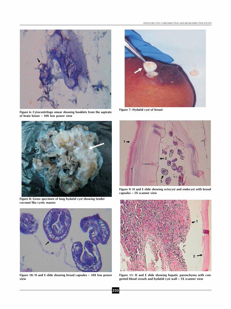
Figure 6: Cytocentrifuge smear showing hooklets from the aspirate
of brain lesion – 10X low power view
Figure 7: Hydatid cyst of breast
Figure 11: H and E slide showing hepatic parenchyma with con-
gested blood vessels and hydatid cyst wall – 5X scanner view
Figure 10: H and E slide showing brood capsules – 10X low power
view
Figure 8: Gross specimen of lung hydatid cyst showing tender
coconut like cystic masses
HYDATID CYST A PROSPECTIVE AND RETROSPECTIVE STUDY
Figure 9: H and E slide showing ectocyst and endocyst with brood
capsules – 5X scanner view

234
DISCUSSION
In our study comprising of 34 cases of hydatid disease, the
incidence of which was found to have a male predominance
(Magath, 1941). It has been postulated that the sex
predominance may be determined by which sex has more
contact with the usual definitive host in that country (Jidejian,
1953; Somily et al., 2005). We found that the liver was the
most common affected organ, followed by lung, subcutaneous
tissue, spleen, breast, kidney, brain, synovial tissue and
retrouterine (Magath, 1941; Yuksel et al., 2007; Bal et al.,
2008). Pulmonary involvement was predominantly seen in
children and young adults (Miller, 1953; Polat et al., 2003).
Renal involvement is rare involving a single case (Polat et al.,
2003). In our study, cases with liver involvement, presented
with right upper quadrant pain. Majority of the cases with
lung involvement were asymptomatic and were detected
during routine examination. We came across a single case of
hydatid cyst of the breast, which is a rare site of occurrence
(Farrokh, 2000; Arikan et al., 2004; Saluja et al., 2005). To the
best of our knowledge retro uterine and synovial tissue hydatid
cysts are also rarer sites.
We have recently reported a single case involving brain
(Awasthy et al., 2006). The patient was a 25 year old female,
who presented with headache, weakness of the left side of the
body, with deviation of the mouth to the right side and difficulty
in eating. CT scan report was, ? brain abscess, ? tuberculoma,
? cystic tumor. Cytological diagnosis was hydatid cyst
confirmed by histopathology. Histologically all cases showed
inner nucleated germinal layer, an outer anucleated chitinous
layer, with innumerable delicate laminations covered by an
adventitial layer. In addition are seen brood capsules and
scolices which constitute hydatid sand (Robbins and Cotran
Pathologic Basis of Diseases, 2004).
CONCLUSION
The cases recorded in our Institute may encompass a smaller
percentage, as many cases being asymptomatic, may go
undetected. The study was presented as we have come across
some rare sites of presentation such as the breast, synovial
tissue and retro uterine.
REFERENCES
Amir Jahed, A. K., Fardin, R., Farzad, A. and Bakshandeh, K. 1975.
Clinical echinococcosis. Ann Surg. 182(5): 541-6.
Anderson’s Pathology. 1996. 10th Ed. St Louis (Missouri): MosbyPublishers; Ivan Dam Janov, James Linder, editors. Kamal G. Ishak,Rodney S. Markin. Liver 57. pp. 1830.
Arikan, S., Yucel, A. F., Barut, G. and Kocakusak, A. 2004. HydatidDisease in the Breast: a Case Report. Acta chir belg. 104: 473-5.
Bal, N., Kocer, N. E., Arpasi, R., Ezer, A. and Kayaselcuk, F. 2008.
Uncommon locations of hydatid cyst. Saudi Med. J. 29(7): 1004-8.
Farrokh, D. 2000. Hydatid Cysts of the Breast: A Report of ThreeCases. Irn. J. Med. Sci. 25(1 and 2): 72-5.
Jidejian, Y. 1953. Hydatid disease. Surgery. 34(1): 155-67.
Magath, T. B. 1941. Hydatid diseases in North America. Pennsylvania
Med. J. 44: 813.
Miller, M. J. 1953. Hydatid infectio in Canada. Can. Med. Assoc. J.
68: 423-49.
Neeraj, Awasthy., Karam, Chand. and Singh, A. P. 2006. GiganticIntracranial hydatid cyst: An unusual case report. J. Pediatric
Neurology. 4(2):115-9.
Polat, P., Kantarci, M., Alper, F., Suma, S., Koruyucu, M. B. and Okur,
A. 2003. Hydatid disease from head to toe. Radiographics. 23: 475-94.
Robbins and Cotran Pathologic Basis of Diseases. 2004. 7th Ed.
Thompson Press (India): Saunders An imprint of Elsevier; Kumar,Abbas, Fausto, editors. Alexander J. McAdam, Arlene H. Sharpe.Infectious Diseases Chapter 8. pp 406 - 7.
Saluja, J. G., Ajinkya, M. S., Mehta, H. T., Vivekanand, S. Katti.,
Yuvaraj, J. Patole. and Jain, R. 2005. Echinococcus Cystic Disease ofthe Breast and Literature Review. http://www.bhj.org/ journal/april2005/htm/ case_echinococcuus_212.htm.
Somily, A., Robinson, J. L., Miedzinski, L. J., Bhargava, R. and Marrie,
T. J. 2005. Echinococcal disease in Alberta, Canada: more than acalcified opacity. BMC Infect Dis. 5(1): 34.
Yuksel, M., Demirpolat, G., Sever, A., Bakaris, S., Bulbuloglu, E. and
Elmas, N. 2007. Hydatid disease involving some rare locations in thebody: a pictorial essay. Korean J. Radiol. 8(6): 531-40.
N. RUKMANGADHA et al.,