satellite cells maintain regenerative capacity but fail to
TRANSCRIPT

RESEARCH Open Access
Satellite cells maintain regenerativecapacity but fail to repair disease-associated muscle damage in mice withPompe diseaseGerben J. Schaaf1,2,3, Tom J. M. van Gestel1,2,3, Stijn L. M. in ‘t Groen1,2,3, Bart de Jong1,2,3, Björn Boomaars1,2,3,Antonietta Tarallo4,5, Monica Cardone4,5,6, Giancarlo Parenti4,5, Ans T. van der Ploeg2,3
and W. W. M. Pim Pijnappel1,2,3*
Abstract
Pompe disease is a metabolic myopathy that is caused by glycogen accumulation as a result of deficiency of thelysosomal enzyme acid alpha glucosidase (GAA). Previously, we showed that adult muscle stem cells termed satellite cellsare present at normal levels in muscle from patients with Pompe disease, but that these are insufficiently activated torepair the severe muscle pathology. Here we characterized the muscle regenerative response during disease progressionin a mouse model of Pompe disease and investigated the intrinsic capacity of Gaa−/− satellite cells to regenerate muscledamage. Gaa−/− mice showed progressive muscle pathology from 15 weeks of age as reflected by increased lysosomalsize, decreased fiber diameter and reduced muscle wet weight. Only during the first 15 weeks of life but not thereafter,we detected a gradual increase in centrally nucleated fibers and proliferating satellite cells in Gaa−/− muscle, indicating amild regenerative response. The levels of Pax7-positive satellite cells were increased in Gaa−/− mice at all ages, most likelyas result of enhanced satellite cell activation in young Gaa−/− animals. Surprisingly, both young and old Gaa−/− miceregenerated experimentally-induced muscle injury efficiently as judged by rapid satellite cell activation and completerestoration of muscle histology. In response to serial injury, Gaa−/− mice also regenerated muscle efficiently andmaintained the satellite cell pool. These findings suggest that, similar to human patients, Gaa−/− mice haveinsufficient satellite cell activation and muscle regeneration during disease progression. The initial endogenous satellitecell response in Gaa−/− mice may contribute to the delayed onset of muscle wasting compared to human patients.The rapid and efficient regeneration after experimental muscle injury suggest that Gaa−/− satellite cells are functionalstem cells, opening avenues for developing muscle regenerative therapies for Pompe disease.
Keywords: Satellite cells, Muscle regeneration, Pompe disease, Lysosomal storage disease, Glycogenosis type II
IntroductionPompe disease is a metabolic myopathy that is caused bydeficiency of acid alpha glucosidase (GAA), a lysosomalenzyme responsible for the degradation of glycogen [38].Pompe patients develop progressive skeletal muscle weak-ness due to lysosomal expansion, followed by lysosomal
disruption and myofiber death. Affected muscles in-clude those involved in mobility and respiration, and asa result Pompe patients become wheelchair and ventila-tor dependent [59]. The most severe classic infantileform of Pompe disease is caused by complete absenceof GAA enzyme activity and results in death within thefirst year of life, if left untreated [52]. In milder formsof Pompe disease, residual GAA activity exists, and pa-tients develop symptoms later in life [17, 54]. A treat-ment for Pompe disease is available in the form of enzymereplacement therapy (ERT). ERT improves muscle functionand prolongs survival [2, 4, 21, 24, 33, 36, 37, 51, 57], but
* Correspondence: [email protected] of Clinical Genetics, Erasmus MC, University Medical Center,Rotterdam, the Netherlands2Department of Pediatrics, Erasmus MC, University Medical Center,Rotterdam, the NetherlandsFull list of author information is available at the end of the article
© The Author(s). 2018 Open Access This article is distributed under the terms of the Creative Commons Attribution 4.0International License (http://creativecommons.org/licenses/by/4.0/), which permits unrestricted use, distribution, andreproduction in any medium, provided you give appropriate credit to the original author(s) and the source, provide a link tothe Creative Commons license, and indicate if changes were made. The Creative Commons Public Domain Dedication waiver(http://creativecommons.org/publicdomain/zero/1.0/) applies to the data made available in this article, unless otherwise stated.
Schaaf et al. Acta Neuropathologica Communications (2018) 6:119 https://doi.org/10.1186/s40478-018-0620-3

the heterogeneous response among patients has urged thedevelopment of alternative treatment options [5].Skeletal muscle has the capacity to regenerate upon
damage. Genetic ablation of Pax7-expressing cells inmice has shown that this process is dependent on adultmuscle stem cells termed satellite cells [25, 40]. Inhealthy muscle, satellite cells reside in a quiescent statelocated in between the sarcolemma and the basal lamina[30]. Upon muscle damage, satellite cells become acti-vated and enter the cell cycle. Proliferating satellite cellshave two fates: to repair muscle fibers, or to replenishthe satellite cell pool [7]. Given the regenerative proper-ties of skeletal muscle a major unresolved question inthe field remains why satellite cells are apparently unableto efficiently repair disease-induced muscle damage.Several explanations have been proposed, including ex-haustion of the satellite cell pool [39] or intrinsic failureof satellite cells to regenerate muscle [3, 10]. For ex-ample, in Duchenne Muscular Dystrophy, both satellitecell depletion/exhaustion and intrinsic failure of satellitecells to regenerate have been proposed [10, 39]. Previ-ously, we have analyzed muscle biopsies from patientswith Pompe disease [41]. Our study demonstrated a lackof muscle regeneration to the severe damage observed inbiopsies from Pompe patients, even in those from severelyaffected classic infantile patients. We found that satellitecells were present at similar levels as in healthy controls,arguing against satellite cell depletion in Pompe disease[41]. However, satellite cells were mostly inactive, in agree-ment with the lack of detectable muscle regeneration.The maintenance of the satellite cell pool in patients
with Pompe disease suggested the possibility that en-dogenous satellite cells represent a therapeutic target forPompe disease. A prerequisite for this idea is that satel-lite cells are intrinsically capable of regenerating muscle.So far, this remained unclear given the low level ofmuscle regeneration in Pompe patients. To address this,in the present study we used two knockout mousemodels for Pompe disease on different genetic back-grounds [6, 35]. We characterized the satellite cell re-sponse in Gaa−/− mice, and related this to myofiberpathology. We then applied a single and serial externalinjury and characterized muscle regeneration and thesatellite cell response. Our results indicate that Gaa−/−
mice have activated satellite cells and low levels ofmuscle regeneration only during the first 15 weeks oflife. Single and serial experimentally induced muscle in-juries provoked efficient satellite cell activation andmuscle regeneration in Gaa−/− mice. These results indi-cate that satellite cells in Gaa−/− mice have the intrinsiccapacity to efficiently regenerate muscle and self-renew.These findings suggest that satellite cell activation maybe explored as a therapeutic strategy to promote muscleregeneration in Pompe disease.
Material and methodsMice and animal proceduresAge-matched wildtype and Gaa−/− animals on an FVB/N [6] inbred, or mixed C57/Bl6 and 129/Sv [35] back-ground were used between 2 and 70 weeks of age. Wildtype FVB/N breeder animals were obtained from Envigo,and were used to start a colony that is maintained at theErasmus MC animal facility. Gaa−/−(FVB/N) animalshad been generated previously by targeted disruption ofexon 13 of the Gaa gene [6]. We performed homozy-gous breedings to generate both the wildtype and Gaa−/−
animals in the FVB/N background during the duration ofthis project. Wildtype control and Gaa−/− animals in themixed C57/Bl6 and 129/Sv background were obtained aslittermates from heterozygous breedings and maintainedat the Cardarelli Hospital’ s Animal Facility (Naples, Italy).Gaa−/−(Bl6) animals obtained by insertion of a neo cas-sette into exon 6 of the Gaa gene [35] were purchasedfrom Charles River Laboratories (Wilmington, MA). Allmice in experiment were housed under a light–dark cycle(12 h) and under defined pathogen-free conditions, withaccess to food and water ad libitum.Muscle injury was induced by intramuscular injection
of 1.2% (w/v in PBS) BaCl2 or cardiotoxin (CTX; 10 μmolin PBS). Animals were allowed to recover for the timeindicated in the figures. Serial injury experiments wereperformed by injecting BaCl2, as described above, threetimes at monthly intervals into the Tibialis Anterior(TA) muscle. Three weeks after the last BaCl2 injectionthe animals were sacrificed for tissue collect.At the end of experiments animals were sacrificed by
cervical dislocation during daytime without a fixed time-point. Tissue wet weight was determined by weighingfreshly dissected tissue that was blotted dry. All animalexperiments were approved by the local and nationalanimal experiment authorities in compliance with theEuropean Community Council Directive guidelines (EUDirective 86/609), regarding the protection of animalsused for experimental purposes, and according to Insti-tutional Animal Care and Use Committee (IACUC)guidelines for the care and use of animals in research.The study was approved by the local and national au-thorities in the Netherlands and Italy, respectively. Allprocedures with the animals were performed with theaim of ensuring that discomfort, distress, pain, and in-jury would be minimal.
Determination of glycogen levelsTo measure tissue glycogen concentrations 20 30 μmcryosections were collected for each sample. The sec-tions were homogenized using 5 mm stainless steelbeads (Qiagen NV) in the Qiagen Retsch MM300 Tis-sueLyser (Qiagen NV) at 30 Hz for 5 min. Glycogen wasquantified in tissue supernatant by measuring the
Schaaf et al. Acta Neuropathologica Communications (2018) 6:119 Page 2 of 16
amount of glucose released from glycogen after conver-sion by amyloglycosidase and amylase (Roche Diagnostics)for 1 h as previously described [58]. Spectral absorbanceof the products was measured on a Varioskan spec-trometer (Thermo Scientific) at 414 nm. Results fromthe glycogen measurements were normalized for pro-tein content using the Pierce BCA protein assay kit(Thermo Scientific).
Histology and immunofluorescent analysesHematoxylin and Eosin (HE) staining and Masson’s tri-chrome staining were performed using routine histologyprotocols as described previously [41]. For immunostain-ing, Tissue-Tek OCT-embedded tissue was snap-frozenin liquid nitrogen-cooled isopentane. 10 μm cryosectionswere cut and fixated in ice-cold aceton. A heated antigenretrieval procedure with 10 mM citrate buffer was usedfor the detection of Pax7. Sections were stained essen-tially as described previously [41], but using the M.O.M.kit from Vector laboratories for blocking endogenousmouse immunogens. Primary antibodies used wereeMyHC (F1.652; DSHB; 1:300), Ki67 (Ab15580; Abcam;1:50), laminin (L9393; Sigma; 1:500 or LS-C (6142; LSBIO; 1:500)), Lamp1 (Ab24170; Abcam; 1:150). Hoechst(H33258, Sigma) was used at 1 μg/ml. To detect cen-trally nucleated myofibers aceton-fixed 10 μm cryosec-tions were stained for laminin using a primary antibodyand Hoechst for nuclei, as described above, and imagedby fluorescent microscopy.
Image acquisition and analysisHistological sections were scanned with 4x and 20x ob-jectives on a Hamamatsu NanoZoomer 2.0 (HamamatsuPhotonics). Images were analyzed using NDP view soft-ware (NDP View 1.2.31 Eng, Hamamatsu Photonics).Sections used for immunofluorescence were scanned onZeiss LSM700 (Carl Zeiss B.V.) using tile-scan modalitywith a 20x objective. Image analysis and processing wasperformed using Fiji (fiji.sc/Fiji) and Adobe Photoshop.Quantification of myofiber diameter was performedusing cross sections by measuring the longest diagonal(in μm) in at least 100 fibers per sample, randomly se-lected throughout the whole section.
Flow cytometryPreparation of limb muscle for flow cytometric analysiswas adapted from Liu et al. [28]. In short, dissected tis-sue was minced thoroughly to small pieces in F10medium (Lonza) containing collagenase II (750 U/ml;Fisher Scientific) using scalpels. Minced tissue was disso-ciated for 70 min in F10 medium containing 750 U/mlcollagenase II), then for 30 min in F10 medium contain-ing collagenase II (100 U/ml) and dispase (1.1 U/ml;Fisher Scientific). Cell suspensions were filtered over a
40 μM cell strainer (Falcon) and a small sample was re-trieved to determine total mononuclear cell count usinga hematocytometer. The cell suspensions were stainedwith CD31-APC (1:100), CD45-APC (1:100), Sca1-FITC(1:100) and Vcam-biotin (1:50) primary antibodies. Vcamwas visualized using streptavidin-PECY7 (1:100). Allantibodies for flowcytometry were derived from BD Bio-sciences. Cell viability was determined by staining with1 μg/ml Hoechst 33258 (Sigma). Samples were analyzedon a BD-ARIAIII (BD biosciences).
Statistical analysisFor all experiments normal distribution of data was de-termined based on calculated residuals. Normally dis-tributed data from two groups was tested using a2-tailed t-test. For experiments with three groups ormore a one-way ANOVA of independent samples withTukey or Games-Howell Post Hoc multiple correction(depending on homogeneity of variance) was used.Non-normal distributed data was statistically testedusing a Mann-Whitney non-parametric test (two groups)or a Kruskal-Wallis test of independent samples withBonferoni multiple comparison for three or moregroups. For all tests a p-value less than 0.05 was consid-ered significant. Data was analyzed using IBM SPSS sta-tistics version 25.
ResultsCharacterization of muscle pathology during diseaseprogression in Gaa−/− micePrevious work has shown that Gaa−/− non-inbred mice(on FVB or C57/Bl6 backgrounds mixed with the 129background) display hallmarks of Pompe disease, in-cluding progressive skeletal muscle wasting and glyco-gen accumulation. We now performed a more in-depthquantitative analysis of the timing of myofiber path-ology and muscle wasting in FVB inbred mice (indicatedas Gaa−/−(FVB)). Key results throughout this reportwere confirmed on Gaa−/− mice in the C57/Bl6non-inbred background (indicated as Gaa−/−(Bl6)).Glycogen accumulation was detected in Tibialis Anterior
(TA) muscle from Gaa−/− mice at the age of 2 weeks, andincreased further during aging (Fig. 1a). Maximal glycogenlevels were reached in animals of about 25 weeks andremained stable thereafter. To examine abnormalities inlysosomal size, immunofluorescent staining of Lamp1 wasperformed (Fig. 1b). No Lamp1 staining was detected inTA muscle sections from wild type mice due to the smallsize of lysosomes. In Gaa−/− muscle, TA sections showed apunctated Lamp1 pattern from 15 weeks of age and on-wards indicating increased lysosomal size. The number ofLamp1-positive spots per fiber did not increase further inolder Gaa−/− mice (Fig. 1b-c). TA wet weight was reducedin Gaa−/− mice compared to wild type mice, starting at
Schaaf et al. Acta Neuropathologica Communications (2018) 6:119 Page 3 of 16

A
B C
D
FE
Fig. 1 (See legend on next page.)
Schaaf et al. Acta Neuropathologica Communications (2018) 6:119 Page 4 of 16

25 weeks of age, and decreased further with age with 54%at 70 weeks of age (Fig. 1d). Histological analysis of TA sec-tions showed a decrease in fiber diameter in the Gaa−/−
mice compared to wild type mice that started between 15and 25 weeks of age (Fig. 1e-f). Fiber diameter distributionwas determined by quantifying the number of fibers in dif-ferent fiber size categories. This showed enrichment ofsmaller-sized fibers in muscles of 15-week Gaa−/− mus-cles relative to those from age-matched wild type ani-mals (Additional file 1: Figure S1A). Gaa−/− QuadricepsFemoris (QF) muscles at 3 months of age in the C57/Bl6 non-inbred background also displayed enrichmentfor smaller fibers (Additional file 1: Figure S1B). Theseresults indicate that Gaa−/−(FVB) mice show progres-sive myofiber pathology reminiscent of Pompe diseasestarting at 15 weeks of age.
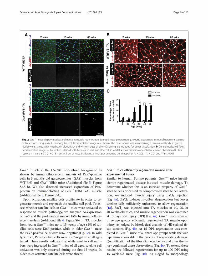
Modest and transient muscle regeneration during diseaseprogression in Gaa−/− miceTo assess whether Gaa−/− (FVB) mice regenerate TAmuscle in response to Pompe disease-induced musclepathology, we first performed immunofluorescent stainingof embryonic Myosin Heavy Chain (eMyHC), a markerfor actively regenerating myofibers [42] (Fig. 2a). Fewsmall-sized eMyHC-positive fibers were detected in mus-cles from Gaa−/− animals between 2 and 60 weeks of age(< 0.7% of myofibers). Representative examples are shownin Fig. 2a. A similar result was obtained for Gaa−/− animalsin the C57/Bl6 non-inbred background (Additional file 2:Figure S2A). Muscle from wild type mice did not showeMyHC-positive fibers in either background.In murine muscle, regenerated myofibers can be iden-
tified by the presence of centrally located nuclei [27].We used Hoechst to stain nuclei in TA sections (Fig. 2b),and this showed an increased percentage of fibers withcentral nuclei in Gaa−/− TA muscle from 15 weeks of age(Fig. 2c). At 25 weeks of age, the percentage of centrally nu-cleated fibers reached a plateau of 15% that remained stableuntil 60 weeks of age. In comparison, published results inthe mdx mouse, a model for Duchenne Muscular Dys-trophy, showed that already at 12 weeks of age > 70% oflower limb muscle myofibers were centrally nucleated [1],
indicating that the disease-mediated muscle-regenerativeresponse is relatively mild in Gaa−/− muscle. Modestcentral nucleation was also detected in GAS muscleat 3 month-old Gaa−/−(Bl6) mice (Additional file 2:Figure S2B). We conclude that Gaa−/− mice have arelatively mild and limited muscle regenerative re-sponse during disease progression.
Satellite cells are increased in number but are onlytransiently activated during disease progression in Gaa−/−
limb muscleGenetic ablation of Pax7-expressing cells demonstratedthat satellite cells are indispensable for muscle regener-ation [25, 40]. Satellite cells are marked by expression ofPax7, which is a master transcription factor that regu-lates survival and expression of myogenic transcriptionfactors involved in muscle differentiation and regeneration[23, 43]. To assess the consequence of Gaa-deficiency andthe related muscle pathology on satellite cell dynamics, weperformed immunofluorescent staining of Pax7 in TA sec-tions (Fig. 3a). The number of Pax7-positive cells was sta-bly increased in Gaa−/− TA muscle relative to wild typemuscle at all ages tested (2–60 weeks), and varied between20 and 50 Pax7-positive cells/mm2 (Fig. 3b). The increasein Pax7-positive cells in Gaa−/− muscle was equally pro-nounced when expressed as satellite cell per myofiber,with a ~ 5 fold increase at 15 weeks and ~ 7.1 fold increaseat 25 week animals (Additional file 3: Figure S3), indicat-ing that the difference in satellite cell density was inde-pendent of changes in fiber diameter.The number of Pax7-positive cells in wild type TA
muscle decreased from 8 to 2 Pax7-positive cells/mm2
during the same period (Fig. 3b). To confirm increased sat-ellite cell levels in Gaa−/− mice, we analysed the number ofsatellite cells by flow cytometry using a satellite cell surfaceprofile based on expression of Vcam [28] (Additional file 4:Figure S4A). Using this profile we could detect a > 93%pure population of Pax7-positive cells (Additional file 4:Figure S4B). The number of Vcam-positive cells was stablyincreased in Gaa−/− TA muscle between 15 and 70 weeksof age relative to wild type TA muscle (Additional file 4:Figure S4C). Satellite cell numbers were also increased in
(See figure on previous page.)Fig. 1 Characterization of lysosomal and muscle wasting pathology during disease progression in Gaa−/− mice. a. Glycogen accumulation.Glycogen levels were measured biochemically in TA muscles at the indicated ages. b. Lysosomal pathology. Immunofluorescent analysis of TAsections using a Lamp1 antibody (in green). Representative images are shown. The basal lamina was stained using a Laminin antibody (in red).Nuclei were stained with Hoechst (in blue). Black and white images of Lamp1 staining are included for better visualization. c. Quantification ofthe number of Lamp1-positive spots per fiber from B. Data are from two TA muscles derived from two different animals per genotype pertimepoint, and are expressed as mean ± SD. ***p < 0.001. d. Wet weight of TA muscles. Each dot represents TA wet weight from one muscle ofone animal. Means ± SD are indicated as lines (n = 4–12 animals per genotype per timepoint). *p < 0.05 and ***p < 0.001. e. HE staining of TAsections. Representative images are shown. f. Quantification of fiber size from E. Data from individual mice are plotted (n = 2–4 animals pergenotype per timepoint). Means ± SD are indicated. *p < 0.05 and **p < 0.01
Schaaf et al. Acta Neuropathologica Communications (2018) 6:119 Page 5 of 16
Gaa−/−muscle in the C57/Bl6 non-inbred background asshown by immunofluorescent analysis of Pax7-positivecells in 3 months old gastrocnemius (GAS) muscles fromWT(Bl6) and Gaa−/−(Bl6) mice (Additional file 5: FigureS5A-B). We also detected increased expression of Pax7protein by immunoblotting of Gaa−/−(Bl6) GAS muscle(Additional file 5: Figure S5C).Upon activation, satellite cells proliferate in order to re-
generate muscle and replenish the satellite cell pool. To as-sess whether satellite cells in Gaa−/− mice were activated inresponse to muscle pathology, we analysed co-expressionof Pax7 and the proliferation marker Ki67 by immunofluor-escent analysis (Additional file 6: Figure S6). In TA musclesfrom young Gaa−/− mice up to 15 weeks of age ± 5% of sat-ellite cells were Ki67-positive, while in older Gaa−/− micethe Pax7-positive cells were Ki67-negative (Fig. 3c). In wildtype mice, Pax7-positive cells were Ki67-negative at all agestested. These results indicate that while satellite cell num-bers were increased in Gaa−/− mice of all ages, satellite cellactivation was only observed during the first 15 weeks. Inolder mice activated satellite cells were absent.
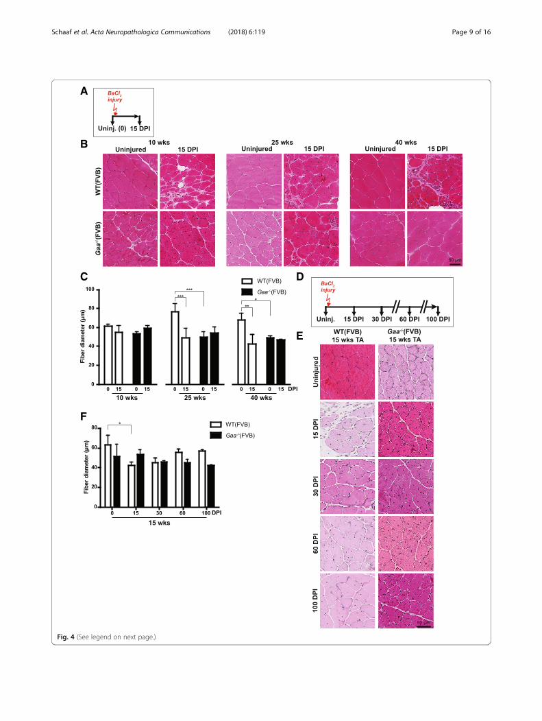
Gaa−/− mice efficiently regenerate muscle afterexperimental injurySimilar to human Pompe patients, Gaa−/− mice insuffi-ciently regenerated disease-induced muscle damage. Todetermine whether this is an intrinsic property of Gaa−/−
satellite cells or caused by compromised satellite cell activa-tion, we induced muscle injury using BaCl2 injection(Fig. 4a). BaCl2 induces myofiber degeneration but leavessatellite cells sufficiently unharmed to allow regeneration[18]. BaCl2 was injected into TA muscles in 10, 25, or40 weeks-old mice, and muscle regeneration was examinedat 15 days post injury (DPI) (Fig. 4a). Gaa−/− mice from allthree age groups efficiently regenerated TA muscle uponinjury, as judged by histological analysis of HE-stained tis-sue sections (Fig. 4b). At 15 DPI, regeneration was com-pleted in Gaa−/− mice of all three age groups while the wildtype muscle was still in the process of regenerating damage.Quantification of the fiber diameter before and after the in-jury confirmed these observations (Fig. 4c). To extend theseresults, we followed regeneration for up to 100 DPI using15 week-old mice (Fig. 4d). As judged by morphology,
A B
C
Fig. 2 Gaa−/− mice display modest and transient muscle regeneration during disease progression. a. eMyHC expression. Immunofluorescent stainingof TA sections using a MyHC antibody (in red). Representative images are shown. The basal lamina was stained using a Laminin antibody (in green).Nuclei were stained with Hoechst (in blue). Black and white images of eMyHC staining are included for better visualization. b. Central nucleated fibers.Representative images of TA sections stained with Laminin (in red) and Hoechst (in white). c. Quantification of central nucleated fibers from B. Datarepresent means ± SD (n = 2–3 muscles from at least 2 different animals per genotype per timepoint). *p < 0.05. **p < 0.01 and ***p < 0.001
Schaaf et al. Acta Neuropathologica Communications (2018) 6:119 Page 6 of 16

A
B C
Fig. 3 (See legend on next page.)
Schaaf et al. Acta Neuropathologica Communications (2018) 6:119 Page 7 of 16

muscle regeneration was complete in Gaa−/− mice at 15DPI in Gaa−/− mice, and between 30 and 60 DPI in wildtype mice (Fig. 4e). Quantification of fiber diameter con-firmed these observations (Fig. 4f). To examine the devel-opment of chronic tissue fibrosis that may result fromincomplete regeneration as is observed in dystrophicmuscle [9], we performed trichrome staining on regener-ated muscle. This showed some trichrome-positive areas attime points before regeneration was completed in animalsof both genotypes reflecting the transient expansion offibro/adipogenic progenitors that is part of normal muscleregeneration [20] (Additional file 7: Figure S7). At 15 DPIlittle or no trichrome staining was detected in Gaa−/− orwild type mice at 10, 15, or 40 weeks of age (Additional file 8:Figure S8), suggesting successful tissue remodelling and ab-sence of fibrotic muscle tissue replacement. In mice in theC57/Bl6 non-inbred background, injury was induced usingcardiotoxin (CTX) injection into Quadriceps Femoris (QF)muscle at 3 months and 11 months of age, or into the GASmuscle at 3 months of age. All these tissues showed effi-cient regeneration in Gaa−/− mice at similar efficienciescompared to wild type mice, as judged by morphology(Additional file 9: Figure S9). We conclude that Gaa−/−
mice have efficient intrinsic capacity to regenerate muscleafter experimentally-induced injury.
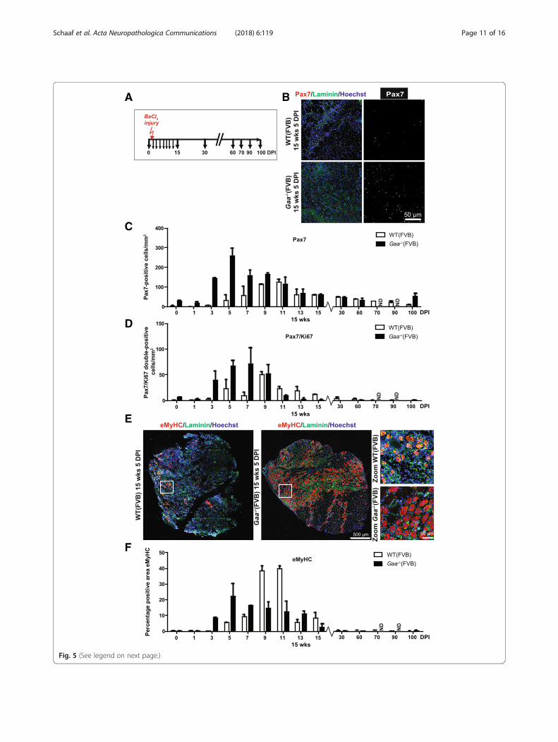
Satellite cell response in Gaa−/− mice after experimentalinjuryTo determine whether the efficient muscle regenerationupon induced injury in Gaa−/− mice is accompanied bysatellite cell activation, we quantified the number ofPax7-positive and Pax7/Ki67 double-positive cells at sev-eral time points following BaCl2 injection in the TAmuscle (Fig. 5a-b). In both Gaa−/− and wild type mice,the number of Pax7-positive cells was transiently andstrongly induced upon injury. In Gaa−/− mice, satellitecell numbers were already increased at 3 DPI, peaked at5 DPI, and then slowly returned to pre-injury numbersduring 7–60 DPI (Fig. 5c). Wild type mice showedslightly slower kinetics, with increased satellite cell num-bers that started increasing at 5 DPI and peaked at 9–11DPI, and slowly returned to pre-injury levels during 11–100 DPI. The kinetics of the number of Pax7/Ki67double-positive cells paralleled those of Pax7-single posi-tive cells in both Gaa−/− and wild type mice (Fig. 5d).
In the same experiment, active muscle regenerationwas examined using immunofluorescent staining ofeMyHC (Fig. 5e). The kinetics of eMyHC expression inGaa−/− mice paralleled the changes in number of Pax7-expressing cells after injury (Fig. 5f; compare with Fig. 5c).Together, these data show that the efficient regenerativeresponse of Gaa−/− muscle after BaCl2-induced injury ismediated by a rapid satellite cell response and furtherconfirms that Gaa−/− satellite cells are not functionallycompromised.
Gaa−/− satellite cells regenerate muscle and self-renewafter serial injuryGaa−/− mice showed efficient satellite cell-mediatedmuscle regeneration upon a single induced injury. Todetermine if Gaa−/− satellite cells can self-renew after in-jury, which is essential for long-term muscle regener-ation, we performed a serial injury experiment usingBaCl2 (Fig. 6a). Three consecutive injuries were appliedat 4 week intervals to the same TA muscles and animalswere allowed to regenerate in between injuries. At theend of the experiment, 3 weeks after the last injury(mice were 51 weeks of age at this timepoint), mice weresacrificed and TA muscles were analysed. Histologicalanalysis of HE-stained tissue sections showed completeregeneration from the serial injuries in both Gaa−/− andwild type muscle (Fig. 6b). Quantification of fiber diam-eter showed full restoration of fiber diameter in Gaa−/−
mice 3 weeks after the third injury (Fig. 6c). In wild typemice, fiber diameter was not yet fully restored at this timepoint, consistent with the slower kinetics after a single in-jury (Fig. 6c, see also Fig. 4f). Therefore the parameters ofthis experiment did not allow to fully assess the capacityof wild type mice to regenerate after serial injury. Thenumber of Pax7-positive cells in three-times regeneratedGaa−/− muscle was, although slightly lower, not signifi-cantly different from satellite cell levels in pre-injury mus-cles or in muscles at 60 weeks of age (Fig. 6d). In wildtype mice, the number of Pax7-positive cells was still en-hanced at 3 weeks after the third injury, in line with theslower regeneration kinetics after a single injury (seeFig. 4f ). The levels of Pax7/Ki67 double-positive cells at3 weeks after the third injury were very low in bothGaa−/− and wild type TA muscle, consistent with theirlevels at 3 weeks after a single injury (compare with
(See figure on previous page.)Fig. 3 Satellite cells are increased in number but are only transiently activated during disease progression in Gaa−/− limb muscle. a. pax7 expression.Immunofluorescent (IF) staining of TA sections using a Pax7 antibody (in red). Representative images are shown. The basal lamina was stained using aLaminin antibody (in green). Nuclei were stained with Hoechst (in blue). Black and white images of Pax7 staining are also shown for better visualization.Zooms of selected areas (white squares) are shown below the entire sections. b. Quantification of the number of Pax7-positive cells/mm2 from A. Dataare means ± SD from 2 muscles derived from 2 different animals per genotype per timepoint. *p < 0.05. **p < 0.01 and ***p < 0.001. c. Quantification ofthe number of Pax7/Ki67 double-positive cells by immunofluorescent staining of TA sections using Pax7 and Ki67 antibodies. Representative stainings areshown in Additional file 5: Figure S5. Data represent means ± SD from 2 TA muscles derived from 2 different animals. *p < 0.05
Schaaf et al. Acta Neuropathologica Communications (2018) 6:119 Page 8 of 16
A
B
C
F
D
E
Fig. 4 (See legend on next page.)
Schaaf et al. Acta Neuropathologica Communications (2018) 6:119 Page 9 of 16

Fig. 5d). This showed that also after repeated injury,satellite cells in Gaa−/− TA muscle returned within anormal timeframe to their quiescent state. We concludethat Gaa−/− mice have a robust capacity to regeneratemuscle via satellite cells even after repeated injury andthat Gaa−/− satellite cells retain the capacity to self-renewupon injury.
DiscussionIn this study, we have used mouse models for Pompedisease to assess the muscle regenerative capacity of sat-ellite cells. We first determined the timing of musclepathology, and found the following sequence of events:glycogen accumulation (starting at 2 weeks), enlarged ly-sosomes (starting at 15 weeks of age), reduced fiberdiameter (starting at 15–25 weeks of age), and reducedwet weight (starting at 25 weeks of age). Gaa-deficientmice display a mild muscle regenerative response shortlyafter birth up to 25 weeks of age, indicated by a gradual in-crease in central nucleation, detection of some eMyHC-positive myofibers and low-level satellite cell activation.This correlated with the detection of proliferating satellitecells during this period, but not thereafter. Satellite cellproliferation during the first 15 weeks of age resulted instably increased levels of satellite cells in animals up to atleast 60 weeks of age. Induced muscle injury in Gaa−/−
mice using BaCl2 or CTX resulted in very efficient satellitecell response and muscle regeneration. In addition, Gaa−/−
muscle regenerated completely after three consecutiverounds of injury and regeneration, indicating that Gaa−/−
satellite cells are capable of self-renewal. These results in-dicate that, similar to human Pompe patients, Gaa−/−
mice lack an efficient muscle regenerative response duringdisease progression but maintain the satellite cell pool des-pite the developing muscle damage. Importantly, satellitecells in mice with Pompe disease have the intrinsic cap-acity to efficiently regenerate after damage, suggesting thatthe lack of a satellite cell response in Pompe disease iscaused by deficient satellite cell activation.The mouse models for Pompe disease offers the op-
portunity to study the early stages of disease onset andto link these to the muscle regenerative response. Wehave used an inbred FVB strain as well as Gaa−/− miceon a mixed C57/Bl6 and 129/Sv background to investi-gate this. The key aspects of muscle regeneration activity
in Gaa-deficient muscle were observed in both thesemouse models, including the mild regenerative responseduring disease progression, reflected by the gradual in-crease in central nucleation and detection of feweMyHC-expressing myofibers, together with increasedsatellite cell levels and an efficient regenerative responseafter experimental injury. This strengthens the conclu-sion that the regenerative response during Pompe dis-ease progression is inefficient and disturbed. To addressthis point in more detail, we used Gaa−/− animals in theFVB background to extensively characterize the regener-ation response as well as the ability to regenerate after(serial) experimental injury. This extended on earlier re-ports [6, 35] that the Gaa−/− mouse models developssymptoms more slowly compared to classic infantilePompe patients, even though in both human and mousecases, Gaa activity was completely disrupted. Classic in-fantile Pompe patients show symptoms shortly afterbirth [55] and these include generalized muscle weak-ness evident by decreased muscle tone and strength.Muscle biopsies from classic infantile Pompe patientsshow severely damaged muscle fibers [41, 48, 56]. At thesame time, satellite cells in classic infantile patients arenot activated and muscle regeneration is undetectable[41]. In contrast, Gaa−/− mice developed cellular path-ology at adulthood, starting at 15–25 weeks of age, as in-dicated by increased lysosomal size and decreased fiberdiameter and wet weight. Interestingly, satellite cells inGaa−/− mice were activated until the age of 15 weeks, asindicated by the detection of Pax7-positive satellite cellsthat co-express Ki67. Proliferating satellite cells were notdetected in older animals. This may suggest that the en-dogenous satellite cells response in the first 15 weeksafter birth contributed to the delayed onset of musclewasting in Gaa−/− mice compared to that in human clas-sic infantile patients. It is interesting to speculate that amodest satellite cells response and muscle regenerationactivity may prevent the development of muscle fiberpathology. Future work is required to test this notion.It remains unclear why the satellite cell response in
Pompe disease is so modest (mouse) or not detectable(human). In certain other neuromuscular disorders, sat-ellite cells and muscle regeneration have a markedly dif-ferent behaviour. For example, in Duchenne MuscularDystrophy, studies using the mdx mouse model or the
(See figure on previous page.)Fig. 4 Gaa−/−mice regenerate muscle efficiently after experimental injury. a. Schematic representation of the injury experiment. Black arrowsindicate the time at which TA muscles were collected for analysis, the red arrow indicates the time of injury. b. HE staining of TA sections before(Uninjured, 0 days post injury (DPI)) and at 15 days DPI with BaCl2 at three ages. Representative images are shown. c. Quantification of fiberdiameter from (b). d. Schematic representation of injury experiment with a longer follow up after injury. Black arrows indicate the time at whichTA muscles were collected for analysis, red arrow indicate the time of injury. e. HE staining of TA sections of the injury experiment with long follow-up.Representative images are shown. f. Quantification of fiber diameter from E. Data in C and F are means ± SD from at least 3 muscles derived from 2 ormore different animals. *p < 0.05; **p < 0.01 and ***p < 0.001
Schaaf et al. Acta Neuropathologica Communications (2018) 6:119 Page 10 of 16
A B
C
D
E
F
Fig. 5 (See legend on next page.)
Schaaf et al. Acta Neuropathologica Communications (2018) 6:119 Page 11 of 16

(See figure on previous page.)Fig. 5 Rapid satellite cell response in Gaa−/− mice after experimental injury. a. Experimental schedule. Black arrows indicate the time at which TAmuscles were collected for analysis, the red arrow indicates the time of injury. b. pax7 expression. Immunofluorescent (IF) staining of TA sectionsusing a Pax7 antibody (in red). Representative images are shown. The basal lamina was stained using a Laminin antibody (in green). Nuclei werestained with Hoechst (in blue). Black and white images of Pax7 staining are included for better visualization. c. Quantification of the number ofPax7-positive cells/mm2 from B. Data are means ± SD from at least 2 muscles from 2 different animals. d. As C, but now for the number of Pax7/Ki67 double-positive cells/mm2. e. eMyHC expression at 5 DPI. Immunofluorescent staining using a MyHC antibody (in red). Representativeimages are shown. The basal lamina was stained using a Laminin antibody (in green). Nuclei were stained with Hoechst (in blue). Zooms ofselected areas (white squares) are shown on the right. f. Quantification of the eMyHC-positive area in TA sections. Data are means ± SD from atleast 2 muscles derived from 2 different animals
E
D
C
A B
Fig. 6 Gaa−/− satellite cells regenerate muscle and self-renew after serial injury. a. Experimental schedule. Black arrows indicate the time at whichTA muscles were collected for analysis, red arrows indicate the time of injury. b. HE staining of TA sections before and 3 weeks after the thirdinjury. Representative images are shown. c. Quantification of fiber diameter in WT (left) and Gaa−/− (right) TA muscle after serial injury from (b).d. Quantification of the number of Pax7-positive cells by immunofluorescent staining of TA sections using a Pax7 antibody in WT (left) and Gaa−/−
(right) TA muscle after serial injury. e. Quantification of the number of Pax7/Ki67 double-positive cells by immunofluorescent staining of TA sectionsusing Pax7 and Ki67 antibodies in WT (left) and Gaa−/− (right) TA muscle after serial injury. Data in c-e are means ± SD from at least 2 muscles derivedfrom 2 different animals. For comparison with levels of indicated parameter in uninjured age-matched TA muscles, values at 40 weeks of age (tocompare with the start of the experiment) and at 60 weeks of age (to compare with the end of the experiment) are included in (c-e)
Schaaf et al. Acta Neuropathologica Communications (2018) 6:119 Page 12 of 16

more severe mdx/utrophin double-knockout model havereported both exhaustion and depletion of satellite cellsas result of continuous satellite cell activation duringdisease progression [29, 39]. In muscle biopsies fromDuchenne patients, elevated muscle regeneration activityis observed [8, 22, 31, 39, 41]. Increased satellite cell ac-tivation is attributed to sarcolemmal fragility as result ofloss of functional dystrophin. Loss of sarcolemmal integ-rity triggers the release of signals from the muscle envir-onment [62], from the damaged myofiber itself [16] orfrom other cell types that are recruited and activatedafter damage [19, 20]. These events promote the transi-ent infiltration of immune cells that are essential forproper regeneration and participate in satellite cell acti-vation [49]. However, chronic inflammation in dys-trophic muscle contributes to disease progression anddysregulated satellite cell activation, as has also beenproposed for inflammatory myopathies [61]. In Pompedisease the primary defect is not at the sarcolemma [15]and an aberrant immune response is generally absent inPompe disease [41], indicating that satellite cell functionand activation are differently regulated in Gaa-deficientmuscle. It is tempting to speculate that the lysosomaldamage as result of glycogen accumulation and/or thesubsequent block of autophagy [13, 32] interfere withsatellite cell activation. It has been established that au-tophagy is crucial for securing the bioenergetics de-mands associated with satellite cell activation [46].Deficiency of SIRT1, a nutrient sensor that regulates au-tophagic flux in satellite cell progeny, was found to delaysatellite cell activation [46]. Inhibition of autophagic fluxhas been reported to occur in Gaa−/− myofibers [12, 36],but whether this affects satellite cell activation remainsto be determined.Satellite cells in Gaa−/− muscle do not respond to the
progressive tissue damage, in Pompe patients as well asin mice of 15 weeks and older. Our finding thatexperimentally-induced muscle injury evokes an efficientmuscle regenerative response suggests that once satellitecells are activated, downstream processes such as myo-genic differentiation and myoblast fusion are unaffectedby Gaa deficiency. This is in agreement with the normalmyogenic differentiation of induced pluripotent stemcells established from Pompe patients’ fibroblasts, eventhose generated from a severely affected classic infantilepatient [60]. We speculate that the maintenance of satel-lite cell function and number, as well as a functional re-generation machinery offers opportunities for developinga muscle regenerative therapy for Pompe disease throughstimulation of endogenous satellite cells. Satellite cells canbe safely and efficiently activated through exercise [45].Previous exercise programs in our and other centers werefound to be well tolerated by and beneficial for adultPompe patients [11, 26, 34, 44, 47, 50]. It can be predicted
that induced satellite cell activation would be less favor-able in untreated classic infantile patients, since these pa-tients display severe pathology directly after birth, andnewly regenerated muscle fibers likely develop pathologyrapidly. However, treatment of classic infantile patientswith ERT can significantly improve muscle function andmorphology [53] and delay the severity of symptoms. Thiswould suggest that combining ERT with induced satellitecell activation might be beneficial for these patients. Thedevelopment of pathology can take years in patients witha more slowly progressing disease course. Based on ourfindings in the mouse model of Pompe disease, restorationof the muscle condition via induced regeneration wouldbe predicted to delay disease progression. We consider itworthwhile to extend research on satellite cell activationin Pompe disease and other neuromuscular disorders thatharbor functional yet inactive satellite cells. Identificationof additional muscle diseases with such a profile may re-sult in the development of a more generic therapeuticstrategy. If successful, such strategy would be highly valu-able given the scarcity of treatment options for neuromus-cular disorders.
ConclusionThe current study shows that in Gaa−/− mice satellitecell activation and muscle regeneration was insufficientto repair the disease-mediated damage, similar as wasobserved in human patients. However, Gaa-deficient sat-ellite cells were intrinsically capable of regeneratingmuscle and harbored self-renewal potential. Our findingssuggest that the muscle phenotype in Pompe diseasemay be ameliorated by regenerative therapies directed atsatellite cell activation.
Additional files
Additional file 1: Figure S1. Reduced fiber diameter in GAAKO animals.A. Fiber diameter frequency distribution plot of WT(FVB) and Gaa−/−(FVB)at 15 weeks of age. B. WT(Bl6) and GAAKO(Bl6) at 3 months of age showingreduced fiber diameter in Gaa−/− (Bl6). These data suggest muscle atrophy isobserved in GAA-deficient animals on both FVB/N and C57/Bl6 backgrounds.(PDF 122 kb)
Additional file 2: Figure S2. Modest muscle regeneration in Gaa−/−(Bl6)animals. A.eMyHC staining of QF sections from WT(Bl6) and Gaa−/−(Bl6)animals. The figure shows selected areas of eMyHC (red)/laminin(green)/Hoechst(blue) stained QF sections from 4, 12 and 36 week old WT(Bl6)and Gaa−/− (Bl6) animals. eMyHC-positive were rare and very small inGaa−/− (Bl6) muscle, in line with findings in GAA−/−(FVB) (see Fig. 2A).B. Examples of HE-stained section from GAS muscle from 3 months oldWT(Bl6) and Gaa−/−(Bl6) animals. (PDF 320 kb)
Additional file 3: Figure S3. Satellite cell numbers are increased inGaa−/− TA muscle. The number of Pax7-positive cells in 15 week (A) and25 week (B) WT and Gaa−/− from Fig. 3 expressed as Pax7-positive cells/myofiber. Data are means ± SD from 2 muscles derived from 2 different ani-mals per genotype per timepoint. *p < 0.05 and **p < 0.01. (PDF 103 kb)
Additional file 4: Figure S4. Identification of Pax7-positive satellite cellsby flow cytometry. A. Representative dot plots from CD31-APC/CD45-APC/Sca1-FITC/Vcam-PeCY7 stained muscle cell suspensions according to
Schaaf et al. Acta Neuropathologica Communications (2018) 6:119 Page 13 of 16
the procedure described previously by Liu et al. [28]. The gating strategy isdepicted by the green arrow. The colors of the box/plot outlines correspondwith the gated populations. Satellite cells are in the CD45neg/CD31neg/sca1-neg/Vcam-positive gate (green box). B. pax7 (red)/Hoechst(blue) staining ofFACS-sorted satellite cells after 24 h culture using the procedure shown in(A). The lower panel shows the zoom of the insert in the upper panel.Counting Pax7 expressing cells indicated that sorting was performed at> 93% purity. C. Quantification of the percentage of Vcam-positive cellsby flow cytometry. Data from individual mice are plotted as single dots.(PDF 1056 kb)
Additional file 5: Figure S5. Satellite cell numbers are increased inGaa−/−(Bl6) muscle. A. Satellite cells were detected in 3 months old WT(Bl6)and Gaa−/−(Bl6) gastrocnemius (GAS) cryosections by immunofluorescentstaining of Pax7 (red). Myofibers were visualized using a laminin antibody(green) and nuclei with Hoechst (blue). White arrows point to Pax7-positivesatellite cells. B. Quantification of A. The figure depicts the mean percentageof Pax7-positive Satellite cells per field ± SD. C. Western blot analysis of Pax7expression in GAS muscle from 13 week old WT(Bl6) and Gaa−/−(Bl6)animals. Western blot analysis was performed as previously described[14]. (PDF 952 kb)
Additional file 6: Figure S6. Detection of proliferating satellite cells inGAA-deficient limb muscle. Representative images from TA limb musclesections co-stained for Pax7 (red) and Ki67 (green) to detect proliferatingsatellite cells (arrow). Nuclei are visualized with Hoechst (blue). The arrowheadpoints to a Pax7-positive/Ki67-negative quiescent satellite cell. (PDF 286 kb)
Additional file 7: Figure S7. Detailed histological evaluation ofregenerating WT and Gaa−/− muscle. The figure depicts HE- andtrichrome stained histological sections from 15 week WT(FVB) andGaa−/− (FVB) TA muscles at multiple time-points during the first 15 daysafter BaCl2-induced muscle regeneration. Gaa−/− (FVB) muscle regeneratesefficiently and completely (left panels). The trichrome stain shows absence ofresidual fibrotic tissue after completing a regeneration cycle (right panels). Asexplained in the text WT(FVB) has a regeneration cycle of more than 30 daysand was therefore still actively remodelling at 15 DPI. Arrows point to smallde novo myofibers detected as early as 3 DPI in regenerating Gaa−/−(FVB)muscle. (PDF 569 kb)
Additional file 8: Figure S8. Gaa−/− muscle regenerates completelywithout tissue remodeling Depicted are images from trichrome stainingof TA muscle of WT(FVB) and Gaa−/−(FVB) at 10, 25 and 40 weeks beforeand 15 days after BaCl2 injury. (PDF 348 kb)
Additional file 9: Figure S9. Efficient regeneration of GAAKO-muscleafter cardiotoxin-induced injury in GAA-deficient animals on a C57/Bl6background. The figure shows histological sections from TA HE-stainedsections from 12 and 48 week old WT(Bl6) and Gaa−/− (Bl6) animals atindicated time points after injury uisng cardiotoxin (CTX)-injection. Theupper panels show HE-stained sections from QF muscle, while the middlepanels show images from regenerating GAS. The lower panels depictregenerating QF from 11 months old WT(Bl6) and Gaa−/− (Bl6) animals.These data demonstrate that the capacity to regenerate after experimentalinjury is also maintained in GAA-deficient muscle on a C57/Bl6 background.(PDF 386 kb)
AbbreviationsBaCl2: Barium chloride; CNF: Central nucleated fibers; CTX: Cardiotoxin;DPI: Days post injury; eMyHC: Embryonic myosin heavy chain; ERT: Enzymereplacement therapy; GAA: Acid alpha glucosidase; GAS: Gastrocnemius;OCT: Optimal cutting temperature; PFA: Paraformaldehyde; QF: QuadricepsFemoris; TA: Tibialis Anterior
AcknowledgementsThe authors wish to acknowledge all members of the Molecular Stem CellBiology group at the Center for Lysosomal and Metabolic Diseases, ErasmusMC, Netherlands, for discussion; Sander van Hooff, Philip Lijnzaad andDimitris Rizopoulos for advice with the statistical analysis. The work is fundedthrough the Center for Lysosomal and Metabolic Diseases at Erasmus MC,and the Prinses Beatrix Spierfonds/Stichting Spieren voor Spieren (projectnumber W.OR13-21).
FundingThe work is funded through the Center for Lysosomal and MetabolicDiseases at Erasmus MC, and the Prinses Beatrix Spierfonds/Stichting Spierenvoor Spieren (project number W.OR13–21).
Availability of data and materialsAll data generated or analyzed during this study are included in thispublished article and its supplementary information file.
Authors’ contributionsGS and WP conceived the project and designed the experiments; GS, TvG, SitG,BdJ, BB, AT, MC performed experiments; all authors analyzed and interpreted thedata; GS, AvdP, and WP obtained funding, GS and WP wrote the manuscript. Allauthors read and approved the final manuscript.
Ethics approval and consent to participateNot applicable
Consent for publicationNot applicable.
Competing interestsAvdP has provided consulting services for various industries in the field ofPompe disease under an agreement between these industries and ErasmusMC, Rotterdam, The Netherlands. The other authors declare that they haveno conflict of interest.
Publisher’s NoteSpringer Nature remains neutral with regard to jurisdictional claims inpublished maps and institutional affiliations.
Author details1Department of Clinical Genetics, Erasmus MC, University Medical Center,Rotterdam, the Netherlands. 2Department of Pediatrics, Erasmus MC,University Medical Center, Rotterdam, the Netherlands. 3Center for Lysosomaland Metabolic Diseases, Erasmus MC, University Medical Center, Rotterdam,the Netherlands. 4Department of Translational Medical Sciences, Federico IIUniversity, Naples, Italy. 5Telethon Institute of Genetics and Medicine,Pozzuoli, Italy. 6Present address: Department of Genetics, St Jude ChildrenResearch Hospital, Memphis, TN, USA.
Received: 17 September 2018 Accepted: 15 October 2018
References1. Ahmad A, Brinson M, Hodges BL, Chamberlain JS, Amalfitano A (2000) Mdx
mice inducibly expressing dystrophin provide insights into the potential ofgene therapy for duchenne muscular dystrophy. Hum Mol Genet 9:2507–2515. https://doi.org/10.1093/hmg/9.17.2507
2. Angelini C, Semplicini C, Ravaglia S, Bembi B, Servidei S, Pegoraro E et al(2012) Observational clinical study in juvenile-adult glycogenosis type 2patients undergoing enzyme replacement therapy for up to 4 years. JNeurol. 259:952–958
3. Attia M, Maurer M, Robinet M, Le Grand F, Fadel E, Le Panse R et al (2017)Muscle satellite cells are functionally impaired in myasthenia gravis:consequences on muscle regeneration. Acta Neuropathol 134:869–888
4. Bembi B, Pisa FE, Confalonieri M, Ciana G, Fiumara A, Parini R et al (2010)Long-term observational, non-randomized study of enzyme replacementtherapy in late-onset glycogenosis type II. J Inherit Metab Dis 33:727–735
5. Bergsma AJ, van der Wal E, Broeders M, van der Ploeg AT, Pim PijnappelWWM (2018) Alternative Splicing in Genetic Diseases: Improved Diagnosisand Novel Treatment Options. In: International Review of Cell and MolecularBiology, vol 335, pp 85–141
6. Bijvoet AGA, Van De Kamp EHM, Kroos MA, Ding JH, Yang BZ, Visser P et al(1998) Generalized glycogen storage and cardiomegaly in a knockoutmouse model of Pompe disease. Hum Mol Genet 7:53–62
7. Brack AS, Rando TA (2012) Tissue-specific stem cells: Lessons from theskeletal muscle satellite cell. Cell Stem Cell 10:504–514
8. Decary S, Ben Hamida C, Mouly V, Barbet JP, Hentati F, Butler-Browne GS(2000) Shorter telomeres in dystrophic muscle consistent with extensiveregeneration in young children. Neuromuscul Disord 10:113–120
Schaaf et al. Acta Neuropathologica Communications (2018) 6:119 Page 14 of 16

9. Desguerre I, Mayer M, Leturcq F, Barbet J-P, Gherardi RK, Christov C (2009)Endomysial fibrosis in Duchenne muscular dystrophy: a marker of pooroutcome associated with macrophage alternative activation. J NeuropatholExp Neurol 68:762–773
10. Dumont NA, Wang YX, von Maltzahn J, Pasut A, Bentzinger CF, Brun CE etal (2015) Dystrophin expression in muscle stem cells regulates their polarityand asymmetric division. Nat Med 21:1455–1463. https://doi.org/10.1038/nm.3990
11. Favejee MM, van den Berg LE, Kruijshaar ME, Wens SC, Praet SF, PimPijnappel WW et al (2015) Exercise training in adults with Pompe disease:the effects on pain, fatigue, and functioning. Arch Phys Med Rehabil 96:817–822 doi:S0003-9993(14)01284-2 [pii]10.1016/j.apmr.2014.11.020
12. Fukuda T, Ewan L, Bauer M, Mattaliano RJ, Zaal K, Ralston E et al (2006b)Dysfunction of endocytic and autophagic pathways in a lysosomal storagedisease. Ann Neurol 59:700–708
13. Fukuda T, Roberts A, Ahearn M, Zaal K, Ralston E, Plotz PH et al (2006a)Autophagy and lysosomes in Pompe disease. Autophagy 2:318–320
14. Gatto F, Rossi B, Tarallo A, Polishchuk E, Polishchuk R, Carrella A et al (2017)AAV-mediated transcription factor EB (TFEB) gene delivery amelioratesmuscle pathology and function in the murine model of Pompe Disease. SciRep 8(15089):1–12
15. Griffin JL (1984) Infantile acid maltase deficiency. I. Muscle fiber destructionafter lysosomal rupture. Virchows Arch B Cell Pathol Incl Mol Pathol 45:23–36
16. Guerci A, Lahoute C, Hébrard S, Collard L, Graindorge D, Favier M et al(2012) Srf-dependent paracrine signals produced by myofibers controlsatellite cell-mediated skeletal muscle hypertrophy. Cell Metab 15:25–37
17. Hagemans MLC, Hop WJC, Van Doom PA, Reuser AJJ, Van Der Ploeg AT(2006) Course of disability and respiratory function in untreated late-onsetPompe disease. Neurology 66:581–583
18. Hardy D, Besnard A, Latil M, Jouvion G, Briand D, Thépenier C et al (2016)Comparative Study of Injury Models for Studying Muscle Regeneration inMice. PLoS One 11:e0147198
19. Heredia JE, Mukundan L, Chen FM, Mueller AA, Deo RC, Locksley RM et al(2013) Type 2 innate signals stimulate fibro/adipogenic progenitors tofacilitate muscle regeneration. Cell 153:376–388
20. Joe AW, Yi L, Natarajan A, Le Grand F, So L, Wang J et al (2010) Muscleinjury activates resident fibro/adipogenic progenitors that facilitatemyogenesis. Nat Cell Biol 12:153–163. https://doi.org/10.1038/ncb2015
21. Kishnani PS, Corzo D, Nicolino M, Byrne B, Mandel H, Hwu WL et al (2007)Recombinant human acid [alpha]-glucosidase: major clinical benefits ininfantile-onset Pompe disease. Neurology 68:99–109
22. Kottlors M, Kirschner J (2010) Elevated satellite cell number in Duchennemuscular dystrophy. Cell Tissue Res 340:541–548
23. Kuang S, Chargé SB, Seale P, Huh M, Rudnicki MA (2006) Distinct roles forPax7 and Pax3 in adult regenerative myogenesis. J Cell Biol 172:103–113.https://doi.org/10.1083/jcb.200508001
24. Kuperus E, Kruijshaar ME, Wens SCA, de Vries JM, Favejee MM, van derMeijden JC et al (2017) Long-term benefit of enzyme replacement therapyin Pompe disease. Neurology. 89:2365–2373. https://doi.org/10.1212/WNL.0000000000004711
25. Lepper C, Partridge TA, Fan C-M (2011) An absolute requirement for Pax7-positive satellite cells in acute injury-induced skeletal muscle regeneration.Development 138:3639–3646
26. Leutholtz BC, Ripoll I (1996) The effects of exercise on a patient with severeacid maltasae deficiency. Eur J Phys Med Rehabil 6:185–187
27. Lexell J, Jarvis J, Downham D, Salmons S (1992) Quantitative morphology ofstimulation-induced damage in rabbit fast-twitch skeletal muscles. CellTissue Res 269(2):195–204
28. Liu L, Cheung TH, Charville GW, Rando TA (2015) Isolation of skeletal musclestem cells by fluorescence-activated cell sorting. Nat Protoc 10:1612–1624.https://doi.org/10.1038/nprot.2015.110
29. Lu A, Poddar M, Tang Y, Proto JD, Sohn J, Mu X et al (2014) Rapid depletionof muscle progenitor cells in dystrophic mdx/utrophin-/-mice. Hum MolGenet 23:4786–4800
30. Mauro A (1961) Satellite cell of skeletal muscle fibers. J Biophys BiochemCytol 9:493–495
31. Morgan JE, Zammit PS (2010) Direct effects of the pathogenic mutation onsatellite cell function in muscular dystrophy. Exp Cell Res 316:3100–3108
32. Nascimbeni AC, Fanin M, Masiero E, Angelini C, Sandri M (2012) The role ofautophagy in the pathogenesis of glycogen storage disease type II (GSDII).Cell Death and Differentiation 19:1698–1708
33. Papadopoulos C, Orlikowski D, Prigent H, Lacour A, Tard C, Furby A et al(2017) Effect of enzyme replacement therapy with alglucosidase alfa(Myozyme(R)) in 12 patients with advanced late-onset Pompe disease. MolGenet Metab. 22:80–85
34. Preisler N, Laforêt P, Madsen KL, Husu E, Vissing CR, Hedermann G et al(2017) Skeletal muscle metabolism during prolonged exercise in pompedisease. Endocr Connect 6:384–394
35. Raben N, Nagaraju K, Lee E, Kessler P, Byrne B, Lee L et al (1998) Targeteddisruption of the acid alpha-glucosidase gene in mice causes an illness withcritical features of both infantile and adult human glycogen storage diseasetype II. J Biol Chem 273:19086–19092. https://doi.org/10.1074/jbc.273.30.19086
36. Raben N, Takikita S, Pittis MG, Bembi B, Marie SKN, Roberts A et al (2007)Deconstructing pompe disease by analyzing single muscle fibers: To see aworld in a grain of sand. Autophagy 3:546–552
37. Regnery C, Kornblum C, Hanisch F, Vielhaber S, Strigl-Pill N, Grunert B et al(2012) 36 months observational clinical study of 38 adult Pompe diseasepatients under alglucosidase alfa enzyme replacement therapy. J InheritMetab Dis 35:837–845
38. Reuser A, Hirschhorn R, Kroos MA. Pompe Disease: Glycogen StorageDisease Type II, Acid α-Glucosidase (Acid Maltase) Deficiency. In: The onlineMetabolic and Molecular Bases of Inherited Disease. David Valle, MD, Editor-in-Chief, Arthur L. Beaudet, MD, Editor, Bert Vogelstein, MD, Editor, KennethW. Kinzler, Ph.D., Editor, Stylianos E. Antonarakis, MD, D.Sc., Editor, AndreaBallabio, M. 2018. p. 1–72.
39. Sacco A, Mourkioti F, Tran R, Choi J, Llewellyn M, Kraft P et al (2010) Shorttelomeres and stem cell exhaustion model duchenne muscular dystrophyin mdx/mTR mice. Cell 143:1059–1071
40. Sambasivan R, Yao R, Kissenpfennig A, Van Wittenberghe L, Paldi A,Gayraud-Morel B et al (2011) Pax7-expressing satellite cells are indispensablefor adult skeletal muscle regeneration. Development 138:3647–3656. https://doi.org/10.1242/dev.067587
41. Schaaf GJ, van Gestel TJ, Brusse E, Verdijk RM, de Coo IF, van Doorn PA et al(2015) Lack of robust satellite cell activation and muscle regeneration duringthe progression of Pompe disease. Acta Neuropathol Commun 3:1–11
42. Schiaffino S, Rossi AC, Smerdu V, Leinwand LA, Reggiani C (2015)Developmental myosins: expression patterns and functional significance.Skelet Muscle 5:22. https://doi.org/10.1186/s13395-015-0046-6
43. Seale P, Sabourin LA, Girgis-Gabardo A, Mansouri A, Gruss P, Rudnicki MA(2000) Pax7 is required for the specification of myogenic satellite cells. Cell102:777–786 doi:S0092-8674(00)00066-0 [pii]
44. Slonim AE, Bulone L, Goldberg T, Minikes J, Slonim E, Galanko J et al (2007)Modification of the natural history of adult-onset acid maltase deficiency bynutrition and exercise therapy. Muscle Nerve 35:70–77. https://doi.org/10.1002/mus.20665
45. Snijders T, Verdijk LB, Beelen M, McKay BR, Parise G, Kadi F et al (2012) Asingle bout of exercise activates skeletal muscle satellite cells duringsubsequent overnight recovery. Exp Physiol 97:762–773. https://doi.org/10.1113/expphysiol.2011.063313
46. Tang AH, Rando TA (2014) Induction of autophagy supports the bioenergeticdemands of quiescent muscle stem cell activation. EMBO J 33:2782–2797
47. Terzis G, Dimopoulos F, Papadimas GK, Papadopoulos C, Spengos K,Fatouros I et al (2011) Effect of aerobic and resistance exercise training onlate-onset Pompe disease patients receiving enzyme replacement therapy.Mol Genet Metab 104:279–283 doi:S1096-7192(11)00165-X [pii]10.1016/j.ymgme.2011.05.013
48. Thurberg BL, Lynch Maloney C, Vaccaro C, Afonso K, Tsai AC-H, Bossen Eet al (2006) Characterization of pre- and post-treatment pathology afterenzyme replacement therapy for Pompe disease. Lab Invest 86:1208–1220
49. Tidball JG (2017) Regulation of muscle growth and regeneration by theimmune system. Nat Rev Immunol 17:165–178
50. van den Berg LE, Favejee MM, Wens SC, Kruijshaar ME, Praet SF, Reuser AJet al (2015) Safety and efficacy of exercise training in adults with Pompedisease: evalution of endurance, muscle strength and core stability beforeand after a 12 week training program. Orphanet J Rare Dis 10:87. https://doi.org/10.1186/s13023-015-0303-010.1186/s13023-015-0303-0 [pii]
51. Van den Hout H, Reuser AJ, Vulto AG, Loonen MC, Cromme-Dijkhuis A, Vander Ploeg AT (2000) Recombinant human alpha-glucosidase from rabbitmilk in Pompe patients. Lancet 356:397–398
52. van den Hout HM, Hop W, van Diggelen OP, Smeitink JA, Smit GP, Poll-TheBT et al (2003) The natural course of infantile Pompe’s disease: 20 originalcases compared with 133 cases from the literature. Pediatrics 112:332–340
Schaaf et al. Acta Neuropathologica Communications (2018) 6:119 Page 15 of 16

53. Van den Hout JMP, Kamphoven JHJ, Winkel LPF, Arts WFM, De Klerk JBC,Loonen MCB et al (2004) Long-term intravenous treatment of Pompedisease with recombinant human alpha-glucosidase from milk. Pediatrics113:e448–e457
54. van der Beek NA, de Vries JM, Hagemans ML, Hop WC, Kroos MA, Wokke JHet al (2012) Clinical features and predictors for disease natural progressionin adults with Pompe disease: a nationwide prospective observationalstudy. Orphanet J Rare Dis 7:88. https://doi.org/10.1186/1750-1172-7-88
55. van der Beek NAME, Hagemans MLC, van der Ploeg AT, Reuser AJJ, vanDoorn PA (2006) Pompe disease (glycogen storage disease type II): clinicalfeatures and enzyme replacement therapy. Acta Neurol Belg. 106:82–86
56. van der Ploeg A, Carlier PG, Carlier RY, Kissel JT, Schoser B, Wenninger S etal (2016) Prospective exploratory muscle biopsy, imaging, and functionalassessment in patients with late-onset Pompe disease treated withalglucosidase alfa: The EMBASSY Study. Mol Genet Metab 119:115–123
57. van der Ploeg AT, Clemens PR, Corzo D, Escolar DM, Florence J, GroeneveldGJ et al (2010) A randomized study of alglucosidase alfa in late-onsetPompe’s disease. N Engl J Med 362:1396–1406
58. van der Ploeg AT, Kroos M, van Dongen JM, Visser WJ, Bolhuis PA, LoonenMCB et al (1987) Breakdown of lysosomal glycogen in cultured fibroblastsfrom glycogenosis type II patients after uptake of acid α-glucosidase. JNeurol Sci 79:327–336
59. van der Ploeg AT, Reuser AJ (2008) Pompe’s disease. The Lancet 372:1342–1353
60. van der Wal E, Herrero-Hernandez P, Wan R, Broeders M, In’t Groen SLM,van Gestel TJM et al (2018) Large-Scale Expansion of Human iPSC-DerivedSkeletal Muscle Cells for Disease Modeling and Cell-Based TherapeuticStrategies. Stem Cell Reports 10:1975–1990
61. Wanschitz JV, Dubourg O, Lacene E, Fischer MB, Höftberger R, Budka H et al(2013) Expression of myogenic regulatory factors and myo-endothelialremodeling in sporadic inclusion body myositis. Neuromuscul Disord 23:75–83
62. Yin H, Price F, Rudnicki MA (2013) Satellite cells and the muscle stem cellniche. Physiol Rev 93:23–67. https://doi.org/10.1152/physrev.00043.2011
Schaaf et al. Acta Neuropathologica Communications (2018) 6:119 Page 16 of 16