sar computation inside fetus by rf coil during mr ...takahashi/pdf/a57.pdfresonance imaging (mri)...
TRANSCRIPT
IEICE TRANS. COMMUN., VOL.E92–B, NO.2 FEBRUARY 2009431
PAPER Special Section on Medical Information and Communications Technologies
SAR Computation inside Fetus by RF Coil during MR ImagingEmploying Realistic Numerical Pregnant Woman Model
Satoru KIKUCHI†a), Student Member, Kazuyuki SAITO††, Masaharu TAKAHASHI††, Members,Koichi ITO†, Fellow, and Hiroo IKEHIRA†††, Nonmember
SUMMARY This paper presents the computational electromagneticdosimetry inside an anatomically based pregnant woman models exposedto electromagnetic wave during magnetic resonance imaging. The twotypes of pregnant woman models corresponding to early gestation and 26weeks gestation were used for this study. The specific absorption rate(SAR) in and around a fetus were calculated by radiated electromagneticwave from highpass and lowpass birdcage coil. Numerical calculation re-sults showed that high SAR region is observed at the body in the vicinityof gaps of the coil, and is related to concentrated electric field in the gapsof human body such as armpit and thigh. Moreover, it has confirmed thatthe SAR in the fetus is less than International Electrotechnical Commissionlimit of 10 W/kg, when whole-body average SARs are 2 W/kg and 4 W/kg,which are the normal operating mode and first level controlled operatingmode, respectively.key words: magnetic resonance imaging (MRI), birdcage coil, pregnantwoman, fetus, specific absorption rate (SAR)
1. Introduction
In recent years, various types of imaging systems have beenemployed for diagnosis of diseases on the medical field.They are ultrasonic pulse-echo technique and X-ray com-puted tomography, positron emission tomography, magneticresonance imaging (MRI) etc. In those techniques, MRI isone of diagnostic modalities to obtain detail images of thehuman tissues and anatomical structures, without using ion-izing radiation. The MRI system is composed of several im-portant units including radio frequency (RF) technologiessuch as RF coil [1]. Several kinds of RF coils are developedand selected according to the imaging portion of the body.During the magnetic resonance (MR) imaging, the RF coilradiates pulsing electromagnetic (EM) waves (RF pulses) tothe human body and in response receives the nuclear mag-netic resonance signals emitted from the nuclei, which con-stitutes the human body. Here, according to shape of theRF pulses, various types of images inside the body can beobtained. In general, although widths of the RF pulses arenarrow, their amplitudes are not so low. Therefore, it is nec-essary to estimate the specific absorption rate (SAR) in the
Manuscript received April 17, 2008.Manuscript revised August 18, 2008.†The authors are with the Graduate School of Engineering,
Chiba University, Chiba-shi, 263-8522 Japan.††The authors are with the Research Center for Frontier Medical
Engineering, Chiba University, Chiba-shi, 263-8522 Japan.†††The author is with the National Institute of Radiological Sci-
ences, Chiba-shi, 263-8555 Japan.a) E-mail: [email protected]
DOI: 10.1587/transcom.E92.B.431
human body due to the radiated EM energy from the RFcoils.
Until now, the SAR evaluation in the human head hasbeen studied during MR imaging [2]–[6], and the restrictionof SAR for the safety of patients has been established asthe International Electrotechnical Commission (IEC) stan-dard [7]. Recently, MR imaging is employed not only forthe head but also for various portions of the body, becausethe MRI system tends to generation of high quality imagesand reduction of imaging time. Especially, in this paper, theSAR distributions in a pregnant woman and her fetus areinvestigated. In general, an ultrasound diagnostic systemis used for the medical examination of the pregnant woman.However, the MR imaging is sometimes chosen for the med-ical reasons, when the diagnosis with ultrasound is unclearfor diagnosis of fetal anomaly [8]–[10]. Moreover, severalresearchers have been suggested that there is uncertain re-garding the risk posed by MR imaging to the fetus [11].
Therefore, over the past few years, several studies havebeen made on the SAR evaluation of pregnant woman andher fetus due to the radiated EM energy from the RF coil[12]–[14]. In these studies, the SAR estimation was inves-tigated about the local average SAR and the average SARof fetus by employing abdomen model. Hence, the eval-uation on the local peak SAR in fetus and pregnant womanwas hardly investigated. Furthermore, in some studies, fetusmodel used for the calculation was a simple structure modelthat had not anatomical structure. Meanwhile, a realisticwhole-body pregnant woman model including an anatomi-cally fetus model for EM dosimetry has been developed bythe National Institute of Information and CommunicationsTechnology (NICT), Japan and Chiba University [15]. Theauthors studied the fundamental investigations on the SARcalculation using the whole-body pregnant woman model byRF coil [16]. Additionally, our previous study regarding theSAR evaluation in pregnant woman exposed to the EM wavefrom communication devices are described in [17]. Conse-quently, the SAR inside a fetus is calculated using a whole-body voxel model of a pregnant woman positioned close toa dipole antenna and planar inverted F antenna.
In this paper, the SAR distributions in the pregnantwoman and her fetus exposed to the EM waves from theRF coil were calculated employing the two types of differ-ent pregnancy stage high-resolution whole-body pregnantwoman models. As the purpose of this paper is concerned,it is necessary to discuss both viewpoints of the local SARs
Copyright c© 2009 The Institute of Electronics, Information and Communication Engineers
432IEICE TRANS. COMMUN., VOL.E92–B, NO.2 FEBRUARY 2009
and the average SARs in detail. In addition, two typesof birdcage coil were employed for these calculations. InSect. 2, the two types of woman models of different preg-nancy stage and two types of configurations of a birdcagecoil for MRI system are explained. In Sect. 3, the SAR dis-tributions in these models are discussed. Finally, conclu-sions are presented in Sect. 4.
2. Numerical Calculation Method and CalculationModels
2.1 Calculation Method
In the numerical calculation, the electric field in and aroundthe coil is analyzed by the finite difference time domain(FDTD) method [18] and the EM energy absorption rate(SAR) is calculated from the following equation:
SAR =σ
ρE2 [W/kg] (1)
where σ is the conductivity of the tissue [S/m], ρ is the den-sity of the tissue [kg/m3], and E is the electric field (rms)[V/m]. The SAR takes a value proportional to the squareof the electric field generated inside the human body andis equivalent to the heating source generated by the electricfield in the human tissue. In addition, it has been confirmedthat the result of numerical calculation corresponded withmeasurement result employing tissue-equivalent solid phan-tom [19].
2.2 Realistic Woman Models
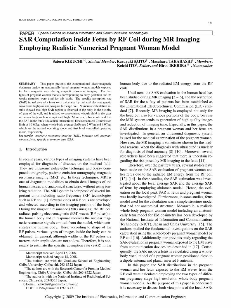
As stated above in this paper, the SAR distributions on twotypes of realistic woman models are calculated. Here, the re-alistic high-resolution whole-body voxel model of an adultJapanese female average figure, developed at NICT [20] isused for the calculation of the pregnant woman in early pe-riod (Model A). The size of fetus (embryo) in early period isvery small, hence it is difficult to confirm pregnancy status.Therefore, it is considered that the person, who does not no-tice her own pregnancy state, uses MRI. Moreover, the 26thgestational week pregnant woman model (Model B) [15]–[17] is employed as one example of late pregnancy stages.In order to develop this model, first, the fetus model is devel-oped from the MR images of a pregnant woman in the 26thgestational week. The fetus model consists of six organs in-cluding fetal body, fetal brain, fetal eyes, amniotic fluid, pla-centa, and uterus wall. Next, the abdomen of the Model A isexpanded following the standard shape and structure of thepregnant woman. Finally, these two models are combinedand adjusted following the comments from medical doctors.In addition, the more detailed explanations regarding the de-velopment of pregnant woman model are described in [15].
Figures 1(a) and (b) show Model A and B, respec-tively. These high resolution models are composed of 2 mm× 2 mm × 2 mm cubical voxel. The physical properties of
Fig. 1 Realistic woman models from [16].
these models are determined from [13], [21]–[23], except di-electric properties of fetal brain and fetal body. Here, it hasbeen found that dielectric properties at microwave frequencyband depended on tissue water contents [24]. Consequently,the dielectric properties of fetal brain and fetal body ad-justed to account for the higher water content in comparisonwith similar adult tissues. These properties were decidedwith correction method by the difference of water content inthe tissue. The procedure for determining values were pre-viously discussed by Hand et al. [13]. Table 1 summariesa part of the parameters around the abdomen of pregnancyand her fetus.
2.3 Calculation Models of RF Coil
In this paper, a birdcage coil is employed as one of the mostfundamental RF coils for MRI system. The birdcage coilis often used as both the transmission of RF pulse and re-

KIKUCHI et al.: SAR COMPUTATION INSIDE FETUS BY RF COIL433
Table 1 Example of physical properties of pregnant woman and herfetus at 64 MHz.
Density Relative Conductivityρ[kg/m3] permitivity εr [S/m]
Maternal bodyMuscle 1,040 72.0 0.71Fat 928 13.6 0.07Skin 1,100 84.4 0.46Blood 1,060 86.5 1.21Bone cortical 1,990 16.7 0.06Bone cancellous 1,040 18.2 0.11Lung 655 56.2 0.41Liver 1,050 80.6 0.45Stomach 1,050 85.8 0.88Colon 1,044 94.7 0.64Small intestine 1,044 118.3 1.59Ovary 1,048 106.8 0.69Uterus wall 1,052 92.1 0.91Amniotic fluid 1,000 97.3 2.07Placenta 1,060 86.5 1.21
Fetal bodyBody 1,040 94.2 0.92Eyes 1,009 69.1 1.50Brain 1,030 97.2 0.76
ception of NMR signal, and is categorized into two typesby the difference of the position of loaded capacitors; oneis a highpass birdcage coil, and the other is a lowpass bird-cage coil [1]. Figures 2(a) and (b) show the highpass bird-cage coil with RF shield and lowpass birdcage coil with RFshield, respectively. The operating frequency of the coil isaround 64 MHz in both cases, which is used in the generic1.5 T MRI systems to excite the nuclei in the human bodyfor imaging. These coils consist of two end rings and eightlegs, whose widths are 10 mm, and are composed of perfectelectric conductors for calculations. The diameter and thelength of the coil are 600, 700 mm, respectively, so that therealistic woman model can be inserted. In order to reduceradiation of RF energy to outside of the coil, the coils aresurrounded by RF shield which is also modeled as perfectelectric conductor. A cylindrical RF shield, with an internaldiameter of 740 mm and a length of 1,260 mm, has been lo-cated lateral to the coil. The dimensions of the coil and theRF shield were determined based on [13], [14], [25].
Here, the capacitors were loaded into the end rings onthe highpass birdcage coil in Fig. 2(a), and into the legs onthe lowpass birdcage coil in Fig. 2(b). In order to determinethe capacitances, “birdcage builder [26],” which calculatesthe resonance frequency of the birdcage coil by the equiva-lent electrical circuit model, was employed. Values for ca-pacitors to resonate both coils at 64 MHz were 15.27 pF forthe highpass birdcage coil and 4.47 pF for the lowpass bird-cage coil. In addition, two feeding points were employedin these calculations and the phase difference between twoports is 90 degree. This excitation method is called “Quadra-ture excitation” and can generate clear MR image by usingcircular polarized field [1].
(a) Highpass type (b) Lowpass type
Fig. 2 Two types of birdcage coil with RF shield.
Fig. 3 FDTD calculation model.
2.4 Numerical Calculation Model
Figure 3 indicates FDTD calculation model including therealistic woman model. As an example of the model, theModel B is inserted into highpass birdcage coil. There areseveral possibilities for the coil position, because it is con-sidered that the position of the coil is not the same for eachimaging. Previously, the relationship of several position ofthe RF coil to the SAR was calculated [27]. However, inthis paper, the coil was placed the center of uterus in ModelA, and placed center of fetal brain in Model B. In order tocalculate the whole-body of the woman models, a large ana-lytical region was required and a super technical server (Hi-tachi SR11000) in the Institute of Media and InformationTechnology, Chiba University was employed. In the numer-ical calculations, the uniform grid size inside of the shieldincluding the woman model is 2.0 mm. The non-uniformmesh is used for the outside of the shield, because most ofthe EM waves are not emitted outward by the shield.
The parameters used in the FDTD calculations arelisted in Table 2. In addition, steady state analysis is per-formed by enforcing a continuous sinusoidal wave of elec-tric field on the feeding gaps to calculate the SAR distribu-tion in the model. Moreover, the coordinate origin is thecenter of calculation model. In the previous study, we inves-tigated on the SAR evaluation inside the tissue-equivalentsolid phantom using surface coil, the effectiveness of calcu-lation technique is confirmed by comparison with an exper-iment result [28].
434IEICE TRANS. COMMUN., VOL.E92–B, NO.2 FEBRUARY 2009
Table 2 Parameters for FDTD calculations.
Cell size [mm] Δx, Δy 2.0, 2.0(Minimum) Δz 2.0 (const.)Cell size [mm] Δx, Δy 5.6, 5.6(Maximum) Δz 2.0 (const.)Analytical space x× y× z [cell] 534 × 534 × 984Time step [ps] 3.8Absorbing boundary condition PML (8 layers)
3. SAR Distribution in the Abdomen of PregnantWoman Models
In the actual MRI system, the various pulse sequences areemployed according to the imaging region and type. How-ever, in this paper, the SARs in the pregnant woman and herfetus were calculated by exciting the continuous sinusoidalwave at feeding point, because the radiation power of eachpulse sequence was not clear. Here, in order to simplify con-version of radiation power, the SAR values are normalizedby 1.0 W radiation power from the coil in all cases. In addi-tion, the radiation power was calculated by a surface integralof poynting vector on closed surface surrounding the coil.
3.1 Comparison of the SAR Distributions in Model A withthe Two Types of RF Coils
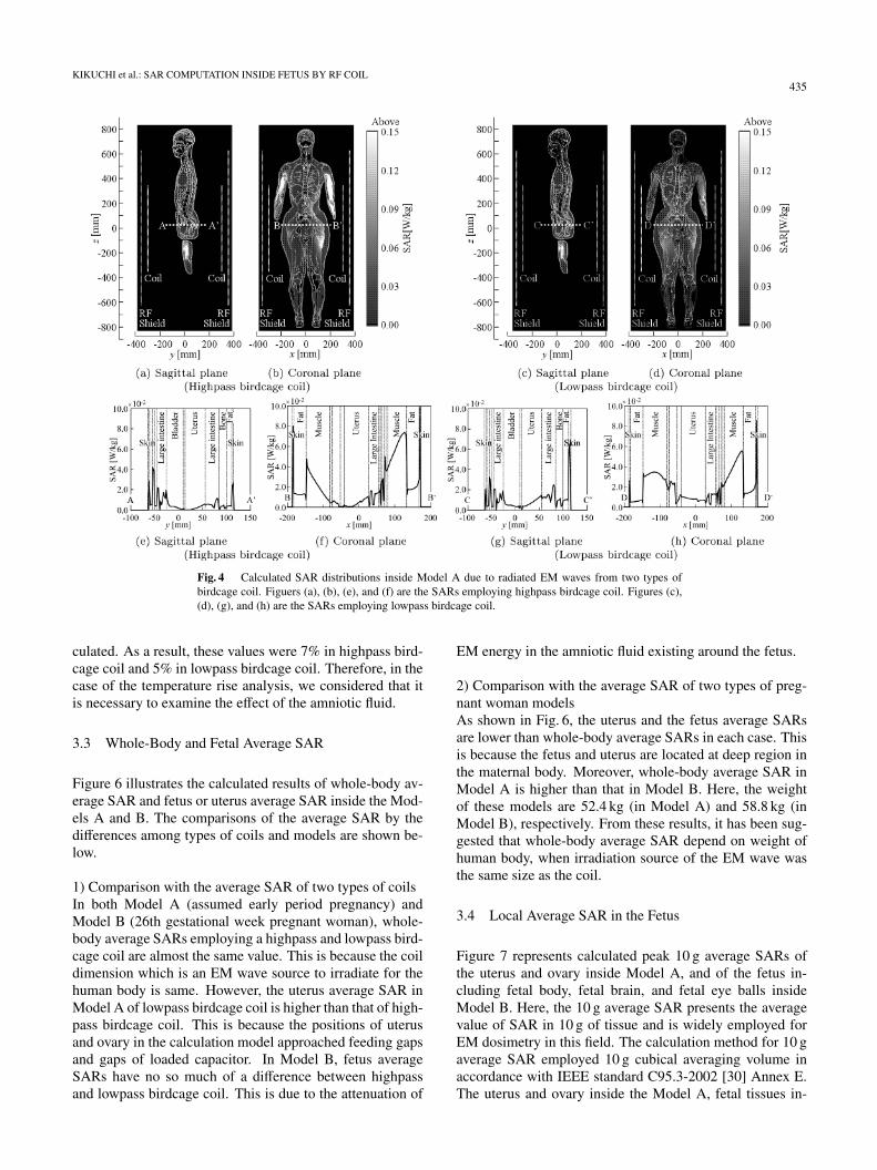
Figures 4(a)–(h) show the calculated SAR distributions in-side the Model A by employing the highpass and lowpassbirdcage coil, respectively. The observation planes are thesagittal plane (yz-plane) including the uterus, and the coro-nal plane (xz-plane) around the center of the uterus. More-over, positions of observation line A-A’, B-B’, C-C’, and D-D’ are indicated in the Figs. 4(a)–(d). In this paper, ModelA (the non-pregnant woman model) is assumed as the earlyperiod of pregnancy. Therefore, the SAR inside the uterusand ovary were observed in place of fetal tissue, because thefetus or embryo was very small in the early period [29].
As shown in Figs. 4(b) and (d), the SAR distributionson the coronal plane are observed unsymmetrical tendencyin comparison with the left side (−225 mm < z < 0) and rightside(0 < z < 225 mm). Moreover, it was confirmed that atendency of the SAR distribution changes when we observethe coronal plane of other positions. Because it was consid-ered that the tendency is influenced by two points of feeding,a symmetry of SAR distributions was confirmed in the caseof one point of feeding model. Moreover, from Figs. 4(a)–(d), relatively high SAR values are observed around the skin,muscle, etc which have a high electrical conductivity andlocated close to the surface of the maternal body. The ten-dency can be confirmed in Figs. 4(e)–(h), except fat tissuewhich is low electrical conductivity. Moreover, a high SARvalues are observed inside the thigh (x = 0, −100 < z <−300 mm in Figs. 4(b) and (d)) and armpit (x = ±200 mm,z = 400 mm in Figs. 4(b) and (d)). This is due to the con-centrated electric field at those narrow gaps. In comparisonwith the SAR distributions due to the EM energy from the
highpass and lowpass birdcage coil, we observed compara-tively high SAR values in the vicinity of each positions offeeding point and loaded capacitor. Here, according to theresult that confirmed all SAR distributions, it was found thatthis phenomenon is dependent on whether an electric fieldconcentrates on existing gap of the human body and a coil.
Meanwhile, the SAR in the uterus and the ovary(around 15 < y < 60 mm, −50 < z < 50 mm in Figs. 4(a)and (c), around −40 < x < 25 mm, −50 < z < 50 mm inFigs. 4(b) and (d)) are low compared to the maternal surfaceof skin and muscle tissue. In addition, low SAR value isobserved at the uterus (around 15 < y < 60 mm in Figs. 4(e)and (f), around −40 < x < 25 mm in Figs. 4(g) and (h)) dueto the attenuation of EM energy in the muscle tissue (−150 <x < −80 mm, 80 < x < 140 mm in Figs. 4(f) and (h)). More-over, compared with the SAR in the uterus and the ovary bydifferent coil type, it is observed that there is hardly a differ-ence. From these result, it has been confirmed that the SARsin the uterus and the ovary are low, because these areas areplaced in deep region of the body.
3.2 Comparison of the SAR Distributions in Model B withthe Two Types of RF Coils
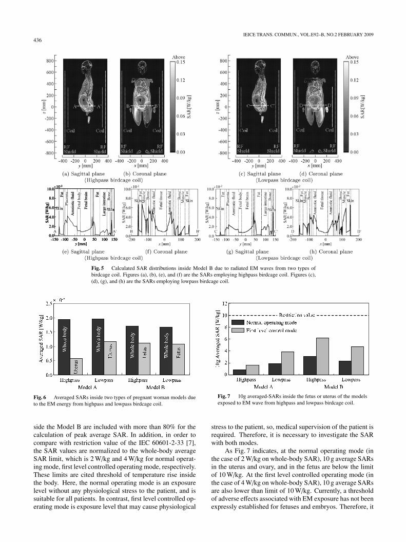
Figures 5(a)–(h) show the calculated SAR distributions in-side the Model B by use of the highpass and lowpass bird-cage coil, respectively. The observation planes are the sagit-tal plane (yz-plane) including the center of the fetal head,and are the coronal plane (xz-plane) around the center offetal body. Moreover, positions of observation line A-A’,B-B’, C-C’, and D-D’ are indicated in the Figs. 5(a)–(d).
As shown in Fig. 5, relatively high SAR values areobserved around the maternal surface and unsymmetricaltendency are observed by the effect of two points feeding.Moreover, it is observed that the SAR at the amniotic fluidis also relatively high. Especially, relatively high SAR val-ues are observed at the upper and lower portions of amnioticfluid (around y = −100, z = −50, 150 mm in Figs. 5(a) and(c), around x = 0, z = −50 mm and x = 100 mm, 50 < z <200 mm in Figs. 5(b) and (d)). In addition, as the Figs. 5(e)–(h) indicate, it has found that the SAR in the amniotic fluidnear the fetal tissue is relatively high. This is because theelectrical conductivity of amniotic fluid is almost 1.5–2.0times higher than other tissues, as listed in Table 1. In ad-dition, the SAR distributions around the boundary of eachtissue were precipitously varied due to the heterogeneousstructure.
However, the SAR in the fetus (around −20 < y <50 mm at Figs. 5(e) and (g), around −50 < x < 15 mm inFigs. 5(f) and (h)) is the low, which is compared to the valuesof maternal body. Moreover, maximum SAR value withinfetus is less than 1.0% of that of the maternal body. Fromthese results, it has been confirmed that the SAR in the fetusis attributed to the attenuation of EM energy in the amnioticfluid which is high conductivity. Here, we focused attentionon the power deposition of amniotic fluid, and the powerdeposition of the amniotic fluid for the total values was cal-
KIKUCHI et al.: SAR COMPUTATION INSIDE FETUS BY RF COIL435
Fig. 4 Calculated SAR distributions inside Model A due to radiated EM waves from two types ofbirdcage coil. Figuers (a), (b), (e), and (f) are the SARs employing highpass birdcage coil. Figures (c),(d), (g), and (h) are the SARs employing lowpass birdcage coil.
culated. As a result, these values were 7% in highpass bird-cage coil and 5% in lowpass birdcage coil. Therefore, in thecase of the temperature rise analysis, we considered that itis necessary to examine the effect of the amniotic fluid.
3.3 Whole-Body and Fetal Average SAR
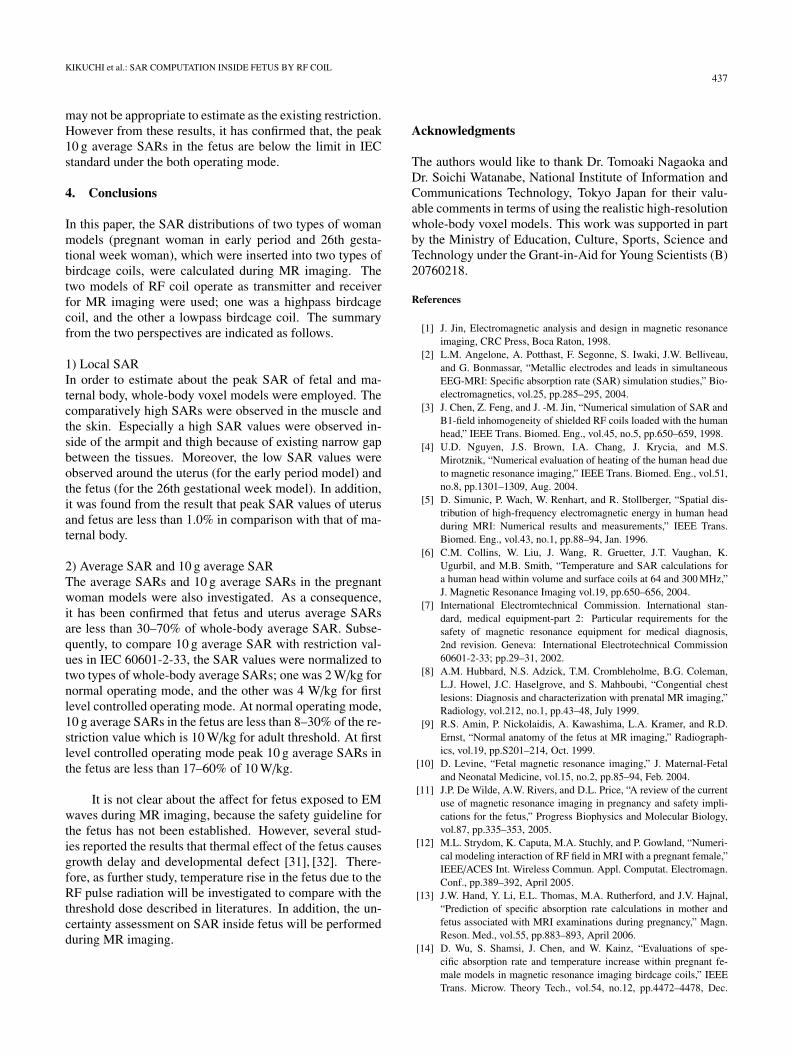
Figure 6 illustrates the calculated results of whole-body av-erage SAR and fetus or uterus average SAR inside the Mod-els A and B. The comparisons of the average SAR by thedifferences among types of coils and models are shown be-low.
1) Comparison with the average SAR of two types of coilsIn both Model A (assumed early period pregnancy) andModel B (26th gestational week pregnant woman), whole-body average SARs employing a highpass and lowpass bird-cage coil are almost the same value. This is because the coildimension which is an EM wave source to irradiate for thehuman body is same. However, the uterus average SAR inModel A of lowpass birdcage coil is higher than that of high-pass birdcage coil. This is because the positions of uterusand ovary in the calculation model approached feeding gapsand gaps of loaded capacitor. In Model B, fetus averageSARs have no so much of a difference between highpassand lowpass birdcage coil. This is due to the attenuation of
EM energy in the amniotic fluid existing around the fetus.
2) Comparison with the average SAR of two types of preg-nant woman modelsAs shown in Fig. 6, the uterus and the fetus average SARsare lower than whole-body average SARs in each case. Thisis because the fetus and uterus are located at deep region inthe maternal body. Moreover, whole-body average SAR inModel A is higher than that in Model B. Here, the weightof these models are 52.4 kg (in Model A) and 58.8 kg (inModel B), respectively. From these results, it has been sug-gested that whole-body average SAR depend on weight ofhuman body, when irradiation source of the EM wave wasthe same size as the coil.
3.4 Local Average SAR in the Fetus
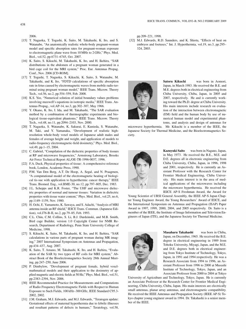
Figure 7 represents calculated peak 10 g average SARs ofthe uterus and ovary inside Model A, and of the fetus in-cluding fetal body, fetal brain, and fetal eye balls insideModel B. Here, the 10 g average SAR presents the averagevalue of SAR in 10 g of tissue and is widely employed forEM dosimetry in this field. The calculation method for 10 gaverage SAR employed 10 g cubical averaging volume inaccordance with IEEE standard C95.3-2002 [30] Annex E.The uterus and ovary inside the Model A, fetal tissues in-
436IEICE TRANS. COMMUN., VOL.E92–B, NO.2 FEBRUARY 2009
Fig. 5 Calculated SAR distributions inside Model B due to radiated EM waves from two types ofbirdcage coil. Figures (a), (b), (e), and (f) are the SARs employing highpass birdcage coil. Figures (c),(d), (g), and (h) are the SARs employing lowpass birdcage coil.
Fig. 6 Averaged SARs inside two types of pregnant woman models dueto the EM energy from highpass and lowpass birdcage coil.
side the Model B are included with more than 80% for thecalculation of peak average SAR. In addition, in order tocompare with restriction value of the IEC 60601-2-33 [7],the SAR values are normalized to the whole-body averageSAR limit, which is 2 W/kg and 4 W/kg for normal operat-ing mode, first level controlled operating mode, respectively.These limits are cited threshold of temperature rise insidethe body. Here, the normal operating mode is an exposurelevel without any physiological stress to the patient, and issuitable for all patients. In contrast, first level controlled op-erating mode is exposure level that may cause physiological
Fig. 7 10g averaged-SARs inside the fetus or uterus of the modelsexposed to EM wave from highpass and lowpass birdcage coil.
stress to the patient, so, medical supervision of the patient isrequired. Therefore, it is necessary to investigate the SARwith both modes.
As Fig. 7 indicates, at the normal operating mode (inthe case of 2 W/kg on whole-body SAR), 10 g average SARsin the uterus and ovary, and in the fetus are below the limitof 10 W/kg. At the first level controlled operating mode (inthe case of 4 W/kg on whole-body SAR), 10 g average SARsare also lower than limit of 10 W/kg. Currently, a thresholdof adverse effects associated with EM exposure has not beenexpressly established for fetuses and embryos. Therefore, it
KIKUCHI et al.: SAR COMPUTATION INSIDE FETUS BY RF COIL437
may not be appropriate to estimate as the existing restriction.However from these results, it has confirmed that, the peak10 g average SARs in the fetus are below the limit in IECstandard under the both operating mode.
4. Conclusions
In this paper, the SAR distributions of two types of womanmodels (pregnant woman in early period and 26th gesta-tional week woman), which were inserted into two types ofbirdcage coils, were calculated during MR imaging. Thetwo models of RF coil operate as transmitter and receiverfor MR imaging were used; one was a highpass birdcagecoil, and the other a lowpass birdcage coil. The summaryfrom the two perspectives are indicated as follows.
1) Local SARIn order to estimate about the peak SAR of fetal and ma-ternal body, whole-body voxel models were employed. Thecomparatively high SARs were observed in the muscle andthe skin. Especially a high SAR values were observed in-side of the armpit and thigh because of existing narrow gapbetween the tissues. Moreover, the low SAR values wereobserved around the uterus (for the early period model) andthe fetus (for the 26th gestational week model). In addition,it was found from the result that peak SAR values of uterusand fetus are less than 1.0% in comparison with that of ma-ternal body.
2) Average SAR and 10 g average SARThe average SARs and 10 g average SARs in the pregnantwoman models were also investigated. As a consequence,it has been confirmed that fetus and uterus average SARsare less than 30–70% of whole-body average SAR. Subse-quently, to compare 10 g average SAR with restriction val-ues in IEC 60601-2-33, the SAR values were normalized totwo types of whole-body average SARs; one was 2 W/kg fornormal operating mode, and the other was 4 W/kg for firstlevel controlled operating mode. At normal operating mode,10 g average SARs in the fetus are less than 8–30% of the re-striction value which is 10 W/kg for adult threshold. At firstlevel controlled operating mode peak 10 g average SARs inthe fetus are less than 17–60% of 10 W/kg.
It is not clear about the affect for fetus exposed to EMwaves during MR imaging, because the safety guideline forthe fetus has not been established. However, several stud-ies reported the results that thermal effect of the fetus causesgrowth delay and developmental defect [31], [32]. There-fore, as further study, temperature rise in the fetus due to theRF pulse radiation will be investigated to compare with thethreshold dose described in literatures. In addition, the un-certainty assessment on SAR inside fetus will be performedduring MR imaging.
Acknowledgments
The authors would like to thank Dr. Tomoaki Nagaoka andDr. Soichi Watanabe, National Institute of Information andCommunications Technology, Tokyo Japan for their valu-able comments in terms of using the realistic high-resolutionwhole-body voxel models. This work was supported in partby the Ministry of Education, Culture, Sports, Science andTechnology under the Grant-in-Aid for Young Scientists (B)20760218.
References
[1] J. Jin, Electromagnetic analysis and design in magnetic resonanceimaging, CRC Press, Boca Raton, 1998.
[2] L.M. Angelone, A. Potthast, F. Segonne, S. Iwaki, J.W. Belliveau,and G. Bonmassar, “Metallic electrodes and leads in simultaneousEEG-MRI: Specific absorption rate (SAR) simulation studies,” Bio-electromagnetics, vol.25, pp.285–295, 2004.
[3] J. Chen, Z. Feng, and J. -M. Jin, “Numerical simulation of SAR andB1-field inhomogeneity of shielded RF coils loaded with the humanhead,” IEEE Trans. Biomed. Eng., vol.45, no.5, pp.650–659, 1998.
[4] U.D. Nguyen, J.S. Brown, I.A. Chang, J. Krycia, and M.S.Mirotznik, “Numerical evaluation of heating of the human head dueto magnetic resonance imaging,” IEEE Trans. Biomed. Eng., vol.51,no.8, pp.1301–1309, Aug. 2004.
[5] D. Simunic, P. Wach, W. Renhart, and R. Stollberger, “Spatial dis-tribution of high-frequency electromagnetic energy in human headduring MRI: Numerical results and measurements,” IEEE Trans.Biomed. Eng., vol.43, no.1, pp.88–94, Jan. 1996.
[6] C.M. Collins, W. Liu, J. Wang, R. Gruetter, J.T. Vaughan, K.Ugurbil, and M.B. Smith, “Temperature and SAR calculations fora human head within volume and surface coils at 64 and 300 MHz,”J. Magnetic Resonance Imaging vol.19, pp.650–656, 2004.
[7] International Electromtechnical Commission. International stan-dard, medical equipment-part 2: Particular requirements for thesafety of magnetic resonance equipment for medical diagnosis,2nd revision. Geneva: International Electrotechnical Commission60601-2-33; pp.29–31, 2002.
[8] A.M. Hubbard, N.S. Adzick, T.M. Crombleholme, B.G. Coleman,L.J. Howel, J.C. Haselgrove, and S. Mahboubi, “Congential chestlesions: Diagnosis and characterization with prenatal MR imaging,”Radiology, vol.212, no.1, pp.43–48, July 1999.
[9] R.S. Amin, P. Nickolaidis, A. Kawashima, L.A. Kramer, and R.D.Ernst, “Normal anatomy of the fetus at MR imaging,” Radiograph-ics, vol.19, pp.S201–214, Oct. 1999.
[10] D. Levine, “Fetal magnetic resonance imaging,” J. Maternal-Fetaland Neonatal Medicine, vol.15, no.2, pp.85–94, Feb. 2004.
[11] J.P. De Wilde, A.W. Rivers, and D.L. Price, “A review of the currentuse of magnetic resonance imaging in pregnancy and safety impli-cations for the fetus,” Progress Biophysics and Molecular Biology,vol.87, pp.335–353, 2005.
[12] M.L. Strydom, K. Caputa, M.A. Stuchly, and P. Gowland, “Numeri-cal modeling interaction of RF field in MRI with a pregnant female,”IEEE/ACES Int. Wireless Commun. Appl. Computat. Electromagn.Conf., pp.389–392, April 2005.
[13] J.W. Hand, Y. Li, E.L. Thomas, M.A. Rutherford, and J.V. Hajnal,“Prediction of specific absorption rate calculations in mother andfetus associated with MRI examinations during pregnancy,” Magn.Reson. Med., vol.55, pp.883–893, April 2006.
[14] D. Wu, S. Shamsi, J. Chen, and W. Kainz, “Evaluations of spe-cific absorption rate and temperature increase within pregnant fe-male models in magnetic resonance imaging birdcage coils,” IEEETrans. Microw. Theory Tech., vol.54, no.12, pp.4472–4478, Dec.
438IEICE TRANS. COMMUN., VOL.E92–B, NO.2 FEBRUARY 2009
2006.[15] T. Nagaoka, T. Togashi, K. Saito, M. Takahashi, K. Ito, and S.
Watanabe, “An anatomically realistic whole-body pregnant-womanmodel and specific absorption rates for pregnant-woman exposureto electromagnetic plane wave from 10 MHz to 2 GHz,” Phys. Med.Biol., vol.52, pp.6731–6745, Oct. 2007.
[16] K. Saito, S. Kikuchi, M. Takahashi, K. Ito, and H. Ikehira, “SARdistributions in the abdomen of a pregnant woman generated in abird cage coil for the MRI system,” Proc. Eur. Antennas Propag.Conf., Nov. 2006 [CD-ROM].
[17] T. Togashi, T. Nagaoka, S. Kikuchi, K. Saito, S. Watanabe, M.Takahashi, and K. Ito, “FDTD calculations of specific absorptionrate in fetus caused by electromagnetic waves from mobile radio ter-minal using pregnant woman model,” IEEE Trans. Microw. TheoryTech., vol.56, no.2, pp.554–559, Feb. 2008.
[18] K.S. Yee, “Numerical solution of initial boundary values problemsinvolving maxwell’s equations in isotropic media,” IEEE Trans. An-tennas Propag., vol.AP-14, no.3, pp.302–307, May 1966.
[19] Y. Okano, K. Ito, I. Ida, and M. Takahashi, “The SAR evaluationmethod by a combination of thermographic experiments and bio-logical tissue-equivalent phantoms,” IEEE Trans. Microw. TheoryTech., vol.48, no.11, pp.2094–2103, Nov. 2000.
[20] T. Nagaoka, S. Watanabe, K. Sakurai, E. Kunieda, S. Watanabe,M. Taki, and Y. Yamanaka, “Development of realistic high-resolution whole-body voxel models of Japanese adult males andfemales of average height and weight, and application of models toradio-frequency electromagnetic-field dosimetry,” Phys. Med. Biol.,vol.49, pp.1–15, 2004.
[21] C. Gabriel, “Compilation of the dielectric properties of body tissuesat RF and microwave frequencies,” Armstrong Laboratory, BrooksAir Force Technical Report AL/OE-TR-1996-0037, 1996.
[22] F.A. Duck, Physical properties of tissue: A comprehensive referencebook, London, Academic Press, 1990.
[23] P.M. Van Den Berg, A.T. De Hoop, A. Segal, and N. Praagmen,“A computational model of the electromagnetic heating of biologi-cal tis-sue with application to hyperthermic cancer therapy,” IEEETrans. Biomed. Eng., vol.BME-30, no.12, pp.797–805, Dec. 1983.
[24] J.L. Schepps and K.R. Foster, “The UHF and microwave dielec-tric properties of normal and tumour tissues: Variation in dielectricproperties with tissue water content,” Phys. Med. Biol., vol.25, no.6,pp.1149–1159, Nov. 1980.
[25] H. Ochi, E. Yamamoto, K. Sawaya, and S. Adachi, “Analysis of MRIantenna inside an RF shield,” IEICE Trans. Commun. (Japanese Edi-tion), vol.J76-B-II, no.2, pp.79–85, Feb. 1993.
[26] C.L. Chin, C.M. Collins, S. Li, B.J. Dardzinski, and M.B. Smith,Bird cage Builder, version 1.0 Copyright Center for NMR Re-search, Department of Radiology, Penn State University College ofMedicine, 1998.
[27] S. Kikuchi, K. Saito, M. Takahashi, K. Ito, and H. Ikehira, “SARcalculations in various parts of pregnant woman during MR imag-ing,” 2007 International Symposium on Antennas and Propagation,pp.434–437, Aug. 2007.
[28] K. Saito, T. Amano, M. Takahashi, K. Ito, and H. Ikehira, “Evalu-ation of the SAR by two types of RF coils for MRI system,” Ab-stract Book of the Bioelectromagnetics Society 28th Annual Meet-ing, pp.247–250, June 2006.
[29] P. Dimbylow, “Development of pregnant female, hybrid voxel-mathmatical models and their application to the dosimetry of ap-plied magnetic and electric fields at 50 Hz,” Phys. Med. Biol., vol.51,pp.2383–2394, Nov. 2006.
[30] IEEE Recommended Practice for Measurements and Computationsof Radio Frequency Electromagnetic Fields with Respect to HumanExposure to Such Fields, 100 kHz–300 GHz, IEEE Standard C95.3-2002, 2002.
[31] J.M. Graham, M.J. Edwards, and M.J. Edwards, “Teratogen update:Gestational effects of maternal hyperthermia due to febrile illnessesand resultant patterns of defects in humans,” Teratology, vol.58,
pp.209–221, 1998.[32] M.J. Edwards, R.D. Saunders, and K. Shiota, “Effects of heat on
embryos and foetuses,” Int. J. Hyperthermia, vol.19, no.3, pp.295–324, 2003.
Satoru Kikuchi was born in Aomori,Japan, in March 1983. He received the B.E. andM.E. degrees both in electrical engineering fromChiba University, Chiba, Japan, in 2005 and2007, respectively. He and is currently work-ing toward the Ph.D. degree at Chiba University.His main interests include research on evalua-tion of the interaction between electromagnetic(EM) field and the human body by use of nu-merical human model and experimental phan-toms, and analysis and design of antennas for
microwave hyperthermia. Mr. Kikuchi is a member of the IEEE, theJapanese Society for Thermal Medicine, and the Bioelectromagnetics So-ciety.
Kazuyuki Saito was born in Nagano, Japan,in May 1973. He received the B.E., M.E. andD.E. degrees all in electronic engineering fromChiba University, Chiba, Japan, in 1996, 1998and 2001, respectively. He is currently an As-sistant Professor with the Research Center forFrontier Medical Engineering, Chiba Univer-sity. His main interest is in the area of med-ical applications of the microwaves includingthe microwave hyperthermia. He received theIEICE AP-S Freshman Award, the Award for
Young Scientist of URSI General Assembly, the IEEE AP-S Japan Chap-ter Young Engineer Award, the Young Researchers’ Award of IEICE, andthe International Symposium on Antennas and Propagation (ISAP) PaperAward in 1997, 1999, 2000, 2004, and 2005 respectively. Dr. Saito is amember of the IEEE, the Institute of Image Information and Television En-gineers of Japan (ITE), and the Japanese Society for Thermal Medicine.
Masaharu Takahashi was born in Chiba,Japan, on December, 1965. He received the B.E.degree in electrical engineering in 1989 fromTohoku University, Miyagi, Japan, and the M.E.and D.E. degrees both in electrical engineer-ing from Tokyo Institute of Technology, Tokyo,Japan, in 1991 and 1994 respectively. He was aResearch Associate from 1994 to 1996, an As-sistant Professor from 1996 to 2000 at MusashiInstitute of Technology, Tokyo, Japan, and anAssociate Professor from 2000 to 2004 at Tokyo
University of Agriculture and Technology, Tokyo, Japan. He is currentlyan Associate Professor at the Research Center for Frontier Medical Engi-neering, Chiba University, Chiba, Japan. His main interests are electricallysmall antennas, planar array antennas, and electromagnetic compatibility.He received the IEEE Antennas and Propagation Society (IEEE AP-S) To-kyo chapter young engineer award in 1994. Dr. Takahashi is a senior mem-ber of the IEEE.
KIKUCHI et al.: SAR COMPUTATION INSIDE FETUS BY RF COIL439
Koichi Ito received the B.S. and M.S. de-grees from Chiba University, Chiba, Japan, in1974 and 1976, respectively, and the D.E. de-gree from the Tokyo Institute of Technology, To-kyo, Japan, in 1985, all in electrical engineering.From 1976 to 1979, he was a Research Asso-ciate at the Tokyo Institute of Technology. From1979 to 1989, he was a Research Associate atChiba University. From 1989 to 1997, he was anAssociate Professor at the Department of Elec-trical and Electronics Engineering, Chiba Uni-
versity, and is currently a Professor at the Graduate School of Engineer-ing, Chiba University. He has been appointed as one of the Deputy Vice-Presidents for Research, Chiba University, since April 2005. In 1989, 1994,and 1998, he visited the University of Rennes I, France, as an Invited Pro-fessor. Since 2004 he has been appointed as an Adjunct Professor to In-stitute of Technology Bandung (ITB), Indonesia. His main research inter-ests include analysis and design of printed antennas and small antennas formobile communications, research on evaluation of the interaction betweenelectromagnetic fields and the human body by use of numerical and exper-imental phantoms, microwave antennas for medical applications such ascancer treatment, and antennas for body-centric wireless communications.Dr. Ito is a Fellow of the IEEE, a member of the American Associationfor the Advancement of Science, the Institute of Image Information andTelevision Engineers of Japan (ITE) and the Japanese Society for Ther-mal Medicine (formerly, Japanese Society of Hyperthermic Oncology). Heserved as Chair of the Technical Group on Radio and Optical Transmis-sions, ITE from 1997 to 2001 and Chair of the Technical Group on HumanPhantoms for Electromagnetics, IEICE from 1998 to 2006. He also servedas Chair of the IEEE AP-S Japan Chapter from 2001 to 2002 and TPCCo-Chair of the 2006 IEEE International Workshop on Antenna Technol-ogy (iWAT2006). He currently serves as General Chair of the iWAT2008to be held in Chiba, Japan in 2008, Vice-Chair of the 2008 InternationalSymposium on Antennas and Propagation (ISAP2008) to be held in Tai-wan in 2008 and as an Associate Editor for the IEEE Transactions on An-tennas and Propagation. He also serves as a Distinguished Lecturer andan AdCom member for the IEEE Antennas and Propagation Society sinceJanuary 2007.
Hiroo Ikehira was born in Kyoto, Japan,in February 1955. He received the Medical Li-cense degree from Kobe University in 1980, theM.D. and Ph.D. degree in medicine from ChibaUniversity in 1987. He was a Researcher from1980 to 1988, a Senior Researcher from 1989to 1998, a General Manager at the Laboratoryof Molecule Information, the National Instituteof Radiological Sciences (NIRS). From 1990to 1997, he became an assistant professor withthe Department of Radiology, Chiba University
Hospital. From 1998–2000, he was an associate professor with the Facultyof Medicine, Chiba University. In 1982, he visited Faculty of Medicineat the University of Aberdeen, UK, as Resident. In 1987, he was a Vis-iting Scientist with the Lawrence Berkeley National Laboratory, Califor-nia, USA. He is currently a Deputy Director at the Biophysics Group ofMolecular Imaging Center, NIRS, an affiliate professor with the GraduateSchool of Medicine, Chiba University, and a lecturer with Graduate Schoolof Frontier Sciences, University of Tokyo. His main research interest is thebioimaging with magnetic resonance imaging (MRI) system. Dr. Ikehira ismember of the International Society for Magnetic Resonance in Medicine(ISMRM).