safety qualification of impurities in biopharmaceutical drugs
TRANSCRIPT

Safety Qualification of Impurities inSafety Qualification of Impurities in Biopharmaceutical Drugsp g
Impurities in Drugs:Monitoring, Safety and Regulationg y g
The Israel Chapter of PDA
July, 15 – 16, 2008July, 15 16, 2008
David Jacobson-Kram, Ph.D. DABTOffice of New DrugsOffice of New Drugs
Center for Drug Evaluation and ResearchFood and Drug Administration
Food and Drug Administration

Examples of Commonly Used Cell Substrates and Biologics ProducedSubstrates and Biologics Produced
CHO (hamster) – recombinant proteinsNS0 (mouse myeloma) – monoclonal antibodiesMouse hybridoma – monoclonal antibodiesVero (monkey) – viral vaccinesMRC 5 (human) viral vaccinesMRC-5 (human) – viral vaccines293 (human) – adenoviral vectors for gene therapyPER.C6 (human) – adenoviral vectors for gene therapyPER.C6 (human) adenoviral vectors for gene therapyMDCK (canine) – viral vaccines (e.g., Influenza)Insect – recombinant proteins
(Microbial expression systems also used – not focus of this presentation)
Food and Drug Administration
presentation)

Examples of Adventitious Agent p gContamination of Biologics
SV40 in early poliovirus and adenovirus vaccinesAvian leukosis virus and Hepatitis B in yellow fever vaccineCreutzfeldt-Jakob disease from growth hormoneH titi B H titi C HIV d B19 i bl dHepatitis B, Hepatitis C, HIV and B19 in blood productsMMV (Mouse parvovirus) in CHO cellsMMV (Mouse parvovirus) in CHO cells
Food and Drug Administration

Key Guidance Documents Points to Consider in the Characterization of Cell Lines Used to Produce Biologicals (1993)ICH Q5D D i ti d Ch t i ti f C ll S b t t U d fICH Q5D. Derivation and Characterization of Cell Substrates Used for Production of Biotechnological/Biological Products (1998)Points to Consider in the Manufacturing and Testing of MonoclonalPoints to Consider in the Manufacturing and Testing of Monoclonal Antibody Products for Human Use (1997)ICH Q5A. Viral Safety Evaluation of Biotechnology Products Derived f C ll Li f H A i l O i i (1998)from Cell Lines of Human or Animal Origin (1998)Guidance for Human Somatic Cell Therapy & Gene Therapy (1998)European Pharmacopoeia 5 0 section 5 2 3 Cell substrates for theEuropean Pharmacopoeia 5.0, section 5.2.3 – Cell substrates for the production of vaccines for human use (2005)Characterization and Qualification of Cell Substrates and Other Biological Starting Materials Used in the Production of Viral Vaccines for the Prevention and Treatment of Infectious Diseases (FDA, CBER, Draft Guidance, September, 2006)
Food and Drug Administration
Guidance, September, 2006)

Biosafety Testing Across Biologics Spectrum
PRECLINICAL PHASE I PHASE II PHASE III LICENSED PRODUCTDEVELOPMENT
IND BLA
PRODUCT STAGE
TOXICOLOGY
CELL LINE CHARACTERIZATION
(CLC) of MCB WCB(CLC) of MCB, WCB, and EPC
Raw material testing
VIRAL CLEARANCE (VC)
LOT RELEASE (LRT)( )
ANALYTICAL
Food and Drug Administration

Biologics Production: Flow and Testing PointsPoints
Master Cell Bank (CLC)
Working Cell Bank (CLC)
Final Product(LRT)
Working Cell Bank (CLC)
End of Production Cells (CLC)
Purified BulkProduction Unprocessed Bulk (LRT) Purification (VC)
Purified Bulk(LRT)
Food and Drug Administration
CLC = cell line characterization ; LRT = lot release testing ; VC = viral clearance studies

Key Questions to Determine Appropriate Testing for Cell and Virus Banks
Is it a FDA or a global submission?Is it a FDA or a global submission?What is the species of the cells for banking or the cells used for virus production?W t i d d i th hi t f th llWas serum or trypsin used during the history of the cell line?Was cell line exposed to cells or ingredients from otherWas cell line exposed to cells or ingredients from other species (e.g., mouse feeder cells and human stem cells)?Are the cells grown in medium containing antibiotics or ingredients (e g methotrexate) possibly inhibitory or toxicingredients (e.g., methotrexate) possibly inhibitory or toxic to adventitious agents or to cells in the test system?Is the sample matrix osmotically and pH compatible with th t t t ?the test system?What is the source/history of the cell line and virus stock?
Food and Drug Administration

General Categories of Characterization Testing for Cell Banks and EPC and VirusTesting for Cell Banks and EPC and Virus
BanksPurity - Microbial Contaminants
Bacteria, Fungiac e a, u gMycoplasma (Spiroplasma for insect cells and Baculovirus)Vi Ad titi R t i E dViruses : Adventitious, Retroviruses, Endogenous
IdentityIdentitySpecies for cells
Genetic Stability (for transfected cell substrates)
Food and Drug Administration

General Testing Categories Performed MCB WCB EPC d Vi b kon MCB, WCB, EPC, and Virus banks
T MCB/MVB WCB/WVB EPCTests MCB/MVB WCB/WVB EPC (CAL)
Sterility + + +y
Mycoplasma + + +
Virus: -Adventitious
Retrovirus
+ +
−/+
+ + - Retrovirus
- Specific (species/virus)
++
− −
+ −/+
Species identification)- for cells
+ (cells) + (cells) +
Food and Drug Administration
for cells

Characterization Testing of Cell and Virus Banks d EPC St ilit d M l T tiand EPC: Sterility and Mycoplasma Testing
Sterility testing for bacteria and fungi F ll ICH d ti t ti f d t t f- Follow ICH recommendations – testing performed on contents of individual containers (1% of total bank but not less than two containers or vials).)
- Bacteriostasis and Fungistasis (B&F) assay recommended prior to performing the Sterility assay to assess sample matrix for inhibition.
C ( ) ( )Mycoplasma testing - PTC (28 day assay) or EP (28 day assay) - Mycoplasma agar plate and semi-solid broth bottle procedures for agar-
cultivable mycoplasma and the VERO cell culture/H stain procedure forcultivable mycoplasma and the VERO cell culture/H stain procedure for non-agar cultivable mycoplasma
- Qualification (Mycoplasmastasis) – Stasis assay is not referenced in any FDA regulation or guidance document, while the EP requires it. It is good science to perform a stasis assay to assess the sample matrix for any inhibitory effect, which could prevent the detection of an
Food and Drug Administration
y y , padventitious mycoplasma.

28-Day Mycoplasma Culture MethodSample
Hoechst stain1.0 ml
Vero Cells 3-5 days
Hoechst stain
Observe DNA fluorescing staining pattern
0.2 ml 14 days Observe for mycoplasma coloniesA l t
10 ml Day 3 Day 7 Day 14
Agar plate
10 ml
Mycoplasma Broth
Ob f l l iDay 17 Day 21 Day 28
14 days 14 days 14 days
Food and Drug Administration
Observe for mycoplasma colonies

Characterization Testing of CellCharacterization Testing of Cell and Virus Banks and EPC: Virus Assays
In vitro adventitious virus- In vitro adventitious virus screening assay
- In vivo adventitious virus screening assayscreening assay
- In vivo species-specific assays - In vitro virus-specific assays
Food and Drug Administration

Assay for Adventitious Virus Testing -ssay o d e t t ous us est g14-Day and 28-Day Virus Screens
28-Day Adventitious Virus assay 14-Day Adventitious Virus assay
Observations Observations_______________________ _____________________________________________ ______________________Day 0 Day 14 Day 28Inoculate 0.5 ml/well HA/HAD HA/HADMRC-5, Vero, CHO-K1 Blind Passage End of study
(P iti t l i MRC 5 l V PI 3 CHO K1 SV 5)(Positive control viruses: MRC-5 = measles; Vero = PI-3; CHO-K1 = SV-5)
Food and Drug Administration

Morphological Changes (CPE)i 324K C ll I f t d ith REO Viin 324K Cells Infected with REO Virus
REO-infected 324K cellsControl 324K cellsDay 20Day 20
Food and Drug Administration

Hemadsorption (HAD) – Vero cells and Rhesus RBCsRhesus RBCsRBC stickage and clumping to infected assay cells
Food and Drug Administration

Hemagglutination (HA)Positive HA result
Negative HA result
Food and Drug Administration

Sample cytotoxicity effect – non-viral CPEControl MRC-5 Cells Sample Treated MRC-5 Cells
Ascitic fluid samplecontains lipids.
Viewing plane above cell monolayer
p
Food and Drug Administration
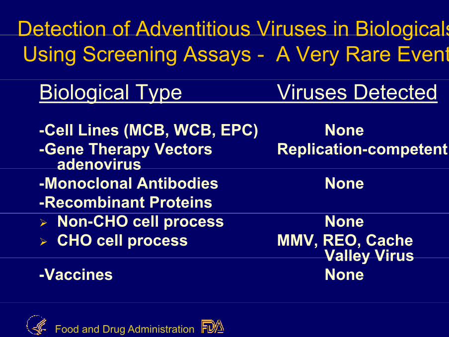
Detection of Adventitious Viruses in BiologicalsDetection of Adventitious Viruses in Biologicals Using Screening Assays - A Very Rare Event
Biological Type Viruses Detected
C ll i (MCB WCB EPC) N-Cell Lines (MCB, WCB, EPC) None-Gene Therapy Vectors Replication-competent
adenovirusadenovirus-Monoclonal Antibodies None-Recombinant Proteins
Non-CHO cell process NoneCHO cell process MMV, REO, Cache
Valley VirusValley Virus-Vaccines None
Food and Drug Administration

Susceptibility of CHO Cells to ViViruses
1) CHO cells have a limited susceptibility to viruses.2) Not susceptible to the following virus groups:
Ad i C i Pi i (C kiAdenovirus, Coronavirus, Picornavirus (Coxsackie, Rhinovirus), Herpes (HSV 1&2, CMV, VZV), Orthomyxo (Influenza A&B), Togavirus (BVDV)(Influenza A&B), Togavirus (BVDV)
3) CHO cells are not susceptible to Retroviruses (no infectious retrovirus isolated from CHO cells – to date)
4) CHO cells are susceptible to Reovirus, (1,2,3), Paramyxo (Parainfluenza 1,2,3 and SV-5), Bunya (Cache Valley) Parvo (MMV)(Cache Valley), Parvo (MMV)
Food and Drug Administration
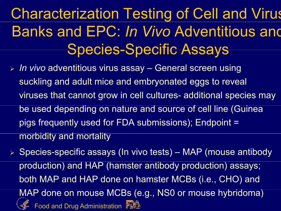
Characterization Testing of Cell and Virus Banks and EPC: In Vivo Adventitious and
Species Specific AssaysSpecies-Specific AssaysIn vivo adventitious virus assay – General screen using suckling and adult mice and embryonated eggs to reveal viruses that cannot grow in cell cultures- additional species may b d d di t d f ll li (G ibe used depending on nature and source of cell line (Guinea pigs frequently used for FDA submissions); Endpoint =
bidit d t litmorbidity and mortality
Species-specific assays (In vivo tests) – MAP (mouse antibody production) and HAP (hamster antibody production) assays; both MAP and HAP done on hamster MCBs (i.e., CHO) and
C ( S )Food and Drug Administration
MAP done on mouse MCBs (e.g., NS0 or mouse hybridoma)

Mouse Antibody Production y(MAP) Assay
BasisDetection of murine viruses based upon generation of p gspecific antibody (or serum enzyme) in response to virus infection
ProcedureInoculate cell lysate into mice via intracranial, intranasal, intraperitoneal and per os routeTest serum for elevated lactic dehydrogenase level ft 3 dafter 3 days
Bleed mice after 28 days and test for virus-specific antibody by ELISA and/or immunofluorescent staining
Food and Drug Administration
antibody by ELISA and/or immunofluorescent staining

Characterization Testing of Cell and gVirus Banks and EPC: Virus-
S ifi ASpecific AssaysVirus-Specific Assays
PCR and in vitro infectivity assays – MMV (mouse parvovirus) for CHO cell lines(mouse parvovirus) for CHO cell linesBovine polyoma virus (PCR) expected by EP reviewers; Bovine polyoma infects human and simian cell lines; no evidence for infecting CHOsimian cell lines; no evidence for infecting CHOPCR panel for simian viruses for simian cell lines such as VeroPCR panel for human viruses for human cell substrates
Food and Drug Administration

Characterization of Cell and Virus Banks:Banks:
Standard PCR panel for a Human MCB andand
Virus Banks Produced in Human CellsSpecific Human VirusesSpecific Human Viruses
HIV 1&2HTLV 1&2HTLV 1&2CMVEBVEBVHAV, HBV, HCVHHV-6,7,8, ,B19SV-40
Food and Drug AdministrationOthers

Characterization Testing of Cell and Virus Banks: Animal Sourced MaterialsBanks: Animal Sourced Materials –
Testing ConsiderationsCells exposed to serum or additives derived from animal sources (at any time in its history) should be certified to be free from adventitious agents (i e 9CFR bovine virusfree from adventitious agents (i.e., 9CFR bovine virus testing) and BSE (may require re-establishment of cell bank if appropriate serum source documentation is absent).pp p )If porcine trypsin used in harvesting cells, cells should be tested for adventitious viruses including porcine parvovirus (9 CFR porcine virus testing)(9 CFR porcine virus testing).9CFR virus screening of FBS and trypsin is part of a good cGMP production program. 9CFR bovine virus test includescGMP production program. 9CFR bovine virus test includes PI3 and IBR viruses for EP.There is an increasing expectation to account for anti -BVDV
Food and Drug Administration
antibodies in serum.

In Vitro Assay for BovineIn Vitro Assay for Bovine Viruses – 9CFR testingg
BasisDetection of wide variety of bovine viruses based upon y pdevelopment of cytopathology, hemadsorption of red blood cells, and specific immunofluorescent staining
ProcedureInoculate serum or clarified cell lysate into bovine turbinate (BT) and VERO cellsMonitor microscopically for 21 days, subculturing twiceTest for hemadsorptionStain fixed cells with anti-bovine virus fluorescein-l b l d tib diFood and Drug Administration
labeled antibodies

Characterization Testing of Cell Banks and EPC : Retrovirus (RV)and EPC : Retrovirus (RV) -
Adventitious and EndogenousMouse and hamster cell lines produce endogenous retrovirus-like particles (Type A and Type C). Mouse cells are inherently capable of producing i f ti RV P iti t ti lt b bt i d ithinfectious mouse RV. Positive testing results may be obtained with mouse cell lines (e.g., NS0)CHO cell lines express defective RV particles. To date, it has not been h th t h t ll li i f ti RVshown that hamster cell lines can express infectious RV.
Cocultivation assays (assess tropism for human cells); Extended S+L- focus (mink lung or feline cell) assays; and Extended XC plaque assays detect infectious RV based on amplification in culture followed by appropriate endpoint assays. RV infectivity tests not required for murine hybridoma cell lines for FDA submissions.Endogenous RVs in porcine tissues (i.e., PERVs) – Concern of regulators for xenotransplantation or devices utilizing porcine cells/tissues.
Food and Drug Administration

Characterization Testing of Cell and Virus Banks and EPC : Retrovirus (RV)–AdventitiousBanks and EPC : Retrovirus (RV) Adventitious and Endogenous - continued
Reverse transcriptase (RT) assay for detecting RT activityReverse transcriptase (RT) assay for detecting RT activity associated with RVs. PERT testing is done on human and simian MCBs and EPCs and not appropriate for rodent MCBs or EPCs. PERT testing is req ired b the FDA for iral accinePERT testing is required by the FDA for viral vaccine manufacturers. Samples for RT testing need to be prepared carefully to prevent DNA polymerase contamination, which can result in a false positive.For MCB and EPC, Transmission Electron Microscopy (TEM) analysis is performed for detecting the presence of virus-likeanalysis is performed for detecting the presence of virus like particles or other microbial agents; to characterize any retrovirus-like particles present; to enumerate the number of particles associated with the cells; and to provide an ultrastructuralassociated with the cells; and to provide an ultrastructural evaluation of cellular morphology.
Food and Drug Administration

Characterization Testing of Cell B k d EPC S iBanks and EPC: Species
IdentificationIdentificationEither phenotypic (e.g., isoenzyme analysis) or p yp ( g y y )genotypic (e.g., DNA fingerprinting) methods may be used for most FDA submissions. The EP requires both fingerprinting and another identity test (e gboth fingerprinting and another identity test (e.g., isoenzyme).
Karyology is recommended for new cell lines and for diploid cell lines (not necessary for well characterized cell lines such as MRC 5 WI 38 and FRhl 2 cellcell lines such as MRC-5, WI-38 and FRhl-2 cell lines).
Food and Drug AdministrationPerform on MCB, WCB and EPC.
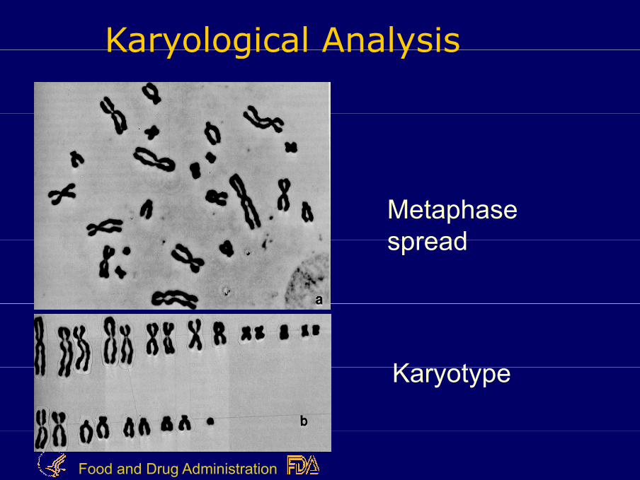
Karyological Analysis Karyological Analysis
Metaphase spreadspread
K tKaryotype
Food and Drug Administration

DNA fingerprinting analysisDNA fingerprinting analysis
A A A B C D D E E E
Food and Drug Administration
Individual

Isoenzyme analysisIsoenzyme analysis
Food and Drug Administration

Characterization Testing of Cell Banks and EPC (CAL) :Banks and EPC (CAL) :
Genetic Stability
Genetic stability (i.e., determine gene copy #; sequencing of the insert of the MCB and EPC) typically performed during Phase 3 once the process is defined.
C ll d t th i d t t bCells used to synthesize products must be genetically stable enough to produce the
t d t i th h t thexpected protein throughout the fermentation period.
Food and Drug Administration

Characterization Testing of Cell Banks and EPC (CAL): Genetic Stability
Molecular studies required to show that:Molecular studies required to show that:Correct sequence made and incorporated into host cellStructure and copy # maintained to end of productionStructure and copy # maintained to end of productionStability of the expression system – in the EPC and at least once in MCBonce in MCB- Gene copy # (QPCR)
Deletions/Insertions (Southern blotting)- Deletions/Insertions (Southern blotting)- Protein produced – can be analyzed at the mRNA level (mRNA sequencing)( seque c g)
- Number of integration sites (FISH/Southern)- mRNA transcript size distribution (Northern blotting)
Food and Drug Administration
- mRNA transcript size distribution (Northern blotting)

Lot Release Testing: Unprocessed BulLot Release Testing: Unprocessed BulTESTING ASSAYMicrobial - B&F and Sterility
- MycoplasmaMycoplasma
Adventitious Viruses - In Vitro Virus Assay- MMV assay (infectivity or PCR) for CHO cells
Viral Quantitation TEM particle count (required for determining(required for determining average retroviral load; links with VC studies) –
Food and Drug Administration
)three lots tested

Lot Release Testing Purified Bulkg
TESTING ASSAYTESTING ASSAYMicrobial - B&F and Sterility
Oth C t i t H t C ll DNA H tOther Contaminants Host Cell DNA, Host Cell Protein (Residual t ti )testing)
Analytical - PurityCharacterization - Potency
- StabilityFood and Drug Administration
Stability

Final Filled Product Testingg
TESTING ASSAYMicrobial B&F and SterilityMicrobial - B&F and Sterility
Safety - Endotoxin: RabbitSafety Endotoxin: Rabbit Pyrogen or LAL
General Safety- General SafetyAnalytical Ch t i ti
- PurityCharacterization - Potency
- Stability
Food and Drug Administration
y

ConclusionConclusion
Sponsors need to review carefully their testing plan to assure it meetstesting plan to assure it meets regulatory expectations..
PreIND meeting is a good time for thatPreIND meeting is a good time for that discussion.
Food and Drug Administration

Food and Drug Administration