safety and efficacy of 68ga-dotatoc positron emission
TRANSCRIPT

IND # 114,398 for 68Ga-DOTATOC O’Dorisio, M Sue
1
Safety and Efficacy of 68Ga-DOTATOC Positron Emission Tomography (PET) for Diagnosis, Staging, and Measurement of Response to
Treatment in Somatostatin Receptor Positive Tumors
Principal Investigator: M. Sue O’Dorisio, MD, PhD
Co-Investigators: David Bushnell, MD Yusuf Menda, MD
Michael Schultz, PhD, Laura Ponto, PhD
John Sunderland, PhD Thomas O’Dorisio, MD
Michael Graham, PhD, MD
Document type: PROTOCOL FOR IND# 114,398 Version number: 7
Date: November 13, 2014
IND # 114,398 for 68Ga-DOTATOC O’Dorisio, M Sue
2
Summary of Changes Version 7 11/13/2014 Page Protocol Changes: 1 Changed Version number and date of protocol. 3 Revised page numbers. 10 Clarified that one nuclear medicine physician will report locations of suspected
tumor deposits, instead of two physicians. 11 Added the number “1” in front of “Adverse Event” for consistency. 13 Deleted DSM information as there is no medical monitor for this study.
Changed risk level of study from 3 to 4. Added information about DSMC audits 14 Removed hyperlink to IRB guidelines for AE reporting. 14 Added Expedited AE Reporting table. 27-31 Added the Data and Safety Monitoring Plan as Appendix 1

IND # 114,398 for 68Ga-DOTATOC O’Dorisio, M Sue
3
Safety and Efficacy of
68Ga-DOTATOC Positron Emission Tomography (PET) for Diagnosis, Staging, and Measurement of Response to Treatment in Somatostatin
Receptor Positive Tumors
TABLE OF CONTENTS 1. Introduction…………………………………………………………. 4 2. General Investigational Plan……………………………………… 6 3. Clinical Protocol……………………………………………………. 7 4. Chemistry, Manufacturing and Control………………………….. 17 5. Labeling…………………………………………………………….. 19 6. Pharmacology and Toxicology…………………………………… 19 7. Previous Human Experience …………………………………….. 20 8. References………………………………………………………..... 23 Appendices:
I. Menda et al., J Nuc Med article on 90Y-DOTATOC therapy in children II. Bushnell et al., J Clin Oncol article on 90Y-DOTATOC therapy III. Sample Case Report Form IV. Materials Specification for 0.1 N HCl V. Receiving Documentation for 0.1 N HCl VI. RDRC approved 68Ga-DOTATOC Biodistribution & Reproducibility
protocol VII. IRB approval letter for RDRC protocol using 68Ga-DOTATOC VIII. Email communications with FDA in regards to Amendment to IND #

IND # 114,398 for 68Ga-DOTATOC O’Dorisio, M Sue
4
Project Goal Demonstrate the safety and efficacy of [68Ga]-DOTA-tyr3-Octreotide ([68Ga]-DOTATOC) as an accurate imaging technique for diagnosis, staging, and monitoring of response to
treatment in patients with somatostatin receptor expressing tumors.
1. Introduction: Somatostatin Receptor Positive Tumors Neuroendocrine tumors are solid malignant tumors that arise from dispersed neuroendocrine cells found throughout the body. Gastroenteropancreatic neuroendocrine tumors (NETs) can be divided into two groups: Carcinoid tumors that may arise from the lungs, stomach, small bowel or colon and pancreatic neuroendocrine tumors (also known as pancreatic islet cell tumors). The clinical behavior of NETs is extremely variable; some may cause hormone hypersecretion and others may not, the majority of them are slow-growing tumors (well-differentiated NETs), whereas some NETs are highly aggressive (poorly differentiated NETs). The incidence of NETs is increasing, from 1.1/100,000 per year in 1973 to 5.3/100,000 per year in 20041. Among NETs, 25% have distant metastases and 25% have regional involvement at the time of initial diagnosis1. Other tumors that express high levels of somatostatin receptors include neuroblastoma, medulloblastoma, and Ewing’s sarcoma2-4. The radiological detection and staging of these tumors is challenging and requires a multimodality approach. Somatostatin receptor imaging with In-111 Pentetreotide (OctreoScan) and multiphase CT are the most commonly used modalities although the use of endoscopic ultrasound and MRI is also increasing. Surgery is the only curative option for NETs. However, curative surgery in malignant NET is possible in less than 30% of patients with recurrence identified in the majority of patients as late as 15 years after initial surgery. Treatment with somatostatin analogs, which include the short acting subcutaneous and long acting release (LAR) octreotide, are effective in stabilizing NETs and have been recently demonstrated to prolong the time to progression of disease5. Chemotherapy is generally not effective in low grade NETs, but it may be helpful in high grade and pancreatic NETs. On the other hand, neuroblastoma, medulloblastoma, and Ewing’s sarcoma are initially responsive to chemotherapy, but relapses are common and salvage therapies are not very effective, resulting in <30% overall survival at 5 years6-8.
Somatostatin Receptor Targeted Imaging and Therapy Tumors that express somatostatin receptors can be targeted with radiolabeled somatostatin analogues for imaging and treatment (Figure 1). Somatostatin receptor gamma camera imaging with
Figure 1: Conceptual representation of targeted molecular imaging and therapy of cancerous tumors. A cell surface receptor is identified that is upregulated in cancerous tumor cell relative to normal surrounding tissue and organs. A peptide vector is developed that binds with high affinity and specificity to the molecular target. A chelator is added to the targeting vector via a molecular linker. The chelator can be used to couple a positron emitter as the reporter signal for PET, or with a beta emitting radionuclide (e.g., 90Y) for molecular therapy. To be effective, the high binding affinity of the vector for the target antigen and the stable (high specific activity radiometallation must be maintained after placement of the chemical linker.

IND # 114,398 for 68Ga-DOTATOC O’Dorisio, M Sue
5
In-111 DTPA-octreotide (OctreoScan) targeting somatostatin receptor 2 (sstr2), is used routinely for imaging of neuroendocrine tumors with a detection rate >90% for well-differentiated carcinoid tumors and majority of pancreatic NETs, but only a 50% detection rate for insulinomas, which may show a weaker expression of sstr29. Given the clinical efficacy of this radiolabeled peptide as a diagnostic agent, studies to test if therapeutic radiation could be targeted to tumors in a similar manner was a logical next step. Attempts to utilize In-111 DTPA Octreotide as a therapeutic agent have been minimally effective due to short range of auger electrons utilized in this therapy. The efficacy of this treatment was improved with the development of somatostatin analogues labeled with beta emitting radioisotopes. Further studies have identified DOTA as a superior chelator compared to DTPA, increasing the stability and receptor targeting of somatostatin analogues10. There is now a large clinical experience with Yttrium-90 DOTA-tyr3-Octreotide peptide radioreceptor therapy (PRRT) in Europe, primarily in adults with neuroendocrine tumors 11. An international Phase II clinical trial then followed and included several trial sites in the United States, notably the University of Iowa, where we entered 40 subjects 12. With its low toxicity profile, the significant improvement in symptoms and quality of life and the lack of effective alternative therapies, PRRT has been suggested as possible first-line therapy in adult patients with gastroenteropancreatic neuroendocrine tumor. Recent data have also demonstrated a significant survival benefit with PRRT compared to historical controls in this population. We have also now conducted a Phase I trial of 90Y-DOTA-tyr3-Octreotide in children and young adults at the University of Iowa, which also shows promise of efficacy of this treatment in pediatric patient population 13. We now propose a new imaging agent for use in diagnosis and therapy of Somatostatin receptor positive tumors as shown in Figure 2.
Figure 2 Chemical identity of [68Ga]DOTATOC IND # 114,398 with current IND #61,907 compound [90Y]DOTATOC
Somatostatin Receptor PET Imaging with Ga-68 DOTA0-Tyr3-octreotide More recently, positron emission tomography (PET) radiopharmaceuticals have been developed that can be labeled with Gallium-68 (Ga-68). Gallium-68 is a generator product with a half-life of 68 min (compared to 67 hours for In-111 in OctreoScan). The parent nuclide of Ga-68 is Germanium-68, which has a half-life of 270.8 days. Ga-68 decays by 89% through positron emission and 11% by electron capture. Its parent, A number of Ga-68 DOTA-conjugated peptides have been introduced, including Ga-68 DOTA0-Tyr3]octreotide (Ga-68 DOTATOC), Ga-68 DOTA0-1NaI3-octreotide (Ga-68 DOTANOC) and [Ga-68 DOTA0-Tyr3]octreotate (Ga-68 DOTATOC). All of these radiolabeled peptides bind to sstr2, although DOTANOC also binds to sstr 3 and sstr 5, and DOTATOC to sstr514. The primary advantage of Ga-68 based somatostatin receptor PET imaging over OctreoScan SPECT is the higher imaging resolution and accurate quantitation of uptake due to robust

IND # 114,398 for 68Ga-DOTATOC O’Dorisio, M Sue
6
attenuation correction. The improved resolution and quantitation of uptake obtained with Ga-68 DOTATOC PET should provide a more accurate assessment of somatostatin receptor density, which will lead to a more accurate prediction of treatment response to somatostatin analogues. A recent study from Europe comparing Ga-68 DOTATOC with Octreoscan found Ga-68 DOTATOC to be superior in detection of skeletal and pulmonary involvement of neuroendocrine tumors15. 2. Rationale and overall study design Rationale: 68Ga-DOTATOC positron emission tomography (PET) scanning and 90Y-DOTATOC peptide receptor radionuclide therapy (PRRT) are readily available in Europe, but neither radiopharmaceutical is approved for use in the United States. IND #61,907 is currently active under the above named investigators for 90Y-DOTATOC PRRT in somatostatin receptor positive tumors. We have conducted a single institution Phase I trial of 90Y-DOTATOC therapy in children and young adults (Appendix I) and we have participated in a Phase II trial of 90Y-DOTATOC PRRT in adults (also in Appendix II).
The purpose of this amendment is to test the efficacy of 68Gallium-DOTATOC in diagnosis, staging, and determination of response to90Y-DOTATOC PRRT in children and adults with known or suspected somatostatin receptor positive tumors, including, but not limited to neuroendocrine tumors, neuroblastoma, and medulloblastoma. 68Ga-DOTATOC PET would replace 111In-DTPA-Octreotide single photon emission tomography (SPECT) imaging. Whereas, Octreoscan uses a 222 MBq imaging dose of Indium (2.8 day half life) resulting in an effective dose equivalent (HE) equal to 2.61 rads, [68Ga]DOTATOC (68 min half life) uses 185 MBq with an effective dose equivalent of 0.46 rads. In addition, [68Ga]DOTATOC PET/CT can be completed within 2 hours compared to an Octreoscan which requires 3 visits over 24 hours, making [68Ga]DOTATOC a much more convenient imaging choice for patients. The data obtained in this Ga-68 DOTATOC PET study will be used to support the use of 68Ga-DOTATOC PET for diagnosis and staging in patients with suspected or proven somatostatin receptor positive tumors.
Overall Study Goal and Study Design Project Goal This study is planned to demonstrate the safety and efficacy of [68Ga]-DOTA-tyr3-Octreotide ([68Ga]-DOTATOC) as an accurate imaging technique for diagnosis, staging, and monitoring of response to treatment in patients with Somatostatin receptor expressing tumors. Project Design 68Ga-DOTATOC Positron Emission Tomography (PET) for Diagnosis, Staging, and Measurement of Response to Treatment in Somatostatin Receptor Positive Tumors is a prospective, Phase 1-2, single center, open-label study in a total of 112 subjects with known or suspected somatostatin receptor positive tumor. Eligible participants will undergo baseline assessments at enrollment. Study participants will receive 68Ga-DOTATOC and undergo a PET/CT imaging study with an option to receive a second 68Ga-DOTATOC PET/CT if they begin a new treatment (surgery, hepatic embolization, Sandostatin LAR, chemotherapy, targeted biological therapy, or peptide receptor radiotherapy) within 30 days of the first scan. The second scan will be performed at a time recommended by the treating physician as optimal interval to observe results from treatment. Participant Enrollment Participants will be recruited from the University of Iowa Comprehensive Cancer Center Clinics and the University of Iowa Children’s Hospital. Participants who will be approached regarding the study are those individuals who are being seen for known or suspected malignancies.

IND # 114,398 for 68Ga-DOTATOC O’Dorisio, M Sue
7
Schema
Informed Consent
Review pathology report, Octreoscan (if previously performed), CT or MRI, and laboratory tests
Physical exam with vital signs Blood tests (CBC, differential, AST, ALT, bilirubin, BUN, creatinine
Pregnancy test if indicated Injection of 68Ga-DOTATOC
PET/CT scan vital signs
Phone call follow-up 18-36 hrs after PET/CT
Physical exam and blood tests 7-28 days after PET/CT scan
Selected patients will undergo second scan following therapy as recommended by treating physician
using above schedule.
2. Clinical Protocol Inclusion Criteria:
• Signed informed consent. • Age ≥ 2 years. • Known or suspected somatostatin receptor positive tumor such as carcinoid;
neuroendocrine tumor; neuroblastoma; medulloblastoma; pheochromocytoma. Supporting evidence may include MRI, CT, biochemical markers, and or pathology report.
• Karnofsky performance status or Lansky Play Scale status of ≥ 50 (or ECOG/WHO equivalent).
• Subject is male; or is a female who is either pre-menarchal, surgically sterile (has had a documented bilateral oophorectomy and/or documented hysterectomy), postmenopausal (> 1 years without menses), non-lactating, or of childbearing potential for whom a serum pregnancy test (with the results known prior to investigational product administration) is negative. A negative serum pregnancy test will be required for all female subjects with child bearing potential. If a false pregnancy test is suspected, e.g., perimenopausal condition, an obstetrician will be consulted to determine if she is/is capable of becoming pregnant.
Exclusion Criteria
• Subject weighs more than 400 pounds (Subjects who weigh more than 400 pounds will not be able to fit inside the imaging machines).
• Inability to lie still for the entire imaging time (e.g. cough, severe arthritis, etc.) • Inability to complete the needed investigational and standard-of-care imaging examinations
due to other reasons (severe claustrophobia, radiation phobia, etc.) • Does the subject have any additional medical condition, serious intercurrent illness, or other
extenuating circumstance that, in the opinion of the Investigator, may significantly interfere with study compliance?

IND # 114,398 for 68Ga-DOTATOC O’Dorisio, M Sue
8
Procedures Informed Consent Subjects meeting eligibility criteria will be consented. After consent, subjects are considered actively enrolled in the study and a study log will be maintained. Record of consent will be scanned into the patient’s electronic chart. Physical Examination and Medical History Physical examination will be performed to ensure suitability according to the inclusion and exclusion criteria at screening and baseline and to document the health status and toxicity at the time points specified in the Schedule of Events (Table 1).
Table 1. Schedule of Study Events Visit
1a Scan 1b
1 Day (18-36 hours)
1 wk (7-28 days)
Scan 2
1 day 1 wk (7-28 days)
Informed consent X Medical history X X Physical examination Xc X X X Concomitant meds X X X X X Blood pressure, temp, respiratory rate, heart rate, weight
X X X X X
CBC, differential, AST, ALT, bilirubin, BUN, creatinine
X X X X
Pregnancy testd X X X 68Ga-DOTATOC PET/CT X X Adverse events X X X X X X X a To facilitate scheduling, some screening procedures may occur up to 1 month prior to the first dose of study drug. b Baseline procedures must be performed no more than 7 days prior to study drug. c Visit 1 physical exam to include height and performance status. d A urine pregnancy test may be done in PET if Visit 1 and Scan 1 are on separate days. Screening results must be available and baseline procedures performed before the first dose of study drug is administered. To avoid repeat tests, some screening values may be counted as baseline values if obtained within specified time window.
The physical examination comprises measurement of height and body weight, vital signs of temperature, pulse, respiratory rate, and blood pressure. A medical history will be recorded at screening and prior to second scan. The medical history will elicit information concerning existing medical conditions, major illnesses, and related surgical procedures. Any prescribed or over-the counter medications that the subject received within the past 30 days should be recorded on the case report form (CRF Appendix III). Pregnancy Test Female subjects of childbearing potential will have a pregnancy test (serum) before exposure to research-related radiation. A woman who is pregnant may not participate in this study and must tell us if she may have become pregnant during the previous 14 days because the pregnancy test is
IND # 114,398 for 68Ga-DOTATOC O’Dorisio, M Sue
9
unreliable during this time. If a false pregnancy test is suspected, e.g., perimenopausal condition, an obstetrician will be consulted to determine if she is/is capable of becoming pregnant.
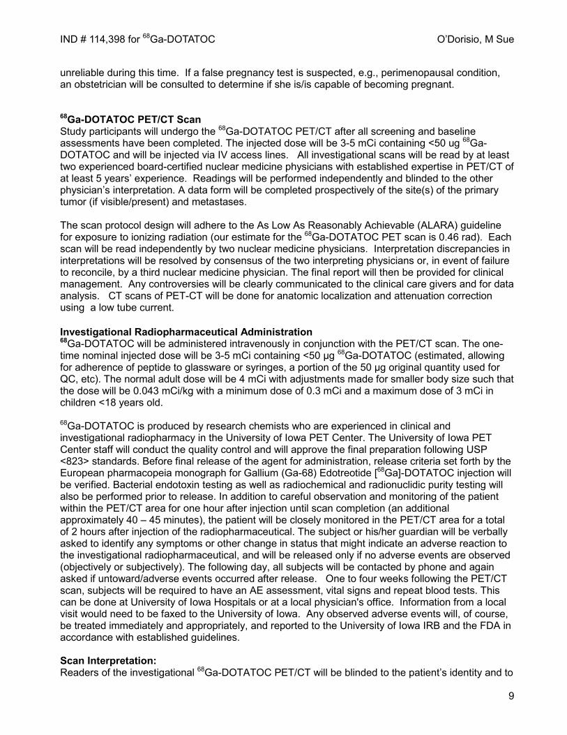
68Ga-DOTATOC PET/CT Scan Study participants will undergo the 68Ga-DOTATOC PET/CT after all screening and baseline assessments have been completed. The injected dose will be 3-5 mCi containing <50 ug 68Ga-DOTATOC and will be injected via IV access lines. All investigational scans will be read by at least two experienced board-certified nuclear medicine physicians with established expertise in PET/CT of at least 5 years’ experience. Readings will be performed independently and blinded to the other physician’s interpretation. A data form will be completed prospectively of the site(s) of the primary tumor (if visible/present) and metastases. The scan protocol design will adhere to the As Low As Reasonably Achievable (ALARA) guideline for exposure to ionizing radiation (our estimate for the 68Ga-DOTATOC PET scan is 0.46 rad). Each scan will be read independently by two nuclear medicine physicians. Interpretation discrepancies in interpretations will be resolved by consensus of the two interpreting physicians or, in event of failure to reconcile, by a third nuclear medicine physician. The final report will then be provided for clinical management. Any controversies will be clearly communicated to the clinical care givers and for data analysis. CT scans of PET-CT will be done for anatomic localization and attenuation correction using a low tube current. Investigational Radiopharmaceutical Administration 68Ga-DOTATOC will be administered intravenously in conjunction with the PET/CT scan. The one-time nominal injected dose will be 3-5 mCi containing <50 μg 68Ga-DOTATOC (estimated, allowing for adherence of peptide to glassware or syringes, a portion of the 50 μg original quantity used for QC, etc). The normal adult dose will be 4 mCi with adjustments made for smaller body size such that the dose will be 0.043 mCi/kg with a minimum dose of 0.3 mCi and a maximum dose of 3 mCi in children <18 years old.
68Ga-DOTATOC is produced by research chemists who are experienced in clinical and investigational radiopharmacy in the University of Iowa PET Center. The University of Iowa PET Center staff will conduct the quality control and will approve the final preparation following USP <823> standards. Before final release of the agent for administration, release criteria set forth by the European pharmacopeia monograph for Gallium (Ga-68) Edotreotide [68Ga]-DOTATOC injection will be verified. Bacterial endotoxin testing as well as radiochemical and radionuclidic purity testing will also be performed prior to release. In addition to careful observation and monitoring of the patient within the PET/CT area for one hour after injection until scan completion (an additional approximately 40 – 45 minutes), the patient will be closely monitored in the PET/CT area for a total of 2 hours after injection of the radiopharmaceutical. The subject or his/her guardian will be verbally asked to identify any symptoms or other change in status that might indicate an adverse reaction to the investigational radiopharmaceutical, and will be released only if no adverse events are observed (objectively or subjectively). The following day, all subjects will be contacted by phone and again asked if untoward/adverse events occurred after release. One to four weeks following the PET/CT scan, subjects will be required to have an AE assessment, vital signs and repeat blood tests. This can be done at University of Iowa Hospitals or at a local physician's office. Information from a local visit would need to be faxed to the University of Iowa. Any observed adverse events will, of course, be treated immediately and appropriately, and reported to the University of Iowa IRB and the FDA in accordance with established guidelines. Scan Interpretation: Readers of the investigational 68Ga-DOTATOC PET/CT will be blinded to the patient’s identity and to

IND # 114,398 for 68Ga-DOTATOC O’Dorisio, M Sue
10
any other clinical information prior to initial interpretation. A board certified nuclear medicine physician who is experienced and credentialed in interpretation of clinical PET/CT scan will independently report all locations of suspected tumor deposits without discussion with other healthcare providers or with correlation with other imaging. In addition to localizing all areas of tumor, quantification of size and uptake of up to 5 “target” lesions will be performed (no more than two in a given organ), with SUV and cross sectional measurements in long axis and short axis, in mm, recorded for each lesion visible on CT. All of these interpretations (the original and whether or not adjudication was needed, and the results of adjudication if performed) will be retained for statistical analysis. Readers will then be asked to review all other available imaging (see next paragraph) to see if there are sites of tumor seen on conventional imaging not seen with 68Ga-DOTATOC on the initial interpretation, and, if so, to determine if this impacted treatment decisions. These results will be recorded for statistical analysis. After the investigational 68Ga-DOTATOC PET/CT has been independently interpreted, comparison will be made with other, conventional imaging (CI), as available. CI used for comparison will include CT scans, MRI scans, 111In-octreotide scans, ultrasounds and x-ray plain films. Part of data analysis specific to imaging will include: Whe the r the re we re s ite s of tumor s e en with 68Ga-DOTATOC PET/CT not seen with CI, and, if so, these data will be recorded for statistical analysis Whe the r or not the 68Ga-DOTATOC PET/CT resulted in a change in stage relative to CI, and, if so, whether it would be up- vs. down- staging Whe the r or not the 68Ga-DOTATOC PET/CT scan result caused the clinical care givers to change treatment, e.g. change within modality (e.g. change chemotherapy, change planned surgery) or across modalities (e.g. add chemotherapy or radiation therapy to surgery, etc.). Risks Risks of Study Drug Research radiation dose estimate: Our calculations estimate that the effective whole-body dose of the 68Ga-DOTATOC PET/CT is approximately 1.6 rem, less than the radiation dose from a conventional 18F-FDG PET/CT scan of about 2.4 rem, assuming the standard use of a “low-dose” CT for anatomic localization and attenuation correction, as described in our proposed imaging protocol. The U.S. national annual background dose for humans is approximately 0.36 rem. The maximum annual permissible whole body radiation dose to a career radiation worker (e.g., a radiologist, nuclear medicine technologist, etc.) is 5.0 rem. Thus, the one-time dose to the patient for the investigational PET/CT will be approximately one-third of the annual permissible dose to a career radiation worker and about 5 years’ worth of background radiation in the US. Given the average age of the patients in this proposed investigation (about 60 years of age), the increased risk to the patient, who will have a known, current life-threatening malignancy, for a second, radiation-induced malignancy from this investigational radiation exposure is extremely low (estimated < 1%). Given the risk of death from either delay in diagnosis or under-treatment of these known malignancies, we feel the added radiation burden to the patient is well within the patient’s best interest if 68Ga-DOTATOC PET/CT imaging adds value. Risks of Study Procedures Risks of Investigational PET/CT: An adverse reaction to the investigational radiopharmaceutical is possible though not reported. Additional risks include radiation exposure from the investigational PET/CT scan (discussed above), dosage infiltration, risks from IV access. blood tests, and the inconvenience of the investigational PET/CT scan. Children may require sedation or anesthesia to remain still during the PET scanning procedure. The risks of sedation include a reaction to the medication used for sedation or anesthesia. If anesthesia is required, there is a risk of injury to the throat or airways from insertion of the breathing tube.
IND # 114,398 for 68Ga-DOTATOC O’Dorisio, M Sue
11
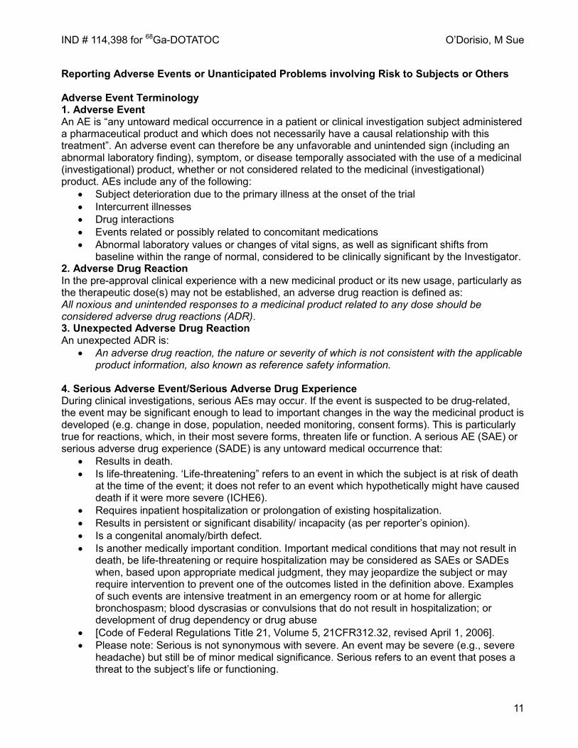
Reporting Adverse Events or Unanticipated Problems involving Risk to Subjects or Others Adverse Event Terminology 1. Adverse Event An AE is “any untoward medical occurrence in a patient or clinical investigation subject administered a pharmaceutical product and which does not necessarily have a causal relationship with this treatment”. An adverse event can therefore be any unfavorable and unintended sign (including an abnormal laboratory finding), symptom, or disease temporally associated with the use of a medicinal (investigational) product, whether or not considered related to the medicinal (investigational) product. AEs include any of the following:
• Subject deterioration due to the primary illness at the onset of the trial • Intercurrent illnesses • Drug interactions • Events related or possibly related to concomitant medications • Abnormal laboratory values or changes of vital signs, as well as significant shifts from
baseline within the range of normal, considered to be clinically significant by the Investigator. 2. Adverse Drug Reaction In the pre-approval clinical experience with a new medicinal product or its new usage, particularly as the therapeutic dose(s) may not be established, an adverse drug reaction is defined as: All noxious and unintended responses to a medicinal product related to any dose should be considered adverse drug reactions (ADR). 3. Unexpected Adverse Drug Reaction An unexpected ADR is:
• An adverse drug reaction, the nature or severity of which is not consistent with the applicable product information, also known as reference safety information.
4. Serious Adverse Event/Serious Adverse Drug Experience During clinical investigations, serious AEs may occur. If the event is suspected to be drug-related, the event may be significant enough to lead to important changes in the way the medicinal product is developed (e.g. change in dose, population, needed monitoring, consent forms). This is particularly true for reactions, which, in their most severe forms, threaten life or function. A serious AE (SAE) or serious adverse drug experience (SADE) is any untoward medical occurrence that:
• Results in death. • Is life-threatening. ‘Life-threatening” refers to an event in which the subject is at risk of death
at the time of the event; it does not refer to an event which hypothetically might have caused death if it were more severe (ICHE6).
• Requires inpatient hospitalization or prolongation of existing hospitalization. • Results in persistent or significant disability/ incapacity (as per reporter’s opinion). • Is a congenital anomaly/birth defect. • Is another medically important condition. Important medical conditions that may not result in
death, be life-threatening or require hospitalization may be considered as SAEs or SADEs when, based upon appropriate medical judgment, they may jeopardize the subject or may require intervention to prevent one of the outcomes listed in the definition above. Examples of such events are intensive treatment in an emergency room or at home for allergic bronchospasm; blood dyscrasias or convulsions that do not result in hospitalization; or development of drug dependency or drug abuse
• [Code of Federal Regulations Title 21, Volume 5, 21CFR312.32, revised April 1, 2006]. • Please note: Serious is not synonymous with severe. An event may be severe (e.g., severe
headache) but still be of minor medical significance. Serious refers to an event that poses a threat to the subject’s life or functioning.
IND # 114,398 for 68Ga-DOTATOC O’Dorisio, M Sue
12
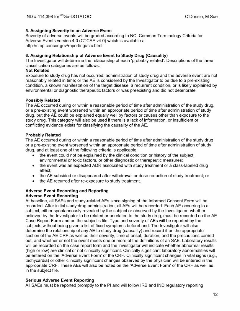
5. Assigning Severity to an Adverse Event Severity of adverse events will be graded according to NCI Common Terminology Criteria for Adverse Events version 4.0 (CTCAE v4.0) which is available at http://ctep.cancer.gov/reporting//ctc.html. 6. Assigning Relationship of Adverse Event to Study Drug (Causality) The Investigator will determine the relationship of each ‘probably related’. Descriptions of the three classification categories are as follows: Not Related Exposure to study drug has not occurred; administration of study drug and the adverse event are not reasonably related in time; or the AE is considered by the Investigator to be due to a pre-existing condition, a known manifestation of the target disease, a recurrent condition, or is likely explained by environmental or diagnostic therapeutic factors or was preexisting and did not deteriorate. Possibly Related The AE occurred during or within a reasonable period of time after administration of the study drug, or a pre-existing event worsened within an appropriate period of time after administration of study drug, but the AE could be explained equally well by factors or causes other than exposure to the study drug. This category will also be used if there is a lack of information, or insufficient or conflicting evidence exists for classifying the causality of the AE. Probably Related The AE occurred during or within a reasonable period of time after administration of the study drug or a pre-existing event worsened within an appropriate period of time after administration of study drug, and at least one of the following criteria is applicable:
• the event could not be explained by the clinical condition or history of the subject, environmental or toxic factors, or other diagnostic or therapeutic measures;
• the event was an expected ADR associated with study treatment or a class-labeled drug effect;
• the AE subsided or disappeared after withdrawal or dose reduction of study treatment; or • the AE recurred after re-exposure to study treatment.
Adverse Event Recording and Reporting Adverse Event Recording At baseline, all SAEs and study-related AEs since signing of the Informed Consent Form will be recorded. After initial study drug administration, all AEs will be recorded. Each AE occurring to a subject, either spontaneously revealed by the subject or observed by the Investigator, whether believed by the Investigator to be related or unrelated to the study drug, must be recorded on the AE Case Report Form and on the subject’s file. Type and severity of AEs will be reported by the subjects without being given a list of fixed symptoms beforehand. The Investigator will also determine the relationship of any AE to study drug (causality) and record it on the appropriate section of the AE CRF as well as their severity, time of onset, duration, and the precautions carried out, and whether or not the event meets one or more of the definitions of an SAE. Laboratory results will be recorded on the case report form and the investigator will indicate whether abnormal results (high or low) are clinical or not clinically significant. Clinically significant laboratory abnormalities will be entered on the ‘Adverse Event Form’ of the CRF. Clinically significant changes in vital signs (e.g., tachycardia) or other clinically significant changes observed by the physician will be entered in the appropriate CRF. These AEs will also be noted on the ‘Adverse Event Form’ of the CRF as well as in the subject file. Serious Adverse Event Reporting All SAEs must be reported promptly to the PI and will follow IRB and IND regulatory reporting
IND # 114,398 for 68Ga-DOTATOC O’Dorisio, M Sue
13
requirements. SAEs should be followed to resolution, even if this is after the study reporting period. Data and Safety Monitoring Assessment of Risk Level: This investigation involves risk-level 4 procedures, in a population that is at risk of death> 5% or grade 4 – 5 SAE >15%. Purpose: The Data Safety Monitoring Plan for this study (see Appendix 1) is written to ensure the safety of subjects and to verify the validity and integrity of the data. DSMC Audits: An independent Study Monitor or the DSMC Chair (or designee), will review study data (provided by the PI and CRSO) and communicate with the PI at least quarterly. A copy of this communication will be forwarded to the DSMC and PRMC Chairs. This communication may coincide with the CRSO’s quarterly audit when possible. If an audit has not been conducted—due to lack of research activity--the quarterly summary letter will occur as scheduled.
- Subject accrual data will be obtained from OnCore on an ongoing basis by the CTRM during accrual review for the PRMC.
- The research coordinator or data manager for the study will register new subjects in OnCore - Adverse events will be reported to the CRSO as defined in the DSMP for the study - Serious adverse events will be communicated to the CRSO and recorded in OnCore. - The CRSO will contact the research team every three months to coordinate an audit of the
trial's research records. Records for a minimum of 25% of subjects will be audited. OnCore auditing functionality will be used to conduct, evaluate, and report on each audit. Audits will:
o verify eligibility of patients accrued to the study, o check for the presence of a signed informed consent, o determine compliance with protocol’s study plan, o determine whether SAEs are being appropriately reported to internal and external
regulatory agencies, o compare accuracy of data in the research record with the primary source documents, o review investigational drug processing and documentation, o assess cumulative AE/SAE reports for trends and compare to study stopping rules.
Assessing Toxicity: Toxicity will be graded according to NCI’s Common Toxicity Criteria (CTCAE v4). The principal investigator will responsible for determining the attribution of toxicity as it is related to the investigational drug. All grades of toxicity will be noted and reported appropriately. Adverse Event Reporting: An adverse event (AE) is defined in the CTEP, NCI Guidelines [2005] as “any unfavorable and unintended sign (including an abnormal laboratory finding), symptom or disease temporally associated with the use of a medical treatment or procedure (attribution of unrelated, unlikely, possible, probably or definite).”
IND # 114,398 for 68Ga-DOTATOC O’Dorisio, M Sue
14
All adverse events associated with the experimental procedures will be reported to the Data Safety Monitoring Committee (DSMC) via the Clinical Research Safety Officer (CRSO), as well as the appropriate federal regulatory authority, following established guidelines. Reports to DSMC will be sent to the CRSO, entered into OnCore and communicated to the DSMC in a timely manner. Only Grade 4 and 5 events that are unexpected, with at least possible attribution to 68Ga-DOTATOC PET, will require an expedited report. The investigator will continue to follow or obtain documentation until the resolution of such an event. Adverse events may also require reporting to the IRB per HSO-established guidelines. The clinical research coordinator is responsible for collecting and recording all clinical data. As these results are collected, all toxicities and adverse events will be identified and reported to the principal investigator. The principal investigator will determine relationship of the event to the study drug and decide course of action for the study participant. As part of the ongoing monitoring of this study, the PI will maintain an AE summary table to be reviewed during the quarterly audits.
Guidelines for Routine Adverse Event Reporting
Attribution Adverse Event Grade 1 Grade 2 Grade 3 Grade 4 Grade 5
Unrelated Not required Not required CRSO CRSO CRSO Unlikely Not required Not required CRSO CRSO CRSO Possible CRSO CRSO CRSO CRSO CRSO Probable CRSO CRSO CRSO CRSO CRSO Definite CRSO CRSO CRSO CRSO CRSO
Expedited Adverse Event Reporting Only Grade 4 and 5 events that are unexpected with at least “possible” attribution to the 68Ga-DOTATOC PET will require an expedited report to the CRSO/DSMC (24-hour notification followed by complete report within 5 calendar days). All expedited adverse event reports will be reviewed by the DSMC.
1. EXPEDITED ADVERSE EVENT REPORTING
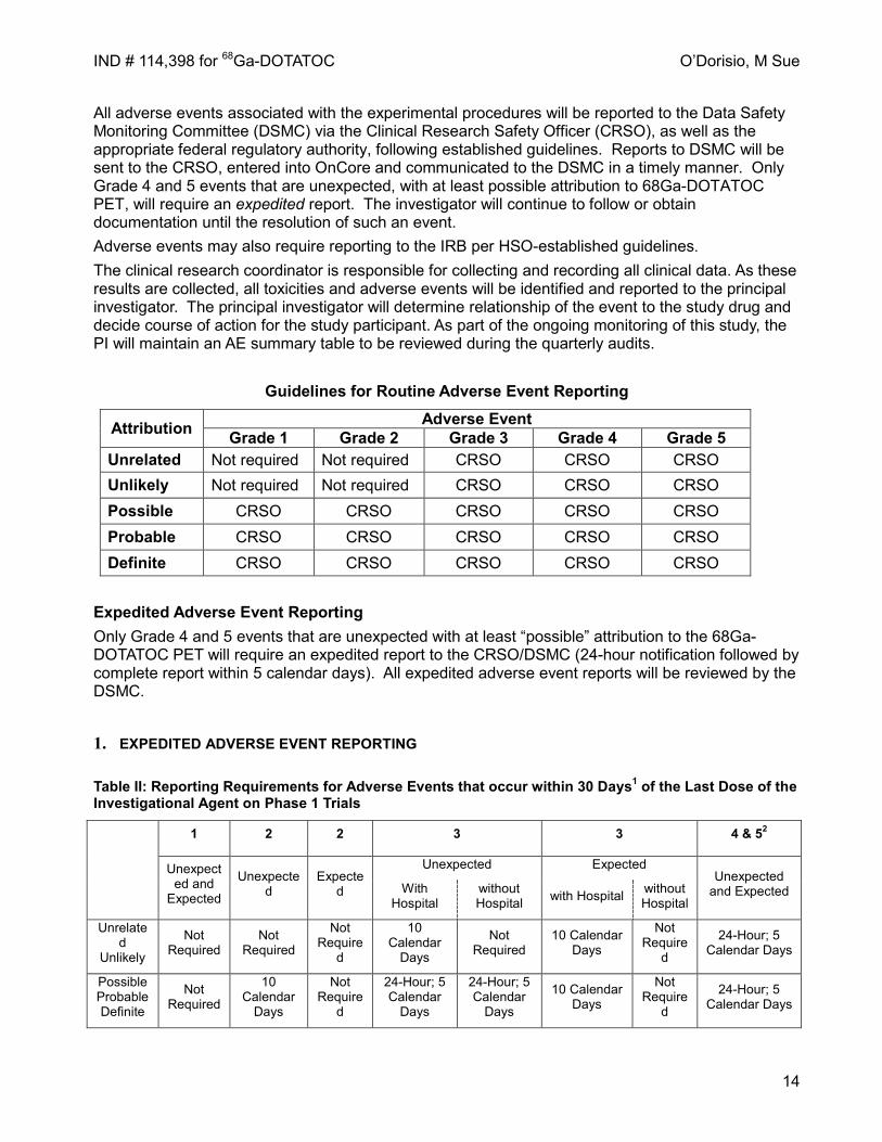
Table II: Reporting Requirements for Adverse Events that occur within 30 Days1 of the Last Dose of the Investigational Agent on Phase 1 Trials
1 2 2 3 3 4 & 52
Unexpected and
Expected
Unexpected
Expected
Unexpected Expected Unexpected
and Expected With Hospital
without Hospital with Hospital without
Hospital
Unrelated
Unlikely
Not Required
Not Required
Not Require
d
10 Calendar
Days
Not Required
10 Calendar Days
Not Require
d
24-Hour; 5 Calendar Days
Possible Probable Definite
Not Required
10 Calendar
Days
Not Require
d
24-Hour; 5 Calendar
Days
24-Hour; 5 Calendar
Days
10 Calendar Days
Not Require
d
24-Hour; 5 Calendar Days
IND # 114,398 for 68Ga-DOTATOC O’Dorisio, M Sue
15
1Adverse events with attribution of possible, probable, or definite that occur greater than 30 days after the last dose of treatment with an agent under a CTEP IND require reporting as follows: AdEERS 24-hour notification followed by complete report within 5 calendar days for:
Grade 3 unexpected events with hospitalization or prolongation of hospitalization Grade 4 unexpected events Grade 5 expected and unexpected events
2Although an AdEERS 24-hour notification is not required for death clearly related to progressive disease, a full report is required as outlined in the table. Study Withdrawal/Discontinuation This is a low risk imaging protocol; the amount of radioactive peptide is below physiological effects level and the radiation dose is below allowable levels. No adverse events are expected. Adverse events are recorded on the CRF at the following times: immediately following the PET/CT at the conclusion of a 2 hrs observation time from time of injection of radiopharmaceutical during phone call 18-24 hrs after PET/CT one to four weeks following PET/CT at outpatient visit with physical exam and labs Subjects will be entered in groups of three; if one of the first 3 subjects experiences a grade 3 or 4 adverse event, another 3 will be entered. If two of six subjects experience grade 3 or 4 adverse event, the study will be stopped. Otherwise an additional 14 subjects will be entered. If three of 20 subjects experience grade 3 or 4 adverse event, the study will be stopped. if < 3/20 subjects experience grade 3 or 4 adverse event, the radiopharmaceutical will be considered safe. At any time, subjects may withdraw from the study (i.e., withdraw consent to participate) at their own request. A subject’s participation in the study may be discontinued at any time at the discretion of the Investigator and in accordance with his/her clinical judgment. No disadvantage will arise for any subject who withdraws consent for participation at any time or who is withdrawn from the study by the Investigator. Reasons for discontinuation of study treatment will be recorded on the appropriate page of the CRF in any case and may include the following:
• Subject's request for withdrawal • Investigator's decision that discontinuation is in the best interest of the subject • Non-compliance with the regimen and timing that might result in dropping out from the study • Development of an intolerable AE due to study participation as determined by the
Investigator, subject, or both. • Development of an intercurrent illness, condition, or procedural complication, which would
interfere with the subject's continued participation. • Subject is lost to follow-up.
Subjects who discontinue treatment will be asked to complete a follow-up examination prior to leaving the study, if possible. Any subject who discontinues treatment for medical reasons, e.g. because of adverse events (AEs) or clinical laboratory abnormalities, should be followed up at medically appropriate intervals in order to evaluate the course and to ensure reversibility or stabilization of the abnormality or event. If a subject fails to return for a scheduled visit, a documented effort must be made to determine the reason. This information should be recorded in the study records. Statistical Considerations The distribution of patients will be an important element of statistical consideration. Several types of patients will be studied, including patients with:

IND # 114,398 for 68Ga-DOTATOC O’Dorisio, M Sue
16
• Primary pancreatic, small bowel, or lung neuroendocrine tumor • Metastatic pancreatic, small bowel or lung neuroendocrine tumor • Primary or metastatic neuroblastoma • Primary or metastatic medulloblastoma • Primary or metastatic pheochromocytoma
Given the distribution of patients seen at the University of Iowa Hospitals, an excellent distribution of patients will be enrolled. We predict that with a total of 153 subjects, each of the above types of diagnoses will be captured and accuracy of the 68Ga-DOTATOC PET/CT can be adequately studied. We will observe for adverse events related to the use of the 68Ga-DOTATOC radiopharmaceutical in accordance with NCI toxicity guidelines (described below) in order to provide the FDA and ourselves with important, traceable toxicity/safety data regarding the use of this investigational radiopharmaceutical. In those patients with available conventional imaging (Octreoscan, high-resolution, contrast CT or MRI), we will determine if the 68Ga-DOTATOC PET/CT scan alters care of patients and whether or not conventional imaging demonstrated negative or equivocal evidence of tumor size and location in the same patients. Many of our patients will be referred from outside centers with conventional imaging (CT, MRI and 111In-Octreotide scans) already accomplished. We will assess for an impact on treatment by determining if a given patient’s care is altered after the results of the 68Ga-DOTATOC PET/CT scan are available. For those patients who have available CT, MRI or Octreoscan, we will determine equivalence of conventional imaging and 68Ga-DOTATOC PET using percentage of concordance in tumor detection. We hypothesize that less than 80% agreement on their assessment would be inconclusive while more than 90% agreement on their assessment would favor equivalence. We define an indicator variable for [68Ga]DOTATOC PET/CT findings (+/-) and another indicator variable for Octreoscan findings (+/-). Positive findings are labeled (+) while negative findings are (-); with concordance [68Ga]DOTATOC PET/CT vs. Octreoscan occurring at the combinations (+/+) and (-/-), and discordance occurring at (+/-) or (-/+).
Since the same patient is measured under both tumor detection methods (conventional imaging and 68Ga-DOTATOC PET/CT), we base our sample size and power justification on the proportion of concordance and on the proportion of discordance. This outcome is of direct benefit to patient population. This analysis requires 112 patients to decide whether the proportion of agreement between the two assessment methods is less than or equal to 80% or greater than or equal to 90%. If 97 or more subjects result in the same decision on both [68Ga]DOTATOC PET/CT and conventional imaging, the null hypothesis that P <= 80% would be rejected in favor of the alternative. The expected error rate is alpha = 0.05 and the actual error rate of 0.047. If this number of subjects with concordant decision is 96 or less, the hypothesis that P >= 90% is rejected with a target error rate of 10% and an actual error rate of 0.092. The power of the test is 90%.
In case the null of P <= 80% is accepted, we have at least 20% chance of assessing discordance. This percentage (~22 subjects) is then used to search for false positives and false negatives. Pilot data suggests more subjects may fall within the category +/- (i.e. positive [68Ga]DOTATOC PET/CT, negative Octreoscan).
Note: There is a source of bias in this design. Subjects receive treatment based on positive findings from Octreoscan + diagnostic CT, the composite gold standard, regardless of their discordance with [68Ga]DOTATOC PET/CT. Bias came about because the cohort -/+ (i.e. [68Ga]DOTATOC PET/CT (-) and composite gold standard (+)) would receive treatment regardless of [68Ga]DOTATOC PET/CT finding. This bias should not create a commotion because the scenario -/+ would be rare based on pilot finding. The discordance +/- (i.e. [68Ga]DOTATOC PET/CT (+) and composite gold standard (-)) is more important at showing the advantage of [68Ga]DOTATOC PET/CT. This discordance would yield a population greater than 22 if concordance is less than 80% and a population <22 otherwise.
Binomial exact tests would be used on this discordant population after biopsy and after 6-month follow up to detect whether the proportion discovered after biopsy (or follow up) is significantly

IND # 114,398 for 68Ga-DOTATOC O’Dorisio, M Sue
17
smaller or comparable to the population claimed by [68Ga]DOTATOC PET/CT findings. This will also serve as false positive rate assessment if not confirmed at follow up. On a more general note for discordance/concordance study design, a sample of size 112 (or a set of 112 pairs of assessment) will achieve 99% power to detect an odds ratio of 12.33 using a two-sided McNemar test with a significance level 0.05. The odds ratio is equivalent to a difference between 2 paired proportions of 17% which occurs when the proportion of -/+ is 1.5% (i.e. [68Ga]DOTATOC PET/CT negative; composite gold standard positive) and the proportion in +/- is 18.5% (i.e. [68Ga]DOTATOC PET/CT positive; composite gold standard negative ), and the total discordance is 20%.
Power Table
Sample Size
Power Odds Ratio
-/+ Rate
+/-Rate Alpha 112 99% 12.33 1.5% 18.5% 0.05 112 96% 7.00 2.5% 17.5% 0.05 112 86% 4.71 3.5% 16.5% 0.05 112 79% 4.00 4.0% 16.0% 0.05
Privacy/Confidentiality Issues All routine medical data will be entered into the patient’s electronic medical record. These records are only available to those with direct clinical duties to the patient. For all other records, the Principal investigator will collect data and enter it into password protected computer in a locked office. Each patient will have a unique identifier number, with the key to the patient’s medical record number kept in a locked cabinet in the office. Only research associates or those individuals directly involved with the study will have access to data. Information is for research purposes only and when used for publication purposes, all participants will have their names concealed. Access to identified patient information will be limited to the investigators listed within the IRB application. De-identified information with HIPAA identifiers removed will be available to other investigators following IRB approval. Confidentiality and security will be maintained for the database. The database is stored behind a firewall (in addition to the institutional firewall) with the highest level of protection, i.e. the same level of protection as the on-line hospital information systems at the University of Iowa Hospitals . This means that users must logon to a web server that sits between the institutional firewall and the firewall to the database, and only this application server is allowed to query the database. Only users approved through our institutional review boards will be allowed access to patient identifiers. Other levels of authorization may exist for future approved users following IRB approval, e.g. access to de-identified data. Data is initially collected in the medical record for each individual study participant. The information will be extracted from the patient’s medical record and then transferred into the Case Report Form (CRF). The study data will be kept on site and in a securely locked room to protect patient confidentiality. The CRFs do not include personal identifiers for any participant. Numbers and initials are assigned for each participant and these become the identifying information for each study participant. A master list is kept separately that identifies which names go with which numbers and initials. Study personnel (PI and co-investigators) and government regulatory agencies have access to all research records as required by law. Others (such as law enforcement agencies) may have access to records as defined by law. Record Retention The PI must retain all study records by the applicable regulations in a secure and safe facility. The institution must consult with the PI before disposal of any study records, and must notify the PI of any change in the location, disposition or custody of the study files. The PI/institution must take

IND # 114,398 for 68Ga-DOTATOC O’Dorisio, M Sue
18
measures to prevent accidental or premature destruction of essential documents, that is, documents that individually and collectively permit evaluation of the conduct of a study and the quality of the data produced, including paper copies of study records (e.g., subject charts) as well as any original source documents that are electronic as required by applicable regulatory requirements. Records and documents pertaining to the conduct of this study, including Case Report Forms (Appendix III), source documents, consent forms, laboratory test results, and medication inventory records, must be retained by the investigator for at least 2 years following submission of a New Drug Application. No study records will be destroyed without prior authorization from the Principal Investigator. 4. Chemistry, Manufacturing, and Control The University of Iowa PET Center Radiopharmaceutical Laboratory maintains an environment that is adherent to USP <823>. Reagents used for the preparation of [68Ga]-DOTATOC are received and inspected per specification sheets (e.g., see 0.1 N HCl Specification Sheet, Appendix IV) and accepted or rejected. Accepted reagents are labeled with appropriate expiration dates, initialed, assigned identification numbers (e.g., see Receiving Document 0.1 N HCl Appendix V), and quarantined in specified locations in the radiopharmaceutical laboratory. The University of Iowa (UI) PET Center Radiopharmaceutical Laboratory is staffed with expert radiochemists that oversee the preparation of the radiopharmaceuticals and are experienced in the compounding of the proposed investigational radiopharmaceutical because of its ongoing use under Radioactive Drug Research Committee approval (Biodistribution and Reproducibility of Ga-68 DOTATOC Positron Emission Tomography in Patients with Somatostatin Receptor Positive Tumors: A Feasibility Study; University of Iowa IRB # 201012803). Because Ga-68 has a 68 minute half life, production of [Ga-68]DOTATOC requires the use of a Germanium-68 (Ge-68) to Ga-68 generator. The eluted Ga-68 is purified following elution using an ion-exchange column prior to introduction into the radiolabeling vessel with DOTATOC to remove zinc-68 (the decay product of Ga-68), which builds up in the generator with the decay of Ga-68. According to the [Ga-68]DOTATOC Batch Record, the generator is connected to the ModularLab PharmTracer module and eluted with 0.1 N HCl directly to an ion exchange column for purification of the Ga-68 from Zn-68 (daughter nuclide of Ga-68 decay). The system includes a disposable GMP-certified (sterile, pyrogen-free) “kit” that is snapped in place and manages (via software) the flow of reagents for purification, radiolabeling, and transfer of the final product to the product vial via sterilizing (0.22 μm filter). A new kit is used for each preparation and preparations are usually used for a single patient study. At the end of the process, the generator line is disconnected, capped, and the generator stands ready for connection to a new kit for a new preparation. The system then elutes Ga-68 with acetone/0.02 N HCl solution directly to a reaction vessel containing 40 micrograms of DOTATOC (obtained commercially) dissolved in 2 mL of sodium acetate buffer (pH 4). The solution is heated to 95 °C for 7 minutes at which time the heating element is turned off and the vessel is cooled with the addition of 2 mL isotonic saline for injection. Acetone is removed by distillation during the radiolabeling process and a test for residual is included in subsequent quality control prior to release. The solution is next passed over a disposable C-18 cartridge, which retains [Ga-68]DOTATOC, while allowing any remaining free Ga-68 to pass. The cartridge is rinsed with isotonic saline for injection to remove any residual free Ga-68, followed by elution of [Ga-68]DOTATOC with 1 mL 47.5% ethanol in isotonic saline for injection directly through a sterile 0.22 micron membrane filter into the final collection vial.

IND # 114,398 for 68Ga-DOTATOC O’Dorisio, M Sue
19
A 0.8 mL aliquot is then taken for quality control testing and the product line and vent filter are removed. Radiochemical yields of approximately 55% (uncorrected for decay) are routinely achieved using this process. The filter is retained for a QC pressure test to ensure filter integrity for sterilization. The production process can be conducted by a single person and requires approximately 1 hr to complete. Quality control procedures are usually carried out by two persons to expedite delivery of the final product vial for human use when QC parameter testing has been completed. The QC process takes approximately 20 minutes. The quality control tests are performed and compared to release criteria prior to release of the final product as shown in Table 2.
Table 2. Analytical Specifications Test Release Criteria Appearance Clear and no visible impurities Filter Test 2 bar for 1 minute pH 3.0 – 7.0 Radiochemical >90% Endotoxins <17.5 EU per mL Acetone <5000 ppm Ethanol <10% Radionuclidic Half life +/- 90 seconds
Filter Test: A filter pressure test is conducted by applying pressure to the sterilizing filter at 2 bar inert gas (nitrogen or argon) for 1 minute to ensure the integrity of the filter for sterilization. Bacterial Endotoxins: Bacterial endotoxin test following a 50:1 dilution. Endotoxin/Limulus Amebocyte Lysate (LAL) testing using Endosafe-PTS Cartridges or alternative gel-clot vials as backup. pH: The pH of the final product should fall within the limits of 4.0-7.0 and is measured by spotting pH paper. Radiochemical Purity: The system utilizes ITLC-SG media and 0.2 M Citric acid: ethanol (90:10) as the solvent. When using this TLC system, colloidal Ga-68, Ga68-DOTATOC, and free Ga-68 can be differentiated at rf – 0.0, 0.5 and 1.0, respectively. Sterility: Following release, Tryptic Soy Broth (TSB) and Fluid Thioglycollate Media (FTM) are inoculated, incubated (TSB at room temp, FTM at 36 C) and checked for growth during the following 14 days. At the completion of the sterility test, the batch record is completed and filed. Acetone: When acetone is used in the purification process, a gas chromatography test is used for analytical specification. Ethanol: When ethanol is used in the purification process, a gas chromatography test is used for analytical specification. Radionuclidic Purity: Half life measurement is determined by continuous measurement of radioactivity signal over 5 minutes and evaluation of the slope of the decay curve. The half life

IND # 114,398 for 68Ga-DOTATOC O’Dorisio, M Sue
20
specification is obtained from Evaluated Nuclear Structure Data File (ENSDF) at the National Nuclear Data Center (NNDC) which can be accessed at www.nndc.gov . Additional standard operating procedures include out of specification investigations and reprocessing in the event of a 0.22 micron filter failure.
CMC REFERENCES: 1. Ocak et al., Full automation of 68Ga labelling of DOTA-peptides including a cation exchange pre-
purification. (2010). Appl. Rad. Isot., 68;297-302. 2. Velikyan I., Positron Emitting 68Ga-Based Imaging Agents: Chemistry and Diversity. Med. Chem.
2011 Jun 28. [Epub ahead of print]. 3. Capello et al., Cancer Biother. Radiopharm., 18, 761 (2003) 4. Breeman et al., Eur. J. Nucl. Med. Mol. Imaging, 30, 917 (2003) 5. de Jong et al., J. Nucl. Med., 46, 13S (2005) 6. Valkema et al., J. Nucl. Med., 46, 83S (2005) 7. L.Kolby et al., Br. J. Cancer, 93, 1144 (2005) 8. Gallium-68 Half Life. Evaluated Nuclear Structure Data File (ENSDF); www.nndc.gov.
5. Labeling Each unit dose will be made at the time of use, within one hour, due to the 68 minute half-life of the 68Ga radiopharmaceutical, and labeled with the patient’s name and ID in accordance with the usual and customary practice of radiopharmacy at the University of Iowa PET Center. The standard label includes patient name, medical identification number, time/date of completion of compounding, and identity of the radiopharmaceutical; all necessary information will be available. The amount of radioactivity injected into the patient’s IV will be recorded immediately prior to injection, with residual activity in the syringe recorded immediately after injection. The dose calibrators used in the University of Iowa are regularly maintained and calibrated in accordance with current NRC standards. An investigational record will be maintained with a copy of each drug label, also documenting time of injection, pre- and post-injection activity, time and date of injection, and patient identification information. Investigational radiopharmaceutical QA/QC information will be maintained for each unit dose for at least six years after the closure of this study. These data will provide important information should there be need to perform a rootcause analysis for a failure-to-perform of a given investigational PET/CT scan.
6. Pharmacology and Toxicology Information The below summary of human experience provides strong evidence that the amount of investigational drug (DOTATOC) in mass quantity and the amount of radiation (as also provided via our measured radiation dosimetry) are acceptable in terms of risk. The NOAEL level of DOTATOC has never been established in humans, despite some of the below studies reporting much larger mass quantity use than we propose, with some using multiple large doses5. The radiation toxicity from the investigational radioisotope is also, accordingly, within the acceptable range for patients with life-threatening malignancies whose treatment we believe will benefit from the use of the proposed investigational imaging procedure. Accordingly, we believe the risk/benefit of our proposed investigation to be justified. 7. Previous Human Experience Experience at the University of Iowa Although there is extensive use of [68Ga]DOTATOC PET/CT in Europe, the limited US experience is outlined here. The University of Iowa Radioactive Drug Research Committee and Institutional Review Board approved a study entitled “Biodistribution and Reproducibility of Ga-68 DOTATOC

IND # 114,398 for 68Ga-DOTATOC O’Dorisio, M Sue
21
Positron Emission Tomography in Patients with Somatostatin Receptor Positive Tumors: A Feasibility Study” in March, 2011 (Appendices VI and VII. Subjects received two [68Ga]-DOTATOC PET/CT scans within 36 hours on each of five subjects who were selected based on biopsy proven NET with a positive Octreoscan™ within the past 3 months. [68Ga]DOTATOC was prepared with GMP grade DOTATOC from Molecular Insight Pharmaceuticals using the Eckert-Zeigler ModularLab PharmTracer system. A comparison study is shown in Figure 3; the number of lesions is clearly greater with [68Ga]DOTATOC PET compared to Octreoscan; biodistribution and reproducibility for these five subjects is summarized in Fig 4 &Table 3.
Figure 3. [68Ga]-DOTATOC PET/CT in patient with biomarkers and liver mets suggestive of ileal NET, but primary not seen on either high-resolution, contrast CT or Octreoscan. Arrows indicate ileal lesion clearly demonstrated on PET/CT with [68Ga]-DOTATOC that was (in retrospect) faintly visible on Octreoscan. Unknown primary lesion in ileum confirmed by surgical exploration. Also note addition liver lesions seen on [68Ga]-DOTATOC PET/CT.
Table 3. SUVmax reproducibility of [68Ga]-DOTATOC PET lesions
in patients with metastatic NETs
Subject 1
Subject 2 Subject 3 Subject 4 Subject 5 Total
Organs Involved Pancreas, bone, LN, peritoneum
Lung, bone, LN, liver
Heart, liver, LN Bone, liver, LN
Lung, bone, LN, liver, sm bowel
% Error * 5 target lesions
7.6 ± 7.3 11.8 ± 9.9 11.8 ± 7.8 4.6 ± 4.0 9.2 ± 5.9 9.0 ± 7.1
% Error * 10 target lesions
10.4 ± 10.4 15.7 ± 16.3 7.4 ± 7.4 5.0 ± 3.9 10.7 ± 6.2 9.8 ± 10.1
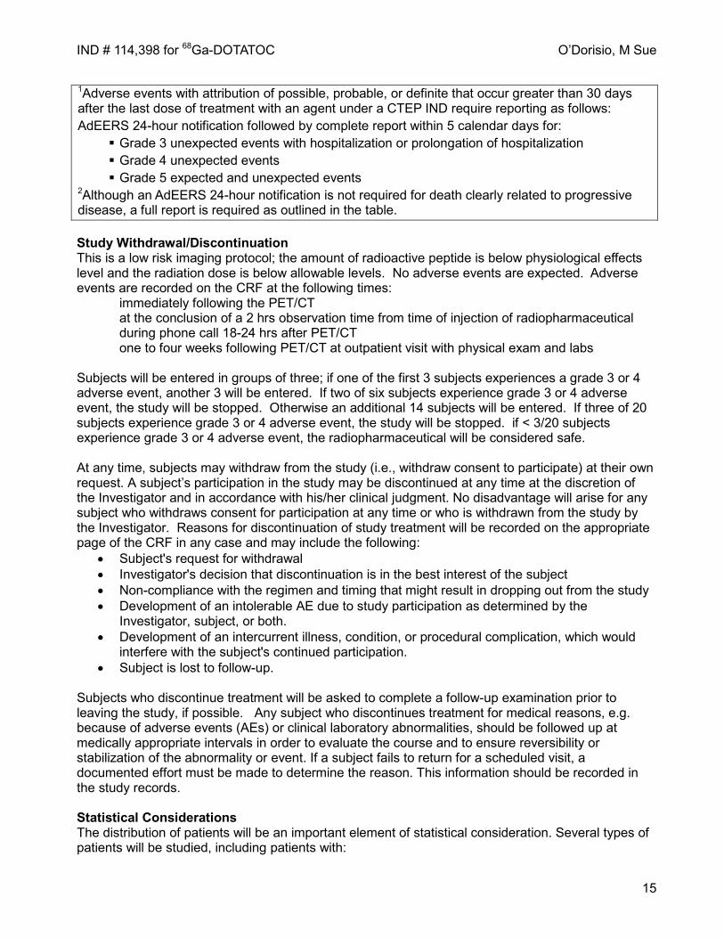
To evaluate the reproducibility of measurement of uptake of 68Ga DOTATOC, 5 patients with metastatic NET underwent two DOTATOC PET scans. Standardized uptake values (SUVmax) were measured for 5 target lesions (max 2 per organ) and 10 target lesions (max 5 per organ) per subject. Target lesions included representative lesions from all involved organs and the lesions with the highest activity for each involved organ. Percentage error (i.e. reproducibility) was calculated for each lesion as Percentage Error = 100*ABS
IND # 114,398 for 68Ga-DOTATOC O’Dorisio, M Sue
22
(Scan 2-Scan 1)/((Scan 2 + Scan 1)/2))16. *mean + standard deviation
Production of [68 Ga]DOTATOC at University of Iowa The cGMP grade DOTATOC was purchased from Bachem with the written permission of Molecular Insight Pharmaceuticals, Inc. Radiolabeling was performed using a TiO2 germanium-gallium generator and automated ModularLab PharmTracer, both from Eckert-Zeigler Eurotope GmbH. This system utilizes a sterile cassette that is replaced for each radiolabeling procedure. Performance has been very reproducible with radiochemical, radionuclic, sterility as shown in Table 4.
Figure 4. Reproducibility of [68Ga]DOTATOC PET in patients with NET. Results for each subject and all target lesions in Table 2 are shown. The average (mean ± SD) reproducibility was 10% ±10% for 50 target lesions. The scatter graph (excluding 2 lesions with SUV>50 for display purposes) shows the high correlation of SUVmax of all target lesions on Scan 1 and 2 (R2=0.99).

IND # 114,398 for 68Ga-DOTATOC O’Dorisio, M Sue
23
European Experience in Humans The potential of [68Ga]DOTATOC PET/CT for improved imaging of somatostatin receptor positive tumors has been recognized since early comparative studies noted improvements in detection sensitivity and image quality in comparison to [111In]DTPA-octreotide SPECT17. Although we have successfully employed [111In]DTPA-octreotide SPECT/CT (Octreoscan) + a diagnostic CT for diagnosis, staging, and monitoring of NET in children and adults2,18, our RDRC approved studies (Figures 3 and 4) as well as comparisons of [68Ga]DOTATOC and Octreoscan by European investigators, support our hypothesis that [68Ga]DOTATOC will provide at least equivalent, and likely objective improvements in, diagnosis and staging of neuroendocrine tumors and other somatostatin receptor positive tumors19-21. Several advantages of [68Ga]DOTATOC PET/CT have been pointed out through these published studies. For example, Gabriel and colleagues concluded that [68Ga]DOTATOC PET/CT shows a significantly higher detection rate (higher sensitivity) compared to conventional Octreoscan SPECT/CT based on a randomized prospective study. These findings are corroborated by a prospective study concluding that [68Ga]DOTATOC PET was superior to Octreoscan in detection of metastases in the lung, skeleton, liver, and brain15. A study demonstrating that [68Ga]DOTATOC PET influences patient management in ≥ 33% of patients further supports the introduction of this imaging technique into the United States22. Comparisons of [68Ga]DOTATOC and Octreoscan by European investigators, support our early observations that [68Ga]DOTATOC will provide at least equivalent, and likely objective improvements in, diagnosis and staging of NETs19-21. Several advantages of [68Ga]DOTATOC PET/CT have been pointed out through these published studies. For example, Gabriel and colleagues concluded that [68Ga]DOTATOC PET/CT shows a significantly higher detection rate (higher sensitivity) compared to conventional Octreoscan SPECT/CT based on a randomized prospective study. These findings are corroborated by a prospective study concluding that [68Ga]DOTATOC PET was superior to Octreoscan in detection of NET metastases in the lung, skeleton, liver, and brain15. A study demonstrating that [68Ga]DOTATOC PET influences patient management in ≥ 33% of patients further supports our proposed research22.
IND # 114,398 for 68Ga-DOTATOC O’Dorisio, M Sue
24
Request for Permission to Charge for an Investigational Drug under an IND This application is seeking FDA approval to charge for the cost of producing 68Ga-DOTATOC to be used as a PET tracer. Costs are justified under Title 21, Chapter 1, Subchapter D Drugs for Human Use, Part 312, IND application as outlined below: 312.8b (i). 68Ga-DOTATOC PET will be used in this clinical trial as a replacement for Octreoscan SPECT imaging. As stated in our IND application, the primary advantage of Ga-68 based somatostatin receptor PET imaging over OctreoScan SPECT is the higher imaging resolution and accurate quantitation of uptake due to robust attenuation correction. The improved resolution and quantitation of uptake obtained with Ga-68 DOTATOC PET should provide a more accurate assessment of somatostatin receptor density, which will lead to a more accurate prediction of treatment response to somatostatin analogues. A recent study from Europe comparing Ga-68 DOTATOC with Octreoscan found Ga-68 DOTATOC to be superior in detection of skeletal and pulmonary involvement of neuroendocrine tumors15. Additional advantages for patients include the lower radiation dose and the shorter imaging time. Whereas, Octreoscan uses a 222 MBq imaging dose of Indium (2.8 day half life) resulting in an effective dose equivalent (HE) equal to 2.61 rads, [68Ga]DOTATOC (68 min half life) uses 185 MBq with an effective dose equivalent of 0.46 rads. In addition, [68Ga]DOTATOC PET/CT can be completed within 2 hours compared to an Octreoscan which requires 3 visits over 24 hours, making [68Ga]DOTATOC a much more convenient imaging choice for patients. 312.8b (ii). The imaging trial proposed under this IND will enroll up to 153 subjects, ages 2 – 100 years of age with Somatostatin receptor positive tumors, many of whom will have had a recent Octreoscan and/or high-resolution, contrast-enhanced CT or MRI. The tumor types include neuroendocrine tumors, neuroblastoma, medulloblastoma, and pheochromocytoma. The study will thus test both efficacy and safety in adults and children with multiple tumor types. 312.8b(iii). [68Ga]DOTATOC will be produced at the University of Iowa PET Center. The extraordinary cost of producing [68Ga]DOTATOC precludes performance of the study without charging due to manufacturing complexity. The direct costs include purchase and storage of the synthetic peptide, DOTATOC; a gallium generator every 6 months to produce the short lived isotope; chemicals required in the radiolabeling procedure; a specialized ModularLab to ensure reproducibility of the product and safety of the radiochemists; single use radiolabeling cassettes to ensure sterility of the product; personnel costs for the radiochemists; and the base cost of the PET/CT imaging. 312.8c. Because [68Ga]DOTATOC has a 68 minute half-life, it will need to be produced locally when trials are expanded to other centers. We intend to provide the protocol as well as all required Standards of Procedure associated with this IND to the Society of Nuclear Medicine Clinical Trials Network. We will apply for expanded access charging at that time, if appropriate. Charges will be paid by the subject or the subject’s insurance company with the exception that the scans for children < 17 yrs with brain tumors may be paid from a childhood brain tumor research account in the University of Iowa Foundation. References: 1. Yao JC, Hassan M, Phan A, et al. One hundred years after "carcinoid": epidemiology of and prognostic factors for neuroendocrine tumors in 35,825 cases in the United States. Journal of Clinical Oncology. 2008;26(18):3063-3072.

IND # 114,398 for 68Ga-DOTATOC O’Dorisio, M Sue
25
2. O'Dorisio MS, Khanna G, Bushnell D. Combining anatomic and molecularly targeted imaging in the diagnosis and surveillance of embryonal tumors of the nervous and endocrine systems in children. Cancer Metastasis Review. 2008;27(4):665-677. 3. Fruhwald MC, O'Dorisio MS, Pietsch T, Reubi JC. High expression of somatostatin receptor subtype 2 (sst2) in medulloblastoma: implications for diagnosis and therapy. Pediatric Research. 1999;45(5 Pt 1):697-708. 4. Juweid ME, Menda Y, O'Dorisio MS, et al. 111In-pentetreotide versus bone scintigraphy in the detection of bony metastases of neuroblastoma. NuclMed Commun. 2002;23(10):983-989. 5. Rinke A, Muller HH, Schade-Brittinger C, et al. Placebo-controlled, double-blind, prospective, randomized study on the effect of octreotide LAR in the control of tumor growth in patients with metastatic neuroendocrine midgut tumors: a report from the PROMID Study Group. J Clin Oncol. 2009;27(28):4656-4663. 6. DuBois SG, Messina J, Maris JM, et al. Hematologic toxicity of high-dose iodine-131-metaiodobenzylguanidine therapy for advanced neuroblastoma. J Clin Oncol. 2004;22(12):2452-2460. 7. Pomeroy SL, Tamayo P, Gaasenbeek M, et al. Prediction of central nervous system embryonal tumour outcome based on gene expression. Nature. 2002;415(6870):436-442. 8. Thacker MM, Temple HT, Scully SP. Current treatment for Ewing's sarcoma. ExpertRevAnticancer Ther. 2005;5(2):319-331. 9. Kwekkeboom DJ, de Herder WW, van Eijck CH, et al. Peptide receptor radionuclide therapy in patients with gastroenteropancreatic neuroendocrine tumors. Semin Nucl Med;40(2):78-88. 10. Kwekkeboom DJ, Bakker WH, Kooij PP, et al. [177Lu-DOTAOTyr3]octreotate: comparison with [111In-DTPAo]octreotide in patients. EurJNuclMed. 2001;28(9):1319-1325. 11. Forrer F, Waldherr C, Maecke HR, Mueller-Brand J. Targeted radionuclide therapy with 90Y-DOTATOC in patients with neuroendocrine tumors. Anticancer Res. 2006;26(1B):703-707. 12. Bushnell DL, Jr., O'Dorisio TM, O'Dorisio MS, et al. 90Y-edotreotide for metastatic carcinoid refractory to octreotide. J Clin Oncol. 2010;28(10):1652-1659. 13. Menda Y, O'Dorisio MS, Kao S, et al. Phase I trial of 90Y-DOTATOC therapy in children and young adults with refractory solid tumors that express somatostatin receptors. J Nucl Med. 2010;51(10):1524-1531. 14. Wild D, Macke HR, Waser B, et al. 68Ga-DOTANOC: a first compound for PET imaging with high affinity for somatostatin receptor subtypes 2 and 5. EurJNuclMedMolImaging. 2005;32(6):724. 15. Buchmann I, Henze M, Engelbrecht S, et al. Comparison of 68Ga-DOTATOC PET and 111In-DTPAOC (Octreoscan) SPECT in patients with neuroendocrine tumours. EurJNuclMedMolImaging. 2007;34(10):1617-1626. 16. Shields AF, Lawhorn-Crews JM, Briston DA, et al. Analysis and reproducibility of 3'-Deoxy-3'-[18F]fluorothymidine positron emission tomography imaging in patients with non-small cell lung cancer. Clin Cancer Res. 2008;14(14):4463-4468. 17. Hofmann M, Maecke H, Borner R, et al. Biokinetics and imaging with the somatostatin receptor PET radioligand (68)Ga-DOTATOC: preliminary data. EurJNuclMed. 2001;28(12):1751-1757. 18. Khanna G, O'Dorisio MS, Menda Y, Kirby P, Kao S, Sato Y. Gastroenteropancreatic Neuroendocrine Tumors in Children and Young Adults. Pediatric Radiology. 2008;38:251-259. 19. Gabriel M, Decristoforo C, Kendler D, et al. 68Ga-DOTA-Tyr3-octreotide PET in neuroendocrine tumors: comparison with somatostatin receptor scintigraphy and CT. JNuclMed. 2007;48(4):508-518. 20. Jindal T, Kumar A, Venkitaraman B, Dutta R, Kumar R. Role of (68)Ga-DOTATOC PET/CT in the evaluation of primary pulmonary carcinoids. Korean J Intern Med. 2010;25(4):386-391. Prepublished on 2010/12/24 as DOI 10.3904/kjim.2010.25.4.386. 21. Jindal T, Kumar A, Kumar R, Dutta R. Role of 68Ga-DOTATOC PET/CT in carcinoids. Pathol Int. 2010;60(2):143-144.

IND # 114,398 for 68Ga-DOTATOC O’Dorisio, M Sue
26
22. Ruf J, Heuck F, Schiefer J, et al. Impact of Multiphase 68Ga-DOTATOC-PET/CT on therapy management in patients with neuroendocrine tumors. Neuroendocrinology. 2010;91(1):101-109.

IND # 114,398 for 68Ga-DOTATOC O’Dorisio, M Sue
27
Appendix 1 HCCC Clinical Trial Data and Safety Monitoring Plan (DSMP)*
Date: 11/12/14
IRB#: 201110718 Title: Safety and Efficacy of 68Ga-DOTATOC Positron Emission Tomography (PET) for Diagnosis,
Staging, and Measurement of Response to Treatment in Somatostatin Receptor Positive Tumors
PI: M. Sue O'Dorisio, PHD, MD Co-I: David Bushnell, MD, Yusuf Menda, MD, Michael Schultz, PhD, Laura Ponto, PhD, John Sunderland, PhD, Thomas O’Dorisio, MD and Michael Graham, PhD, MD
* All investigator-initiated protocols will be subject to ongoing monitoring of accrual, subject eligibility, protocol modifications and continuing reviews. Active studies will be audited for the DSMC by the CRSO, following guidelines based on level of risk to subjects. Audits will be conducted by reviewing subject files provided by clinical research coordinators, as well as original source documentation provided by online medical records and research pharmacists. The CRSO will review adverse events, eligibility of subjects, and adherence to the IRB- and PRMC-approved protocol. Protocols found to have discrepancies will be require a response from the PI and an action plan for correcting identified deficiencies. The DSMC will provide a schedule of audit and report dates if requested.
Type of Clinical Trial:
Investigator-initiated (UI/HCCC) Investigator-initiated, participating site
Pilot study Phase I
Phase I/II Phase II
Phase III Compassionate-use drug protocol
Interventional Treatment Interventional Non-Treatment
Non-Interventional

IND # 114,398 for 68Ga-DOTATOC O’Dorisio, M Sue
28
Study risk-level:
Level 1—low risk of morbidity or death, * <<1% of death or any adverse event
Level 2—risk of death* <1% or any adverse event 1% – 5%
Level 3—risk of death* 1% – 5% or grade 4 – 5 SAE 1% – 5%
Level 4—risk of death* >5% or grade 4 – 5 SAE >15%
Drugs being used on a “compassionate” basis
* Risk of death” refers specifically to 100-day treatment-related mortality
___________________________ Subject inclusion criteria:
• Signed informed consent. • Age ≥ 2 years. • Known or suspected somatostatin receptor positive tumor such as carcinoid;
neuroendocrine tumor; neuroblastoma; medulloblastoma; pheochromocytoma. Supporting evidence may include MRI, CT, biochemical markers, and or pathology report.
• Karnofsky performance status or Lansky Play Scale status of ≥ 50 (or ECOG/WHO equivalent).
• Subject is male; or is a female who is either pre-menarchal, surgically sterile (has had a documented bilateral oophorectomy and/or documented hysterectomy), postmenopausal (> 1 years without menses), non-lactating, or of childbearing potential for whom a serum pregnancy test (with the results known prior to investigational product administration) is negative. A negative serum pregnancy test will be required for all female subjects with child bearing potential. If a false pregnancy test is suspected, e.g., perimenopausal condition, an obstetrician will be consulted to determine if she is/is capable of becoming pregnant.
• Subject exclusion criteria:
• Subject weighs more than 400 pounds (Subjects who weigh more than 400 pounds will not be able to fit inside the imaging machines).
• Inability to lie still for the entire imaging time (e.g. cough, severe arthritis, etc.) • Inability to complete the needed investigational and standard-of-care imaging examinations
due to other reasons (severe claustrophobia, radiation phobia, etc.) • Does the subject have any additional medical condition, serious intercurrent illness, or other
extenuating circumstance that, in the opinion of the Investigator, may significantly interfere with study compliance?
• Stopping rules (subject and study): This is a low risk imaging protocol; the amount of radioactive peptide is below physiological effects level and the radiation dose is below allowable levels. No adverse events are expected. Adverse events are recorded on the CRF at the following times:
• immediately following the PET/CT • at the conclusion of a 2 hrs observation time from time of injection of radiopharmaceutical • during phone call 18-24 hrs after PET/CT • one to four weeks following PET/CT at outpatient visit with physical exam and labs

IND # 114,398 for 68Ga-DOTATOC O’Dorisio, M Sue
29
Risks of Study Drug: Research radiation dose estimate: Our calculations estimate that the effective whole-body dose of the 68Ga-DOTATOC PET/CT is approximately 1.6 rem, less than the radiation dose from a conventional 18F-FDG PET/CT scan of about 2.4 rem, assuming the standard use of a “low-dose” CT for anatomic localization and attenuation correction, as described in our proposed imaging protocol. The U.S. national annual background dose for humans is approximately 0.36 rem. The maximum annual permissible whole body radiation dose to a career radiation worker (e.g., a radiologist, nuclear medicine technologist, etc.) is 5.0 rem. Thus, the one-time dose to the patient for the investigational PET/CT will be approximately one-third of the annual permissible dose to a career radiation worker and about 5 years’ worth of background radiation in the US. Given the average age of the patients in this proposed investigation (about 60 years of age), the increased risk to the patient, who will have a known, current life-threatening malignancy, for a second, radiation-induced malignancy from this investigational radiation exposure is extremely low (estimated < 1%). Given the risk of death from either delay in diagnosis or under-treatment of these known malignancies, we feel the added radiation burden to the patient is well within the patient’s best interest if 68Ga-DOTATOC PET/CT imaging adds value. Risks of Study Procedures Risks of Investigational PET/CT: An adverse reaction to the investigational radiopharmaceutical is possible though not reported. Additional risks include radiation exposure from the investigational PET/CT scan (discussed above), dosage infiltration, risks from IV access. blood tests, and the inconvenience of the investigational PET/CT scan. Children may require sedation or anesthesia to remain still during the PET scanning procedure. The risks of sedation include a reaction to the medication used for sedation or anesthesia. If anesthesia is required, there is a risk of injury to the throat or airways from insertion of the breathing tube.
Risk Level 4 Interventional treatment trials involving investigational agents or devices with a risk of death* >5% or grade 4 – 5 SAE >15% (all investigator initiated INDs, and most Phase I/II trials, gene therapy, gene manipulation or viral vector systems high-risk clinical procedures if performed solely for research purposes. The use of a new chemical or drug for which there is limited or no available safety data in humans. An independent Study Monitor or the DSMC Chair (or designee), will review study data (provided by the PI and CRSO) and communicate with the PI at least quarterly. A copy of this communication will be forwarded to the DSMC and PRMC Chairs. This communication may coincide with the CRSO’s quarterly audit when possible. If an audit has not been conducted—due to lack of research activity--the quarterly summary letter will occur as scheduled. Subject accrual data will be obtained from OnCore on an ongoing basis by the CTRM during accrual review for the PRMC.
- Subject accrual data will be obtained from OnCore on an ongoing basis by the CTRM during accrual review for the PRMC.
- The research coordinator or data manager for the study will register new subjects in OnCore - Adverse events will be reported to the CRSO as defined in the DSMP for the study - Serious adverse events will be communicated to the CRSO and recorded in OnCore. - The CRSO will contact the research team every three months to coordinate an audit of the
trial's research records. Records for a minimum of 25% of subjects will be audited. OnCore auditing functionality will be used to conduct, evaluate, and report on each audit. Audits will:
o verify eligibility of patients accrued to the study,

IND # 114,398 for 68Ga-DOTATOC O’Dorisio, M Sue
30
o check for the presence of a signed informed consent, o determine compliance with protocol’s study plan, o determine whether SAEs are being appropriately reported to internal and external
regulatory agencies, o compare accuracy of data in the research record with the primary source documents, o review investigational drug processing and documentation, o assess cumulative AE/SAE reports for trends and compare to study stopping rules.
ROUTINE ADVERSE EVENT REPORTING
Table I: Routine Reporting of Adverse Events for Phase 1 and 2 Trials
Attribution Adverse Event Grade 1 Grade 2 Grade 3 Grade 4 Grade 5 Unrelated Not required Not required Not
required CRSO CRSO
Unlikely Not required Not required Not required CRSO CRSO
Possible CRSO CRSO CRSO CRSO CRSO
Probable CRSO CRSO CRSO CRSO CRSO
Definite CRSO CRSO CRSO CRSO CRSO

IND # 114,398 for 68Ga-DOTATOC O’Dorisio, M Sue
31
EXPEDITED ADVERSE EVENT REPORTING
Table II: Reporting Requirements for Adverse Events that occur within 30 Days1 of the Last Dose of the Investigational Agent on Phase 1 Trials
1 2 2 3 3 4 & 52
Unexpected and
Expected
Unexpected
Expected
Unexpected Expected Unexpected
and Expected With Hospital
without Hospital with Hospital without
Hospital
Unrelated
Unlikely
Not Required
Not Required
Not Require
d
10 Calendar
Days
Not Required
10 Calendar Days
Not Require
d
24-Hour; 5 Calendar Days
Possible Probable Definite
Not Required
10 Calendar
Days
Not Require
d
24-Hour; 5 Calendar
Days
24-Hour; 5 Calendar
Days
10 Calendar Days
Not Require
d
24-Hour; 5 Calendar Days
1Adverse events with attribution of possible, probable, or definite that occur greater than 30 days after the last dose of treatment with an agent under a CTEP IND require reporting as follows: AdEERS 24-hour notification followed by complete report within 5 calendar days for:
Grade 3 unexpected events with hospitalization or prolongation of hospitalization Grade 4 unexpected events Grade 5 expected and unexpected events
2Although an AdEERS 24-hour notification is not required for death clearly related to progressive disease, a full report is required as outlined in the table.
IND # 114,398 for 68Ga-DOTATOC O’Dorisio, M Sue
32
Appendix 2
CRF: Data Collection for 68Ga-DOTATOC PET/CT
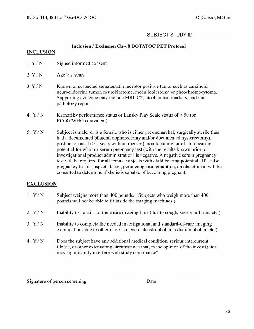
IND # 114,398 for 68Ga-DOTATOC O’Dorisio, M Sue
33
SUBJECT STUDY ID:_____________
Inclusion / Exclusion Ga-68 DOTATOC PET Protocol INCLUSION 1. Y / N Signed informed consent 2. Y / N Age > 2 years 3. Y / N Known or suspected somatostatin receptor positive tumor such as carcinoid, neuroendocrine tumor, neuroblastoma, medulloblastoma or pheochromocytoma. Supporting evidence may include MRI, CT, biochemical markers, and / or pathology report 4. Y / N Karnofsky performance status or Lansky Play Scale status of > 50 (or ECOG/WHO equivalent) 5. Y / N Subject is male; or is a female who is either pre-menarchal, surgically sterile (has had a documented bilateral oophorectomy and/or documented hysterectomy), postmenopausal (> 1 years without menses), non-lactating, or of childbearing potential for whom a serum pregnancy test (with the results known prior to investigational product administration) is negative. A negative serum pregnancy test will be required for all female subjects with child bearing potential. If a false pregnancy test is suspected, e.g., perimenopausal condition, an obstetrician will be consulted to determine if she is/is capable of becoming pregnant. EXCLUSION 1. Y / N Subject weighs more than 400 pounds. (Subjects who weigh more than 400 pounds will not be able to fit inside the imaging machines.) 2. Y / N Inability to lie still for the entire imaging time (due to cough, severe arthritis, etc.) 3. Y / N Inability to complete the needed investigational and standard-of-care imaging examinations due to other reasons (severe claustrophobia, radiation phobia, etc.) 4. Y / N Does the subject have any additional medical condition, serious intercurrent illness, or other extenuating circumstance that, in the opinion of the investigator, may significantly interfere with study compliance? _________________________________________ ____________________ Signature of person screening Date
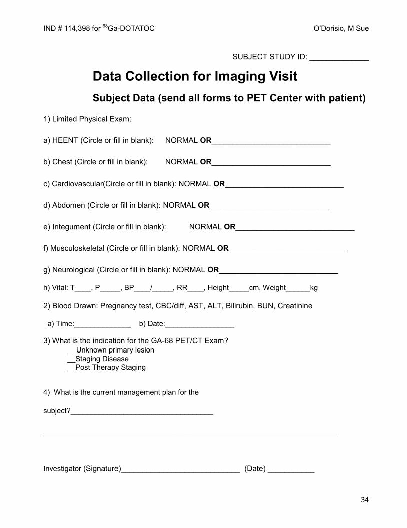
IND # 114,398 for 68Ga-DOTATOC O’Dorisio, M Sue
34
SUBJECT STUDY ID: ______________ Data Collection for Imaging Visit Subject Data (send all forms to PET Center with patient)
1) Limited Physical Exam:
a) HEENT (Circle or fill in blank): NORMAL OR____________________________
b) Chest (Circle or fill in blank): NORMAL OR____________________________
c) Cardiovascular(Circle or fill in blank): NORMAL OR____________________________
d) Abdomen (Circle or fill in blank): NORMAL OR____________________________
e) Integument (Circle or fill in blank): NORMAL OR____________________________
f) Musculoskeletal (Circle or fill in blank): NORMAL OR____________________________
g) Neurological (Circle or fill in blank): NORMAL OR____________________________
h) Vital: T____, P_____, BP____/_____, RR____, Height_____cm, Weight______kg
2) Blood Drawn: Pregnancy test, CBC/diff, AST, ALT, Bilirubin, BUN, Creatinine
a) Time:______________ b) Date:_________________ 3) What is the indication for the GA-68 PET/CT Exam? __Unknown primary lesion __Staging Disease __Post Therapy Staging
4) What is the current management plan for the
subject?___________________________________
__________________________________________________________________________ Investigator (Signature)____________________________ (Date) ___________

IND # 114,398 for 68Ga-DOTATOC O’Dorisio, M Sue
35
SUBJECT STUDY ID: ______________________
Data Collection for Imaging
This page will be completed in the PET Center: 1) Pregnancy test (urine) required? Y / N Results_________________ 2) IV inserted (location): ____________________________________
3) Alprazolam Y/N Dose __________mg @ Time _______:________ a) Who is the responsible person with the subject (name)?___________________
5) Ga-68 DOTATOC Dose: _____________mCi @ Injection Time_______:________
6) PET Scan Start Time _________:__________ End Time__________:___________
7) Vital Signs (supine) at PET Completion: P _____, BP ______/______, RR________
8) Patient Observed from time of Ga-68 injection until discharged @ (time) ______:_______
Signature of PET staff: ___________________________ Date: __________________
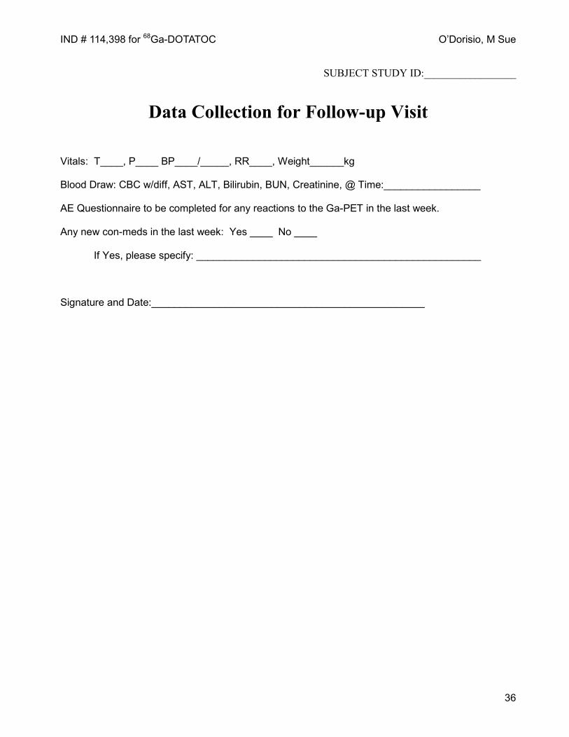
IND # 114,398 for 68Ga-DOTATOC O’Dorisio, M Sue
36
SUBJECT STUDY ID:__________________
Data Collection for Follow-up Visit
Vitals: T____, P____ BP____/_____, RR____, Weight______kg Blood Draw: CBC w/diff, AST, ALT, Bilirubin, BUN, Creatinine, @ Time:_________________ AE Questionnaire to be completed for any reactions to the Ga-PET in the last week. Any new con-meds in the last week: Yes ____ No ____ If Yes, please specify: __________________________________________________ Signature and Date:________________________________________________

IND # 114,398 for 68Ga-DOTATOC O’Dorisio, M Sue
37
SUBJECT STUDY ID:__________________
Ga-68 DOTATOC PET Adverse Event Questionnaire Please only list AEs that are new and have happened since the Ga-PET scan.
POSSIBLE ADVERSE EVENTS YES/NO COMMENTS/NOTES
BODY AS A WHOLE Pain (abdominal) Pain (chest/breast) Pain (other/site) Fever Injection site reaction CARDIOVASCULAR SYSTEM Flushing (vasodilatation) DIGESTIVE SYSTEM Nausea Diarrhea Vomiting RESPIRATORY SYSTEM Dyspnea (shortness of breath) SKIN & APPENDAGES Rash Itching (pruritus) Hives (urticaria) Sweating Cyanosis (finger or toe
discoloration) CENTRAL NERVOUS SYSTEM Visual disturbances Numbness of feet Numbness of fingers/hands Weakness of feet Weakness of fingers Burning sensation in feet Burning sensation in fingers
Signature:___________________________ Date:________________ Time:____________

IND # 114,398 for 68Ga-DOTATOC O’Dorisio, M Sue
38
SUBJECT STUDY ID:__________________
Karnofsky Performance Scale
KARNOFSKY PERFORMANCE STATUS SCALE DEFINITIONS RATING (%) CRITERIA
Able to carry on normal activity and to work; no special care needed
100 Normal no complaints; no evidence of disease.
90 Able to carry on normal activity; minor signs or symptoms of disease.
80 Normal activity with effort; some signs or symptoms of disease.
Unable to work; able to live at home and care for most personal needs; varying amount of assistance needed.
70 Cares for self; unable to carry on normal activity or to do active work.
60 Required occasional assistance, but is able to care for most of his personal needs.
50 Required considerable assistance and frequent medical care.
Unable to care for self; required equivalent of institutional or hospital care; disease may be progressing rapidly.
40 Disabled; required special care and assistance.
30 Severely disabled; hospital admission is indicated although death not imminent.
20 Very sick; hospital admission necessary; active supportive treatment necessary.
10 Moribund; fatal processes progressing rapidly.
0 Dead
Signature:___________________________ Date:________________ Time:____________