saccharomyces cerevisiae–based platform for rapid
TRANSCRIPT

Saccharomyces cerevisiae–Based Platform for RapidProduction and Evaluation of Eukaryotic NutrientTransporters and Transceptors for Biochemical Studiesand CrystallographyPeter Scharff-Poulsen1,2*, Per Amstrup Pedersen1*
1 Department of Biology, University of Copenhagen, Copenhagen, Denmark, 2 Center for Microbial Biotechnology, Department of Systems Biology, Technical University of
Denmark, Kongens Lyngby, Denmark
Abstract
To produce large quantities of high quality eukaryotic membrane proteins in Saccharomyces cerevisiae, we modified a high-copy vector to express membrane proteins C-terminally-fused to a Tobacco Etch Virus (TEV) protease detachable GreenFluorescent Protein (GFP)-8His tag, which facilitates localization, quantification, quality control, and purification. Using thisexpression system we examined the production of a human glucose transceptor and 11 nutrient transporters andtransceptors from S. cerevisiae that have not previously been overexpressed in S. cerevisiae and purified. Whole-cell GFP-fluorescence showed that induction of GFP-fusion synthesis from a galactose-inducible promoter at 15uC resulted in stableaccumulation of the fusions in the plasma membrane and in intracellular membranes. Expression levels of the 12 fusionsestimated by GFP-fluorescence were in the range of 0.4 mg to 1.7 mg transporter pr. liter cell culture. A detergent screenshowed that n-dodecyl-ß-D-maltopyranoside (DDM) is acceptable for solubilization of the membrane-integrated fusions.Extracts of solubilized membranes were prepared with this detergent and used for purifications by Ni-NTA affinitychromatography, which yielded partially purified full-length fusions. Most of the fusions were readily cleaved at a TEVprotease site between the membrane protein and the GFP-8His tag. Using the yeast oligopeptide transporter Ptr2 as anexample, we further demonstrate that almost pure transporters, free of the GFP-8His tag, can be achieved by TEV proteasecleavage followed by reverse immobilized metal-affinity chromatography. The quality of the GFP-fusions was analysed byfluorescence size-exclusion chromatography. Membranes solubilized in DDM resulted in preparations containingaggregated fusions. However, 9 of the fusions solubilized in DDM in presence of cholesteryl hemisuccinate and specificsubstrates, yielded monodisperse preparations with only minor amounts of aggregated membrane proteins. In conclusion,we developed a new effective S. cerevisiae expression system that may be used for production of high-quality eukaryoticmembrane proteins for functional and structural analysis.
Citation: Scharff-Poulsen P, Pedersen PA (2013) Saccharomyces cerevisiae–Based Platform for Rapid Production and Evaluation of Eukaryotic NutrientTransporters and Transceptors for Biochemical Studies and Crystallography. PLoS ONE 8(10): e76851. doi:10.1371/journal.pone.0076851
Editor: Vladimir N. Uversky, University of South Florida College of Medicine, United States of America
Received June 19, 2013; Accepted September 2, 2013; Published October 4, 2013
Copyright: � 2013 Scharff-Poulsen, Pedersen. This is an open-access article distributed under the terms of the Creative Commons Attribution License, whichpermits unrestricted use, distribution, and reproduction in any medium, provided the original author and source are credited.
Funding: This study was supported by grants from the Danish Research Council for Technology and Production to Morten Kielland-Brandt (IVC) and to PerAmstrup Pedersen and a grant from the Danish National Advanced Technology Foundation (IBISS) to Per Amstrup Pedersen. The funders had no role in studydesign, data collection and analysis, decision to publish, or preparation of the manuscript.
Competing Interests: The authors have declared that no competing interests exist.
* E-mail: [email protected] (PSP); [email protected] (PAP)
Introduction
Nutrient transporters are the gatekeepers controlling transport
of essential nutrients such as sugars and amino acids across the
plasma membrane of cells. From a medical and a pharmaceutical
perspective, human nutrient transporters are of great importance
because (i) various gene defects in nutrient transporters have been
identified and shown to cause human diseases, (ii) nutrient
transporters are potential drug targets, (iii) drugs are transported
into cells using nutrient transporters (as reviewed by [1], see also
Genomic Transporter Database of SLC (Solute Carrier) gene
tables at web-site http://www.pharmaconference.org/slctable.
asp). Obviously, structural information about nutrient transporters
is of great interest to both academia and the pharmaceutical
industry. Nevertheless, structures are only known for a few
nutrient transporters of bacterial origin, whereas structures of
eukaryotic transporters are not yet available.
Nutrient transporters from yeast constitute straight-forward
targets for gaining valuable insight into structure-function
relationships of similar transporters from higher eukaryotic
organisms. In yeast, sugars and amino acids are transported
across the plasma membrane by transporters of the major
facilitator superfamily (MFS) and of the amino acid-polyamine-
organocation (APC) superfamily. As shown in the TransportDB
database (http://www.membranetransport.org/) the yeast MFS
comprises 85 members, of which 20 have functions in hexose
transport, and the yeast APC family comprises 24 members, of
which 18 have functions in amino acid transport.
An interesting aspect of the nutrient transporters from yeast is
the finding that some of them also have receptor functions
involved in signal transduction processes (reviewed in [2]). These
PLOS ONE | www.plosone.org 1 October 2013 | Volume 8 | Issue 10 | e76851

so-called ‘‘transceptors’’ constitute a novel concept in signaling,
and comprise both transporting and non-transporting transcep-
tors. Non-transporting transceptors include the glucose sensors
Snf3 and Rgt2 (reviewed in [3]) and the amino acid sensor Ssy1
(reviewed in [4]). Transporting transceptors include the Gap1
amino acid transporter [5,6], the Pho84 phosphate transporter [7]
and the Mep2 ammonium transporter [8]. Growing evidence for
transporters functioning as transceptors in humans, fruit flies and
plants, suggests that transceptors are widespread in nature and
that we may only have recognized the tip of the iceberg [2,9].
Recent studies of the amino acid transceptor Ssy1 and the
glucose transceptor Snf3 from yeast [10,11,12,13] revealed that
Ssy1 and Snf3 are able to sense both extracellular and intracellular
nutrients, and a mechanistic model that explains how these
transceptors may participate in maintaining intracellular homeo-
stasis for nutrients in yeast cells was proposed. This model may be
of importance for understanding how for instance pertubations in
amino acid availability affect growth of cancer cells, and assist in
identification of new anti-cancer targets and development of
cancer treatment therapies [14]. Similarly, the results obtained
with Snf3 may add to the understanding of the human glucose
transceptor GLUT2 [15] which has been proposed to be involved
in regulation of food intake by the hypothalamus [16].
Members of the Proton-dependent Oligopeptide Transporter
(POT) family, which include the S. cerevisiae Ptr2 oligopeptide
transporter, are also of great interest in nutrient uptake. In
addition to their function in peptide transport in organs such as the
gastrointestinal tract, the kidney and the central nervous system,
they are also responsible for uptake of a large variety of
pharmaceuticals with peptide-like structures such as penicillins,
antivirals and anticancer agents [17].
Although the yeast nutrient transporters and transceptors have
high physiological importance and implications for the function of
similar mammalian proteins they have not yet been purified and
characterized biochemically and structurally. Therefore, to deepen
our understanding of eukaryotic nutrient transporters and
transceptors, we here report on the production of a selection of
these membrane proteins using a high-copy vector expression
system [18] combined with the GFP-fusion methodology devel-
oped by Drew et al 2008 [19]. We show that reasonable amounts
of transporters and transceptors can be produced in a high quality.
With these results, we may provide biochemical characterizations
and make the first moves towards crystallization and structure
determination of this class of eukaryotic membrane proteins, with
significant perspectives in physiology and drug development.
Materials and Methods
Yeast strains and culture conditionsOverexpression of membrane protein-GFP fusions in S. cerevisiae
was performed in strain PAP1500 (MATa ura3-52 trp1:: GAL10-
GAL4 lys2-801 leu2D1 his3D200 pep4::HIS3 prb1D1.6R can1 GAL)
[18] as follows: PAP1500 transformed with various expression
constructs was inoculated in 5 ml of synthetic minimal (SD) media
[20,21] supplemented with glucose (20 g/L), leucine (60 mg/L)
and lysine (30 mg/L) and incubated over night at 30uC with
shaking. The over-night culture was diluted 50 times in SD media
supplemented with lysine (30 mg/L) and incubated for 2 days at
30uC with shaking to increase plasmid copy number during
leucine-limitation. Next, the expression strain was cultivated in SD
media supplemented with glucose (5 g/L), glycerol (3% v/v),
alanine (20 mg/L), arginine (20 mg/L), aspartic acid (100 mg/L),
cysteine (20 mg/L), glutamic acid (100 mg/L), histidine (20 mg/
L), lysine (30 mg/L), methionine (20 mg/L), phenylalanine
(50 mg/L), proline (20 mg/L), serine (375 mg/L), threonine
(200 mg/L), tryptophan (20 mg/L), tyrosine (30 mg/L), and
valine (150 mg/L). Thus, 1 liter of this medium was inoculated
with the leucine-starved culture to give an OD450 of 0.05 and
cultivation was continued at 30uC with shaking until OD450
reached 1.0. Cultures were thermo-equilibrated to 15uC (unless
otherwise stated) and induction of GFP-fusion synthesis was
initiated with addition of 110 ml 20% (w/v) galactose dissolved in
the above described medium lacking glucose. Incubation at 15uCwas continued with shaking for 48 hours. Cells were harvested by
centrifugation at 3,000 rpm in a SLA-3000 rotor (Sorvall) for 5
minutes at 5uC. A complete list of strains used is available in Table
S1.
Construction of expression plasmidsTransporter and transceptor genes were PCR amplified from
chromosomal DNA with AccuPol DNA polymerase (Ampliqon
DK) and primers (TAG Copenhagen) shown in Table S2. Each
GFP-8His expression plasmid was generated by in vivo homologous
recombination in S. cerevisiae by transforming strain PAP1500 with
a transporter PCR fragment, a GFP PCR fragment and BamHI
and HindIII digested pEMBLyex4 [22]. A TEV site (GEN-
LYFQSQF) was introduced between the transporter and the GFP
tag. GFP was C-terminally fused to a sequence of 8 His moieties.
PAP1500 transformants were selected on SD plates with leucine
(60 mg/L) and lysine (30 mg/L). All plasmid constructs were
checked by DNA sequencing at Eurofins MWG Operon
(Germany).
Membrane preparationSmall scale preparation of crude yeast membranes was carried
out by a glass bead disruption method. A cell pellet from a 1 liter
culture (usually 3 g wet weight) was resuspended in 10 ml ice-cold
lysis buffer (25 mM imidazole adjusted to pH 7.5 with HCl, 10%
sucrose (w/v), 1 mM EDTA, 1 mM EGTA, and protease
inhibitors (1 mM PMSF, leupeptin (1 mg/ml), pepstatin (1 mg/
ml) and chymostatin (1 mg/ml)). Approximately 10 ml of glass
beads (425 to 600 microns, Sigma) were added to the cell
suspension. Cells were disrupted by 4 times 1 minute high speed
whirlimixing interrupted by 1 minute of cooling on ice. The
supernatant was collected and glassbeads were washed two times
in 10 ml ice-cold lysisbuffer. Unbroken cells and cell debris were
removed from the combined supernatants by centrifugation at
10,000 rpm in a SS-34 rotor (Sorvall) for 10 minutes at 5uC. This
method typically disrupted 85% of the PAP1500 cells as measured
by GFP fluorescence. Membranes were collected by centrifugation
of the final supernatant at 40,000 rpm in a 70 TI rotor (Beckman)
for 1.5 hour at 5uC. Membrane pellets were resuspended in 3 ml
buffer (20 mM phosphate pH 7.0, 200 mM NaCl, 10% glycerol,
10 mM imidazole, 1 mM PMSF and 1 mg/ml of leupeptin,
pepstatin and chymostatin, respectively) using a rotating Potter-
type homogenizer.
Protein quantificationThe protein concentration in crude membranes was determined
by the Lowry assay [23]. The concentration of proteins in purified
fractions was measured using a Nanodrop apparatus (Thermo
Scientific).
Bioimaging of live yeast cellsS. cerevisiae cells used for bioimaging were grown and induced for
production of GFP-fusions as described [24]. Fluorescence was
visualized at 1,000 x magnification with a Nikon Eclipse E600
Nutrient Transporter and Transceptor Purification
PLOS ONE | www.plosone.org 2 October 2013 | Volume 8 | Issue 10 | e76851

fluorescence microscope equipped with an Optronics Magnafire
model S99802 camera.
Whole-cell fluorescenceFive ml of yeast culture with a known optical density was
harvested by centrifugation at 3000 rpm for 3 minutes at 5uC in a
Multifuge 3 S-R (Heraus). The supernatant was carefully removed
and cells were re-suspended in 100 ml sterile water and transferred
to a Nucleon Nunc white micro plate (Cat. No. 136101).
Fluorescence was measured in a microplate spectrofluorometer
(Fluoroskan Ascent, Thermo Labsystems) using 485 nm excitation
and 520 nm emission.
In-gel fluorescenceIn-gel fluorescence was carried out on an Image Station
4000 MM (Carestream) using 465 nm excitation and 535 nm
emission or an ImageQuant LAS 4010 imager (GE Healthcare).
Detergent Screen180 ml membrane extract from yeast cells overexpressing the
indicated membrane protein-GFP fusions diluted in solubilization
buffer (20 mM phosphate pH 7.0, 200 mM NaCl, 10% glycerol,
1 mM PMSF and 1 mg/ml of leupeptin, pepstatin and chymos-
tatin, respectively) to a concentration of 3.5 mg protein/ml was
mixed with 20 ml detergent (100 mg/ml) yielding a protein to
detergent ratio of 1:3 (mg/ml). The mixture was incubated at 4uCfor 1 hour with mild agitation. Non-solubilized material was
pelleted by centrifugation in an Airfuge (Beckman) fitted with an
A-100/30 rotor for 12 minutes at 92,000 rpm at 30 psi air pressure
(167,000 g). The supernatant was transferred to a Nucleon Nunc
white microplate and GFP fluorescence was measured in a
Fluoroskan Ascent spectrofluorometer (Thermo Labsystems) using
485 nm excitation and 520 nm emission. Solubilization efficiency
was estimated by the ratio of GFP fluorescence of the supernatant
and of the detergent-solubilized membranes before centrifugation.
Solubilization of membranes in DDM and cholesteryl hemi-
succinate (CHS) was carried out as described above using a 20 ml
mixture of DDM (100 mg/ml) and of CHS (20 mg/ml), and
addition of substrates to a final concentration of: 100 mM glucose
for the Hxt1-, Hxt2-, Hxt3-, Hxt4-, Rgt2-, Snf3- and GLUT2-
GFP fusions; 10 mM glutamine for the Agp1-GFP fusion; 2 mM
leucine for the Ssy1- and Tat1-GFP fusions; 10 mM Gly-Leu
dipeptide for the Ptr2-GFP fusion; 10 mM NH4Cl for the Mep2-
GFP fusion.
Anagrade quality detergents and CHS were purchased from
Affymetrix: DM, n-decyl-b-D-maltopyranoside; DDM, n-dodecyl-
b -D-maltopyranoside; OG, n-octyl-b -D-glucopyranoside;
CHAPS, 3-[(3-cholamidopropyl)-dimethylammonio]-1-propane
sulfonate/N,N-dimethyl-3-sulfo-N-[3-[[3a,5b,7a,12a)-3,7,12-tri-
hydroxy-24-oxocholan-24-yl]amino]propyl]-1-propanaminium
hydroxide; CYMAL-5, 5-cyclohexyl-1-pentyl-b-D-maltoside; FC-
12, n-dodecylphosphocholine.
Fluorescence-detection size-exclusion chromatographyFluorescence-detection size-exclusion chromatography (FSEC)
was carried out essentially as described [25]. Total membranes
were solubilized as described in the detergent screen section and
loaded onto a Superose 12 10/300 column (GE Healthcare Life
Science) mounted on an AKTA purifier (GE Healthcare Life
Science). The column was pre-equilibrated with SEC buffer
(20 mM Na+-phosphate pH 7.0, 200 mM NaCl, and 0.3 mg/ml
DDM) and run with a flow rate of 0.5 ml/min. After 5 ml elution,
fractions of 0.2 ml were collected and GFP fluorescence was
measured in a Fluoroskan Ascent spectrofluorometer (Thermo
Labsystems) using 485 nm excitation and 520 nm emission.
A high molecular weight gel filtration calibration kit (GE
Healthcare Life Science) was used for determination of the void
volume and molecular weight estimations.
Purification of transporter-GFP fusions by Ni-NTA affinitypurification
Crude membranes isolated from a 1-liter induced culture were
diluted to a concentration of 3.5 mg protein/ml in lysis buffer
(20 mM phosphate pH 7.0, 200 mM NaCl, 10% glycerol, 10 mM
imidazole, 1 mM PMSF and 1 mg/ml of leupeptin, pepstatin and
chymostatin, respectively). DDM solution (100 mg/ml) was added
to a final concentration of 10 mg/ml, yielding a protein to DDM
ratio of approximately 1:3. Solubilization was carried out for 1
hour at 4uC with mild agitation.
Insoluble material was removed by centrifugation at 40,000 rpm
and 4uC for 1.5 hours in a Sorvall T-845 rotor. Fluorescence was
measured before and after centrifugation to measure solubilization
efficiency.
Solubilized membranes were incubated over night with 0.5 ml
Ni-NTA Superflow resin (Qiagen) for binding of the transporter-
GFP fusions in a glass beaker at 4uC with magnetic stirring. The
resin was transferred to a 2 ml CellThru disposable column
(Clontech) and washed in 40 column volumes of washing buffer
(20 mM phosphate pH 7.0, 200 mM NaCl, 10% glycerol, 50 mM
imidazol, and 0.3 mg/ml DDM). The fusion proteins were then
eluted with elution buffer (20 mM phosphate pH 7.0, 200 mM
NaCl, 10% glycerol, 450 mM imidazol, and 0.3 mg/ml DDM).
Fluorescence was measured throughout the purification procedure
to monitor the presence of fusion protein.
GFP purification and quantificationYeast-enhanced GFP-8His was produced in E. coli BL21(DE3)-
pLysS from plasmid pET20bGFP-8His (a generous gift from Dr.
David Drew, Imperial College London, England). Histidine-
tagged GFP was purified using Supplementary Protocol 2 in [26].
A set of concentrations of the purified GFP-8His was used to
establish a correlation between fluorescence and the amount of
GFP. The correlation was used to quantify the amount of
transporter-GFP fusion in whole cells and cell extracts [19].
TEV protease purificationE. coli strain BL21(DE3) Codon Plus was transformed with
MBP-TEV-His protease expression plasmid (obtained from Dr.
David Drew) to yield PAP8350. TEV protease was produced as
follows (essentially as described in a protocol for TEV purification
obtained from Dr. David Drew). Strain PAP8350 was grown at
30uC in LB medium containing 100 mg/ml ampicillin and 50 mg/
ml chloramphenicol. At OD600 = 0.6 the culture was transferred
to 25uC and IPTG was added to a final concentration of 0.4 mM
to induce synthesis of the MBP-TEV fusion, which is self-cleaved
at a TEV cleavage site between MBP and TEV. After shaking for
22 hours at 25uC, cells were harvested by centrifugation. Cells
were re-suspended in 20 ml of 20 mM phosphate pH 7.0,
200 mM NaCl, 20% glycerol and disrupted by sonication on a
Bandelin-Sonopuls 3100 apparatus for 15 minutes (Pulse on 5
seconds, Pulse off 5 seconds, amplitude 50%). Cell debris was
removed by centrifugation at 24,000 rpm and 5uC for 10 minutes
in a SS34 rotor. TEV protease was bound to Ni-NTA resin
(Qiagen) which had been equilibrated in purification buffer
(20 mM phosphate pH 7.0, 200 mM NaCl, 20% glycerol,
3 mM DTT) with 20 mM imidazole. The resin loaded with
Nutrient Transporter and Transceptor Purification
PLOS ONE | www.plosone.org 3 October 2013 | Volume 8 | Issue 10 | e76851

TEV protease was washed in 20 column volumes purification
buffer with 50 mM imidazole, and the protease was eluted with
purification buffer with 250 mM imidazole. Fractions containing
TEV were dialyzed against (20 mM phosphate pH 7.0, 200 mM
NaCl, 30% glycerol, 3 mM DTT). Aliquots of the TEV protease
preparation were stored at – 80uC in the dialysis buffer with 50%
glycerol.
TEV protease digestionGFP-fusions were cleaved with His-tagged TEV protease during
dialysis over night at 4uC in Snake Skin dialysis tubes (Thermo
Scientific). The dialysis buffer consisted of 20 mM phosphate
pH 7.0, 200 mM NaCl and 0.03% DDM (w/v).
A fusion to TEV ratio of 1:3 was used for cleavage.
Results
High-copy expression vector for production ofmembrane protein-GFP fusions in S. cerevisiae
To facilitate overexpression of nutrient transporters and
transceptors fused to GFP, we generated plasmid constructs as
outlined in Figure 1. A collection of 12 plasmids was constructed
with the following nutrient transporters and transceptors: (i) six
members of the yeast MFS comprising the glucose transceptors
Snf3 and Rgt2 and the hexose transporters Hxt1, Hxt2, Hxt3, and
Hxt4, (ii) three yeast APC family members comprising the amino
acid transceptor Ssy1 and the amino acid transporters Agp1 and
Tat1, (iii) the yeast POT family peptide transporter Ptr2, (iv) the
yeast Amt family ammonium transceptor Mep2 and (v) the human
glucose transceptor GLUT2.
The expression vector provides: (i) a strong galactose inducible
CYC-GAL promoter, which is enhanced by overexpressing the
Gal4 transcriptional activator in the host strain PAP1500 [18], (ii)
a poorly expressed leu2-d gene, which brings about a plasmid copy
number in the range of 200 to 400 per haploid genome, when cells
are starved for leucine [27,28] (iii) prevention of plasmid-loss due
to the presence of the URA3 and leu2-d selection markers, (iv) the
ability to monitor the localization, quality and quantity of fusion
proteins throughout the overexpression and purification steps by
fluorescence measurements. Strain PAP1500 was transformed
with the various high-copy expression constructs to form the
PAP1500-GFP expression system. Propagation and induction of
the strains were carried out as described in Materials and
Methods.
Optimization of induction conditions for production oftransporter- and transceptor-GFP fusions
To explore the optimal induction time and temperature for
overexpression of GFP-fusions produced in shake cultures, we
measured whole-cell fluorescence to monitor accumulation of the
Agp1-GFP and Ssy1-GFP fusions during induction. Figure 2A
shows that accumulation of the Ssy1-GFP fusion had an optimum
after induction for 48 hours at 15uC. At 20uC and 25uC an
optimum was found after 24 hours of induction. However, the
stability of the fusion was persistent for a longer period of time at
15uC than at 20uC and 25uC. The Agp1-GFP fusion had an
optimum of accumulation after 24 hours of induction at 25uC,
between 24 to 48 hours at 20uC and 48 hours at 15uC. Similarly
to the Ssy1-GFP fusion, accumulation of the Agp1-GFP fusion was
more stable at 15uC than at the higher temperatures. Due to these
results and our experience with production of other membrane
proteins, which accumulate GFP-fusions even after 300 hours of
induction at 15uC, we decided to carry out expression screening of
the 12 transporter and transceptor fusions using induction at 15uCfor 48 hours. This induction condition will most probably ensure
high production of the fusions, whereas fusions that are only
transiently expressed at 20uC and 25uC may result in low
production.
Finally, the induction profiles indicated that optimization of
growth conditions must be carried out individually for each fusion
protein to obtain the highest possible expression level, at a later
stage when production of the fusions is carried out in large
fermentors.
Localization, quantity, quality and integrity of theoverexpressed transporter- and transceptor-GFP fusions
As a first quality assessment of the overexpressed fusions we
analyzed live yeast cells by fluorescence microscopy. Since the C-
terminal GFP only becomes folded and fluorescent when the
upstream membrane protein integrates into the membrane,
fluorescence visualizes that the fusions are membrane-integrated
[19]. The results in Figure 3 show that all fusions accumulated in
the plasma membrane. Although membrane-integrated expression
is no guarantee of function [19] this is a promising result in the
empirical process towards obtaining useful membrane proteins for
functional and structural work.
In addition, accumulation in intracellular compartments can
also be observed for the majority of fusions. To unravel these
localization patterns we made the following analysis. Firstly, to rule
out that any of the produced GFP-fusions accumulated in the
vacuole and potentially are destined for degradation, we compared
their localization to a GFP fusion of the Pmc1 Ca2+-ATPase that
exclusively localizes to the vacuolar membrane [29]. As evident
from Figure 3, none of the produced transporter/transceptor-GFP
fusions localize to the vacuolar membrane visualized by the Pmc1-
GFP fusion. To further approach the intracellular localization
observed for most of the transporters, we made comparisons to
bioimages of yeast producing a Pmr1-GFP fusion. Pmr1 is the
secretory pathway Ca2+, Mn2+-ATPase that localizes to a Golgi
compartment [30]. Since the dot-like appearance observed for
Pmr1-GFP is also found for most of the transceptor/transporter
Figure 1. Structural map of the plasmids used for expression ofmembrane protein-GFP-8His fusion proteins. Abbreviations used:CG-P, a hybrid promoter carrying the GAL10 upstream activatingsequence fused to the 5’ non-translated leader of the cytochrome-1gene; TRA, transporter/transceptor gene; T, Tobacco Etch Virus (TEV)cleavage site; GFP-His, yeast enhanced GFP cDNA fused to eighthistidine codons; 2m, the yeast 2 micron origin of replication; leu2-d, apoorly expressed allele of the b-isopropylmalate dehydrogenase gene;bla, a b-lactamase gene; pMB1, the pMB1 origin of replication; URA3, theyeast orotinin-5’-P decarboxylase gene. Rapid construction of theexpression plasmids were carried out by insertion of transporter/transceptor and GFP PCR fragments into linearized expression vectorpEMBLyex4 by in vivo homologous recombination in S. cerevisiae.doi:10.1371/journal.pone.0076851.g001
Nutrient Transporter and Transceptor Purification
PLOS ONE | www.plosone.org 4 October 2013 | Volume 8 | Issue 10 | e76851
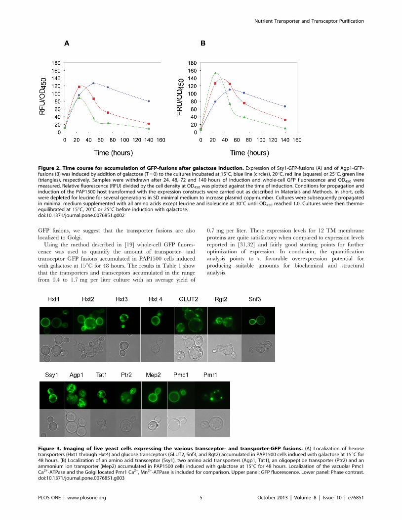
GFP fusions, we suggest that the transporter fusions are also
localized to Golgi.
Using the method described in [19] whole-cell GFP fluores-
cence was used to quantify the amount of transporter- and
transceptor GFP fusions accumulated in PAP1500 cells induced
with galactose at 15uC for 48 hours. The results in Table 1 show
that the transporters and transceptors accumulated in the range
from 0.4 to 1.7 mg per liter culture with an average yield of
0.7 mg per liter. These expression levels for 12 TM membrane
proteins are quite satisfactory when compared to expression levels
reported in [31,32] and fairly good starting points for further
optimization of expression. In conclusion, the quantification
analysis points to a favorable overexpression potential for
producing suitable amounts for biochemical and structural
analysis.
Figure 2. Time course for accumulation of GFP-fusions after galactose induction. Expression of Ssy1-GFP-fusions (A) and of Agp1-GFP-fusions (B) was induced by addition of galactose (T = 0) to the cultures incubated at 15uC, blue line (circles), 20uC, red line (squares) or 25uC, green line(triangles), respectively. Samples were withdrawn after 24, 48, 72 and 140 hours of induction and whole-cell GFP fluorescence and OD450 weremeasured. Relative fluorescence (RFU) divided by the cell density at OD450 was plotted against the time of induction. Conditions for propagation andinduction of the PAP1500 host transformed with the expression constructs were carried out as described in Materials and Methods. In short, cellswere depleted for leucine for several generations in SD minimal medium to increase plasmid copy-number. Cultures were subsequently propagatedin minimal medium supplemented with all amino acids except leucine and isoleucine at 30uC until OD450 reached 1.0. Cultures were then thermo-equilibrated at 15uC, 20uC or 25uC before induction with galactose.doi:10.1371/journal.pone.0076851.g002
Figure 3. Imaging of live yeast cells expressing the various transceptor- and transporter-GFP fusions. (A) Localization of hexosetransporters (Hxt1 through Hxt4) and glucose transceptors (GLUT2, Snf3, and Rgt2) accumulated in PAP1500 cells induced with galactose at 15uC for48 hours. (B) Localization of an amino acid transceptor (Ssy1), two amino acid transporters (Agp1, Tat1), an oligopeptide transporter (Ptr2) and anammonium ion transporter (Mep2) accumulated in PAP1500 cells induced with galactose at 15uC for 48 hours. Localization of the vacuolar Pmc1Ca2+-ATPase and the Golgi located Pmr1 Ca2+, Mn2+-ATPase is included for comparison. Upper panel: GFP fluorescence. Lower panel: Phase contrast.doi:10.1371/journal.pone.0076851.g003
Nutrient Transporter and Transceptor Purification
PLOS ONE | www.plosone.org 5 October 2013 | Volume 8 | Issue 10 | e76851

The purified crude membrane fractions were found to contain
67 to 157 pmol transporter/mg membrane protein corresponding
to a membrane density in the range from 0.4 to 1.5% of the total
membrane protein content (Table 1). This may be appropriate for
structural studies, since successful crystallization was achieved with
recombinant G-protein coupled receptors (GPCRs) purified from
membranes with approximately 50 pmol/mg membrane protein
corresponding to 0.2% of the total membrane protein content
[33,34].
Crude membranes isolated from the expression strains were
analyzed by SDS-PAGE and in-gel fluorescence to assess the
quality of the recombinant transceptor- and transporter-GFP
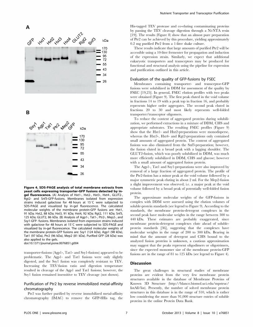
fusions. The results in Figure 4 show that all tested strains
accumulated full-length GFP-fusions in accordance with their
calculated molecular weights including the contribution of 10 to
15 kDa of correctly folded GFP [35]. None of the fusions show
significant degradation and release of free GFP; only a negligible
sign of degradation can be seen for the Hxt4-, Agp1- and Tat1-
fusions. The integrity of the Ssy1-GFP fusion was also investigated
by western analysis using an antibody against Ssy1. A full-length
protein of 130 kDa similar to that identified by in-gel fluorescence
was observed with no sign of degradation (not shown).
Detergent screen of recombinant GFP fusionsA quick detergent screen was performed to identify appropriate
detergents for solubilization of each fusion protein. The results
presented in Figure 5 show that DDM, DM, FC-12 and CYMAL-
5 solubilized the GFP fusions with an acceptable efficiency of
about 50 to 100%, whereas OG and CHAPS resulted in poor
solubilization; except for the Mep2-GFP fusion which solubilized
well in OG. Since DDM is particularly compatible with TEV
protease cleavage of fusion proteins (personal communication with
D. Drew) we decided to use this detergent for solubilization and
purification.
Purification of transporter- and transceptor-GFP fusionsby Ni-affinity chromatography
Purification of the 12 GFP-fusions included in this study was
carried out once using the following procedure: Crude membranes
isolated from 1-liter shake cultures of the various expression strains
induced at 15uC for 48 hours were solubilized in DDM and
purified by Ni-NTA chromatography using a one-step block
gradient procedure as outlined in Materials and Methods. GFP
fluorescence was measured throughout the purification steps to
monitor the yield and the elution profile. As evident from the
fluorescence measurements the binding efficiency to the Ni-NTA
was on average 70% and the recovery in the elution peak was on
average 50%. SDS-PAGE analysis by Coomassie Blue staining
and in-gel fluorescence of the peak fractions from each purification
show that partial purifications of the various fusions were
accomplished (Figure 6).
Comparisons of the protein bands detected show that the
intense Coomassie stained bands correspond to the fluorescent
bands visible by in-gel fluorescence. In addition, the prominent
protein bands migrated with molecular weights in agreement with
that calculated for each fusion protein. Interestingly, Rgt2 and
Snf3 appeared as discrete double and triple bands, respectively,
suggesting posttranslational modifications of these transceptors.
Because of the distinct character of the Rgt2 and Snf3 bands,
posttranslational modifications like glycosylation and ubiquityla-
tion may be excluded; rather phosphorylations may be suggested.
In brief, the results show that considerable amounts of partially
purified transporter- and transceptor-fusion proteins can easily be
isolated using a one-step block gradient procedure.
Removal of GFP-8His tags using TEV proteaseWe analyzed the option to remove the GFP-8His tag from the
fusion proteins by protease cleavage at a TEV site joining the
membrane protein and the GFP-8-His tag (Figure 1). Undigested
and TEV digested fusion proteins were analyzed by SDS-PAGE
and in-gel fluorescence as shown in Figure 7. In the absence of
TEV, intense protein bands corresponding to the full-length
fusions are present on the gel; in addition, high molecular weight
bands presumably representing oligomeric states like dimers and
trimers are observed.
Exposure to TEV protease easily released the GFP-8His tag
from the Hxt1-, Hxt2-, Hxt3-, Hxt4-, Rgt2-, Snf3-, GLUT2-,
Ptr2-, and Mep2-fusions (Figure 7). However, the APC family
Table 1. Yields of transporters and transceptors in whole-cells and crude membranes.
Strain GFP-fusion Expression in whole-cells Expression in membranes
mg transporter/L pmol transporter/mg % of total membrane proteins
PAP7910 Ssy1-GFP-His 0.53 157 1.5
PAP8004 Agp1-GFP-His 0.60 155 1.1
PAP8143 Tat1-GFP-His 0.53 71 0.5
PAP8141 Ptr2-GFP-His 0.51 101 0.7
PAP8139 Mep2-GFP-His 0.68 148 0.8
PAP8131 Hxt1-GFP-His 0.47 67 0.4
PAP8133 Hxt2-GFP-His 0.78 126 0.8
PAP8135 Hxt3-GFP-His 0.71 84 0.5
PAP8137 Hxt4-GFP-His 0.68 126 0.8
PAP8145 Rgt2-GFP-His 1.65 98 0.8
PAP8147 Snf3-GFP-His 1.06 69 0.7
PAP7913 GLUT2-GFP-His 0.36 131 0.8
Accumulation of transporters and transceptors in expression strains induced with galactose at 15uC for 48 hours were quantified using purified yeast-enhanced GFP as astandard to correlate fluorescence to the amount of GFP protein in whole-cells and in membrane extracts as outlined [19].doi:10.1371/journal.pone.0076851.t001
Nutrient Transporter and Transceptor Purification
PLOS ONE | www.plosone.org 6 October 2013 | Volume 8 | Issue 10 | e76851
transporter-fusions (Agp1-, Tat1- and Ssy1-fusions) appeared to be
problematic. The Agp1- and Tat1 fusions were only slightly
digested, and the Ssy1 fusion was completely resistant to TEV.
Increasing the TEV:fusion ratio and digestion temperature
resulted in cleavage of the Agp1 and Tat1 fusions; however, the
Ssy1 fusion remained insensitive to TEV cleavage (not shown).
Purification of Ptr2 by reverse immobilized metal-affinitychromatography
Ptr2 was further purified by reverse immobilized metal-affinity
chromatography (IMAC) to remove the GFP-8His tag, the
His-tagged TEV protease and co-eluting contaminating proteins
by passing the TEV cleavage digestion through a Ni-NTA resin
[19]. The results (Figure 8) show that an almost pure preparation
of Ptr2 can be achieved by this procedure, yielding approximately
0.2 mg purified Ptr2 from a 1-liter shake culture.
These results indicate that large amounts of purified Ptr2 will be
accessible using a 10-liter fermenter for propagation and induction
of the expression strain. Similarly, we expect that additional
eukaryotic transporters and transceptors may be produced for
functional and structural analysis using the pipeline for expression
and purification outlined in this article.
Evaluation of the quality of GFP-fusions by FSECMembranes containing transporter- and transceptor-GFP
fusions were solubilized in DDM for assessment of the quality by
FSEC [19,25]. In general, FSEC elution profiles with two peaks
were obtained (Figure 9). The first peak eluted in the void volume
in fractions 14 to 19 with a peak top in fraction 16, and probably
represents higher order aggregates. The second peak eluted in
fractions 20 to 30 and most likely represents well-folded
transporter/transceptor oligomers.
To reduce the content of aggregated proteins during solubili-
zation, we performed extractions in a mixture of DDM, CHS and
appropriate substrates. The resulting FSEC profiles (Figure 9)
show that the Hxt1- and Hxt2-preparations were monodisperse,
whereas the Hxt3-, Hxt4- and Rgt2-preparations only contained
small amounts of aggregated protein. The content of aggregated
fusions was also eliminated from the Snf3-preparation; however,
the fusion eluted in a broad peak with a lagging shoulder. The
GLUT2-fusion, which was poorly solubilized in DDM, was much
more efficiently solubilized in DDM, CHS and glucose; however
with a small amount of aggregated fusion protein.
The Agp1-, Tat1 and Ssy1-preparations were also improved by
removal of a large fraction of aggregated protein. The profile of
the Ptr2-fusion has a minor peak at the void volume followed by a
major symmetric peak eluting in about 2 ml. For the Mep2-fusion
a slight improvement was observed; i.e. a major peak at the void
volume followed by a broad peak of potentially well-folded fusion
protein.
The approximate molecular weights of the GFP-fusions in
complex with DDM were assessed using the elution volumes of
soluble-protein standards (see legend to Figure 9). According to the
standards, the membrane protein-detergent complexes in the
second peak have molecular weights in the range between 300 to
440 kDa. These estimates are probably exaggerated, since
membrane protein-detergent complexes elute ahead of soluble-
protein standards [36], suggesting that the complexes have
molecular weights in the range of 200 to 300 kDa. Bearing in
mind that the amount of detergent and CHS bound to the
analyzed fusion proteins is unknown, a cautious approximation
may suggest that the peaks represent oligodimers or oligotrimers,
since the expected monomer size of the membrane protein-GFP
fusions are in the range of 81 to 125 kDa (see legend to Figure 4).
Discussion
The great challenges in structural studies of membrane
proteins are evident from the very few membrane protein
structures available in the database of Membrane Proteins of
Known 3D Structure (http://blanco.biomol.uci.edu/mpstruc/
listAll/list). Presently, the number of solved membrane protein
structures in this database is in the range of 310, which is rather
low considering the more than 91,000 structure entries of soluble
proteins in the online Protein Data Bank.
Figure 4. SDS-PAGE analysis of total membrane extracts fromyeast cells expressing transporter-GFP fusions detected by in-gel fluorescence. (A) Analysis of Hxt1-, Hxt2-, Hxt3-, Hxt4-, GLUT2-,Rgt2- and Snf3-GFP-fusions. Membranes isolated from expressionstrains induced galactose for 48 hours at 15uC were subjected toSDS-PAGE and visualized by in-gel fluorescence. The calculatedmolecular weights of the membrane protein-GFP fusions are: Hxt1,91 kDa; Hxt2, 88 kDa; Hxt3, 91 kDa; Hxt4, 92 kDa; Rgt2, 111 kDa; Snf3,125 kDa; GLUT2, 86 kDa. (B) Analysis of Agp1-, Tat1-, Ptr2-, Mep2-, andSsy1-GFP- fusions. Membranes isolated from expression strains inducedwith galactose for 48 hours at 15uC were subjected to SDS-PAGE andvisualized by in-gel fluorescence. The calculated molecular weights ofthe membrane protein-GFP fusions are: Ssy1 (124 kDa), Agp1 (98 kDa),Tat1 (97 kDa), Ptr2 (96 kDa), Mep2 (81 kDa). Purified GFP (28 kDa) wasalso applied to the gels.doi:10.1371/journal.pone.0076851.g004
Nutrient Transporter and Transceptor Purification
PLOS ONE | www.plosone.org 7 October 2013 | Volume 8 | Issue 10 | e76851

At present, the list of structures of transporters of the MFS and
of the APC superfamily in the database of Membrane Proteins of
Known 3D Structure includes only 10 and 6 structures of
prokaryotic origin, respectively, whereas no eukaryotic structures
are yet available. Examples of crystal structures of bacterial
members of the APC family comprise the arginine-dependent
arginine:agmatine antiporter AdiC from E. coli [37,38] and a
broad-specificity amino acid transporter ApcT from Methanocaldo-
coccus jannaschii [39]. Examples of crystal structures of bacterial
members of the MFS comprise lactose permease from E. coli [40]
and the PepTSo oligopeptide-proton symporter from Shewanella
oneidensis [41].
As discussed in recent reports, the isolation of eukaryotic
membrane proteins produced in S. cerevisiae is challenging, and the
limited number of membrane protein structures is due to
difficulties encountered with production, solubilization and puri-
fication of appropriate amounts of membrane proteins that are
able to form crystals diffracting at a high resolution [42,43]. To
explore the possibilities of improving production of eukaryotic
membrane proteins in S. cerevisiae we set forth to combine our
know-how in a high-copy expression system [18] with contempo-
rary membrane protein-GFP fusion technologies [19]. This
initiative is motivated by our interest in understanding the
molecular mechanisms of the yeast amino acid and glucose
transceptors and their related amino acid and glucose transporters.
Figure 5. Detergent screen of transporter- and transceptor-GFP fusions. Membranes of yeast cells overexpressing the indicated (A) Hxt1-,Hxt2-, Hxt3-, Hxt4-, GLUT2-, Rgt2- and Snf3-GFP-fusions and (B) Agp1-, Tat1-, Ptr2-, Mep2-, and Ssy1-GFP- fusions were solubilized with the sixdetergents indicated. Detergent solubilization efficiency was estimated by GFP fluorescence of the 92,000 rpm supernatant and of the detergent-solubilized membranes before centrifugation as described in Materials and Methods.doi:10.1371/journal.pone.0076851.g005
Figure 6. SDS-PAGE analysis of GFP-fusions isolated by Ni-NTA affinity purification. Peak-fractions from purification of Hxt1-, Hxt2-, Hxt3-,Hxt4-, Rgt2-, Snf3-, GLUT2-, Ssy1-, Agp1-, Tat1-, Ptr2-, and Mep2-GFP-fusions were subjected to SDS-PAGE and visualized by Coomassie Blue staining(upper panel)and in-gel fluorescence (lower panel). The positions of fusion proteins are indicated by arrows. Marker: PageRuler prestained proteinladder (Fermentas). The calculated molecular weights of membrane protein GFP-fusions are: Hxt1, 91 kDa; Hxt2, 88 kDa; Hxt3, 91 kDa; Hxt4, 92 kDa;Rgt2, 111 kDa; Snf3, 125 kDa; GLUT2, 86 kDa, Ssy1 (124 kDa), Agp1 (98 kDa), Tat1 (97 kDa), Ptr2 (96 kDa), Mep2 (81 kDa).doi:10.1371/journal.pone.0076851.g006
Nutrient Transporter and Transceptor Purification
PLOS ONE | www.plosone.org 8 October 2013 | Volume 8 | Issue 10 | e76851

Due to our interest in nutrient transporters and transceptors we
here made constructs that overexpress four hexose transporters
(Hxt1, Hxt2, Hxt3 and Hxt4), two glucose transceptors (Snf3 and
Rgt2), two amino acid transporters (Agp1 and Tat1), an amino
acid transceptor (Ssy1), an oligo peptide transporter (Ptr2), and an
ammonium transceptor (Mep2) from S. cerevisiae as fusions to GFP.
To test expression of a mammalian transceptor we also included
the human glucose transceptor GLUT2 in this study.
We found that the twelve membrane proteins were expressed as
full-length GFP-fusions and located to the plasma membrane and/
or intracellular membranes (Figure 3, Figure 4). As discussed in the
results section some of the transporters and transceptors may
accumulate in Golgi. This may be considered a draw-back leading
to inactive transporters. However, as observed in [44] a plant
plasma membrane H+-ATPase overexpressed in yeast was
localized to the endoplasmatic reticulum and not the plasma
membrane as expected; yet it was fully acticve. In accordance with
this finding, we propose that the highly overexpressed transporters
and transceptors produced in the present study challenges the
capacity of the secretory pathway and leads to accumulation of
functional fusions in intracellular membranes such as Golgi.
The expression vector used in this study has the advantages of a
strong promoter, a very high-copy-number feature and selection
markers that prevent plasmid-loss. Furthermore, the host strain
PAP1500 is tailored to overproduce the Gal4 transcription factor
simultaneously with induction of membrane protein production to
maximize transcription from the extraordinary high number of
plasmids.
Using this expression system we were able to produce the 12
targets in this study in the range of 0.4 mg to 1.7 mg transporter/
transceptor pr. liter culture, when expression strains are grown in
shake flasks (Table 1). The capacity of our expression system is
high-lighted by the overexpression of the Ssy1-GFP fusion. We
obtained a level of Ssy1-GFP of 0.5 mg/L cell culture (Table 1). In
contrast, Ssy1 could not be detected in other studies [45,46], and
only very low expression was reported in [47]. Similarly, we were
unable to detect any expression of Ssy1 using the pYES2
overexpression vector (Invitrogen) (unpuplished results). Obvious-
ly, the features of the PAP1500-GFP expression system make a
huge difference for stable overexpression of Ssy1-GFP fusions. To
our knowledge, the 12 transporters and transceptors in the present
study have not previously been overexpressed to levels that allow
Figure 7. TEV protease digests of Ni-NTA purified transporter- and transceptor GFP-fusions. TEV protease digestions of the indicatedGFP-fusions purified by Ni-NTA affinity chromatography. Digestions were analyzed by SDS-PAGE using in-gel fluorescence to visualize the GFP-fusionsand the released GFP-8His. (2) undigested; (+) digested with TEV protease. Released GFP-8His is indicated with an arrow.doi:10.1371/journal.pone.0076851.g007
Figure 8. Purification of the Ptr2 transporter. Membranes from a 1 litre shake culture were solubilised in DDM and applied to a Ni-NTA column.Ptr2-GFP-His fusions were eluted from the column and digested with TEV protease. Purification of Ptr2 and removal of the released GFP-8His tag wascarried out by reverse IMAC. Samples from the 3-step procedure were analysed by SDS-PAGE in a 10% gel. (A) Detection by in-gel fluorescence. (B)Detection by Coomassie staining. Lane 1: Ni-NTA elution. Lane 2: Cleavage by TEV protease. Lane 3: Flow-through of reverse Ni-NTA chromatographyafter cleavage with TEV protease. M: PageRuler prestained protein ladder (Fermentas). Positions of the Ptr2-GFP-His fusion, Ptr2 transporter, cleavedoff GFP-8His and the TEV protease are indicated.doi:10.1371/journal.pone.0076851.g008
Nutrient Transporter and Transceptor Purification
PLOS ONE | www.plosone.org 9 October 2013 | Volume 8 | Issue 10 | e76851

purification. Presumeably, because yeast expression systems with
an efficiency comparable to our system have not been available.
In the present study we decided to investigate production of the
GFP-fusions in shake flask cultures induced for 48 hours at 15uC.
As evident from the optimization experiments with two fusions in
Figure 2, it is very likely that individual optimization of growth and
induction conditions for each transporter tested in our expression
system may improve the yield. For example the Ssy1-GFP-fusion
accumulated stably at 15uC with a 48 hour induction, whereas the
Agp1-GFP-His fusion peaked after 24 hours at 25uC. Further-
more, when production is scaled up in computer controlled
fermenters, the yield will presumably be increased considerably
Figure 9. FSEC profiles of transporter and transceptor GFP-fusions. FSEC analysis of total membranes solubilized in DDM (blue line) or inDDM, CHS and specific substrates (red line). Membranes were isolated from yeast cells, which were induced for overexpression of the GFP-fusions at15uC for 48 hours. The solubilized membranes were separated on a Superose 12 10/300 column. Fractions were collected and measured for their GFPfluorescence. Molecular weight markers (GE Healthcare Life Science) separated on the same column peaked as follows: Blue Dextran 2000, 2000 kDa,fraction 14, corresponding to a void volume of 7.6 ml; thyroglobulin, 669 kDa, fraction 15; ferritin, 440 kDa, fraction 23; aldolase, 158 kDa, fraction 32;conalbumin, 75 kDa, fraction 36; purified yEGFP (this work), 28 kDa, fraction 45.doi:10.1371/journal.pone.0076851.g009
Nutrient Transporter and Transceptor Purification
PLOS ONE | www.plosone.org 10 October 2013 | Volume 8 | Issue 10 | e76851

according to our experience with the high-copy vector system
[18].Thus, our induction protocol usually produce about 3 grams
of wet weight cells in 1 liter shake cultures, whereas a 10-liter
fermenter produces 250 grams. Taken together, we expect that the
yield of membrane proteins may be increased in the range of 10
times after individual induction optimizations and production in
fermenters.
We also report a fast detergent screen based on centrifugations
in a Beckman airfuge, which reduces the consumption of
membranes and detergent as well as the centrifugation time.
Generally, we found that DDM, DM, FC-12 and CYMAL-5
solubilized the GFP fusions with an acceptable efficiency of about
50 to 100%, whereas OG and CHAPS gave poor solubili-
zation (Figure 5). This screening method may be used in pre-
crystallization screenings for selection of detergents that produce
monodisperse and stable membrane proteins suitable for obtaining
high-resolution X-ray structures.
The monodispersity of the GFP-fusions solubilized either in
DDM or in DDM with CHS and specific substrates was analyzed
by FSEC [19,25]. For most of the fusions solubilization in DDM
resulted in a high content of aggregated protein (Figure 9).
However, solubilization in DDM supplemented with CHS and
specific substrates resulted in highly improved FSEC profiles with
small amounts of aggregated fusions (Figure 9). In particular,
Hxt1-, Hxt2-, Hxt3-, Hxt4-, Rgt2-, GLUT2- and Ptr2-fusions
gave preparations that can be used straight away for purification
and crystallization trials. The remaining fusions need further
screenings for improved profiles in more suitable detergents and
panels of additives such as ligands and lipids. This may rescue
aggregated proteins and give the desired monodisperse prepara-
tions. Alternatively, aggregated material may be resolved from the
presumably well-folded soluble protein by preparative size
exclusion chromatography to deliver monodisperse preparations
ideal for crystallization trials. As discussed in for example
[19,42,32,48] monodisperse preparations are not easily obtained
and often numerous optimization attempts must be carried out.
However, using the PAP1500-GFP expression system with
induction at 15uC for 48 hours a high degree of monodispersity
was easily obtained for the majority of membrane proteins in this
study. Thus, application of our expression system combined with
low temperature induction conditions appears to be favorable for
producing a variety of membrane proteins.
In the present study we demonstrate that partially purified
transporter- and transceptor-GFP-fusions can easily be obtained
from DDM solubilized membranes using a simple one-step Ni-
NTA affinity procedure (Figure 6). Ni-NTA resin binding
efficiencies were in the range of 70% and the recovery was on
average 50%. To our knowledge, these results are the first reports
on purification of these transporters and transceptors.
The purified Hxt1-, Hxt2-, Hxt3-, Hxt4-, Rgt2-, Snf3-,
GLUT2-, Ptr2-, and Mep2-GFP-His fusions were readily cleaved
by TEV protease for removal of the GFP-His tag (Figure 7). On
the contrary, the Agp1- and Tat1-GFP fusions needed higher
incubation temperatures for cleavage, whereas the Ssy1-GFP
fusion was insensitive to cleavage at the employed conditions.
Perhaps steric hindrance in the three APC family-GFP fusions
makes the TEV protease site less accessible.
An example of successful removal of the GFP-tag from the
transporter by TEV cleavage and reverse immobilized metal-
affinity chromatography (IMAC) is shown for the Ptr2-GFP fusion
(Figure 8). Using this approach approximately 0.2 mg purified
Ptr2 was obtained from a 1-liter culture. Following this purifica-
tion scheme, similar results will most likely be achievable for the
other TEV cleavable GFP-fusions in this study; except for the APC
family-GFP fusions, for which an alternative approach for
proteolytic removal of the GFP tag must be explored.
Interestingly, the glucose transceptors Rgt2 and Snf3 migrated
as double and triple bands, respectively, in SDS-PAGE analysis
(Figure 6). These observations have not been reported previously,
and may suggest posttranslational modifications such as phos-
phorylations active in sensing or trafficking mechanisms [49]
during the high-galactose induction conditions.
Taken together, we show that the PAP1500-GFP expression
system has the potential to deliver mg amounts of endogenous
yeast nutrient transceptors and transporters with acceptable FSEC
profiles. We have recently published the use of our expression
system for production of human Aquaporin-1, which accumulated
in unexpected high levels constituting 8.5% of the total membrane
protein content [50]. Similarly, we were able to produce high
quality of rat Adenosine A1 receptor in our system yielding 1.9%
of the total membrane protein content (unpublished data). With
these results, we demonstrate that application of the PAP1500-
GFP expression system and low temperature induction conditions
may facilitate production of high amounts of a wide variety of
eukaryotic membrane proteins in a quality that pave the way for
biophysical, functional and structural studies.
Supporting Information
Table S1 Strains used in this study.
(DOCX)
Table S2 Primers used in this study. Nucleotide sequences
shown in bold are complementary to the template. Human
GLUT2 was amplified from a plasmid with GLUT2 cDNA
obtained from Dr. Armelle Leturque. The sequence shown in
italics is the Kozak sequence from the yeast PMR1 gene used for
GLUT2. All other sequences are used for homologous recombi-
nation.
(DOCX)
Acknowledgments
We thank David Drew for providing plasmid pET20bGFP-8His, a MBP-
TEV-His protease expression plasmid, a protocol for TEV protease
purification and helpful discussions. Armelle Leturque is thanked for
providing a plasmid with human GLUT2 cDNA. David Sørensen is
thanked for excellent technical assistance. We are grateful to Morten
Kielland-Brandt for critical reading of an early version of this paper.
Author Contributions
Conceived and designed the experiments: PSP PAP. Performed the
experiments: PSP PAP. Analyzed the data: PSP PAP. Contributed
reagents/materials/analysis tools: PSP PAP. Wrote the paper: PSP PAP.
References
1. Hediger MA, Romero MF, Peng J, Rolfs A, Takanaga H, et al. (2004) The
ABCs of solute carriers: physiological, pathological and therapeutic implications
of human membrane transport proteins. Eur J Physiol 447: 465–468.
2. Rubio-Texeira M, Van Zeebroeck G, Voordeckers K, Thevelein JM (2010)
Sacchacomyces cerevisiae plasma membrane nutrient sensors and their role in PKA
signaling. FEMS Yeast Res 10: 134–149.
3. Gancedo JM (2008) The early steps of glucose signalling in yeast. FEMS
Microbiol Rev 4: 673–704.
4. Ljungdahl PO (2009) Amino-acid-induced signalling via the SPS-sensing
pathway in yeast. Biochem Soc Trans 37: 242–247.
5. Donaton MC, Holsbeeks I, Lagatie O, Van Zeebroeck G, Crauwels M, et al.
(2003) The Gap1 general amino acid permease acts as an amino acid sensor for
Nutrient Transporter and Transceptor Purification
PLOS ONE | www.plosone.org 11 October 2013 | Volume 8 | Issue 10 | e76851

activation of protein kinase A targets in the yeast Saccharomyces cerevisiae. Mol
Microbiol 50: 911–929.6. Van Zeebroeck, G, Bonini BM, Versele M, Thevelein JM (2009) Transport and
signaling via the amino acid binding site of the yeast Gap1 amino acid
transceptor. Nat Chem Biol 5: 45–52.7. Giots F, Donaton MC, Thevelein JM (2003) Inorganic phosphate is sensed by
specific phosphate carriers and acts in concert with glucose as a nutrient signalfor activation of the protein kinase A pathway in the yeast Saccharomyces cerevisiae.
Mol Microbiol 47: 1163–1181.
8. Van Nuland A, Vandormael P, Donaton M, Alenquer M, Lourenco A, et al.(2006) Ammonium permease-based sensing mechanism for rapid ammonium
activation of the protein kinase A pathway in yeast. Mol Microbiol 59: 1485–505.
9. Hundal HS, Taylor PM (2009) Amino acid transceptors: gate keepers of nutrientexchange and regulators of nutrient signaling. Am J Physiol Endocrinol Metab
296: E603–E613.
10. Poulsen P, Wu B, Gaber RF, Ottow K, Andersen HA, et al. (2005) Amino acidsensing by Ssy1. Biochem Soc Trans 33: 265–268.
11. Wu B, Ottow K, Poulsen P, Gaber RF, Albers E, et al. (2006) Competitive intra-and extracellular nutrient sensing by the transporter homologue Ssy1p. J Cell
Biol 173: 327–331.
12. Poulsen P, Gaber RF, Kielland-Brandt MC (2008) Hyper- and hypo-responsivemutant forms of the Saccharomyces cerevisiae Ssy1 amino acid sensor. Mol
Membrane Biol 25: 164–176.13. Karhumaa K, Wu B, Kielland-Brandt MC (2010) Conditions with high
intracellular glucose inhibit sensing through glucose sensor Snf3 in Saccharomyces
cerevisiae. J Cell Biochem 110: 920–925.
14. Lamb RF (2012) Amino acid sensing mechanisms: an Achilles heel in cancer?
FEBS Journal 279: 2624–2631.15. Leturque A, Brot-Laroche E, Le Gall M (2009) GLUT2 mutations, translocation
and receptor function in diet sugar managing. Am J Physiol Endocrinol Metab296:E985–E992.
16. Stolarczyk E, Guissard C, Even PC, Grosfeld A, Serradas P, et al. (2010)
Detection of extracellular glucose by GLUT2 contributes to hypothalamiccontrol of food intake. Am J Physiol Endocrinol Metab 298: E1078–E1087.
17. Newstead S (2011) Towards a structural understanding of drug and peptidetransport within the proton-dependent oligopeptide transporter (POT) family.
Biochem Soc Trans 39:1353–1358.18. Pedersen PA, Rasmussen JH, Jorgensen PL (1996) Expression in high yield of pig
alpha 1 beta 1 Na,K-ATPase and inactive mutants D369N and D807N. J. Biol.
Chem. 27: 2514 – 2522.19. Drew D, Newstead S, Sonoda Y, Kim H, von Heijne G, et al. (2008) GFP-based
optimization scheme for the overexpression and purification of eukaryoticmembrane proteins in Saccharomyces cerevisiae. Nat Protoc 3: 784–798.
20. Wickersham LJ (1951) Taxonomy of yeasts. U. S. Dept. Agric. Tech. Bull. 1029.
21. Sherman F (1991) Basic methods of yeast genetics. In: Guthrie C, Fink GR,editors. Guide to yeast genetics and molecular biology, Methods in Enzymology,
vol. 194. Waltham, MA: Academic Press. pp. 3–21.22. Cesareni G, Murray JAH (1987) Plasmid vectors carrying the replication origin
of filamentous single stranded phages. In: Setlow JK, Hollaender A, editors.Genetic Engineering, Principles and Methods, vol 9. New York: Plenum Press.
pp. 135–149.
23. Lowry OH, Rosebrougn NJ, Farr L, Randall RJ (1951) Protein measurementwith the Folin phenol reagent. J Biol Chem 193: 265–275.
24. Jørgensen JR, Pedersen PA (2001) Role of phylogenetically conserved aminoacid residues in folding of Na+, K+-ATPase. Biochemistry 40: 7301–7308.
25. Kawate T, Gouaux E (2006) Fluoroscence-detection size-exclusion chromatog-
raphy for precrystallization screening of integral membrane proteins. Structure14: 673–681.
26. Drew D, Lerch M, Kunji E, Slotboom DJ, de Gier JW (2006) Optimization ofmembrane protein overexpression and purification using GFP fusions. Nat
Methods 3: 303–313.
27. Erhart E, Hollenberg CP (1983) The presence of a defective LEU2 gene on 2 muDNA recombinant plasmids of Saccharomyces cerevisiae is responsible for curing and
high copy number. J Bacteriol 156: 625–635.28. Romanos MA, Scorer CA, Clare JJ (1992) Foreign Gene Expression in Yeast: a
Review. Yeast 8: 423–488.
29. Cunningham K, Fink GR (1994) Calcineurin-dependent growth control inSaccharomyces cerevisiae mutants lacking PMC1, a homolog of plasma membrane
Ca2+ -ATPases. J Cell Biol 124: 351–63.
30. Antebi A, Fink GR (1992) The yeast Ca2+-ATPase homologue, PMR1; isrequired for normal Golgi function and localizes to a novel Golgi-like structure.
Mol Biol Cell 3: 633–654.
31. Drew D, Kim H (2012) Optimizing Saccharomyces cerevisiae induction regimes. In:
Bill RM, editor. Recombinant Protein Production in Yeast: Methods andProtocols, Methods in Molecular Biology, vol. 866. Berlin: Springer Science+-Business Media. pp. 191–195
32. Shiroishi M, Tsujimoto H, Makyio H, Asada H, Yurugi-Kobayashi T, et al.
(2012) Platform for the rapid construction and evaluation of GPCRs forcrystallography in Saccharomyces cerevisiae. Microb Cell Fact 11: 78.
33. Jaakola VP, Griffith MT, Hanson MA, Cherezov V, Chien EYT, et al. (2008)
The 2.6 Angstrom Crystal Structure of a Human A2A Adenosine ReceptorBound to an Antagonist. Science 322: 1211–1217.
34. Shiroishi M, Kobayashi T, Ogasawara S, Tsujimoto H, Ikeda-Suno C, et al.(2011) Production of the stable human histamine H1 receptor in Pichia pastoris for
structural determination, Methods 55: 281–286.
35. Geertsma ER, Groeneveld M, Slotboom DJ, Poolman B (2008) Quality controlof overexpressed membrane proteins. Proc Natl Acad Sci USA 105: 5722 –
5227.
36. Fang Y, Kolmakova-Partensky L, Miller C (2007) A bacterial arginine agmatine
exchange transporter involved in extreme acid resistance. J Biol Chem 282: 176–182.
37. Gao X, Lu F, Zhou L, Dang S, Sun L, et al. (2009) Structure and mechanism of
an amino acid antiporter. Science 324: 1565–1568.
38. Fang Y, Jayaram H, ShanT, Kolmakova-Partensky L, Wu F, et al. (2009)
Structure of a prokaryotic virtual proton pump at 3.2A resolution. Nature 460:1040–1043.
39. Shaffer PL, Goehring A, Shankaranarayanan A, Gouaux E (2009) Structure and
Mechanism of a Na+-Independent Amino Acid Transporter. Science 325: 1010–
1014.
40. Abramson J, Smirnova I, Kasho V, Verner G, Kaback HR, et al. (2003)Structure and mechanism of the lactose permease of Escherichia coli. Science 310:
610–615.
41. Newstead S, Drew D, Cameron AD, Postis VL, Xia X (2011) Crystal structure
of a prokaryotic homologue of the mammalian oligopeptide–proton symporters,PepT1 and PepT2. EMBO J 30, 417–426.
42. Sonoda Y, Cameron A, Newstead S, Omote H, MoriyamaY, et al. (2010) Tricks
of the trade used to accelerate high-resolution structure determination ofmembrane proteins. FEBS Letters 584: 2539–2547.
43. Clark KM, Fedoriw N, Robinson K, Conelly SM, Randles J, et al. (2010)Purification of transmembrane proteins from Saccharomyces cerevisiae for X-ray
crystallography. Protein Expr Purif 71: 207–223.
44. Villalba JM, Palmgren MG, Berberian GE, Ferguson C, Serrano R (1992)Functional expression of plant plasma membrane H+-ATPase in yeast
endoplasmic reticulum. J Biol Chem 267: 12341–12349.
45. Ghaemmaghami S, Huh WK, Bower K, Howson RW, Belle A, et al. (2003)
Global analysis of protein expression in yeast. Nature 425: 737–741.
46. Gelperin DM, White MA, Wilkinson ML, Kon Y,1 Kung LA, et al. (2005)Biochemical and genetic analysis of the yeast proteome with a movable ORF
collection. Genes Dev 19: 2816–2826.
47. Spira F, Mueller NS, Beck G, von Olshausen P, Beig J, et al. (2012) Patchwork
organization of the yeast plasma membrane into numerous coexisting domains.Nat Cell Biol 14: 640–648.
48. Hattori M, Hibbs RE, Gouaux E (2012) A fluorescence-detection size-exclusion
chromatography-based thermostability assay for membrane protein precrystalli-
zation screening. Structure 20: 1293–1299.
49. Kriel J, Haesendonckx S, Rubio-Texeira M, Van Zeebroeck G, Thevelein JM(2011) From transporter to transceptor: Signaling from transporters provokes re-
evaluation of complex trafficking and regulatory controls. Bioessays 33: 870–879.
50. Bomholt J, Helix-Nielsen C, Scharff-Poulsen P, Pedersen PA (2013) Recombi-
nant production of human Aquaporin to an exceptional high membrane density
in Saccharomyces cerevisiae PlosOne 8(2):e56431.
Nutrient Transporter and Transceptor Purification
PLOS ONE | www.plosone.org 12 October 2013 | Volume 8 | Issue 10 | e76851