rui kano, dvm. phd department of pathobiology, … · department of pathobiology, nihon university...
TRANSCRIPT

Rui Kano, DVM. PhDDepartment of Pathobiology, Nihon University School of
Veterinary Medicine
Aspergillosis of the dog and cat

Cases of aspergillosis are increasing in small animals in Japan
Because of increasing;agingchemotherapy, inbreeding ?

Molecular identification for Aspergillus species-Why ?
Identification of clinical isolates in species level of Aspergillus is important, in relation with the susceptibilities of these organisms to various antifungal drugs.
A. fumigatus and N. fischeri produce the toxic metabolite, gliotoxin which has been implicated in infectivity.
Molecular identification has received wide recognition as a rapid and easy identification system.

Currently accepted classification of Aspergillus section Fumigati, including only
isolates implicated in human diseaseAntifungal susceptibilities; MIC (g/ml)
Species AMB ITZ VRZ CAPA. fumigatus 0.5 0.5 0.25 0.125A. lentulus 1-2 0.5-1 1-2 2-16A. viridinutansN. pseudofischeri 0.5 1 2 0.015N. fischeriN. udagawae 0.5-4 0.125-0.5 0.25-1 0.015-0.06N. hiratsukae
Balajee et al., J. Clin. Microbiol. 2005; 43, 5996-5999Balajee et al., Eukaryot. Cell. 2006; 5, 1705-1712

Morphological identification vs. molecular identification
Aspergillus species can be identified by morphological characteristics, however this is limited because morphology differences are small and largely dependent on growth conditions.
Molecular analysis is a rapid and easy identification method. However, it has not been proved what is the most useful system to identify the species.
A
TG
C
A
TG
C
A
TG
C
A
TG
C
A
TG
C
A
TG
C
A
TG
C
A
TG
C

Orbital aspergillosis has been reported in humans whoreceived steroid treatment and chemotherapy, and were suffering from immuno-suppressive diseases, including acquired immunodeficiency syndrome, but is extremely rare in cats.
A. fumigatus is reported as the most frequent cause of sino-orbital aspergillosis in humans and small animals.
Orbital aspergillosis

Case 1:A 1 year and eleven months old spayed female American short Hair cat.Chief complaint: progressive protrusion of the left third eyelid and serous ocular and nasal discharge of 3 months duration.
Case presentation

Computerized tomography (CT) revealed a soft-tissue mass within the left orbit.
Histopathologic examination of this mass from the left eye orbit revealed granulomatousinflammation with many branching hyphal filaments in the granules.
Serum Aspergillus antigen titers measured by the latex galactomannan agglutination test (Pastorex Aspergillus; SanofiDiagnostics Pasteur, Marnes-la-coquette, France) was positive.
LDiagnosis

Velvety, grayish-greencolored colonies developed from this mass samples after 1-week incubation on Sabouraud's dextrose agar at 25oC; based on gross and microscopic characters, this isolate was identified asA. fumigatus.
The case was diagnosed as proven aspergillosis due toA. fumigatus
Identification

Schema for molecular identification procedure of the isolate
Cultured on SDA agar at 30 oC for 2-3 days
DNA extraction
PCR amplification ITS and -tublin regions
Sequencing
ITS1 ITS218S 5.8S 28S
About 550-bp
Homology search by BLAST data base analysis
About 450-bp
b-tubulin geneITS region

Results of comparative sequence analyses in GenBank
ITS region sequence of the clinical isolate was 100 % identical to both A. udagawaeand N. fischeri, and < 98% identical to A. fumigatus.
-tubulin region sequence analyses of clinical isolate was 99% identical to A. udagawae.

Drug Species Dose (mg/kg) Route Interval AMB Cat 0.25 IV 48 h
Dog 025-0.5 IV 48 h
AMB (lipid) Cat 1 IV 3 d/weekDog 2-3 IV 3 d/week
ITZ Cat 10 PO 24 hDog 2.5-5.0 PO 12 h
Therapy for disseminated Aspergillosis
Green CE. Infectious Diseases of Dog and Cat, 3rd ed. 2006. Sunders Elsevier
AMB; Ampotericin B, ITZ; Itraconazole

ITZ 20 mg / kg orally once a day AMB 0.2 mg / kg, 3 days per week
Therapy for this case
ITZ was discontinuedAMB was continued
Micafungin1 mg / kg, 3 days per week
The mass continued to increase
4 weeks
No response to therapy was seen and the cat continued to deteriorate and expired.
5 weeks

The in vitro susceptibilities of the isolate against the antifungal drugs
AMB ITZ VRZ POSIsolate >32 g/ml 0.38 g/ml 0.19 g/ml 0.19 g/ml
AMB; Ampotericin B, ITZ; Itraconazole, VRZ; Voriconazole, POS; Posaconazole
Minimal Inhibitory Concentration

ConclusionTo our knowledge, this is the first case of the recovery of A. udagawae from orbital aspergillosisin a cat.
The cat failed therapy with AMB and this correlated with in vitro low susceptibility of A.udagawae to AMB.
It is important to identify the isolate correctly as to guide therapeutic decisions canine and feline aspergillosis.
Kano R,Itamoto K, Okuda M, Inokuma H, Hasegawa A, Balajee SA (2008) ::Isolation of Aspergillus udagawae from a fatal case of feline orbital aspergillosis.Mycoses 51, 360-361.

A. terreus infectionA. terreus has been recognized to be an etiological argent of disseminated infection due to Aspergillus species in dogs and cats. Human and animal aspergillosis due to A. terreus is poor to response against AMB therapy.
We isolated Aspergillus sp. from a canine case with renal disease and identified it as A. terreus by the molecular analysis.

Case 2:A 6 year old male Shibainu dog
Chief complaint: anorexia and weakness of 4 days duration.
Clinical findings:On physical examination, clinical abnormality sing was not find. Hematologic and serum biochemistry analysis showed leukocytosis, high levels of CRP, BUN, creatinin and liver enzymes.
Case presentation
PCV 40 %WBC 29400 /lCRP 18 mg/dlTP 9.8 g/dlBUN 70 mg/dlCreatinin 5.4 mg.dlGlucose 120 mg/dlALT 46 U/lALP 240 U/l
Hematology and serum biochemistry in patient dog

Bilateral renomegalieswere on the lateral view of radiograph.
Radiographic evaluation could not find skeletal and respiratory system abnormality.
Dilation of renal pelvis and mass were on the view of ultrasound.
Radiology and ultra-sonography
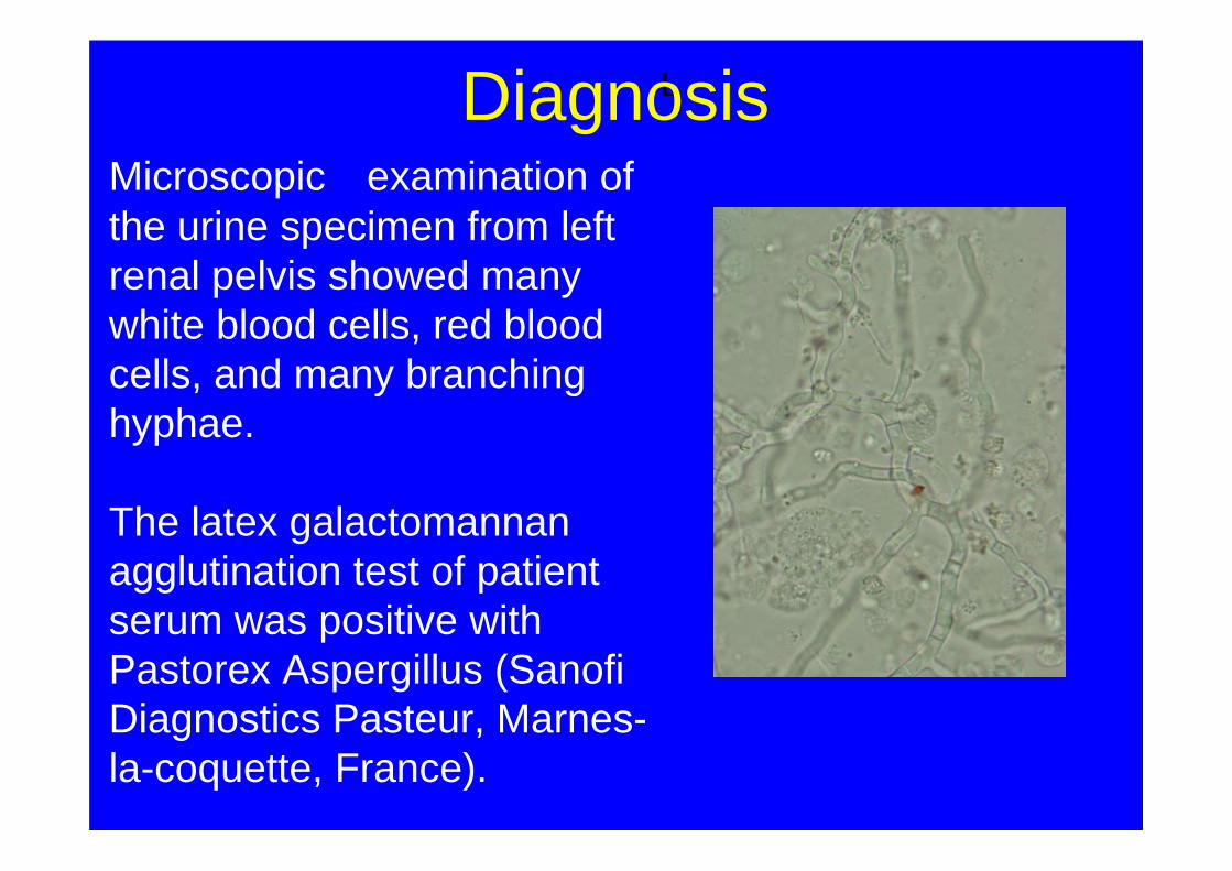
Microscopic examination of the urine specimen from left renal pelvis showed many white blood cells, red blood cells, and many branching hyphae.
The latex galactomannanagglutination test of patient serum was positive with Pastorex Aspergillus (SanofiDiagnostics Pasteur, Marnes-la-coquette, France).
LDiagnosis

From this urine samples, velvety, brown-cinnamon–colored colonies grew after 5-day incubation on potato dextrose agar at 25oC; basedon gross and microscopic characters, this isolate was identified as A.terreus.
ITS and -tubulin sequence of the clinical isolate was 100 % identical to A. terreus.
The case was diagnosed as proven aspergillosis due to A. terreus
Isolation and identification

The in vitro susceptibilities of the isolateagainst the antifungal drugs
ITZ VRZ POSIsolate 0.19 g/ml 0.047 g/ml 0.19 g/ml
Minimal Inhibitory Concentration
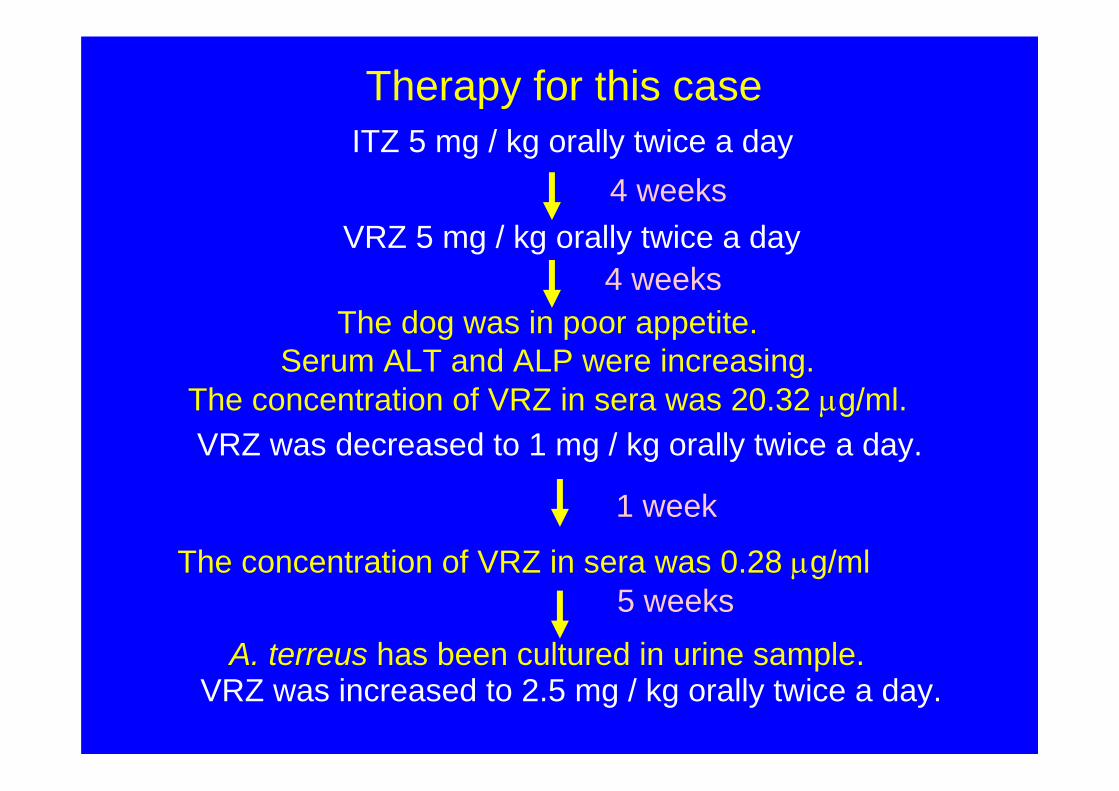
VRZ 5 mg / kg orally twice a day
Therapy for this case
VRZ was decreased to 1 mg / kg orally twice a day.
The dog was in poor appetite.Serum ALT and ALP were increasing.
The concentration of VRZ in sera was 20.32 g/ml.
4 weeks
A. terreus has been cultured in urine sample.
5 weeksThe concentration of VRZ in sera was 0.28 g/ml
VRZ was increased to 2.5 mg / kg orally twice a day.
1 week
ITZ 5 mg / kg orally twice a day 4 weeks

ConclusionThe case had been treated with ITZ (5 mg/kg BID for 4 weeks), however, A. terreus was still isolated from urine sample.
VRZ was administered to the case, since antifungal susceptibility testing revealed that this isolate had higher in vitro MIC to ITZ rather than VRZ.
However, the primary focus of A. terreusinfection was not determined in this patient.

For appropriate therapy on animal aspergillosis, the isolates should be identified molecularly and their susceptibility to antifungal drugs should be determined by MIC testing.
General conclusion