rt-7300 auto hematology analyzer · manual calibration in main screen, ... service rt-7300...
TRANSCRIPT
1
INTRODUCTION:
Automated cell counters sample the blood, and quantify, classify, and describe cell populations
using both electrical and optical techniques. Electrical analysis involves passing a dilute solution
of the blood through an aperture across which an electrical current is flowing. The passage of
cells through the current changes the impedance between the terminals (the Coulter principle).[ A
lytic reagent is added to the blood solution to selectively lyse the red cells (RBCs), leaving
only white cells (WBCs), and platelets intact. Then the solution is passed through a second detector.
This allows the counts of RBCs, WBCs, and platelets to be obtained. The platelet count is easily
separated from the WBC count by the smaller impedance spikes they produce in the detector due
to their lower cell volumes.
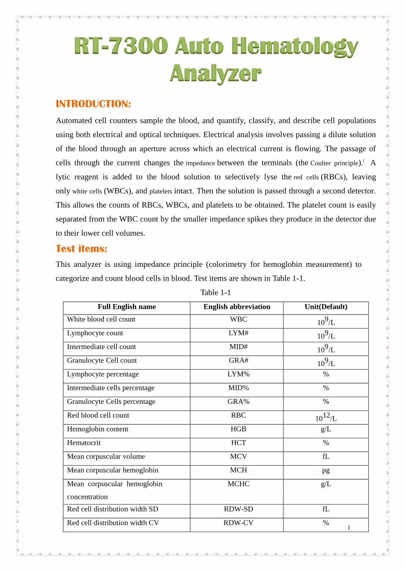
Test items:
This analyzer is using impedance principle (colorimetry for hemoglobin measurement) to
categorize and count blood cells in blood. Test items are shown in Table 1-1.
Table 1-1
Full English name English abbreviation Unit(Default)
White blood cell count WBC 109/L
Lymphocyte count LYM# 109/L
Intermediate cell count MID# 109/L
Granulocyte Cell count GRA# 109/L
Lymphocyte percentage LYM% %
Intermediate cells percentage MID% %
Granulocyte Cells percentage GRA% %
Red blood cell count RBC 1012
/L
Hemoglobin content HGB g/L
Hematocrit HCT %
Mean corpuscular volume MCV fL
Mean corpuscular hemoglobin MCH pg
Mean corpuscular hemoglobin
concentration
MCHC g/L
Red cell distribution width SD RDW-SD fL
Red cell distribution width CV RDW-CV %
RT-7300 Auto Hematology
Analyzer
2
Platelet count PLT 109/L
Mean platelet volume MPV fL
Platelet distribution width PDW %
Plateletcrit PCT %
Platelet–large cell ratio P-LCR %
White blood cell histogram WBC Histogram
Red blood cell histogram RBC Histogram
Platelet histogram PLT Histogram
RT 7300 technical Specification
Test principle: WBC/RBC/PLT: Impedance method; HGB: colorimetry
Aspiration volume: 9.8μL(Whole Blood), 9.8μL(Anticoagulant Peripheral
Blood), 20μL(Pre-diluted Peripheral Blood)
Test rate: About 1 min /ea.
Working environment: 15°C~35°C, relative humidity ≤80%
Store environment: 0°C~40°C, relative humidity ≤85%
Power supply: A.C.110V~220V, 50/60Hz
Input power: 96VA
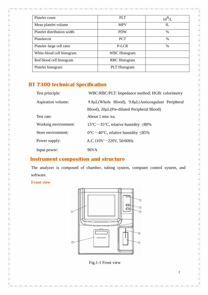
Instrument composition and structure
The analyzer is composed of chamber, tubing system, computer control system, and
software.
Front view
Fig.1-1 Front view
3
Display screen: Display the software interface
Indicator light: green at startup, turn red when starting sample test, turn green after test.
Feed key: built-in printer feeds paper outward
Aspiration key: in sample analysis, press this key, the instrument will aspirate sample
Printer cover: print paperinstalling position for built-in printer
Sampling needle: use sampling needle to aspirate sample into chamber
Aspiration key: in sample analysis, press this key, the instrument will aspirate sample
Reagent
The instrument needs to use Lyse, cleanser, diluents for measurement and maintenance. To
ensure accuracy of test result, please use reagents accompanied with instrument.
Preparation before startup
Before startup, the operator shall check by following steps to make sure system is ready:
Check if the diluent, Lyse and cleanser meet the need of current test. Check if the waste
bottle is full, tubing system is ok and without folded, all the connection is ok.
Whether instrument power adapter is safely and stably connected to power socket.
Check whether built-in or external printer paper is sufficiently installed in right
position.
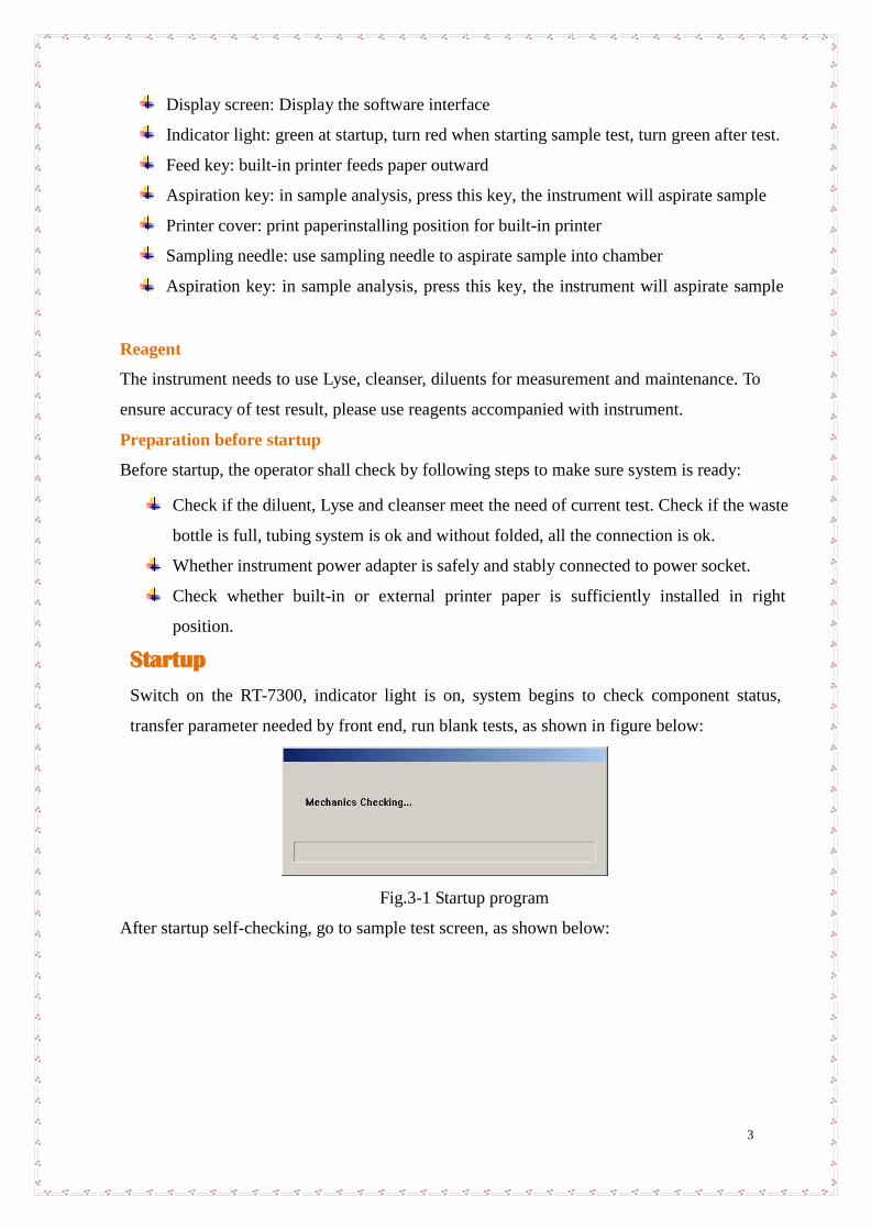
Startup
Switch on the RT-7300, indicator light is on, system begins to check component status,
transfer parameter needed by front end, run blank tests, as shown in figure below:
Fig.3-1 Startup program
After startup self-checking, go to sample test screen, as shown below:

4
Parameter
Reference Range
WBC
≤ 0.2 × 10⁹
/ L
RBC
≤ 0.03 × 10¹²
/ L
HGB
≤ 2 g / L H
CT ≤ 0.5
% PLT
≤ 10 × 109
/ L
Fig.3-2 Blank test result
Blank test only shows test results of WBC, RBC, HGB, HCT and PLT. The operator can run
blank test at any time required. Press “Aspiration Key” to start blank test. During test, the
indicator light turns red, status column on top of screen will display test status.
Blank test acceptable range is as follows:
If the results of blank test are not in this acceptable range, please repeat the steps above until the
results are good. If the results are still out of range after 5 times’ tests, please check if the
reagent and connections of pipeline are good. And please try to use functions like “Remove
Blockage”, “Back Flush”, “Cleaning” and “Concentrated Cleanser Soaking” in the software
Service Menu to solve possible aperture blockage problem.
Blood sample collection
Blood sample collection is classified as venous blood and peripheral blood.
Venous blood collection
Venous blood can be collected by using vacuum negative pressure tube or ordinary method
under atmospheric pressure, anticoagulant must be dropped to all venous blood collection
containers in advance, usually EDTA.K2.2H2O is adopted as anticoagulant, with content of
1.5-2.2mg/ml.

5
Peripheral blood collection
Blood sampling position:
For adults, the inner side of middle finger or ring finger tips of left hand is better; for children
above half a year old, middle finger is better; for infant below half a year old, blood is usually
sampled from thumb or outer side of foot bottom.
Blood sampling method:
It shall be carried out in accordance with peripheral blood collection standard of health authority.
Peripheral blood collection normally adopts local centesis; the typical collection method is
to pierce through finger end. Blood tube uses 20μL constant volume blood tube or bullet
blood tube. It is recommended to collect no less than 30μL blood to facilitate double check.
During blood sampling, if blood flow stagnates, one may slightly press farther end of wound,
never force around piercing hole. Avoid tissue fluid mixed with blood, hampering test analysis
result accuracy.
Blood sample counting
You can select blood type by “Mode” button or select form the box at sample data edit menu.
There are three kind of blood type for selection, which are whole blood, anticoagulant peripheral
blood and pre-diluted peripheral blood.
If select pre-diluted peripheral blood mode, first add 20μl peripheral blood at the diluting cup,
then click “Diluent” button, a message box as following will appear:
Fig.3-5 Add diluent
Press the aspiration key will add 700μl diluent to the diluting cup to complete the dilution out
of instrument, the analyzer will aspirate 300μl diluent the sample for counting.
The test procedures are as following:
1. Put sample cup under sampling needle, press aspiration key, the instrument aspirate
blood sample, wait until sampling needle rises inside instrument, remove sample cup.
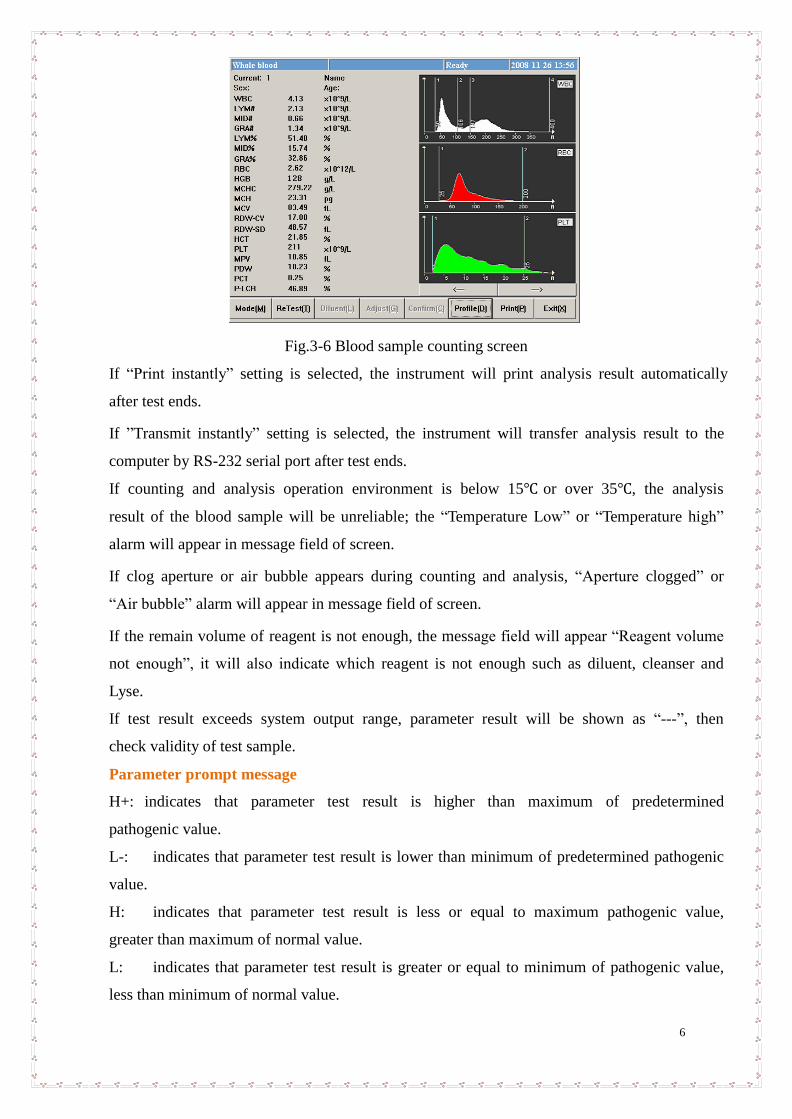
2. Instrument begins to analyze sample, frame top message box shows “Testing…”, after
test, parameter, test result and histogram will be shown as follows:
6
Fig.3-6 Blood sample counting screen
If “Print instantly” setting is selected, the instrument will print analysis result automatically
after test ends.
If ”Transmit instantly” setting is selected, the instrument will transfer analysis result to the
computer by RS-232 serial port after test ends.
If counting and analysis operation environment is below 15℃ or over 35℃, the analysis
result of the blood sample will be unreliable; the “Temperature Low” or “Temperature high”
alarm will appear in message field of screen.
If clog aperture or air bubble appears during counting and analysis, “Aperture clogged” or
“Air bubble” alarm will appear in message field of screen.
If the remain volume of reagent is not enough, the message field will appear “Reagent volume
not enough”, it will also indicate which reagent is not enough such as diluent, cleanser and
Lyse.
If test result exceeds system output range, parameter result will be shown as “---”, then
check validity of test sample.
Parameter prompt message
H+: indicates that parameter test result is higher than maximum of predetermined
pathogenic value.
L-: indicates that parameter test result is lower than minimum of predetermined pathogenic
value.
H: indicates that parameter test result is less or equal to maximum pathogenic value,
greater than maximum of normal value.
L: indicates that parameter test result is greater or equal to minimum of pathogenic value,
less than minimum of normal value.
7
Histogram prompt message
LF1: prompts that region on the left side of lymphocyte peak is abnormal, possibly because
of: platelet coagulation, giant platelet, plasmodium, nucleated red blood cell, non-lysed red
blood cell, abnormal lymphocyte, cryoglobulin.
LF2: prompts that lymphocyte peak and intermediate cell region are abnormal, possibly
because of: heteromorphic lymphocyte, plasma cell, atypical cell, initial cell, eosinophils and
basophils population.
LF3:prompts that the region between intermediate cell area and neutrophil peak is abnormal,
possibly because of: immature granulocyte, abnormal cell and eosinophils.
LF4: the region on the right side of neutrophil is abnormal, possibly because of
granulocytosis.
PF1:the region on the right side of platelet is abnormal, indicating probable existence of
large platelet, platelet aggregate, small red blood cell, cell fragment and fibrous protein.
PF2:the region on the left side of platelet is abnormal, indicating probable existence of: small
platelet cell fragment, red blood cell inclusion body and electronic noise interference.
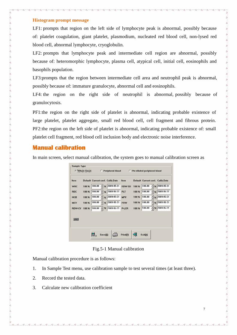
Manual calibration
In main screen, select manual calibration, the system goes to manual calibration screen as
Fig.5-1 Manual calibration
Manual calibration procedure is as follows:
1. In Sample Test menu, use calibration sample to test several times (at least three).
2. Record the tested data.
3. Calculate new calibration coefficient
8
Calculate new calibration coefficient as per formula below:
4. In Manual Calibration screen, select sample type, input new calibration coefficient
in calibration input box, input calibration time in calibration time box. If running
auto-calibration, the parameter calibration time will be automatically updated to the time of
automatic calibration.
5. Click “Save” button to save current calibration result. Click “Exit” button, the system
will not save result, and directly return to main screen.
Auto-calibration
When automatic calibration is selected, after testing calibration sample, the instrument can
automatically calculate new calibration coefficient. Auto-calibration procedure is as follows:
1. Set up calibration sample, select parameters to calibrate;
2. Test calibration sample;
3. Determine calibration coefficient. Auto-calibration screen is as follows:
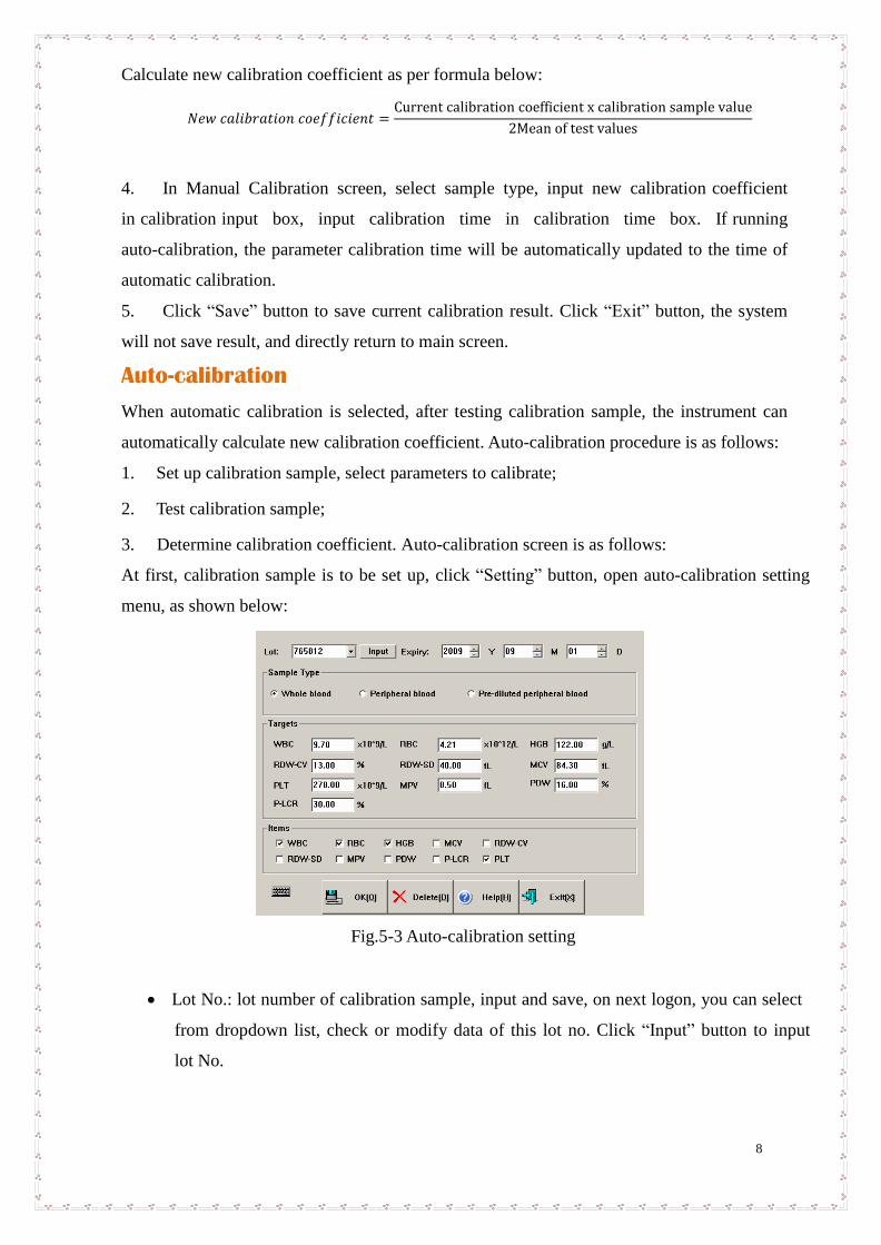
At first, calibration sample is to be set up, click “Setting” button, open auto-calibration setting
menu, as shown below:
Fig.5-3 Auto-calibration setting
Lot No.: lot number of calibration sample, input and save, on next logon, you can select
from dropdown list, check or modify data of this lot no. Click “Input” button to input
lot No.
9
Expiry: validity period of calibration sample, if validity period is less than current
system date, when confirmed to return, the instrument will give alarm of using
another calibration sample to calibrate.
Sample type: blood type of calibration sample.
Targets: reference value of each parameter of calibration sample.
Calibration parameter: parameter to calibrate.
Click “Delete” button, current calibration sample setting data can be deleted. Click “OK” to
save current lot number data and exit. Click “Cancel” button, the system will not function,
and directly return to Auto-calibration running screen.
After setting, return to screen as shown in Fig.5-2, start calibration test, the process is as
follows:
1) Have calibration sample ready, press Aspiration key to start test.
2) After test, calibration result will be shown in current test number column.
3) Repeat test, the instrument allows 20 runs at most.
4) Statistic results and new calibration coefficient will be shown in table below frame after
each test automatically.
5) Unsatisfactory test data rows can be deleted at any time.
If deciding to use new calibration coefficient, click “Save” button, the system will save new
calibration coefficient.
Service
RT-7300 Fully-automatic Hematology Analyzer is a clinical precision analyzer; in order to
maintain good instrument condition, get reliable test result, and reduce instrument fault rate, the
instrument shall be maintained routinely, this chapter introduces related maintenance operation.
Routine maintenance
Startup and shutdown process
At startup, the instrument will run mechanical component test and blank test, for user to
discover problem as early as possible. While running shutdown operation, daily shutdown
maintenance program will be automatically performed. After shutting off instrument power, just
clean workbench and instrument surface.
Automatic rinse
If number of samples instrument tests has reached the number preset by user, then the
instrument will run automatic rinse program. User can adjust automatic rinse frequency at will.
Refer to Chapter 7, System Setting. Rinse can also be done in “Maintenance” menu.
10
Clean instrument surface
Keep instrument working environment clean.
Instrument surface can be cleaned with neutral detergent and wet cloth.
Maintenance program
Back flush
Back flush gem aperture, together with the function of “High voltage pulse”, to prevent and
eliminate clogging of aperture.
High voltage pulse
This function is to burn the gem hole to prevent and eliminate jam of hole.
Drain chambers
Drain off the diluent in WBC and RBC chamber.
Drain pipeline
Drain off liquid in the pipeline.
Remove Blockage
Particular procedure is for eliminate the blockage of hole.
Prime
System will prime automatically during sample test. If you have performed drain operation or
replaced reagent, you shall perform the operation of priming.
1) All reagents: fill diluent, lyse and cleanser into related pipeline.
2) Diluent: fill diluent into related pipeline.
3) Lyse solution: fill lyse solution into related pipeline.
4) Cleanser: fill cleanser into related pipeline.
Cleaning
When you think that the chamber has been polluted or the blank test result keeps unacceptable,
you shall use this function.
Concentrated cleanser soaking
Concentrated cleanser is an alkalescent wash solution. It is used for cleaning pipeline and
chamber. At the service menu, click “Bougie Fluid” to perform the concentrated cleanser
soaking procedure. When the system gives the message to add concentrated cleanser, add it
manually into the chamber. You shall perform this operation once every three days.
Check mechanics
In Service menu, click “Check Mechanics” key, open mechanical test menu as below:
11
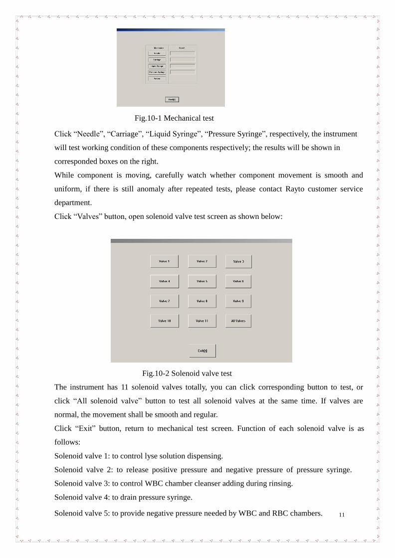
Fig.10-1 Mechanical test
Click “Needle”, “Carriage”, “Liquid Syringe”, “Pressure Syringe”, respectively, the instrument
will test working condition of these components respectively; the results will be shown in
corresponded boxes on the right.
While component is moving, carefully watch whether component movement is smooth and
uniform, if there is still anomaly after repeated tests, please contact Rayto customer service
department.
Click “Valves” button, open solenoid valve test screen as shown below:
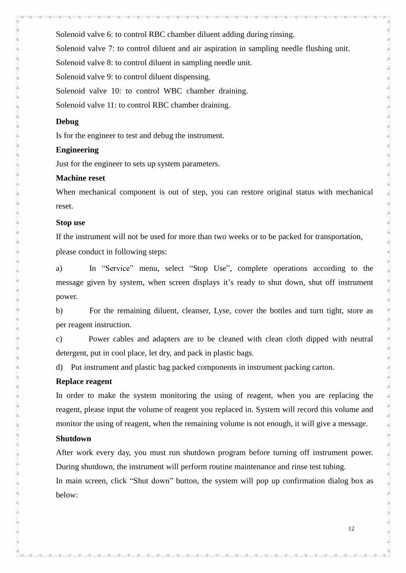
Fig.10-2 Solenoid valve test
The instrument has 11 solenoid valves totally, you can click corresponding button to test, or
click “All solenoid valve” button to test all solenoid valves at the same time. If valves are
normal, the movement shall be smooth and regular.
Click “Exit” button, return to mechanical test screen. Function of each solenoid valve is as
follows:
Solenoid valve 1: to control lyse solution dispensing.
Solenoid valve 2: to release positive pressure and negative pressure of pressure syringe.
Solenoid valve 3: to control WBC chamber cleanser adding during rinsing.
Solenoid valve 4: to drain pressure syringe.
Solenoid valve 5: to provide negative pressure needed by WBC and RBC chambers.
12
Solenoid valve 6: to control RBC chamber diluent adding during rinsing.
Solenoid valve 7: to control diluent and air aspiration in sampling needle flushing unit.
Solenoid valve 8: to control diluent in sampling needle unit.
Solenoid valve 9: to control diluent dispensing.
Solenoid valve 10: to control WBC chamber draining.
Solenoid valve 11: to control RBC chamber draining.
Debug
Is for the engineer to test and debug the instrument.
Engineering
Just for the engineer to sets up system parameters.
Machine reset
When mechanical component is out of step, you can restore original status with mechanical
reset.
Stop use
If the instrument will not be used for more than two weeks or to be packed for transportation,
please conduct in following steps:
a) In “Service” menu, select “Stop Use”, complete operations according to the
message given by system, when screen displays it’s ready to shut down, shut off instrument
power.
b) For the remaining diluent, cleanser, Lyse, cover the bottles and turn tight, store as
per reagent instruction.
c) Power cables and adapters are to be cleaned with clean cloth dipped with neutral
detergent, put in cool place, let dry, and pack in plastic bags.
d) Put instrument and plastic bag packed components in instrument packing carton.
Replace reagent
In order to make the system monitoring the using of reagent, when you are replacing the
reagent, please input the volume of reagent you replaced in. System will record this volume and
monitor the using of reagent, when the remaining volume is not enough, it will give a message.
Shutdown
After work every day, you must run shutdown program before turning off instrument power.
During shutdown, the instrument will perform routine maintenance and rinse test tubing.
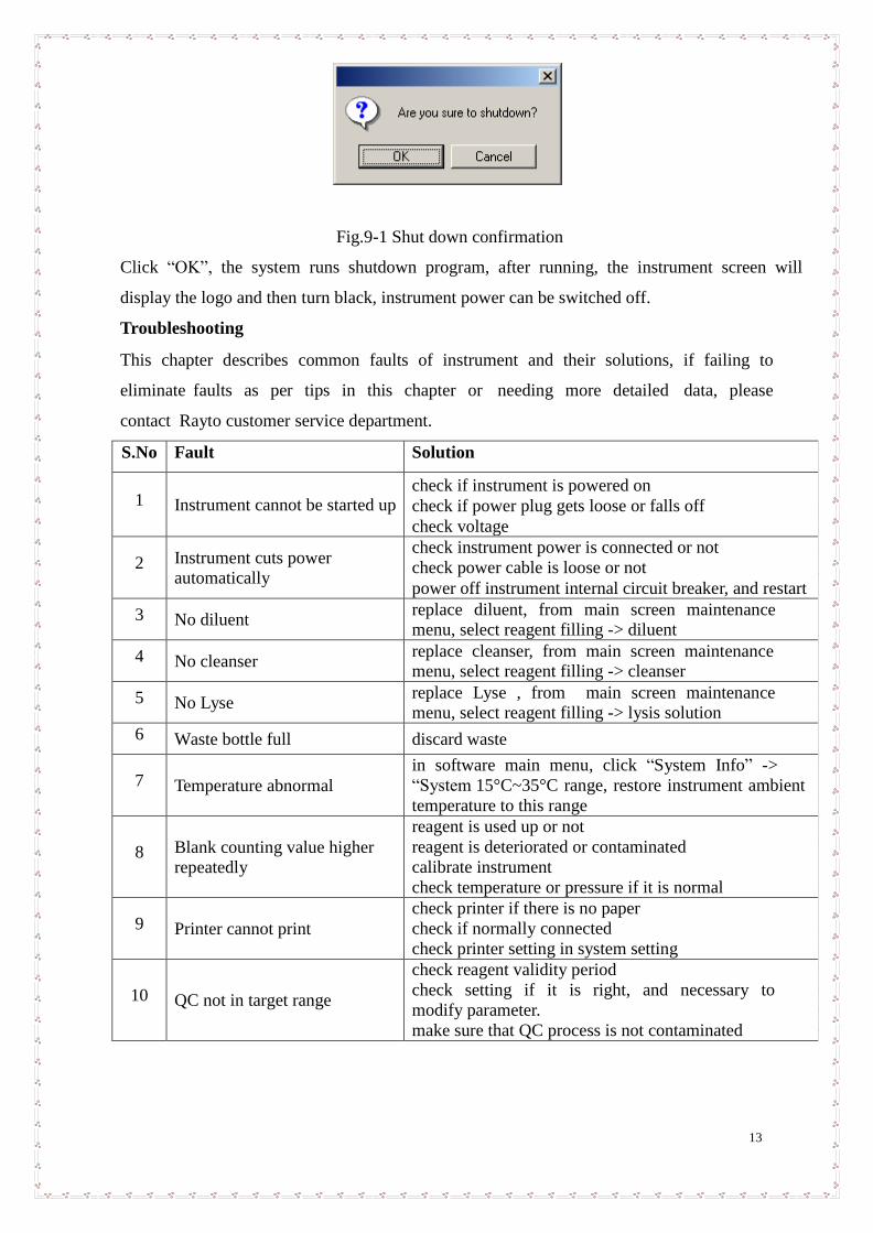
In main screen, click “Shut down” button, the system will pop up confirmation dialog box as
below:
13
Fig.9-1 Shut down confirmation
Click “OK”, the system runs shutdown program, after running, the instrument screen will
display the logo and then turn black, instrument power can be switched off.
Troubleshooting
This chapter describes common faults of instrument and their solutions, if failing to
eliminate faults as per tips in this chapter or needing more detailed data, please
contact Rayto customer service department.
S.No Fault Solution
1 Instrument cannot be started up check if instrument is powered on
check if power plug gets loose or falls off
check voltage
2 Instrument cuts power
automatically
check instrument power is connected or not
check power cable is loose or not
power off instrument internal circuit breaker, and restart
3 No diluent replace diluent, from main screen maintenance
menu, select reagent filling -> diluent
4 No cleanser replace cleanser, from main screen maintenance
menu, select reagent filling -> cleanser
5 No Lyse replace Lyse , from main screen maintenance
menu, select reagent filling -> lysis solution
6 Waste bottle full discard waste
7 Temperature abnormal
in software main menu, click “System Info” ->
“System 15°C~35°C range, restore instrument ambient
temperature to this range
8 Blank counting value higher
repeatedly
reagent is used up or not
reagent is deteriorated or contaminated
calibrate instrument
check temperature or pressure if it is normal
9 Printer cannot print
check printer if there is no paper
check if normally connected
check printer setting in system setting
10 QC not in target range
check reagent validity period
check setting if it is right, and necessary to
modify parameter.
make sure that QC process is not contaminated