rotograph evo - villa radiology systemsknowledge.villaus.com/product manuals/older products... ·...
TRANSCRIPT

Version September, 27 2011 (Rev. 1)
Rotograph EVO 0051
(110-120V version)
User's Manual

USER'S MANUAL Revision history
(Rev. 1) Rotograph EVO (110-120V)
Revision history
Rev. Date Page/s Modification description
0 31.07.09 - Document approval.
1 28.09.11 All New wall fixing brackets. "Rotograph EVO" no more displayed at device switching ON. Release FW version 1.05 on CPU board. New laser model for patient centering. New CARPUS default esposure parameters. EMC tables on "Warnings" paragraph added. Bite protective sleeves added. Film/screen combinations table updated. New TMJ positioner. Release FW version 1.06 on CPU board (DAP value added). Patient positioning images improvement.
(Ref. RDM 7086, RDM 7094, RDM 7113, RDM 7178, RDM 7201, RDM 7249, RDM 7312, RDM 7367, RDM 7400, RDM 7402, RDM 7437, RDM 7461)

USER'S MANUAL Revision history
Rotograph EVO (110-120V) (Rev. 1)
THIS PAGE IS INTENTIONALLY LEFT BLANK
USER'S MANUAL Contents
(Rev. 1) Rotograph EVO (110-120V) i
Contents
1. INTRODUCTION 1
1.1 Icons appearing in the manual ............................................................... 1
2. SAFETY INFORMATION 2
2.1 Warnings ................................................................................................ 3 2.1.1 Electromagnetic emissions ..................................................................6
2.1.2 Electromagnetic immunity...................................................................7 2.1.3 Recommended separation distances for non-life
supporting equipment .........................................................................8
2.2 Environmental risks and displacement................................................... 9
2.3 Symbols used ....................................................................................... 10
3. CLEANING AND DISINFECTION 11
4. DESCRIPTION 12
4.1 Identification labels and laser labels ..................................................... 12
4.2 Functions, models and versions............................................................ 14 4.2.1 Film / Screen combinations .............................................................. 15 4.2.2 Basic version..................................................................................... 16 4.2.3 Version with cephalometric device ..................................................... 16 4.2.4 EVO XP (Extended Projection Package) - Optional .............................. 17
5. TECHNICAL CHARACTERISTICS 18
5.1 Dimensions........................................................................................... 23
5.2 Loading curve of the tube and cooling curve of the anode ..................... 25
5.3 Applied safety regulations..................................................................... 27
5.4 Note on constant magnification for dental arch X-ray and TMJ (mouth open/closed) examinations ............................................... 28
5.5 Measurement method of technical factors (paragraph for authorised personnel) ........................................................................... 29
5.6 Verification method of technical factors (paragraph for authorised personnel) ........................................................................... 30
6. GENERAL INSTRUCTIONS FOR USE 32
6.1 Control panel - description and functions............................................. 32 6.1.1 Key function description.................................................................... 35
6.2 Switching on the device ........................................................................ 36
6.3 Positioning of chin support ................................................................... 38

USER'S MANUAL Contents
Rotograph EVO (110-120V) (Rev. 1) ii
6.4 Panoramic examination ........................................................................ 40 6.4.1 Preparation of the device ................................................................... 41 6.4.2 Anatomic / manual exposure ............................................................ 44
6.4.2.1 Anatomic exposure .............................................................. 45 6.4.2.2 Manual exposure ................................................................. 46
6.4.3 Preparation of the patient.................................................................. 47 6.4.4 Making an exposure.......................................................................... 50
6.5 TMJ examination .................................................................................. 55 6.5.1 Preparation of the device ................................................................... 57 6.5.2 Anatomic / Manual Exposure............................................................ 58
6.5.2.1 Anatomic exposure .............................................................. 59 6.5.2.2 Manual exposure ................................................................. 60
6.5.3 TMJ closed mouth............................................................................. 61 6.5.3.1 Preparation of the patient..................................................... 61 6.5.3.2 Carrying out the first exposure (mouth closed)...................... 63
6.5.4 TMJ open mouth............................................................................... 65 6.5.4.1 Preparation of the patient..................................................... 66 6.5.4.2 Carrying out the second exposure (mouth open) ................... 68
6.6 SINUS examination............................................................................... 71 6.6.1 Anatomic / Manual Exposure............................................................ 72
6.6.1.1 Anatomic exposure .............................................................. 73 6.6.1.2 Manual exposure ................................................................. 74
6.6.2 Preparation of the patient.................................................................. 75 6.6.3 Making an exposure.......................................................................... 77
6.7 Cephalometric examination .................................................................. 80 6.7.1 Preparation of the device ................................................................... 81 6.7.2 Anatomic / Manual Exposure............................................................ 84
6.7.2.1 Anatomic exposure .............................................................. 85 6.7.2.2 Manual exposure ................................................................. 86
6.7.3 Preparation of the patient.................................................................. 87 6.7.4 Making an exposure.......................................................................... 88
6.8 Examination to assess bone growth (Carpus)........................................ 91 6.8.1 Preparation of the device ................................................................... 92 6.8.2 Preparation of the patient.................................................................. 95 6.8.3 Making an exposure.......................................................................... 96
6.9 Messages on display ............................................................................. 99 6.9.1 Error message with error code E000 ÷ E199 .................................... 100 6.9.2 Error message with error code E200 ÷ E299 .................................... 100 6.9.3 Error message with error code E300 ÷ E399 .................................... 101
6.9.3.1 E340 - Cassette holder not in PAN position......................... 101 6.9.3.2 E360 / E361 - X-ray button pressed during start up or
axis movement................................................................... 101 6.9.3.3 E362 - X-ray button released during examination............... 101 6.9.3.4 E376 - Timer back up triggered .......................................... 101

USER'S MANUAL Contents
(Rev. 1) Rotograph EVO (110-120V) iii
6.9.4 Error message with error code E700 ÷ E799..................................... 102 6.9.4.1 E774 - X-rays button not pressed....................................... 102 6.9.4.2 E775 - X-rays button released prematurely......................... 102
6.9.5 Error message with error code E850 ÷ E852..................................... 103 6.9.5.1 E850 - One or more keys appear to be pressed on
start-up ............................................................................. 103 6.9.5.2 E851 - Column key pressed................................................ 103 6.9.5.3 E852 - Key "Patient Entrance" pressed during the
movement .......................................................................... 103
6.10 Research and correction of possible defects in dental X-rays .............. 104 6.10.1 Faults due to the wrong positioning of the patient............................ 104 6.10.2 Defects due to wrong data setting and to the dark room ................... 105 6.10.3 Defects on film due to the device...................................................... 106
6.11 Analysis of the problems on the panoramic examinations................... 107 6.11.1 Proper positioning of the patient ...................................................... 108
6.11.1.1 Errors due to poor positioning of patient............................. 110 6.11.1.2 Images with artefacts ......................................................... 117 6.11.1.3 Incorrect film contrast and density ..................................... 121
6.12 Storing of automatic exposure parameters.......................................... 123 6.12.1 Table of pre-set anatomic parameters .............................................. 124
7. MAINTENANCE 125 No part of this publication can be reproduced, transmitted, transcribed or translated without the approval of VILLA SISTEMI MEDICALI S.p.A.
This manual in English is the original version.

USER'S MANUAL Contents
Rotograph EVO (110-120V) (Rev. 1) iv
THIS PAGE IS INTENTIONALLY LEFT BLANK

USER'S MANUAL Introduction
(Rev. 1) Rotograph EVO (110-120V) 1
1. INTRODUCTION NOTE: The present manual is updated for the product it is sold with, in order to guarantee an adequate reference to use the product properly and safely. The manual may not reflect changes to the product that do not affect operating modes or safety. Rotograph EVO, produced by VILLA SISTEMI MEDICALI S.p.A., is an X-ray device for the radiographic analysis of the maxillo-facial complex. The basic version of the Rotograph EVO performs Panoramic, Sinus and TMJ examinations of the maxillo-facial complex. The following options are available and must be ordered separately:
• EVO XP (Extended Projection Package); it allow the execution of the following examinations: Emi-panoramic, Reduced dose Panoramic, Frontal dentition, Improved orthogonality Panoramic
• CEPH; it allows the execution of the following examinations:
- CEPH exam in different formats
- CARPUS exam.
The aim of this publication is to instruct the user on the safe and effective use of the device.
The device must be used complying with the procedures described and never be used for purposes different from those herewith indicated.
Please read this manual thoroughly before starting to use the unit; it is advisable to keep the manual near the device, for reference while operating.
Rotograph EVO is an electro-medical device and it can be used only under the supervision of a physician or of highly qualified personnel, with the necessary knowledge on X-ray protection.
The user is liable as concerns legal fulfilment related to the installation and the operation of the device.
1.1 Icons appearing in the manual
This icon indicates a “NOTE”: please read the items marked by this icon thoroughly.
This icon indicates a “WARNING”: the items marked by this icon refer to the safety aspects of the patient and/or the operator.
�
�

USER'S MANUAL Safety information
Rotograph EVO (110-120V) (Rev. 1) 2
2. SAFETY INFORMATION WARNING: Please read this chapter thoroughly. VILLA SISTEMI MEDICALI designs and builds its devices in compliance with the safety requirements; furthermore it supplies all information necessary for correct use, and the warnings related to danger associated with X-ray generating units. Villa Sistemi Medicali cannot be held responsible for:
• the use of Rotograph EVO different from the intended use
• damage to the unit, the operator or the patient, caused both by installation and maintenance procedures different from those described in this Manual and in the Service Manual supplied with the unit, and by wrong operations
• mechanical and/or electrical modifications performed during and after the installation, different from those described in the service manual.
Installation and any technical intervention must only be performed by qualified technicians authorised by Villa Sistemi Medicali. Only authorised personnel can remove the covers and/or have access to the components under tension.

USER'S MANUAL Safety information
(Rev. 1) Rotograph EVO (110-120V) 3
2.1 Warnings This device has not been designed to be used in environments where vapours, anaesthetic mixtures flammable with air, or oxygen and nitrous oxide, can be detected. Do not let water, or other liquids, into the device, as this could cause short-circuits and corrosion. Before cleaning the device, be sure the that main power supply has been disconnected from the equipment. Pushing the ON/OFF button on the basement of the equipment, it mustn't switch on. Wherever necessary, use the appropriate accessories, such as the leaded aprons, to protect the patient from radiation. While performing the radiography, no-one, apart from the operator and the patient, must remain in the room. Rotograph EVO has been built to support a continuous operation at intermittent load; therefore please follow the described use cycles to enable the device to cool down. Rotograph EVO must be switched off while using devices such as electrical lancets or similar apparatus. Please clean and disinfect, when necessary, all parts that can be in contact with the patient. The centering bite or the bite protective sleeve and the ear-centring devices of the Cephalostat must be replaced after each examination in which they were used. Never try to rotate the moving arm manually when the unit is switched on, to avoid permanent damage to the unit. Movement is only possible in case of Error 362 because motors are disabled to permit the patient exit. Although the dose supplied by dental X-ray units is quite low and distributed on a fairly small surface, the operator must adopt the precautions and/or suitable protection for the patient and himself, during the execution of radiography. It is advisable to control the X-ray emission from a protected area, by means of a remote control. If it is necessary to operate near the patient, stay as far as the remote control cable allows, or at least 1.5m (4.92') both from the X-ray source and from the patient, as shown in Figure 1 and Figure 2.

USER'S MANUAL Safety information
Rotograph EVO (110-120V) (Rev. 1) 4
Protected area
Minimum distance fromX-ray source 1.5m
Figure 1 - Dental arch X-ray version
Minimum distance fromX-ray source 1.5m
Protected area
Figure 2 - Cephalometric version

USER'S MANUAL Safety information
(Rev. 1) Rotograph EVO (110-120V) 5
WARNING: PRECAUTIONS WHILE USING LASER CENTRING DEVICES:
• Always keep the room well lit.
• Do not look into the output windows of laser centring units.
• Do not stare at the reflections of laser pointers.
• Instruct the patient to keep his/her eyes closed as long as the laser pointers are active.
• Before starting an examination, the patient must remove earrings, glasses, necklaces and whatever else could reflect the laser beam or be impressed on the radiographic image.
• Do not clean the openings of laser centring devices with tools that could modify the optics. Any cleaning must be performed only by
authorised technicians. Operations other than those indicated could cause the ejection of dangerous non-ionising radiation.
NOTE: When the unit is switched on, do not move the rotating arm or the tube-head.
�

USER'S MANUAL Safety information
Rotograph EVO (110-120V) (Rev. 1) 6
2.1.1 Electromagnetic emissions
In accordance with the IEC 60601-1-2 standard, the Rotograph EVO is suitable for use in the specified electromagnetic environment. The purchaser or user of the system should assure that it is used in an electromagnetic environment as described below.
Emissions test Compliance Electromagnetic environment
Group I Rotograph EVO uses RF energy only for its internal function. Therefore, the R.F. emissions is very low and not likely to cause any
interference in nearby electronic equipment.
Radiated and conducted
RF emissions
CISPR 11 Class B Rotograph EVO is suitable for use in domestic
establishments and in establishments directly connected to the low voltage power supply network which supplies buildings used for
domestic purposes.
Harmonics emissions IEC 61000-3-2
Complies Rotograph EVO is suitable for use in establishments directly connected to a public
low voltage power supply network.
Voltage fluctuations/ flicker emissions
IEC 61000-3-3
Complies Rotograph EVO is suitable for use in establishments directly connected to a public
low voltage power supply network.

USER'S MANUAL Safety information
(Rev. 1) Rotograph EVO (110-120V) 7
2.1.2 Electromagnetic immunity
In accordance with the IEC 60601-1-2 standard, the Rotograph EVO is suitable for use in the specified electromagnetic environment. The purchaser or user of the system should assure that it is used in an electromagnetic environment as described below.
Immunity test
IEC 60601-1-2
Test level
Compliance
level
Electromagnetic Environment
Electrostatic discharge (ESD)
IEC 61000-4-2
6 kV contact 8 kV air
IEC 60601-1-2 Test level
Residential / Hospital
Radiated RF IEC 61000-4-3
Non-life-supporting equipment 3 V/m 80 MHz to 2.5 GHz Life-supporting equipment 10 V/m 80 MHz to 2.5 GHz
IEC 60601-1-2 Test level
IEC 60601-1-2
Test level
Residential / Hospital
Conducted RF IEC 61000-4-6
Non-life-supporting equipment 3 V 150 kHz to 80 MHz Life-supporting equipment 3 V outside ISM band 10 V
inside ISM band
IEC 60601-1-2
Test level
Electrical fast transient/burst
IEC 61000-4-4
2 kV for power supply lines 1 kV for input/output lines > 3 m
IEC 60601-1-2 Test level
Residential / Hospital
Surge IEC 61000-4-5
1 kV differential mode
2 kV common mode
IEC 60601-1-2 Test level
Residential / Hospital
Voltage dips, short interruptions and voltage variations on power supply input lines
IEC 61000-4-11
0 % Un for 0.5 cycles 40 % Un for 5 cycles 70 % Un for 25 cycles
0 % Un for 5 s
IEC 60601-1-2 Test level
Residential / Hospital
Power frequency (50/60 Hz) magnetic field
IEC 61000-4-8
3 A/m IEC 60601-1-2 Test level
Residential / Hospital

USER'S MANUAL Safety information
Rotograph EVO (110-120V) (Rev. 1) 8
2.1.3 Recommended separation distances for non-life supporting
equipment
R.F. source Typical Rated
Power (W) Distance
(m)
Microcellular phone CT1, CT2, CT3 0.01 0.4
DECT cellular phone, wireless information technology equipment (modems, LANs)
0.25 2
Cellular phone, hand held (USA) 0.6 3
Cellular phone, hand held (e.g. GSM and NMT, Europe; DECS 1800)
2 8
6 11
Walkie-talkie (rescue, police, fire, maintenance) 5 9
Cellular phone, bag 16 16
Mobile radio (rescue, police, fire) 100 40
For transmitters using frequencies below 800 MHz, the DISTANCE can be estimated using Equation A:
d = 4 √ P For transmitters using frequencies between 800 MHz and 2.5 GHz, the DISTANCE can be estimated using Equation B:
d = 2.3 √ P where P is the reted power of the transmitter in watt (W) according to the transmitter manufacturer.

USER'S MANUAL Safety information
(Rev. 1) Rotograph EVO (110-120V) 9
2.2 Environmental risks and displacement In some of its parts, the device contains materials and liquids that, at the end of the lifespan of the unit, must be disposed of at the appropriate disposal centres. In particular, the device contains the following materials and/or components:
• Tube-head: dielectric oil, lead, copper, iron, aluminium, glass, tungsten.
• Control Panel: iron, copper, aluminium, glass-resin, non-
biodegradable plastic material packaging.
• Column, rotating arm and extensions: iron, lead, aluminium, copper, glass-resin, and non-biodegradable plastic material.

USER'S MANUAL Safety information
Rotograph EVO (110-120V) (Rev. 1) 10
2.3 Symbols used In this manual and on the Rotograph EVO itself, apart from the symbols indicated on the control panel, the following icons are also used (see Chapter 6):
Symbol Description
Device with type B applied parts
In some of its parts, the device contains materials and liquids that, at the end of the lifespan of the unit, must be disposed of at the appropriate disposal centres
∼∼∼∼ A.C.
N Connection point to the neutral conductor
L Connection point to the line conductor
Protection earthing
Operation earthing
OFF; device not connected to the mains
ON; device connected to the mains
Laser
Laser source output
Dangerous voltage
Consult instruction for use
0051
Conformity to the EC 93/42 Directive and its revised version

USER'S MANUAL Cleaning and disinfection
(Rev. 1) Rotograph EVO (110-120V) 11
3. CLEANING AND DISINFECTION In order to guarantee a good level of hygiene and cleaning, it is necessary to respect the following procedures.
WARNING: Disconnect the unit from the mains before performing any cleaning.
Do not let water or other liquids enter the unit, as these could cause corrosion or short-circuiting.
Use only a wet cloth and a mild detergent to clean the painted surfaces, the accessories and the connection cables, and then wipe with a dry cloth; do not use corrosive, abrasive solvents (alcohol, benzine, trichloro-ethylene).
To clean the rare earths scintillating screens in the cassette, please follow the indication given by the manufacturer, do not use detergents, solvent (alcohol, benzine), corrosive or abrasive stuffs.
The centring bite or the bite protective sleeve and the ear centring devices of the cephalostatus must be replaced after each examination in which they have been used.
Thoroughly clean the chin support, TMJ positioner, resting handles, nose-rest and temple clasps group any time these are used.
The chin support, TMJ positioner, resting handles, nose-rest and temple clasps group should be disinfected (when considered necessary) with a solution of 2% glutaraldehyde.

USER'S MANUAL Description
Rotograph EVO (110-120V) (Rev. 1) 12
4. DESCRIPTION
4.1 Identification labels and laser labels
5, 6
3
4
1
2
7

USER'S MANUAL Description
(Rev. 1) Rotograph EVO (110-120V) 13
1b ETL
certification label
1a Rotograph EVO
identification label
2 Tube-head
identification label
3 EVO XP – Extended Projection Package
identification label
5 (N° 2) Spot Laser
identification label
4 CEPHALOMETRIC device
identification label
6 (N° 2) Laser symbol label
7 WARNING
label

USER'S MANUAL Description
Rotograph EVO (110-120V) (Rev. 1) 14
4.2 Functions, models and versions Rotograph EVO, produced by VILLA SISTEMI MEDICALI S.p.A., is a complete panoramic system, which enables to perform all X-rays commonly necessary in dental field (except for endoral x-rays). In some versions, certain examination modes are not available but the device (thanks to its computerised control system) can be expanded and updated with new releases, directly at the Dentist premises.
The cassettes used for all examinations are flat. For cephalometry the following cassette sizes, to be selected in the order, are available: 8x10", 18x24 cm and 24x30 cm. For the other exams there is one single cassette with standard size 15x30cm; an optional version with a 24x30cm cassette to be used on CR systems is available.
The basic version performs Panoramic, Sinus, and TMJ examinations. Optional functions enable the system to perform the following additional examinations:
• EVO XP (Extended Projection Package)
Allows you to carry out the following additional examinations: Emi-panoramic, Reduced dose Panoramic, Frontal dentition and Improved orthogonality Panoramic.
• CEPH
Allows you to carry out the following examinations:
− CEPH exam in different formats
− CARPUS exam.

USER'S MANUAL Description
(Rev. 1) Rotograph EVO (110-120V) 15
4.2.1 Film / Screen combinations
To get a good image quality, it is advisable to match the intensifying screens and the films, as indicated hereafter:
Films Sensibility Screen
KONICA MG Green KIRAN Green 400
KONICA MGH Green KIRAN Green 400
KODAK T-MAT G/RA
Green KODAK
Lanex Regular
AGFA HTA Green AGFA Medium
FUJI HR-G Green FUJI G8
IMATION XDA Green IMATION T 16
KODAK T-MAT G/RA
Green KODAK
Lanex Medium
KONIKA MG Green KONIKA KR II
KONIKA MGH Green KONIKA KR II
STERLING ULTRAVISION
Blu STERLING
Ultravision Rapid
Table 1
NOTE: It is advisable to use always films and screens of the same brand. Combinations of films and screen of different manufacturer are possible so long as the same sensitivity is maintained. Never combine films and screens with different sensibility (green and blue).
The factory set values of the exposure factors listed in paragraph 6.12.1 as default, are indicative and optimised for the combination film/screen supplied with the device (film T-MAT G/RA and screens Lanex Regular or film KONIKA MG and KIRAN screens). For the other combinations listed in the table or for further combinations, the exposure factors have to be modified accordingly by acting as described in paragraph 6.12. The real adjustment of these values depends on different conditions, such as the preference of the user for very/little exposed images. The quality of the image, therefore, does not exclusively depend on the device, but it is also extremely important to pay attention to the processing procedure of the films and the materials related.
NOTE: Perform the maintenance of the film processor as described in the related instruction manual. Regularly check the levels of the used chemical substances; replace them regularly as indicated by the manufacturer (or according to the number of processed films).
�
�

USER'S MANUAL Description
Rotograph EVO (110-120V) (Rev. 1) 16
4.2.2 Basic version
The basic version enables to perform the following examinations:
• Panoramic Adult or Child, with 3 Sizes and 3 Types of biting for a total of 18 combinations in Automatic selection; in manual selection it is possible to select high voltage between 60kV and 86kV, in 2kV steps and anodic current from 6 mA to 10 mA in 1 mA steps.
• Sinus enables to perform images of the paranasal sinuses with front projection (postero/anterior).
• TMJ mouth closed/open in lateral projection.
4.2.3 Version with cephalometric device
The version with cephalometric device allows you to perform the following examinations:
• Panoramic, Sinus, and TMJ, Adult and Child, with the same characteristics described for the base version.
• Cephalometry for Adult and Children with 3 Sizes for up to 6 combinations in automatic selection. In Manual selection it is possible to vary the tension from 60kV to 86kV, with 2kV steps, the anodic current from 6mA to 12mA with 1mA steps, and the exposure time from 0.2s to 3s. The exams are performed on 8"x10", 18x24cm or 24x30cm flat cassettes. The positioning of the collimators is automatic and depends on the type of cassette and on the selected projection; the Soft Tissues Filter is fixed and can be positioned at pleasure to obtain the best possible prominence of the face profile (operation carried out by an authorised technician).
• Examination to evaluate the bone growth (Carpus) only Child with 3 selectable sizes.

USER'S MANUAL Description
(Rev. 1) Rotograph EVO (110-120V) 17
4.2.4 EVO XP (Extended Projection Package) - Optional
The unit, both the base and the version with cephalometric device, is prearranged to be fitted with the EVO XP (Extended Projection Package) function, which enables to perform the following examinations:
• The right or left Emi-panoramic is used when the patient is known to have a problem only on one side of the arch, in order to reduce the radiation
• The reduced dose Panoramic reduces the dose radiated on the dentition by excluding the TMJ's ascending rami from the exams
• The frontal dentition enables to perform examinations of the front part (roughly from canine to canine)
• The Panoramic with improved orthogonality reduces the overlap of the teeth, thereby improving the diagnosis of interproximal decay.
NOTE: All these examinations can be added to Rotograph EVO systems already installed in the field. NOTE: The code inserted into Rotograph EVO to enable the optional examinations is protected by a Unique Identification Code (UIC); in the event the UIC is not present or is faulty, an error E107 will be shown. Pressing the "Patient Entrance" (7) push-button will reset such condition, although at the end of the start-up position, only the Panoramic, Sinus and TMJ functions will remain active. The UIC can be visualised on the display by pressing concurrently the "Arrow right/Arrow left" keys during power on sequence. The UIC is simply an identifier of the single Rotograph EVO unit; in order to enable the optional functions it is necessary to request the activations code from Villa Sistemi Medicali, which derives from the Unique Identification Code or from the device serial number.
�
�

USER'S MANUAL Technical characteristics
Rotograph EVO (110-120V) (Rev. 1) 18
5. TECHNICAL CHARACTERISTICS
General features
Type Rotograph EVO
Manufacturer VILLA SISTEMI MEDICALI Buccinasco (MI) Italia
Class Class II B for European Directive for Medical Devices 93/42 Class II for Canadian MDR Class I with type B applied parts according to IEC 60601-1 Class II according to 21CFR-subchapter J
Protection degree IPX0 standard device
Rated line voltage 110-120V∼
Line frequency 50/60Hz
Maximum line current 15 A @ 115V∼ 50/60Hz
Power consumption 1.7 kVA @ 115V∼ 50/60Hz
Protection fuse (F1) 15 A T
Switching supply protection fuse (F2) 3 A T
HF Generator board protection fuses F1: 10 A F F2: 5 A HF F3: 2 A T
Line voltage regulation < 3 % at 99 V∼
Rated output voltage (kVp) 60 ÷ 86 kVp, with 2 kVp steps
Anodic current 6 ÷ 10 mA, with 1 mA steps for PAN, TMJ and Sinus 6 - 12 mA in 1 mA steps for Ceph
Additional filtration 0.15 mm Al eq. (for PAN, TMJ and Sinus)

USER'S MANUAL Technical characteristics
(Rev. 1) Rotograph EVO (110-120V) 19
Exposure times
Panoramic (PAN) 13.8 s Adult / Child
EmiPanoramic 7.4 s Adult / 7.3 s Child
Improved orthogonality Panoramic 11.9 s Adult / Child
Reduced dose Panoramic 11.4 s Adult / Child
Frontal dentition 4.4 s Adult / Child
TMJ mouth closed/open 4.8 s per image for left and right joint in open and closed condition
Sinus P/A projection 9.4 s
Cephalometry (Ceph) 0.8 - 2 s
Exposure time accuracy ± 10 %
Examination modes
Examination selection • Automatic selection for Adult and Child, 3 Sizes
• 3 biting modes (in Panoramic)
• Manual selection
• Collimator with automatic positioning
Panoramic
NOTE: Some of these exams are optional and depend on the system configuration.
• Standard Panoramic
• Half Panoramic L/R
• Improved orthogonality Panoramic
• Reduced dose Panoramic
• Frontal dentition
TMJ (Temporal Mandibular Joint) TMJ open and closed mouth
Sinus Sinus P/A projection
Cephalometry • Cassette 8" x 10"
• Cassette 18 x 24 cm
• Cassette 24 x 30 cm
• Fixed Soft Tissues Filter

USER'S MANUAL Technical characteristics
Rotograph EVO (110-120V) (Rev. 1) 20
Image magnification
Panoramic 1: 1.23 (constant)
TMJ 1: 1.20 (medium)
Sinus 1: 1.30 (medium)
Ceph 1: 1.10 on the sagittal medial plane in LL projection
Number of images in TMJ (open/closed mouth)
4
Tube-head characteristics
Model MRE 05
Manufacturer Villa Sistemi Medicali S.p.A. 20090 Buccinasco (MI) Italia
Maximum tube voltage 86 kVp
kVp accuracy ± 8 %
Maximum anodic current 12 mA
Anodic current accuracy ± 10 %
Duty cycle 1 : 16
Nominal power 1.032 kW (86 kVp - 12 mA)
Total filtration 2.5mm Al eq. @ 70 kVp
HVL (Half value layer) > 3.1mm Al eq. @ 86 kVp
Transformer insulation Oil bath
Cooling By convection
Leakage radiation at 1 m < 0.5 mGy/h @ 86 kVp - 12 mA - 3s duty cycle 1/16
Tube-head maximum thermic capacity 310 kJ

USER'S MANUAL Technical characteristics
(Rev. 1) Rotograph EVO (110-120V) 21
X-ray tube characteristics
Manufacturer CEI Bologna (Italy)
Type OPX 105
Nominal focus size 0.5 IEC 60336
Inherent filtration 0.5mm Al eq.
Anode tilt 5°
Anode material Tungsten
Nominal maximum voltage 105 kVp
Filament max current 4 A
Filament max voltage 8 V
Anode thermal capacity 30 kJ
Anod thermic capacity in continuous operation
250 W
Laser centring devices
2 laser beams are used for the patient positioning; beams that align the sagittal and Frankfurt planes (please refer to relevant paragraphs for detailed explanation).
Wave length 650 nm
Divergence < 2.0 mRad
Optical power on the working surface < 1 mW
Laser class Class 1 laser product according to IEC standard 60825-1:1993 + A1:1997 + A2:2001

USER'S MANUAL Technical characteristics
Rotograph EVO (110-120V) (Rev. 1) 22
Mechanical characteristics
Focus-film distance (PAN, TMJ and Sinus)
50 cm (20")
Film size (PAN, TMJ and Sinus) 15 x 30 cm flat cassette
Focus film distance (CEPH) 165 cm (65")
Film size (CEPH) 8" x 10", 18 x 24 cm or 24 x 30 cm
Telescopic motorised column run 85 cm (33.5")
Maximum total height 245 cm (96.2")
Weight • 157 kg (346 lbs) base version
• 177 kg (390 lbs) version with Ceph
Column weight 87 kg (192 lbs)
Weight of arm support, rotating arm and tube head
74 kg (163 lbs)
CEPH arm 20 kg (44 lbs)
Legs (optional) 30 kg (66 lbs)
Working conditions
Minimum room size (please refer to the Service Manual)
• 130x120 cm (51.2"x47.2") without CEPH
• 145x202 cm (57"x78.7") with CEPH
Recommended room size (please refer to the Service Manual)
• 130x140 cm (51.2"x55.1") without CEPH
• 160x220 cm (63"x86.6") with CEPH
Maximum working temperature range + 10° ÷ + 40°
Relative working humidity (RH) range 30% ÷ 75%
Temperature range for transport and storing
- 20° ÷ + 70°
Humidity range for transport and storing
< 95% without condense
Minimum atmospheric pressure for transport and storing
630 hPa

USER'S MANUAL Technical characteristics
(Rev. 1) Rotograph EVO (110-120V) 23
5.1 Dimensions
with free standing base
905
(35,
63")
175
5 (6
9,10
")÷
÷16
00 (
63,0
0")
2
450
(96,
46")
1020 (40,16")
Ø 1040 (40,95")Ø 1140 (44,88")
1271
(50
,04"
)
Figure 3 Rotograph EVO dimensions Base version

USER'S MANUAL Technical characteristics
Rotograph EVO (110-120V) (Rev. 1) 24
90
5 (3
5,63
")
1
755
(69,
10")
÷
927
(36,
50")
177
7 (6
9,96
")÷
÷16
00 (
63,0
0")
2
450
(96,
46")
1020 (40,16")
Ø 1040 (40,95")
1271
(50
,04"
)
1910 (75,20")
Ø 1140 (44,88")
1970 (77,56") with free standing base
with free standing base
Figure 4 - Rotograph EVO dimensions version equipped with cephalometric unit

USER'S MANUAL Technical characteristics
(Rev. 1) Rotograph EVO (110-120V) 25
5.2 Loading curve of the tube and cooling curve of the
anode
Tube "CEI - OPX/105" (0.5 IEC 336)
Load
Anode cooling curve

USER'S MANUAL Technical characteristics
Rotograph EVO (110-120V) (Rev. 1) 26
Tube-head heating and cooling curve
0
50
100
150
200
250
300
350
400
0 100 200 300 400 500 600
min
E[k
J]

USER'S MANUAL Technical characteristics
(Rev. 1) Rotograph EVO (110-120V) 27
5.3 Applied safety regulations Rotograph EVO complies with the following standards:
0051 The symbol CE grants that Rotograph EVO D complies with directives 93/42 and its revised version for medical devices issued by the European Community.
• Canadian Medical Device Regulations
• 21 CFR Subchapter J
• General safety: IEC 60601-1:1988 + A1:1991 + A2:1995 IEC 60601-2-7:1998 IEC 60601-2-28:1993 IEC 60601-2-32:1994
• Electromagnetic compliance: IEC 60601-1-2:2001
• Protection against radiation: IEC 60601-1-3:1994 IEC 60825-1:1993 + A1:1997 + A2:2001
Classification
Rotograph EVO D is an electro-medical X-ray device belonging to Class 1 and Type B as per classification IEC 60601-1, foreseen for a continuous working at intermittent load. According to CE 93/42 directive for medical devices, the equipment belongs to class II B. According to Canadian MDR, the equipment belongs to class II. According to FDA 21 CFR, the equipment belongs to class II.

USER'S MANUAL Technical characteristics
Rotograph EVO (110-120V) (Rev. 1) 28
5.4 Note on constant magnification for dental arch
X-ray and TMJ (mouth open/closed) examinations NOTE: Rotograph EVO is based on a standard dentition and ascending rami shape. This shape, based on statistical studies, establishes a form for the dentomaxillofacial complex, adopted as "standard". Rotograph EVO follows a rototranslation path which maintains constant the magnification factor stated in the Technical Characteristics of each type of exam along this "standard" shape only along the dentition area. The patient’s anatomy can differ significantly from the statistical model, so the magnification factor is not maintained and can be different from the value stated. Based on his experience and competence, the user has to judge this variation. IN ANY CASE, THE TMJ RADIOGRAPHY CANNOT BE USED TO PERFORM CALCULATIONS OF DISTANCES, ANGLES ETC. ON THE FILM.
�

USER'S MANUAL Technical characteristics
(Rev. 1) Rotograph EVO (110-120V) 29
5.5 Measurement method of technical factors
(paragraph for authorised personnel) WARNING: These measurements require the removal of the HF group covers; this means to gain access to internal parts where high voltage are normally present. For the measurement of the exposure parameters with the invasive method, please follow the procedure described in the Service manual. WARNING: During the panoramic examination, the set value of kV and tube current varies according to a fixed curve, to compensate the variations in absorption by the patient's tissues; in this way, it is possible to obtain a good uniformity of the image contrast. In particular, the chosen value is lowered during the initial phase, and increased on the canine/incisor zone, in order to compensate the effect of greater attenuation owing to the spine. The value displayed during the panoramic examination corresponds to the one chosen by the user, while the real value can be different; this fact must be considered if the exposure parameters are checked using standard diagnostic mode. The accuracy of the exposure parameters kV and mA, stated in the Technical Data section, refers to the accuracy compared with the instantaneous value set by the system. In any case, the manufacturer guarantees that the accuracy of the exposure parameters is always in compliance with the international standards for the safety of medical devices IEC 60601-1. In particular, in accordance with IEC 60601-2-7, the maximum deviation (including the
correction and instrumental doubt) is less than or equal to ±10 for kV,
while for tube current it is less than or equal to ±15%.

USER'S MANUAL Technical characteristics
Rotograph EVO (110-120V) (Rev. 1) 30
5.6 Verification method of technical factors (paragraph
for authorised personnel) The exposure parameters (kV, time and dose) can be checked using the so-called "non-invasive method". WARNING: The device collimator gives a narrow X-ray beam. Measurements taken with a non-invasive instrument and a narrow beam can be difficult and/or unreliable; it is therefore necessary to use a special probe with a reduced sensitive area. It may be helpful to use a fluorescent screen to locate the X-ray beam, and consequently position the probe of the kV meter. The procedure to measure the exposure parameters by a non-invasive kV meter is the following: 1. With the unit switched on, select the Panoramic Examination mode
by pressing key "Examination mode Selection - M" (11) .
2. Press the keys increase (4) - decrease (5) and F1 at
the same time and release them; the LEDs relating to "Patient type", "Patient size" and "Biting" switch off and the display shows:
x x k V x x m A x x . x s E M I S S I O N P R O G R A M
WARNING: The following operations involve the emission of X-rays, so the Authorised Technician must pay the greatest attention and respect the protection regulations in force in that country. NOTE: This program allows you to carry out the measuring of the exposure parameters with the tube-head arm in a fixed position (not rotating) without variation due to spine compensation. 3. Place the measuring instrument.
�

USER'S MANUAL Technical characteristics
(Rev. 1) Rotograph EVO (110-120V) 31
4. To set the exposure parameters, press key (3) , the display will show respectively one of the following three indications:
> x x k V x x m A x x . x s
x x k V > x x m A x x . x s
x x k V x x m A > x x . x s
The symbol ">" indicates which parameter is being changed. The selected parameter can be modified by pressing the increase key (4) and the decrease key (5) . The parameters can vary within the limits shown in the following table:
Parameter Minimum value Maximum value
kV 60 86
mA 6 12
s 0.2 15
Table 2 5. Perform an exposure; technical factors can be read on the measuring
instrument. NOTE: The performance is guaranteed only if the measurement of kV and time is done with the invasive method, due to the fact that non-invasive method may introduce errors for instruments tolerance or wrong measurement condition. 6. To end the control program, press key "Test" (6) ; the display
will indicate:
x x k V x x m A x x . x s P A N O R A M I C - S T D
and the unit will return to standard mode.
�

USER'S MANUAL General instructions for use
Rotograph EVO (110-120V) (Rev. 1) 32
6. GENERAL INSTRUCTIONS FOR USE
6.1 Control panel - description and functions The Rotograph EVO keyboard is divided into function areas, plus a display to view the operative messages and error signals. The next figure shows a general view of the keyboard, while details on each functional area are provided in the following pages.
Figure 5
"Adult/Child selection" button
"Size Selection" button
"Type of mastication selection" button
"Exposure Parameters Variation" button
"Centring/ Patient Entrance" button
"Exam mode selection" buttons
"Angulation" button
"Centring devices ON" button
"Test" button
Light signalling "X-rays in progress"
Messages display Light signalling "Ready for x-rays"
"DAP values" button

USER'S MANUAL General instructions for use
(Rev. 1) Rotograph EVO (110-120V) 33
The "Centring/Patient Entrance" button is used to:
- start/stop the start examination procedures
- bring the rotation arm to the patient entrance position at the end of the exam.
The "Examination Selection Mode" takes place by means of three keys: the first one, identified by the symbol "M" helps select the exam mode between Panoramic, TMJ, Sinus, and Cephalometric. The other two, identified by the arrows, help navigate within the exams of each mode.
It is possible to select the anatomic mode examinations (anatomic selection), using prefixed exposure values. This kind of selection enables to choose between Adult/Child, each with three different sizes (small, medium, large).
The Panoramic mode enables to select the patient's type of biting between: protruded, standard or retracted, marked by the lighting up of one of the three available LEDs. The arch selection does not influence the values of kV and mA but acts on the position of the focus layer.
Child
Adult
Small
Medium
Large
Protruded
Standard
Retracted

USER'S MANUAL General instructions for use
Rotograph EVO (110-120V) (Rev. 1) 34
Furthermore there is the possibility to manually select the exposure parameters; in this case, it is necessary to select first the parameter to be modified with the key identified by the symbol "P" and then, using the increase / decrease keys, identified by the arrows, it is possible to set the parameter with the desired value. The parameters available are kV and mA.
There are two light indicators; the first one on the left indicates the condition "Machine Ready", indicating the user that by pressing the X-ray button key once more, X-rays emission will start; the second indicates the effective emission of X-rays.
The movement of the column is controlled by the appropriate keys. The speed has two set values. The movements are enabled during equipment setting.
The key "Luminous centring device" helps turn ON/OFF the laser centring devices that allow the correct positioning of the medial-sagittal and Frankfurt planes, by adapting Rotograph EVO to the patient's anatomy.
The key "Test" is used to avoid the X-rays emission, in order to check the absence of collisions with the patient.
The key "F1 - DAP values" is used to display the DAP (Dose per Area Product) values relative to the selected exam and exposure parameter.

USER'S MANUAL General instructions for use
(Rev. 1) Rotograph EVO (110-120V) 35
6.1.1 Key function description
Figure 6 - Control panel LEGEND:
Messages Display: indicates operative messages, warnings and exposure parameters.
Signal lights 1 - Light indicating the machine is ready for X-ray
emission (green LED) 2 - Yellow LED indicating X-ray emission
Manual setting of exposure parameters 3 - Parameter selection key: kV mA 4 - kV or mA increase key 5 - kV or mA decrease key
Preparation functions 6 - Key to set Test function
(green LED) 7 - Key for:
> Resetting and realigning the device's axes (in case of collision with patient or in case of release of rays button) > Repositioning the rotation group (to bring the group to the initial position after the examination and to exit from the "making an exposure") mode > Confirmation
8 - Key to display DAP values
Anatomic selection 9 - Patient selection key: Adult or Child
(green LED) 10 - Size selection key: Small, Normal, or Large
(green LED) 11 - Arch selection key: Protruded, Standard or
Retracted (for panoramic execution) (green LED)
Examination mode 12 - Exam mode selection key 13 +14 - Type of exam selection keys
Centring devices 15 - Sagittal and Frankfurt plane centring device
ON/OFF key
Column height adjustment 16 - Column up key 17 - Column down key
Display
1 2
3 5
4
6 7
9
10
11
12
13+14
15
16
17
8

USER'S MANUAL General instructions for use
Rotograph EVO (110-120V) (Rev. 1) 36
6.2 Switching on the device Press the green button on the base of the column to switch the system on; the display shows:
H W = x . x x S W = x x . x x
This message will be present for about 20 seconds. After this time the LEDs on the control panel start blinking and on the display will be present the following message:
R E L E A S E * . * *
After 3 seconds, the display shows the following message:
> T E S T <
NOTE: During this phase, Rotograph EVO does not perform any movement, it just performs a series of checks which, in the event of negative result, could require the intervention of the technician. The only problem that can be solved by the user is related to the position of the PAN cassette holder; in this case, the following message will be displayed:
C L O S E C A S S E T T E T O P A N O R A M I C
When the self-diagnosis is completed, the following appears on the display:
M A C H I N E S E T T I N G P R E S S > 0 <
Press key (7) to start the device alignment phase. Once the key has been pressed, the message disappears and the display shows the following message during the alignment of the axes:
W A I T F O R . . . M A C H I N E S E T T I N G
�

USER'S MANUAL General instructions for use
(Rev. 1) Rotograph EVO (110-120V) 37
WARNING: During this phase, the machine checks for possible obstacles that may create collisions simulating the movements performed during the examination. During this phase the machine also carries out the following actions: counts the motor steps of the cassette path and then position the cassette in such a way to be accessible to the operator; moves the Y axis slide to a position suitable for the patient's centring. Once the alignment is done, the software indicates the position of the cassette holder and the presence of the cassette into its holder. If the cassette is found in place, the following message is displayed:
R E P L A C E C A S S E T T E
NOTE: No operations are available if cassette is not removed. WARNING: The position of the identification characters "R" (right side) and "L" (left side) are correct if the cassette is fit into the unit with the opening positioned upward. After 3 seconds, the following configuration will be automatically set by the system:
• ADULT with the lighting up of the corresponding LED
• MEDIUM SIZE with the lighting up of the corresponding LED
• STANDARD DENTITION with the lighting up of the corresponding LED
and the display shows (for instance):
x x k V x x m A 1 3 . 8 s P A N O R A M I C - S T D
THE MACHINE IS READY
NOTE: The above mentioned position is chosen also in the event that, for any reason, the device repeats the initialisation phase.
�
�

USER'S MANUAL General instructions for use
Rotograph EVO (110-120V) (Rev. 1) 38
6.3 Positioning of chin support Rotograph EVO is equipped with three types of supports: a standard support fitted with a special removable appendix for edentulous patients, a lower one for SINUS examinations and a third one, to be used for TMJ examinations.
The standard chin support must be used, in Panoramic mode, with all the people who can assure a tight grip on the centring bite. The appendix for edentulous patients must be applied only for patients who cannot assure a tight grip on the bite or are not co-operating and might move during the examination.
For the SINUS examinations, there is special support that, being in a lower position, ensures a better centring of the interested area in the rays field.
For TMJ examinations, a specific positioner is included, allowing the patient to open and close the mouth without touching any positioner with the chin. NOTE: A fourth chin support, at a low height for standard Panoramic, is provided to ensure a better view of the lower section of the chin for patients with particular anatomy. This chin support is marked by a down arrow "▼" on the front of the chin support itself.
Panoramic standard chin support
Edentulous patients appendix
SINUS chin support
TMJ positioner
�

USER'S MANUAL General instructions for use
(Rev. 1) Rotograph EVO (110-120V) 39
NOTE: Always remove the chin support when performing Ceph examinations.
�

USER'S MANUAL General instructions for use
Rotograph EVO (110-120V) (Rev. 1) 40
6.4 Panoramic examination When making a panoramic examination, the tube-head support arm (X-rays generator) and the film cassette holder make a continuously rotating movement. After an initial acceleration, rotation is made at a constant speed. The movement of the film cassette holder is not made at constant speed since the relative movement of the cassette with respect to the rotation speed determines the position and thickness of the focused layer. At the same time the arm carries out a scanning towards the support column. The X-rays are emitted only when the rotating arm is at constant rotation speed. The patient's centring is assisted by two linear luminous laser beams, which indicate the position of the sagittal medial plane; the corresponding patient's plane needs to be aligned with this plane. The latter is held in place, during the examination phase, by means of temple clasps rods and of a forehead support. A fourth fixing point is determined by the support.
45
46
Legend of Reference Lines 45 Mid-Sagittal line
46 Frankfurt plane line: plane that identifies a line that ideally connects the hole in the auricular canal - external auditory meatus - with the bottom edge of the orbital fossa
Figure 7

USER'S MANUAL General instructions for use
(Rev. 1) Rotograph EVO (110-120V) 41
6.4.1 Preparation of the device
When the unit is switched on, the Panoramic Examination is selected as standard. If the operator has previously made another kind of examination to select Panoramic use the key "Examination Mode Selection - M" (11) . By doing so, it is possible to modify the type of examination between STD PANORAMIC, TMJ O/C, SINUS, CEPH; such variation takes place with continuous rotation, therefore, to move from the TMJ O/C mode to the STD PANORAMIC the key needs to be pressed 3 times. After selection Panoramic, the system positions itself with the following configuration:
• ADULT with the lighting up of the corresponding LED
• MEDIUM SIZE with the lighting up of the corresponding LED
• STANDARD DENTITION with the lighting up of the corresponding LED
and the display showing the default exposure parameters (if this is the first panoramic exposure), or the exposure parameters (kV and mA) of the last exposure performed. For example:
x x k V x x m A 1 3 . 8 s P A N O R A M I C - S T D
Once the settings have been completed, the chin support must be placed in position (see the operative notes in paragraph 6.3). The key "Examination Mode Selection - M" (11) enables the selection of specific submodes, selectable by means of the keys "Arrow right" (13) and "Arrow left" (12), enabling the sliding in one direction or another.
Previous examination Next examination For the Panoramic examination, where the option EVO XP (Extended Projection Package) is enabled, the following selections are possible: STD Panoramic -> Right Emi-panoramic -> Left Emi-panoramic -> Reduced dose Panoramic -> Improved orthogonality dentition -> Frontal dentition -> STD Panoramic. This selection is cyclic, so pressing the button repeatedly will change the selected mode.

USER'S MANUAL General instructions for use
Rotograph EVO (110-120V) (Rev. 1) 42
• Right / Left Emi-panoramic
The Emi-panoramic mode, right or left, means that only the corresponding half arch is irradiated; the emission will start from the beginning, to just after the mid sagittal plane for the right part. For the left, it will start just before the mid sagittal plane and continue until the end of the rotation. These two kinds of examinations are usually used when it is already known that the patient has a problem on only one half of the arch, so it is possible to reduce the irradiation of the patient. Follow the instructions for normal Panoramic for patient positioning.
• Reduced dose Panoramic
The reduced dose Panoramic examination makes an X-ray only of the dental arch, excluding from the image the ascending rami of the temporo-mandibular joint; the examination is performed with the same trajectory of the standard Panoramic, by reducing the rays emission time. This examination is used, for instance, during the treatment continuation phases or where the lack of pathologies of the same joint is already known. Follow the instructions for normal Panoramic for patient positioning.
• Improved orthogonality dentition
The improved orthogonality Panoramic delivers the image of the pure dental arch cutting out from the image the ascending rami branches of the temporo mandibular joint; the trajectory of the rotating arms is, however, optimised for a better orthogonality between the X-ray beam and the incident sections of near teeth. Thus the image has reduced overlapping of the teeth, improving the diagnosis of interproximal decay. As a consequence of the different trajectory, the focus layer, mainly in the front teeth area, is smaller and the patient positioning for this examination needs more care. Follow the instructions for normal Panoramic for patient positioning.

USER'S MANUAL General instructions for use
(Rev. 1) Rotograph EVO (110-120V) 43
• Frontal dentition
The Frontal dentition examination performs an X-ray of the dentition frontal area (roughly from canine to canine). Follow the instructions for normal Panoramic for patient positioning.
NOTE: Rotograph EVO is based on a standard dentition and ascending rami shape. This shape, based on statistic study, establishes a form for the dentomaxillofacial complex that it is assumed as "standard". Rotograph EVO follows a rototranslation path which maintains constant the magnification factor stated in the technical characteristics of each type of exam along this "standard" shape and in the dentition area. The patient’s anatomy can differ significantly from the statistical model, so the magnification factor is not maintained and can be different from the value stated. Based on his experience and competence, the user has to judge this variation. IN ANY CASE, THE PANORAMIC RADIOGRAPHY CANNOT BE USED TO PERFORM CALCULATIONS OF DISTANCES, ANGLES ETC. ON THE FILM.
�

USER'S MANUAL General instructions for use
Rotograph EVO (110-120V) (Rev. 1) 44
6.4.2 Anatomic / manual exposure
NOTE: If the previous exam was carried out manually, just press the key "Size Selection" (9) or the key "Selection Examination Mode - M" (11) . After setting the machine, it is possible to choose between the following two operating modes:
• ANATOMIC: with the values of kV and mA programmed on the basis of the type of patient and the size.
• MANUAL: with the possibility to vary the kV and mA values already set.
NOTE: In manual condition, the "Adult/Child selection" (8) key LED and the "Type of Biting Selection" (10) key LED light up to indicate the it is
possible to change the selection; press key (8) to change from
Adult to Child and press key (10) to modify the type of biting
from Normal to Protruded to Retracted.
�
�

USER'S MANUAL General instructions for use
(Rev. 1) Rotograph EVO (110-120V) 45
6.4.2.1 Anatomic exposure
Select the type of patient with the Adult/Child key (8). Select the type of build with the Size (9) key (small - medium - large). On the basis of these selections, the display will visualise the kV and mA settings as in the table.
Exposure values in PAN mode
Adult Patient (13.8 seconds)
Child Patient (13.8 seconds)
kV mA kV mA
Small 70 10 66 10
Medium 74 10 68 10
Large 76 10 70 10
Table 3 The pre-set value has been chosen based on the suggested combination of film/screen. Select the type of biting with the key "Type of Biting Selection"
(10) .
NOTE: The type of biting does not affect the kV and mA values, but it affects the position of the focus layer, by adapting the rotation movement to the patient's anatomy.
�

USER'S MANUAL General instructions for use
Rotograph EVO (110-120V) (Rev. 1) 46
6.4.2.2 Manual exposure
If the kV and mA combinations of the Table 3 are not considered suitable for a specific examination, it will be possible to set new parameters using the manual mode. To modify the kV or mA values, press key (3) , the LEDs of the "Adult/Child Selection" (8) and "Type of Biting Selection" (10) keys will flash, and the LED of the "Size Selection" (9) will go off; the display will show respectively one of the following two indications:
> x x k V x x m A 1 3 . 8 s P A N O R A M I C - S T D
or
x x k V > x x m A 1 3 . 8 s P A N O R A M I C - S T D
The symbol ">" indicates which parameter is being changed; to modify the type of parameter to work on, press repeatedly key (3) . The selected parameter can be modified by pressing the increase key (4) and the decrease key (5) . The kV value can vary between 60 and 86 kV, with 2 kV steps. The value of mA can vary between 6 and 10 mA, with 1 mA steps. NOTE: To change the values rapidly, keep the increase key (4) or decrease key (5) pressed. Select the type of mouth with the key "Type of Biting Selection"
(10) .
�

USER'S MANUAL General instructions for use
(Rev. 1) Rotograph EVO (110-120V) 47
6.4.3 Preparation of the patient
1. Ask the patient to remove all metallic objects located in the area to be
X-rayed (necklaces, earrings, glasses, hairpins, removable dental prosthesis, etc.). Ensure that there are no thick garments in the area to be X-rayed (coats, jackets, ties, etc.).
2. Ask the patient to put on the protective apron, or something similar, making sure that it does not interfere with the trajectory of the X-ray beams.
3. Place the patient in a standing position at the chin support. With the keys "Column Movement" (15/16) lift/lower the column until the chin support is aligned with the patient's chin.
4. Position the patient with the temple clasps (Figure 8) ensuring that the chin rests on the special support; the hands should rest on the front handles. Ask the patient to bite the reference notch of the bite with his incisors. In case of edentulous patients, he/she must rest the chin against the reference shoulder of the edentulous chin support.
5. Instruct the patient to close his eyes.
6. Press the key "Centring devices ON" (14) . Two laser beams will light up the sagittal medial plane line and the horizontal line for the Frankfurt plane reference (the plane that identifies a line that ideally links the ear hole - the auditory meatus - with the lower part of the orbital fossa in Figure 7). Position the patient's head in such a way as to ensure that the luminous beams fall in correspondence with the respective anatomical references; The luminous beam of the Frankfurt plane can be adjusted according to the patient's height; this can be adjusted by means of the laser knob on the side of the mirror.
NOTE: The laser centring devices remain on for approximately 1 minute; shutdown can be anticipated by pressing the "Centring Device On" key (14) or, with alignment complete, by pressing the "Patient entrance" key (7) to begin preparation for exposure.
�

USER'S MANUAL General instructions for use
Rotograph EVO (110-120V) (Rev. 1) 48
2
45
1
46
34
Legend of Reference Lines
45 Sagittal medial line 46 Frankfurt plane line Legend positioning devices and patient centring
1 Panoramic chin rest 2 Centring bite 3 Forehead support closing/release knob 4 Temple clasps closing/release knob
Figure 8: Panoramic positioning
7. At this point, the patient must move his feet towards the column, making sure to keep his head within the pre-aligned anatomical references. In this way, you will have a greater extension of the spine in the cervical area, improving the darkening of the X-ray in the apical area of the incisors, and avoiding the collision of the tube-head with the patient's shoulders. Check that the Frankfurt plane is still horizontal.
8. Close the temple clasps (Figure 8) to help the patient keep a correct position; bring also the forehead support close to the patient's forehead and ensure that, in this phase, the patient has not changed position.

USER'S MANUAL General instructions for use
(Rev. 1) Rotograph EVO (110-120V) 49
9. Press the key "Patient Entrance" (7) to confirm the
parameters. The luminous centring devices switch off and the rotating arm goes to its examination start position. Once alignment has been completed, the following message will be displayed:
x x k V x x m A 1 3 . 8 s > S T A R T E X A M <
x = value defined by the settings The green LED "Ready of X-rays" lights up to indicate that pressing the X-ray button once more will start the radiation phase.
10. Ask the patient to: keep the lips closed, bring the tongue towards the palate, keep perfectly still and do not look at the rotating arm during the movements.

USER'S MANUAL General instructions for use
Rotograph EVO (110-120V) (Rev. 1) 50
6.4.4 Making an exposure
NOTE: When the key "Test" (6) is pressed the Test function is activated. In this condition, it will be possible to make the unit perform all the movements made during the examination without emitting X-rays. Once the cycle is completed, deactivate the "Test" function by pressing the key again. WARNING: During the emission of X-rays, the protection procedures for the operator and personnel in the area must be in compliance with the local regulations. In all cases, it is recommended that during the emission of X-rays, only the patient and operator be present in the room. If the operator is not protected by suitable screens, he must stand at least 1.5m (4.92') away from the emission of the rays (see the Figure 1 and Figure 2). 1. Verify once again that the exposure data are correct. If not, correct
them as described in paragraph 6.4.2.2; ensure that the machine's indicator light "Ready for X-rays" will come on, so press the X-ray button for the entire duration of the exposure, checking the simultaneous working of the ray indicator light "X-rays in progress" (if you are within sight of the machine) and the acoustic ray signal. The following message will be displayed first:
> S T A R T E X A M < P R E - H E A T I N G . . .
and then (after 2 seconds), the following message will be displayed:
x x k V x x m A 1 3 . 8 s > X - R A Y <
x = value defined by the settings
NOTE: If the machine is in the "Test" mode, the display will show:
T E S T X R A Y N O T A C T I V E
�
�

USER'S MANUAL General instructions for use
(Rev. 1) Rotograph EVO (110-120V) 51
NOTE: If there is no cassette, the following message will be displayed:
E X A M S E T T I N G I N S E R T C A S S E T T E
The examination will not be continued until the cassette has been inserted. WARNING: The position of the identification characters "R" (right side) and "L" (left side) are correct if the cassette is fit into the unit with the opening positioned upward. NOTE: The rotation of the arm and the emission of the X-rays will start with a delay of 2 seconds from when the X-ray button is pressed. Since the X-ray button is a "dead man's switch", it must be kept pressed until the end of the exposure. 2. Once the exposure is completed, the system will rotate back. When it
has completed this movement, the display shows the message:
P A T I E N T E X I T P R E S S > 0 <
and it will be necessary to free the patient from the positioning device.
NOTE: If the examination is made in "Test" mode with the patient already in position, he must not be removed from the temple clasp group, to avoid having to reposition him. Press the "Patient Entrance" (7) key to return the unit to its initial position. This movement can be stopped by pressing the same key. Now the system is ready to perform a new examination. 3. Press the key "Patient Entrance" (7) , the unit will move back
to the starting position showing the message:
A X I S P O S I T I O N I N G P L E A S E W A I T . . .
�
�
�

USER'S MANUAL General instructions for use
Rotograph EVO (110-120V) (Rev. 1) 52
Then, the following message will be displayed:
R E P L A C E C A S S E T T E
At the end of the removal, the following message is displayed:
x x k V x x m A 1 3 . 8 s P A N O R A M I C - S T D
x = value defined by the settings that shows the values set for that last exposure. A new exposure can now be made.
NOTE: If you try to perform a new exam before the cooling period has elapsed (4 minutes), the following message will be displayed indicating the time to wait before performing a new examination:
T U B E C O O L I N G P L E A S E W A I T x x x s
The waiting time allows the anode in the radiogenic tube to cool down. 4. Once the cassette has been removed, it can be opened in a dark room
and the film developed. WARNING: After every examination, clean the chin support, the handles and the temple clasps group thoroughly and change the centring bite or the bite protective sleeve.
�

USER'S MANUAL General instructions for use
(Rev. 1) Rotograph EVO (110-120V) 53
NOTE: If, during the exposure, the patient moves, or the machine collides with the patient himself (or with any object), or you realise that the parameters set are not correct, you must release the X-ray button immediately, interrupting the emission of X-rays and the movement of the arm. If this occurs, the following message will be displayed:
E 3 6 2 P R E S S > 0 <
All the motors will switch off, and it will be possible, if necessary, to manually rotate the arm, allowing the patient to come out; it is recommended that this movement has to be made with great care in order to prevent damage to the machine. Then press the "Patient Entrance" (7) key and the display will show:
W A I T F O R . . . M A C H I N E S E T T I N G
and then:
R E P L A C E C A S S E T T E
The system now returns to its initial position and the unit must be reset (cassette and parameters) and the patient repositioned. NOTE: Remove the cassette and change the film to prevent a double exposure of the film that would then provide non-diagnostic results.
�
�

USER'S MANUAL General instructions for use
Rotograph EVO (110-120V) (Rev. 1) 54
NOTE: During the Panoramic, the value of the expository parameters varies according to a fixed curve, to compensate the variations in absorption by the patient's tissues. In this way, it is possible to obtain a good uniformity of the image contrast. In particular, the chosen value of the kV is lowered in the initial and end sections of the panoramic and increased on the incisors/canine zone. The tube current varies according to the kV, also if the set value is slightly increased on the initial/end sections. These variations have the effect of compensating the higher absorption of X-ray in the zone of the spinal column. As an example, the variation of the parameters follows the curve below:
Set value
Actual value mA
Actual value kV
The values displayed during the panoramic examination correspond to the ones chosen by the user, while the real value in the various positions of the examination cycle can be different; in any case, the system guarantees that the accuracy of the exposure parameters is always within the limits set by the international standards for the safety of medical devices, IEC 60601-1. In particular, in accordance with IEC 60601-2-7, the maximum deviation (including the correction
according to the above curve and instrumental doubt) is within ±10% for
the kV, while for the tube current it is within ± 15%.
�

USER'S MANUAL General instructions for use
(Rev. 1) Rotograph EVO (110-120V) 55
6.5 TMJ examination The TMJ examination with open or closed mouth is similar to panoramic; the only difference is that the exposure is performed only on the involved area (the temporo mandibular joint), then it stops, and starts again on the second joint. The operation sequence of the examination is therefore identical to the one described for the panoramic. The temporo-mandibular joint examination makes use of a projection geometry giving an image of the X-rayed condyle along a direction almost parallel with its major axis, in order to achieve a clear view of its positioning inside the cavity. This TMJ function enables to obtain 4 different images on the same film, by performing two rotational movements. The 4 images represent the right and left condyle of the temporo-mandibular arch (TMJ) with closed mouth and open mouth. Selecting close mouth exam only the external sectors of the film are exposed, while selecting open mouth exam, the exposure occurs on the inner sectors. The exposed film sectors are separated between then by thin non-exposed sectors. The position of the images couples the images corresponding to the same condyle to help a diagnosis. Figure 9 shows the information related to the single sectors.
RIGHT condyle with closed mouth
1st exposure
RIGHT condyle with open mouth
3rd exposure
LEFT condyle with open mouth
4th exposure
LEFT condyle with closed mouth
2nd exposure
Figure 9
L R

USER'S MANUAL General instructions for use
Rotograph EVO (110-120V) (Rev. 1) 56
NOTE: During the TMJ examination, the emission of X-rays is intermittent (it is interrupted during the transition phases between the various exposures), but it is necessary to keep the X-ray button pressed for the whole rotation time. Do not release the X-ray button during the emission interruption if not necessary. The cooling phase of the tube-head occurs at the end of all 4 exposures. In the CHILD position, exposure start is delayed by a few degrees with respect to the ADULT position.
�

USER'S MANUAL General instructions for use
(Rev. 1) Rotograph EVO (110-120V) 57
6.5.1 Preparation of the device
To select the TMJ examination, press key "Examination Mode Selection - M" (11) until the following message is displayed:
7 4 k V 1 0 m A 9 . 7 0 s T M J O / C - > C l o s e
The system is positioned in the following configuration:
• ADULT with the lighting up of the corresponding LED
• MEDIUM SIZE with the lighting up of the corresponding LED
and the display showing the default exposure parameters (if this is the first TMJ exposure), or the exposure parameters (kV and mA) of the last exposure performed. For example:
7 0 k V 1 0 m A 9 . 7 0 s T M J O / C - > C l o s e
Once the settings have been completed, the chin support must be placed in position if it has been removed (see the operative notes in paragraph 6.3). NOTE: Rotograph EVO is based on a standard dentition and ascending rami shape. This shape, based on statistical data, establishes a standard shape for the dentomaxillofacial complex, defining also the position and the direction of the condyles. The patient anatomy can differ significantly from the statistical model; based on his experience and competence, the user has to judge this variation.
IN ANY CASE, THE TMJ RADIOGRAPHY CANNOT BE USED TO PERFORM CALCULATIONS OF DISTANCES, ANGLES ETC. ON THE FILM.
�

USER'S MANUAL General instructions for use
Rotograph EVO (110-120V) (Rev. 1) 58
6.5.2 Anatomic / Manual Exposure
NOTE: If the previous exam was carried out manually, just press the key "Size
Selection" (9) or the key "Examination Mode Selection - M" (11) After setting the machine, it is possible to choose between the following two operating modes:
• ANATOMIC: with the values of kV and mA programmed on the basis of the type of patient and the size.
• MANUAL: with the possibility to vary the kV and mA values already set.
NOTE: In the manual mode, the LED of the "Adult/Child Selection" (8) key flashes to indicate that it is possible to change the selection; use
key (8) to change from Adult to Child.
�
�

USER'S MANUAL General instructions for use
(Rev. 1) Rotograph EVO (110-120V) 59
6.5.2.1 Anatomic exposure
Select the type of patient with the Adult/Child key (8). Select the type of build with the Size (9) key (small - medium - large). On the basis of the selections made, the display will visualise the kV and mA settings as in the table.
Exposure values in TMJ examination (9.7 sec)
Examination Adult Child
TMJ mouth closed/open kV mA kV mA
Small 70 10 60 10
Medium 74 10 66 10
Large 78 10 70 10
Table 4 The pre-set value has been chosen based on the suggested combination of film/screen. The time (9.7 sec.) refers to the sum of the four exposures (2 closed mouth exposures and 2 open mouth exposures).

USER'S MANUAL General instructions for use
Rotograph EVO (110-120V) (Rev. 1) 60
6.5.2.2 Manual exposure
If the kV and mA combinations of the Table 4 are not considered suitable for a specific examination, it will be possible to set new parameters using the manual mode. To modify the kV or mA values, press key (3) , the LEDs of the "Adult/Child Selection" (8) key will flash; the LED of the "Size Selection" (9) will go off. The display will show respectively one of the following two indications:
> x x k V x x m A 9 . 7 0 s T M J O / C - > C l o s e
or
x x k V > x x m A 9 . 7 0 s T M J O / C - > C l o s e
The symbol ">" indicates which parameter is being changed. The selected parameter can be modified by pressing the increase key (4) and the decrease key (5) . The kV value can vary between 60 and 86 kV, with 2 kV steps. The value of mA can vary between 6 and 10 mA, with 1 mA steps. NOTE: To change the values rapidly, keep the increase key (4) or decrease key (5) pressed.
�

USER'S MANUAL General instructions for use
(Rev. 1) Rotograph EVO (110-120V) 61
6.5.3 TMJ closed mouth
6.5.3.1 Preparation of the patient
1. Ask the patient to remove all metallic objects located in the area to be
X-rayed (necklaces, earrings, glasses, hairpins, removable dental prosthesis, etc.). Ensure that there are no thick garments in the area to be X-rayed (coats, jackets, ties, etc.).
2. Ask the patient to put on the protective apron, or something similar, making sure that it does not interfere with the trajectory of the X-ray beams.
3. Place the patient in a standing position at the TMJ positioner. With the keys "Column movement" (15/16) lift/lower the column until the TMJ positioner is aligned with the patient's nose.
4. Position the patient with the temple clasps (Figure 10) asking him to place his hands on the front support.
45
1
34
Figure 10 – TMJ closed mouth positioning
Legend of Reference Lines 45 Sagittal medial line Legend positioning devices and patient centring 1 TMJ positioner 3 Forehead support
closing/release knob 4 Temple clasps
closing/release knob

USER'S MANUAL General instructions for use
Rotograph EVO (110-120V) (Rev. 1) 62
5. Instruct the patient to close his eyes. 6. Press the key "Centring devices ON" (14) .
Two laser beams will light up the sagittal medial plane line and the horizontal line for the Frankfurt plane reference (the plane that identifies a line that ideally links the ear hole - the auditory meatus - with the lower part of the orbital fossa in Figure 7). Using as reference the sagittale medial plane laser, position the patient's head in such a way that the sagittal medial plane is lit by the corresponding laser beam as in Figure 10. The reference of the Frankfurt plane can be used to make sure the head of the patient is remaining in the same position when examination is taken with eighter open or closed mouth.
NOTE: The laser centring devices remain on for approximately 1 minute; shutdown can be anticipated by pressing the "Centring Device On" key (14) or, with alignment complete, by pressing the "Patient entrance" key (7) to begin preparation for exposure.
7. Close the temple clasps and bring the forehead support close. This
will help the patient to stay in a correct position. Check that, during this phase, the patient has not changed position.
8. Press the key "Patient Entrance" (7) to confirm the
parameters. The luminous centring devices switch off and the rotating arm goes to its examination start position. Once alignment has been completed, the following message will be displayed:
x x k V x x m A 9 . 7 0 s > S T A R T E X A M <
x = value defined by the settings The green LED "Ready of X-rays" lights up to indicate that pressing the X-ray button once more will start the radiation phase.
9. Ask the patient to: keep the lips closed, keep perfectly still and do not look at the rotating arm during the movements.
�

USER'S MANUAL General instructions for use
(Rev. 1) Rotograph EVO (110-120V) 63
6.5.3.2 Carrying out the first exposure (mouth closed)
WARNING: During the emission of X-rays, the protection procedures for the operator and personnel in the area must be in compliance with the local regulations. In all cases, it is recommended that during the emission of X-rays, only the patient and operator be present in the room. If the operator is not protected by suitable screens, he must stand at least 1.5m (4.92') away from the emission of the rays (see the Figure 1 and Figure 2). NOTE: If deemed necessary, it is possible to check the interference of the rotation movement with the shoulder of the patient; it is possible, by pressing the key "Test" (6) , to activate the Test function. In this condition, it will be possible to make the machine perform all the movements made during the examination, but without emitting rays. The test function of the TMJ closed/open mouth is the same as for the panoramic mode and so there will not be a second rotation corresponding to the open mouth exam. Once the cycle is completed, deactivate the "Test" function by pressing key again. 1. Check once again that the exposure data are correct. If not, correct
them as described in paragraph 6.5.2.2. ensure that the machine's indicator light "Ready for X-ray" will come on, so press the X-ray button for the entire duration of the exposure, checking the simultaneous working of the ray indicator light "X-rays emission" (if you are within sight of the machine) and the acoustic ray signal. The following message will be displayed first:
> S T A R T E X A M < P R E - H E A T I N G . . .
and then (after 2 seconds), the following message will be displayed:
x x k V x x m A 9 . 7 0 s > X - R A Y <
x = value defined by the settings
NOTE: If the machine is in the "Test" mode, the display will show:
T E S T X R A Y N O T A C T I V E
�
�

USER'S MANUAL General instructions for use
Rotograph EVO (110-120V) (Rev. 1) 64
NOTE: If there is no cassette, the following message will be displayed:
> I N I T E X A M < I N S E R T C A S S E T T E
The examination will not be continued until the cassette has been inserted. WARNING: The position of the identification characters "R" (right side) and "L" (left side) are correct if the cassette is fit into the unit with the opening positioned upward. NOTE: The rotation of the arm and the emission of the X-rays will start with a delay of 2 seconds from pressing the X-ray button. As the X-ray button is a "dead man's switch", it is necessary to keep it pressed until the end of the exposure. The X-ray emission to the central part of the dental arch is suspended during the examination phase, so the relative signals (sound and visual) are therefore also suspended. 2. Once the exposure is completed, the system will carry out a short
return rotation and the following message will be displayed:
P A T I E N T E X I T P R E S S > 0 <
It will then be possible to set up the system for the open mouth examination, keeping the patient in position or releasing him from the working area.
3. Press the key "Patient Entrance" (7) . The machine will
reposition itself back to the starting position displaying the message:
A X I S P O S I T I O N I N G P L E A S E W A I T . . .
The end of the movement, the display will show the message:
I N S T R U C T P A T I E N T T O O P E N M O U T H !
�
�

USER'S MANUAL General instructions for use
(Rev. 1) Rotograph EVO (110-120V) 65
6.5.4 TMJ open mouth
NOTE: It is possible to carry out this examination with a new exposure on the same film used for closed mouth TMJ.
�

USER'S MANUAL General instructions for use
Rotograph EVO (110-120V) (Rev. 1) 66
6.5.4.1 Preparation of the patient
1. The patient must be prepared following the operations described in
paragraph 6.5.3.1. The following message will be displayed:
I N S T R U C T P A T I E N T T O O P E N M O U T H !
2. Press the key "Patient Entrance" (7) to confirm. The following message will be displayed:
x x k V x x m A 9 . 7 0 s T M J O / C - > O p e n
x = value defined by the settings
3. Position the patient again if he has been removed from the centring device. Tell him to open his mouth (helping him to keep in position using appropriate mechanical devices - not supplied - if necessary).
45
1
34
Figure 11 – Open mouth examination positioning
Legend of Reference Lines 45 Sagittal medial line Legend positioning devices and patient centring 1 TMJ positioner 3 Forehead support
closing/release knob 4 Temple clasps
closing/release knob

USER'S MANUAL General instructions for use
(Rev. 1) Rotograph EVO (110-120V) 67
4. Instruct the patient to close his eyes. 5. Press the key "Centring devices ON" (14) .
Two laser beams will light up the sagittal medial plane line and the horizontal line for the Frankfurt plane reference (Figure 7). Using as reference the sagittal medial plane laser, position the patient's head in such a way that the sagittal medial plane is lit by the corresponding laser beam as in Figure 11. The reference of the Frankfurt plane can be used to make sure the head of the patient is remaining in the same position when examination is taken with either open or closed mouth.
NOTE: The laser centring devices remain on for approximately 1 minute; shutdown can be anticipated by pressing the "Centring Device On" key (14) or, with alignment complete, by pressing the "Patient entrance" key (7) to begin preparation for exposure. 6. Close the temple clasps and bring the forehead support close. This
will help the patient to stay in a correct position. Check that, during this phase, the patient has not changed position.
7. Advise the patient to remain perfectly still and not look at the
rotating arm during the movements.
�

USER'S MANUAL General instructions for use
Rotograph EVO (110-120V) (Rev. 1) 68
6.5.4.2 Carrying out the second exposure (mouth open)
WARNING: During the emission of X-rays, the protection procedures for the operator and personnel in the area must be in compliance with the local regulations. In all cases, it is recommended that during the emission of X-rays, only the patient and operator be present in the room. If the operator is not protected by suitable screens, he must stand at least 1.5m (4.92') away from the emission of the rays (see the Figure 1 and Figure 2). WARNING: Using the laser centring devices, check that the system is still aligned with the patient's sagittal medial plane. 1. Press the key "Patient Entrance" (7) . The display will show:
x x k V x x m A 9 . 7 0 s > S T A R T E X A M <
Check again that the exposure data are correct (see paragraph 6.5.2).
NOTE: The Adult/Child and Size small - medium - large selection keys are deactivated. The exposure parameters can be changed as described in paragraph 6.5.2.
Press the X-ray button for the entire duration of the exposure, checking the concurrent working of the ray indicator light "X-rays emission" (if you are within sight of the machine) and the acoustic ray signal. The following message will be displayed first:
> S T A R T E X A M < P R E - H E A T I N G . . .
and then (after 2 seconds), the following message will be displayed:
x x k V x x m A 9 . 7 0 s > X - R A Y <
x = value defined by the settings
�

USER'S MANUAL General instructions for use
(Rev. 1) Rotograph EVO (110-120V) 69
NOTE: The rotation of the arm and the emission of the X-rays will start with a delay of 2 seconds from when the X-ray button is pressed. As the X-ray button is a "dead man's switch", it is necessary to keep it pressed until the end of the exposure. During the examination, the emission of rays in correspondence with the central part of the dental arch is suspended; the relative signals (sonant and visual) are also suspended. 2. Once the exposure is completed, the system will rotate back. When it
has completed this manoeuvre, the display shows the message:
P A T I E N T E X I T P R E S S > 0 <
and it will be necessary to free the patient from the positioning device.
3. Press the "Patient Entrance" (7) key ; the machine will return
to the patient entry position and the following message will be displayed:
P L E A S E W A I T . . .
Then, the following message will be displayed:
R E P L A C E C A S S E T T E
WARNING: After every examination, clean the TMJ positioner, the handles and the temple clasps group thoroughly and change the protective sleeve if used.
�

USER'S MANUAL General instructions for use
Rotograph EVO (110-120V) (Rev. 1) 70
NOTE: If, during the exposure, the patient moves, or the machine collides with the patient himself (or with any object), or you realise that the parameters set are not correct, you must release the X-ray button immediately, interrupting the emission of X-rays and the movement of the arm. If this occurs, the following message will be displayed:
E 3 6 2 P R E S S > 0 <
All the motors will switch off, and it will be possible, if necessary, to manually rotate the arm, allowing the patient to come out; it is recommended that this movement be made with great care in order to prevent damage to the machine. Then press the "Patient Entrance" (7) key and the display will show:
P L E A S E W A I T . . . M A C H I N E S E T T I N G
and then:
R E P L A C E C A S S E T T E
The system now returns to its initial position and the unit must be reset (cassette and parameters) and the patient can be repositioned. NOTE: If the open mouth exposure is not completed, the closed mouth exposure must be repeated or the four complete pictures will not appear on the same plate. No warning message will be given if the cassette is removed but the film is not replaced, and another exposure is made. However, the film would provide non-diagnostic results due to the double exposure.
�
�

USER'S MANUAL General instructions for use
(Rev. 1) Rotograph EVO (110-120V) 71
6.6 SINUS examination To select the SINUS examination, press key "Examination Mode Selection - M" (11) until the following message is displayed:
x x k V x x m A 9 . 4 0 s S I N U S
x = value defined by the settings During the examination, one single rotation of the rotating arm is to be expected, with the X-rays emission limited to the interested area. NOTE: Rotograph EVO is based on a standard dentition and ascending rami shape. This shape, based on statistical data, establishes a standard shape for the dentomaxillofacial complex, defining also the position and the direction of the condyles. The patient anatomy can differ significantly from the statistical model; based on his experience and competence, the user has to judge this variation.
IN ANY CASE, RADIOGRAPHY IMAGES CANNOT BE USED TO PERFORM CALCULATIONS OF DISTANCES, ANGLES ETC. ON THE FILM.
�

USER'S MANUAL General instructions for use
Rotograph EVO (110-120V) (Rev. 1) 72
6.6.1 Anatomic / Manual Exposure
NOTE: If the previous exam was carried out manually, just press the key "Size
Selection" (9) or the key "Examination Mode Selection - M" (11) After setting the machine, it is possible to choose between the following two operating modes:
• ANATOMIC: with the values of kV and mA programmed on the basis of the type of patient and the size.
• MANUAL: with the possibility to vary the kV and mA values already set.
NOTE: In the manual mode, the LED of the "Adult/Child Selection" (8) key flashes to indicate that it is possible to change the selection; use
key (8) to change from Adult to Child.
�
�
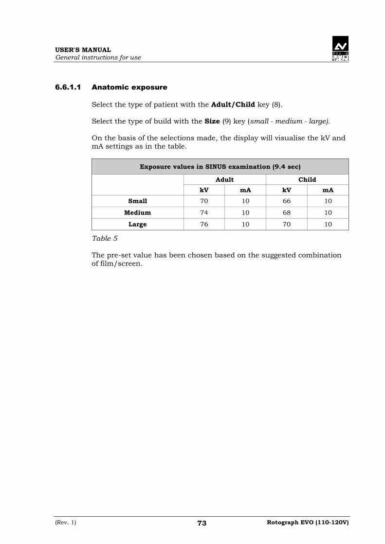
USER'S MANUAL General instructions for use
(Rev. 1) Rotograph EVO (110-120V) 73
6.6.1.1 Anatomic exposure
Select the type of patient with the Adult/Child key (8). Select the type of build with the Size (9) key (small - medium - large). On the basis of the selections made, the display will visualise the kV and mA settings as in the table.
Exposure values in SINUS examination (9.4 sec)
Adult Child
kV mA kV mA
Small 70 10 66 10
Medium 74 10 68 10
Large 76 10 70 10
Table 5 The pre-set value has been chosen based on the suggested combination of film/screen.

USER'S MANUAL General instructions for use
Rotograph EVO (110-120V) (Rev. 1) 74
6.6.1.2 Manual exposure
If the kV and mA combinations of the Table 5 are not considered suitable for a specific examination, it will be possible to set new parameters using the manual mode. To modify the kV or mA values, press key (3) , the LEDs of the "Adult/Child Selection" (8) key will flash; the LED of the "Size Selection" (9) will go off. The display will show respectively one of the following two indications:
> x x k V x x m A 9 . 4 0 s S I N U S
or
x x k V > x x m A 9 . 4 0 s S I N U S
The symbol ">" indicates which parameter is being changed. The selected parameter can be modified by pressing the increase key (4) and the decrease key (5) . The kV value can vary between 60 and 86 kV, with 2 kV steps. The value of mA can vary between 6 and 10 mA, with 1 mA steps. NOTE: To change the values rapidly, keep the increase key (4) or decrease key (5) pressed.
�

USER'S MANUAL General instructions for use
(Rev. 1) Rotograph EVO (110-120V) 75
6.6.2 Preparation of the patient
1. Ask the patient to remove all metallic objects located in the area to be
X-rayed (necklaces, earrings, glasses, hairpins, removable dental prosthesis, etc.). Ensure that there are no thick garments in the area to be X-rayed (coats, jackets, ties, etc.).
2. Ask the patient to put on the protective apron, or something similar, making sure that it does not interfere with the trajectory of the X-ray beams.
3. Place the patient in a standing position at the SINUS chin support With the keys "Column movement" (15/16) raise/lower the column until the chin support rest is aligned with the patient's chin.
4. Position the patient with the temple clasps (Figure 12) ensuring that the chin rests on the special support; ask the patient to place his hands on the front supports. Ensure that the patient rests his chin on the chin support for SINUS.
45
1
46
34
Figure 12 – SINUS positioning
Legend of Reference Lines 45 Sagittal medial line 46 Frankfurt plane line Legend positioning devices and patient centring 1 SINUS chin support 3 Forehead support
closing/release knob 4 Temple clasps
closing/release knob

USER'S MANUAL General instructions for use
Rotograph EVO (110-120V) (Rev. 1) 76
5. Instruct the patient to close his eyes. 6. Press the key "Centring devices ON" (14) .
Two laser beams will light up the sagittal medial plane line and the horizontal line for the Frankfurt plane reference (the plane that identifies a line that ideally links the ear hole - the auditory meatus - with the lower part of the orbital fossa in Figure 7). Position the patient's head in such a way as to ensure that the first two luminous beams fall in correspondence with the respective anatomical references. The luminous beam of the Frankfurt plane can be adjusted according to the patient's height; this can be adjusted by means of the laser knob on the side of the mirror.
NOTE: The laser centring devices remain on for approximately 1 minute; shutdown can be anticipated by pressing the "Centring Device On" key (14) or, with alignment complete, by pressing the "Patient entrance" key (7) to begin preparation for exposure.
7. Close the temple clasps and bring the forehead support close. This
will help the patient to stay in a correct position. Check that, during this phase, the patient has not changed position.
8. Press the key "Patient Entrance" (7) to confirm.
The luminous centring devices switch off and the rotating arm goes to its examination start position. Once alignment has been completed, the following message will be displayed:
x x k V x x m A 9 . 4 0 s > S T A R T E X A M <
x = value defined by the settings 9. Ask the patient to: close his mouth, remain perfectly still and
not look at the rotating arm during the movement.
�

USER'S MANUAL General instructions for use
(Rev. 1) Rotograph EVO (110-120V) 77
6.6.3 Making an exposure
WARNING: During the emission of X-rays, the protection procedures for the operator and personnel in the area must be in compliance with the local regulations. In all cases, it is recommended that during the emission of X-rays, only the patient and operator be present in the room. If the operator is not protected by suitable screens, he must stand at least 1.5m (4.92') away from the emission of the rays (see the Figure 1 and Figure 2). NOTE: Before performing a lateral Sinus examination, because of the specific trajectory described by the rotating arm, it is recommended to check for possible mechanical interferences with the patient's shoulder during the rotation. By pressing the key "Test" (6) , to activate the Test function. In this condition, it will be possible to make the machine perform all the movements made during the examination, but without emitting rays. Once the cycle is completed, deactivate the "Test" function by pressing the key again. 1. Verify once again that the exposure data are correct. If not, correct
them as described in paragraph 6.4.2.2; ensure that the machine's indicator light "Ready for X-ray" will come on, so press the X-ray button for the entire duration of the exposure, checking the simultaneous working of the ray indicator light "X-ray emission" (if you are within sight of the machine) and the acoustic ray signal. The following message will be displayed first:
> S T A R T E X A M < P R E - H E A T I N G . . .
and then (after 2 seconds), the following message will be displayed:
x x k V x x m A 9 . 4 0 s > X - R A Y <
x = value defined by the settings NOTE: If the machine is in the "Test" mode, the display will show:
T E S T X R A Y N O T A C T I V E
�
�

USER'S MANUAL General instructions for use
Rotograph EVO (110-120V) (Rev. 1) 78
NOTE: If there is no cassette, the following message will be displayed:
> I N I T E X A M < I N S E R T C A S S E T T E
The examination will not be continued until the cassette has been inserted. WARNING: The position of the identification characters "R" (right side) and "L" (left side) are correct if the cassette is fit into the unit with the opening positioned upward. NOTE: The rotation of the arm and the emission of the X-rays will start with a delay of 2 seconds from when the X-ray button is pressed. As the X-ray button is a "dead man's switch", it is necessary to keep it pressed until the end of the exposure. During the examination, the emission of rays in correspondence with the central part of the dental arch is suspended; the relative signals (sonant and visual) are also suspended. 2. Once the exposure is completed, the system will rotate back. When it
has completed this manoeuvre, the display shows the message:
P A T I E N T E X I T P R E S S > 0 <
and it will be necessary to free the patient from the positioning device.
3. Press the key "Patient Entrance" (7) . The machine will
reposition itself back to the starting position displaying the message:
A X I S P O S I T I O N I N G P L E A S E W A I T . . .
Then, the following message will be displayed:
R E P L A C E C A S S E T T E
At the end of the removal, the following message is displayed:
x x k V x x m A 9 . 4 0 s S I N U S
x = value defined by the settings
that shows the values set for that last exposure. A new exposure can now be made.
�
�

USER'S MANUAL General instructions for use
(Rev. 1) Rotograph EVO (110-120V) 79
NOTE: If you try to perform a new exam before the cooling period has elapsed (4 minutes), the following message will be displayed indicating the time to wait before performing a new examination:
T U B E C O O L I N G P L E A S E W A I T x x x s
The waiting time allows the anode in the radiogenic tube to cool down. 4. Once the cassette has been removed, it can be opened in a dark room
and the film developed. WARNING: After every examination, clean the chin support, the handles and the temple clasps group thoroughly. NOTE: If, during the exposure, the patient moves, or the machine collides with the patient himself (or with any object), or you realise that the parameters set are not correct, you must release the X-ray button immediately, interrupting the emission of X-rays and the movement of the arm. If this occurs, the following message will be displayed:
E 3 6 2 P R E S S > 0 <
all the motors will switch off, and it will be possible, if necessary, to manually rotate the arm, allowing the patient to come out. It is recommended that this movement be made with great care in order to prevent damage to the machine.
Then press the "Patient Entrance" (7) key and the display
will show:
P L E A S E W A I T . . . M A C H I N E S E T T I N G
and then:
R E P L A C E C A S S E T T E
The system now returns to its initial position and the unit must be reset (cassette and parameters) and the patient repositioned. NOTE: Remove the cassette and change the film to prevent a double exposure of the film that would then provide non-diagnostic results.
�
�
�

USER'S MANUAL General instructions for use
Rotograph EVO (110-120V) (Rev. 1) 80
6.7 Cephalometric examination There is no rotation of the tube-head (X-ray generator) support arm and cassette holder for the cephalometric examination. Various projections are possible for the cephalometric examination. The diaphragm positions automatically according the cassette format and the projection selected. The Cephalometric examination is fitted with a Soft Tissues Filter (STF); this filter reduces the dose in areas with low bone content and highlights the patient's profile which, under normal conditions, would be overexposed and so not visible. Rotograph EVO makes different kinds of exposures, according to the original configuration and the type of cassette used:
• INCHES
8"x10" Asymmetrical for Latero-Lateral
"8x10" Symmetrical for Antero-Posterior and Postero-Anterior
10"x8" Asymmetrical
for Latero-Lateral
• 18 x 24 cm
18x24 Asymmetrical for Latero-Lateral
18x24 Symmetrical for Antero-Posterior and Postero-Anterior
24x18 Asymmetrical
for Latero-Lateral
• 24 x 30 cm
24x30 Asymmetrical for Latero-Lateral
24x30 Symmetrical for Antero-Posterior and Postero-Anterior
30x24 Asymmetrical
for Latero-Lateral
NOTE: The size of the cassette is not automatically recognised by the unit but must be input by the operator.
�

USER'S MANUAL General instructions for use
(Rev. 1) Rotograph EVO (110-120V) 81
6.7.1 Preparation of the device
To select the CEPH examination, press key "Examination Mode Selection - M" (11) until the following message is displayed:
x x k V x x m A 0 . 8 0 s C E P H 8 " x 1 0 " L L
x = value defined by the settings 1. Press the "Patient Entrance" (7) key ; the display will show
alternatively the following messages:
C E P H - R E M O V E C H I N R E S T
and
C E P H - C L O S E T E M P L E S U P P O R T
The first message tells the operator to remove the chin support, while the second message tells him to close the temple clasps. These operations are necessary to prevent interference with the rays beam and with the panoramic cassette holder when the arm is being positioned.
WARNING: Neither of the two messages are controlled by the system and they can therefore appear even if the unit has been set correctly. In the same way, these messages can be reset, even if the machine has not been suitably prepared. In this case an E 340 error might be displayed. WARNING: There is no need to position any type of chin support for the cephalometric examination. The chin support used for panoramic examinations must be removed as indicated on the display. If the chin support is not removed, it will collide with the cassette holder during alignment and can obscure some anatomical parts of the patient during the examination. At the same time, the temple clasps must be closed, in order to avoid collision with the rotating arm.

USER'S MANUAL General instructions for use
Rotograph EVO (110-120V) (Rev. 1) 82
2. Once what was required is performed, press the key "Patient
Entrance" (7) messages will disappear and the machine will align automatically with respect to the cassette and the following message will be displayed:
C E P H P L E A S E W A I T . . .
Once alignment is completed, the following message will be displayed:
C E P H - O P E N C A S S E T T E U N I T
requesting the operator to open the cassette holder for panoramic examination.
NOTE: The position of the cassette holder for panoramic examination is controlled by two micro-switches, it must therefore be completely opened.
The following message will be displayed:
x x k V x x m A 0 . 8 0 s C E P H 8 " x 1 0 " L L
NOTE: The system is positioned in the following configuration:
• ADULT with the lighting up of the corresponding LED
• MEDIUM SIZE with the lighting up of the corresponding LED. No LED of the key "Type of Mastication Selection" (10) is lit and the key is disabled. 3. Insert the cassette ensuring that it is aligned with the required
format/projection references and that it is fastened with the circular lock.
NOTE: The size and the orientation of the cassette is not automatically recognised by the unit but must be correctly inserted by the operator.
�
�
�

USER'S MANUAL General instructions for use
(Rev. 1) Rotograph EVO (110-120V) 83
4. By means of the keys "Arrow right" (13) and "Arrow left" (12)
select the size of the cassette and the type of associated projection (see the table at the beginning of the Chapter).
NOTE: Exposure cannot be made if a cassette has not been inserted. NOTE: If the presence of a cassette is detected the system will ask for its replacement before the exposure. By pressing the key "Patient Entrance" (7) it is possible to cancel the request in case the cassette was previously replaced. NOTE: The system does not detect if the film has already been exposed; if the cassette has been removed, but the film has not been replaced the exposure will be allowed, but the result will not be significant from the diagnostic point of view.
�
�
�

USER'S MANUAL General instructions for use
Rotograph EVO (110-120V) (Rev. 1) 84
6.7.2 Anatomic / Manual Exposure
NOTE: If the previous exam was carried out manually, just press the key "Examination Mode Selection - M" (11) to change to Anatomic exposure. After setting the machine accordingly, the following two operating modes may be selected:
• ANATOMIC: with the kV, mA and s values programmed according to the type of patient and size
• MANUAL: with the possibility of changing the kV, mA and s values set.
NOTE: In the manual mode, the LED of the "Adult/Child Selection" (8) key flashes to indicate that it is possible to change the selection; use
key (8) to change from Adult to Child.
�
�

USER'S MANUAL General instructions for use
(Rev. 1) Rotograph EVO (110-120V) 85
6.7.2.1 Anatomic exposure
Select the type of patient with the Adult/Child key (8). Select the type of build with the Size (9) key (small - medium - large). The kV, mA and s values will be displayed according to the selections made as per the following table:
Adult Child
kV mA s kV mA s
Small 74 12 0,8 72 12 0,7
Medium 76 12 0,8 74 12 0,9
Large 78 12 0,8 76 12 0,9
Table 6: Latero-Lateral projection These values have been set for latero-lateral projection, according to the recommended film-screen combination. For antero-posterior projections, it is recommended to set the following values:
Adult Child
kV mA s kV mA s
Small 84 12 2 80 12 1,5
Medium 84 12 2 80 12 1,5
Large 86 12 2 82 12 1,5
Table 7: Antero-Posterior projection

USER'S MANUAL General instructions for use
Rotograph EVO (110-120V) (Rev. 1) 86
6.7.2.2 Manual exposure
If the kV, mA and s combinations in the Table 6 and Table 7 are not considered suitable for a specific examination, it will be possible to set new parameters using the manual mode. To modify the kV or mA values, press key (3) , the LED of the key "Adult/Child Selection" (8) will flash; the LED of the "Size Selection" (9) will go off; the display will show respectively one of the following three indications:
> x x k V x x m A x . x x s C E P H 8 " x 1 0 " L L
x x k V > x x m A x . x x s C E P H 8 " x 1 0 " L L
x x k V x x m A > x . x x s C E P H 8 " x 1 0 " L L
The symbol ">" indicates which parameter is being changed. The selected parameter can be modified by pressing the increase key (4) and the decrease key (5) . The "kV" value can vary between 60 and 80 kV, with 2 kV steps. The "mA" value can vary between 4 and 12 mA, with 1 mA steps. The "s" value can vary between 0.20 s and 3.00 s as per the following table:
Exposure times selectable in manual mode
0,20 - 0,23 - 0,26 - 0,30 - 0,33 - 0,36 - 0,40 - 0,45 - 0,50 - 0,60 - 0,70 - 0,80- 0,90- 1,00 - 1,30- 1,60 - 2,00 - 3,00
NOTE: To change the values rapidly, keep the increase key (4) or decrease key (5) pressed.
�

USER'S MANUAL General instructions for use
(Rev. 1) Rotograph EVO (110-120V) 87
6.7.3 Preparation of the patient
1. Ask the patient to remove all metallic objects located in the area to be
X-rayed (necklaces, earrings, glasses, hairpins, removable dental prosthesis, etc.). Ensure that there are no thick garments in the area to be X-rayed (coats, jackets, ties, etc.).
2. Ask the patient to put on the protective apron, or something similar, making sure that it does not interfere with the trajectory of the X-ray beams.
3. Open the ear centring device (Figure 13) to its maximum span by using the upper part of the rods of the centring device itself. Move the nose rest (Figure 13) away outwardly to its maximum extension. Manually rotate the craniostat group according to the cephalometric projection to be made, moving the upper part of the ear centring device (Figure 13).
4. Position the patient upright near the auricular centring device. With the keys "Column Movement" (15/16) lift/lower, the column till the centring pins (Figure 13) are close to the ear to clasp the patient's head so that the pivots penetrate the ear (Figure 13) moving the upper part of the rods. If a Latero-Lateral examination is performed, position the nose rest.
5. By selecting an "asymmetric" projection, the Soft Tissues Filter (STF) will be automatically inserted.
2
3
1
Figure 13
Legend 1 Nose rest 2 Ear centring device 3 Pins for ear centring
device

USER'S MANUAL General instructions for use
Rotograph EVO (110-120V) (Rev. 1) 88
6.7.4 Making an exposure
WARNING: During the emission of X-rays, the protection procedures for the operator and personnel in the area must be in compliance with the regulations in force in the country where the machine is used. In all cases, it is recommended that during the emission of X-rays, only the patient and operator be present in the room. If the operator is not protected by suitable screens, he must stand at least 1.5m (4.92') away from the emission of the rays (see Figure 1 and Figure 2). 1. Verify once again that the exposure data are correct (see paragraph
6.7.2). Advise the patient to remain still and to keep his mouth closed, with the teeth touching, throughout the duration of the exposure. Press the "Patient Entrance" (7) key ; the collimator corresponding to the selected cassette will be positioned and the LED "Ready for X-rays" (1) will light up, indicating that the machine is ready to produce X-rays.
NOTE: If you want to cancel the operation, press key "Patient Entrance" (7) 2. Press the X-ray button for the entire duration of the exposure,
checking the concurrent working of the ray indicator light "X-rays emission" (if you are within sight of the machine) and the acoustic ray signal. The following message will be displayed first:
> S T A R T E X A M < P R E - H E A T I N G . . .
and then (after 2 seconds), the following message will be displayed:
x x k V x x m A x . x x s > X - R A Y <
x = value defined by the settings
�

USER'S MANUAL General instructions for use
(Rev. 1) Rotograph EVO (110-120V) 89
NOTE: If there is no cassette, the following message will be displayed:
C E P H I N S E R T C A S S E T T E
NOTE: If, during the preparation of the first cephalometric examination the system detects a cassette which has not been replaced, the following message will be displayed:
C E P H R E P L A C E C A S S E T T E
The examination will not be continued until a new cassette has been inserted. NOTE: X-rays are emitted with a delay of two seconds from pressing the X-ray button to allow the heating of the filament and the control of all set parameters. Since the X-ray button is a "dead man's switch", it must be kept pressed until the end of the exposure. 3. Once the exposure is completed, the following message will be
displayed:
C E P H R E P L A C E C A S S E T T E
4. Remove the cassette; the cassette size, kV, mA and s values of the
just done exposure will appear. NOTE: If you want to carry out a new exposure after inserting the cassette, but the necessary waiting time for the cooling of the anode hasn't yet passed, the display will show a message indicating the time you still have to wait before carrying out the new test:
T U B E C O O L I N G P L E A S E W A I T x x x s
This time enables the X-ray tube's anode to cool down.
�
�
�
�

USER'S MANUAL General instructions for use
Rotograph EVO (110-120V) (Rev. 1) 90
5. The cassette can now be opened in a dark room and the film developed.
NOTE: If the patient moves during the exposure, or if you realise that incorrect parameters have been set, it will be necessary to stop pressing the X-ray button immediately, to interrupt the emission of rays. The following message will be displayed:
E 3 6 2 P R E S S > 0 <
Then press the "Patient Entrance" (7) key and the display will show:
C E P H R E P L A C E C A S S E T T E
The system now returns to the position for Ceph exam and the unit starts the procedure for the new examination. NOTE: No warning message will be given if the cassette is removed but the film is not replaced, and another exposure is made. However, the film would provide non-diagnostic results due to the double exposure. NOTE: After every examination, clean the ear centring device and temple clasps thoroughly.
�
�
�

USER'S MANUAL General instructions for use
(Rev. 1) Rotograph EVO (110-120V) 91
6.8 Examination to assess bone growth (Carpus) The cephalometric device can also be used to carry out X-rays to evaluate the state of calcification and bone growth, X-raying the hand/wrist complex to obtain an X-ray that contains the anatomic details necessary to evaluate the patient's bone growth trend. To perform this kind of examination, the exposure parameters allowed by current regulations can give an overexposed film. It is recommended to use cassettes with medium-type screens.

USER'S MANUAL General instructions for use
Rotograph EVO (110-120V) (Rev. 1) 92
6.8.1 Preparation of the device
1. Select the CEPH examination, pressing key "Examination Mode
Selection - M" (11) until the following message is displayed:
x x k V x x m A x . x x s C E P H 8 " x 1 0 " L L
x = value defined by the settings
2. By means of the keys "Arrow right" (13) and "Arrow left" (12) select the Carpus examination:
6 0 k V 0 6 m A 0 . 2 0 s C A R P U S 8 " x 1 0 " P A
x = value defined by the settings
Press the "Patient Entrance" (7) key ; the display will show
alternatively the following messages:
C A R P U S - R E M O V E C H I N R E S T
and
C A R P U S - C L O S E T E M P L E S U P P O R T
The first message tells the operator to remove the chin support, while the second message tells him to close the temple clasps. These operations are necessary to prevent interference with the rays beam and with the panoramic cassette holder when the arm is being positioned.
WARNING: Neither of the two messages are controlled by the system and they can therefore appear even if the unit has been set correctly. In the same way, these messages can be reset, even if the machine has not been suitably prepared. In this case an E 340 error might be displayed.

USER'S MANUAL General instructions for use
(Rev. 1) Rotograph EVO (110-120V) 93
WARNING: There is no need to position any type of chin support for the Carpus examination. The chin support used for panoramic examinations must be removed as indicated on the display. If the chin support is not removed, it will collide with the cassette holder during alignment and can obscure some anatomical parts of the patient during the examination. At the same time, the temple clasps must be closed, in order to avoid collision with the rotating arm. 5. Once what was required is performed, press the key "Patient
Entrance" (7) messages will disappear and the machine will align automatically with respect to the cassette and the following message will be displayed:
C A R P U S 8 " x 1 0 " P A P L E A S E W A I T . . .
Once alignment is completed, the following message will be displayed:
C A R P U S - O P E N C A S S E T T E U N I T
requesting the operator to open the cassette holder for panoramic examination.
NOTE: The position of the cassette holder for panoramic examination is controlled by two micro-switches, it must therefore be completely opened.
The following message will be displayed:
x x k V x x m A x . x x s C A R P U S 8 " x 1 0 " P A
NOTE: The system is positioned in the following configuration:
• ADULT with the lighting up of the corresponding LED
• MEDIUM SIZE with the lighting up of the corresponding LED. No LED of the key "Type of Mastication Selection" (10) is lit and the key is disabled.
�
�

USER'S MANUAL General instructions for use
Rotograph EVO (110-120V) (Rev. 1) 94
6. Insert the cassette ensuring that it is aligned with the required format/projection references and that it is fastened with the circular lock.
NOTE: The size and the orientation of the cassette is not automatically recognised by the unit but must be correctly inserted by the operator. NOTE: Exposure cannot be made if a cassette has not been inserted. NOTE: If the presence of a cassette is detected the system will ask for its replacement before the exposure. By pressing the key "Patient Entrance" (7) it is possible to cancel the request in case the cassette was previously replaced. NOTE: The system does not detect if the film has already been exposed; if the cassette has been removed, but the film has not been replaced the exposure will be allowed, but the result will not be significant from the diagnostic point of view. 7. Regulate the exposure parameters as required, using the pre-set
values or manual selection; the display will show the kV, mA and second settings as per the following table.
Child
kV mA s
Small 60 6 0,2
Medium 60 6 0,3
Large 60 6 0,4
�
�
�
�

USER'S MANUAL General instructions for use
(Rev. 1) Rotograph EVO (110-120V) 95
6.8.2 Preparation of the patient
1. Turn the ear centring device to the Antero-posterior position; bring
the nose-rest to a parking position. 2. Place the patient slightly to the side of the cephalometry device. 3. Insert a cassette in symmetric position, identified by the relative
serigraphy on the support. Place the hand on the support surface. The patient's hand and arm must form a vertical line.
Figure 14

USER'S MANUAL General instructions for use
Rotograph EVO (110-120V) (Rev. 1) 96
6.8.3 Making an exposure
WARNING: During the emission of X-rays, the protection procedures for the operator and personnel in the area must be in compliance with the regulations in force in the country where the machine is used. In all cases, it is recommended that during the emission of X-rays, only the patient and operator be present in the room. If the operator is not protected by suitable screens, he must stand at least 1.5m (4.92') away from the emission of the rays (see Figure 1 and Figure 2). 1. Press the "Patient Entrance" (7) key .
The unit will move into the selected examination start position. The signalling LED "Ready for X-rays" will light up, indicating that the machine is ready to produce X-rays.
NOTE: If you want to cancel the operation, press key "Patient Entrance" (7) 2. Press the X-ray button for the entire duration of the exposure,
checking the concurrent working of the ray indicator light "X-rays emission" (if you are within sight of the machine) and the acoustic ray signal. The following message will be displayed first:
> S T A R T E X A M < P R E - H E A T I N G . . .
and then (after 2 seconds), the following message will be displayed:
x x k V x x m A x . x x s > X - R A Y <
x = value defined by the settings NOTE: If there is no cassette, the following message will be displayed:
C E P H I N S E R T C A S S E T T E
�
�

USER'S MANUAL General instructions for use
(Rev. 1) Rotograph EVO (110-120V) 97
NOTE: If, during the preparation of the first examination the system detects a cassette which has not been replaced, the following message will be displayed:
C E P H R E P L A C E C A S S E T T E
The examination will not be continued until a new cassette has been inserted. NOTE: X-rays are emitted with a delay of two seconds from pressing the X-ray button to allow the heating of the filament and the control of all set parameters. Since the X-ray button is a "dead man's switch", it must be kept pressed until the end of the exposure. 3. Once the exposure is completed, the following message will be
displayed:
C E P H R E P L A C E C A S S E T T E
4. Remove the cassette; the cassette size, kV, mA and s values of the
just done exposure will appear. NOTE: If you want to carry out a new exposure after inserting the cassette, but the necessary waiting time for the cooling of the anode hasn't yet passed, the display will show a message indicating the time you still have to wait before carrying out the new test:
T U B E C O O L I N G P L E A S E W A I T x x x s
This time enables the X-ray tube's anode to cool down. 5. The cassette can now be opened in a dark room and the film
developed.
�
�
�

USER'S MANUAL General instructions for use
Rotograph EVO (110-120V) (Rev. 1) 98
NOTE: If the patient moves during the exposure, or if you realise that incorrect parameters have been set, it will be necessary to stop pressing the X-ray button immediately, to interrupt the emission of rays. The following message will be displayed:
E 3 6 2 P R E S S > 0 <
Then press the "Patient Entrance" (7) key and the display will show:
C E P H R E P L A C E C A S S E T T E
The system now returns to the position for the Ceph exam and the unit starts the procedure for the new examination. NOTE: No warning message will be given if the cassette is removed but the film is not replaced, and another exposure is made. However, the film would provide non-diagnostic results due to the double exposure.
�
�

USER'S MANUAL General instructions for use
(Rev. 1) Rotograph EVO (110-120V) 99
6.9 Messages on display Rotograph EVO is fully controlled by a microprocessor which controls the programming of the emission parameters and signals the various conditions of the machine, the possible abnormalities and errors via displayed messages. The messages can be divided into two groups:
• operation messages: these messages tell the operator how to set up the unit for the examination
• error messages: these messages are displayed when an error occurs; there are two kinds of error messages as follows:
1 - Messages prompted when the X-ray emission button is released by
the operator or by pressing key "Patient Entrance" (7) . The message displayed will be as follows
E X X X P R E S S > 0 <
xxx = error message code number
Operations are reset by pressing key (7) .
2 - Messages generated by a system error. In this case, the Technical
Service must be called. Messages that require the intervention of the Technical Service are displayed as follows:
E X X X C A L L T E C H A S S .
xxx = error message code number
3 - Messages related to H.F. board problems. If this occurs, switch off
the unit. Wait a few minutes for the capacitors of the relative circuit to discharge, and then switch the machine on again. If the problem persists, call the Technical Service.
E X X X S W I T C H P O W E R O F F
xxx = error message code number Following are reported the different error messages and the relative controls and operations to be performed.

USER'S MANUAL General instructions for use
Rotograph EVO (110-120V) (Rev. 1) 100
6.9.1 Error message with error code E000 ÷÷÷÷ E199 NOT resettable errors. These are internal errors of the control system; it is necessary to call the Technical Assistance Service.
6.9.2 Error message with error code E200 ÷÷÷÷ E299 This category of errors apply to the rotation motor; of these only the error "E206 - Collision with patient", caused by a possible collision between the rotation arm and the patient, it is an actual reversible. Press the key "Patient Entrance" (7) to reset the error and to perform the axes centring operation. For all other cases, call the Technical Assistance Service.

USER'S MANUAL General instructions for use
(Rev. 1) Rotograph EVO (110-120V) 101
6.9.3 Error message with error code E300 ÷÷÷÷ E399
6.9.3.1 E340 - Cassette holder not in PAN position
A Panoramic type examination was requested, but the cassette holder does not appear to be closed; close it in the PAN position and press the key "Patient Entrance" (7) to reset the error condition.
6.9.3.2 E360 / E361 - X-ray button pressed during start up or axis
movement
Release the X-ray button if pressed; press the key "Patient Entrance" (7) to reset the error condition. If the error does not disappear, call the Technical Assistance Service.
6.9.3.3 E362 - X-ray button released during examination
NOTE: The X-ray button has the so-called "dead man's switch" function, i.e. it must be kept pressed for the whole time of the examination, also during the phases of the examination with emission interruption (for instance, in open/close mouth TMJ).
This message signals that the button was released during the examination phase; the motors are unlocked, therefore the patient can get out of the system. Repeat the system centring phase and repeat the examination.
WARNING: In case the emission has already started, replace the film to avoid double exposures that make the image non-diagnostic.
6.9.3.4 E376 - Timer back up triggered
This error signals that the emission has not completed correctly, but it was interrupted but the safety timer backup that cuts in to stop the emission also in case of failure of the control system. Turn off immediately the system as an unwanted emission could be present.
�

USER'S MANUAL General instructions for use
Rotograph EVO (110-120V) (Rev. 1) 102
6.9.4 Error message with error code E700 ÷÷÷÷ E799 WARNING: These error codes refer to the X-rays generation, therefore, they can also indicate a safety problem. With error code E759, turn off immediately the system as a not requested X-ray emission was detected. In this case, call immediately the Technical Assistance Service.
6.9.4.1 E774 - X-rays button not pressed
The lack of the button is signalled also if the emission software control is present. The error signals a possible failure on the connection of the X-rays button with the generator card.
6.9.4.2 E775 - X-rays button released prematurely
The release of the X-rays button during the emission phase is signalled; this signalling has a different meaning from that of the corresponding E362 error, as this message is generated by the HF card, which signals a possible failure on the connection of the X-rays button with the card itself.
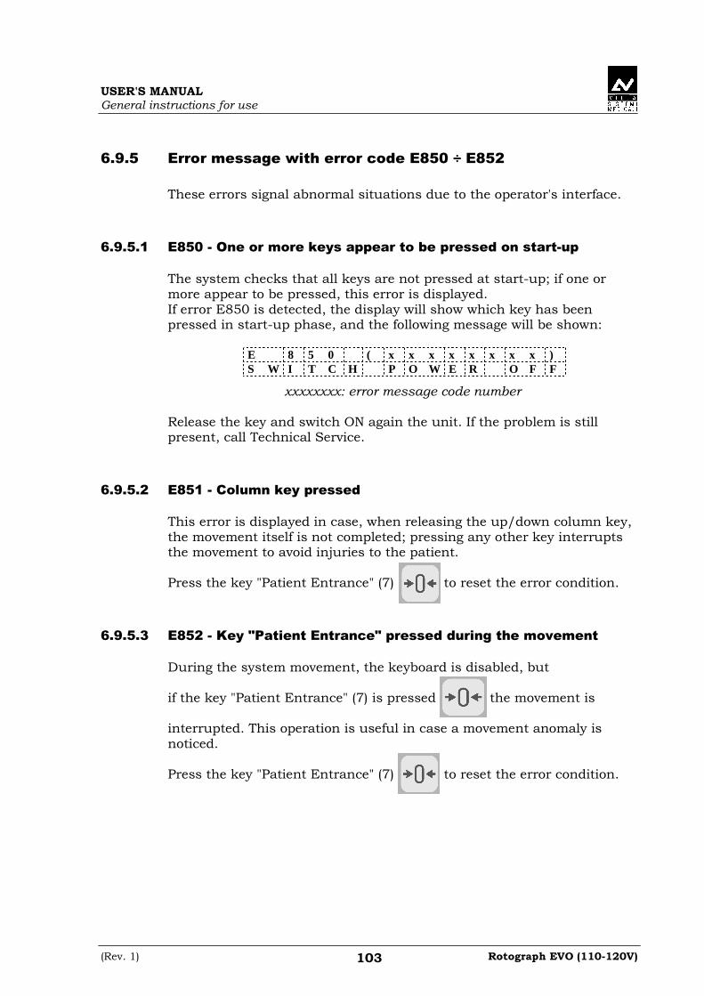
USER'S MANUAL General instructions for use
(Rev. 1) Rotograph EVO (110-120V) 103
6.9.5 Error message with error code E850 ÷÷÷÷ E852 These errors signal abnormal situations due to the operator's interface.
6.9.5.1 E850 - One or more keys appear to be pressed on start-up
The system checks that all keys are not pressed at start-up; if one or more appear to be pressed, this error is displayed. If error E850 is detected, the display will show which key has been pressed in start-up phase, and the following message will be shown:
E 8 5 0 ( x x x x x x x x ) S W I T C H P O W E R O F F
xxxxxxxx: error message code number Release the key and switch ON again the unit. If the problem is still present, call Technical Service.
6.9.5.2 E851 - Column key pressed
This error is displayed in case, when releasing the up/down column key, the movement itself is not completed; pressing any other key interrupts the movement to avoid injuries to the patient. Press the key "Patient Entrance" (7) to reset the error condition.
6.9.5.3 E852 - Key "Patient Entrance" pressed during the movement
During the system movement, the keyboard is disabled, but if the key "Patient Entrance" (7) is pressed the movement is interrupted. This operation is useful in case a movement anomaly is noticed. Press the key "Patient Entrance" (7) to reset the error condition.

USER'S MANUAL General instructions for use
Rotograph EVO (110-120V) (Rev. 1) 104
6.10 Research and correction of possible defects in
dental X-rays
6.10.1 Faults due to the wrong positioning of the patient
Problem Description Solution
Overlarge and blurred incisors.
The patient is not positioned correctly. He is too far from
the optimal focal plane.
Position the patient correctly, check that he holds the bite with the incisors on the appropriate notch and that the bite holder rod is vertical.
Over-small and blurred incisors.
The patient is not positioned correctly. He is too near the
optimal focal plane.
Position the patient correctly, check that he holds the bite with the incisors on the appropriate notch and that the bite holder rod is vertical.
Radiography with blank central area.
The spine of the patient inhibits the passage of the X-ray as it is too
compressed.
Check the alignment of the Frankfurt plane, try to stretch the cervical part of the spine by moving the patient's feet forward (see paragraph 6.4.3 points 3/4/6/7) and, if necessary, correct the height
of the chin support.
Asymmetric dental
arch.
The sagittal medial line does not correspond to the laser centring beam.
Realign the patient (see paragraph
6.4.3 point 6).
Upper apical area too dark.
The patient does not keep his lips shut and the tongue
is not against the palate.
See paragraph 6.4.3 point 8.
Upper central apical area out of focus.
The patient keeps his head rotated backwards (Frankfurt plane not
aligned).
Position the patient again and realign the Frankfurt plane.
The image is slanted in comparison with the longitudinal axis of the film and some anatomical structures
are not symmetric.
The patient's head is slanted (not vertical).
Position the patient again, correcting the position of the sagittal plane.
The teeth on one side are bigger than those
on the other side.
The patient's head is rotated with respect to the axis of
the bite.
Position the patient again, correcting the position of the sagittal plane and controlling that his head does not
rotate.
Presence (in CEPH examination) of a white area in the lower
part of the image.
Panoramic chin-rest still mounted.
Perform the exam again, removing the PAN chin-rest.

USER'S MANUAL General instructions for use
(Rev. 1) Rotograph EVO (110-120V) 105
6.10.2 Defects due to wrong data setting and to the dark room
Problem Description Solution
Under or overexposed film.
The set kV value is not fit for the size of the patient. Possible error during
development.
Check the development system, or repeat the exam varying the kV value: see paragraph 6.4.4.Should this solution fail, call the Technical Service.
Completely black film. The film caught too much light or underwent an
involuntary radiation.
Verify the locking of the dark room and do not keep new films near
radiogene sources or light sources.
Veiled film. This defect is checked looking at the film edges not impressed by the radiation: if they are not perfectly transparent, thus causing a
poor definition on all film.
Verify that the film is not expired and that the storing indications given by the manufacturer have been followed. Verify the working of
the non-actinic lamp in dark room.
Film with stripes. Being rapidly scratched against its box the film accumulated electrostatic charges or the scintillating screens have not been treated with antistatic
liquid.
Clean the screens with the antistatic liquid.
Film with blurred details and good
contrast.
The film is not correctly pressed between the
screens.
Replace the cassette.
Film completely white. Missing X-ray emission or wrong development.
Verify the emission of the X-rays by acoustical and luminous signal, if the error persists, check the development procedure. If no solution can be found, call the Technical Service.
Soft Tissue not (or poorly) visible in L-L
projection.
A symmetric cassette size was selected.
Select an asymmetrical cassette size.

USER'S MANUAL General instructions for use
Rotograph EVO (110-120V) (Rev. 1) 106
6.10.3 Defects on film due to the device
1. Should the film show non irradiated areas or be completely white,
this can mean that there is a defect in the alignment between X-ray beams and film or a partial or total missing of irradiation; in any case, call the Technical Service.
2. In the event the soft tissue of the patient is not highlighted while
performing a cephalometry, in a latero-lateral, let the technician verify the adjustment of the Soft Tissue Filter.

USER'S MANUAL General instructions for use
(Rev. 1) Rotograph EVO (110-120V) 107
6.11 Analysis of the problems on the panoramic
examinations The panoramic radiography is the examination of the maxillo-facial region normally used to view the dental region inside the complete head and sinuses-orbital complex. In a good Panoramic, you can distinguish the main anatomic structures that are simplified in the diagram below (which indicates only the main ones, and is not complete).
Figure 15
Ref. Anatomic structure
1 Palatal plane
2 Maxillary sinus
3 Maxilla and maxillary tuberosity
4 Temporo mandibular condyle
5 Ascending ramus of the TMJ
6 Coronoid process (overlap with maxilla)
7 Mandibular canal
8 Chin foramen
9 Anterior nasal spine
10 Nasal cavities
11 Ioid bone (normally duplicated)

USER'S MANUAL General instructions for use
Rotograph EVO (110-120V) (Rev. 1) 108
6.11.1 Proper positioning of the patient
The proper positioning of the patient during the panoramic examination is very important in order to get good quality radiography. This is due to the fact that the shape of the focussed area, e.g. of the layer clearly shown on the film, tends to follow the dental arch and has a non-constant deepness. The objects outside this focused area will therefore appear blurred on the radiography. 1. The patient should not wear clothes that may interfere with the X-ray
beams, also to leave more space between the patient’s shoulders and the rotating arm of the machine. Care must be taken in order to avoid interference between the X-ray beam and the protective apron worn by the patient.
2. Metal objects (necklaces, earrings etc.) must be avoided; these objects not only create radio-opaque images in their own position, but also false images projected in other parts of the radiography, so disturbing the correct view of the anatomy.
3. The patient’s head must be slightly tilted downward in order to make the Frankfurt plane horizontal. In this way, the hard palatal ceiling will be projected slightly over the superior apex of the anterior teeth. If the patient has a low palatal ceiling, slightly increase the downward tilting.
4. Align the sagittal medial plane with the centre of the chin support, normally indicated by the relevant light beam.
Figure 16
5. The patient must extend his spine; this is normally obtained by
asking the patient to step forward, making sure that all other conditions are unchanged. If not properly extended, the spine will cause the appearing of a lower exposed area (clearer) in the front part of the film.

USER'S MANUAL General instructions for use
(Rev. 1) Rotograph EVO (110-120V) 109
6. The patient’s tongue must be positioned against his palate. Otherwise, the air between the tongue and the palate forms an area of lower absorption, which leads to a darker area that hides the apex of the teeth of the maxilla.
The result of all the above listed actions will be a radiography where all the parts are properly exposed and are well identifiable as in the diagram of Figure 17.
Figure 17 It must be noted that the radiography is quite symmetrical, with the ascending rami of the temporo mandibular joints almost parallel. The occlusal plane is shown slightly tilted upward, the palatal plane does not overlap the apex of the upper arch and therefore allows a good view of the apex itself.

USER'S MANUAL General instructions for use
Rotograph EVO (110-120V) (Rev. 1) 110
6.11.1.1 Errors due to poor positioning of patient
• The film shows the anterior teeth with reduced magnification
and not well defined. The cervical spine is shown as evident
white shadow.
In addition, on the molar zone there are too many shadows, disturbing the reading.
The resulting film is similar to the schema shown on Figure 18.
Figure 18 Possible causes:
The patient it positioned too much forward. Solution:
Check the patient's positioning by using luminous beams.
If, after the correct positioning of the patient, the problem still remains, check the alignment of the centring laser lights, simply switching on the centring lights and checking their position. The sagittal medial luminous beam must hit the centre of the chin support.

USER'S MANUAL General instructions for use
(Rev. 1) Rotograph EVO (110-120V) 111
• Anterior teeth are enlarged and blurred
Figure 19 shows the result of this error.
Figure 19 Possible causes:
The patient it positioned too much backward. Solution:
Check the patient's positioning by using luminous beams.

USER'S MANUAL General instructions for use
Rotograph EVO (110-120V) (Rev. 1) 112
• Part of the image is enlarged while the other is reduced
The schema described on Figure 20 the film obtained; it is possible to observe that one part of the radiography is blurred and enlarged, while the other is reduced and seems to be in focus; the two condylar rami are at the same height on the X-ray.
Figure 20 Possible cause:
This effect can be due to two different causes. In the first one, the sagittal medial plane is not aligned with the relevant centring light beam, which falls at the centre of the chin support. In the second case, the centre of the sagittal medial plane corresponds with the centre of the chin support, but the patient’s head is rotated.
In both cases, one side is closer to the film plane than the other, thus resulting in a different magnification of the two sides; the part more distant from the film will be more magnified while the part closer to the film plane will result smaller. The result will be an image as shown in Figure 20; the left-hand area of the image shows a bigger magnification that can be noticed both on the teeth and on the ascending rami of the TMJ. Solution:
Check the positioning of the sagittal medial plane by using the relevant centring light beam. Check also the position of the sagittal medial beam; lighted, it must fall both on the centre of the chin rest and also on the centre of the bite.

USER'S MANUAL General instructions for use
(Rev. 1) Rotograph EVO (110-120V) 113
• The image shows the upper vertex of the condylar rami of
different heights;
Figure 21 shows the result of this error.
Figure 21 Possible causes:
The sagittal medial plane is not vertical. This can be the patient’s problem, but if the defect is always present, check the laser beam. Solution:
Verify that the laser beam is vertical; this check can be performed very quickly by using the laser beam and verifying that it falls on the centre of the chin support; remove the chin support itself and check that the beam falls in the centre of the two holes used to fix the support itself.
If not, a possible cause can be the imperfect horizontality of the chin support arm, that must be adjusted using the relevant screws.

USER'S MANUAL General instructions for use
Rotograph EVO (110-120V) (Rev. 1) 114
• The image shows undulated teeth rows
As can be seen in Figure 22, the upper teeth are magnified and unfocused, with the shadow of the hard palate positioned over the superior apex. The temporo-mandibular joints are exposed outward, with lines divergent upward. In some cases, the condylar vertices might not appear on the image.
Figure 22 Possible causes:
A Frankfurt plane tilted too much upward produces different anomalies that may also appear simultaneously. A chin support plane too high during the patient positioning, or when extending the spine, may generate this mistake. In this condition, the rear side of the patient’s head may also interfere with the rotating arm of the panoramic equipment.

USER'S MANUAL General instructions for use
(Rev. 1) Rotograph EVO (110-120V) 115
• The radiographic image shows the teeth row too curved
upward with the lower incisor not focused
Figure 23 shows the result of this type of error. The temporo-mandibular joints are shown very high up, with lines converging towards the top. In some cases the upper condyle might not be visible in the image.
Figure 23 Possible causes:
Patient’s head tilted downward, as on the diagram alongside. Solution:
Check the positioning of the patient by aligning the Frankfurt plane with the corresponding light beam.

USER'S MANUAL General instructions for use
Rotograph EVO (110-120V) (Rev. 1) 116
NOTE: In some cases, the positioning of the Frankfurt plane too tilted downward produces a correct image of the lower incisors, but the projection of the palate falls on the upper teeth apex, as shown in Figure 24.
Figure 24 In this case, a light tilting forward and downward of the Frankfurt plane causes the palate to be projected over and far enough from the roots of the teeth of the maxilla arch, without distortion of the incisor teeth, as in Figure 25.
Figure 25
�

USER'S MANUAL General instructions for use
(Rev. 1) Rotograph EVO (110-120V) 117
6.11.1.2 Images with artefacts
• Radiographs that show images with soft tissues or artefacts
The radiographs may show anatomical parts of the soft tissues or show radiographic artefacts. Normally the soft tissues might be more or less present, depending on the patient positioning, while the presence of artefacts is strictly dependent on the presence of foreign objects on the trajectory of the X-ray beam. The next figure shows these cases; please consider that all structures have a bilateral duplicate.
Figure 26
Soft tissue Description Artefacts Description
2 Ear soft tissue 1 Space between tongue and palate. All the structures of the oropharynx cavity can be shown
3 Nose soft tissue 4 Spinal column
7 Epiglottis 5 Image of the patient's leaded protective apron (light area)
The part identified with "6" in Figure 26 represents the image of the controlateral mandible (the other side of the mandible). That therefore results as a clearer area overlapped with the real image. Very often the resulting darker area in the bottom corner is noticed and is considered as an artefact of the radiological image.

USER'S MANUAL General instructions for use
Rotograph EVO (110-120V) (Rev. 1) 118
This is not true, because it is derived from the projection geometry used to obtain the panoramic image. The effect can be more evident if the film in underexposed due to wrong radiological parameters. With reference to Figure 26 above, let’s analyse some errors.
• Wrong positioning of the spine
In the event the image shows an over-bright and unfocused part in the central area (see point "4" - Figure 26), this is probably caused by the wrong position of the spine that has not been properly extended by the patient. In this case, the spine absorbs an excessive quantity of radiation that therefore causes the image to be over-bright. This excessive brightness can be seen above all in the lower part, but is less visible in the upper part of the X-ray. Solution:
Ask the patient to step forward, thus extending his spine, in order to reduce X-ray absorption.
• Shadows or bright artefacts
The most common cause for the presence of these artefacts is the presence of metal objects worn by the patient (earrings, necklaces, etc.). The necklaces worn by the patient normally result in a radio-opaque arch positioned in the chin area. This arch normally overlaps the chin itself and the shadow of the spine, disturbing the diagnosis of possible problems in the chin area and in the area of the apices of the mandibular incisors.
The earrings, on the other hand, create real images in the proper position and shadow images projected in the contro-lateral area, thus hiding possible problems or generating bright areas within the paranasal sinuses.
In some cases, that may depend either on the trajectory of the panoramic machine or on the position of the metal objects, they can generate up to three images (one real and two shadows), thus further disturbing the correct diagnosis. This situation may occur especially if the patient has large prothesis or metal fillings, and is associated with a positioning error, that projects the shadow of the metal part on wide areas of the image.

USER'S MANUAL General instructions for use
(Rev. 1) Rotograph EVO (110-120V) 119
• Non-exposed area in the lower-central part of the image
If the problem appears as shown in point "5" of Figure 26 above, it indicates that there has been interference between the leaded apron worn by the patient and the X-ray beam. Solution:
Properly position the leaded apron (tight around the patient's shoulders and neck) then carry out a new examination.
• The teeth rows are overexposed
As already described, if the tongue is not positioned against the palate during the exposure, it will create an air chamber between the tongue and the palate; this air gap creates a less absorbing area that overlaps the teeth, often in the apex area. This area is identified as reference "1" in Figure 26. Solution:
Ask the patient to position his tongue against the palate during the exposure.
• Presence of bright spots or rows
If these artefacts are always in the same position on the film, they are probably generated by dirty or damaged intensifying screen. Solution:
Check and clean/replace the screen.
• Film with images of the cassette locking device
The cassette has been fit into the machine on the wrong side. The obtained film shows a general underexposure caused by the presence of the secondary lead screening. Solution:
Insert the cassette correctly.

USER'S MANUAL General instructions for use
Rotograph EVO (110-120V) (Rev. 1) 120
• Damaged films
– Presence of scratches or residuals.
Possible causes:
The problems are probably due to the developer (ruler worn out or dirty). Solution:
Check the film processor; check for possible scratches on the rollers; replace the developing liquids.
– The film shows an unexposed area 10x50mm wide. Possible causes:
In this case a cassette with patient identification window has been used. Solution:
Use a cassette without the identification label.
• The film shows spots similar to lightning
Possible causes:
They might be caused by electrostatic charges produced by the intensifying screens or the scratching of the intensifying screens against the film. Solution:
Check the humidity of the dark room and place the film into the cassette without scratching it against the intensifying screen.

USER'S MANUAL General instructions for use
(Rev. 1) Rotograph EVO (110-120V) 121
6.11.1.3 Incorrect film contrast and density
• Images too clear
Normally this indicates that the exposure parameters are too low with respect to the patient size. Another possible cause is a wrong film development process, especially if a manual developer is used. Another possibility is the misalignment of the X-ray beam with respect to the alignment of the two collimators. Solution:
– Increase the exposure parameters
– Check the development process (liquids, temperatures, duration, etc.)
– Check that the panoramic cassette holder is fully closed in panoramic position (check that it is really fully pushed against the mechanical stop)
• Images too dark
Possible causes:
Exposure parameters too high with respect to the patient size. Solution:
– Decrease the exposure parameters
– Check that the development process (liquids, temperatures, duration, etc.).
• Image without contrast (flat image)
In the case the film has a poor contrast (flat image), there can be different causes.
Possible cause Solution
kV value too high Decrease the kV value
Improper film-screen combination Check film-screen compatibility
Edge of the film (non-exposed area) blurred
Check the film (expiration date, storage)

USER'S MANUAL General instructions for use
Rotograph EVO (110-120V) (Rev. 1) 122
• Blurred film
Possible causes:
This problem is normally coupled to the presence of poor brightness in the areas with metal parts. This may indicate a problem of the dark room (the film has been exposed to the light during the handling), or to the wrong storage of the film package or, the films themselves are expired. Also the film package has been unintentionally exposed to X-ray. Solution:
Avoid the above conditions.
• One side of the film is overexposed
Possible cause Solution
The film was already exposed Check the film package, if exposed by mistake or casually, replace it with a new one; take care of not expose it to X-ray or light
Light filters through the dark room or in the cassette
Check for the presence of light in the dark room and eliminate it. Use special lamps in the dark room
The cassette has not been properly closed
Close correctly the cassette before exiting from the dark room with a new film

USER'S MANUAL General instructions for use
(Rev. 1) Rotograph EVO (110-120V) 123
6.12 Storing of automatic exposure parameters The pre-set technical exposure factors can be varied according to the needs of the user, for example the combinations of screens and films, or the aim for somewhat contrasted images. To modify the automatic exposure parameters, please follow the indicated procedure: 1. Select the examination, the type of patient and the size to be
modified. 2. Press key (3) to select the parameter to modify; on the display,
on the left of the value, the reference character ">" will appear.
3. Press increase (4) or decrease (5) keys to set the value to store. At the end of the modifications, the new parameters are stored by pressing concurrently the decrease (5) and function "F1" key After pressing the keys, the display will show the following message:
S T O R E ? > 0 < = S , T = N
and the LEDs relating to the keys for patient and size (that are being modified) will light up; press the "Patient Entrance" (7) key to confirm or the "Test" (6) key to cancel the setting.

USER'S MANUAL General instructions for use
Rotograph EVO (110-120V) (Rev. 1) 124
6.12.1 Table of pre-set anatomic parameters
PANORAMIC TMJ open/close mouth
Adult
Child
Adult
Child
Small
70
10
kV
mA
66
10
kV
mA
Small
70
10
kV
mA
60
10
kV
mA
Medium
74
10
kV
mA
68
10
kV
mA
Medium
74
10
kV
mA
66
10
kV
mA
Large
76
10
kV
mA
70
10
kV
mA
Large
78
10
kV
mA
70
10
kV
mA
SINUS
Adult
Child
Small
70
10
kV
mA
66
10
kV
mA
Medium
74
10
kV
mA
68
10
kV
mA
Large
76
10
kV
mA
70
10
kV
mA
CEPHALOMETRY (L.L.) CEPHALOMETRY (A/P - P/A)
Adult
Child
Adult
Child
Small
74
12
0,8
kV
mA
s
72
12
0,7
kV
mA
s
Small
84
12
2
kV
mA
s
80
12
1,5
kV
mA
s
Medium
76
12
0,8
kV
mA
s
74
12
0,9
kV
mA
s
Medium
84
12
2
kV
mA
s
80
12
1,5
kV
mA
s
Large
78
12
0,8
kV
mA
s
76
12
0,9
kV
mA
s
Large
86
12
2
kV
mA
s
82
12
1,5
kV
mA
s

USER'S MANUAL Maintenance
(Rev. 1) Rotograph EVO (110-120V) 125
7. MAINTENANCE This unit, like all other electrical appliances, must be used correctly and also serviced and controlled at regular intervals. This precaution ensures a safe and efficient performance. The periodical maintenance consists in checks performed by the operator himself and/or by a qualified technician. The operator can control the following items:
• check that the plates are complete and well fixed
• check possible oil leaks from the tube-head
• check that the X-ray button cable does not show breaking or wearing signs
• check that the unit is not damaged externally as to compromise the safety of protection from radiation.
WARNING: It is recommended that the operator performs the checks before each session. In the event the operator detects faults or abnormalities, he must immediately call the Technical Service.

USER'S MANUAL Maintenance
Rotograph EVO (110-120V) (Rev. 1) 126
MAINTENANCE LOGBOOK Installation: Date ........... Technician .................... Maintenance: Date ............. Technician .................... Cause ............................................. Maintenance: Date ............. Technician .................... Cause ............................................. Maintenance: Date ............. Technician .................... Cause ............................................. Maintenance: Date ............. Technician .................... Cause ............................................. Maintenance: Date ............. Technician .................... Cause ............................................. Maintenance: Date ............. Technician .................... Cause ............................................. Maintenance: Date ............. Technician .................... Cause .............................................

Cod. 6907911503_Rev.1 0051 VILLA SISTEMI MEDICALI S.p.A. Via Delle Azalee, 3 20090 Buccinasco (Milan) - ITALY Tel. (+39) 02 48859.1 Fax (+39) 02 4881844