romana stehlik - göteborgs universitet · romana stehlik. doctoral thesis . ... like sleepiness...
TRANSCRIPT

Restless Legs Syndrome among women with Chronic Widespread Pain
Romana Stehlik
Doctoral thesis
Department of Internal Medicine and Clinical Nutrition Institute of Medicine
Sahlgrenska Academy
UNIVERSITY OF GOTHENBURG
2016

Restless Legs Syndrome among women with Chronic Widespread Pain © 2016 Romana Stehlik [email protected] ISBN 978-91-628-9794-9 (PDF) 978-91-628-9795-6 (Print) Cover illustration by Jan Tichy Printed by Ineko, Gothenburg, Sweden 2016

“wherefore to some, when being a bed they betake themselves to sleep, presently in
the arms and legs leapings and contractions of the tendons, and so great a restlessness and tossing of their members ensure, that the diseased are no more able
to sleep, than if they were in a place of the greatest torture”
Sir Thomas Willis, 1672
Till min familj som stöttade mig under många år.

4 Abstract

Abstract 5
Abstract
Aims: The main aim of this thesis was to explore the prevalence, incidence and severity of Restless Legs Syndrome (RLS) in Chronic Widespread Pain (CWP). In addition, the thesis aimed to analyse the impact of pain and RLS on sleep as well as on measures of the Hypothalamic-Pituitary-Adrenal (HPA) -axis function and skin sympathetic activity. Methods: Study I: In this cross-sectional cohort study 232 patients with fibromyalgia (FMS) answered a questionnaire on RLS symptoms and sleep quality. Studies II and III: Females in the age range 18-65 years were randomly recruited from the general population in Dalarna county (final analysis cohort n=3060 in study II and n=2727 in study III, respectively). Participants answered questions on A) the quality, quantity and spreading of pain and B) the occurrence of RLS based on established diagnostic criteria (study II). In study III, the independent influences of RLS and pain spreading on sleep quality and daytime symptoms like sleepiness and body fatigue were analysed. Study IV: In a longitudinal study, subjects with (n=27 cases) and without widespread pain (n=27 controls) were randomly recruited 2 years after entry in studies II/III. Prevalence and incidence of RLS, pain, and occurrence of depressive symptoms were captured. Morning cortisol and fasting glucose concentrations were analysed. A sleep study addressing sleep disordered breathing and skin sympathetic activity using finger pulse wave amplitude (PWA) analysis was performed. Results: Study I; The prevalence of RLS in women with FMS was high (63.8%) and difficulties initiating sleep were more prevalent in FMS patients with RLS compared with FMS patients without RLS (49.3% vs. 31.6%, p=0.01). Study II; There was a linear relationship between pain spreading and RLS, the prevalence of RLS was 9.6% in subjects with no reported pain and 23.9% and 54.8% in those reporting 1 and 5 pain areas, respective-ly (p<0.001) and this association was independent of cofounders like age, body mass index, and psychiatric disease. Study III; Intensity and spreading of pain as well as RLS diagnosis were all independently associated with impaired quantity and quality of sleep (p<0.001). The occurrence of daytime sleepiness increased with both pain spreading and RLS (p<0.001) but only pain spreading predicted body fatigue (p<0.001). Study IV; The two-year RLS incidence was higher in those with CWP (n=12) compared with controls without CWP (n=2). In subjects with CWP, morning cortisol (464±141 vs. 366±111 nmol/l, p=0.011) and finger vasoconstriction events during sleep (PWA 50% 11.2±8 vs. 6.1±2 events/hour, p=0.048) were elevated compared with controls suggesting an activated sympatico-adrenal system in subjects with CWP. However, RLS status did not influence those parameters. Conclusions: RLS is highly prevalent in females with CWP. Our data suggest that CWP may trigger RLS development. Both CWP and RLS have a deteriorating effect on sleep quality and daytime sleepiness. Comorbid anxiety and depression were frequently present in both CWP and RLS. Further analysis suggests that CWP is associated with activations of both the HPA axis and the sympathetic system, which may influence the development of RLS.

6 Sammanfattning på Svenska
Sammanfattning på Svenska
Mål: Avhandlingen beskriver förekomst, incidens och svårighetsgrad av rastlösa ben (restless legs syndrom/RLS) hos individer med kronisk spridd smärta (chronic widespread pain, CWP). Effekten av smärta och RLS på sömnkvalitét och aktivitetsnivån av stresshor-moner och det sympatiska nervsystemet analyseras. Metoder: Studie I: I en tvärsnittsstudie, 232 patienter med fibromyalgi (FMS) som besvarade ett frågeformulär kring RLS symtom och sömnkvalitet. Studier II och III: Kvinnor i åldern 18-65 år rekryterades slumpmässigt från den allmänna befolkningen i Dalarna (analyserad kohort n = 3060 i studie II och n = 2727 i studie III). Deltagarna besvarade frågor kring A) kvalitet, kvantitet och utbredning av smärta och B) förekomst av RLS baserat på fyra diagnostiska kriterier (studie II). I studie III undersöktes med frågeformulär hur RLS och CWP påverkar sömnkvalitet uttryckt som sömnighet och fysisk trötthet under dagen. Studie IV: Försökspersoner med (n = 27) och utan (n = 27) spridd smärta) återrekryterades slumpmässigt 2 år efter deltagande i ovan nämnda populationsbaserade studie. Prevalens och incidens av RLS, smärta, och förekomst av depressiva symtom registrerades. Morgonkortisol och fasteglukos analyserades. Samtliga genomgick en sömnstudie för analys av nattlig andning och hudsympatisk aktivitet med hjälp av finger pulsvåg analys (PWA). Resultat: Studie I; Förekomsten av RLS hos kvinnor med FMS var hög (63,8%) och svårigheter att initiera sömnen var vanligare hos FMS patienter med RLS jämfört med FMS patienter utan RLS (49,3% och 31,6%, p = 0,01) . Studie II; Förhållandet mellan RLS och CWP var linjär, RLS prevalensen var 9,6% hos patienter utan angiven smärta samt 23,9% och 54,8% hos de som rapporterande 1 eller 5 smärtområden, respektive, p <0,001. Associationen var oberoende av potentiella störfaktorer som ålder, body mass index, och psykisk samsjuklighet. Studie III; Intensitet och spridning av smärta samt RLS diagnos utgjorde alla oberoende prediktorer för minskad sömnmängd och sämre sömnkvalitet (p <0,001). Förekomsten av dagsömnighet ökade med både CWP och RLS (p <0,001) men bara CWP predikterade fysisk trötthet (p <0,001). Studie IV; Utveckling av RLS under 2 år var mer frekvent hos CWP individer (n = 12) jämfört med kontroller utan CWP (n = 2). Hos patienter med CWP var såväl morgon kortisol (464±141 och 366±111 nmol / l, p = 0,011) som antalet kraftiga kärlsammandragningar under sömn (PWA 50% 11,2±8 vs. 6,1±2 händelser/timme, p = 0,048) förhöjda jämfört med kontroller vilket tyder på ett aktiverat sympathikoadrenalt system hos individer med CWP. RLS status påverkade inte dessa parametrar. Slutsatser: RLS är kraftigt överrepresenterat hos kvinnor med CWP. Våra data antyder att RLS symptom utvecklas efter det att CWP redan har etablerats. Såväl CWP som RLS har ett negativt inflytande på sömnkvalitet och dagtidssömnighet. Psykiatrisk samsjuklighet i form av ångest och depression var vanligt förekommande vid både CWP och RLS. En vidare analys tyder på att CWP är associerad med en aktivering av både den HPA-axeln och det sympatiska systemet, vilket möjligtvis kan påverka utvecklingen av RLS.

List of papers 7
List of papers
This thesis is based on the following four studies.
I. Stehlik R., Arvidsson L, Ulfberg J
Restless legs syndrome is common among female patients with fibromyalgia. Eur Neurol. 2009; 61(2): 107-11
II. Stehlik R., Hedner J, Ulfberg J, Grote L
High prevalence of restless legs syndrome among women with multi-site pain: A population-based study in Dalarna, Sweden. Eur J Pain. 2014; 18(10): 1402-9
III. Stehlik R., Ulfberg J, Zou D, Hedner J, Grote L
Restless Legs Syndrome and pain deteriorate daytime sleepiness, body fatigue and sleep - a population based survey. Submitted
IV. Stehlik R, Ulfberg J, Hedner J, Grote L
Incidence of Restless Legs Syndrome in females with chronic widespread pain and the associations with sleep, mood, and biomarkers In manuscript

8 Content
Content
Abbreviation 10
1. Introduction 13
1.1 Pain
1.1.1 Classification of pain 13
1.1.2 Chronic pain- mechanisms 15
1.1.3 Chronic pain – risk factors 16
1.1.4 Chronic pain – treatment 17
1.2 Restless legs syndrome 17
1.2.1 Definition 17
1.2.2 Epidemiology 18
1.2.3 Pathogenesis 18
1.2.4 Treatment 19
1.3 Association between Restless Legs Syndrome
and Chronic Widespread Pain 20
1.3.1 Pain and RLS – classification
and epidemiology 20
1.3.2 Pathomechanisms explaining the asso-
ciation between CWP and RLS 20
2. Aims of the thesis 27
3. Hypothesis 27

Content 9
4. Methods 29
4.1 Design and study population 30
4.2 Methods used in study protocols I-IV 31
4.3 Description of the questionnaires and investigations 35
4.4 Statistical analysis 40
4.5 Ethical approval 42
5. Results
5.1 Pain characteristics 43
5.2 Restless legs prevalence 43
5.3. RLS incidence 45
5.4. Impact of RLS and pain on sleep 47
5.5. Impact of RLS and pain on daytime symptoms 51
5.6. The impact of CWP on HPA axis and the
autonomic nervous system 52
5.7. Psychiatric comorbidity in CWP and RLS 54
5.8. Pharmacological treatment of RLS and pain 56
6. Discussion 57
7. Conclusions 64
8. Future perspectives 65
9. Acknowledgements 66
10. References 68

10 Abbreviations
Abbreviations
ACTH Adrenocorticotropic Hormone ANOVA Analysis of Variance AHI Apnea–Hypopnea Index ATC Anatomical Therapeutic Chemical BMI Body Mass Index BPI Brief Pain Inventory CAT Cochrane-Armitage Trend CBT Cognitive Behavioral Therapy CMP Chronic Multisite Pain CPM Conditioned Pain Modulation CNS Central Nervous System COPD Chronic Obstructive Pulmonary Disease CRH Corticotropin-Releasing Hormone CSF Cerebrospinal Fluid CWP Chronic Widespread Pain DAG Directed Acyclic Graph DSM IV Diagnostic and Statistical Manual of Mental
Disorders, 4th Edition ESS Epworth Sleepiness Scale FMS Fibromyalgia Syndrome GH Growth Hormone GLM Generalised Linear Model HADS Hospital Anxiety and Depression Scale HPA Hypothalamic-Pituitary-Adrenal 5-HT 5-Hydroxytryptamine or Serotonin IGF-1 Insulin-like Growth Factor-1 IL Interleukin IRLS International Restless Legs Scale IRLSSG International Restless Legs Syndrome Study Group LTP Long Term Potentiation

Abbreviations 11
MRI Magnetic Resonance Imaging NGF Neural Growth Factor NMDA N-Methyl-D-Aspartate nNOS Neuronal Nitric Oxide Synthase NSAID Non-Steroidal Anti-Inflammatory Drugs NSQ Nordic Sleep Questionnaire OSA Obstructive Sleep Apnea PET Positron Emission Tomography RLS Restless Legs Syndrome PLM Periodic Limb Movement PSQI Pittsburgh Sleep Quality Index PWA Pulse Wave Amplitude SaO2 Oxygen Saturation SAM Sympathetic Adrenal Medullary SD Standard Deviation SEM Standard Error of the Mean SNRI Serotonin Noradrenaline Reuptake Inhibitor SP Substance P TMD Temporomandibular Joint Dysfunction TNF-α Tumor Necrosis Factor Alpha VAS Visual Analog Scale VEGF Vascular Endothelial Growth Factor

12

Introduction 13
1. Introduction
My interest in the Fibromyalgia Syndrome (FMS) started when I worked as a sleep specialist at the sleep center in Avesta. I was surprised to see how preva-lent Restless Legs Syndrome (RLS) was among women with FMS. To my even greater surprise, I could only find one small study addressing this question at that time. Nevertheless, FMS and RLS are both more prevalent in women compared with men and both conditions have been well established. My clinical observation was that patients with widespread pain developed RLS several weeks or even years after the onset of pain spreading. It became increas-ingly evident that these conditions are likely to be linked to each other and this triggered the concept of this thesis to systematically explore their possible asso-ciation.
1.1. Pain
Pain is an unpleasant and subjective sensation that can be evoked by both patho-logical and physiological processes. Pain represents a combination of chemical and neuronal network activities and is often generated by stimulation of specific nociceptive receptors, for example for pressure, temperature or chemical stimuli.
Further, in the central nervous system (CNS), pain involves endogenous opioid and dopaminergic modulation as well as thalamo-cortical integration with limbic system influences. Thereby, the perception of pain is influenced by mood (de-pression, anxiety), context (belief, expectation, and placebo) and state of cogni-tion (hypervigilance, attention) [1]. Different pain types can be classified according to the duration (acute versus chronic), the type of stimulus (nocicep-tive versus neurogenic), or the involvement of multiple areas and functional sys-tems of the body (e.g. local versus widespread pain).
1.1.1. Classification of pain
Acute pain occurs in more extensive tissue damage associated with e.g. trauma, surgery, tumor, or certain medical conditions. Acute pain is temporary and dis-appears after healing of the injury. Hyperactivity of the autonomic nervous sys-tem is present [1].

14 Introduction
Chronic pain is defined as a pain that has lasted for more than 3- 6 months and persists despite the fact that the primary cause has healed. Chronic pain is a very common condition that affects more than 19% of the general population with a strong female dominance (80-90%) [2]. The peak incidence is in the age range of 30-50 year. Moreover, 20-40% of all patients seeking primary care suffer from chronic pain. Chronic pain, unlike acute pain, does not have a function as a warning signal, and it becomes unpleasant and troublesome [3]. Thus, this condi-tion is directly disabling for the affected person. Chronic pain reduces quality of life and leads to negative health consequences in the long-term. Costs in Sweden for treating chronic pain and its consequences are estimated to exceed SEK 87 billion annually [2].
Nociceptive pain is caused by the stimulation of peripheral nociceptive nerve fibers each responding to specific stimuli like heat or cold (thermal receptors), tearing or shearing (mechanical receptors) or local irritation caused by chemicals or inflammation (chemical receptors) [4]. Depending on the localization of the specific receptors, nociceptive pain can be divided into "visceral", "deep somat-ic" and "superficial somatic" pain. For example, activation of nociceptors in the skin by a needle or a knife can induce a sharp, well-defined somatic acute pain.
Neuropathic pain is caused by alteration of the somatosensory nervous system. The typical description of peripheral neuropathic is a feeling of "burning", "elec-trical", or "pins and needles". A typical complication of diabetes mellitus is neu-ropathic pain in the extremities staring in the distal somatosensory system (“burning feet”).
Chronic pain conditions including several body locations. Widepread pain has been defined in several terms like Chronic Widespread Pain (CWP), Chronic Multisite Pain (CMP) and Fibromyalgia Syndrome (FMS). CWP is defined as pain that lasts for three months or more, affecting both sides of the body, sites above and below the diaphragm plus pain in the axial skeleton [5] and with vari-able symptom severity and impact on quality of life. Psychological comorbidity and disturbed sleep are very frequent. CMP is defined as a chronic pain spread over different location of the body. However, the definition of CMP does not include the description of the different pain locations like the term CWP. Finally, FMS is an internationally well-defined pain syndrome which includes a hetero-geneous group of patients with chronic and widespread pain [5]. Common clini-cal features of this disorder include fatigue and sleep disturbance, neurocognitive deficit and comorbid conditions such as headache, irritable bowel syndrome, vulvodynia, temporomandibular disorder, anxiety, and depression. There are

Introduction 15
altered physiological responses to painful stimulation at the spinal and su-praspinal levels in FMS patients. Altered central nervous system (CNS) pain processing including temporal summation and descending inhibition have been reported [6]. CWP, including FMS and CMP, is a highly prevalent condition affecting up to 10-24% of the general population [7].
There is ongoing work to categorize the population of patients with widespread pain into subgroups based on clinical symptoms in order to individualize and improve treatment. Generalized pain and multisite pain are common pain disor-ders and the borders between generalized pain, multisite pain, and FMS are over-lapping. These conditions most likely remain very similar in terms of pathogenesis and treatment. However, in the context of this thesis, widespread pain was labelled slightly different in the four studies. The term FMS was used in the patient group of study I. The term CMP was used in studies II and III be-cause the questionnaire data allowed for the identification of widespread pain but not in the detailed manner required for a CWP or FMS diagnosis. In study IV, all cases met the pain expert for an exact pain classification and the term CWP was applied for subjects with different aetiologies of widespread pain. In order to improve readability, we frequently used the term “CWP” to describe widespread pain in the running text of this thesis (if not otherwise stated).
1.1.2. Chronic pain - mechanisms
Chronic pain is not merely a result of increased continuous nociceptive activity but also of other CNS functional changes. A number of mechanisms underlying the development of chronic pain including central sensitization, long-term po-tentiation (LTP), disinhibition, pain facilitation and central plasticity have been identified [6]. Sensitization of CNS may occur because of chronic C-fiber activa-tion [8] following the repetitive flow of nociceptive stimuli. These pain signals are not filtered out, as they would be in the case for repeated visual-, auditory- or olfactory stimuli. Moreover, these signals induce an increased excitability of secondary neurons including activation of N-Methyl-D-Aspartate (NMDA) re-ceptor-mediated mechanisms.
The phenomenon of LTP is defined as a reinforcement of signal transmission between two neurons, which increase cell communication and in the context of pain may stabilize painful memories. Disinhibition describes the mechanisms behind the function of inhibition on pain impulses in the descending pain path-ways. Serotonin and dopamine have important roles in this process as major pain-modulatory neurotransmitters. According to the theory of pain facilitation,

16 Introduction
pain sensation transmitted in afferent neurons from the viscera overlap with so-matic afferent neurons of the dorsal horn receiving pain sensation from the re-ferred site (so called “subliminal fringe effect”) [8]. As a result, stimuli of very low intensity in the somatic afferents will be potentiated by visceral pain and the increased signal is transferred to the CNS to evoke a pain sensation [9].
Central plasticity is a neuroanatomical restructuring of the CNS during pro-longed nociception, e.g through synapse and/or dendrite growth [10]. The neu-ronal plasticity of secondary neurons results in a decrease of the pain threshold or an increased activity at the spinal cord level. Both mechanisms have been proposed to cause allodynia and hyperalgesia that persist although the primary tissue injury already has healed. Chronic pain and spreading of pain sensation could also be induced and maintained by glial cell activation [11]. Most Positron Emission Tomography (PET) imaging studies investigating chronic conditions show an activation of sensory and affective brain structures, suggesting a high degree of complexity of pain perception [12]. It is also known that cognitive and emotional factors provide a strong influence on the perception of pain [13].
1.1.3. Chronic pain - risk factors
Female gender represents a risk factor for development of chronic pain. Indeed, data suggest that female sex hormones could account for this risk increase [14]. This hypothesis is also supported by the fact that gender-related differences in the pain response are not present before puberty and after menopause [15]. Other hypotheses state that gender-related differences in CNS-serotonin production may explain increased prevalence of chronic pain development in women [16]. The prevalence of chronic pain increases with age. Progressive degenerative processes in the musculoskeletal system (e.g. decline in muscle mass, arthrosis) may play a role in this context. Further, the efficiency of pain-inhibitory mecha-nisms is compromised with age and could explain the higher prevalence of chronic pain seen in older age [17].
Sleep disturbances are very common in CWP with a prevalence of >90%. In-somnia and sleep disordered breathing are the most clinical relevant sleep disor-ders [18]. The impact of chronic pain on sleep can be described as a vicious circle with mutual deleterious influences between pain and sleep-associated symptoms. Sleep in patients with musculoskeletal pain is often reported to be nonrestorative or of poor quality [19] [20].

Introduction 17
Various co-morbidities and risk factors have been associated with the presence of chronic pain. These include premature birth, pain earlier in live, and a history of substance abuse. Premature infants exposed to painful clinical procedures may develop a deficient pain inhibitory activity. Mental illness during childhood has been associated with increased incidence of chronic pain in adulthood [21]. Pa-tients with substance abuse developed a higher probability to suffer from chronic pain even when the substance abuse was terminated [22]. In addition, psychiatric comorbidity is frequently observed in subjects with CWP [23] and it has been suggested that pain modulating CNS processes are modified in depressive and anxiety disorders leading to an enhanced pain response. Finally, underlying ge-netic factors have also been identified in chronic pain syndromes [24].
1.1.4. Chronic pain - treatment
A symptomatic approach is the core principle for the therapy of chronic pain. Treatment of CWP includes a combination of cognitive behavioural therapy (CBT), pharmacological treatment and exercise [25]. The therapeutic goals in-clude improvement of both quality of life and symptoms, not primarily a reduc-tion of pain intensity. The pharmacological treatment is based on pain relievers including Non-Steroidal Anti-Inflammatory Drugs (NSAID), opioids, antide-pressants, and antiepileptic substances. Specific recommendations apply in the drug treatment of CWP [26]. The improvement of sleep by treatment of insom-nia and sleep apnea has been demonstrated to further reduce pain symptoms and to increase quality of life [27]. Importantly, CWP patients do not constitute a homogeneous group and long-term therapy of this chronic condition needs indi-vidualized treatment plans.
1.2. Restless Legs Syndrome 1.2.1. Definition
RLS is a neurological and sleep related disorder that involves a sensorimotor disturbance most frequently reported in the legs or arms [28]. RLS usually mani-fests as an unpleasant and sometimes painful tingling sensation (paraesthesia). The diagnosis of RLS is based on the medical history and four key symptom complaints [29] 1. An urge to move limbs, usually accompanied or caused by uncomfortable and unpleasant sensations in the limbs; 2. A precipitation or worsening of symptoms during rest or inactivity; 3. A temporary relief of symp-toms following movement; 4. The evening or early night-time period is associat-

18 Introduction
ed with an appearance or worsening of symptoms. Characteristic symptoms in-clude significantly impaired sleep due to difficulties to initiate and/or maintain sleep. Periodic limb movements (PLM) occur in approximately 80% of subjects with RLS and can considerably disrupt sleep architecture. Myalgia, oedema, arthritis, leg cramps, positional discomfort, neuropathies, vascular disease and akathisia need to be ruled out. RLS has recently been also named Willis-Ekbom Disease.
Primary RLS is hereditary in up to two thirds of patients. RLS may also be sec-ondary to a number of conditions including iron deficiency, pregnancy, end-stage renal failure, and neuropathy [30].
1.2.2. Epidemiology
The prevalence of RLS is high and has been reported to range between 5 and 15% in the adult population. RLS occurs more frequently in women and in ad-vanced age. The peak prevalence is in the 60-79 year old age group but RLS may start at any age and may occur in young children [31]. A minority of the RLS population experiences daily or severe symptoms [32]. The prevalence of RLS may vary between geographical regions and has been reported in 3% of individuals from the Mediterranean or Middle Eastern region, and in 1–5% of those from the Far East. It is unknown if this regional difference is explained by study methodology, genetic, or environmental factors [29]. RLS is also common in pregnancy and 26% of women are affected. The symptoms are more strongly linked to the third trimester of pregnancy and tend to disappear at the time of delivery [33].
1.2.3. Pathogenesis
The pathogenesis of RLS remains incompletely understood. In fact, links have been made to dopaminergic dysfunction, reduced iron levels in the CNS, genetic traits, altered hypocretin/orexin or endorphin activity, immunological dysfunc-tion, inflammatory mechanisms as well as local hypoxic and vascular mecha-nisms [34] [35]. Iron insufficiency is a major cause of RLS [36] and especially low CNS iron levels may be of significance since iron is needed as a cofactor for tyrosine hydroxylase (the rate limiting enzyme in the synthesis of dopamine). Further, the dopaminergic D2-receptor is a protein containing iron. There are reports of patients, assessed with imaging techniques, with hemochromatosis and severe RLS showing decreased iron content in the substantia nigra, red nucleus, and pallidum when compared with controls [37]. This means that even if iron
Introduction 19
levels can be normal (ferritin 10-50 µg/L) in the blood, CNS iron levels may be deficient and may trigger RLS. Several studies have demonstrated that low lev-els of iron in the cerebrospinal fluid and in the brain are overrepresented in pa-tients with RLS compared with matched controls [36]. Data suggest that altered central pain processing [38] and the endogenous opioid system are involved in RLS pathogenesis [38] [39].
RLS symptoms possibly depend on abnormal sensorimotor integration at the spinal cord level as well as abnormal central somatosensory processing [40]. PLM during sleep appears to depend on increased excitability of the spinal cord and a decreased supraspinal inhibitory mechanism from the A-11 diencephalic dopaminergic system [41]. A positive family history for RLS has been reported by 40% to 60% of RLS patients. Five loci (RLS 1: 12q, RLS 2: 14q, RLS 3: 9p, RLS 4: 2q, RLS 5: 20p) have been described to associate with various RLS symptoms. Genome-wide association studies have identified RLS specific vari-ants within the intronic and intergenetic regions of MEIS1 (2p), LBXCOR1/MAP2K5 (15q), BTBD9 (6p), neuronal nitric oxide synthase (NOS1) (12q) and protein tyrosine phosphatase receptor type delta (9p) genes [42].
There is higher prevalence of RLS among subjects with obstructive sleep apnea (OSA) [43]. The underlying pathomechanisms remain unknown but it has been suggested that peripheral hypoxia precipitates RLS symptoms [44] and treatment with CPAP can improve RLS.
1.2.4. Treatment
Treatment of primary and secondary RLS is quite similar, but treatment of sec-ondary RLS involves even treatment of the underlying cause [45]. Non-pharmacological therapy includes mental alerting activities, abstaining from caf-feine, nicotine, alcohol, and an evaluation of medications that may provoke or worsen RLS. Iron replacement may be considered if S-ferritin is below 50 µg/L [36]. First line pharmacological treatment includes a dopamine agonist (pramipexol, ropinirol, rotigotine). The most troublesome side effect during long-term treatment with a dopamine agonist is augmentation [46]. Augmenta-tion is defined as a re-appearance of symptoms earlier during the day, an exten-sion of symptoms to body areas not originally affected by RLS, a shorter latency to symptoms and/or more severe symptoms once they appear during rest. Second and third lines of treatment include anti-epileptics (e.g. gabapentin, pregabaline) and opioids, both are established in chronic pain treatment [47].

20 Introduction
1.3. Association between Restless Legs Syn-drome and Chronic Widespread Pain
1.3.1. Pain and RLS – classification and epidemiology
Painful RLS The majority of RLS patients (80%) do not report pain as part of their RLS symptomatology. However, a small subgroup of approximately 10-20% of RLS patients report a painful experience of the RLS-symptoms [48]. There is a high prevalence of painful RLS in neuropathy, which includes any damage or impairment of the nerves in the legs or feet caused by for instance diabetes mellitus, radiculopathy, and polyneuropathy.
RLS in other (pain) syndromes A number of studies of patients with localised pain syndromes like in arthritis, have shown an increased prevalence of RLS. RLS may be present in up to 30% of patients with rheumatoid arthritis [49]. There are also elevated prevalence rates of RLS in patients with other connective tissue diseases such as Sjogren’s syndrome, scleroderma, and lupus [50].
RLS in fibromyalgia In the meanwhile, RLS is recognised as a common condi-tion in FMS. The prevalence may vary between 33% and 64% according to re-cent studies [51] [52] [53] [54]. Sleep disruption caused by restless legs syndrome may exacerbate the symptoms of FMS and impair the quality of life.
1.3.2. Pathomechanisms explaining the association between CWP and RLS
The pathogenesis of CWP and RLS remains incompletely understood. However, recent evidence suggests that mechanisms like an activated hypothalamic-pituitary-adrenal (HPA) axis, altered neurotransmitter function, increased in-flammatory responses, dopaminergic dysfunction, local hypoxia and vasomotor dysfunction, as well as disturbed sleep may contribute to the association between CWP and RLS.
Alteration of the stress system and neurotransmitters
Both up- and downregulation of the sympathetic nervous system and the HPA axis has been reported in CWP [55] [56] [57]. Chronic stress due to pain may lead to a maladaptive HPA axis function. The enhanced central drive on hor-

Introduction 21
mone production and a flattened circadian rhythm of cortisol secretion may re-sult in initial hyper- and subsequent hypofunction of the HPA axis. Indeed, the presence of at least one major stressful episode during the year prior to the CWP diagnosis has been reported [58]. Intensity of clinical pain is associated with elevated concentrations of corticotropin-releasing hormone (CRH) in the cere-bro-spinal fluid (CSF) [57] and a higher salivary cortisol level [56]. Other hor-monal imbalance like low serum growth hormone (GH) and insulin-like growth factor-1 (IGF-1) levels have been demonstrated in FMS patients [59].
Modified activity of neurotransmitter systems involving serotonin, noradrena-line, glutamate, and dopamine may cause pain spreading like in CWP [60] [61]. As mentioned above, serotonin and noradrenalin are important signalling neuro-transmitters in central pain inhibitory pathways [62] [60] and serotonin plays an essential role in descending pain facilitation via the 5-HT3 receptor [63] [64]. Substance P (SP) is a neuropeptide that is important for spinal nociception. It coexists with the excitatory neurotransmitter glutamate in primary nociceptive afferents [65] and causes sensitization of dorsal horn neurons [66] [67]. Gluta-mate, which plays an important role in nociception, has excitatory and sensitiz-ing effects [68]. However, glutamate also modulates some inhibitory effects in descending pain pathways [63]. The CNS concentrations of serotonin and nora-drenalin may be lower in the CSF and in the peripheral blood [69] [70] [71] [72]. These changes may contribute to dysfunctional descending pathways and result in attenuated descending inhibition [73]. Conversely, a group of patients with FMS reported good or very good clinical pain relief following treatment with selective Serotonin-Noradrenaline Reuptake Inhibitors (SNRI) [74]. In contrast, CSF concentrations of substance P and glutamate have repeatedly been found to be increased in FMS patients [75] [76] [77]. Proton magnetic resonance spec-troscopy studies could show that glutamate is elevated in pain processing regions such as the insula, amygdala, and cingulate cortex [78] [79] [80] [81]. These findings may support the hypothesis that hyperactive glutamate-related mecha-nisms may contribute to increased pain sensitivity. Indeed, an elevated glutamate level in the insular cortex correlated with a high sensitivity for pressure-induced pain [82].
CSF levels of dopamine and presynaptic dopamine function are reduced and dopamine responses to acute pain are diminished in FMS patients [83]. Inactiva-tion of dopaminergic D2-receptors leads to hyperalgesia [83] and some data demonstrate that the development of chronic pain involves the plasticity of do-paminergic neurotransmission in the mesolimbic system [84]. Additionally, opi-oid activity is high in FMS as indicated by elevated CSF and blood levels of

22 Introduction
opioid peptides [39]. There appears to be an upregulation of opioid receptors [85] and reduced cerebral mµ-receptor binding at rest [86]. However, elevated opioids might be a consequence of pain, rather than a cause [87] [88].
Only few studies on stress mechanisms and neurotransmitter dysfunction in RLS are available. In a recent study, Magnetic Resonance Imaging (MRI) studies of the brain found abnormally high levels of glutamate in RLS patients [89]. There was an inverse relationship between glutamate concentration and sleep quality. Dysregulation of both dopamine and glutamate may be involved in the sleep disturbance caused by RLS. Although mechanistic data are sparse the clinical findings clearly suggest that stress activation worsens RLS and dyssomnia, which – in a vicious circle - further increases stress and RLS symptom burden.
Pro-inflammatory and neuro-immunoendocrine mechanisms
Pro-inflammatory interleukins (IL) released from the lymphocytes, macrophages and endothelial cells in tissue damage lead to the production of bradykinin, pros-taglandin E, SP, and Neural Growth Factor (NGF). These signalling mediators are known to cause sensitization in acute pain [90]. Increased levels of CRH and SP have been shown in the CSF of FMS patients, as well as increased SP, IL-6 and IL-8 in the serum [91]. Therefore, the hypothesis has been proposed that FMS is a neuro-immunoendocrine disorder with increased release of CRH and SP from neurons in specific muscular sites that trigger local mast cells to release proinflammatory and neurosensitising molecules. Certain pro-inflammatory cy-tokines such as IL-1β, IL-6, and tumor necrosis factor-alpha (TNF-α) are also involved in inflammation-induced central sensitization [92]. Further, CNS glial cells can be activated by excitatory neurotransmitters released from nearby neu-rons and may cause neuro-inflammation and promotion of maladaptive neural plasticity resulting in neuropathy. Indeed, a strong correlation has been estab-lished between inflammation and spinal glia cell activation on one hand and in-duction of neuropathic pain state on the other hand [92] [93] [94] [95].
In conclusion there is cumulating evidence that in FMS, as well as in other pain syndromes, levels of pro- inflammatory cytokines like IL-6 and IL-8 are in-creased whereas anti-inflammatory cytokines are decreased and several chemo-kines appear to be increased in FMS patients [96] [97] [98] [91] [99] [94].
A recent review article investigated health conditions reported to cause or exa-cerbate RLS symptoms, approximately 95% of the 38 different health conditions that were strongly associated with RLS have an inflammation or immune com-
Introduction 23
ponent [100]. Further evidence for a link between RLS and inflammation can be derived from the studies showing an elevated blood level of C-reactive protein (a marker of systemic inflammation) which was associated with RLS severity [101]. Moreover, a small crossover trial found that infusion of hydrocortisone, which dampened systemic inflammation, also reduced RLS symptoms [102]. Further, iron deficiency, which is very common in RLS, may be caused by in-flammation. Sleep deficit as a common phenomena in RLS, may act as a pro- inflammatory processes and has been associated with elevated IL-6 and TNF-a level in RLS [101]. Finally, increased inflammation or immunological modula-tion in RLS may be caused by genetic factors (see also above) [100].
Hypoxia and vascular changes in CWP and RLS
There is increasing evidence that hypoxia may pay a role in the development of chronic pain. For example, there is a high prevalence of chronic pain in patients with chronic obstructive pulmonary disease (COPD) or with sleep disordered breathing (snoring, obstructive sleep apnea (OSA)) all leading to reduced oxy-genation during sleep[18] [103] [104]. Patients with FMS showed a high preva-lence of sleep disordered breathing with repetitive desaturations and increased nocturnal time at oxygen saturation (SaO2) levels below 90% when compared with controls [105]. Possible mechanisms could be an activation of the sympa-thetic nervous system by hypoxia. In a study by Zamunér [106], sympathetic activity was increased in FMS patients at rest. A higher sympathetic drive was linked to a higher pain intensity. Indeed, the causal direction between the two conditions could be further elucidated by interventional studies showing that treatment of sleep apnea by continuous positive airway pressure reduced pain sensitivity in elderly patients [27]. Further studies in FMS patients have demon-strated reduced microcirculation, mitochondrial damage and a reduced content of high energy phosphates - all indicating a potential increase in hypoxic load to local tissue [107].
Several pathomechanisms related to an increased exposition to hypoxia have been described in RLS [44, 108] [109]. One study [108] showed that both OSA and RLS had an increased capillary network in the skeletal muscle and the per-centage of capillaries expressing vascular endothelial growth factor (VEGF) was higher in both OSA and RLS patients compared with controls. Both findings indicated a potential increased exposure to hypoxia not only in OSA but also in RLS. Indeed, another study could demonstrate a decrease in oxygenation of the limbs in RLS patients compared with controls and this local hypoxia was re-versed by dopaminergic treatment [44]. A thermography analysis also suggested

24 Introduction
an impaired microvascular circulation in patients with RLS compared with matched controls [109]. Pramipexol, a dopamine agonist, is known to alter ther-moregulation and to increased blood flow in peripheral tissue [110]. This may in part explain the effectiveness of dopamine agonists in the treatment of RLS symptoms [44]. Recently, studies have demonstrated variants of the neuronal nitric oxide synthase (nNOS) gene that are associated with restless legs syn-drome [111]. Nitric oxide (NO) is a major signalling and effector molecule that modulates the physiological response to hypoxia, for example as a vasodilator and modulator of energetic metabolism. The hypoxic pathway is activated in multiple cell types in RLS subjects [112]. Increased levels of nNOS and nitro-tyrosine in RLS suggest that NO is involved in this activation, which further might strengthen the hypothesis of a hypoxic mechanism in RLS. Additionally, a recent study demonstrated that RLS patients have poorer vascular endothelial function than healthy subjects indicating a dysfunction of the NO mediated vas-odilatory function [113].
Dopaminergic mechanisms
In RLS, dysfunction of the central dopaminergic system has been established as a hallmark of the disease. Interestingly, dopaminergic dysfunction has also been described for FMS in a number of studies suggesting a significant link between dopaminergic mechanisms and pain regulation [114]. Dopamine is an important regulator of prolactin secreted from the pituitary gland and the highest levels of prolactin have been recorded during sleep early in the morning [115] [116]. Pa-tients with FMS showed an increased prolactin secretion after a single dose of buspirone suggesting a deficient dopaminergic control of prolactin levels [117]. Indeeed, PET studies showed reduced dopaminergic neurotransmission in FMS patients when compared with controls [118]. Lower levels of dopamine metabo-lites as well as norepinephrine and serotonin have been identified in the CSF of patients with FMS compared with a control group [114] [119] [120]. Several studies have demonstrated good analgesic effect of NMDA-receptor agonists in FMS and this effect has been attributed to a dopamine D2-receptor agonist prop-erties of these drugs [121] [122] [123]. Data indicate that the analgesic potential of drugs that bind to mµ-opioid receptors in the nucleus accumbens requires a functioning of the dopaminergic neurotransmission [124]. Inversely, a reduced mµ-opioid binding in the nucleus accumbens was shown in FMS patients and mµ-opioid receptor binding was negatively correlated with the degree of affec-tive pain [86].

Introduction 25
The impact of sleep and sleep disorders on pain and RLS
The relationship between pain conditions and sleep disorders has been investi-gated in several studies. Already in 1930:s the research group of Kleitman showed a positive relationship between sleep deficit and increased pain sensitivi-ty and the potential development of fibromyalgia-like symptoms in a series of healthy subjects [125]. The avenue for research on the association between pain and sleep was paved but, unfortunately, these areas of research developed sepa-rately. Recent studies have clearly demonstrated that sleep deprivation in healthy pain-free volunteers enhances pain sensitivity and lower pain threshold [126]. Other recent studies including subjective and objective assessments of pain and sleep showed that impaired sleep is a stronger, more reliable predictor of pain than the opposite [127]. Indeed, it was nicely demonstrated in several clinical studies that disturbed sleep has a deleterious effect on pain inhibitory function [128] [127]. In a polysomnography study of temporomandibular joint dysfunc-tion (TMD) patients, poor sleep efficiency was significantly associated with di-minished Conditioned Pain Modulation (CPM - a way to study the endogenous pain inhibition ability of a subject) efficacy, and impaired overall pain inhibition [129]. Self-reported sleep efficiency correlated inversely with CPM efficacy in FMS patients [128]. Further, CPM efficacy was reduced in rheumatoid arthritis patients compared with healthy controls, and appeared to be mediated by self-reported sleep disturbance in that patient group [130]. Experimentally induced sleep disturbance, even after a single night, has the potential to increase both clinical pain and the response to quantitative sensory tests. Hyperalgesic effects following experimental disruption of sleep continuity may be particularly rele-vant in patients with chronic pain, because it effects endogenous pain modulato-ry pathways and increases the vulnerability to central sensitization and persistent pain [127]. Another study with FMS patients could demonstrate that disturbed sleep quality mediated the relationship between pain and symptoms of depres-sion [131]. Depression and anxiety are very common conditions in both chronic pain and sleep deficit and there is a potential bidirectional relationship.
Subjective and objective measurements show disturbed sleep quality and quanti-ty in RLS due to difficulties initiating (dysesthesia in limbs) and maintaining (awakenings at night by PLM and dysesthesia) sleep [132]. Insomnia like symp-toms are very common in pain syndromes with an prevalence of 50-53% [133] [134]. Several sleep disorders like snoring, OSA [106], and circadian rhythm disorders [107] [108] may coexist. Objective sleep studies indicate that FMS patients have 50% more cyclical alternating pattern as a surrogate marker of cor-tical arousals (changes in heart rate, electroencephalography, and electrooculog-

26 Introduction
raphy from sleep [135] [136]). Disturbed sleep leads to further activation of the HPA axis, and increased levels of pro-inflammatory markers (IL6, IL8) have been described in insomnia patients [137]. The absence of a circadian pattern in serum TNF-α and IL-6 levels was observed in FMS patients and TNF-α was significantly increased in FMS patients [138]. Cytokines are fatigue-inducing factors. Serotonin and noradrenalin mechanisms have been implied in circadian rhythm regulation [139], and serotonin is recognized as a mediator of the for-mation of sleep-promoting factors in the brain. However, serotonin is also in-volved in arousal and maintenance of higher brain functions during wakefulness. Moreover, deficient serotonin mechanisms are strongly associated with major depression [140] and may be linked to symptoms of insomnia. Dopamine is also an important neurotransmitter in the promotion and maintenance of arousal states [127]. Finally, two recent studies in Alzheimer’s disease [141] and nico-tine withdrawal [142] suggest that genetic variation in dopaminergic function may play a role in the development of an insomnia disorder.
Neuropathic components
Recent evidence suggests a higher frequency of neuropathic pain features in pa-tients with both CWP and RLS. A recent study found neuropathic pain as part of CWP and it was found to be heritable [143]. A significant association of RLS with positive sensory symptoms of peripheral neuropathy has been found, sug-gesting that a disorder of the sensory inputs may be involved in the pathogenesis of at least a subgroup of RLS patients [144] [145] [146].

Aims of the thesis 27
2. Aims of the thesis
The aim of the current thesis was to map the associations between widespread pain, RLS and disturbed sleep in middle-aged female subjects. We further aimed to investigate the incidence of RLS in females with widespread pain and to study potential pathomechanisms linked to pain spreading and RLS.
3. Hypothesis
Starting from the clinical observation and a literature review we hypothesized that RLS is a common phenomenon in individuals with widespread pain. In more detail (figure 1), we hypothesized that
1. RLS is overrepresented in a cohort of female patients with FMS and RLS is associated with frequent sleep complaints.
2. Chronic pain and pain spreading are independent risk factors for a RLS diag-nosis in a population-based cohort of middle-aged females.
3. Intensity, duration and spreading of pain as well as the diagnosis of RLS are independent predictors for sleep deficit and sleep fragmentation in a population based cohort of middle-aged females.
4. The incidence of RLS during a 2-year observation period is elevated in fe-males with widespread pain compared with controls.
5. Widespread pain and RLS both activate the hormonal stress axis and the sym-pathetic nervous system during sleep.

28 Hypothesis
Figure 1: RLS, pain and sleep are interrelated and may share pathomecha-nisms.

Methods 29
4. Methods
This thesis is based on four studies performed in three study cohorts. A brief
overview of the study cohorts is given in table 1.
Table 1 Summary of methodologies applied in the studies.
Study I II III IV Design Cross sec-
tional co-hort study
Cross-sectional cohort study
See study II Prospective case control study
Population Patients with FMS (n=232)
Population based sample of middle aged females (n=3060)
Population based sample of middle aged females (n= 2727)
Recruited from cohort II, CWP n=27, Controls n=27
Outcomes Prevalence of RLS, association with sleep disturbance
Prevalence of and risk factors for RLS in females with/ without pain
Sleep quality and quantity, daytime sleepiness in fe-males with/ with-out pain and RLS
RLS incidence in CWP, HPA axis activity and breath-ing/sympathe-tic activity in sleep
Data analysis
Chi-square Student´s t-test
Chi-square Cochrane-Armitage trend test, GLM
Chi-square, ANOVA, Kruskal Wallis, GLM
Student´s t-test Kruskal Wallis Chi-square, GLM
GLM=Generalised linear model, CWP=Chronic Widespread Pain

30 Methods
4.1. Design and study population Study I Female patients with FMS (n=266) were included in a questionnaire based study. Thirty four females did not answer despite two reminders. The response rate was 87.2%. The final study cohort of female patients with FMS (n=232) was investi-gated and diagnosed by a trained specialist in rehabilitation medicine at a Pain clinic in Säter, Dalarna County. The inclusion criteria were women with FMS according to the American College of Rheumatology 1990 criteria [5], the diag-nosis was established between 2002 and 2007. Study II and III Women (n=10000) aged between 18 and 64 in the Swedish county of Dalarna (approximately 81 000 female inhabitants in the targeted age range) received a questionnaire by mail. The study population was almost evenly distributed with respect to age and was randomly selected from the population census. The selec-tion process was automated (logistics provided by the Swedish Post office) without any involvement of the investigators. From the 5960 non- responders a random subgroup (n=3000) received one reminder letter. From the 4040 re-sponses received, 980 individuals were excluded from the analysis due to in-complete information on pain status, pain spreading and RLS symptoms (study II, analysis population n=3060). In study III, sleep data from 333 individuals were incomplete and the final study population comprised 2727 individuals. A non-responder analysis including a telephone interview (n=30) and a review of written information provided in the returned but unanswered questionnaires was performed (n=70) (Flow chart for studies II and III, figure 2). Study IV
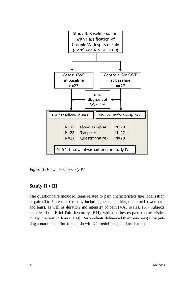
Study participants were recruited from the previously described population based study described in study II (figure 2). Females with CWP (n=27) and controls (n=27) without CWP were randomly selected from 3060 participants with valid RLS and CWP classification (flow chart, figure 3). Subjects with CWP under-went a personal interview and a clinical examination by one trained pain expert.

Methods 31
Figure 2: Flow chart for study II and III
4.2. Methods used in study protocols I-IV
Study I
Patients completed the Swedish version of a validated questionnaire containing four questions addressing Restless Legs according to the international RLS study group criteria [29]. In addition, questions concerning symptoms of insomnia (parts of Nordic Sleep Questionnaire (NSQ) [147]) and daytime sleepiness (Epworth Sleepiness Scale [148]) were answered.
32 Methods
Figure 3: Flow-chart in study IV
Study II + III
The questionnaire included items related to pain characteristics like localisation of pain (0 to 5 areas of the body including neck, shoulder, upper and lower back and legs), as well as duration and intensity of pain (VAS scale). 1677 subjects completed the Brief Pain Inventory (BPI), which addresses pain characteristics during the past 24 hours [149]. Respondents delineated their pain area(s) by put-ting a mark on a printed manikin with 20 predefined pain localisations.
Methods 33
Diagnosis and symptoms related to RLS were assessed using the same method-ology as that applied in study I. In RLS subjects, the frequency of RLS symp-toms was captured by a four point scale ranging from “rarely” to “always”. Anthropometrics (Body Mass Index (BMI), smoking habits, alcohol use), socio-economic status, exercise habits, family history of RLS, co-morbid dis-ease/complaints and ongoing medication were assessed. Age was reported by 1543 subjects in study II and by 1357 in study III.
Information about sleep was collected by means of the NSQ. Daytime sleepiness was classified by the Epworth Sleepiness scale (ESS) and body fatigue was as-sessed in terms of frequency (5 point scale) and intensity (5 point scale).
All patients received a study information letter with the heading “Study of the prevalence of chronic pain and RLS in females from the Dalarna region” that was attached to the mailed questionnaire.
Study IV
All subjects completed a series of questionnaires: (HADS, ESS, Pain Detect, and IRLS) described in detail below. Anthropometric data (age, height, body weight, current smoking, and alcohol consumption (units/week) and comorbidities (car-diovascular, metabolic, neurological, and psychiatric disease) were assessed in the medical interview. Concomitant medication was assessed and classified ac-cording to the Anatomical Therapeutical Chemical (ATC) classification system. Blood samples (for analysis of ferritin, morning cortisol, fasting glucose, cobal-amin and creatinine) were obtained at 8:00 am in all study subjects.
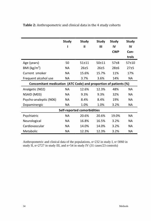
Anthropometric data and clinical information including comorbidities and medi-cation were obtained from the study participants (table 2).
34 Methods
Table 2: Anthropometric and clinical data in the 4 study cohorts
Anthropometric and clinical data of the populations, n=232 in study I, n=3060 in study II, n=2727 in study III, and n=54 in study IV (31 cases/23 controls)
Study I
Study II
Study III
Study IV CWP
Study IV
Con-trols
Age (years) 50 51±11 50±11 57±8 57±10 BMI (kg/m2) NA 26±5 26±5 28±6 27±5 Current smoker NA 15.6% 15.7% 11% 17% Frequent alcohol use NA 3.7% 3.6% 14% NA
Concomitant medication (ATC Code) and proportion of patients (%) Analgetic (N02) NA 12.6% 12.3% 48% NA NSAID (M03) NA 9.3% 9.3% 32% NA Psycho-analeptic (N06) NA 8.4% 8.4% 19% NA Dopaminergic NA 1.0% 1.0% 3.2% NA
Self-reported comorbidities Psychiatric NA 20.6% 20.6% 19.0% NA Neurological NA 16.8% 16.5% 3.2% NA Cardiovascular NA 14.0% 14.0% 3.2% NA Metabolic NA 12.3% 12.3% 3.2% NA
Methods 35
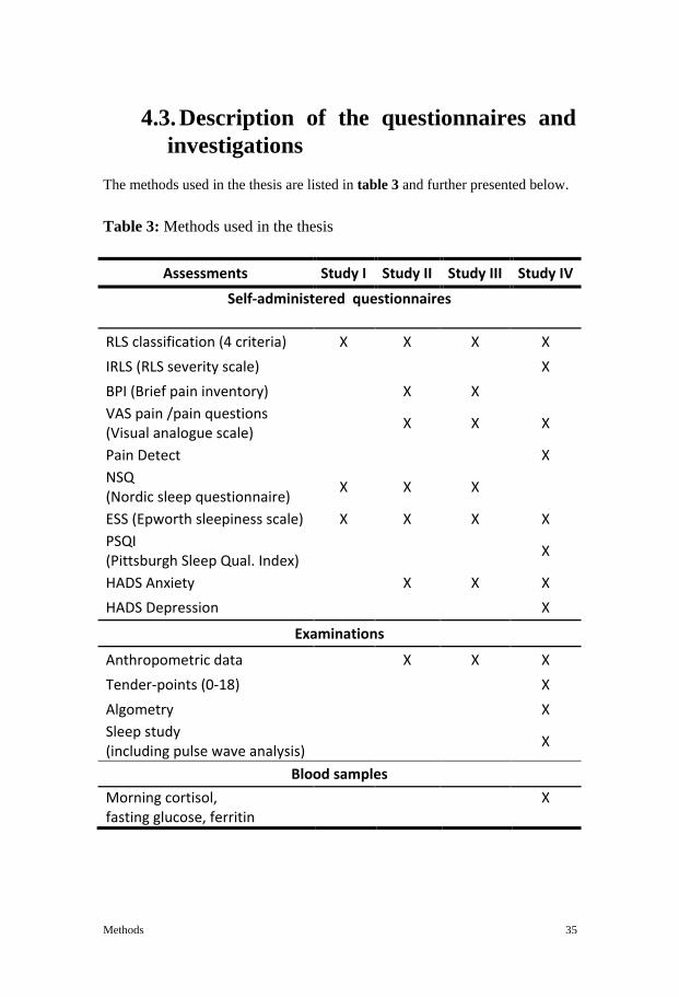
4.3. Description of the questionnaires and investigations
The methods used in the thesis are listed in table 3 and further presented below.
Table 3: Methods used in the thesis
Assessments Study I Study II Study III Study IV
Self-administered questionnaires RLS classification (4 criteria) X X X X IRLS (RLS severity scale) X BPI (Brief pain inventory) X X VAS pain /pain questions (Visual analogue scale) X X X
Pain Detect X NSQ (Nordic sleep questionnaire) X X X
ESS (Epworth sleepiness scale) X X X X PSQI (Pittsburgh Sleep Qual. Index) X
HADS Anxiety X X X HADS Depression X
Examinations
Anthropometric data X X X Tender-points (0-18) X Algometry X Sleep study (including pulse wave analysis) X
Blood samples Morning cortisol, fasting glucose, ferritin
X

36 Methods
Nordic Sleep Questionnaire: The Scandinavian Sleep Research Society devel-oped in 1988 a standardised questionnaire for sleep assessment. The NSQ ap-plies a five-point quantitative scale for many variables as well as self-reported means of sleep latency, sleep length, number of nocturnal awakenings and the amount of sleep considered as “optimal”. [147].
Restless legs syndrome: RLS symptoms were assessed by four standardized criteria for the diagnosis of RLS; (A) Dysesthesia and/or urge to move the limbs, (B) difficulties in resting, (C) worsening of symptoms at rest and improvement by movement, (D) worsening of symptoms at night. These criteria were defined by the International Restless Legs Syndrome Study Group (IRLSSG) and used in the Diagnostic and Statistical Manual for Mental Disorders, Fourth Edition (DSM IV) for the diagnosis of RLS [150]. A questionnaire based classification of RLS was assigned if all 4 criteria were met [150]. The actual number of crite-ria fulfilled (0-4) was also used for analysis. A RLS diagnosis in study IV was confirmed by an experienced physician. Changes in RLS diagnosis from baseline (population based survey) to follow up (2 years period) were evaluated for all participants. Baseline RLS status was not available in 15 control subjects with-out pain due to an unintentional recoding of the subject´s identification number.
IRLS: A ten-question scale that measures severity of RLS designed by the IRLSSG. The IRLS consists of 10 questions rated from 0 to 4 for each question adding to a maximum score of 40 [28]. A score of ≥20 was considered as severe RLS.
The Hospital Anxiety and Depression Scale (HADS). The HADS contains 14 statements, with a rating from 0 to 3, in which a higher score indicates a higher degree of distress [151]. The scores build two subscales, for anxiety (HADS-A) and depression (HADS-D), ranging from 0 to 21 [152]. The cut-off score of eight is suggested to indicate possible anxiety and depression [153]. Several studies have shown that HADS is not adversely affected by chronic pain and have strong associations with the clinical variables of interest [154].
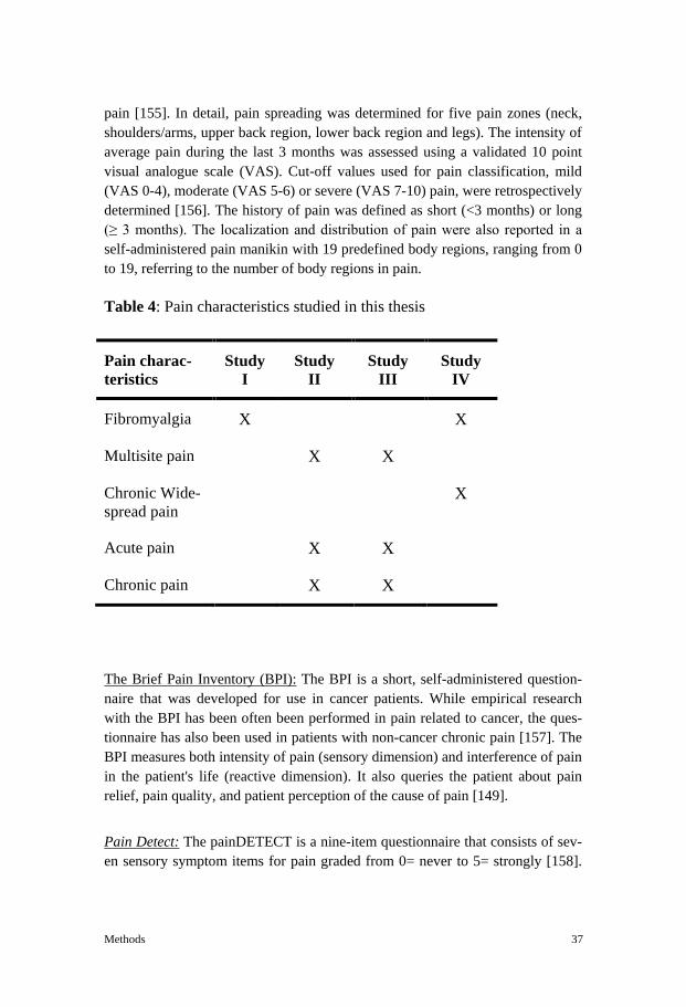
Pain severity and localization. Several dimensions of pain have been investigated in this thesis (table 4). Localization and degree of pain were assessed by a vali-dated screening questionnaire, which addresses intensity, timing, and location of
Methods 37
pain [155]. In detail, pain spreading was determined for five pain zones (neck, shoulders/arms, upper back region, lower back region and legs). The intensity of average pain during the last 3 months was assessed using a validated 10 point visual analogue scale (VAS). Cut-off values used for pain classification, mild (VAS 0-4), moderate (VAS 5-6) or severe (VAS 7-10) pain, were retrospectively determined [156]. The history of pain was defined as short (<3 months) or long (≥ 3 months). The localization and distribution of pain were also reported in a self-administered pain manikin with 19 predefined body regions, ranging from 0 to 19, referring to the number of body regions in pain.
Table 4: Pain characteristics studied in this thesis
Pain charac-teristics
Study I
Study II
Study III
Study IV
Fibromyalgia X X
Multisite pain X X
Chronic Wide-spread pain
X
Acute pain X X
Chronic pain X X
The Brief Pain Inventory (BPI): The BPI is a short, self-administered question-naire that was developed for use in cancer patients. While empirical research with the BPI has been often been performed in pain related to cancer, the ques-tionnaire has also been used in patients with non-cancer chronic pain [157]. The BPI measures both intensity of pain (sensory dimension) and interference of pain in the patient's life (reactive dimension). It also queries the patient about pain relief, pain quality, and patient perception of the cause of pain [149].
Pain Detect: The painDETECT is a nine-item questionnaire that consists of sev-en sensory symptom items for pain graded from 0= never to 5= strongly [158].

38 Methods
One temporal item on pain-course pattern graded −1 to +1, and one spatial item on pain radiation graded 0 for no radiation or +2 for radiating pain is included. A total score (range from −1 to 38) can be calculated from the nine items, with higher scores indicating higher levels of neuropathic pain. PainDETECT was originally developed for evaluating low back pain but is currently used also in the evaluation of the neuropathic component in patients with CWP [143].
Sleep assessment: Study I; Sleep quality and quantity was assessed for the fol-lowing symptoms (5 item scale): Insomnia, early awakening and awakenings at night, snoring and fatigue assessment. Study II and III; Sleep history was as-sessed by means of the NSQ [147]. The questions included mean subjective sleep latency, sleep duration, and number of nocturnal awakenings, loud snoring, and witnessed apnea. Daytime sleepiness (ESS score) and body fatigue (have you felt body fatigue daytime during the last three weeks – always, 4-5 times a week, 2-3 times a week, 1-2 times a week and 1 time or less and how much have you been troubled by body fatigue during the last three days – very much, mod-erately and mild) were also captured. Significant body fatigue was defined as symptoms of fatigue with high frequency (4-5 days a week – always) in combi-nation with severe symptoms during the past 3 days.
The Pittsburgh Sleep Quality Index (PSQI): The PSQI [159] is a self-rated ques-tionnaire that assesses sleep quality and disturbances over a 1-month time inter-val and contains 19 self-rated questions generating seven "component" scores: subjective sleep quality, sleep latency, sleep duration, habitual sleep efficiency, sleep disturbances, use of sleeping medication, and daytime dysfunction. The sum of scores for these seven components yields one global score. Higher scores represent worse sleep quality: component scores range from 0 to 3, and global scores range from 0 to 21 [160].
Medical examination for characterisation of pain
All subjects in the CWP group were examined physically and the 18 tender points were assessed by trained physiotherapist by manual palpation according to established criteria for the diagnosis of CWP [5]. A Somedic algometer (Somedic Production AB, Sollentuna, Sweden) was used to examine the pain pressure threshold by muscle tenderness [161].

Methods 39
Polygraphic sleep study
All study participants in study IV were invited to undergo a sleep study (cardio-respiratory polygraphy, Embletta, Embla, Canada or T3, Noxturnal, Iceland) at home within the time window 10 pm to 7 am. The device continuously assessed breathing by means of a respiratory plethysmography, nasal flow, and oxygen saturation. The body position, limb movement (electromyography), pulse rate and digital pulse wave amplitude (PWA) signals were also obtained. Apneas, hypopneas, desaturations (4% cut off), and episodes with air flow limitation (single breath flow contour analysis) were manually scored by a single experi-enced scorer blinded to subjects group status. PWA attenuations of 10%, 30%, 50% or more from the preceding baseline (PWA 10%, 30%, 50%, for details see figure 4) and PLM´s were automatically scored. All event frequencies were in-dexed for analysis time (Apnea Hypopnea Index (AHI), Oxygen Desaturation Index, PLM index, PWA 10%, 30% 50% index).
Figure 4: A continuous recording of air flow, oxygen saturation (SaO2) and the beat to beat pulse wave. Pulse wave amplitude (PWA) is automatically analysed by recognizing an attenuation of the amplitude from a given baseline (moving window 20 sec). In the lower part a PWA attenuation event of >50% amplitude reduction (PWA50%) is illustrated.

40 Methods
4.4. Statistical Analysis An overview of the statistical tests used in the thesis is shown in Table 5. All tests were two-tailed and conducted at the 5% significance level. Descriptive statistics are presented as mean, standard deviation (SD), standard error of the mean (SEM), range for continuous variables and as number and percent for cate-gorical variables. Statistical analysis was performed using IBM-SPSS version 17.0 and 20.0 software (IBM, Illionois, USA). A p-value of <0.05 was consid-ered as statistically significant.
Descriptive statistics was used to assess prevalences of RLS and FMS and to assess sleep disturbance characteristics in study I. In study III, sleep parameters were categorized based on pain duration, intensity and spreading. Similarly, sleep quality and quantity was evaluated for RLS characteristics (diagnosis yes/no, frequency of symptoms).
Chi square tests were used to test significant differences in the distribution be-tween groups. The test was applied on categorical variables from a single popu-lation and to determine whether there is a significant association between the two distributions. The Cochrane-Armitage Trend (CAT) test was used to analyse a significant trend in the prevalence of RLS across the number of pain are-as/severity of pain. An analysis of variance (ANOVA) was used to compare con-tinuous variables across the classification of RLS and pain when data were normally distributed. The Kruskal Wallis test was used to compare continuous variables when data were not normally distributed. The tests were applied to evaluate significant group differences between groups as well as between cases and controls in study II, III and IV.
Directed Acyclic Graphs (DAG) were constructed in order to select important confounders for the analysis and to detect potential bias (figure 5).
Generalised linear models (GLM) were computed with RLS diagnosis as the dependent variable and pain spreading, - localization, - duration (acute vs. chron-ic), -comorbid psychiatric disease, psychoanaleptic medication (ATC code N06), BMI, marital status and education level were added as independent variables (categorized with exception to BMI) in study II. In study III independent asso-ciations were analysed for continuous sleep parameters, the ESS score and body fatigue. RLS diagnosis, pain spreading (0-5 pain zones), pain intensity (3 catego-ries mild to severe), and pain duration (short- and long-term) were entered into the model as predicting factors. Frequent snoring and witnessed apnea, psychiat-

Methods 41
ric, neurologic, metabolic, and cardiovascular co-morbidity as well as intake of medication according to ATC codes N05 (psycholeptics), N06 (psychoanalep-tics), N02 (analgesics), M01A (anti-inflammatory/NSAID), frequent alcohol consumption (at least 4 days/week), and current smoking were added as potential confounders. BMI was included as a predicting covariate in all analyses. Data on age as a cofounder was available in less than 50% of cases. Therefore, we re-calculated all analysis models for continuous sleep variables in the remaining 1146 females with actual information on age as a covariate. Generalised linear modelling was used in study IV to evaluate the independent contribution of CWP- and RLS status (Factor) on the level of daytime sleepiness (ESS score), depressive and anxiety related symptoms (HADS scores), sleep quality (PSQI), and morning cortisol level after adjustment of confounders like age and BMI (covariates).
Figure 5: Relation between widespread pain, RLS status, and important, non-biased confounders in the DAG (for study II).

42 Methods
Table 5. Overview of statistical tests in the thesis
Statistics Study I
Study II
Study III
Study IV
Descriptive statistics X X X X Pearson´s Chi square X X X ANOVA X Kruskal-Wallis X X Logistic regression model X Generalised Linear Model X X Cochrane-Armitage Trend (CAT) test
X
Student´s T-test X X Fisher´s exact test X
4.5. Ethical approval
Study I
The study was reviewed at the Ethics Committee at Uppsala University in 2006. The study's character was perceived in a way that an ethics quota was not need-ed. A written decision letter was obtained on 20th September 2006. (Uppsala University, application number 2006-210).
Studies II-IV
The studies were reviewed and approved by the regional Ethical Review Board (Uppsala University, application number 2010-124).

Results 43
5. Results
5.1. Pain characteristics
A total of 232 women with FMS were investigated in study I. Further details on severity and duration of the pain disorder were unknown.
In study II, 2453 women (80.2% of the entire study population) reported any occurrence of pain. Pain duration was short (<3 months) in 790 (25.8%) and long (≥3 months) in 1663 (54.4%) subjects. A total of 230 women reported pain in all 5 body areas and were classified as multisite pain (7.5%). 516 women (21.0% out of 2453) reported severe pain according to a VAS scale referring to pain experienced in the last 3 months. Severe pain during the past 24 hours was reported in 287 subjects (16.5% of 1738 responders to this VAS scale). 607 (19.8%) women were free of pain. The distribution of pain was equivalent in study III.
In study IV, 31 women with widespread pain and 23 women without pain were recruited from study II (see also below). FMS was diagnosed in 24 cases and multisite pain/spread musculoskeletal pain in the remaining 7 cases.
5.2. Restless legs prevalence
All four studies showed that the prevalence of RLS in widespread/multisite pain was significantly higher than expected from the general population (Table 6). In study I, RLS prevalence was 63.8% in patients with a physician verified diagno-sis of FMS. In study II, 774 (25.3%) out of 3060 women answered all four RLS questions positively and were classified as RLS positive. The RLS prevalence in study cohort II/III was comparable to previous data from the population for indi-viduals without pain (12.2%, 8.6%, or 9.6% depending on the different classifi-cation systems of pain). The prevalence further increased linearly with the reported number of pain areas: One (23.9%), two (26.4%), three (39.2%), four (44.9%), and finally in five pain areas (54.8%), respectively (p<0.001 for trend, unadjusted Odds ratio 5.6 for multisite pain compared with no pain). Likewise, RLS prevalence increased from 9.6% (no pain) to 27.9%, 37.9% and 42.4% in subjects with mild, moderate, and severe pain according to classes of VAS score (p<0.001). Finally, RLS prevalence was higher in women with long-term pain

44 Results
(35.3%) when compared with those reporting a pain history of <3 months (14.3%, p<0.001).
Table 6: RLS prevalence in women with and without CWP
RLS in CWP
RLS in “No pain”
Study I 64% NA
Study II/III 55% 10%
Study IV 77% 26%
Figure 6: Prevalence of RLS stratified into three age classes in relation to the degree of pain spreading

Results 45
In a sub-analysis including 1357 women stratified by age classes, RLS frequency increased with age: 24% for age 18-45 years, 27% for age 46-55 years, and 31% for age 56-64 years, p=0.014. In 317 pain-free individuals, corresponding num-bers were and 8.2, 5.9, and 15.7%, respectively, p=0.07. However, when pain spreading was taken into account, this age related pattern was reversed in multi-site pain: In 107 women with reported pain in five areas, the highest RLS preva-lence of 73.3% was seen in the youngest age group and vice versa (see figure 6, p=0.017).
The influence of pain on RLS prevalence was independent of several confound-ers evaluated in the GLM model (see also DAG model). Pain spreading, pain localization in the leg, pain duration and co-morbid psychiatric disease were independently predictors of RLS diagnosis in the final model. Other confound-ing factors like BMI, pain intensity, medication, marital status, education level, and smoking were not independent predictors. A separate GLM model including age was generated in a subgroup of 1587 women and the results were essentially unchanged.
The results are consistent in study IV and substantiates earlier results. Prevalence of RLS was higher in subjects with CWP compared with controls (77% vs. 26%, p<0.001) and the severity of symptoms was more pronounced (IRLS scale score 15.1±10.4 vs. 4.8±9.0, <0.001). Severe RLS (IRRLS score >20) were found in 45% of CWP and 13% of controls (p=0.02).
Despite the high prevalence of restless legs in women with CWP active treat-ment with dopaminergic drugs was very rare (study I < 1%, study II and III 1%, and 3% in study IV).
The prevalence of neuropathic pain defined by the criteria of the Pain Detect questionnaire was found significantly higher in RLS sufferers with CWP com-pared with RLS women without pain ( Kruskal Wallis test, p<0,001).
5.3. RLS incidence
In study IV, the diagnosis of CWP at baseline was verified in 27 out of 27 wom-en (100%) at follow up. Additional 4 women reported the incidence of wide-spread pain in the last 24 months (figure 3). Likewise, RLS at baseline was verified in 11 out of 14 subjects (77%) including one individual with very typi-

46 Results
cal symptoms of RLS at baseline but no longer at follow up. In addition, 14 inci-dent cases of RLS were diagnosed at follow-up visit (figure 7) and 12 of those had comorbid CWP. 5 out of those 12 had scored 3 out of 4 RLS criteria already at baseline. Based on the score of ≥20 in the IRRLS scale, RLS severity was classified as “severe” in 45% of the CWP women and 13% in the pain-free controls (p<0.001).
Figure 7: RLS incidence at follow-up, shown are the number of subjects with RLS diagnosis at baseline (left part of the diagram, study II) and at 24 months follow up (right part, study IV).

Results 47
5.4. Impact of RLS and pain on sleep RLS and CWP independently impaired sleep quality and quantity. In study I, FMS patients with RLS reported a higher frequency of difficulties initiating (49.3% vs 31.6%, p<0.01) and maintaining sleep (78.0% vs 63.3%, p=0.02) when compared with FMS without RLS (figure 8). Likewise, RLS patients with FMS more frequently complained about non restorative sleep upon awakening (70.7% vs 56.4%, p<0,04) when compared to women without RLS (figure 8).
Figure 8: Frequency of RLS and sleep complaints (study I, n=232 women with FMS, n=148 classified as RLS patients)
In study III, multisite pain and RLS had worsening effect on sleep latency, sleep duration and the number of nocturnal awakenings from sleep (figure 9). In par-

48 Results
ticular, the impact of both diseases was significant on the difference between desired and actual sleep time - labelled as “sleep deficit” (factor pain spreading and RLS p<0,001, figure 9). Chronic pain and pain spreading (no pain/ 1-2 are-as/3-5 pain areas) had a strong influence on sleep quantity (sleep duration 425.6± 55.6, 409.5±67.9, 384.9±79.1 min, p<0.001respectively) and quality (nocturnal awakenings – 1.5±1.3, 2.1±1.5, 3.0±1.7 events/night, p<0.001 respectively).
Figure 9: Number of awakenings from sleep (events/night) and mean self-reported sleep deficit (in minutes) in relation to the number of pain areas (p<0.001 for both variables, mean and SEM).
RLS was associated with a worsening of sleep variables when compared with women without RLS complaints. Sleep time was shorter (392.4±78.9 vs 415.9±63.1min), sleep latency was increased (37.9±46.9 vs 20.8±25.5 min) and the number of awakenings was increased (2.8±1.7 vs 1.9±1.5n/night), all p<0.001, respectively. In addition, sleep deficit was more pronounced in subjects with RLS (-76.4±74.5 vs -60.9±47.7, p<0.001, figure 10).

Results 49
Several factors and diseases were associated with modification of total sleep time and CWP was one of the strongest (figure 11). After adjustment for im-portant confounders like psychiatric comorbidity, cardiovascular disease, alco-hol, smoking, anthropometrics, pain duration and medication, RLS status and widespread pain still remained independent predictors of the four sleep variables stated above.
Figure 10: Number of awakenings from sleep (events/night) and mean self-reported sleep deficit (minutes) in women with and without a RLS diagnosis (p<0.001 for both variables, mean and SEM).

50 Results
Figure 11: Mean sleep time for several factors (age classes 18-45, 46-55, 56-64 years, BMI classes <25, 25-<30, ≥30 kgm-2, cardiovascular and psychiatric dis-ease, RLS diagnosis and number of pain areas, Y=Yes, N=No).
In study IV, sleep quality was worsened (PSQI questionnaire) in women with CWP when compared with controls (PSQI score 10.6±3.6 vs. 5.5±2.9, p<0.001) and 92% of women with CWP rated their sleep quality as poor. Comorbid RLS did not further influence sleep quality in CWP subjects.
The objective sleep study showed a trend towards a higher number of PLM´s in women with RLS compared with RLS free individuals (8.0±12 vs. 5.6±12 events/hour, n.s.). Significantly more flow limitation was scored in CWP indi-viduals as a sign of mild obstructive sleep disordered breathing (time in flow limitation 35±22 vs. 21±34 min, p=0.022, The AHI prevalence defined by a cut off ≥5 events/hour was 45% and 33%, respectively, in the two groups ( n.s.).

Results 51
5.5. Impact of RLS and pain on daytime symptoms
In study I, ESS score was significantly higher in FMS patients with than without comorbid RLS (17.2±4.5 and 15.9±4.2, p<0.03).
In study III, daytime sleepiness assessed by the ESS score was associated with both pain spreading and RLS. In the GLM model, ESS score increased with RLS diagnosis (0.74, p<0.01) and pain spreading (0.5, 1.7, and 1.8 for 1, 3, and 5 pain areas, respectively, p<0.001) independently from confounders like history of psychiatric disease, frequent snoring (>3 nights/week), increased sleep defi-cit/sleep latency, marital status, and intake of NSAID medication (ATC Code M01A). An ESS score >10, often considered as a threshold for clinically rele-vant hypersomnia, was linearly related to increased RLS symptom frequency (figure 12) and with the number of pain areas (figure 13). Comorbid psychiatric disease was also an important factor increasing the ESS score by 0.76 (95% con-fidence interval 0.35 - 1.17, p<0.001) units in the final GLM model.
Figure 12: Impact of RLS symptom frequency on the prevalence of an ESS score >10.

52 Results
Figure 13: Impact of the number of pain areas on the prevalence of an ESS score >10.
5.6. The impact of CWP on HPA axis and the autonomic nervous system
In study IV several blood tests and the overnight PWA signal were analysed in order to evaluate the impact of CWP and RLS status on the stress axis and the autonomic nervous system. Morning cortisol levels and fasting blood glucose were higher in CWP compared with controls (Table 7). Blood glucose correlated significantly with current pain (VAS, r=0.42, p=0.005) as well as with mean pain during the past 7 days (VAS, r=0.41, p=0.006). Morning cortisol tended to corre-late with current pain (VAS, r=0.27, p=0.07) as well as with mean pain during the past 7 days (VAS, r=0.26, p=0.08). A GLM analysis, adjusting for age, BMI, and RLS status, showed that a CWP diagnosis was associated with a 101±46 nmol increase in morning cortisol, p=0.026. RLS status and RLS symptom se-verity did associate with morning cortisol or glucose level. Ferritin levels were not influenced by RLS or CWP status.

Results 53
Episodes with strong PWA attenuation during the sleeping period (vasocon-striction of 50% and more) were more frequent in subjects with CWP compared to controls (PWA50%, 11.2±8 vs. 6.1±2 events/h, p=0.048). Conversely, small vasoconstrictive events (PWA10-50%) were less frequent in CWP compared with controls (PWA10-50% index 42.3±13 vs 70.9±10 events/h, p<0.002). In addition, mean heart rate tended to be slightly elevated in the CWP group (66.3±8.7 vs. 61.2±8.2 beats/min, p=0.08). There were no significant correla-tions between RLS severity or RLS prevalence on one hand and PWA event frequency on the other.
Table 7: Analysis results of the blood samples taken in cases (CWP) and controls
Parameter Chronic wide-spread pain
(n=25)
Controls
(n=23)
P-value
Morning cortisol (nmol/L)
464±141 366±111 0.011
Fasting blood glucose (mmol/L)
6.0±0.8 5.4±0.7 0.007
Ferritin 89.3±68 87.8±45 n.s.
% Ferritin <50 ng/ml 36% 26% n.s.
% Ferritin <30 ng/ml 16% 13% n.s.

54 Results
5.7. Psychiatric comorbidity in CWP and RLS
In study II and III the most prevalent reported comorbidity was psychiatric dis-eases (20. 6%). In the GLM, a history of psychiatric disease independently influ-enced quantitative sleep parameters (β value in sleep latency 21.0 min, total sleep time -22.1 min, nocturnal awakenings 0.7 events/night, sleep deficit -35.0 min, all p<0.001, respectively). A comorbid diagnosis of psychiatric disease independently predicted daytime sleepiness, measured with ESS score, in the final GLM (β value 0.76, CI 95% 0.35-1.17, p<0.0001).
Psychiatric comorbidity in terms of depression and anxiety was more prevalent in the CWP group. Widespread pain was associated with symptoms of anxiety independent of age, BMI, or RLS comorbidity (GLM Model, β=3.0±1.3, p<0.02 for CWP). RLS diagnosis was associated with the HADS depression score inde-pendent of comorbid CWP (GLM Model, β=3.9±1.1, p<0.001 for RLS). The effects of CWP and RLS diagnoses on the HADS depression and anxiety scores are illustrated in figures 14 and 15.
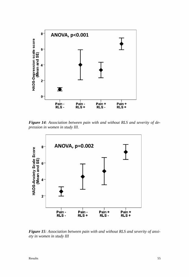
Results 55
Figure 14: Association between pain with and without RLS and severity of de-pression in women in study III.
Figure 15: Association between pain with and without RLS and severity of anxi-ety in women in study III
ANOVA, p<0.001
ANOVA, p=0.002
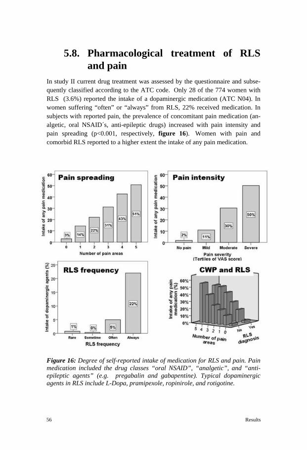
56 Results
5.8. Pharmacological treatment of RLS and pain
In study II current drug treatment was assessed by the questionnaire and subse-quently classified according to the ATC code. Only 28 of the 774 women with RLS (3.6%) reported the intake of a dopaminergic medication (ATC N04). In women suffering “often” or “always” from RLS, 22% received medication. In subjects with reported pain, the prevalence of concomitant pain medication (an-algetic, oral NSAID´s, anti-epileptic drugs) increased with pain intensity and pain spreading (p<0.001, respectively, figure 16). Women with pain and comorbid RLS reported to a higher extent the intake of any pain medication.
Figure 16: Degree of self-reported intake of medication for RLS and pain. Pain medication included the drug classes “oral NSAID”, “analgetic”, and “anti-epileptic agents” (e.g. pregabalin and gabapentine). Typical dopaminergic agents in RLS include L-Dopa, pramipexole, ropinirole, and rotigotine.
Discussion 57
6. Discussion
The aim of the current thesis was to map the associations between widespread pain, RLS and disturbed sleep in middle-aged women. Another aim was to study potential pathomechanisms linked to pain spreading and RLS. The main findings in this thesis include the dose response relationship between pain spreading and RLS frequency, the independent influence of both pain and RLS on impaired sleep quality, and the subsequent increase in daytime sleepiness. Potential mech-anisms behind pain spreading and RLS include activation of the HPA axis and the sympathetic nervous system. Prevalence and incidence of RLS in multisite- and widespread pain The high RLS prevalence of 64% in women with CWP was described for the first time in a larger FMS patient cohort in study I of this thesis. Subsequently, we were able to confirm this finding in a large population based cohort. Other investigators, who also reported a strong association between prevalence of RLS and FMS, confirmed our findings. In these smaller cohorts with FMS patients the RLS prevalence was found to be 42% [54], 33% [53] and 43% [162]. A very recent study in Australian twin families by Hoogwout et al. [163] confirmed findings from study II – the authors reported a strong dose response relationship between spreading of musculoskeletal pain and restless legs frequency in 1072 twins in the age of 22 years. The fully adjusted odds ratio for RLS was 4.9 (95% CI 1.6-14.6) in individuals with three of more pain sites in the body, a number very similar to our findings in a middle age female population cohort. A RLS diagnosis in studies I to III was based on questionnaire data alone. How-ever, in study IV RLS diagnosis was based on several measures including the four validated RLS questions, the IRLS scale for symptom severity and the ex-amination and interview performed by a trained specialist in the assessment of a RLS diagnosis. RLS prevalence was higher in the CWP group (77%) compared with the controls (26%). More important, in study IV we could show for the first time that the incidence of RLS in women with CWP is higher than in the control group. Further, 5 out of 12 incident cases of RLS reported already 2 to 3 RLS criteria at the baseline assessment. This data is consistent with the clinical obser-vation that patients often report an onset of RLS when CWP already has been established. Pain intensity is higher in women with RLS than without. Pain thresholds are reduced and reported clinical pain scores are higher in subjects with RLS relative

58 Discussion
to controls [164]. In the same study, RLS patients had an enhanced temporal summation of heat-induced pain, which may reflect aberrant central nervous system facilitation of pain transmission. RLS is characterized by complaints of an "urge to move" frequently associated with dysesthesias and these sensations may have painful characteristics [165]. Possible shared pathomechanisms in-clude the dysregulation of neurotransmitters with a significant participation of monoaminergic dysfunctional circuits [166]. The main pathomechanism in RLS is a dysregulation in the dopamine and iron systems, as recognised in current research, and the pharmacological treatment data strongly support a dopaminer-gic abnormality for RLS. Our findings may therefore indirectly suggest a dopa-minergic involvement in modulating of pain perception. In fact, a small pilot study including six female patients showed a reduced presynaptic dopamine ac-tivity in FMS [118]. A subsequent study showed that FMS patients also had an abnormal dopamine response to pain [167]. Further, a dysregulation in the neu-rotransmitter glutamate may also be a shared pathomechanism in CWP and RLS according to a limited number of studies. Glutamate receptors play a crucial role in the pain pathways and a recent study showed increased glutamatergic activity in RLS linked to frequent arousals and the disruptive sleep in RLS [89]. This thesis did not explore specific CNS dopaminergic or glutaminergic pathways. However, our data show a strong association and temporal relationship between RLS and CWP which strongly suggest that both diseases share common patho-physiological pathways. Sleep disturbance, RLS and widespread pain Another important focus of this thesis was the analysis of the impact of pain characteristics and RLS on sleep quality. The studies I, III and IV show a robust and consistent finding that pain and RLS have a negative influence on sleep quality and daytime function. In particular, pain spreading reduces and frag-ments sleep in a dose dependent manner. In addition, sleep is already affected by mild to moderate symptom frequency of RLS. As expected, daytime sleepiness and body fatigue were linked to the presence of disturbed sleep. Sleep is known to interact with RLS and pain. Disturbed sleep continuity may contribute to the experience of a lower pain tolerance threshold [126] and sleep deprivation produces hyperalgesia [168]. An arousal enhancing function of pain is the prevailing explanation of sleep deprivation but there is increasing evidence that sleep deprivation in fact could be a pain trigger. The Norwegian HUNT study found that sleep problems were dose-dependently associated with an in-creased risk of pain in the lower back and the neck/shoulder regions of both

Discussion 59
women and men. Those individuals reporting sleep problems 'sometimes' and 'often/always' had a higher risk for the development of chronic pain compared to normal sleepers, 23-32% and 51-66%, respectively [169]. Recent studies show complex and bidirectional relationship between pain and sleep [170] [171] [172] and there is increasing evidence that sleep management and reduction of sleep deficit may improve CWP and reduce pain intensity [173] [174]. Another study suggests that individuals with both short (<6 hours) and long (>9 hours) sleep time are more likely to report pain [175]. Not only sleep length but also sleep quality interact with pain ratings as shown in several studies. Selective slow wave sleep disruption increased musculoskeletal pain and fatigue in middle aged women [176]. Growth hormone is secreted mostly during slow wave sleep and there is strong evidence of decreased GH secretion at least in a subset of FMS patients [177] [59]. In contrast, GH replacement in low IGF-1 patients can sig-nificantly improve symptoms and quality of life in FMS patients suggesting that GH dysfunction may be a link between disturbed sleep and pain load in FMS [178]. Sleep problems caused by sensory symptoms and the presence of PLMs are very well known to be present in patients with RLS [47]. Indeed, Edwards et al. showed that pain processing may be amplified in RLS, perhaps in part as a con-sequence of sleep disruption and sleep deprivation [164]. Neuropathic pain in RLS and CWP Recently, efforts have been made to define several RLS phenotypes including a RLS phenotype associated with pain syndromes [47]. It is known, that pain per-ception is altered in RLS patients and a neuropathic component may be involved in this RLS phenotype. Indeed, a significant association of RLS with positive sensory symptoms of peripheral neuropathy has been found, suggesting that a disorder of the sensory inputs may be involved in the pathogenesis of RLS for a subgroup of patients [179] [144]. One important factor for the development of RLS by a neuropathic process could be the involvement of small fibres. A small fibre neuropathy in RLS has been demonstrated by means of quantitative sensory testing [180] or by skin biopsy with quantification of intraepidermal nerve fibres [181]. In addition, static mechanical hyperalgesia to local stimuli was found to be altered in RLS patients [48]. Thus, central sensitization of the nociceptive system may be present in RLS and mechanical hyperalgesia is a hallmark sign of neuropathic pain. Finally, a high degree of neuropathic pain has also been identi-fied in women with CWP. According to a recent study, the frequency of neuro-pathic pain features in CWP patients was as high as 15.9% [143]. Thus, the high
60 Discussion
component of neuropathic pain in RLS patients shown in study IV of this thesis may be an additional underlying factor explaining the high association between RLS and CWP. Altered HPA axis and sympathetic activity in CWP and RLS Study IV could identify markers for an activation of the HPA axis and the Sym-pathetic Adrenal Medullary (SAM) system in subjects with CWP. In multivari-ate analysis, an almost 25% increase in morning cortisol was identified irrespective of the confounders assessed. Dysregulation of the HPA axis in CWP has previously been described. Several studies have shown both hyper- and hy-pocortisolemia [182] [183]. HPA axis function was studied in 40 FMS patients, 28 patients suffering from chronic low back pain and in 14 controls by means of CRH- and adrenocorticotropic hormone (ACTH) stimulation tests as well as 24-hour urinary cortisol. FMS patients showed an ACTH hyper-responsiveness and a relative adrenal hypo-responsiveness, indicating a tertiary (hypothalamic or supra-hypothalamic level) hypoadrenalism [184]. A review by Crofford and col-leagues [185] discussed the major observations to date related to the stress re-sponse dysfunction in FMS patients. The authors noted that the common clinical features of FMS like widespread pain and fatigue may relate to an underlying dysfunction of the HPA axis. Interestingly, both stress systems, HPA and SAM, appear to be altered [185]. Clinical pain severity was associated with concentra-tions of CRH in the CSF [57] and with salivary cortisol levels [56]. These data are in line with our observation (study IV) that morning cortisol levels tended to correlate with pain ratings at the same day and in the preceding week. The hy-pothesis of an activated SAM system is supported by the finding of an elevated fasting blood glucose levels in CWP subjects of our study. The data suggest al-tered insulin sensitivity; a finding observed both in chronic stress [186] as well as in disturbed sleep [187]. Data on the HPA axis function in RLS patients are sparse. One study found evidence for a nocturnal activation of the hypothalamic-pituitary-adrenal system [188]. In study IV, however, there was no evidence showing any effect of RLS on morning cortisol or fasting glucose. In study IV the novel method of PWA analysis was applied for the assessment of overnight skin vascular reactivity [189]. PWA measures finger vascular tone, which is strongly influenced by skin sympathetic nerve activity via modulation of vascular alpha-receptors [190]. PWA is used both in the diagnosis of sleep, arousal from sleep, and sleep disordered breathing. In the current context, PWA analysis mirrored overnight sympathetic activity and vascular function. The in-creased event index of strong vasoconstrictions in the finger (PWA 50% events)

Discussion 61
observed in CWP women suggest an elevated sympathetic vascular tone during sleep. Indeed, sympathetic blockade by phentolamine and doxazosine (pharma-cological blockade of vascular alpha-receptors) was able to substantially reduce PWA attenuations [190] [191]. In opposite, an increase in small PWA attenua-tions in the range of 10-30% was associated with better cardiovascular function and lower cardiovascular risk [192]. This is in line with the findings in study IV that controls had an higher PWA event index in the lower attenuation range (PWA 10-50%). Women with CWP showed significantly increased time in flow limitation indicating mild degree of obstructive sleep disordered breathing. In-deed, patients with OSA may have an activation of the stress system and an in-crease in sympathetic activity [193] [194]. RLS are often linked to medical disorders like end-stage renal disease [195] [196], iron deficiency [36] [197] or neurological diseases like axonal neuropa-thy [198], cryoglobulinemic neuropathy [179], familial amyloid polyneuropathy [199], Charcot-Marie-Tooth disease type 2 [144], small- fiber neuropathies [200], radiculopathies [201], myelopathies such as multiple sclerosis [202] or syringomyelia [203]. The data in study II did not indicate if any of those comor-bidities could explain the association between RLS and pain. Common factors discussed in the etiology of RLS like metabolic dysfunction, iron deficiency or increases in overnight hypoxic load did not appear in our limited data set of study IV. This may be in part explained by the fact that the case control design was created from the CWP and not from the RLS status. In summary, the data from study IV showed an activation of the HPA axis and the SAM system in women with CWP. Comorbid RLS did not appear to further influence this association. Stress also contributes to the aetiology of depressive disorders and it is increas-ingly accepted that chronic stress, such as in chronic pain conditions, play an important role in the development of depression and anxiety. Conversely, anti-depressants have been shown to attenuate HPA axis activity and normalising of HPA dysfunction immediately precedes successful therapeutic effect of antide-pressants in depressive disorders [204]. Psychiatric comorbidity in CWP and RLS Chronic pain syndromes like FMS, irritable bowel syndrome, low back pain, chronic headaches, and nerve pain overlap frequently with mental disorders like anxiety and depression [205]. Psychiatric disease not only contribute to pain

62 Discussion
intensity but also to an increased risk of disability [206]. In study III women with CWP and RLS had a higher prevalence of depression and anxiety symp-toms compared with RLS-free pain subjects. Study IV used comprehensive methodology for the analysis of mood disorders. Depressive symptoms are mainly linked to a comorbid RLS diagnosis in this study, whereas anxiety symp-toms are stronger linked to CWP subjects. Comparable findings have been re-ported in other studies both for FMS [207] and for RLS [208] [209]. In studies III and IV, psychiatric comorbidity was independently associated with disturbed sleep, increased daytime sleepiness, and body fatigue independently of pain and RLS. These findings suggest that mood disorders may play an im-portant role in pain modulation, occurrence of RLS symptoms and a strong in-hibitory influence on sleep. Indeed, chronic pain, depression and sleep disturbances are known to reciprocally influence each other in a vicious circle of cause and effect. For example, disturbed mood and pain ratings augments after sleep loss in patients with rheumatoid arthritis when compared with controls [210]. A study conducted by Chiu [211] demonstrated that poor sleep and de-pression are independently associated with a reduced pain threshold. The results remained consistent even after adjustment for confounders in the multivariate analysis. Disturbed sleep appears to precede pain spreading in FMS and sleep problems among patients with CWP seem to initiate depressive symptoms [212] [213]. According to findings in the HUNT study, insomnia is a significant factor for the incidence of CWP, anxiety and a range of both mental and physical con-ditions [214]. In a large study [215] insomnia at baseline was associated with a 3 fold risk for incident major depressive and/or anxiety disorder during a 3-year follow-up period. Several longitudinal studies demonstrate that insomnia symp-toms significantly increase the risk of future chronic pain disorders and psychiat-ric distress in previously pain-free individuals [216] [217], whereas existing pain is not a strong predictor of incident insomnia. These data are in strong contrast to the DSM-IV assumption that psychiatric disorder causes sleep problems [218].
Study limitations Study I – The main study limitation is the lack of a control group. In addition, the RLS status was assessed by a questionnaire and not by clinical examination and interview. Study II and III – Response rate was low and there was a substantial loss of data for analysis. This limitation induced a potential inclusion bias towards individu-

Discussion 63
als with pain and/or RLS. In fact, the non-responder analysis indicated an over-sampling of subjects with pain and RLS. However, the prevalence of RLS in the pain-free group was approximately 10%, which is very close to the RLS preva-lence of 11.4% reported in the same population 10 years earlier by Ulfberg et al. [219]. The current study aimed to describe the association between pain pheno-types and the presence of RLS symptoms rather than the prevalence of RLS or pain in this particular population. It is therefore unlikely that study participation bias severely limited our conclusions. All data on sleep quality, sleep quantity and daytime symptoms are self-reported. It is known that subjective data on sleep may differ from objective measures of sleep obtained by polysomnogra-phy. However, the thesis aimed to analyse large cohorts of patient- and popula-tion based samples, which excluded the possibility for an objective assessment of sleep and daytime sleepiness. This is a cross sectional study and it is not pos-sible to comment on the causal relationship between RLS, CMP, sleep disorders and daytime sleepiness. Study IV – The main limitation is the restricted number of study participants and the partial incompleteness of data, which has reduced the statistical power in the analyses. Control subjects did not perform the thorough clinical examination and interview with the pain and RLS expert and this may have resulted in a lower incidence of RLS diagnosis in the control group. There is a possibility to com-ment on causal relationship in this study, but our findings can only be general-ized to the female population.
Clinical application
CWP and RLS are common chronic debilitating conditions. Patients often have very limited access to specialized care units despite the fact that chronic pain results in poorer quality of life than that reported by patients with other chronic disorders. Research on the association between RLS and pain is limited. For instance, a search on the PubMed for chronic diseases such as diabetes or hypo-thyroidism provide 537954 and 39217 search results respectively, compared with 9054 for FMS, 2722 for CWP and 3775 for RLS (assessment made on March 30, 2016). Our data also strongly suggest that a substantial number of patients with RLS remain untreated despite a high burden of symptoms. Finally, the thesis suggests that clinical management of each, CWP, RLS or sleep disor-ders, need to consider the other two diseases due to a very high interrelation. The effect size of combined treatment strategies needs to be assessed.

64 Conclusions
7. Conclusions
This thesis has demonstrated that RLS is very common in women with CWP. Pain spreading appears to be the pain characteristic that is most strongly associ-ated with RLS. Pain intensity and pain duration are also related to the occurrence of RLS. In addition, both pain and RLS are linked to a detrimental effect on shorter sleep length, reduced sleep continuity and to a non-restorative function of sleep. Psychiatric comorbidity is highly prevalent in this context and occurs in almost 50% of women suffering from both CWP and RLS. The longitudinal study in this thesis demonstrated that RLS frequently appeared at a later stage when pain spreading had already been established. This thesis provides further evidence of a possible common pathomechanism for RLS development that involves an activated HPA axis in subjects with CWP. Further, the sympathetic system is activated during sleep in CWP. A specific phenotype among patients with RLS may include women with CWP and our data support a possible neuropathic component of RLS in women with CWP. Finally, our data demonstrate that the majority of women with clinically signifi-cant pain syndromes and more than three quarters of subjects with frequent symptoms of RLS did not receive pharmacological treatment. This finding sug-gests that more information about CWP and RLS is needed in order to increase the awareness of the condition among both healthcare professionals and the gen-eral public.
Future Perspectives 65
8. Future Perspectives
RLS is a common condition in CWP and multisite pain, which increases the risk of mental illness and sleep disturbance. However, the condition is still signifi-cantly under-recognized and under-treated. Future studies need to address the effect of RLS treatment on pain intensity in CWP and multisite pain, comorbid sleep disturbances and psychiatric diseases. As our studies suggest, there may be a specific RLS phenotype associated with pain. Future research is needed on the potential neuropathic component in women with CWP and RLS. The risk for cardiovascular disease in RLS is well established and future studies need to ad-dress the influence of HPA axis and SAM system activation on the risk for car-diovascular and metabolic complications in patients with CWP and RLS. Finally, further research is warranted to evaluate the effect of different measures to improve sleep quantity and sleep quality on pain intensity, pain spreading and the symptom load of RLS.

66 Acknowledgements
9. Acknowledgements
During the work with the present thesis there have been many events and chang-es in my life – I have changed place of work and got new colleagues, moved to a new city, have watched my children grow and come into puberty. There are many people who have supported me through these years and encour-aged me to continue the journey of this thesis, and I wish to express my gratitude to them: Ludger Grote, my main supervisor, for the patience over the years, tremendous commitment, energy, vast clinical and scientific knowledge, pushing me further in my development and giving me concrete, professional and invaluable advice. Jan Hedner, my co-supervisor, for believing in me, sharing the clinical and sci-entific knowledge, valuable opinions, advice and feedback on my manuscripts. Jan Ulfberg, my co-superisor and former colleague at sleep laboratory, who introduced me in the research and helped me to get started with it, believed in my project and helped me to launch it, has given me important advice and shared vast clinical and scientific experiences and knowledge. Ding Zou, colleague in the vigilance laboratory, for sharing the knowledge in clinical research and for feedback on my manuscripts and valuable opinions. Karin Koch-Schmidt, my former boss from Skonvik Pain Rehabilitation clinic in Dalarna, for help and support over the years and for having made it possible to implement both the population-based studies and the prospective part of the the-sis that involved help from the staff at the clinic with blood tests, preparation of materials, callings of patients. Your exuberant energy and humor that made eve-ryday life more enjoyable and work burden easier to bear. My past and present colleagues at Pain Rehabilitation clinic and Pain Center at Uppsala Academic Hospital for friendship, support, encouragement especially following, Mathias Andersson at Skonvik Pain Rehabilitation clinic, for many deep, interesting conversations and discussions over the years, support during tough periods as well as many laughs together and fun and not least the enor-mous amount of energy and deep involvement in patients with chronic pain. Ros-Marie Envall, Max Mildenberger, Mona Jacobs-Lindén, Lisa Friberg at Skonvik and Eva-Britt Hysing, Rolf Karlsten and Torsten Gordh at Pain

Acknowledgements 67
Center at Uppsala Academic Hospital for sharing your valuable clinical and scientific knowledge; Mia (sekr) and Ellen (sekr) at Skonvik Pain Rehabilitation for helt with collecting of data and help in solving all kinds of problems with administrative tasks over the years and support; Marie Essermark at Pain Cen-ter for helping me by means of transferring and collecting blood samples. Lennart Arvidsson, co-author and skilled and proficient colleague at Skonvik Pain Rehabilitation with vast clinical experience, for very interesting, deep and inspiring discussions over the years and sharing the knowledge. Jan Wesström, colleague and traveling companion on many congresses and conferences and interesting debate partner with many brilliant ideas. Ronny Ravna, always friendly and incredibly accommodating, for help with technical issues and problem-solving sleep screening equipment Jan Tichy, academic painter and family member, who allowed me use one of his amazing paintings as the cover illistration. My family: my mother for believing in me and teaching me the important value of commitment and hard work, my deceased father who taught me to read books as a young child and thirst for new knowledge and to not feel limited by the out-er frames, my beloved children Patricia and Lukas who tolerated all working weekends and holidays that I have been forced to dedicate to the study and last but not least my husband Leo who supported me during years, always believed in me, helped me through difficult times and made it possible for me to get through the entire doctoral period. The study was supported by the Center for Clinical Research in Dalarna, the Pain Center in Säter, the Swedish Heart and Lung Foundation, the Swedish Scociety for Sleep Research and Sleep Medicine, and the Pain Center at Uppsala University (head Professor Torsten Gordh).

68 References
10. References
1. Cross, S.A., Pathophysiology of pain. Mayo Clin Proc, 1994. 69(4): p.
375-83. 2. Breivik, H., et al., Survey of chronic pain in Europe: prevalence, impact
on daily life, and treatment. Eur J Pain, 2006. 10(4): p. 287-333. 3. McCarberg, B.H., et al., The impact of pain on quality of life and the
unmet needs of pain management: results from pain sufferers and physicians participating in an Internet survey. Am J Ther, 2008. 15(4): p. 312-20.
4. Dubin, A.E. and A. Patapoutian, Nociceptors: the sensors of the pain pathway. The Journal of Clinical Investigation, 2010. 120(11): p. 3760-3772.
5. Wolfe, F., et al., The American College of Rheumatology 1990 Criteria for the Classification of Fibromyalgia. Report of the Multicenter Criteria Committee. Arthritis Rheum, 1990. 33(2): p. 160-72.
6. Markenson, J.A., Mechanisms of chronic pain. Am J Med, 1996. 101(1a): p. 6s-18s.
7. Mansfield, K.E., et al., A systematic review and meta-analysis of the prevalence of chronic widespread pain in the general population. Pain, 2016. 157(1): p. 55-64.
8. Latremoliere, A. and C.J. Woolf, Central sensitization: a generator of pain hypersensitivity by central neural plasticity. J Pain, 2009. 10(9): p. 895-926.
9. Woolf, C.J., Central sensitization: Implications for the diagnosis and treatment of pain. Pain, 2011. 152(3 Suppl): p. S2-15.
10. Vanderah, T.W. and M.H. Ossipov, Plasticity of the Central Nervous System and Chronic Pain. Journal of Neuropathic Pain & Symptom Palliation, 2005. 1(2): p. 27-36.
11. Loggia, M.L., et al., Evidence for brain glial activation in chronic pain patients. Brain, 2015. 138(Pt 3): p. 604-15.
12. Tracey, I. and P.W. Mantyh, The cerebral signature for pain perception and its modulation. Neuron, 2007. 55(3): p. 377-91.
13. Bushnell, M.C., M. Čeko, and L.A. Low, Cognitive and emotional control of pain and its disruption in chronic pain. Nature reviews. Neuroscience, 2013. 14(7): p. 502-511.
14. Bartley, E.J. and R.B. Fillingim, Sex differences in pain: a brief review of clinical and experimental findings. BJA: British Journal of Anaesthesia, 2013. 111(1): p. 52-58.
15. Robinson, J.E. and R.V. Short, Changes in breast sensitivity at puberty, during the menstrual cycle, and at parturition. Br Med J, 1977. 1(6070): p. 1188-91.

References 69
16. Nishizawa, S., et al., Differences between males and females in rates of serotonin synthesis in human brain. Proc Natl Acad Sci U S A, 1997. 94(10): p. 5308-13.
17. Rustoen, T., et al., Age and the experience of chronic pain: differences in health and quality of life among younger, middle-aged, and older adults. Clin J Pain, 2005. 21(6): p. 513-23.
18. Gold, A.R., et al., Inspiratory airflow dynamics during sleep in women with fibromyalgia. Sleep, 2004. 27(3): p. 459-66.
19. Drewes, A.M., et al., The effect of cutaneous and deep pain on the electroencephalogram during sleep--an experimental study. Sleep, 1997. 20(8): p. 632-40.
20. Smith, M.T., et al., Sleep quality and presleep arousal in chronic pain. J Behav Med, 2000. 23(1): p. 1-13.
21. Auerbach, S.M., et al., Depression, pain, exposure to stressful life events, and long-term outcomes in temporomandibular disorder patients. J Oral Maxillofac Surg, 2001. 59(6): p. 628-33; discussion 634.
22. Savage, S.R., K.L. Kirsh, and S.D. Passik, Challenges in Using Opioids to Treat Pain in Persons With Substance Use Disorders. Addiction Science & Clinical Practice, 2008. 4(2): p. 4-25.
23. Bair, M.J., et al., Depression and pain comorbidity: a literature review. Arch Intern Med, 2003. 163(20): p. 2433-45.
24. Buskila, D., Genetics of chronic pain states. Best Pract Res Clin Rheumatol, 2007. 21(3): p. 535-47.
25. Carville, S.F., et al., EULAR evidence-based recommendations for the management of fibromyalgia syndrome. Ann Rheum Dis, 2008. 67(4): p. 536-41.
26. Lee, J., et al., Chronic widespread pain, including fibromyalgia: a pathway for care developed by the British Pain Society. Br J Anaesth, 2014. 112(1): p. 16-24.
27. Onen, S.H., et al., Pain tolerance and obstructive sleep apnea in the elderly. J Am Med Dir Assoc, 2010. 11(9): p. 612-6.
28. Allen, R., et al., Relation of the International Restless Legs Syndrome Study Group rating scale with the Clinical Global Impression severity scale, the restless legs syndrome 6-item questionnaire, and the restless legs syndrome-quality of life questionnaire. Sleep Med, 2013. 14(12): p. 1375-80.
29. Gamaldo, C.E. and C.J. Earley, Restless legs syndrome: a clinical update. Chest, 2006. 130(5): p. 1596-604.
30. Oka, Y. and Y. Ioue, [Secondary restless legs syndrome]. Brain Nerve, 2009. 61(5): p. 539-47.
31. Allen, R.P. and C.J. Earley, The peak prevalence is in the 60-79 year old age group but RLS may start at any age and may occur in young children. J Clin Neurophysiol, 2001. 18(2): p. 128-47.
32. Allen, R.P., et al., Restless legs syndrome prevalence and impact: REST general population study. Arch Intern Med, 2005. 165(11): p. 1286-92.

70 References
33. Manconi, M., et al., Restless legs syndrome and pregnancy. Neurology, 2004. 63(6): p. 1065-9.
34. Allen, R.P., et al., Increased CSF hypocretin-1 (orexin-A) in restless legs syndrome. Neurology, 2002. 59(4): p. 639-41.
35. Nagandla, K. and S. De, Restless legs syndrome: pathophysiology and modern management. Postgrad Med J, 2013. 89(1053): p. 402-10.
36. Allen, R.P. and C.J. Earley, The role of iron in restless legs syndrome. Mov Disord, 2007. 22 Suppl 18: p. S440-8.
37. Haba-Rubio, J., et al., Restless legs syndrome and low brain iron levels in patients with haemochromatosis. J Neurol Neurosurg Psychiatry, 2005. 76(7): p. 1009-10.
38. Walters, A.S., et al., Does the endogenous opiate system play a role in the Restless Legs Syndrome? A pilot post-mortem study. J Neurol Sci, 2009. 279(1-2): p. 62-5.
39. Baraniuk, J.N., et al., Cerebrospinal fluid levels of opioid peptides in fibromyalgia and chronic low back pain. BMC Musculoskelet Disord, 2004. 5: p. 48.
40. Miyamoto, M., et al., [Pathophysiology of restless legs syndrome]. Brain Nerve, 2009. 61(5): p. 523-32.
41. Vetrugno, R., F. Provini, and P. Montagna, Restless legs syndrome and periodic limb movements. Rev Neurol Dis, 2006. 3(2): p. 61-70.
42. Winkelmann, J., et al., Genome-wide association study of restless legs syndrome identifies common variants in three genomic regions. Nat Genet, 2007. 39(8): p. 1000-6.
43. Roux, F.J., Restless legs syndrome: impact on sleep-related breathing disorders. Respirology, 2013. 18(2): p. 238-45.
44. Salminen, A.V., V. Rimpila, and O. Polo, Peripheral hypoxia in restless legs syndrome (Willis-Ekbom disease). Neurology, 2014. 82(21): p. 1856-61.
45. Hening, W., et al., Impact, diagnosis and treatment of restless legs syndrome (RLS) in a primary care population: the REST (RLS epidemiology, symptoms, and treatment) primary care study. Sleep Med, 2004. 5(3): p. 237-46.
46. Williams, A.M. and D. Garcia-Borreguero, Management of restless legs syndrome augmentation. Curr Treat Options Neurol, 2009. 11(5): p. 327-32.
47. Aurora, R.N., et al., The Treatment of Restless Legs Syndrome and Periodic Limb Movement Disorder in Adults—An Update for 2012: Practice Parameters with an Evidence-Based Systematic Review and Meta-Analyses: An American Academy of Sleep Medicine Clinical Practice Guideline. Sleep, 2012. 35(8): p. 1039-1062.
48. Stiasny-Kolster, K., et al., Static mechanical hyperalgesia without dynamic tactile allodynia in patients with restless legs syndrome. Brain, 2004. 127(Pt 4): p. 773-82.

References 71
49. Gjevre, J.A. and R.M. Taylor Gjevre, Restless legs syndrome as a comorbidity in rheumatoid arthritis. Autoimmune Dis, 2013. 2013: p. 352782.
50. Hassan, N., et al., Systemic lupus and risk of restless legs syndrome. J Rheumatol, 2011. 38(5): p. 874-6.
51. Stehlik, R., L. Arvidsson, and J. Ulfberg, Restless legs syndrome is common among female patients with fibromyalgia. Eur Neurol, 2009. 61(2): p. 107-11.
52. Stehlik, R., et al., High prevalence of restless legs syndrome among women with multi-site pain: a population-based study in Dalarna, Sweden. Eur J Pain, 2014. 18(10): p. 1402-9.
53. Viola-Saltzman, M., et al., High prevalence of restless legs syndrome among patients with fibromyalgia: a controlled cross-sectional study. J Clin Sleep Med, 2010. 6(5): p. 423-7.
54. Yunus, M.B. and J.C. Aldag, Restless legs syndrome and leg cramps in fibromyalgia syndrome: a controlled study. Bmj, 1996. 312(7042): p. 1339.
55. Riva, R., et al., Fibromyalgia syndrome is associated with hypocortisolism. Int J Behav Med, 2010. 17(3): p. 223-33.
56. McLean, S.A., et al., Momentary relationship between cortisol secretion and symptoms in patients with fibromyalgia. Arthritis Rheum, 2005. 52(11): p. 3660-9.
57. McLean, S.A., et al., Cerebrospinal fluid corticotropin-releasing factor concentration is associated with pain but not fatigue symptoms in patients with fibromyalgia. Neuropsychopharmacology, 2006. 31(12): p. 2776-82.
58. Clauw, D.J. and G.P. Chrousos, Chronic pain and fatigue syndromes: overlapping clinical and neuroendocrine features and potential pathogenic mechanisms. Neuroimmunomodulation, 1997. 4(3): p. 134-53.
59. Bennett, R.M., Adult growth hormone deficiency in patients with fibromyalgia. Curr Rheumatol Rep, 2002. 4(4): p. 306-12.
60. Yoshimura, M. and H. Furue, Mechanisms for the anti-nociceptive actions of the descending noradrenergic and serotonergic systems in the spinal cord. J Pharmacol Sci, 2006. 101(2): p. 107-17.
61. Becker, S. and P. Schweinhardt, Dysfunctional neurotransmitter systems in fibromyalgia, their role in central stress circuitry and pharmacological actions on these systems. Pain Res Treat, 2012. 2012: p. 741746.
62. Le Bars, D., et al., Diffuse noxious inhibitory controls (DNIC) in animals and in man. Patol Fiziol Eksp Ter, 1992(4): p. 55-65.
63. Millan, M.J., Descending control of pain. Prog Neurobiol, 2002. 66(6): p. 355-474.
64. Suzuki, R., L.J. Rygh, and A.H. Dickenson, Bad news from the brain: descending 5-HT pathways that control spinal pain processing. Trends Pharmacol Sci, 2004. 25(12): p. 613-7.

72 References
65. De Felipe, C., et al., Altered nociception, analgesia and aggression in mice lacking the receptor for substance P. Nature, 1998. 392(6674): p. 394-7.
66. DeVane, C.L., Substance P: a new era, a new role. Pharmacotherapy, 2001. 21(9): p. 1061-9.
67. Snijdelaar, D.G., et al., Substance P. Eur J Pain, 2000. 4(2): p. 121-35. 68. Stratz, T., et al., Influence of tropisetron on the serum substance P
levels in fibromyalgia patients. Scand J Rheumatol Suppl, 2004. 119: p. 41-3.
69. Light, K.C., et al., Adrenergic dysregulation and pain with and without acute beta-blockade in women with fibromyalgia and temporomandibular disorder. J Pain, 2009. 10(5): p. 542-52.
70. Russell, I.J., et al., Cerebrospinal fluid biogenic amine metabolites in fibromyalgia/fibrositis syndrome and rheumatoid arthritis. Arthritis Rheum, 1992. 35(5): p. 550-6.
71. Jaschko, G., et al., Serum serotonin levels are not useful in diagnosing fibromyalgia. Ann Rheum Dis, 2007. 66(9): p. 1267-8.
72. Legangneux, E., et al., Cerebrospinal fluid biogenic amine metabolites, plasma-rich platelet serotonin and [3H]imipramine reuptake in the primary fibromyalgia syndrome. Rheumatology (Oxford), 2001. 40(3): p. 290-6.
73. Julien, N., et al., Widespread pain in fibromyalgia is related to a deficit of endogenous pain inhibition. Pain, 2005. 114(1-2): p. 295-302.
74. Millan, M.J., The induction of pain: an integrative review. Prog Neurobiol, 1999. 57(1): p. 1-164.
75. Liu, Z., et al., A high-recovery extraction procedure for quantitative analysis of substance P and opioid peptides in human cerebrospinal fluid. Peptides, 2000. 21(6): p. 853-60.
76. Russell, I.J., et al., Elevated cerebrospinal fluid levels of substance P in patients with the fibromyalgia syndrome. Arthritis Rheum, 1994. 37(11): p. 1593-601.
77. Vaeroy, H., et al., Elevated CSF levels of substance P and high incidence of Raynaud phenomenon in patients with fibromyalgia: new features for diagnosis. Pain, 1988. 32(1): p. 21-6.
78. Sarchielli, P., et al., Sensitization, glutamate, and the link between migraine and fibromyalgia. Curr Pain Headache Rep, 2007. 11(5): p. 343-51.
79. Peres, M.F., et al., Cerebrospinal fluid glutamate levels in chronic migraine. Cephalalgia, 2004. 24(9): p. 735-9.
80. Fayed, N., et al., Localized 1H-NMR spectroscopy in patients with fibromyalgia: a controlled study of changes in cerebral glutamate/glutamine, inositol, choline, and N-acetylaspartate. Arthritis Res Ther, 2010. 12(4): p. R134.
81. Valdes, M., et al., Increased glutamate/glutamine compounds in the brains of patients with fibromyalgia: a magnetic resonance spectroscopy study. Arthritis Rheum, 2010. 62(6): p. 1829-36.

References 73
82. Harris, R.E., et al., Elevated insular glutamate in fibromyalgia is associated with experimental pain. Arthritis Rheum, 2009. 60(10): p. 3146-52.
83. Magnusson, J.E. and K. Fisher, The involvement of dopamine in nociception: the role of D(1) and D(2) receptors in the dorsolateral striatum. Brain Res, 2000. 855(2): p. 260-6.
84. Dias, E.V., et al., Nucleus accumbens dopaminergic neurotransmission switches its modulatory action in chronification of inflammatory hyperalgesia. Eur J Neurosci, 2015. 42(7): p. 2380-9.
85. Salemi, S., et al., Up-regulation of delta-opioid receptors and kappa-opioid receptors in the skin of fibromyalgia patients. Arthritis Rheum, 2007. 56(7): p. 2464-6.
86. Harris, R.E., et al., Decreased central mu-opioid receptor availability in fibromyalgia. J Neurosci, 2007. 27(37): p. 10000-6.
87. Jones, A.K., et al., Changes in central opioid receptor binding in relation to inflammation and pain in patients with rheumatoid arthritis. Br J Rheumatol, 1994. 33(10): p. 909-16.
88. Maarrawi, J., et al., Differential brain opioid receptor availability in central and peripheral neuropathic pain. Pain, 2007. 127(1-2): p. 183-94.
89. Allen, R.P., et al., Thalamic glutamate/glutamine in restless legs syndrome: increased and related to disturbed sleep. Neurology, 2013. 80(22): p. 2028-34.
90. Mantyh, P.W., et al., Antagonism of Nerve Growth Factor-TrkA Signaling and the Relief of Pain. Anesthesiology, 2011. 115(1): p. 189-204.
91. Wang, H., et al., Circulating cytokine levels compared to pain in patients with fibromyalgia -- a prospective longitudinal study over 6 months. J Rheumatol, 2008. 35(7): p. 1366-70.
92. Watkins, L.R., E.D. Milligan, and S.F. Maier, Glial proinflammatory cytokines mediate exaggerated pain states: implications for clinical pain. Adv Exp Med Biol, 2003. 521: p. 1-21.
93. Watkins, L.R. and S.F. Maier, Beyond neurons: evidence that immune and glial cells contribute to pathological pain states. Physiol Rev, 2002. 82(4): p. 981-1011.
94. Kosek, E., et al., Evidence of different mediators of central inflammation in dysfunctional and inflammatory pain--interleukin-8 in fibromyalgia and interleukin-1 beta in rheumatoid arthritis. J Neuroimmunol, 2015. 280: p. 49-55.
95. Zhang, J.-M. and J. An, Cytokines, Inflammation and Pain. International anesthesiology clinics, 2007. 45(2): p. 27-37.
96. Maes, M., et al., The immune-inflammatory pathophysiology of fibromyalgia: increased serum soluble gp130, the common signal transducer protein of various neurotrophic cytokines. Psychoneuroendocrinology, 1999. 24(4): p. 371-83.

74 References
97. Pay, S., et al., Evaluation of vascular injury with proinflammatory cytokines, thrombomodulin and fibronectin in patients with primary fibromyalgia. Nagoya J Med Sci, 2000. 63(3-4): p. 115-22.
98. Wallace, D.J., et al., Cytokines play an aetiopathogenetic role in fibromyalgia: a hypothesis and pilot study. Rheumatology (Oxford), 2001. 40(7): p. 743-9.
99. Gur, A., et al., Cytokines and depression in cases with fibromyalgia. J Rheumatol, 2002. 29(2): p. 358-61.
100. Weinstock, L.B., A.S. Walters, and P. Paueksakon, Restless legs syndrome--theoretical roles of inflammatory and immune mechanisms. Sleep Med Rev, 2012. 16(4): p. 341-54.
101. Trotti, L.M., et al., Elevated C-reactive protein is associated with severe periodic leg movements of sleep in patients with restless legs syndrome. Brain Behav Immun, 2012. 26(8): p. 1239-43.
102. Hornyak, M., et al., Low-dose hydrocortisone in the evening modulates symptom severity in restless legs syndrome. Neurology, 2008. 70(18): p. 1620-2.
103. Roberts, M.H., et al., Chronic pain and pain medication use in chronic obstructive pulmonary disease. A cross-sectional study. Ann Am Thorac Soc, 2013. 10(4): p. 290-8.
104. Germanowicz, D., et al., Sleep disordered breathing concomitant with fibromyalgia syndrome. J Bras Pneumol, 2006. 32(4): p. 333-8.
105. Alvarez Lario, B., et al., Fibromyalgia syndrome: overnight falls in arterial oxygen saturation. Am J Med, 1996. 101(1): p. 54-60.
106. Zamuner, A.R., et al., Relationship between sympathetic activity and pain intensity in fibromyalgia. Clin Exp Rheumatol, 2015. 33(1 Suppl 88): p. S53-7.
107. Bengtsson, A. and K.G. Henriksson, The muscle in fibromyalgia--a review of Swedish studies. J Rheumatol Suppl, 1989. 19: p. 144-9.
108. Wahlin-Larsson, B., et al., The expression of vascular endothelial growth factor in skeletal muscle of patients with sleep disorders. Muscle Nerve, 2009. 40(4): p. 556-61.
109. Anderson, K.N., C. Di Maria, and J. Allen, Novel assessment of microvascular changes in idiopathic restless legs syndrome (Willis-Ekbom disease). J Sleep Res, 2013. 22(3): p. 315-21.
110. Salminen, A.V., V. Rimpila, and O. Polo, Pramipexole alters thermoregulation in restless legs syndrome. J Clin Sleep Med, 2014. 10(12): p. 1325-9.
111. Winkelmann, J., et al., Variants in the neuronal nitric oxide synthase (nNOS, NOS1) gene are associated with restless legs syndrome. Mov Disord, 2008. 23(3): p. 350-8.
112. Patton, S.M., et al., Hypoxia-inducible factor pathway activation in restless legs syndrome patients. Eur J Neurol, 2011. 18(11): p. 1329-35.
113. Koh, S.Y., et al., Impaired vascular endothelial function in patients with restless legs syndrome: a new aspect of the vascular pathophysiology. J Neurol Sci, 2015. 359(1-2): p. 207-10.

References 75
114. Wood, P.B., Role of central dopamine in pain and analgesia. Expert Rev Neurother, 2008. 8(5): p. 781-97.
115. Monti, J.M. and D. Monti, The involvement of dopamine in the modulation of sleep and waking. Sleep Med Rev, 2007. 11(2): p. 113-33.
116. Monti, J.M. and H. Jantos, The roles of dopamine and serotonin, and of their receptors, in regulating sleep and waking. Prog Brain Res, 2008. 172: p. 625-46.
117. Malt, E.A., et al., Altered dopamine D2 receptor function in fibromyalgia patients: a neuroendocrine study with buspirone in women with fibromyalgia compared to female population based controls. J Affect Disord, 2003. 75(1): p. 77-82.
118. Wood, P.B., et al., Reduced presynaptic dopamine activity in fibromyalgia syndrome demonstrated with positron emission tomography: a pilot study. J Pain, 2007. 8(1): p. 51-8.
119. Clauw, D.J. and L.J. Crofford, Chronic widespread pain and fibromyalgia: what we know, and what we need to know. Best Pract Res Clin Rheumatol, 2003. 17(4): p. 685-701.
120. Pani, L., A. Porcella, and G.L. Gessa, The role of stress in the pathophysiology of the dopaminergic system. Mol Psychiatry, 2000. 5(1): p. 14-21.
121. Sorensen, J., et al., Pain analysis in patients with fibromyalgia. Effects of intravenous morphine, lidocaine, and ketamine. Scand J Rheumatol, 1995. 24(6): p. 360-5.
122. Sorensen, J., et al., Fibromyalgia--are there different mechanisms in the processing of pain? A double blind crossover comparison of analgesic drugs. J Rheumatol, 1997. 24(8): p. 1615-21.
123. Graven-Nielsen, T., et al., Ketamine reduces muscle pain, temporal summation, and referred pain in fibromyalgia patients. Pain, 2000. 85(3): p. 483-91.
124. Altier, N. and J. Stewart, The role of dopamine in the nucleus accumbens in analgesia. Life Sci, 1999. 65(22): p. 2269-87.
125. Cooperman, N.R., F.J. Mullin, and N. Kleitman, Studies on the physiology of sleep. XI. Further observations on the effects of prolonged sleeplessness. American Journal of Physiology, 1934. 107: p. 589-593.
126. Onen, S.H., et al., The effects of total sleep deprivation, selective sleep interruption and sleep recovery on pain tolerance thresholds in healthy subjects. J Sleep Res, 2001. 10(1): p. 35-42.
127. Finan, P.H., B.R. Goodin, and M.T. Smith, The association of sleep and pain: an update and a path forward. J Pain, 2013. 14(12): p. 1539-52.
128. Paul-Savoie, E., et al., Is the deficit in pain inhibition in fibromyalgia influenced by sleep impairments? Open Rheumatol J, 2012. 6: p. 296-302.

76 References
129. Oono, Y., et al., Conditioned pain modulation in temporomandibular disorders (TMD) pain patients. Exp Brain Res, 2014. 232(10): p. 3111-9.
130. Lee, Y.C., et al., The role of sleep problems in central pain processing in rheumatoid arthritis. Arthritis and rheumatism, 2013. 65(1): p. 59-68.
131. Miro, E., et al., When is pain related to emotional distress and daily functioning in fibromyalgia syndrome? The mediating roles of self-efficacy and sleep quality. Br J Health Psychol, 2011. 16(4): p. 799-814.
132. Bogan, R.K., Effects of restless legs syndrome (RLS) on sleep. Neuropsychiatric Disease and Treatment, 2006. 2(4): p. 513-519.
133. Taylor, D.J., et al., Comorbidity of chronic insomnia with medical problems. Sleep, 2007. 30(2): p. 213-8.
134. Tang, N.K., K.J. Wright, and P.M. Salkovskis, Prevalence and correlates of clinical insomnia co-occurring with chronic back pain. J Sleep Res, 2007. 16(1): p. 85-95.
135. Moldofsky, H. and J. Patcai, Chronic widespread musculoskeletal pain, fatigue, depression and disordered sleep in chronic post-SARS syndrome; a case-controlled study. BMC Neurol, 2011. 11: p. 37.
136. Thomas, R.J., Sleep as a window into the world of fibromyalgia syndrome. J Rheumatol, 2011. 38(12): p. 2499-500.
137. Vgontzas, A.N., et al., Chronic insomnia and activity of the stress system: a preliminary study. J Psychosom Res, 1998. 45(1): p. 21-31.
138. Fatima, G., et al., Lack of Circadian Pattern of Serum TNF-alpha and IL-6 in Patients with Fibromyalgia Syndrome. Indian J Clin Biochem, 2012. 27(4): p. 340-3.
139. Gannon, R.L. and M.J. Millan, Evaluation of serotonin, noradrenaline and dopamine reuptake inhibitors on light-induced phase advances in hamster circadian activity rhythms. Psychopharmacology (Berl), 2007. 195(3): p. 325-32.
140. Maletic, V. and C.L. Raison, Neurobiology of depression, fibromyalgia and neuropathic pain. Front Biosci (Landmark Ed), 2009. 14: p. 5291-338.
141. Proitsi, P., et al., Association of serotonin and dopamine gene pathways with behavioral subphenotypes in dementia. Neurobiol Aging, 2012. 33(4): p. 791-803.
142. Vandenbergh, D.J., et al., Dopamine receptor genes (DRD2, DRD3 and DRD4) and gene-gene interactions associated with smoking-related behaviors. Addict Biol, 2007. 12(1): p. 106-16.
143. Momi, S.K., et al., Neuropathic pain as part of chronic widespread pain: environmental and genetic influences. Pain, 2015. 156(10): p. 2100-6.
144. Gemignani, F., et al., Charcot-Marie-Tooth disease type 2 with restless legs syndrome. Neurology, 1999. 52(5): p. 1064-6.
References 77
145. Rutkove, S.B., J.K. Matheson, and E.L. Logigian, Restless legs syndrome in patients with polyneuropathy. Muscle Nerve, 1996. 19(5): p. 670-2.
146. Cho, Y.W., et al., Prevalence and clinical characteristics of restless legs syndrome in diabetic peripheral neuropathy: comparison with chronic osteoarthritis. Sleep Med, 2013. 14(12): p. 1387-92.
147. Partinen, M. and T. Gislason, Basic Nordic Sleep Questionnaire (BNSQ): a quantitated measure of subjective sleep complaints. J Sleep Res, 1995. 4(S1): p. 150-155.
148. Johns, M.W., Reliability and factor analysis of the Epworth Sleepiness Scale. Sleep, 1992. 15(4): p. 376-81.
149. Keller, S., et al., Validity of the brief pain inventory for use in documenting the outcomes of patients with noncancer pain. Clin J Pain, 2004. 20(5): p. 309-18.
150. Allen, R.P., et al., Restless legs syndrome: diagnostic criteria, special considerations, and epidemiology. A report from the restless legs syndrome diagnosis and epidemiology workshop at the National Institutes of Health. Sleep Med, 2003. 4(2): p. 101-19.
151. Bjelland, I., et al., The validity of the Hospital Anxiety and Depression Scale. An updated literature review. J Psychosom Res, 2002. 52(2): p. 69-77.
152. Zigmond, A.S. and R.P. Snaith, The hospital anxiety and depression scale. Acta Psychiatr Scand, 1983. 67(6): p. 361-70.
153. Herrmann, C., International experiences with the Hospital Anxiety and Depression Scale--a review of validation data and clinical results. J Psychosom Res, 1997. 42(1): p. 17-41.
154. Nam, S., et al., Clinical utility of the Hospital Anxiety and Depression Scale (HADS) for an outpatient fibromyalgia education program. Clin Rheumatol, 2014. 33(5): p. 685-92.
155. Andersson, H.I., Increased mortality among individuals with chronic widespread pain relates to lifestyle factors: a prospective population-based study. Disabil Rehabil, 2009. 31(24): p. 1980-7.
156. Price, D.D., et al., The validation of visual analogue scales as ratio scale measures for chronic and experimental pain. Pain, 1983. 17(1): p. 45-56.
157. Furler, L., [Validity and reliability of the pain questionnaire "Brief Pain Inventory". A literature research]. Pflege Z, 2013. 66(9): p. 546-50.
158. Freynhagen, R., et al., painDETECT: a new screening questionnaire to identify neuropathic components in patients with back pain. Curr Med Res Opin, 2006. 22(10): p. 1911-20.
159. Buysse, D.J., et al., The Pittsburgh Sleep Quality Index: a new instrument for psychiatric practice and research. Psychiatry Res, 1989. 28(2): p. 193-213.
160. Knutson, K.L., et al., Stability of the Pittsburgh Sleep Quality Index and the Epworth Sleepiness Questionnaires over 1 year in early middle-aged adults: the CARDIA study. Sleep, 2006. 29(11): p. 1503-6.

78 References
161. Kosek, E., J. Ekholm, and R. Nordemar, A comparison of pressure pain thresholds in different tissues and body regions. Long-term reliability of pressure algometry in healthy volunteers. Scand J Rehabil Med, 1993. 25(3): p. 117-24.
162. Civelek, G.M., P.O. Ciftkaya, and M. Karatas, Evaluation of restless legs syndrome in fibromyalgia syndrome: an analysis of quality of sleep and life. J Back Musculoskelet Rehabil, 2014. 27(4): p. 537-44.
163. Hoogwout, S.J., et al., Musculoskeletal pain is associated with restless legs syndrome in young adults. BMC Musculoskelet Disord, 2015. 16: p. 294.
164. Edwards, R.R., et al., Alterations in pain responses in treated and untreated patients with restless legs syndrome: associations with sleep disruption. Sleep Med, 2011. 12(6): p. 603-9.
165. Cho, Y.W., et al., Prevalence and clinical characteristics of patients with restless legs syndrome with painful symptoms. Sleep Med, 2015. 16(6): p. 775-8.
166. Goulart, L.I., R.N. Delgado Rodrigues, and M.F. Prieto Peres, Restless legs syndrome and pain disorders: what's in common? Curr Pain Headache Rep, 2014. 18(11): p. 461.
167. Wood, P.B., et al., Fibromyalgia patients show an abnormal dopamine response to pain. Eur J Neurosci, 2007. 25(12): p. 3576-82.
168. Lautenbacher, S., B. Kundermann, and J.C. Krieg, Sleep deprivation and pain perception. Sleep Med Rev, 2006. 10(5): p. 357-69.
169. Mork, P.J., et al., Sleep problems, exercise and obesity and risk of chronic musculoskeletal pain: the Norwegian HUNT study. Eur J Public Health, 2014. 24(6): p. 924-9.
170. Alsaadi, S.M., et al., The bidirectional relationship between pain intensity and sleep disturbance/quality in patients with low back pain. Clin J Pain, 2014. 30(9): p. 755-65.
171. Koffel, E., et al., The bidirectional relationship between sleep complaints and pain: Analysis of data from a randomized trial. Health Psychol, 2016. 35(1): p. 41-9.
172. Okifuji, A. and B.D. Hare, Do sleep disorders contribute to pain sensitivity? Curr Rheumatol Rep, 2011. 13(6): p. 528-34.
173. Davies, K.A., et al., Restorative sleep predicts the resolution of chronic widespread pain: results from the EPIFUND study. Rheumatology (Oxford), 2008. 47(12): p. 1809-13.
174. Roehrs, T.A., et al., Pain sensitivity and recovery from mild chronic sleep loss. Sleep, 2012. 35(12): p. 1667-72.
175. Edwards, R.R., et al., Duration of sleep contributes to next-day pain report in the general population. Pain, 2008. 137(1): p. 202-7.
176. Lentz, M.J., et al., Effects of selective slow wave sleep disruption on musculoskeletal pain and fatigue in middle aged women. J Rheumatol, 1999. 26(7): p. 1586-92.
References 79
177. Cuatrecasas, G., et al., High prevalence of growth hormone deficiency in severe fibromyalgia syndromes. J Clin Endocrinol Metab, 2010. 95(9): p. 4331-7.
178. Cuatrecasas, G., C. Alegre, and F.F. Casanueva, GH/IGF1 axis disturbances in the fibromyalgia syndrome: is there a rationale for GH treatment? Pituitary, 2014. 17(3): p. 277-83.
179. Gemignani, F., et al., Cryoglobulinaemic neuropathy manifesting with restless legs syndrome. J Neurol Sci, 1997. 152(2): p. 218-23.
180. Iannaccone, S., et al., Evidence of peripheral axonal neuropathy in primary restless legs syndrome. Mov Disord, 1995. 10(1): p. 2-9.
181. Polydefkis, M., et al., Subclinical sensory neuropathy in late-onset restless legs syndrome. Neurology, 2000. 55(8): p. 1115-21.
182. Griep, E.N., et al., Function of the hypothalamic-pituitary-adrenal axis in patients with fibromyalgia and low back pain. J Rheumatol, 1998. 25(7): p. 1374-81.
183. Holtorf, K., Diagnosis and Treatment of Hypothalamic-Pituitary-Adrenal (HPA) Axis Dysfunction in Patients with Chronic Fatigue Syndrome (CFS) and Fibromyalgia (FM). Journal of Chronic Fatigue Syndrome, 2011. 14(3): p. 59-88.
184. Okifuji, A. and D.C. Turk, Stress and psychophysiological dysregulation in patients with fibromyalgia syndrome. Appl Psychophysiol Biofeedback, 2002. 27(2): p. 129-41.
185. Crofford, L.J., The hypothalamic-pituitary-adrenal stress axis in fibromyalgia and chronic fatigue syndrome. Z Rheumatol, 1998. 57 Suppl 2: p. 67-71.
186. Li, L., et al., Acute psychological stress results in the rapid development of insulin resistance. J Endocrinol, 2013. 217(2): p. 175-84.
187. Tanno, S., et al., Sleep-related intermittent hypoxemia and glucose intolerance: a community-based study. Sleep Med, 2014. 15(10): p. 1212-8.
188. Schilling, C., et al., Restless legs syndrome: evidence for nocturnal hypothalamic-pituitary-adrenal system activation. Mov Disord, 2010. 25(8): p. 1047-52.
189. Sommermeyer, D., et al., The use of overnight pulse wave analysis for recognition of cardiovascular risk factors and risk: a multicentric evaluation. J Hypertens, 2014. 32(2): p. 276-85.
190. Zou, D., et al., Obstructive apneic events induce alpha-receptor mediated digital vasoconstriction. Sleep, 2004. 27(3): p. 485-9.
191. Zou, D., et al., A double-blind, crossover study of Doxazosin and Enalapril on peripheral vascular tone and nocturnal blood pressure in sleep apnea patients. Sleep Med, 2010. 11(3): p. 325-8.
192. Grote, L., et al., Oximeter-based autonomic state indicator algorithm for cardiovascular risk assessment. Chest, 2011. 139(2): p. 253-9.

80 References
193. Broderick, J.E., et al., The association of somatic arousal with the symptoms of upper airway resistance syndrome. Sleep Med, 2014. 15(4): p. 436-43.
194. Hedner, J., et al., Is high and fluctuating muscle nerve sympathetic activity in the sleep apnoea syndrome of pathogenetic importance for the development of hypertension? J Hypertens Suppl, 1988. 6(4): p. S529-31.
195. Winkelman, J.W., G.M. Chertow, and J.M. Lazarus, Restless legs syndrome in end-stage renal disease. Am J Kidney Dis, 1996. 28(3): p. 372-8.
196. Gigli, G.L., et al., Restless legs syndrome in end-stage renal disease. Sleep Med, 2004. 5(3): p. 309-15.
197. Ulfberg, J. and B. Nystrom, Restless legs syndrome in blood donors. Sleep Med, 2004. 5(2): p. 115-8.
198. Shah, R.S. and Y.A. Rajabally, Restless legs syndrome in sensory axonal neuropathy: A case-control study. Rev Neurol (Paris), 2013. 169(3): p. 228-33.
199. Teodoro, T., et al., A peripheral pathway to restless legs syndrome? Clues from familial amyloid polyneuropathy. Parkinsonism Relat Disord, 2015. 21(12): p. 1465-8.
200. Gemignani, F., et al., Restless legs syndrome in diabetic neuropathy: a frequent manifestation of small fiber neuropathy. J Peripher Nerv Syst, 2007. 12(1): p. 50-3.
201. Kocabicak, E., et al., Restless leg syndrome and sleep quality in lumbar radiculopathy patients. Behav Neurol, 2014. 2014: p. 245358.
202. Manconi, M., et al., High prevalence of restless legs syndrome in multiple sclerosis. Eur J Neurol, 2007. 14(5): p. 534-9.
203. Winkelmann, J., et al., Periodic limb movements in syringomyelia and syringobulbia. Mov Disord, 2000. 15(4): p. 752-3.
204. Blackburn-Munro, G., Hypothalamo-pituitary-adrenal axis dysfunction as a contributory factor to chronic pain and depression. Curr Pain Headache Rep, 2004. 8(2): p. 116-24.
205. Understanding Fibromyalgia and Its Related Disorders. Primary Care Companion to The Journal of Clinical Psychiatry, 2008. 10(2): p. 133-144.
206. Woo, A.K.M., Depression and Anxiety in Pain. Reviews in Pain, 2010. 4(1): p. 8-12.
207. Kurtze, N. and S. Svebak, Fatigue and patterns of pain in fibromyalgia: correlations with anxiety, depression and co-morbidity in a female county sample. Br J Med Psychol, 2001. 74(Pt 4): p. 523-37.
208. Szentkiralyi, A., et al., Association between restless legs syndrome and depression in patients with chronic kidney disease. J Psychosom Res, 2009. 67(2): p. 173-80.
209. Sevim, S., et al., Correlation of anxiety and depression symptoms in patients with restless legs syndrome: a population based survey. J Neurol Neurosurg Psychiatry, 2004. 75(2): p. 226-30.

References 81
210. Irwin, M.R., et al., Sleep loss exacerbates fatigue, depression, and pain in rheumatoid arthritis. Sleep, 2012. 35(4): p. 537-43.
211. Chiu, Y.H., et al., Poor sleep and depression are independently associated with a reduced pain threshold. Results of a population based study. Pain, 2005. 115(3): p. 316-21.
212. Affleck, G., et al., Sequential daily relations of sleep, pain intensity, and attention to pain among women with fibromyalgia. Pain, 1996. 68(2-3): p. 363-8.
213. Bigatti, S.M., et al., Sleep disturbances in fibromyalgia syndrome: relationship to pain and depression. Arthritis Rheum, 2008. 59(7): p. 961-7.
214. Mundal, I., et al., Prevalence and long-term predictors of persistent chronic widespread pain in the general population in an 11-year prospective study: the HUNT study. BMC Musculoskelet Disord, 2014. 15: p. 213.
215. Ohayon, M.M., OBSERVATION OF THE NATURAL EVOLUTION OF INSOMNIA IN THE AMERICAN GENERAL POPULATION COHORT. Sleep Med Clin, 2009. 4(1): p. 87-92.
216. Boardman, H.F., et al., The natural history of headache: predictors of onset and recovery. Cephalalgia, 2006. 26(9): p. 1080-8.
217. Lyngberg, A.C., et al., Has the prevalence of migraine and tension-type headache changed over a 12-year period? A Danish population survey. Eur J Epidemiol, 2005. 20(3): p. 243-9.
218. Shanahan, L., et al., Sleep problems predict and are predicted by generalized anxiety/depression and oppositional defiant disorder. J Am Acad Child Adolesc Psychiatry, 2014. 53(5): p. 550-8.
219. Ulfberg, J., et al., Restless Legs Syndrome among working-aged women. Eur Neurol, 2001. 46(1): p. 17-9.