rom j morphol embryol 2014, 55(3):891–903 r j m e · pdf filerom j morphol embryol 2014,...
TRANSCRIPT

Rom J Morphol Embryol 2014, 55(3):891–903
ISSN (print) 1220–0522 ISSN (on-line) 2066–8279
OORRIIGGIINNAALL PPAAPPEERR
An animal model of peripheral nerve regeneration after the application of a collagen–polyvinyl alcohol scaffold and mesenchymal stem cells
SILVIU-ADRIAN MARINESCU1), OTILIA ZĂRNESCU2), IOANA-RUXANDRA MIHAI3), CARMEN GIUGLEA4), RUXANDRA DIANA SINESCU5)
1)Department of Plastic and Reconstructive Microsurgery, “Bagdasar–Arseni” Emergency Hospital, “Carol Davila” University of Medicine and Pharmacy, Bucharest, Romania
2)Department of Histology, Faculty of Biology, University of Bucharest, Romania 3)Department of Plastic and Reconstructive Microsurgery, “Bagdasar–Arseni” Emergency Hospital, Bucharest, Romania 4)Department of Plastic Surgery and Reconstructive Microsurgery, “Sf. Ioan” Emergency Hospital, “Carol Davila” University of Medicine and Pharmacy, Bucharest, Romania
5)Department of Plastic Surgery and Reconstructive Microsurgery, “Elias” Emergency University Hospital, “Carol Davila” University of Medicine and Pharmacy, Bucharest, Romania
Abstract Extensive nerve injuries often leading to nerve gaps can benefit, besides the gold standard represented by autologous nerve grafts, by the inciting field of tissue engineering. To enhance the role of biomaterials in nerve regeneration, the nerve conduits are associated with Schwann or Schwann-like cells. In this study, we evaluated rat sciatic nerve regeneration, by using a biodegradable nerve guide composed of Collagen (COL) and Polyvinyl Alcohol (PVA), associated with mesenchymal stem cells (MSC). After the exposure of the rat sciatic nerve, a nerve gap was created by excising 1 cm of the nerve. Three experimental groups were used for nerve gap bridging: autografts, nerve conduits filled with medium culture and nerve conduits filled with MSC. The methods of sensory and motor assessment consisted of the functional evaluation of sciatic nerve recovery – toe-spread, pinprick tests and gastrocnemius muscle index (GMI). The histological and immunocytochemical analysis of the probes that were harvested from the repair site was performed at 12 weeks. Successful nerve regeneration was noted in all three groups at the end of the 12th week. The functional and immunocytochemical results suggested that COL–PVA tubes supported with mesenchymal stem cells could be considered similar to autologous nerve grafts in peripheral nerve regeneration, without the drawbacks of the last ones. The functional results were better for the autografts and the ultrastructural data were better for the nerve conduits, but there were not noticed any statistical differences.
Keywords: peripheral nerves, nerve conduit, mesenchymal stem cells.
Introduction
Peripheral nerve injuries are a major source of disabilities. The progresses achieved in the domain of microsurgery and a better knowledge of nervous healing led to the improvement of nerve injury repair. Laboratory and translational research continued to influence current surgical indications for the repair of nerve defects [1].
Ray and Mackinnon [1] described more strategies of repair: (1) autologous nerve grafts; (2) nerve transfers; (3) nerve conduits; (4) nerve allografts.
The surgeon is guided by the careful analysis of the advantages and disadvantages of each surgical intervention, depending on the type of the injury, the patient character-istics and his own operative preference. Three specific changes are usually recorded after an axonal injury (Ross and Pawlina, 2011): (1) local changes; (2) anterograde changes; (3) regeneration and retrograde changes [2].
In order to reestablish nerve continuity in the specific case of nerve defects, autologous grafts are gold standard. It was demonstrated that excessive tension has a very deleterious effect on regeneration leading to the deve-lopment of a large quantity of scar tissue and adherence [3, 4]. The tension affects also nerve vascularization
[5, 6]. Autologous grafts act as immune inert scaffolds, delivering adequate neurotrophic factors and viable Schwann cells [1].
The alternative of nerve grafts is represented by tubulization. With a long history, tubulization was intensively studied during the last years, leading to the achievement of a viable series of biologic and synthetic conduits [7, 8]. The XXth century supposed the evaluation of a great variety of scaffolds made of non-biologic materials, such as collagen, cellulose esters, gelatin, rubber and plastics [9]. The most intensive studied and used nerve conduits are made of collagen, with recent reports of low costs and increased efficiency from the Romanian researchers [10].
Artificial nerve conduits are mainly evaluated on animal models, the most profitable being the rat. In rats, the regeneration takes place in defects less than 4 mm, but not in defects longer than 6 mm. The failure of regeneration across longer defects is the consequence of an inadequate initial development of an extracell matriceal scaffold [11]. It is well known that the rat has an excellent regeneration capacity after peripheral nerve injuries, nerve regeneration progressing by 2–3 mm/daily compared to
R J M ERomanian Journal of
Morphology & Embryologyhttp://www.rjme.ro/
Silviu-Adrian Marinescu et al.
892
human regeneration of 1 mm/daily [12]. However, it is still an attractive animal model because it is easy to handle and the maintenance costs are low.
Bioresorbable nerve conduits were also experimental tested in clinical conditions [13, 14]. The next step was represented by the conduits that were enriched with specific neurotrophic factors. Sinis and Gravvanis studies reported promising successes of artificial conduits lined with Schwann cells [15–17]. The specific factors that are frequently used for nerve conduits function are repre-sented by nerve growth factor (NGF) [18], brain derived neurotrophic factor (BDNF) [19] and glial derived nerve growth factor (GDNF) [20].
The effect of including Schwann cells or other cells in nerve conduits was tested in order to find out if nerve conduits performances are improved. Mahay et al., say that, although Schwann cells are essential cells in peripheral nerve regeneration, by guidance and physical support, their use in tissue engineering is not ideal, because they have a slow in vitro growth [21, 22]. By MSC differentiation under the action of a mix of growth factors, including the second glial growth factor, it was demonstrated that these express proteins for the second glial growth receptor, erbB3 and neurotrophic factors (BDNF, NGDF, glial derived nerve growth factor and leukemia inhibitor factor). Thus, it was demonstrated that MSC expressed Schwann cell similar cell and molecular characteristics [21].
In our study, we established an animal model (rat) of peripheral nerve regeneration. We investigated the role of autologous MSC in association with nerve conduits (NC). The NC were manufactured from Polyvinyl Alcohol (PVA) and Collagen in a 1:1 report. The other two groups were represented by simple tubes and autologous grafts. The normal sciatic nerve served as control. At 12 weeks, the rats were investigated by functional, biochemical and immunocytochemical analysis.
Materials and Methods
NC achieving from conditioned mixtures of natural and synthetic polymers
A 10% solution of PVA was prepared and maintained under agitation on water bath at 650C for 24 hours. After a complete dissolution, we made collagen mixtures. The non-denatured Collagen (Col) was achieved from calf tendons, under the form of a gel with a pH 5.5, having a 0.57% dry substance. Once the biodegradable synthetic polymer–biopolymer mixtures are prepared, they are let overnight at 40C, to allow the disappearance of air bubbles formed by mixture homogenization. The PVA mixtures were poured on the PMMA (Poly-methyl-methacrylate) surface and the drying of the mixtures was achieved in the oven at 330C, for 12 hours. The result of the conditioning was the various sizes elastic membranes – nerve conduits.
Membrane cross-linking
First, membrane incubation was done in 40% Ethanol for 30 minutes at room temperature. This was followed by incubation in a 3% Glutaraldehyde medium for 22 hours at room temperature. After cross-linking, the membranes were successively washed in 1 M Sodium chloride solution (two washes which last one hour) and distilled water (six consequent washes which last one hour each). The
eight washes were followed by membrane drying in the same conditions (12 hours, 330C, in the oven).
In vivo testing
For the in vivo testing of NC, we used Wistar rats. The biomaterial variants were implanted in these rats. Probes were harvested at five days, eight weeks, nine weeks and 11 weeks after NC implant.
With a final composition and form determined, the PVAM–Collagen 1:1 NC were finally chosen for in vivo testing on laboratory animal.
The isolation and initiation of rat bone marrow MSC
Harvesting the sources of biologic materials (bones)
MSC were harvested from the long bones of the hind-limb (femur) after animal sacrifice (three young females aged seven weeks). Animals were sacrificed in conformity with the international ethic norms regarding the treatments applied to laboratory animals.
Bones were harvested entirely in sterile drapes inside a laminar flow hood. The harvested bones were maintained in PBS (phosphate buffered saline) and a mixture of antibiotics (Penicillin, Streptomycin and Neomycin) and Amphotericin. After the harvest, muscle and epiphysis were removed and the marrow cavity was washed by pressure injection of PBS. To obtain the primary culture, the cell suspension was suspended again in DMEM (Dulbecco’s Modified Eagle Medium), supplemented with 15% calf fetal serum and a mixture of antibiotics (Penicillin, Streptomycin, Neomycin). The incubation was done in a humid atmosphere, with 5% CO2, at 370C. After 24 hours, while MSC adhered to the plastic surface of the culture plate, the medium is removed and refreshed with DMEM, supplemented with 15% fetal calf serum and a mixture of antibiotics which is changed in a range of time of 3–4 days. When the primary culture arrived at the confluence (~70%, in approximately two weeks), the cells are trypsinized with a 0.25% Trypsin solution and EDTA and then cultured at a 5000–10 000 cell/cm2 density.
The evaluation and characterization of MSC and their plasticity
The stem phenotype of human cells, achieved by adipose tissue enzimatic digestion was emphasized by cell morphology analysis, CFU-F (fibroblast-colony forming units) and the presence of specific surface antigens for mesenchymal stem cells (MSC) (non-hematopoetic) (CDs).
Cell morphology analysis
Human MSC isolated from the lipoaspirate were seeded in T75 culture flasks, in a DMEM/Ham F12 (1:1) medium culture supplemented with 10% fetal calf serum (FCS) and a 1% antibiotic mixture (PSNA). After five days of culture, the non-fixed and non-colored viable cells were analyzed at the inversed microscope with transmitted light and phase (Axio-Observer D1, Zeiss), with digital video camera (AxioCam MRc) and acquisition, archiving and microscope imaging analysis software (AxioVision Rel.4.6). For the observation of morphologic details, it was performed a van Gieson staining method;
An animal model of peripheral nerve regeneration after the application of a collagen–polyvinyl alcohol scaffold…
893
thus the cells were washed three times with PBS, fixed with cold methanol and colored with Giemsa.
Flow cytometry analysis
The identification of the surface markers of the cultured MSC was done with a BD LSR II (BD Biosciences) flow cytometer, by using monoclonal antibodies that were marked with Isothiocyanate Fluorescein (FITC) or PE (Phycoerythrin) specific for CD73, CD90, CD105, CD34 and CD45.
For the analysis, the cells were seeded at a 5×10⁴ cells/cm2 density and incubated, for 48 hours, at 370C, in a humid atmosphere, with 5% CO2, for adherence and division. The cells were collected with 0.25% Trypsin and EDTA, washed three times in PBS with 0.1% calf serum albumin, 0.1% Sodium Azide and fixed with 1% cold Paraformaldehyde. The control was represented by non-colored cells, to evaluate the autofluorescence.
MSC transdifferentiation in Schwann-like cells
In order to transdifferentiate, bone marrow derived MSC were cultured in a growth medium, supplemented with a mixture of specific mediators. The cells from the second passage were detached by trypsinization (0.25% Trypsin and EDTA) and seeded at a 5×10⁴ cell/cm2 density. Initially, the cells were treated with a 1 mM β-Mercaptoetanol in the growth medium, without serum. After 24 hours, the medium was replaced with a fresh growth medium, supplemented with 10% calf fetal serum and 35 ng/mL Retinoic Acid (Sigma). After three more days, the cells were transferred in DMEM medium supplemented with growth factors, respectively 5 mM Forskolyn, 10 ng/mL FGFb (human basic fibroblast growth factor), 5 ng/mL PDGF (platelet-derived growth factor-AA) and 200 ng/mL HRG (heregulin-b1-EGF-domain), for 14 days, with medium changing at each three days.
Immunohistochemical characterization of Schwann cells was done by the indirect immunofluorescence method. This consisted of the following stages: (1) the blocking of the non-specific bound with 2% calf serum albumin; (2) incubation overnight at 40C, with the primary antibody, anti S-100 rat IgG, that was diluted 1:100 in PBS with 2% calf serum albumin; (3) washing with PBS; (4) incubation for one hour at room temperature with the secondary antibody, goat IgG against rat IgG, which was coupled with Texas red, (5) Washing with PBS; (6) contrast with DAPI (7) examination at a Axiostar Pluss (Zeiss) fluorescence microscope.
Surgical procedure
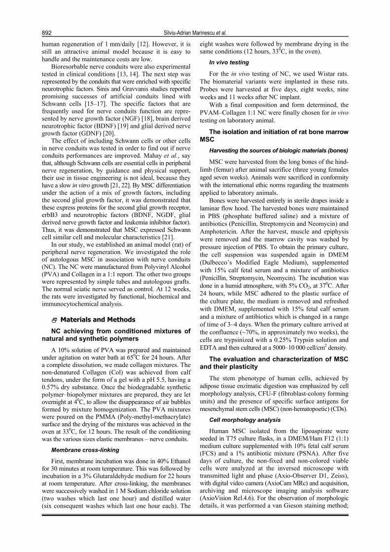
We used for the study 75 Wistar rats. There were selected females weighing 180–220 g. The animals were random divided in three groups of 25 rats: (A) autografts, (B) nerve conduits and (C) NC associated with MSC. All the groups were compared to the normal contralateral sciatic. In Group A, the graft was achieved by the excision of a 10 mm section which was reversed 1800 for suture. In Group B, the sciatic was divided and let to retract. Afterwards a 10 mm segment was excised and the nerve stumps were introduced in the NC and sutured with a 9.0 Prolene suture. For the third group, we used a similar approach, the MSC being introduced in the gap between the NC and the proximal stump (Figure 1).
Figure 1 – (A) Nerve graft. (B) Nerve conduit.
The functional analysis at 12 weeks consisted of sensitive and motor tests. Also, at 12 weeks, it was calculated the gastrocnemius index muscle. Although the sciatic functional index better describes nerve crush injuries, we determined it also at 12 weeks.
Pin-prick test (PP)
The pinprick test (PP) was used for sensitive repair recover. The test consisted in the pinprick with a forceps of the animal operated hindlimb, to evaluate the sensory recover. The animal was pinprick from the toes to the knee, until it was noticed the limb withdrawal. Sensory recover was graded on a scale from 0 to 3. The scale represented 0 – the absence of the response at a pain stimulus, grade I – limb withdrawal after the application of the stimuli above the ankle, grade II – limb withdrawal after the application of stimuli below the ankle, in the heel region, and grade III – limb withdrawal after the application of stimuli in the metatarsal region.
Toe spread test
Toe spread test was used for motor recovery evaluation. It is because the non-operated, normal rat extends and spreads the toes when it is hung by the tail. As in sensory recover, motor recovery was graded on a scale from 0 to 3, where grade 0 – digital movement absence, grade I – any movement, grade II – toe spread and grade III – toe extension and abduction.
Gastrocnemius muscle index
For index calculation, both gastrocnemius muscles from the hindlimbs were excised. Gastrocnemius muscle weighing was done at the end of the 12 weeks of follow-up. This index measured practically muscle atrophy induced by denervation. Gastrocnemius muscle was excised at the muscle and tendon junction and it was immediately weighed using a Sartorius animal weighing balance. Gastrocnemius muscle index (GMI) was calculated by reporting the weight of the operated gastrocnemius weight to the contralateral, non-operated limb. The percent repre-sented the recover from the denervation atrophy of the operated gastrocnemius. The percent of 100% represented full recovery of the operated part. Gastrocnemius muscle is innervated by the sciatic and starts to atrophy after nerve injury. GMI is one of motor function recovery parameter and its raising was correlated with sciatic nerve regeneration and successfully sciatic nerve reinnervation.
Histological and immunohistochemical examination
Histological analysis
Normal or coming from NC sciatic nerve fragments were fixed in the Bouin retainer and in 4% PBS. After

Silviu-Adrian Marinescu et al.
894
fixation, the tissue fragments were dehydrated in increa-sing concentrations of Ethanol, clarified in Toluene and included in paraffin. Five-μm sections were colored with Hematoxylin–Eosin (HE) and observed at Zeiss Axiostar Plus microscope.
Immunohistochemical analysis
Immunohistochemical analysis targeted the evaluation of sciatic nerve regeneration by axon highlighting (neuro-filament protein).
For immunohistochemical analysis, the tissue fragments were fixed 4% PBS, dehydrated in Ethanol, clarified in Toluene and included in paraffin. Five-μm sections were processed by indirect immunoperoxidase method. This consisted of the followings: (1) endogenous peroxidase blocking with 3% H2O2; (2) blocking non-specific binding with 2% serum calf albumin; (3) incubation, overnight at 40C, with the primary antibody, represented by the primary antibody, 160 kD anti-neurofilament rat IgG, diluted 1:80 with 2% calf serum albumin; (4) washing with PBS; (5) incubation with the secondary antibody, goat IgG against rat anti IgG coupled with peroxidase for an hour at room temperature (6) washing with PBS; (7) the development of the immune complex in the presence of 0.05% 3,3’-Diaminobenzidine and 0.003% H2O2 in PBS. The nuclei were contrasted with Hematoxylin.
Results
Mesenchymal stem cells microscopy
Mesenchymal stem cells, obtained in our laboratory from rat bone marrow, by plastic adherence, were evaluated for their morphology by optic microscopy and they were characterized by flow cytometry using specific markers (Figure 2).
Figure 2 – (A and B) Primary culture from rat bone marrow (×10). In the first five days after initiation, the culture has a polymorphic aspect, being also contaminated with other cell type, as endothelial cells, adipocytes or stromal fibroblasts.
Microscopy images allow the highlight of the poly-morphic aspect of the initial culture (Figure 3A), and also the development of some hematopoiesis foci even in the absence of the exogenous cytokines, looking like round cell islands, that grow up and are detached from the substrate (Figure 3B).
There had been many attempts to generate a more homogenous MSC population. This target can be accomplished by supplementing and changing of the culture medium. Thus, the culture media were supple-mented with various growth factors, such as: bFGF, EGF (epidermal growth factor), Tpo, IL-3, SCF (stem cell factor). It was demonstrated that a combination
between bFGF and EGF supported the capacity of MSC multiple differentiation, preventing the differentiation toward hematopoietic lines, osteoblasts or adipocytes.
After a few days of culture and the adding of a fresh medium, we remarked the gradual uniformity of cell’ morphology that turned into fibroblast-like cells, the culture becoming pure (Figure 4).
Figure 3 – The MSC primary culture, first passage: (A) ×10; (B) ×20.
Figure 4 – Bone marrow stem cell culture. Giemsa staining, ×20.
Giemsa staining allowed the highlight of morphologic details, such as the oval form of the vesicular nuclei, which is a characteristic of MSC morphologic phenotype.
Flow cytometry analysis
Flow cytometry analysis was based on the existence of the optical differences between the cells and a specific immune reactivity against Fluorescein marked antibodies. The immune reactivity is conferred by the existence of a certain phenotype characterized by the presence of some molecule-marker configuration.
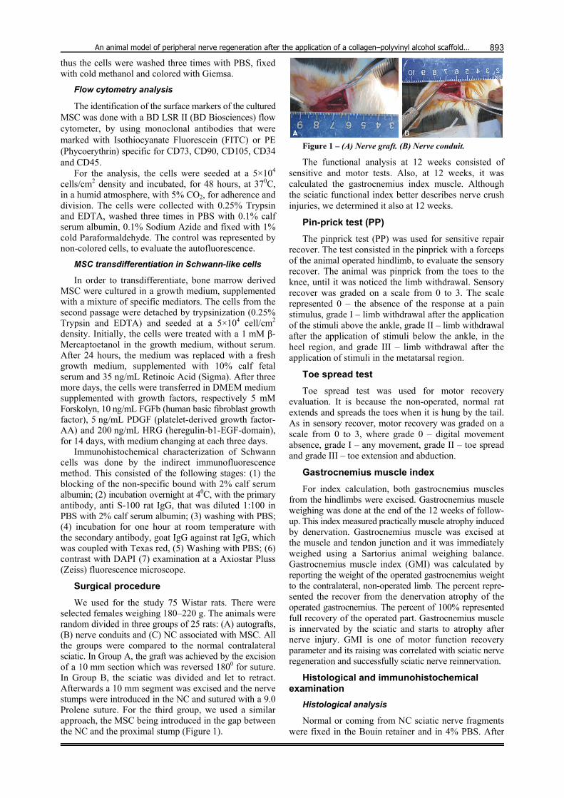
Because of the immunophenotyping by flow cytometry, the cells proved to be positive for CD73, CD90 and CD105 and did not express CD34 and respectively CD45. For the identification and the possibility of interest population evaluation, the gate was put on a FSC/SSC dot plot (Figures 5 and 6).
In Figure 7, one can notice the flow cytometry histo-grams, the fluorescence peak being unbiased, compared to the witness, suggesting the fact that the analyzed cells do not express antigenic markers. Thus, the results achieved following the analysis of marked population with anti-CD34 and anti-CD45 antibodies brought extra proves for giving the interest population cells a MSC status.
An animal model of peripheral nerve regeneration after the application of a collagen–polyvinyl alcohol scaffold…
895
Figure 5 – Control cells, used for parameter set-up.
Figure 6 – Histograms for CD73, CD90 and CD105. It can be noticed that the peak for fluorescence is moved to the right, compared to the control, unmarked, indicating the fact that the population express the three markers.
Figure 7 – Histograms for CD34 and CD45.
Immunohistochemical analyses
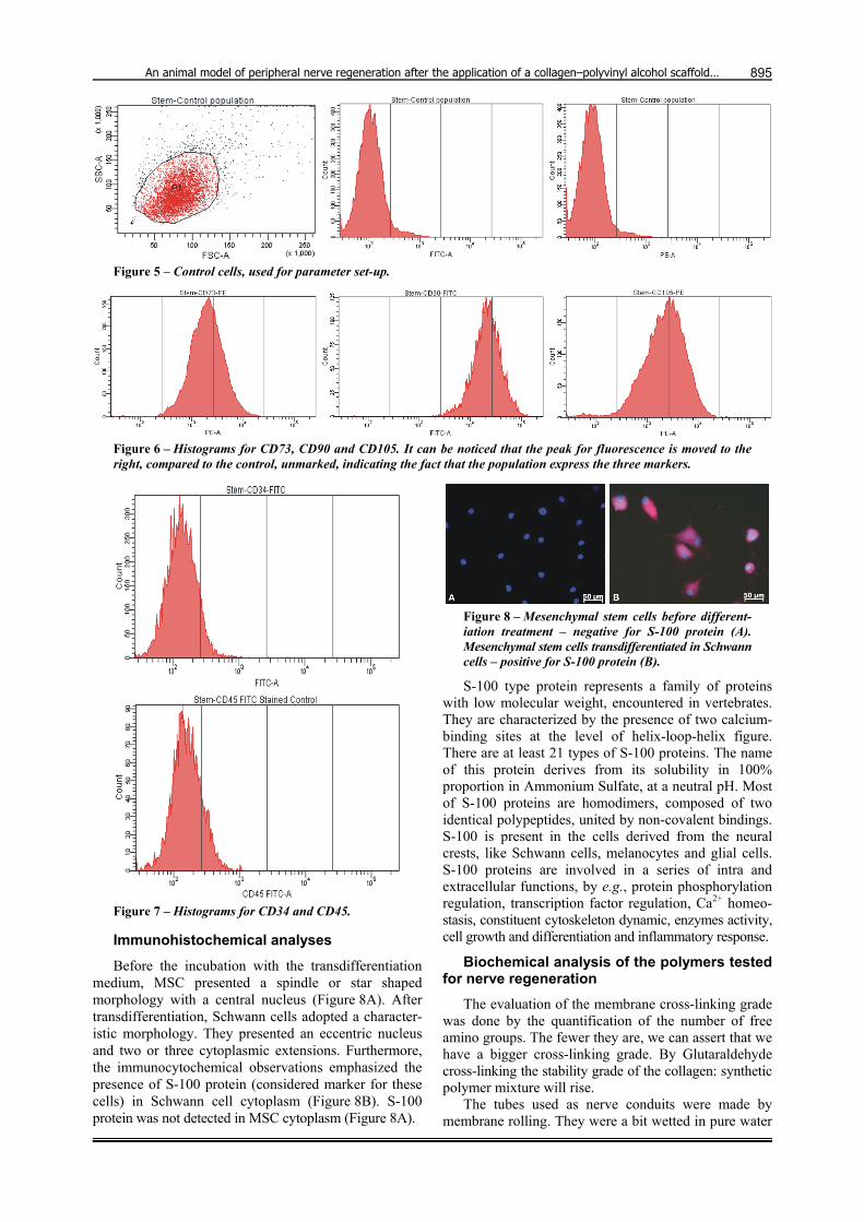
Before the incubation with the transdifferentiation medium, MSC presented a spindle or star shaped morphology with a central nucleus (Figure 8A). After transdifferentiation, Schwann cells adopted a character-istic morphology. They presented an eccentric nucleus and two or three cytoplasmic extensions. Furthermore, the immunocytochemical observations emphasized the presence of S-100 protein (considered marker for these cells) in Schwann cell cytoplasm (Figure 8B). S-100 protein was not detected in MSC cytoplasm (Figure 8A).
Figure 8 – Mesenchymal stem cells before different-iation treatment – negative for S-100 protein (A). Mesenchymal stem cells transdifferentiated in Schwann cells – positive for S-100 protein (B).
S-100 type protein represents a family of proteins with low molecular weight, encountered in vertebrates. They are characterized by the presence of two calcium-binding sites at the level of helix-loop-helix figure. There are at least 21 types of S-100 proteins. The name of this protein derives from its solubility in 100% proportion in Ammonium Sulfate, at a neutral pH. Most of S-100 proteins are homodimers, composed of two identical polypeptides, united by non-covalent bindings. S-100 is present in the cells derived from the neural crests, like Schwann cells, melanocytes and glial cells. S-100 proteins are involved in a series of intra and extracellular functions, by e.g., protein phosphorylation regulation, transcription factor regulation, Ca2+ homeo-stasis, constituent cytoskeleton dynamic, enzymes activity, cell growth and differentiation and inflammatory response.
Biochemical analysis of the polymers tested for nerve regeneration
The evaluation of the membrane cross-linking grade was done by the quantification of the number of free amino groups. The fewer they are, we can assert that we have a bigger cross-linking grade. By Glutaraldehyde cross-linking the stability grade of the collagen: synthetic polymer mixture will rise.
The tubes used as nerve conduits were made by membrane rolling. They were a bit wetted in pure water
Silviu-Adrian Marinescu et al.
896
and then put on bearings with an outer diameter of 1.5 mm and dried in an oven at 330C, for 12 hours. After drying, they were UV sterilized (Figure 9).
Figure 9 – PVA:Col = 1:1 nerve conduit (inner diameter of 1.5 mm).
The combination of native Collagen and Polyvinyl Alcohol, in a 1:1 report, cross-linked proved to be the best candidate for nerve regeneration promotion.
The highlighting of the effect of this material in nerve regeneration at different times of implantation was done by the determination of matriceal enzymes activity (MMP2 and MMP9), that are involved in tissue remodeling process. The variation of those levels was followed by zymography and densitometry.
The bands achieved by zymography were processed and quantitatively analyzed by densitometry (Figure 10).
Figure 10 – Gelatin zymography. Mw: Molecular weight.
Past studies showed that at different times of healing, inflammatory cells, fibroblasts, keratinocytes and endothelial cells are the source of various types of MMP. MMP9 synthesis by keratinocytes is supposed to be involved in their locomotion phenotype and it is inhibited when wound epithelialization is complete. MMP9 activity is noticed in the initial phases, and then it drops rapidly to normal values, being associated with an acute inflammatory response [23]. MMP2 or type A gelatinase acts on type IV collagen. Its activity is registered later (three days). Afterwards, it decreases in conformity with its role, as dominant enzyme involved in the remodeling process [24].
Table 1 – Densitometry results for electrophoresis gels
Sample No.
Harvest time
Latent MMP9
(92 kDa) [%]
Active MMP9
(9286 kDa) [%]
Latent MMP2
(72 kDa) [%]
Active MM2
(66 kDa) [%]
1. 5 days 4.39 4.75 10.17
2. 8 weeks 11.04 11.39
3. 9 weeks 7.22 32.8
4. 11 weeks 7.21 23.43
Reporting the other probes to the witnesses, we noticed a variation of the two categories of metalloproteinases depending on the implant.
Active MMP9, revealed only at five days after the implantation, showed the presence of an inflammatory
process that follows a descendent scale in time, according to the specialty literature. This fact is confirmed in this case by the absence of MMP9 in the probes harvested afterwards. In the dermal tissues, the activity of MMP9 disappears completely after the third day [25].
Due to the complex procedure of implantation and the high grade of tissue invasiveness this intervention involved, the inflammatory process had a different profile, being longer.
MMP2, responsible for the process of tissue regene-ration, exposed in the profile described in Figure 11, reveals the fact that the remodeling process continues also after nine weeks, when it is noticed a maximum point, followed by a decrease.
Figure 11 – MMP2 variation according to implant harvest time.
Functional evaluation results
The animals were followed for 12 weeks by conduct analysis, weight measuring, skin and soft tissue changes. The animals did not suffer any significant weight changes, with the exception of a 3% decrease in the first week, afterwards the weight coming back to normal. The arsenal of denervation injuries includes soft tissue changes such as flexion contractures, chronic ulcerations, postoperative infections, bedsores and autotomy injuries. None of the operated rats developed any bedsores, flexion contractures or chronic ulcerations. From the cohort of the operated rats, that presented sciatic defects, we found out that five from the group of NC, four from the group of grafts and two from the group of NC associated with MSC had had automutilations. Those were similar both in nature and severity.
The functional evaluation of nerve repair was done by clinical analyzes, toe spread, pinprick test and gastro-cnemius muscle index. The animals were followed-up by walking and posture. The pinprick and toe spread tests are reliable and easily reproducible tests [26].
Sensitive testing – pin prick (PP)
There were no significant differences between groups at six and 12 weeks. At six weeks, we noticed a better response of the autografts, followed by the conduits and MSC, but the differences were not statistically significant. All the animals were evaluated by the maximum score of three at 12 weeks (Figure 12).
The motor testing – toe abduction
The motor testing did not reveal any significant differences between the groups for the timing chosen. However, we can remark an augmented motor response in the autograft group at 12 weeks, while at the NC and NC associated with MSC the rise is minimal (Figure 13).

An animal model of peripheral nerve regeneration after the application of a collagen–polyvinyl alcohol scaffold…
897
Figure 12 – Sensitive testing.
Figure 13 – Motor testing.
Animal posture and walking were followed-up. In Figure 14, the paw prints of the hindlimbs were recorded. However, the paw prints were recorded not timely at six respectively 12 weeks, but only at 12 weeks. The sciatic functional index (SFI) was calculated according to Bain
et al. formula [27], considering three parameters, the paw print length (between the heel and the longest operated toe), where: NPL – represents the value for the non-operated limb and EPL – the value for the operated limb, the intermediary interdigital distance (between the second and fourth toes), NIT – the value for the normal limb and EIT – the value for the operated limb, and the inter-digital distance (between the first and fifth toes), NTS – the value for the normal limb, and ETS – the value for the operated limb:
SFI + -38×EPL – NPL/NPL + 109.5×ETS – NTS/NTS + 13.3×EIT – NIT/NIT – 8.8
The normal control values were represented by the values recorded for the normal non-operated paws. The test consisted in the immersion of the hindlimbs in ink and rats follow-up to a black cage, on a band covered with white paper that was changed at each passage. The calculation formula considered a normal function the 0 value and the complete loss of function, value -100. Afterwards, it was calculated the mean value for each experimental group and the calculated mean values were then compared to each other. Unfortunately, SFI seems a more adequate parameter for the crush syndromes and the follow-up of the paw prints in the first weeks is a difficult task [28]. In our study, we measured the SFI at 12 weeks.
The gastrocnemius muscle index
Gastrocnemius reinnervation – measured as we showed by the gastrocnemius muscle index (GMI) – did not showed significant differences between groups at 12 weeks. The best results were achieved by the autografts, followed by the NC associated with MSC. The variations between the three testing groups was not significant (p<0.05) (Figure 15).
Figure 14 – (A and B) Walking track analysis at 12 weeks.
Figure 15 – Gastrocnemius muscle index (GMI).
Electron microscopy and immunohisto-chemical results
Sciatic nerve regeneration after nerve grafting
At 12 weeks, graft histological analysis showed that the myelin nerve fibers had smaller diameters and a thinner myelin sheath compared to the normal sciatic nerve (Figure 16, A and B). In the grafts, the vast majority of the nerve fibers regenerated in the original bundles. There were only a few regenerating fibers outside nerve fascicles. In some rats, we noticed a disorganized arran-gement of nerve fibers and an increasing amount of connective tissue (Figure 16), derived from epineurium integration inside the nerve (Figure 17).
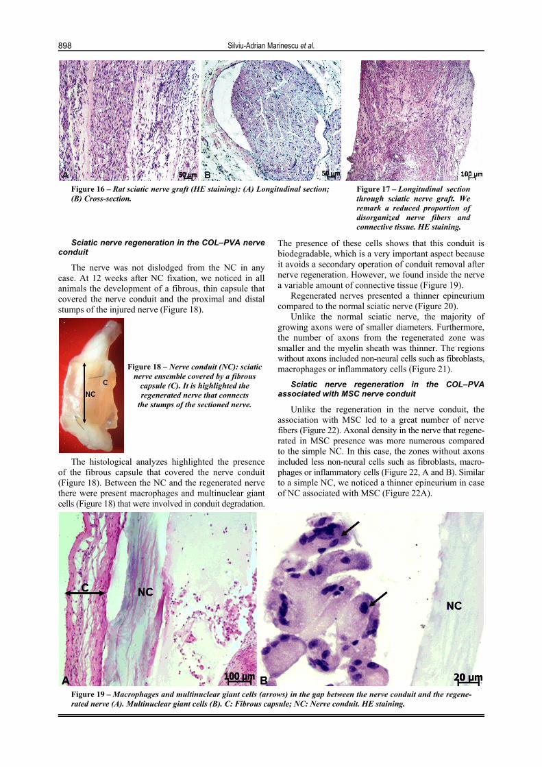
Silviu-Adrian Marinescu et al.
898
Figure 16 – Rat sciatic nerve graft (HE staining): (A) Longitudinal section; (B) Cross-section.
Figure 17 – Longitudinal section through sciatic nerve graft. We remark a reduced proportion of disorganized nerve fibers and connective tissue. HE staining.
Sciatic nerve regeneration in the COL–PVA nerve conduit
The nerve was not dislodged from the NC in any case. At 12 weeks after NC fixation, we noticed in all animals the development of a fibrous, thin capsule that covered the nerve conduit and the proximal and distal stumps of the injured nerve (Figure 18).
Figure 18 – Nerve conduit (NC): sciatic nerve ensemble covered by a fibrous
capsule (C). It is highlighted the regenerated nerve that connects
the stumps of the sectioned nerve.
The histological analyzes highlighted the presence of the fibrous capsule that covered the nerve conduit (Figure 18). Between the NC and the regenerated nerve there were present macrophages and multinuclear giant cells (Figure 18) that were involved in conduit degradation.
The presence of these cells shows that this conduit is biodegradable, which is a very important aspect because it avoids a secondary operation of conduit removal after nerve regeneration. However, we found inside the nerve a variable amount of connective tissue (Figure 19).
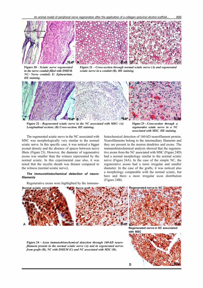
Regenerated nerves presented a thinner epineurium compared to the normal sciatic nerve (Figure 20).
Unlike the normal sciatic nerve, the majority of growing axons were of smaller diameters. Furthermore, the number of axons from the regenerated zone was smaller and the myelin sheath was thinner. The regions without axons included non-neural cells such as fibroblasts, macrophages or inflammatory cells (Figure 21).
Sciatic nerve regeneration in the COL–PVA associated with MSC nerve conduit
Unlike the regeneration in the nerve conduit, the association with MSC led to a great number of nerve fibers (Figure 22). Axonal density in the nerve that regene-rated in MSC presence was more numerous compared to the simple NC. In this case, the zones without axons included less non-neural cells such as fibroblasts, macro-phages or inflammatory cells (Figure 22, A and B). Similar to a simple NC, we noticed a thinner epineurium in case of NC associated with MSC (Figure 22A).
Figure 19 – Macrophages and multinuclear giant cells (arrows) in the gap between the nerve conduit and the regene-rated nerve (A). Multinuclear giant cells (B). C: Fibrous capsule; NC: Nerve conduit. HE staining.
An animal model of peripheral nerve regeneration after the application of a collagen–polyvinyl alcohol scaffold…
899
Figure 20 – Sciatic nerve regenerated in the nerve conduit filled with DMEM. NC: Nerve conduit; E: Epineurium. HE staining.
Figure 21 – Cross-section through normal sciatic nerve (A) and regenerated sciatic nerve in a conduit (B). HE staining.
Figure 22 – Regenerated sciatic nerve in the NC associated with MSC: (A) Longitudinal section; (B) Cross-section. HE staining.
Figure 23 – Cross-section through a regenerative sciatic nerve in a NC associated with MSC. HE staining.
The regenerated sciatic nerve in the NC associated with MSC was morphologically very similar to the normal sciatic nerve. In this specific case, it was noticed a bigger axonal density and the absence of spaces between nerve fibers (Figure 23). However, the diameter of regenerative axons was smaller than the witness represented by the normal sciatic. In this experimental case also, it was noted that the myelin sheath was thinner compared to the witness (normal sciatic nerve).
The immunohistochemical detection of neuro-filaments
Regenerative axons were highlighted by the immuno-
histochemical detection of 160-kD neurofilament protein. Neurofilaments belong to the intermediary filaments and they are present in the neuron dendrites and axons. The immunohistochemical analysis showed that the regenera-tive axons from the NC associated with MSC (Figure 24D) had a normal morphology similar to the normal sciatic nerve (Figure 24A). In the case of the simple NC, the regenerative axons had a more irregular and smaller diameter. In the case of the grafts, it was noticed also a morphology comparable with the normal sciatic, but here and there a more irregular axon distribution (Figure 24B).
Figure 24 – Axon immunohistochemical detection through 160-kD neuro-filament protein in the normal sciatic nerve (A) and in regenerated nerves from grafts (B), NC with DMEM (C) and NC associated with MSC (D).
Silviu-Adrian Marinescu et al.
900
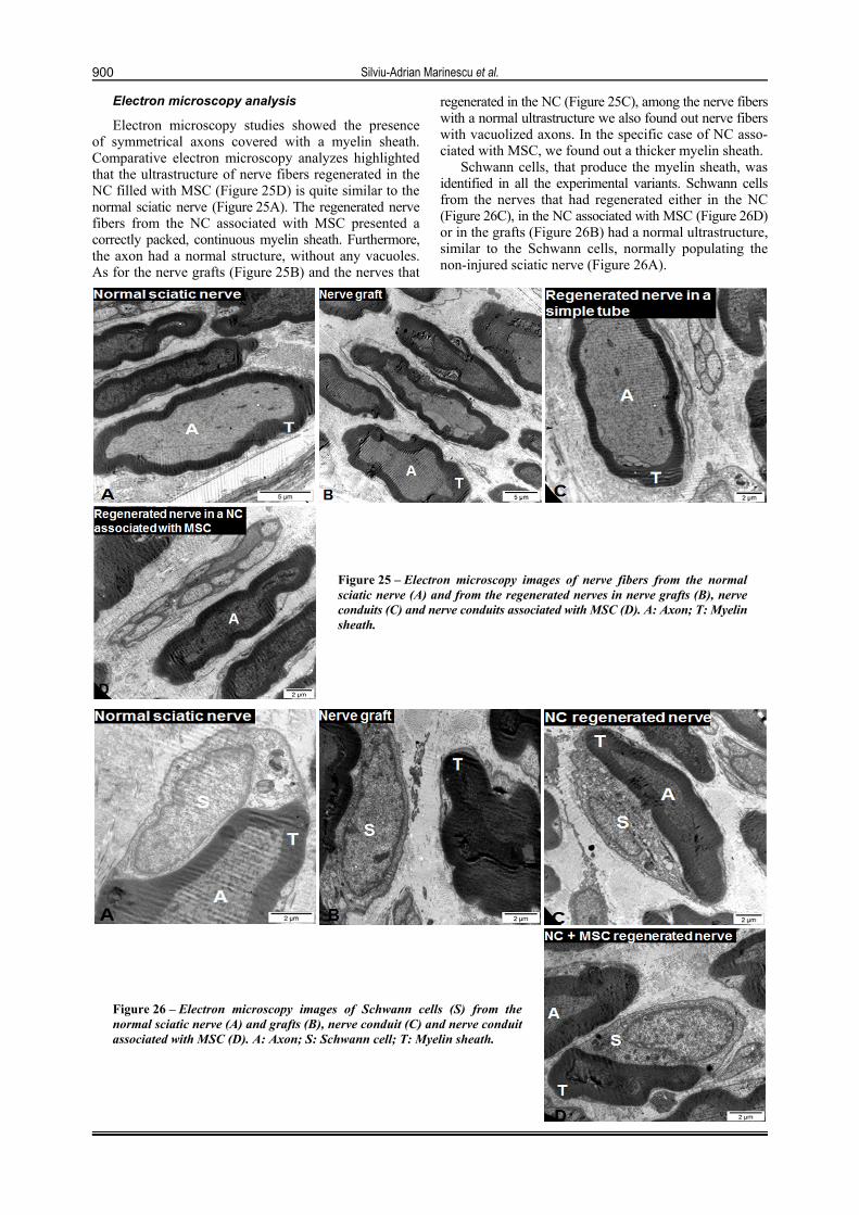
Electron microscopy analysis
Electron microscopy studies showed the presence of symmetrical axons covered with a myelin sheath. Comparative electron microscopy analyzes highlighted that the ultrastructure of nerve fibers regenerated in the NC filled with MSC (Figure 25D) is quite similar to the normal sciatic nerve (Figure 25A). The regenerated nerve fibers from the NC associated with MSC presented a correctly packed, continuous myelin sheath. Furthermore, the axon had a normal structure, without any vacuoles. As for the nerve grafts (Figure 25B) and the nerves that
regenerated in the NC (Figure 25C), among the nerve fibers with a normal ultrastructure we also found out nerve fibers with vacuolized axons. In the specific case of NC asso-ciated with MSC, we found out a thicker myelin sheath.
Schwann cells, that produce the myelin sheath, was identified in all the experimental variants. Schwann cells from the nerves that had regenerated either in the NC (Figure 26C), in the NC associated with MSC (Figure 26D) or in the grafts (Figure 26B) had a normal ultrastructure, similar to the Schwann cells, normally populating the non-injured sciatic nerve (Figure 26A).
Figure 25 – Electron microscopy images of nerve fibers from the normal sciatic nerve (A) and from the regenerated nerves in nerve grafts (B), nerve conduits (C) and nerve conduits associated with MSC (D). A: Axon; T: Myelin sheath.
Figure 26 – Electron microscopy images of Schwann cells (S) from the normal sciatic nerve (A) and grafts (B), nerve conduit (C) and nerve conduit associated with MSC (D). A: Axon; S: Schwann cell; T: Myelin sheath.

An animal model of peripheral nerve regeneration after the application of a collagen–polyvinyl alcohol scaffold…
901
Discussion
The injection of the distal root of the sectioned sciatic nerves with bromodeoxyuridine (BrdU) marked mesenchymal stem cells demonstrated that these cells survived in the zone of injection for at 33 days. Almost 5% from the marked BrdU cells presented the phenotype characteristic for Schwann cells (were positive for the S-100 marker). The tests done at 18 and 33 days after implant showed that the MSC implant led to the functional recovery of the injured nerves [29]. The use of a nerve conduit manufactured from Chitosan and filled with GFP marked mesenchymal stem cells, for the repair of a 5 mm space from rat sciatic nerve demonstrated the significant improvement of axon growth and myelinization and also of sciatic nerve functions compared to the empty nerve conduit. The confocal microscopy confirmed that the mesenchymal stem cells adopted a Schwann cell phenotype [30].
The non-differentiated mesenchymal stem cells concur to nerve regeneration by growth factor secretion and basal lamina components synthesis [31, 32]. Neurotrophic molecule production by mesenchymal stem cells can delay cell death and can induce neural tissue regeneration [33–38]. These results showed the fact that the MSC exert a therapeutic effect by releasing growth factors and other soluble mediators, that action on target tissues and develop a favorable medium for nerve regeneration [34, 39–41].
The data presented in our study refer to the use of a new type of bio composite scaffold based on Collagen and Polyvinyl Alcohol that was associated or not with mesenchymal stem cells to cover a 10 mm nerve defect in rat sciatic nerve. The functional and immunohisto-chemical results confirmed the fact that the single lumen tubes used by us are a method as good as nerve graft. Although the regeneration was noticed in a mixed nerve, it was investigated on a relatively short distance of 1 cm. The graft used by us was harvested from the same nerve, being a truncal graft. The surgical technique adhered to the experimental microsurgical principles, described by Lascăr et al. [42, 43]. At 12 weeks, sensitive recover was equal and maximum at in all three groups. We must emphasize that five rats with NC, four with grafts and two with NC associated with MSC were excluded because the regeneration was unsatisfactory and they presented self-mutilation. The histological examination revealed that adding MSC led to a thicker myelin sheath in the NC associated with MSC group compared to NC with DMEM and even grafts. Nerve fibers width was bigger at 12 weeks. Axonal density was similar in all the three groups. By adding MSC, we hope to repair bigger nerve defects because currently NC application is limited to nerve defects less than 3 cm and sensitive nerves. MSC were eligible for our study because they are deprived of immunogenity in non-immunoprivileged sites [43, 44], thus having a greater experimental value for allogeneic cell therapies. Pereira Lopes et al., in a study concerning biodegradable collagen tubes in which we added MSC for the repair of a nerve defect of 3 mm, did not ascertain function improvement (measured by walking tract test), but they remarked an improvement of ultrastructural parameters [45]. The use of MSC was determined by their ease of isolation. In our study, we harvested human
MSC from the lipoaspirate and rat MSC from the rat femurs and tibia. Recently it was reported that harvested MSC express spontaneous neurotrophic factors, respecti-vely NGF [46]. Isogenic MSC can differentiate in neural-like tissue and can facilitate nerve repair [26].
In the short term, at six weeks, motor recovery was better both for simple tubes and for conduits associated with MSC compared to nerve grafting (higher toe spread scores). Siemionow et al., in 2011 [26], considered that a possible explanation would be the fact that the tubes present as a unique lumen with a greater diameter allowing the rapid growth of a bigger number of axons compared to the grafts. In return, the grafts behave as multi-channel tubes. However, Siemionow et al. reported their dates on a bigger nerve defect of 20 mm, by using either simple epineural tubes or associated with non differentiated MSC. The control was represented by grafts. They demonstrated that simple epineural tubes, that were not associated with MSC, were able to support nerve regeneration by delivering an efficient guidance and the creation of a permissive neural medium, knowing that neuroma are the consequence of an inadequate medium. The literature data are quite heterogeneous, because other authors suggest that the presence of a tube with a unique lumen cannot sufficiently oppose to the dispersion of the regenerative fibers and can lead to the inadequate reinnervation of the target organs. de Ruiter et al. present the concept of the multi-channel nerve conduit concept that can limit nerve fiber dispersion by a correct guidance of the regenerative axon groups. However, the authors did not remark significantly differences between either multi-channel or unique lumen tubes used by them. Also, both kind of tubes presented functional and histological results that were inferior to grafts [47].
Despite the spontaneous axonal regeneration capacity, the recover after traumatic injuries of peripheral nerve injuries is often incomplete and limited to short distances. This situation takes place especially whenever the nerves are injured at a certain distance from the target organs and need repair by grafting. Grafting is encumbered by the donor site morbidity, the absence of a donor graft, incomplete and nonspecific regeneration and at last an unsatisfactory recover both clinically and on animal models [48, 49].
Conclusions
The biopolymeric NC represented adequate supports for MSC embedding. They were tested in vivo for peri-pheral nerve regeneration. In vivo testing showed that: the sensory testing was maximal in all three groups (grafts, NC and NC associated with MSC); the graft group had the better motor recover, translated in spread toe test, GMI and band walking test, but there were not statistically significant differences between the three groups; based on histological, immunohistochemical and electron microscopy studies, it was demonstrated that the injured sciatic nerves regenerated in the case of a conduit filled with MSC similar to autografts. We can conclude that the use of the NC associated with MSC led to a structure of the sciatic nerve similar to the normal sciatic. The achievement of results similar to grafting offered the premises of method transfer in clinical practice.
Silviu-Adrian Marinescu et al.
902
Acknowledgments The experimental studies were done under the auspices
of the PN II Grant REGENES, No. 42–137/2008.
References [1] Ray WZ, Mackinnon SE, Management of nerve gaps: auto-
grafts, allografts, nerve transfers, and end-to-side neurorrhaphy, Exp Neurol, 2010, 223(1):77–85.
[2] Ross MH, Pawlina W, Histology: a text and atlas: with correlated cell and molecular biology, 6th revised international edition, Lippincott Williams & Wilkins, 2011.
[3] Millesi H, Reappraisal of nerve repair, Surg Clin North Am, 1981, 61(2):321–340.
[4] Millesi H, The nerve gap. Theory and clinical practice, Hand Clin, 1986, 2(4):651–663.
[5] Flores AJ, Lavernia CJ, Owens PW, Anatomy and physiology of peripheral nerve injury and repair, Am J Orthop (Belle Mead NJ), 2000, 29(3):167–173.
[6] Lundborg G, Intraneural microcirculation, Orthop Clin North Am, 1988, 19(1):1–12.
[7] Strauch B, Use of nerve conduits in peripheral nerve repair, Hand Clin, 2000, 16(1):123–130.
[8] Chalfoun CT, Wirth GA, Evans GR, Tissue engineered nerve constructs: where do we stand? J Cell Mol Med, 2006, 10(2): 309–317.
[9] Fields RD, Le Beau JM, Longo FM, Ellisman MH, Nerve regeneration through artificial tubular implants, Prog Neurobiol, 1989, 33(2):87–134.
[10] Zegrea I, Chivu LI, Albu MG, Zamfirescu D, Chivu RD, Ion DA, Lascăr I, A Romanian therapeutic approach to peripheral nerve injury, Rom J Morphol Embryol, 2012, 53(2):357–361.
[11] Ceballos D, Navarro X, Dubey N, Wendelschafer-Crabb G, Kennedy WR, Tranquillo RT, Magnetically aligned collagen gel filling a collagen nerve guide improves peripheral nerve regeneration, Exp Neurol, 1999, 158(2):290–300.
[12] Varejão AS, Cabrita AM, Meek MF, Bulas-Cruz J, Melo-Pinto P, Raimondo S, Geuna S, Giacobini-Robecchi MG, Functional and morphological assessment of a standardized rat sciatic nerve crush injury with a non-serrated clamp, J Neurotrauma, 2004, 21(11):1652–1670.
[13] Hoppen HJ, Leenslag JW, Pennings AJ, van der Lei B, Robinson PH, Two-ply biodegradable nerve guide: basics aspects of design, construction and biological performance, Biomaterials, 1990, 11(4):286–290.
[14] Robinson PH, van der Lei B, Hoppen HJ, Leenslag JW, Pennings AJ, Nieuwenhuis P, Nerve regeneration through a two-ply biodegradable nerve guide in the rat and the influence of ACTH4-9 nerve growth factor, Microsurgery, 1991, 12(6):412–419.
[15] Sinis N, Schaller HE, Schulte-Eversum C, Schlosshauer B, Doser M, Dietz K, Rösner H, Müller HW, Haerle M, Nerve regeneration across a 2-cm gap in the rat median nerve using a resorbable nerve conduit filled with Schwann cells, J Neurosurg, 2005, 103(6):1067–1076.
[16] Sinis N, Schaller HE, Schulte-Eversum C, Lanaras T, Schlosshauer B, Doser M, Dietz K, Rösner H, Müller HW, Haerle M, Comparative neuro tissue engineering using different nerve guide implants, Acta Neurochir Suppl, 2007, 100:61–64.
[17] Gravvanis AI, Lavdas AA, Papalois A, Tsoutsos DA, Matsas R, The beneficial effect of genetically engineered Schwann cells with enhanced motility in peripheral nerve regeneration: review, Acta Neurochir Suppl, 2007, 100:51–56.
[18] Pu LL, Syed SA, Reid M, Patwa H, Goldstein JM, Forman DL, Thomson JG, Effects of nerve growth factor on nerve rege-neration through a vein graft across a gap, Plast Reconstr Surg, 1999, 104(5):1379–1385.
[19] Terris DJ, Toft KM, Moir M, Lum J, Wang M, Brain-derived neurotrophic factor-enriched collagen tubule as a substitute for autologous nerve grafts, Arch Otolaryngol Head Neck Surg, 2001, 127(3):294–298.
[20] Piquilloud G, Christen T, Pfister LA, Gander B, Papaloïzos MY, Variations in glial cell line-derived neurotrophic factor release from biodegradable nerve conduits modify the rate of functional motor recovery after rat primary nerve repairs, Eur J Neurosci, 2007, 26(5):1109–1117.
[21] Mahay D, Terenghi G, Shawcross SG, Schwann cell mediated trophic effects by differentiated mesenchymal stem cells, Exp Cell Res, 2008, 314(14):2692–2701.
[22] Ştefănescu O, Enescu DM, Lascăr I, Schwann cell cultures: recent advances and novel approaches to the reconstruction of peripheral nerve defects, Rom J Morphol Embryol, 2012, 53(3):467–471.
[23] Paul RG, Tarlton JF, Purslow PP, Sims TJ, Watkins P, Marshall F, Ferguson MJ, Bailey AJ, Biomechanical and biochemical study of a standardized wound healing model, Int J Biochem Cell Biol, 1997, 29(1):211–220.
[24] Mignatti P, Welgus HG, Rifkin DB, Role of degradative enzymes in wound healing. In: Clark RAF, Henson PM (ed), The molecular and cellular biology of wound repair, Plenum Press, New York, 1988, 497–524.
[25] Utoiu E, Coroiu V, Lungu M, Oancea A, Moldovan L, Assessment of the matrix metalloproteinases induced in hypodermic tissue by implantation of biopolymeric films, Romanian Biological Sciences, 2009, VII(1–4):129–113.
[26] Siemionow M, Duggan W, Brzezicki G, Klimczak A, Grykien C, Gatherwright J, Nair D, Peripheral nerve defect repair with epineural tubes supported with bone marrow stromal cells: a preliminary report, Ann Plast Surg, 2011, 67(1):73–84.
[27] Bain JR, Mackinnon SE, Hunter DA, Functional evaluation of complete sciatic, peroneal, and posterior tibial nerve lesions in the rat, Plast Reconstr Surg, 1989, 83(1):129–138.
[28] Monte-Raso VV, Barbieri CH, Mazzer N, Yamasita AC, Barbieri G, Is the Sciatic Functional Index always reliable and reproducible? J Neurosci Methods, 2008, 170(2):255–261.
[29] Cuevas P, Carceller F, Dujovny M, Garcia-Gómez I, Cuevas B, González-Corrochano R, Diaz-González D, Reimers D, Peripheral nerve regeneration by bone marrow stromal cells, Neurol Res, 2002, 24(7):634–638.
[30] Zhang P, He X, Zhao F, Zhang D, Fu Z, Jiang B, Bridging small-gap peripheral nerve defects using biodegradable chitin conduits with cultured Schwann and bone marrow stromal cells in rats, J Reconstr Microsurg, 2005, 21(8):565–571.
[31] Chen CJ, Ou YC, Liao SL, Chen WY, Chen SY, Wu CW, Wang CC, Wang WY, Huang YS, Hsu SH, Transplantation of bone marrow stromal cells for peripheral nerve repair, Exp Neurol, 2007, 204(1):443–453.
[32] Wang J, Ding F, Gu Y, Liu J, Gu X, Bone marrow mesen-chymal stem cells promote cell proliferation and neurotrophic function of Schwann cells in vitro and in vivo, Brain Res, 2009, 1262:7–15.
[33] Borlongan CV, Lind JG, Dillon-Carter O, Yu G, Hadman M, Cheng C, Carroll J, Hess DC, Bone marrow grafts restore cerebral blood flow and blood brain barrier in stroke rats, Brain Res, 2004, 1010(1–2):108–116.
[34] Chopp M, Li Y, Treatment of neural injury with marrow stromal cells, Lancet Neurol, 2002, 1(2):92–100.
[35] Chopp M, Zhang XH, Li Y, Wang L, Chen J, Lu D, Lu M, Rosenblum M, Spinal cord injury in rat: treatment with bone marrow stromal cell transplantation, Neuroreport, 2000, 11(13): 3001–3005.
[36] Crigler L, Robey RC, Asawachaicharn A, Gaupp D, Phinney DG, Human mesenchymal stem cell subpopulations express a variety of neuro-regulatory molecules and promote neuronal cell survival and neuritogenesis, Exp Neurol, 2006, 198(1): 54–64.
[37] Gu Y, Wang J, Ding F, Hu N, Wang Y, Gu X, Neurotrophic actions of bone marrow stromal cells on primary culture of dorsal root ganglion tissues and neurons, J Mol Neurosci, 2010, 40(3):332–341.
[38] Hofstetter CP, Schwarz EJ, Hess D, Widenfalk J, El Manira A, Prockop DJ, Olson L, Marrow stromal cells form guiding strands in the injured spinal cord and promote recovery, Proc Natl Acad Sci U S A, 2002, 99(4):2199–2204.
[39] Munoz JR, Stoutenger BR, Robinson AP, Spees JL, Prockop DJ, Human stem/progenitor cells from bone marrow promote neurogenesis of endogenous neural stem cells in the hippo-campus of mice, Proc Natl Acad Sci U S A, 2005, 102(50): 18171–18176.
[40] Caplan AI, Dennis JE, Mesenchymal stem cells as trophic mediators, J Cell Biochem, 2006, 98():1076–1084.
[41] Chen X, Li Y, Wang L, Katakowski M, Zhang L, Chen J, Xu Y, Gautam SC, Chopp M, Ischemic rat brain extracts induce
An animal model of peripheral nerve regeneration after the application of a collagen–polyvinyl alcohol scaffold…
903
human marrow stromal cell growth factor production, Neuro-pathology, 2002, 22(4):275–279.
[42] Neuhuber B, Timothy Himes B, Shumsky JS, Gallo G, Fischer I, Axon growth and recovery of function supported by human bone marrow stromal cells in the injured spinal cord exhibit donor variations, Brain Res, 2005, 1035(1):73–85.
[43] Lascăr I, Zamfirescu D, Microchirurgie experimentală, Ed. Paralela 45, Bucureşti, 2000, 120–130.
[44] Hori J, Ng TF, Shatos M, Klassen H, Streilein JW, Young MJ, Neural progenitor cells lack immunogenicity and resist destruction as allografts, Stem Cells, 2003, 21(4):405–416.
[45] Pereira Lopes FR, Camargo de Moura Campos L, Dias Corrêa J Jr, Balduino A, Lora S, Langone F, Borojevic R, Blanco Martinez AM, Bone marrow stromal cells and resorbable collagen guidance tubes enhance sciatic nerve regeneration in mice, Exp Neurol, 2006, 198(2):457–468.
[46] Li N, Yang H, Lu L, Duan C, Zhao C, Zhao H, Spontaneous expression of neural phenotype and NGF, TrkA, TrkB genes in marrow stromal cells, Biochem Biophys Res Commun, 2007, 356(3):561–568.
[47] de Ruiter GC, Spinner RJ, Malessy MJ, Moore MJ, Sorenson EJ, Currier BL, Yaszemski MJ, Windebank AJ, Accuracy of motor axon regeneration across autograft, single-lumen, and multichannel poly(lactic-co-glycolic acid) nerve tubes, Neurosurgery, 2008, 63(1):144–155; discussion 153–155.
[48] De Medinaceli L, Rawlings RR, Is it possible to predict the outcome of peripheral nerve injuries? A probability model based on prospects for regenerating neurites, Biosystems, 1987, 20(3):243–258.
[49] Samii A, Carvalho GA, Samii M, Brachial plexus injury: factors affecting functional outcome in spinal accessory nerve transfer for the restoration of elbow flexion, J Neurosurg, 2003, 98(2):307–312.
Corresponding author Ioana-Ruxandra Mihai, Scientific Researcher, MD, PhD, Department of Plastic and Reconstructive Microsurgery, “Bagdasar–Arseni” Emergency Hospital, 10–12 Berceni Road, 041915 Bucharest, Romania; Phone +4021–334 30 25, Fax +4021–334 49 13, e-mail: [email protected] Received: March 12, 2013
Accepted: October 10, 2014