roles for hedgehog signaling in adult organ homeostasis...
TRANSCRIPT

REVIEW
Roles for Hedgehog signaling in adult organ homeostasis andrepairRalitsa Petrova1,2 and Alexandra L. Joyner1,2,*
ABSTRACTThe hedgehog (HH) pathway is well known for its mitogenic andmorphogenic functions during development, and HH signalingcontinues in discrete populations of cells within many adultmammalian tissues. Growing evidence indicates that HH regulatesdiverse quiescent stem cell populations, but the exact roles that HHsignaling plays in adult organ homeostasis and regeneration remainpoorly understood. Here, we review recently identified functions of HHin modulating the behavior of tissue-specific adult stem andprogenitor cells during homeostasis, regeneration and disease. Weconclude that HH signaling is a key factor in the regulation of adulttissue homeostasis and repair, acting via multiple different routes toregulate distinct cellular outcomes, including maintenance ofplasticity, in a context-dependent manner.
KEY WORDS: Adult stem cells, Hedgehog signaling, Homeostasis
IntroductionDuring vertebrate development, hedgehog (HH) signaling plays anessential role in orchestrating the complex cell specification programsand extensive cell division required to form an organism. In the adult,HH continues to signal to discrete populations of stem and progenitorcells within various organs, including the brain (Ahn and Joyner,2005; Ihrie et al., 2011; Machold et al., 2003), skin (Brownell et al.,2011), prostate (Peng et al., 2013) and bladder (Shin et al., 2011),among others. Whether HH signaling in the adult functions to controlproliferation, specification and/or plasticity, and the mechanism bywhich this occurs, remain unclear. Given that a role for HH signalingin both the maintenance of adult resident stem cells and theprogression of various diseases, including cancer, has emerged inrecent years, it is important to determine how this major signalingpathway can regulate processes that lie on either side of the life-deathspectrum.
The HH signaling pathway in mammalsThe three mammalian HH proteins, called sonic (SHH), Indian(IHH) and desert (DHH) hedgehog, are homologs of theDrosophilasegment polarity gene bearing the same name (Briscoe andTherond, 2013; Echelard et al., 1993). HH proteins undergoextensive post-translational modifications, after which they arereleased by the secreting cell with the help of dispatched, amembrane transporter protein. SHH is the most broadly expressedvertebrate HH and its paracrine activity on adjacent cells is the mostcommon mode of pathway transduction, although HH has also beenproposed to signal in an autocrine manner. HH signaling ispropagated by a receptor complex that includes the G-protein-coupled
receptor smoothened (SMO) and the twelve-pass membrane proteinpatched 1 (PTCH1) (Fig. 1). In the absence of HH ligand, PTCH1inhibits SMOactivation, but whenHH is present this repressive actionis released. In addition to PTCH1, HH interacts with PTCH2 and thecell-surface proteins growth arrest specific (GAS), cell adhesionmolecule-related/downregulated by oncogenes (CDO) and brother ofCDO (BOC), which function as co-receptors. This interaction iscrucial for signal propagation and for establishing a HH gradient(Briscoe and Therond, 2013).
DownstreamofSMO, theGLI (glioma-associatedoncogene familymembers) transcription factors mediate HH signal transduction in aprocess referred to as canonical signaling (reviewed extensively byBriscoe and Therond, 2013; Hui and Angers, 2011). In the absence ofHH ligand,GLI2 andGLI3 undergo limited proteasomal degradation,resulting in the cleavage and removal of the GLI C-terminal activatordomain, which leads to the conversion of GLI3, and to a lesser extentGLI2, into transcriptional repressors (GLI3R andGLI2R) (Fig. 1). GLItranscriptional activators (GLIA), primarilyGLI2A, are formed only inresponse to HH stimulation. Thus, HH signaling functions throughmodulating the balance between GLIA and GLIR. GLIA then triggersexpression of HH target genes such as Gli1, the protein product ofwhich functions only as a transcriptional activator and thus amplifiesHH signaling. SMO and PTCH1 base level expression, much like thatof GLI2 and GLI3, is independent of pathway activity. However,PTCH1 production is upregulated with increasing HH levels, thusforming a negative-feedback loop in the canonical signaling pathway(Ribes and Briscoe, 2009). Recent analysis of the cis-regulatorymodules of HH-regulated genes has revealed that cells interpret thelevels of HH signaling through differential affinity GLI-binding sitesin target genes, whereas tissue specificity is achieved through theparticipation of co-activators (Balaskas et al., 2012; Oosterveen et al.,2012, 2013). Although the required receptors PTCH1/PTCH2 andSMO are committed to propagating canonical HH signaling, in someprocesses pathway activation does not result in GLI-inducedtranscriptional changes and this is referred to as non-canonical HHsignaling (Brennan et al., 2012; Briscoe and Therond, 2013; Jenkins,2009) (see Box 1).
A major distinction between canonical HH signaling invertebrates and flies is the role that the primary cilium plays invertebrate HH signaling (Fig. 1). Most components of the HHpathway transit through the cilium, and depending on whetherPTCH1 or SMO is present, the GLI proteins are processed intotranscriptional activators or repressors, respectively (Goetz andAnderson, 2010). Whereas the primary cilium plays a central role inthe canonical pathway, no evidence has been found to suggest thesame applies to non-canonical HH signaling.
HH signaling: a master regulator of developmentExtensive genetic analyses of HH/GLI signaling mutants havehelped to establish that each of the core components of the canonicalpathway, except GLI1, play a crucial role in mouse embryonic
1Developmental Biology Program, Sloan-Kettering Institute, 1275 York Avenue,NewYork,NY10065,USA. 2BCMBGraduate Program,Weill Cornell Graduate Schoolof Medical Sciences, New York, NY 10065, USA.
*Author for correspondence ( [email protected])
3445
© 2014. Published by The Company of Biologists Ltd | Development (2014) 141, 3445-3457 doi:10.1242/dev.083691
DEVELO
PM
ENT

development (Hui and Angers, 2011). Shh−/− mutant embryossurvive to birth but exhibit a multitude of developmental defects,including malformation of the central nervous system (CNS)starting at embryonic day E8.5, which is later accompanied bysevere abnormalities in the skeletal system as well as defective limb,
foregut and lung development (Chiang et al., 1996; Litingtung et al.,1998; Pepicelli et al., 1998; Varjosalo and Taipale, 2008). Thesedefects are a result of the role of SHH in multiple vertebratepatterning centers and its rather broad pattern of expression. One ofthe major phenotypes associated with developmental loss of SHH iscyclocephaly (cyclopia) – a form of holoprosencephaly resulting inthe formation of a single eye and the development of a proboscisinstead of mouth and nose (Chiang et al., 1996). Ablation of Smo,and thus the ability of cells to propagate all canonical HH signalingduring embryogenesis, results in early embryonic lethality associatedwith arrested somitogenesis, disrupted heart and gut development, andcyclopia (Zhang et al., 2001). In contrast to Shh−/− mutants, Smoknockout embryos exhibit more severe defects overall and do notdevelop to term. This is due to the fact that, during development, SMOplays a role not only in the transduction of SHH-induced signaling butalso that of IHH (Zhang et al., 2001). Inactivating mutations of Ptch1,which result in HH pathway upregulation, are also embryonic lethalwhen homozygous, and Ptch1−/− mouse embryos have open andovergrown neural tubes (Goodrich et al., 1997), which is possibly aresult of GLI-dependent upregulation in cyclin levels (Kenney andRowitch, 2000). Furthermore, in the absence of Ptch1, HH signalingtarget genes such as Gli1 become upregulated in ectodermal andmesodermal tissues but not in the endoderm, suggesting that HHsignaling might not play a major role in the endoderm during earlydevelopment (Goodrich et al., 1997). Unlike SHH, which is requiredfor the development of seemingly all organs, the role of IHH andDHHis restricted to amore limited number of tissue-specific developmentalevents, e.g. bonemorphologyandgonadal differentiation, respectively(Bitgood et al., 1996; St-Jacques et al., 1999).
The requirement for HH signaling components downstream of theligand-receptor complex is perhaps most extensively studied in CNSdevelopment (Fuccillo et al., 2006), where SHH acts initially as amorphogen to pattern the dorsal-ventral axis of the neural tube and toestablish distinct ventral neuron populations in a concentration-dependent manner (Dessaud et al., 2008). Work from a number ofdifferent labs has shown that GLI2A function is crucial for thespecification of the ventral-most neuronal types, whereas the medialspinal cord neurons require the correct level ofGLI3R (Bai et al., 2004;Ding et al., 1998; Matise et al., 1998; Park et al., 2000; Persson et al.,
Box 1. Non-canonical HH signalingThere are a few examples where a subset of the components of thecanonical hedgehog (HH) signaling pathway regulate various basiccellular processes seemingly independently of the full pathway. Forexample, patched 1 (PTCH1) has been implicated in cell cycle regulationthrough interaction with cyclin B1, which acts at the G2/M checkpoint andis required for mitotic progression (Barnes et al., 2001). PTCH1 has alsobeen shown to induce apoptosis independently of the GLI (glioma-associated oncogene family members) proteins when HH ligand isabsent (Thibert et al., 2003). Smoothened (SMO), however, was recentlyfound to function as a G-protein-coupled receptor (GPCR) (Riobo et al.,2006), which allows it to control axon guidance possibly throughmonomeric G proteins (Yam et al., 2009). In terms of the function ofSMO as a GPCR, secondmessengers such as calcium (Ca2+) have alsobeen implicated (Belgacem and Borodinsky, 2011). Finally, although notcommonly described as non-canonical HH signaling, a role for the GLItranscription factors independent of the traditional HH/SMO-signalingcascade has also been reported, particularly in cancer, where othersignaling pathways appear to directly regulate the GLIs (Stecca andRuiz, 2010). It is therefore possible that the progression of oncogenicdisease is somewhat dependent on hijacking GLI activity to override thelimiting step in ligand/receptor-induced HH signaling.
Fig. 1. Mechanism of canonical HH signal transduction in vertebrates.(A) In the absence of hedgehog (HH) ligand, patched 1 (PTCH) localizes to theprimary cilium where it prevents activation of smoothened (SMO), which issequestered into endocytic vesicles (circle). Microtubule motors within thecilium form the intraflagellar transport (IFT) machinery responsible for shuttlingcomponents of the HH signaling pathway, including small amounts of theglioma-associated oncogene proteins (GLIs), in and out of the cilium. At thebase of the cilium, the GLI proteins (GLI2 and GLI3) are phosphorylated byprotein kinase A (PKA), casein kinase 1α (CK1) and glycogen synthase kinase3β (GSK3β), which results in their proteolytic cleavage and removal of theC-terminal ‘activator’ domain (green), generatingGLI2R andGLI3R (red), whichthen suppress transcription of HH target genes in the nucleus. (B) HH signalingis activated upon binding of the ligand to PTCH proteins, which leads to theirexiting the cilium and SMO subsequently entering. With the help of the IFT, theGLIs accumulate in the ciliary tip and then exit the cilium as full-lengthtranscriptional activators (GLI2A and GLI3A). GLIA isoforms translocate to thenucleus, where they induce expression of HH target genes, including thetranscriptional activator Gli1. The PTCH-bound HH ligand is internalized anddegraded.
3446
REVIEW Development (2014) 141, 3445-3457 doi:10.1242/dev.083691
DEVELO
PM
ENT

2002). In contrast to spinal cord development, anterior regions of theCNS that give rise to the forebrain and the midbrain show lessrequirement for GLIA function. Instead, SHH primarily functions byinhibiting GLI3R activity to prevent the dorsalization of ventraldomains andmaintainnormal proliferation (Parket al., 2000),whereasin the midbrain, both GLI3R and GLI2A functions are important forpatterning (Blaess et al., 2006, 2008; Rallu et al., 2002).Apart from tissue patterning, SHH signaling also regulates cell
expansion in the developing neural tube (Rowitch et al., 1999). Here,SHH stimulates cell division in E12.5 embryos, whereas at laterdevelopmental stages SHH inhibits the differentiation of neuralprogenitors, suggesting that HH signaling plays a role in maintainingstem/progenitor cells in a naïve state. Stimulation of cell division andinhibition of differentiation are both consistent with the role of HHsignaling in promoting cancer (Jiang and Hui, 2008), as well as inmaintaining stem cell functions. A recent study established that SHHalso regulates the expansion of multipotent progenitors in thecerebellar white matter that give rise to astrocytes and inhibitoryneurons in the postnatal brain (Fleming et al., 2013). In vitro studies ofcerebellar granule neuron precursor proliferation have helped todetermine that SHH signaling functions through the upregulation ofMYCN and cyclin D1 to further cell cycle progression (Kenney et al.,2003; Kenney andRowitch, 2000). These developmental studies raisethe possibility that HH signaling in the adult could regulate multiplestem cell properties, including proliferation, specification andmaintenance of the undifferentiated state.
HH signaling in the adult central nervous systemNeural stem cellsGiven the role ofHHin embryonicCNSdevelopment, it is perhapsnotsurprising that HH signaling persists as a key regulator of adultneurogenesis (Traiffort et al., 2010). In the adult mammalian brain,new neurons are generated from short-lived transit-amplifying cells(TACs) that derive from self-renewing and largely quiescent neuralstem cells (NSCs) located mainly in the subventricular zone (SVZ) ofthe lateral ventricles (Fig. 2A) and in the subgranular zone (SGZ)of the hippocampal dentate gyrus (DG) (Fuentealba et al., 2012). Stemcell populations in most adult tissues similarly consist of rare long-lived quiescent stem cells that both maintain the stem cell pool andgive rise to TACs, which are committed progenitors that transientlyexpand the cell population as needed. In self-renewing tissues like theadult forebrain and skin, TACs are continuously produced, whereas inmost other organs the quiescent stem cells appearmainly to respond tonatural death of cells in the organ or to injury. Neurogenesis in theadult brain persists throughout the life of mice and is central tomaintaining aspects of brain structure and function.Conditional genetic loss-of-function studies have provided in vivo
evidence that SHH is required for the establishment of the stem andprogenitor cell populations in both the SVZ and SGZ. Midgestationremoval of Shh, Smo or Kif3a – a crucial component of the primarycilium – results in a severe depletion of progenitors in the neurogenicregions (Balordi and Fishell, 2007a; Han et al., 2008;Machold et al.,2003). In these mutant mice, the stem cell compartments suffer fromextensive early postnatal cell apoptosis, resulting in severelyperturbed olfactory bulb (OB) interneuron and DG neuronalproduction, indicating that early SHH signaling is required for thesurvival of NSCs.Once the SVZ and SGZ are formed, SHH is required for the
continuous maintenance of neurogenesis. Early fate-mappingstudies showed that a population of Gli1-expressing cells self-renew and contribute to neuronal production throughout life in bothNSC compartments (Ahn and Joyner, 2005). Complimentary
studies using small molecules showed that SHH gain or loss offunction augments or inhibits proliferation, respectively, in the adultneurogenic regions in vivo, as well as in neural stem/progenitor cellscultured in vitro (Lai et al., 2003; Machold et al., 2003; Palma et al.,2005). Furthermore, SMO removal in the majority of adult SVZNSCs results in reduced SVZ neurogenesis (Balordi and Fishell,2007b; Petrova et al., 2013). This failure to achieve normal levels ofneurogenesis occurs without an obvious increase in cell death ordifferentiation, and thus it is likely that SMO removal insteadinduces a state in which quiescent stem cells cannot generate TACs.Thus, after the initial ‘expansion’ phase of setting up the stem/progenitor cell pool in the two neurogenic niches, SHH may
Fig. 2. Neural stem cells in the mouse forebrain SVZ, like other adult stemcells, produce lineage-restricted progenitors and respond to SHH. (A) Inthe subventricular zone (SVZ) lining the lateral ventricles (LVs), neural stemcells (NSCs; blue) self-renew or divide asymmetrically to generate transit-amplifying cells (TACs, green), progenitors that proliferate and give rise toproliferating neuroblasts (NBs, magenta) that migrate away from the SVZ viathe rostral migratory stream (RMS) to the olfactory bulb (OB). The end feet ofNSCs and astrocytes (purple) often contact blood vessels (BVs), which are anessential component of the neurogenic niche. Multiciliated ependymal cells(brown) form the immediate boundary between the cerebrospinal fluid-filledventricle and the SVZ. Cb, cerebellum; DG, dentate gyrus. (B) Matureastrocytes and NSCs share many molecular and morphologicalcharacteristics, and both cell types respond to sonic hedgehog (SHH)signaling. Whether one cell type can be transformed into the other (dasheddouble-headed arrow), as appears to occur during injury (Sirko et al., 2013),and what role HH signaling may play in this process remain to be determined.NSCs also produce a small number of oligodendrocytes (gray), which isaugmented by SHH signaling (Loulier et al., 2006). Smoothened (SMO) andpatched 1 (PTCH1) are thought to be expressed at all the stages of NSClineage progression. Activation of the canonical HH signaling pathway,however, occurs only at the NSC stage, where it is important formaintaining theundifferentiated and proliferative state of the NSCs (circular arrow). Expressionof the glioma-associated oncogene proteins (GLIs) ends as the lineageprogresses from NSCs to TACs. In addition, whereas GLI2 and GLI3 areexpressed in mature astrocytes and NSCs throughout the SVZ and the rest ofthe brain, GLI1 is present in only a subset of NSCs and astrocytes (hatchedorange line), possibly in regions where HH signaling is the highest (Balordi andFishell, 2007a; Garcia et al., 2010; Petrova et al., 2013).
3447
REVIEW Development (2014) 141, 3445-3457 doi:10.1242/dev.083691
DEVELO
PM
ENT

function to maintain the undifferentiated and proliferation-capablestate of NSCs in the adult forebrain. Consistent with this, it wasrecently found that overactivation of the SHH pathway in SZVNSCs (by deletion of Ptch1) promotes NSC self-renewal at theexpense of TAC and neuron production, through inducing NOTCHsignaling and symmetric cell divisions (Ferent et al., 2014).In contrast to neural tube development, a role for SHH as a
morphogen is yet to be established in the adult brain. Perhaps theonly example of a patterning-like function for SHH is the recentlyproposed involvement of the signaling pathway in influencing theOB fate of NSC-derived progenitors in the adult SVZ (Ihrie et al.,2011; Merkle et al., 2013; Petrova et al., 2013). SVZ NSCspreferentially produce specific subtypes of cells in the OB,depending on their dorsal-ventral and medial-lateral coordinateswithin the SVZ (Merkle et al., 2013, 2007). Genetic SHHconditional loss- and gain-of-function experiments revealed thatthe proportions of different OB interneurons and periglomerularcells are sensitive to the level of SHH signaling (Ihrie et al., 2011;Petrova et al., 2013). Although the ventral enrichment of Gli1expression in the adult SVZ (Ahn and Joyner, 2005; Ihrie et al.,2011) indicates a ventral source of ligand, which is similar to thedeveloping neural tube, there is no proof for the establishment of aSHH gradient along the dorsal-ventral axis of the SVZ. A recentexamination of Gli transcription revealed that SHH signalsprimarily to the slow-cycling NSCs in the adult SVZ, asexpression of Gli1, Gli2 and Gli3 is downregulated as NSCsdifferentiate into progenitor cells (Petrova et al., 2013) (Fig. 2B).Using conditional mouse genetic techniques, it was found that,whereas GLI2 and GLI3 are mostly not required for SVZ NSCs,precise titration of GLIR levels, primarily GLI3R, is crucial for thelong-term maintenance of adult SVZ neurogenesis and for properOB interneuron production (Petrova et al., 2013). Thus, the mannerin which GLI3R/GLI2A are used by SHH signaling appears tobe largely conserved between embryonic and adult forebrainprogenitor and stem cell populations.In addition to neurons, adult SVZ NSCs can also give rise to
oligodendrocyte progenitors (Capilla-Gonzalez et al., 2013; Mennet al., 2006). Augmentation of SHH signaling by in vivo ligandinfusion was shown to increase oligodendrocyte production in theadult forebrain and spinal cord (Bambakidis et al., 2003; Loulieret al., 2006), although the identity of the SHH-responding cellsin the latter case is unclear. The role for SHH signaling inoligodendrogenesis is particularly intriguing in light of a recentstudy indicating that oligodendrocyte progenitor cells can act as thecell of origin for glioblastomas (Liu et al., 2011). Such observationsraise the possibility that atypical activation of SHH signaling couldbe sufficient to transform the ability of the brain to repair itself into amalignant process.Adult NSCs have ultrastructural characteristics and a molecular
profile similar to that of mature parenchymal astrocytes (Doetschet al., 1997), which is the other major population that responds tohigh level SHH signaling in the normal adult brain (Garcia et al.,2010). Both Gli2 and Gli3 are expressed in astrocytes throughoutthe adult brain but only select populations of astrocytes are exposedto high levels of SHH signaling (i.e. express Gli1). Postnatalremoval of Smo in astrocytes results in a partial reactive astrogliosis-like phenotype (Garcia et al., 2010), which would normally occuronly after CNS injury or in disease (Sofroniew, 2009). Recently, theprecise level of SHH signaling was shown to be crucial for theinduction of reactive astrogliosis in response to invasive braininjury, as both inhibition and stimulation of the pathway resultedin reactive astrocytes (Sirko et al., 2013). Interestingly, however,
only SHH augmentation was able to trigger proliferative andneurosphere-forming abilities in cortical astrocytes. This result isparticularly intriguing as a separate study has shown that reactiveastrocytes can be directly converted to functional neurons in vivo(Guo et al., 2013; Sirko et al., 2013). Thus, although the precise roleof SHH signaling in maintaining parenchymal astrocyte function isunclear, SHH seems to act as a modulator of brain plasticity andregeneration. SHH pathway activation in response to injury couldfunction to recruit distinct resident cell populations to achieve tissuerepair by coordinately generating new neurons, astrocytes andoligodendrocytes.
Sources of SHH in the CNSA particularly intriguing question is the identity of the ligand sourcefor neurogenic regions and astrocytes throughout the forebrain.SHH is the only member of the HH family that continues to beexpressed in the adult brain (Traiffort et al., 2010). Under normalconditions, neurons appear to be the main SHH-secreting cell type(as opposed to SHH-responding cells, which are primarily of glialnature) (Garcia et al., 2010; Sirko et al., 2013; Traiffort et al., 1999).Following injury, however, SHH expression has been reported inreactive astrocytes (Amankulor et al., 2009; Sirko et al., 2013).In situ hybridization and genetic fate-mapping techniques to detectShh expression have helped identify neurons in the medial andventral septum of the adult forebrain as a possible ligand source forthe SVZ (Fig. 3A) (Ihrie et al., 2011). Such basal forebrainstructures also project to the adult DG (Amaral and Kurz, 1985), andthus could be a source of SHH for the postnatal SGZ. DispersedSHH-positive neurons have been detected throughout the adultcortex and could also be the source of HH ligand for astrocytes(Garcia et al., 2010). Alternatively or perhaps in addition to thesecells, a population of ShhgfpCre fate-mapped calretinin+ neurons inthe DG hilus region has also been proposed to serve as a local sourceof ligand for the postnatal SGZ region (Li et al., 2013); however, itremains to be determined whether these cells continue to expressSHH in adulthood. Finally, delivery of HH ligand through thecerebrospinal fluid (CSF) in the brain ventricular system has beenreported in the developing brain (Huang et al., 2010), and increasedlevels of SHH protein are detected in the adult CSF following braininjury (Sirko et al., 2013). These observations raise the possibility ofan extraneural source of SHH, as well as a potential novel methodof SHH delivery.
SHH was recently implicated in the reciprocal signaling betweenShh-expressing midbrain dopaminergic neurons and striatal neuronsin the adult forebrain, and was shown to be crucial for themaintenance of the nigrostriatal circuit (Gonzalez-Reyes et al.,2012). Indeed, SHH signaling has previously been demonstrated toprotect dopaminergic neurons from neurotoxic effects (Dass et al.,2005; Hurtado-Lorenzo et al., 2004; Suwelack et al., 2004). Asmidbrain dopaminergic neurons also project to the SVZ(Lennington et al., 2011), along with other neuronal populations(Berg et al., 2013), it is possible that anterograde movement of SHHprotein along axons allows the delivery of the protein to both theSVZ and striatum (Fig. 3A). Release of SHH from both thedendrites and axons of dopaminergic neurons would allowsignaling to two distinct cell populations, as was recentlyproposed to be the case for Purkinje cells in the developingcerebellum (Fleming et al., 2013). However, whether such spatialregulation of SHH release exists for dopaminergic neurons remainsto be determined.
In summary, the apparent dominant relationship between neuronsand glia as ligand-releasing and signal-transducing cells, respectively,
3448
REVIEW Development (2014) 141, 3445-3457 doi:10.1242/dev.083691
DEVELO
PM
ENT

indicates a novel function for HH signaling in neuron-astrocytecommunication. Exploring how such HH-dependent neuroglialrelationships change in the context of injury, neurological disordersand neurodegeneration might prove particularly useful in identifyingputative entry points for therapeutic treatments.
Hedgehog signaling in other adult tissues of ectodermaloriginApart from a role in the CNS, HH signaling has been shown toregulate the long-termmaintenance of other tissues derived from theectoderm, such as the skin and teeth. Consistent with what isobserved in the CNS and indeed in other tissues, an increasingnumber of scientific reports have implicated HH primarily inmaintaining the stem/progenitor cell compartments in both of thesetissues, as well as in the onset of tumorigenesis.
Hair and skinMultiple populations of phenotypically distinct and normallylineage-restricted stem cells exist within the adult mammalianskin (reviewed by Solanas and Benitah, 2013). Mitotically activecells in the epithelial basal layer of the skin constantly produce newinterfollicular skin cells that are later shed as dead squamouskeratinocytes. Bulge stem cells reside in the lower bulged region ofthe hair follicle and are responsible for cyclic regeneration of thefollicle, whereas other stem cell populations located above the bulgecontribute to epidermal compartments such as the sebaceous glandand the infundibulum. Similar to the adult brain, SHH is the mainHH ligand present in postnatal skin. During the expansion (anagen)phase of the hair cycle, Shh expression is readily detectable in theepithelial cells of the lower end of the hair follicle, while thedownstream effectors Gli1 and Ptch1 are more broadly expressed
Fig. 3. Roles of nerve-derived HH signaling in adult organs.(A) (Left) Schematic of the adult rodent forebrain where Shh-expressing neuronal populations (orange) in the medial septalnucleus (MSN) are thought to deliver sonic hedgehog (SHH) to thesubventricular zone (SVZ) neural stem cells (NSCs) and adjacentastrocytes (both depicted in blue). (Right) Dopaminergic neurons inthemidbrain (orange) also expressShh (Gonzalez-Reyes et al., 2012)and are located in close proximity to a midbrain population of GLI1+astrocytes (blue). As these neurons project to the forebrain lateralstriatum (Str) and SVZ lining the lateral ventricles, it is possible thatSHH might travel in an anterograde direction along the axons to bereleased in the SVZ (orange dashed lines), thus serving as analternative distant source of ligand. (B) In adult mouse skin, stem celldomains in the upper and the lower bulge are exposed to SHH ligandduring telogen phase. An unidentified source inducesGli1 in the lowerbulge and dermal papilla (DP), whereas the GLI1+ stem cell domainbelow the isthmus receives SHH from cutaneous nerves (orange line)originating from dorsal root ganglia (DRG) adjacent to the spinal cord(Brownell et al., 2011). A nerve-derived factor sustains the plasticity ofthe latter stem cell population, which after injury can transform intointerfollicular stem cells and contribute to the regeneration of theinterfollicular epidermis (IFE). Upon doing this, these cellsdownregulate Gli1 expression (dark-blue cells; yellow cells areresident interfollicular cells). (C) An adult mouse prostate duct in whichbasal epithelial cells (orange) express SHH and signal to thesurrounding stroma that consists of GLI1+ (blue) and GLI1– (gray)subepithelial (round) and wrapping (crescent) cells surrounding asmooth muscle layer (circled in gray). Interductal fibroblasts (blackovals) are located between the two ducts and also express Gli1,enabling them to respond to SHH. Nerves from the peripheral nervoussystem (PNS) innervate the prostate and signal to stromal cells vianeurotransmitters (Magnon et al., 2013). Although SHH expressionhas not been reported in peripheral nervous system (PNS) nerves(dashed orange lines), these nerves might serve as an exogenoussource of HH ligand for the interductal prostate stroma.
3449
REVIEW Development (2014) 141, 3445-3457 doi:10.1242/dev.083691
DEVELO
PM
ENT
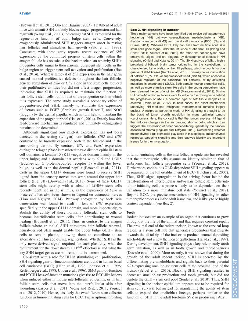
(Brownell et al., 2011; Oro and Higgins, 2003). Treatment of adultmicewith an anti-SHH antibody blocks anagen progression and hairregrowth (Wang et al., 2000), indicating that SHH is required for theregenerative function of adult bulge stem cells. Conversely,exogenously administered SHH triggers anagen onset in restinghair follicles and stimulates hair growth (Sato et al., 1999).Consistent with these early reports, recent evidence of Shhexpression by the committed progeny of stem cells within theanagen follicle has revealed a feedback mechanism whereby SHH+progenitor cells signal to their parental quiescent stem cells in thebulge region to trigger stem cell activation and proliferation (Hsuet al., 2014). Whereas removal of Shh expression in the hair germcaused marked proliferative defects throughout the hair follicle,genetic abrogation of Smo or Gli2 alone in the stem cells reducedtheir proliferative abilities but did not affect anagen progression,indicating that SHH is required to maintain the function ofhair follicle stem cells but not that of the progenitor cells in whichit is expressed. The same study revealed a secondary effect ofprogenitor-secreted SHH, namely to stimulate the expressionof factors such as FGF7 (fibroblast growth factor 7) and NOG(noggin) by the dermal papilla, which in turn help to maintain theexpansion of the progenitor pool (Hsu et al., 2014). Exactly how thisfeed-forward mechanism is regulated and ultimately extinguishedremains to be determined.Although significant Shh mRNA expression has not been
detected in the resting (telogen) hair follicle, Gli2 and Gli3continue to be broadly expressed both in the follicle and in thesurrounding dermis. By contrast, Gli1 and Ptch1 expressionduring the telogen phase is restricted to two distinct epithelial stemcell domains: a keratin 15 (K15)-negative domain mainly in theupper bulge; and a domain that overlaps with K15 and LGR5(leucine-rich G protein-coupled receptor 5) within the lowerbulge, as well as in the dermal papilla (Brownell et al., 2011).Cells in the upper GLI1+ domain were found to receive SHHligand from the sensory nerves that wrap around the upper hairfollicle (Fig. 3B) (Brownell et al., 2011). Some of these GLI1+stem cells might overlap with a subset of LGR6+ stem cellsrecently identified in the isthmus, as the expression of Lgr6 inthese cells has also been shown to depend on cutaneous nerves(Liao and Nguyen, 2014). Pathway abrogation by back skindenervation was found to result in loss of Gli1 expressionspecifically in the upper GLI1+ domain, and more importantly, toabolish the ability of these normally follicular stem cells tobecome interfollicular stem cells after contributing to woundhealing (Brownell et al., 2011). Thus, in contrast to the anagenfollicle where epithelial SHH stimulates hair follicle renewal,neural-derived SHH might enable the upper bulge GLI1+ stemcells to remain plastic, allowing them to contribute to analternative cell lineage during regeneration. Whether SHH is theonly nerve-derived signal required for such plasticity, what therequirement for the downstream GLIA/R effectors is and what thekey SHH target genes are still remain to be determined.Consistent with a role for HH in stimulating cell proliferation,
SHH signaling gain-of-function mutations are found in human basalcell carcinoma (BCC) (Hahn et al., 1996; Johnson et al., 1996;Reifenberger et al., 1998; Unden et al., 1996). SMOgain-of-functionand PTCH1 loss-of-functionmutations give rise to BCC-like lesionswhen induced either in mouse interfollicular epidermis, or in hairfollicle stem cells that move into the interfollicular skin afterwounding (Kasper et al., 2011; Wong and Reiter, 2011; Youssefet al., 2012, 2010). Hence, it is likely that two different stem cells canfunction as tumor-initiating cells for BCC. Transcriptional profiling
of tumor-initiating cells in the interfollicular epidermis has revealedthat the tumorigenic cells assume an identity similar to that ofembryonic hair follicle progenitor cells (Youssef et al., 2012).Furthermore, continuous GLI2-dependent SHH signaling appears tobe required for the full establishment of BCC (Hutchin et al., 2005).Thus, SHH signal upregulation is the driving factor behind thetransformation of interfollicular and/or hair follicle stem cells intotumor-initiating cells, a process likely to be dependent on theirtransition to a more immature cell state (Youssef et al., 2012).Beyond BCC, the precise mechanism of HH signaling in othertumorgenic processes in the adult is unclear, and is likely to be highlycontext dependent (see Box 2).
TeethRodent incisors are an example of an organ that continues to growthroughout the life of the animal and that requires constant repair.The proximal end of the rodent incisor, known as the cervical loopregion, is a stem cell hub that generates progenitors that migratetowards the distal tip of the incisor to produce enamel-depositingamelioblasts and renew the incisor epithelium (Harada et al., 1999).During development, SHH signaling plays a key role in early toothgerm initiation, as well as in tooth growth and morphogenesis(Dassule et al., 2000). More recently, it was shown that during thegrowth of the adult rodent incisor, SHH is secreted by thedifferentiating pre-amelioblasts and signals back to their parentalGli1-expressing amelioblast stem cells at the proximal end of theincisor (Seidel et al., 2010). Blocking SHH signaling resulted indecreased amelioblast production and tooth growth, but did notdeplete the GLI1+ stem cell pool (Seidel et al., 2010). Thus, SHHsignaling in the incisor epithelium appears not to be required forstem cell survival but instead for maintaining the ability of stemcells to expand the amelioblast lineage, perhaps similar to thefunction of SHH in the adult forebrain SVZ in producing TACs.
Box 2. HH signaling in cancerThree major cancers have been identified that involve cell-autonomoushedgehog (HH) pathway over-activation: medulloblastoma (MB),rhabdomyosarcoma (RMS) and basal cell carcinoma (BCC) (Ng andCurran, 2011). Whereas BCC likely can arise from multiple adult skinstem cells gone rogue under the influence of aberrant HH (Wong andReiter, 2011; Youssef et al., 2010), the other two cancer types haveembryonic origins and are triggered by developmental defects in HHsignaling (Onishi and Katano, 2011). The SHH subtype of MB, a highlyprevalent childhood brain tumor originating in the cerebellum, ischaracterized by activation of the HH pathway, which accounts for onequarter of all MB cases (Remke et al., 2013). This may be via inactivationof patched 1 (PTCH1) or suppressor of fused (SUFU), which encodes anegative regulator of the canonical HH pathway, or by activatingmutations in smoothened (SMO). Both granule neuron progenitor cells,as well as more primitive stem-like cells in the young cerebellum havebeen deemed the cell of origin for MB (Manoranjan et al., 2012). SimilarHH gain-of-function mutations were found to be the driving force behindthe progression of RMS, a common type of soft tissue neoplasia inchildren (Roma et al., 2012). In both cases, the exact mechanismunderlying HH-mediated malignant transformation remains largelyunclear. A reciprocal paracrine mode of HH signaling is thought to bethe basis of tumor growth regulation in many epithelial tumors(carcinomas). Here, the concept is that the tumors express HH ligandthat induces changes in the surrounding stromal cells, which in turntriggers the expression of other cancer-altering ligands by the cancer-associated stroma (Teglund and Toftgard, 2010). Determining whethermesenchymal adult stem cells play a role in this epithelial-mesenchymalreciprocal paracrine signaling and their subtype identity are importantissues for further investigation.
3450
REVIEW Development (2014) 141, 3445-3457 doi:10.1242/dev.083691
DEVELO
PM
ENT

SHH signaling was also recently shown to play an important rolein the maintenance of dentin, the mesenchymal compartment of theincisor, which is located under the outer enamel surface (Zhao et al.,2014). The study showed that dentin turnover is dependent on HH-responding periarterial mesenchymal stem cells, which alsocontribute to dentin repair after injury. Much like in the skin, theHH ligand that maintains the GLI1+ stem cell population is secretedby nerves in the neighboring neurovascular bundle. HH inhibitoradministration revealed that, as in the incisor epithelium, thepathway is not required to support stem cell maintenance, survivalor progenitor proliferation, but is necessary for the differentiation ofodontoblasts (Zhao et al., 2014).Although human teeth do not grow continuously, stem cells from
human dental pulp have been isolated (Gronthos et al., 2000) and itremains to be determined whether they respond to canonical SHHsignaling. A multipotent human stem cell population within theperiodontal ligament, which is the connective tissue surrounding thetooth, was shown to express SHH as well as GLI1 and PTCH1, andto respond to exogenous stimulation with recombinant SHH and toinhibition with the SMO inhibitor cyclopamine (Martinez et al.,2011). Understanding how SHH and other signaling pathwaysregulate the function of adult stem cells associated with toothhomeostasis might prove beneficial for the development of dentalimplants and improved dental repair (Nakashima and Iohara, 2014;Nakashima et al., 2009).
HH signaling in adult tissues of mesodermal originUnlike the skin and brain, there is less evidence for HH signaling inhomeostasis and repair of adult tissues of mesodermal origin. Themesoderm forms tissues such as bone, cartilage and muscle, as wellas the circulatory system. As bona fide adult stem or progenitor cellpopulations have yet to be identified in some of these tissues, wefocus this section of our review on the reported roles of HHsignaling in tissue repair following injury.
BoneIHH is one of the main regulators of chondrocyte proliferation andosteoblast differentiation during skeletal development (Long andOrnitz, 2013). In developing long bones, IHH together with theparathyroid hormone-related protein (PTHrP) regulate chondrocytebehavior within the bone growth plate. Disruption of the IHH-PTHrPpathway and upregulation of HH signaling leads to the formation ofchildhood cartilaginous neoplasms, such as enchondromas andosteochondromas (Tiet and Alman, 2003). Throughout adulthood,HH signaling continues to help maintain bone structure as systemicadministration of the SMO inhibitor cyclopamine to adult mice wasshown to result in bone mass reduction. By contrast, adult Ptch1+/−
mutant mice exhibit an increase in bone mass density and osteoblastdifferentiation associated with loss of GLI3R activity (Ohba et al.,2008). Consistent with this, upregulation of HH signaling, either bytransient adenovirus-mediated overexpression of SHH or byconditional deletion of Ptch1 in mature osteoblasts, not only causesincreased osteoblast production but also an indirect increase inosteoclast activity leading to bone resorption and decreased bonestrength (Kiuru et al., 2009; Mak et al., 2008). By contrast, HHsignaling inhibition by partial conditional ablation of Smo in matureosteoblasts results in protection from bone loss in 1-year-old mice(Mak et al., 2008). Taken together, these studies indicate a role forHHsignaling in bone homeostasis through regulating the balance betweenbone formation and bone resorption in a concentration-dependentmanner. In addition to bone homeostatic regulation, HH signaling hasalso been implicated indisease processes, such asosteoporosis, aswell
as during bone repair and revascularization after injury (Fuchs et al.,2012; Wang et al., 2010). Upregulation of both Ihh and Shhtranscription, as well as Ptch1, has been observed immediately afterbone fracture (Ito et al., 1999; Miyaji et al., 2003), indicating that HHsignaling functions in the adult bone to modulate cell behaviorsfollowing disruption of homeostasis to ensure tissue repair. However,no bona fide stem cell population responsible for adult bone repair hasbeen identified to date.
MuscleSkeletal muscle is one of the few mammalian organs in the adultcapable of almost complete regeneration after injury. This is possibledue to the presence of satellite cells, the in situ muscle stem cellpopulation (Lepper et al., 2011). These cells remain mostly inactiveunder normal conditions but in response to injury they can give riseto myogenic cells that reconstitute the myofibers of the muscle (Yinet al., 2013). During embryogenesis, canonical HH signaling tosomites plays a role in the direct induction of myogenic factors suchas MYOD1 (myogenic differentiation 1) and MYF5 (myogenicfactor 5), which are essential for skeletal myogenesis (Pownall et al.,2002). In adult mouse satellite cells, HH signaling continues tofunction as a pro-survival and proliferation factor (Koleva et al.,2005). Intriguingly, upregulation in Shh and Ptch1 transcription hasalso been detected in adult fully differentiated muscle upon theinduction of regeneration following ischemic injury. During thisprocess, HH plays a crucial role in promoting angiogenesis andincreasing satellite cell number at the affected site (Pola et al., 2003,2001; Straface et al., 2009). By contrast, SMO inhibition bycyclopamine treatment in injured animals results in muscle fibrosisand increased inflammation (Straface et al., 2009). Furthermore,although the regenerative ability of skeletal muscle declines in agingmice, intramuscular injection of a Shh-expressing vector was shownto successfully boost muscle repair to levels comparable with thosefound in much younger mice (Piccioni et al., 2013).
The existence of a HH-responding stem cell population in adultmammalian cardiac muscle has not yet been demonstrated;however, HH signaling is nonetheless required for the properfunction of the adult heart. Conditional ablation of Smo in adultsmooth muscle cells surrounding the blood vessels results in loss ofcoronary blood vessels, heart failure and even lethality (Lavineet al., 2008). By contrast, Shh gene transfer in an adult mousemyocardial ischemia model reduces fibrosis and augmentsangiogenesis, thus aiding heart repair (Kusano et al., 2005).
A crucial role for HH in smooth muscle development has beenobserved in many different organs, including gut, bladder (Maoet al., 2010; Tasian et al., 2010) and kidney. During kidney smoothmuscle development, HH functions through interacting withmembers of the bone morphogenic protein family (Yu et al.,2002). Although the role of HH signaling in the adult kidneyremains unclear, upregulation of HH and Gli expression, as wellas an expansion of the α-smooth muscle actin-expressingmyofibroblast population has been reported in a mouse model ofkidney fibrosis (Fabian et al., 2012). Although the role of HH inmediating muscle repair and fibrosis is context dependent, it is clearthat this pathway is an important player in disease progression andtherefore represents a possible therapeutic target for the treatment ofmuscular disorders and possibly for heart failure.
HematopoiesisHematopoietic stem cells (HSCs) are long-lived, largely quiescentstem cells that reside in the bone marrow and constantly replenishthe myeloid (monocytes, macrophages, neutrophils, basophils,
3451
REVIEW Development (2014) 141, 3445-3457 doi:10.1242/dev.083691
DEVELO
PM
ENT

eosinophils, erythrocytes, platelets) and lymphoid (T, B and naturalkiller) cell lineages (Jagannathan-Bogdan and Zon, 2013). WhereasHH has been shown to play a role in the induction of vasculogenesisand hematopoiesis at embryonic stages (Lim and Matsui, 2010),reports on the role of the canonical signaling pathway in the adultare controversial. Ligands of the pathway are expressed in thehematopoietic niche and have been shown to function as survivalsignals for leukemia, lymphoma and myeloma cancer stem cells,whereas inhibition of the pathway appears to suppress diseaseprogression (Dierks et al., 2008, 2007; Zhao et al., 2009). Despitebeing implicated in hematopoietic malignancies, the precise role ofHH signaling during adult homeostasis remains unclear. Much ofthe controversy surrounding HH signaling in hematopoietichomeostasis concerns the varying results obtained from loss-of-function studies. Ptch1+/− adult animals have been shown toundergo accelerated recovery after hematopoietic damage but this isaccompanied by reduced long-term grafting potential when Ptch1+/−
adult HSCs are transplanted into irradiated adult animals (Trowbridgeet al., 2006). Another study using transplantation of fetal liver-derivedHSCs from Ptch1+/− embryos reported an enhancement in the long-term self-renewing potential of Ptch1+/−HSCs, whereas Smo−/− fetalliver-derived HSCs were shown to have normal regeneration capacitywhen placed in an adult wild-type host (Dierks et al., 2008). Bycontrast, CRE-mediated conditional ablation of Smo in both HSCsand their niche from embryonic stages onwardswas shown to result ina profound loss of long-term grafting potential of the HSCs in vivo(Zhao et al., 2009). Yet another set of independent studies based onthe conditional ablation of Smo in adult hematopoietic tissuesconcluded that SMO-mediated HH signaling is not required tomaintain normal adult HSC function (Gao et al., 2009; Hofmannet al., 2009). In summary, the long list of discrepancies with regard tothe requirement for HH in HSC function might result from adifferential requirement for HH signaling in the niche versus theHSCs themselves, and/or from a different role for HH at differentstages of development.
HH signaling in adult tissues of endodermal originThe embryonic endoderm contributes to tissues of the respiratory,gastrointestinal and genitourinary systems. During development,canonical HH signaling is involved in the epithelial-mesenchymalcommunication that regulates the early formation of these systems,during which ligand-releasing epithelial cells signal to the GLI1+mesoderm (Haraguchi et al., 2007; Motoyama et al., 1998;Ramalho-Santos et al., 2000). In the adult, HH continues to signalto the mesenchymal stromal cells but the effect of HH on tissue-specific stem cell populations within these tissues is only starting tobe defined.
Respiratory and gastrointestinal systemsDuring early mammalian embryonic development, both therespiratory and digestive tubes arise from the primitive gut, alsoknown as the archenteron – a cavity within the gastrula. Even at thisearly developmental stage, by instructing mesodermal Hox geneexpression, HH regulates the specification and subdivision of thegut (Sheaffer and Kaestner, 2012). Therefore, due to theircommonality of origin, we review the roles of HH signaling in therespiratory and gastrointestinal systems together.Much like in the adult bone and muscle, localized upregulation of
HH signaling occurs in response to injury in adult lung airways. Inthe normal adult mouse lung, only a fewGli1-expressing fibroblastsare detected around the airways. However, HH signaling isupregulated upon bleomycin-induced lung fibrosis or airway
injury after treatment with naphthalene, as evidenced by anincrease in stromal GLI1+ cells (Liu et al., 2013; Watkins et al.,2003). A similar increase in HH signaling has been detected inhuman lung fibrotic tissue (Stewart et al., 2003), whereas in adultmice Shh overexpression augments collagen deposition and lungfibrosis following airway injury (Liu et al., 2013). Consistent with arole for SHH in lung tissue repair, overexpression of Shh in normaladult mouse airway epithelium can induce cell proliferation andlung tissue modifications similar to those seen in injury (Krauseet al., 2010).
Shh and Ihh expression continues to be detected throughout theadult gastrointestinal tract of both humans and rodents where itsignals to the Gli-expressing mesenchyme (Kolterud et al., 2009;van den Brink et al., 2002, 2001; van Dop et al., 2010). In the adultmurine stomach, HH signaling is thought to be responsible forinhibiting proliferation and stimulating the differentiation of thegastric epithelium (van den Brink et al., 2002, 2001). By contrast,upregulation of HH signaling by conditional removal of Ptch1 inadult colonic mesenchyme results in the depletion of the epithelialprecursor cell pool due to premature differentiation (van Dopet al., 2009). Furthermore, HH signaling has been found to bedownregulated during repair following gastric ulcer induction,whereas inhibition of SMO via cyclopamine treatment of injuredmice further inhibits gastric progenitor cell differentiation (Kanget al., 2009). In mouse models of HH pathway inhibition, atrophy ofthe small intestinal villi is observed resulting from the loss of villussmooth muscle cells, which is also accompanied by inflammationand an increase in proliferation in the epithelial compartment (vanDop et al., 2010; Zacharias et al., 2010). Conversely, upregulationof IHH expression in adult intestine promotes villus smooth muscledifferentiation (Zacharias et al., 2011), indicating that HH signalingin the adult murine intestine regulates tissue homeostasis in aconcentration-dependent manner.
The liver also forms part of the GI system and has the greatestregenerative capacity of any other endoderm organ. Here also, HHsignaling activation is one of the steps towards tissue reconstructionfollowing injury (Omenetti et al., 2007). After partial hepatectomy,canonical HH signaling is required for hepatocyte proliferation;blocking of the HH pathway with the SMO inhibitor cyclopaminedecreases post-operative survival rates in mice (Ochoa et al., 2010).Upregulation of the pathway has been observed in the livers ofindividuals with primary biliary cirrhosis (Jung et al., 2007), alsoimplicating HH signaling in the response to liver damage inhumans. These studies together suggest that the role of HH signalingin cells of the gastrointestinal system is strongly dependent on thecontext of tissue injury or disease state.
Deconstructing the exact mechanism of HH signaling in normaland injured respiratory and gastrointestinal tissues might provemore complex than previously imagined. Results from several newstudies in the adult trachea, stomach and liver have indicated thatdifferentiated non-mitotic cells in these tissues can fully replaceresident stem cells if the latter are selectively ablated (Stange et al.,2013; Tata et al., 2013; Yanger et al., 2013). These findings challengethe importance of an adult resident stem cell population, given thatcommitted cells can replace the stem cells under specific conditions.Whether HH/GLI activity is required to maintain the function ofendogenous putative stromal stem cell populations or plays a role inthe dedifferentiation of mature cells remains to be explored.
Genitourinary systemOne component of the adult genitourinary system that has greatregenerative capacity is the adult prostate. Normally dormant like the
3452
REVIEW Development (2014) 141, 3445-3457 doi:10.1242/dev.083691
DEVELO
PM
ENT

liver, the prostate is capable of multiple rounds of androgen-inducedregeneration following castration-induced involution (degeneration)of the ductal structures (Isaacs and Coffey, 1989). The completeregeneration of the prostate following injury and presence of label-retaining cells (Tsujimura et al., 2002) suggests the presence ofquiescent stem cells in the prostate. A possible role for HH signalingin prostate regeneration was suggested by an experiment in which HHsignaling was blocked during regeneration following castration andandrogen stimulation in adult mice, which resulted in the failure of thetissue to regenerate (Karhadkar et al., 2004). However, the experimenthas not been repeated and the cell type that responded to HH was notidentified. SHH was recently found to be secreted by basal cellswithin the epithelial compartment of the prostate, which are likely tobe themain source of HHwithin the prostate ducts (Peng et al., 2013).In the same study,Gli1was shown to be expressed in four subtypes ofstromal cells, each possibly maintained by a distinct unipotentprogenitor. Followingmultiple rounds of involution and regeneration,GLI1+ stromal cells were shown to continuously self-renew (Penget al., 2013), indicating that epithelial SHH signals to bona fide stemcells in the prostate stroma in a paracrine fashion, much like duringprostate development (Shaw and Bushman, 2007). Determining theidentity of SHH-responding stem cells in the prostate is a priority, asSHH signaling has been implicated in prostate cancer (Chen et al.,2011), and overexpression of SHH ligand in the adult prostate issufficient to induce neoplasia (Chang et al., 2011). A recent report onprostate cancer development revealed that nerve fibers innervating theprostate act as a positive regulator of cancer progression (Magnonet al., 2013). It will be interesting to determine whether any HHproteins are delivered to the prostate through peripheral nerves as isthe case in the adult skin and rodent incisors (Brownell et al., 2011;Zhao et al., 2014) (Fig. 3C).Similar to the prostate, SHH is also involved in the epithelial-
mesenchymal interaction between ligand-secreting basal stem cellsand the underlining GLI1+ mesenchyme in the adult murine bladder(Shin et al., 2011). Upon tissue regeneration following bladder injury,HH signaling becomes upregulated and participates in reciprocalsignaling, leading to the increase in epithelial cell proliferationrequired for the restoration of normal bladder function. Furthermore,in a mouse model of muscle-invasive bladder cancer, theShh-expressing basal cells were recently demonstrated to functionas neoplasia-initiating cells (Shin et al., 2014). After chemicalcarcinogenesis, individual SHH+ cells could give rise to lesions thatquickly progressed to carcinomas, after which Shh expression withinthe tumor was lost. Whether this is the case in humans and whetherthere are roles forHH in the progression of bladder carcinoma remainsto be determined, especially given that constitutive upregulation inHH signaling has been detected in human bladder cancer cell lines(Pignot et al., 2012). There are likely to be interesting parallelsbetween bladder and prostate cancer: in mouse models of prostatecancer, basal cells can also give rise to carcinomas, and the tumor cellsalso lose their basal cell characteristics (Choi et al., 2012;Wang et al.,2013). These few examples clearly demonstrate that, in thegenitourinary system, base levels of HH signaling help maintainhomeostasis and participate in tissue repair; however, in a diseasecontext, the contribution of the HH signaling pathway can havedetrimental and diverse consequences.
ConclusionsIn adult tissue homeostasis, high levels of HH signaling are seen inspecific populations of cells, many of which have stem andprogenitor cell properties. However, the exact mechanism ofcanonical HH signal propagation downstream of the GLIs remains
largely unknown, as specific HH target genes, stemness-inducing orotherwise, have not been identified for most adult tissues. Recentfindings in the developing embryo have indicated that the activatingand repressing effects of the GLIs are enforced throughcollaboration with local master regulators from the SOX (SRYbox containing), FGF and HOX (homeobox) families, and that thiscollaboration allows tissue- or cell-specific interpretation of HHsignaling. Whether this is the case in the adult and which factorsHH is interacting with in various tissues during homeostasis, injuryand regeneration are some of the most exciting and challengingissue the field is facing today.
Following injury, HH signaling can trigger stem and otherresident cells to participate in repair, whereas in diseases, includingcancer, perturbed levels of HH signaling can contribute to diseaseprogression by different routes. Thus, HH upregulation can beviewed as a natural response to injury and a way to achieve tissuerepair by promoting cell survival, proliferation, plasticity ortransdifferentiation. If HH levels are reduced with aging, thedecrease could prove detrimental to tissue homeostasis and repair,and represents a possible mechanism underlying age-related organdegeneration and poor repair. In an attempt to find treatments forvarious human cancers where HH is the suspected driving forcebehind disease progression, a large degree of effort has been spendon developing SMO inhibitors, which seem to be generally welltolerated in pediatric patients (Lin and Matsui, 2012). Althoughpromising, these results are somewhat surprising given the broadregulatory role of HH in multiple tissues during developmentand in adulthood. Further long-term investigations are requiredto completely exclude the possibility that HH inhibitionpharmacologically results in permanent adverse defects later inlife or during aging.
The role of peripheral nerves in delivering signaling factors suchas HH to regulate normal and regenerate non-neuronal tissues isonly just beginning to emerge (Brownell et al., 2011; Zhao et al.,2014). Given this role of the nervous system in delivering HHligands and the involvement of HH signaling in the developingenteric system (Liu and Ngan, 2013), it is interesting to speculatethat HH released by nerves is also involved in transducing thesignaling pathway in the GI system. Thus nerve-derived HH standsout as a putative crucial mediator of organ homeostasis andregeneration with the potential to target stem cell populationslocated in different organs.
In summary, although the HH signaling pathway was originallydiscovered over 20 years ago, there remain exciting avenues ofexploration, particularly in the stem cell and regeneration fieldswhere the exact roles of HH signaling in different cellular anddisease contexts is still unclear. Understanding what regulates HHsignaling at the systemic and local levels, as well as how suchsignals are translated at the transcriptional level in target tissues toallow a context-specific response is likely to be a central challengefor the HH field in the years to come and will ultimately help touncover new therapeutic targets in multiple disease contexts.
AcknowledgementsWe thank all members of the Joyner laboratory over the past decade for helpfuldiscussions about adult stem cells.
Competing interestsThe authors declare no competing financial interests.
FundingThe authors’ research was supported by grants from the National Institute of MentalHealth, National Cancer Institute, National Brain Tumor Society and Geoffrey BeeneCancer Research Center. Deposited in PMC for release after 12 months.
3453
REVIEW Development (2014) 141, 3445-3457 doi:10.1242/dev.083691
DEVELO
PM
ENT

ReferencesAhn, S. and Joyner, A. L. (2005). In vivo analysis of quiescent adult neural stemcells responding to Sonic hedgehog. Nature 437, 894-897.
Amankulor, N. M., Hambardzumyan, D., Pyonteck, S. M., Becher, O. J., Joyce,J. A. andHolland, E. C. (2009). Sonic hedgehog pathway activation is induced byacute brain injury and regulated by injury-related inflammation. J. Neurosci. 29,10299-10308.
Amaral, D. G. and Kurz, J. (1985). An analysis of the origins of the cholinergic andnoncholinergic septal projections to the hippocampal formation of the rat. J. Comp.Neurol. 240, 37-59.
Bai, C. B., Stephen, D. and Joyner, A. L. (2004). All mouse ventral spinal cordpatterning by hedgehog is Gli dependent and involves an activator function ofGli3. Dev. Cell 6, 103-115.
Balaskas, N., Ribeiro, A., Panovska, J., Dessaud, E., Sasai, N., Page, K. M.,Briscoe, J. and Ribes, V. (2012). Gene regulatory logic for reading the SonicHedgehog signaling gradient in the vertebrate neural tube. Cell 148, 273-284.
Balordi, F. and Fishell, G. (2007a). Hedgehog signaling in the subventricular zoneis required for both the maintenance of stem cells and the migration of newbornneurons. J. Neurosci. 27, 5936-5947.
Balordi, F. and Fishell, G. (2007b). Mosaic removal of hedgehog signaling in theadult SVZ reveals that the residual wild-type stem cells have a limited capacity forself-renewal. J. Neurosci. 27, 14248-14259.
Bambakidis, N. C., Wang, R. Z., Franic, L. and Miller, R. H. (2003). Sonichedgehog-induced neural precursor proliferation after adult rodent spinal cordinjury. J. Neurosurg. 99, 70-75.
Barnes, E. A., Kong, M., Ollendorff, V. and Donoghue, D. J. (2001). Patched1interacts with cyclin B1 to regulate cell cycle progression.EMBOJ. 20, 2214-2223.
Belgacem, Y. H. and Borodinsky, L. N. (2011). Sonic hedgehog signaling isdecoded by calcium spike activity in the developing spinal cord. Proc. Natl. Acad.Sci. USA 108, 4482-4487.
Berg, D. A., Belnoue, L., Song, H. and Simon, A. (2013). Neurotransmitter-mediated control of neurogenesis in the adult vertebrate brain. Development 140,2548-2561.
Bitgood, M. J., Shen, L. and McMahon, A. P. (1996). Sertoli cell signaling byDesert hedgehog regulates the male germline. Curr. Biol. 6, 298-304.
Blaess, S., Corrales, J. D. and Joyner, A. L. (2006). Sonic hedgehog regulatesGli activator and repressor functions with spatial and temporal precision in themid/hindbrain region. Development 133, 1799-1809.
Blaess, S., Stephen, D. and Joyner, A. L. (2008). Gli3 coordinates three-dimensional patterning and growth of the tectum and cerebellum by integratingShh and Fgf8 signaling. Development 135, 2093-2103.
Brennan, D., Chen, X., Cheng, L., Mahoney, M. and Riobo, N. A. (2012).Noncanonical Hedgehog signaling. Vitam. Horm. 88, 55-72.
Briscoe, J. and Therond, P. P. (2013). The mechanisms of Hedgehog signallingand its roles in development and disease. Nat. Rev. Mol. Cell Biol. 14, 416–429.
Brownell, I., Guevara, E., Bai, C. B., Loomis, C. A. and Joyner, A. L. (2011).Nerve-derived sonic hedgehog defines a niche for hair follicle stem cells capableof becoming epidermal stem cells. Cell Stem Cell 8, 552-565.
Capilla-Gonzalez, V., Cebrian-Silla, A., Guerrero-Cazares, H., Garcia-Verdugo,J. M. and Quinones-Hinojosa, A. (2013). The generation of oligodendroglialcells is preserved in the rostral migratory stream during aging. Front. Cell.Neurosci. 7, 147.
Chang, H. H., Chen, B. Y., Wu, C. Y., Tsao, Z. J., Chen, Y. Y., Chang, C. P., Yang,C. R. and Lin, D. P.-C. (2011). Hedgehog overexpression leads to the formation ofprostate cancer stem cells with metastatic property irrespective of androgenreceptor expression in the mouse model. J. Biomed. Sci. 18, 6.
Chen, M., Carkner, R. and Buttyan, R. (2011). The hedgehog/Gli signalingparadigm in prostate cancer. Expert Rev. Endocrinol. Metabol. 6, 453-467.
Chiang, C., Litingtung, Y., Lee, E., Young, K. E., Corden, J. L., Westphal, H. andBeachy, P. A. (1996). Cyclopia and defective axial patterning in mice lackingSonic hedgehog gene function. Nature 383, 407-413.
Choi, N., Zhang, B., Zhang, L., Ittmann, M. and Xin, L. (2012). Adult murineprostate basal and luminal cells are self-sustained lineages that can both serve astargets for prostate cancer initiation. Cancer Cell 21, 253-265.
Dass, B., Iravani, M. M., Huang, C., Barsoum, J., Engber, T. M., Galdes, A. andJenner, P. (2005). Sonic hedgehog delivered by an adeno-associated virusprotects dopaminergic neurones against 6-OHDA toxicity in the rat. J. Neural.Transm. 112, 763-778.
Dassule, H. R., Lewis, P., Bei, M., Maas, R. and McMahon, A. P. (2000). Sonichedgehog regulates growth and morphogenesis of the tooth. Development 127,4775-4785.
Dessaud, E., McMahon, A. P. and Briscoe, J. (2008). Pattern formation in thevertebrate neural tube: a sonic hedgehog morphogen-regulated transcriptionalnetwork. Development 135, 2489-2503.
Dierks, C., Grbic, J., Zirlik, K., Beigi, R., Englund, N. P., Guo, G.-R., Veelken, H.,Engelhardt, M., Mertelsmann, R., Kelleher, J. F. et al. (2007). Essential role ofstromally induced hedgehog signaling in B-cell malignancies. Nat. Med. 13,944-951.
Dierks, C., Beigi, R., Guo, G.-R., Zirlik, K., Stegert, M. R., Manley, P., Trussell, C.,Schmitt-Graeff, A., Landwerlin, K., Veelken, H. et al. (2008). Expansion of
Bcr-Abl-positive leukemic stem cells is dependent on Hedgehog pathwayactivation. Cancer Cell 14, 238-249.
Ding, Q., Motoyama, J., Gasca, S., Mo, R., Sasaki, H., Rossant, J. and Hui, C. C.(1998). Diminished Sonic hedgehog signaling and lack of floor plate differentiationin Gli2 mutant mice. Development 125, 2533-2543.
Doetsch, F., Garcia-Verdugo, J. M. and Alvarez-Buylla, A. (1997). Cellularcomposition and three-dimensional organization of the subventricular germinalzone in the adult mammalian brain. J. Neurosci. 17, 5046-5061.
Echelard, Y., Epstein, D. J., St-Jacques, B., Shen, L., Mohler, J., McMahon, J. A.and McMahon, A. P. (1993). Sonic hedgehog, a member of a family of putativesignaling molecules, is implicated in the regulation of CNS polarity. Cell 75,1417-1430.
Fabian, S. L., Penchev, R. R., St-Jacques, B., Rao, A. N., Sipila, P., West, K. A.,McMahon, A. P. andHumphreys, B. D. (2012). Hedgehog-Gli pathway activationduring kidney fibrosis. Am. J. Pathol. 180, 1441-1453.
Ferent, J., Cochard, L., Faure, H., Taddei, M., Hahn, H., Ruat, M. and Traiffort, E.(2014). Genetic activation of Hedgehog signaling unbalances the rate of neuralstem cell renewal by increasing symmetric divisions. Stem Cell Rep. 3, 312-323.
Fleming, J. T., He,W.,Hao, C., Ketova, T., Pan, F. C.,Wright, C.C.V., Litingtung, Y.and Chiang, C. (2013). The purkinje neuron acts as a central regulator of spatiallyand functionally distinct cerebellar precursors. Dev. Cell 27, 278-292.
Fuccillo, M., Joyner, A. L. and Fishell, G. (2006). Morphogen to mitogen: themultiple roles of hedgehog signalling in vertebrate neural development. Nat. Rev.Neurosci. 7, 772-783.
Fuchs, S., Dohle, E. and Kirkpatrick, C. J. (2012). Sonic Hedgehog-mediatedsynergistic effects guiding angiogenesis and osteogenesis. Vitam. Horm. 88,491-506.
Fuentealba, L. C., Obernier, K. and Alvarez-Buylla, A. (2012). Adult neural stemcells bridge their niche. Cell Stem Cell 10, 698-708.
Gao, J., Graves, S., Koch, U., Liu, S., Jankovic, V., Buonamici, S., ElAndaloussi, A., Nimer, S. D., Kee, B. L., Taichman, R. et al. (2009).Hedgehog signaling is dispensable for adult hematopoietic stem cell function.CellStem Cell 4, 548-558.
Garcia, A. D. R., Petrova, R., Eng, L. and Joyner, A. L. (2010). Sonic hedgehogregulates discrete populations of astrocytes in the adult mouse forebrain.J. Neurosci. 30, 13597-13608.
Goetz, S. C. and Anderson, K. V. (2010). The primary cilium: a signalling centreduring vertebrate development. Nat. Rev. Genet. 11, 331-344.
Gonzalez-Reyes, L. E., Verbitsky, M., Blesa, J., Jackson-Lewis, V., Paredes, D.,Tillack, K., Phani, S., Kramer, E. R., Przedborski, S. and Kottmann, A. H.(2012). Sonic hedgehogmaintains cellular and neurochemical homeostasis in theadult nigrostriatal circuit. Neuron 75, 306-319.
Goodrich, L. V., Milenkovic, L., Higgins, K. M. and Scott, M. P. (1997). Alteredneural cell fates and medulloblastoma in mouse patched mutants. Science 277,1109-1113.
Gronthos, S., Mankani, M., Brahim, J., Robey, P. G. and Shi, S. (2000). Postnatalhuman dental pulp stem cells (DPSCs) in vitro and in vivo. Proc. Natl. Acad. Sci.USA 97, 13625-13630.
Guo, Z., Zhang, L., Wu, Z., Chen, Y., Wang, F. and Chen, G. (2013). In vivo directreprogramming of reactive glial cells into functional neurons after brain injury andin an Alzheimer’s disease model. Cell Stem Cell. 14, 188-202.
Hahn, H., Wicking, C., Zaphiropoulous, P. G., Gailani, M. R., Shanley, S.,Chidambaram, A., Vorechovsky, I., Holmberg, E., Unden, A. B., Gillies, S.et al. (1996). Mutations of the human homolog of Drosophila patched in the nevoidbasal cell carcinoma syndrome. Cell 85, 841-851.
Han, Y.-G., Spassky, N., Romaguera-Ros, M., Garcia-Verdugo, J.-M., Aguilar,A., Schneider-Maunoury, S. and Alvarez-Buylla, A. (2008). Hedgehogsignaling and primary cilia are required for the formation of adult neural stemcells. Nat. Neurosci. 11, 277-284.
Harada, H., Kettunen, P., Jung, H.-S., Mustonen, T., Wang, Y. A. and Thesleff, I.(1999). Localization of putative stem cells in dental epithelium and theirassociation with Notch and FGF signaling. J. Cell Biol. 147, 105-120.
Haraguchi, R., Motoyama, J., Sasaki, H., Satoh, Y., Miyagawa, S., Nakagata, N.,Moon, A. and Yamada, G. (2007). Molecular analysis of coordinated bladder andurogenital organ formation by Hedgehog signaling. Development 134, 525-533.
Hofmann, I., Stover, E. H., Cullen, D. E., Mao, J., Morgan, K. J., Lee, B. H.,Kharas, M. G., Miller, P. G., Cornejo, M. G., Okabe, R. et al. (2009). Hedgehogsignaling is dispensable for adult murine hematopoietic stem cell function andhematopoiesis. Cell Stem Cell 4, 559-567.
Hsu, Y.-C., Li, L. and Fuchs, E. (2014). Transit-amplifying cells orchestrate stemcell activity and tissue regeneration. Cell 157, 935-949.
Huang, X., Liu, J., Ketova, T., Fleming, J. T., Grover, V. K., Cooper, M. K.,Litingtung, Y. and Chiang, C. (2010). Transventricular delivery of Sonichedgehog is essential to cerebellar ventricular zone development. Proc. Natl.Acad. Sci. USA 107, 8422-8427.
Hui, C.-C. and Angers, S. (2011). Gli proteins in development and disease. Annu.Rev. Cell Dev. Biol. 27, 513-537.
Hurtado-Lorenzo, A., Millan, E., Gonzalez-Nicolini, V., Suwelack, D., Castro,M. G. and Lowenstein, P. R. (2004). Differentiation and transcription factor genetherapy in experimental parkinson’s disease: sonic hedgehog and Gli-1, but not
3454
REVIEW Development (2014) 141, 3445-3457 doi:10.1242/dev.083691
DEVELO
PM
ENT

Nurr-1, protect nigrostriatal cell bodies from 6-OHDA-induced neurodegeneration.Mol. Ther. 10, 507-524.
Hutchin, M. E., Kariapper, M. S. T., Grachtchouk, M., Wang, A., Wei, L.,Cummings, D., Liu, J., Michael, L. E., Glick, A. and Dlugosz, A. A. (2005).Sustained Hedgehog signaling is required for basal cell carcinoma proliferationand survival: conditional skin tumorigenesis recapitulates the hair growth cycle.Genes Dev. 19, 214-223.
Ihrie, R. A., Shah, J. K., Harwell, C. C., Levine, J. H., Guinto, C. D., Lezameta, M.,Kriegstein, A. R. and Alvarez-Buylla, A. (2011). Persistent sonic hedgehogsignaling in adult brain determines neural stem cell positional identity. Neuron 71,250-262.
Isaacs, J. T. and Coffey, D. S. (1989). Etiology and disease process of benignprostatic hyperplasia. Prostate 15 Suppl. 2, 33-50.
Ito, H., Akiyama, H., Shigeno, C., Iyama, K.-I., Matsuoka, H. and Nakamura, T.(1999). Hedgehog signaling molecules in bone marrow cells at the initial stage offracture repair. Biochem. Biophys. Res. Commun. 262, 443-451.
Jagannathan-Bogdan, M. and Zon, L. I. (2013). Hematopoiesis. Development140, 2463-2467.
Jenkins, D. (2009). Hedgehog signalling: emerging evidence for non-canonicalpathways. Cell. Signal. 21, 1023-1034.
Jiang, J. and Hui, C.-C. (2008). Hedgehog signaling in development and cancer.Dev. Cell 15, 801-812.
Johnson, R. L., Rothman, A. L., Xie, J., Goodrich, L. V., Bare, J. W., Bonifas,J. M., Quinn, A. G., Myers, R. M., Cox, D. R., Epstein, E. H., Jr et al. (1996).Human homolog of patched, a candidate gene for the basal cell nevus syndrome.Science 272, 1668-1671.
Jung, Y., McCall, S. J., Li, Y.-X. and Diehl, A. M. (2007). Bile ductules and stromalcells express hedgehog ligands and/or hedgehog target genes in primary biliarycirrhosis. Hepatology 45, 1091-1096.
Kang, D.-H., Han,M.-E., Song,M.-H., Lee, Y.-S., Kim,E.-H., Kim,H.-J., Kim,G.-H.,Kim, D.-H., Yoon, S., Baek, S.-Y. et al. (2009). The role of hedgehog signalingduring gastric regeneration. J. Gastroenterol. 44, 372-379.
Karhadkar, S. S., Bova, G. S., Abdallah, N., Dhara, S., Gardner, D., Maitra, A.,Isaacs, J. T., Berman, D. M. and Beachy, P. A. (2004). Hedgehog signalling inprostate regeneration, neoplasia and metastasis. Nature 431, 707-712.
Kasper,M., Jaks, V., Are, A., Bergstrom,A., Schwager, A., Svard, J., Teglund, S.,Barker, N. and Toftgard, R. (2011). Wounding enhances epidermaltumorigenesis by recruiting hair follicle keratinocytes. Proc. Natl. Acad. Sci. USA108, 4099-4104.
Kenney, A. M. and Rowitch, D. H. (2000). Sonic hedgehog promotes G(1) cyclinexpression and sustained cell cycle progression in mammalian neuronalprecursors. Mol. Cell. Biol. 20, 9055-9067.
Kenney, A. M., Cole, M. D. and Rowitch, D. H. (2003). Nmyc upregulation by sonichedgehog signaling promotes proliferation in developing cerebellar granuleneuron precursors. Development 130, 15-28.
Kiuru,M., Solomon, J., Ghali, B., vanderMeulen,M., Crystal, R.G. andHidaka,C.(2009). Transient overexpression of sonic hedgehog alters the architecture andmechanical properties of trabecular bone. J. Bone Miner. Res. 24, 1598-1607.
Koleva, M., Kappler, R., Vogler, M., Herwig, A., Fulda, S. and Hahn, H. (2005).Pleiotropic effects of sonic hedgehog on muscle satellite cells. Cell. Mol. Life Sci.62, 1863-1870.
Kolterud, A., Grosse, A. S., Zacharias, W. J., Walton, K. D., Kretovich, K. E.,Madison, B. B., Waghray, M., Ferris, J. E., Hu, C., Merchant, J. L. et al. (2009).Paracrine Hedgehog signaling in stomach and intestine: new roles for hedgehogin gastrointestinal patterning. Gastroenterology 137, 618-628.
Krause, A., Xu, Y., Joh, J., Hubner, R., Gess, A., Ilic, T. and Worgall, S. (2010).Overexpression of sonic Hedgehog in the lung mimics the effect of lung injury andcompensatory lung growth on pulmonary Sca-1 and CD34 positive cells. Mol.Ther. 18, 404-412.
Kusano, K. F., Pola, R., Murayama, T., Curry, C., Kawamoto, A., Iwakura, A.,Shintani, S., Ii, M., Asai, J., Tkebuchava, T. et al. (2005). Sonic hedgehogmyocardial gene therapy: tissue repair through transient reconstitution ofembryonic signaling. Nat. Med. 11, 1197-1204.
Lai, K., Kaspar, B. K., Gage, F. H. and Schaffer, D. V. (2003). Sonic hedgehogregulates adult neural progenitor proliferation in vitro and in vivo.Nat. Neurosci. 6,21-27.
Lavine, K. J., Kovacs, A. and Ornitz, D. M. (2008). Hedgehog signaling is criticalfor maintenance of the adult coronary vasculature in mice. J. Clin. Invest. 118,2404-2414.
Lennington, J. B., Pope, S., Goodheart, A. E., Drozdowicz, L., Daniels, S. B.,Salamone, J. D. and Conover, J. C. (2011). Midbrain dopamine neuronsassociated with reward processing innervate the neurogenic subventricular zone.J. Neurosci. 31, 13078-13087.
Lepper, C., Partridge, T. A. and Fan, C.-M. (2011). An absolute requirement forPax7-positive satellite cells in acute injury-induced skeletal muscle regeneration.Development 138, 3639-3646.
Li, G., Fang, L., Fernandez, G. and Pleasure, S. J. (2013). The ventralhippocampus is the embryonic origin for adult neural stem cells in the dentategyrus. Neuron 78, 658-672.
Liao, X.-H. and Nguyen, H. (2014). Epidermal expression of Lgr6 is dependent onnerve endings and Schwann cells. Exp. Dermatol. 23, 195-198.
Lim, Y. and Matsui, W. (2010). Hedgehog signaling in hematopoiesis. Crit. Rev.Eukaryot. Gene Expr. 20, 129-139.
Lin, T. L. and Matsui, W. (2012). Hedgehog pathway as a drug target: smoothenedinhibitors in development. OncoTargets Ther. 5, 47-58.
Litingtung, Y., Lei, L., Westphal, H. and Chiang, C. (1998). Sonic hedgehog isessential to foregut development. Nat. Genet. 20, 58-61.
Liu, J. A. and Ngan, E. S. (2013). Hedgehog and Notch signaling in enteric nervoussystem development. Neurosignals 22, 1-13.
Liu, C., Sage, J. C., Miller, M. R., Verhaak, R. G. W., Hippenmeyer, S., Vogel, H.,Foreman, O., Bronson, R. T., Nishiyama, A., Luo, L. et al. (2011). Mosaicanalysis with double markers reveals tumor cell of origin in glioma. Cell 146,209-221.
Liu, L., Kugler, M. C., Loomis, C. A., Samdani, R., Zhao, Z., Chen, G. J., Brandt,J. P., Brownell, I., Joyner, A. L., Rom,W. N. et al. (2013). Hedgehog signaling inneonatal and adult lung. Am. J. Respir. Cell Mol. Biol. 48, 703-710.
Long, F. andOrnitz, D. M. (2013). Development of the endochondral skeleton.ColdSpring Harb. Perspect. Biol. 5, a008334.
Loulier, K., Ruat, M. and Traiffort, E. (2006). Increase of proliferatingoligodendroglial progenitors in the adult mouse brain upon Sonic hedgehogdelivery in the lateral ventricle. J. Neurochem. 98, 530-542.
Machold, R., Hayashi, S., Rutlin, M., Muzumdar, M. D., Nery, S., Corbin, J. G.,Gritli-Linde, A., Dellovade, T., Porter, J. A., Rubin, L. L. et al. (2003). Sonichedgehog is required for progenitor cell maintenance in telencephalic stem cellniches. Neuron 39, 937-950.
Magnon, C., Hall, S. J., Lin, J., Xue, X., Gerber, L., Freedland, S. J. and Frenette,P. S. (2013). Autonomic nerve development contributes to prostate cancerprogression. Science 341, 1236361.
Mak, K. K., Bi, Y., Wan, C., Chuang, P.-T., Clemens, T., Young, M. and Yang, Y.(2008). Hedgehog signaling in mature osteoblasts regulates bone formation andresorption by controlling PTHrP and RANKL expression. Dev. Cell 14, 674-688.
Manoranjan, B., Venugopal, C., McFarlane, N., Doble, B. W., Dunn, S. E.,Scheinemann, K. and Singh, S. K. (2012). Medulloblastoma stem cells: wheredevelopment and cancer cross pathways. Pediatr. Res. 71, 516-522.
Mao, J., Kim, B. M., Rajurkar, M., Shivdasani, R. A. and McMahon, A. P. (2010).Hedgehog signaling controls mesenchymal growth in the developing mammaliandigestive tract. Development 137, 1721-1729.
Martinez, C., Smith, P. C., Rodriguez, J. P. and Palma, V. (2011). Sonic hedgehogstimulates proliferation of human periodontal ligament stem cells. J. Dent. Res. 90,483-488.
Matise, M. P., Epstein, D. J., Park, H. L., Platt, K. A. and Joyner, A. L. (1998). Gli2is required for induction of floor plate and adjacent cells, but not most ventralneurons in the mouse central nervous system. Development 125, 2759-2770.
Menn, B., Garcia-Verdugo, J. M., Yaschine, C., Gonzalez-Perez, O., Rowitch, D.and Alvarez-Buylla, A. (2006). Origin of oligodendrocytes in the subventricularzone of the adult brain. J. Neurosci. 26, 7907-7918.
Merkle, F. T., Mirzadeh, Z. and Alvarez-Buylla, A. (2007). Mosaic organization ofneural stem cells in the adult brain. Science 317, 381-384.
Merkle, F. T., Fuentealba, L. C., Sanders, T. A., Magno, L., Kessaris, N. andAlvarez-Buylla, A. (2013). Adult neural stem cells in distinct microdomainsgenerate previously unknown interneuron types. Nat. Neurosci. 17, 207-214.
Miyaji, T., Nakase, T., Iwasaki,M., Kuriyama,K., Tamai, N., Higuchi, C.,Myoui, A.,Tomita, T. andYoshikawa, H. (2003). Expressionand distribution of transcripts forsonic hedgehog in the early phase of fracture repair. Histochem. Cell Biol. 119,233-237.
Motoyama, J., Liu, J., Mo, R., Ding, Q., Post, M. and Hui, C.-C. (1998). Essentialfunction of Gli2 and Gli3 in the formation of lung, trachea and oesophagus. Nat.Genet. 20, 54-57.
Nakashima, M. and Iohara, K. (2014). Mobilized dental pulp stem cells for pulpregeneration: initiation of clinical trial. J. Endod. 40, S26-S32.
Nakashima, M., Iohara, K. and Sugiyama, M. (2009). Human dental pulp stemcells with highly angiogenic and neurogenic potential for possible use in pulpregeneration. Cytokine Growth Factor Rev. 20, 435-440.
Ng, J. M. Y. and Curran, T. (2011). The Hedgehog’s tale: developing strategies fortargeting cancer. Nat. Rev. Cancer 11, 493-501.
Ochoa, B., Syn, W.-K., Delgado, I., Karaca, G. F., Jung, Y., Wang, J., Zubiaga, A.M., Fresnedo, O., Omenetti, A., Zdanowicz, M. et al. (2010). Hedgehogsignaling is critical for normal liver regeneration after partial hepatectomy in mice.Hepatology 51, 1712-1723.
Ohba, S., Kawaguchi, H., Kugimiya, F., Ogasawara, T., Kawamura, N., Saito, T.,Ikeda, T., Fujii, K., Miyajima, T., Kuramochi, A. et al. (2008). Patched1haploinsufficiency increases adult bone mass and modulates Gli3 repressoractivity. Dev. Cell 14, 689-699.
Omenetti, A., Yang, L., Li, Y. X., McCall, S. J., Jung, Y., Sicklick, J. K., Huang, J.,Choi, S., Suzuki, A. and Diehl, A. M. (2007). Hedgehog-mediatedmesenchymal-epithelial interactions modulate hepatic response to bile ductligation. Lab. Invest. 87, 499-514.
Onishi, H. and Katano, M. (2011). Hedgehog signaling pathway as a therapeutictarget in various types of cancer. Cancer Sci. 102, 1756-1760.
3455
REVIEW Development (2014) 141, 3445-3457 doi:10.1242/dev.083691
DEVELO
PM
ENT

Oosterveen, T., Kurdija, S., Alekseenko, Z., Uhde, C. W., Bergsland, M.,Sandberg, M., Andersson, E., Dias, J. M., Muhr, J. and Ericson, J. (2012).Mechanistic differences in the transcriptional interpretation of local and long-rangeShh morphogen signaling. Dev. Cell 23, 1006-1019.
Oosterveen, T., Kurdija, S., Enstero, M., Uhde, C. W., Bergsland, M., Sandberg,M., Sandberg, R., Muhr, J. and Ericson, J. (2013). SoxB1-driven transcriptionalnetwork underlies neural-specific interpretation of morphogen signals. Proc. Natl.Acad. Sci. USA 110, 7330-7335.
Oro, A. E. and Higgins, K. (2003). Hair cycle regulation of Hedgehog signalreception. Dev. Biol. 255, 238-248.
Palma, V., Lim, D. A., Dahmane, N., Sanchez, P., Brionne, T. C., Herzberg, C. D.,Gitton, Y., Carleton, A., Álvarez-Buylla, A. and Ruiz i Altaba, A. (2005). Sonichedgehog controls stem cell behavior in the postnatal and adult brain.Development 132, 335-344.
Park, H. L., Bai, C., Platt, K. A., Matise, M. P., Beeghly, A., Hui, C. C., Nakashima,M. and Joyner, A. L. (2000). Mouse Gli1 mutants are viable but have defects inSHH signaling in combination with a Gli2 mutation.Development 127, 1593-1605.
Peng, Y. C., Levine, C. M., Zahid, S., Wilson, E. L. and Joyner, A. L. (2013). Sonichedgehog signals to multiple prostate stromal stem cells that replenish distinctstromal subtypes during regeneration. Proc. Natl. Acad. Sci. USA. 110,20611-20616.
Pepicelli, C. V., Lewis, P. M. and McMahon, A. P. (1998). Sonic hedgehogregulates branching morphogenesis in the mammalian lung. Curr. Biol. 8,1083-1086.
Persson, M., Stamataki, D., teWelscher, P., Andersson, E., Bose, J., Ruther, U.,Ericson, J. and Briscoe, J. (2002). Dorsal-ventral patterning of the spinal cordrequires Gli3 transcriptional repressor activity. Genes Dev. 16, 2865-2878.
Petrova, R., Garcia, A. D. R. and Joyner, A. L. (2013). Titration of GLI3 repressoractivity by sonic Hedgehog signaling is critical for maintaining multiple adult neuralstem cell and astrocyte functions. J. Neurosci. 33, 17490-17505.
Piccioni, A., Gaetani, E., Neri, V., Gatto, I., Palladino, M., Silver, M., Smith, R. C.,Giarretta, I., Pola, E., Hlatky, L. et al. (2013). Sonic Hedgehog therapy in amouse model of age-associated impairment of skeletal muscle regeneration.J. Gerontol. A Biol. Sci. Med. Sci. 69, 245-52.
Pignot, G., Vieillefond, A., Vacher, S., Zerbib, M., Debre, B., Lidereau, R.,Amsellem-Ouazana, D. and Bieche, I. (2012). Hedgehog pathway activation inhuman transitional cell carcinoma of the bladder. Br. J. Cancer 106, 1177-1186.
Pola, R., Ling, L. E., Silver, M., Corbley, M. J., Kearney, M., Blake Pepinsky, R.,Shapiro, R., Taylor, F. R., Baker, D. P., Asahara, T. et al. (2001). Themorphogen Sonic hedgehog is an indirect angiogenic agent upregulating twofamilies of angiogenic growth factors. Nat. Med. 7, 706-711.
Pola, R., Ling, L. E., Aprahamian, T. R., Barban, E., Bosch-Marce, M., Curry, C.,Corbley, M., Kearney, M., Isner, J. M. and Losordo, D. W. (2003). Postnatalrecapitulation of embryonic hedgehog pathway in response to skeletal muscleischemia. Circulation 108, 479-485.
Pownall, M. E., Gustafsson, M. K. and Emerson, C. P., Jr. (2002). Myogenicregulatory factors and the specification of muscle progenitors in vertebrateembryos. Annu. Rev. Cell Dev. Biol. 18, 747-783.
Rallu, M., Machold, R., Gaiano, N., Corbin, J. G., McMahon, A. P. and Fishell, G.(2002). Dorsoventral patterning is established in the telencephalon of mutantslacking both Gli3 and Hedgehog signaling. Development 129, 4963-4974.
Ramalho-Santos, M., Melton, D. A. and McMahon, A. P. (2000). Hedgehogsignals regulate multiple aspects of gastrointestinal development. Development127, 2763-2772.
Reifenberger, J., Wolter, M., Weber, R. G., Megahed, M., Ruzicka, T., Lichter, P.andReifenberger, G. (1998). Missensemutations in SMOH in sporadic basal cellcarcinomas of the skin and primitive neuroectodermal tumors of the centralnervous system. Cancer Res. 58, 1798-1803.
Remke,M., Ramaswamy, V. and Taylor, M. D. (2013). Medulloblastomamoleculardissection: the way toward targeted therapy. Curr. Opin. Oncol. 25, 674-681.
Ribes, V. and Briscoe, J. (2009). Establishing and interpreting graded SonicHedgehog signaling during vertebrate neural tube patterning: the role of negativefeedback. Cold Spring Harb. Perspect. Biol. 1, a002014.
Riobo, N. A., Haines, G. M. and Emerson, C. P., Jr. (2006). Protein kinase C-deltaand mitogen-activated protein/extracellular signal-regulated kinase-1 control GLIactivation in hedgehog signaling. Cancer Res. 66, 839-845.
Roma, J., Almazan-Moga, A., Sanchez de Toledo, J. and Gallego, S. (2012).Notch, wnt, and hedgehog pathways in rhabdomyosarcoma: from single pathwaysto an integrated network. Sarcoma 2012, 695603.
Rowitch, D. H., St.-Jacques, B., Lee, S. M., Flax, J. D., Snyder, E. Y. andMcMahon, A. P. (1999). Sonic hedgehog regulates proliferation and inhibitsdifferentiation of CNS precursor cells. J. Neurosci. 19, 8954-8965.
Sato, N., Leopold, P. L. and Crystal, R. G. (1999). Induction of the hair growthphase in postnatal mice by localized transient expression of Sonic hedgehog.J. Clin. Invest. 104, 855-864.
Seidel, K., Ahn, C. P., Lyons, D., Nee, A., Ting, K., Brownell, I., Cao, T., Carano,R. A. D., Curran, T., Schober, M. et al. (2010). Hedgehog signaling regulates thegeneration of ameloblast progenitors in the continuously growing mouse incisor.Development 137, 3753-3761.
Shaw, A. and Bushman, W. (2007). Hedgehog signaling in the prostate. J. Urol.177, 832-838.
Sheaffer, K. L. and Kaestner, K. H. (2012). Transcriptional networks in liver andintestinal development. Cold Spring Harb. Perspect. Biol. 4, a008284.
Shin,K., Lee, J.,Guo,N.,Kim,J., Lim,A.,Qu,L.,Mysorekar, I.U. andBeachy,P.A.(2011). Hedgehog/Wnt feedback supports regenerative proliferation of epithelialstem cells in bladder. Nature 472, 110-114.
Shin, K., Lim, A., Odegaard, J. I., Honeycutt, J. D., Kawano, S., Hsieh, M. H. andBeachy, P. A. (2014). Cellular origin of bladder neoplasia and tissue dynamics ofits progression to invasive carcinoma. Nat. Cell Biol. 16, 469-478.
Sirko, S., Behrendt, G., Johansson, P. A., Tripathi, P., Costa, M., Bek, S.,Heinrich, C., Tiedt, S., Colak, D., Dichgans, M. et al. (2013). Reactive glia in theinjured brain acquire stem cell properties in response to sonic hedgehog.[corrected]. Cell Stem Cell 12, 426-439.
Sofroniew, M. V. (2009). Molecular dissection of reactive astrogliosis and glial scarformation. Trends Neurosci. 32, 638-647.
Solanas, G. and Benitah, S. A. (2013). Regenerating the skin: a task for theheterogeneous stem cell pool and surrounding niche. Nat. Rev. Mol. Cell Biol. 14,737-748.
St-Jacques, B., Hammerschmidt, M. and McMahon, A. P. (1999). Indianhedgehog signaling regulates proliferation and differentiation of chondrocytesand is essential for bone formation. Genes Dev. 13, 2072-2086.
Stange, D. E., Koo, B.-K., Huch, M., Sibbel, G., Basak, O., Lyubimova, A.,Kujala, P., Bartfeld, S., Koster, J., Geahlen, J. H. et al. (2013). Differentiatedtroy(+) chief cells act as reserve stem cells to generate all lineages of the stomachepithelium. Cell 155, 357-368.
Stecca, B. and Ruiz i Albata, A. (2010). Context-dependent regulation of the GLIcode in cancer by HEDGEHOG and non-HEDGEHOG signals. J. Mol. Cell Biol. 2,84-95.
Stewart, G. A., Hoyne, G. F., Ahmad, S. A., Jarman, E., Wallace, W. A. H.,Harrison, D. J., Haslett, C., Lamb, J. R. and Howie, S. E. M. (2003). Expressionof the developmental Sonic hedgehog (Shh) signalling pathway is up-regulated inchronic lung fibrosis and the Shh receptor patched 1 is present in circulating Tlymphocytes. J. Pathol. 199, 488-495.
Straface, G., Aprahamian, T., Flex, A., Gaetani, E., Biscetti, F., Smith, R. C.,Pecorini, G., Pola, E., Angelini, F., Stigliano, E. et al. (2009). Sonic hedgehogregulates angiogenesis and myogenesis during post-natal skeletal muscleregeneration. J. Cell. Mol. Med. 13, 2424-2435.
Suwelack, D., Hurtado-Lorenzo, A., Millan, E., Gonzalez-Nicolini, V.,Wawrowsky, K., Lowenstein, P. R. and Castro, M. G. (2004). Neuronalexpression of the transcription factor Gli1 using the Talpha1 alpha-tubulinpromoter is neuroprotective in an experimental model of Parkinson’s disease.Gene Ther. 11, 1742-1752.
Tasian, G., Cunha, G. and Baskin, L. (2010). Smooth muscle differentiation andpatterning in the urinary bladder. Differentiation 80, 106-117.
Tata, P. R., Mou, H., Pardo-Saganta, A., Zhao, R., Prabhu, M., Law, B. M.,Vinarsky, V., Cho, J. L., Breton, S., Sahay, A. et al. (2013). Dedifferentiation ofcommitted epithelial cells into stem cells in vivo. Nature 503, 218-223.
Teglund, S. and Toftgard, R. (2010). Hedgehog beyond medulloblastoma andbasal cell carcinoma. Biochim. Biophys. Acta 1805, 181-208.
Thibert, C., Teillet, M.-A., Lapointe, F., Mazelin, L., Le Douarin, N. M. andMehlen, P. (2003). Inhibition of neuroepithelial patched-induced apoptosis bysonic hedgehog. Science 301, 843-846.
Tiet, T. D. and Alman, B. A. (2003). Developmental pathways in musculoskeletalneoplasia: involvement of the Indian Hedgehog-parathyroid hormone-relatedprotein pathway. Pediatr. Res. 53, 539-543.
Traiffort, E., Charytoniuk, D., Watroba, L., Faure, H., Sales, N. and Ruat, M.(1999). Discrete localizations of hedgehog signalling components in thedeveloping and adult rat nervous system. Eur. J. Neurosci. 11, 3199-3214.
Traiffort, E., Angot, E. and Ruat, M. (2010). Sonic Hedgehog signaling in themammalian brain. J. Neurochem. 113, 576-590.
Trowbridge, J. J., Scott, M. P. and Bhatia, M. (2006). Hedgehog modulates cellcycle regulators in stem cells to control hematopoietic regeneration. Proc. Natl.Acad. Sci. USA 103, 14134-14139.
Tsujimura, A., Koikawa, Y., Salm, S., Takao, T., Coetzee, S., Moscatelli, D.,Shapiro, E., Lepor, H., Sun, T.-T. and Wilson, E. L. (2002). Proximal location ofmouse prostate epithelial stem cells: amodel of prostatic homeostasis. J. Cell Biol.157, 1257-1265.
Unden, A. B., Holmberg, E., Lundh-Rozell, B., Stahle-Backdahl, M.,Zaphiropoulos, P. G., Toftgard, R. and Vorechovsky, I. (1996). Mutations inthe human homologue of Drosophila patched (PTCH) in basal cell carcinomasand the Gorlin syndrome: different in vivo mechanisms of PTCH inactivation.Cancer Res. 56, 4562-4565.
Van Den Brink, G. R., Hardwick, J. C.H., Peppelenbosch, M. P., Van Deventer,S. J. H., Tytgat, G. N. J., Brink, M. A. and TenKate, F. J. (2001). Sonic hedgehogregulates gastric gland morphogenesis in man andmouse.Gastroenterology 121,317-328.
van den Brink, G. R., Hardwick, J. C. H., Nielsen, C., Xu, C., ten Kate, F. J.,Glickman, J., van Deventer, S. J. H., Roberts, D. J. and Peppelenbosch, M. P.
3456
REVIEW Development (2014) 141, 3445-3457 doi:10.1242/dev.083691
DEVELO
PM
ENT

(2002). Sonic hedgehog expression correlates with fundic gland differentiation inthe adult gastrointestinal tract. Gut 51, 628-633.
van Dop, W. A., Uhmann, A., Wijgerde, M., Sleddens-Linkels, E., Heijmans, J.,Offerhaus, G. J., van den Bergh Weerman, M. A., Boeckxstaens, G. E.,Hommes, D. W., Hardwick, J. C. et al. (2009). Depletion of the colonic epithelialprecursor cell compartment upon conditional activation of the hedgehog pathway.Gastroenterology 136, 2195-2203 e2191–2197.
van Dop, W. A., Heijmans, J., Buller, N. V., Snoek, S. A., Rosekrans, S. L.,Wassenberg, E. A., van den BerghWeerman, M. A., Lanske, B., Clarke, A. R.,Winton, D. J. et al. (2010). Loss of Indian Hedgehog activatesmultiple aspects ofa wound healing response in the mouse intestine. Gastroenterology 139,1665-1676 e1661–1610.
Varjosalo, M. and Taipale, J. (2008). Hedgehog: functions and mechanisms.Genes Dev. 22, 2454-2472.
Wang, L. C., Liu, Z.-Y., Gambardella, L., Delacour, A., Shapiro, R., Yang, J.,Sizing, I., Rayhorn, P., Garber, E. A., Benjamin, C. D. et al. (2000). Conditionaldisruption of hedgehog signaling pathway defines its critical role in hairdevelopment and regeneration. J. Invest. Dermatol. 114, 901-908.
Wang, Q., Huang, C., Zeng, F., Xue, M. and Zhang, X. (2010). Activation of the Hhpathway in periosteum-derived mesenchymal stem cells induces bone formationin vivo: implication for postnatal bone repair. Am. J. Pathol. 177, 3100-3111.
Wang, Z. A., Mitrofanova, A., Bergren, S. K., Abate-Shen, C., Cardiff, R. D.,Califano, A. and Shen, M. M. (2013). Lineage analysis of basal epithelial cellsreveals their unexpected plasticity and supports a cell-of-origin model for prostatecancer heterogeneity. Nat. Cell Biol. 15, 274-283.
Watkins, D. N., Berman, D. M., Burkholder, S. G., Wang, B., Beachy, P. A. andBaylin, S. B. (2003). Hedgehog signalling within airway epithelial progenitors andin small-cell lung cancer. Nature 422, 313-317.
Wong, S. Y. and Reiter, J. F. (2011). Wounding mobilizes hair follicle stem cells toform tumors. Proc. Natl. Acad. Sci. USA 108, 4093-4098.
Yam, P. T., Langlois, S. D., Morin, S. and Charron, F. (2009). Sonic hedgehogguides axons through a noncanonical, Src-family-kinase-dependent signalingpathway. Neuron 62, 349-362.
Yanger, K., Zong, Y., Maggs, L. R., Shapira, S. N., Maddipati, R., Aiello, N. M.,Thung, S. N., Wells, R. G., Greenbaum, L. E. and Stanger, B. Z. (2013). Robust
cellular reprogramming occurs spontaneously during liver regeneration. GenesDev. 27, 719-724.
Yin, H., Price, F. and Rudnicki, M. A. (2013). Satellite cells and the muscle stemcell niche. Physiol. Rev. 93, 23-67.
Youssef, K. K., Van Keymeulen, A., Lapouge, G., Beck, B., Michaux, C.,Achouri, Y., Sotiropoulou, P. A. and Blanpain, C. (2010). Identification ofthe cell lineage at the origin of basal cell carcinoma. Nat. Cell Biol. 12,299-305.
Youssef, K. K., Lapouge, G., Bouvree, K., Rorive, S., Brohee, S., Appelstein, O.,Larsimont, J.-C., Sukumaran, V., Van de Sande, B., Pucci, D. et al. (2012).Adult interfollicular tumour-initiating cells are reprogrammed into an embryonichair follicle progenitor-like fate during basal cell carcinoma initiation.Nat. Cell Biol.14, 1282-1294.
Yu, J., Carroll, T. J. and McMahon, A. P. (2002). Sonic hedgehog regulatesproliferation and differentiation of mesenchymal cells in the mouse metanephrickidney. Development 129, 5301-5312.
Zacharias, W. J., Li, X., Madison, B. B., Kretovich, K., Kao, J. Y., Merchant, J. L.and Gumucio, D. L. (2010). Hedgehog is an anti-inflammatory epithelialsignal for the intestinal lamina propria. Gastroenterology 138, 2368-2377e2361–2364.
Zacharias, W. J., Madison, B. B., Kretovich, K. E., Walton, K. D., Richards, N.,Udager, A. M., Li, X. and Gumucio, D. L. (2011). Hedgehog signaling controlshomeostasis of adult intestinal smooth muscle. Dev. Biol. 355, 152-162.
Zhang, X. M., Ramalho-Santos, M. and McMahon, A. P. (2001). Smoothenedmutants reveal redundant roles for Shh and Ihh signaling including regulation of L/R asymmetry by the mouse node. Cell 105, 781-792.
Zhao, C., Chen, A., Jamieson, C. H., Fereshteh, M., Abrahamsson, A., Blum, J.,Kwon, H. Y., Kim, J., Chute, J. P., Rizzieri, D. et al. (2009). Hedgehog signallingis essential for maintenance of cancer stem cells in myeloid leukaemia. Nature458, 776-779.
Zhao, H., Feng, J., Seidel, K., Shi, S., Klein, O., Sharpe, P. and Chai, Y. (2014).Secretion of shh by a neurovascular bundle niche supports mesenchymal stemcell homeostasis in the adult mouse incisor. Cell Stem Cell 14, 160-173.
3457
REVIEW Development (2014) 141, 3445-3457 doi:10.1242/dev.083691
DEVELO
PM
ENT