role of treatments and genetic alterations on ...cholesterol levels often have advanced...
TRANSCRIPT

University of Milano-Bicocca
School of Medicine and School of Science
PhD program in Translational and Molecular
Medicine
DIMET
ROLE OF TREATMENTS AND GENETIC
ALTERATIONS ON ATHEROSCLEROSIS AND
CHOLESTEROL METABOLISM: STUDIES IN
ANIMAL MODELS
Doctor Dellera Federica
Matr. No: 787799
Coordinator: Professor Andrea Biondi
Tutor: Doctor Roberto Giovannoni
Co-tutor: Professor Giulia Chiesa
XXIX CYCLE
ACADEMIC YEAR 2015/2016

2

3
Table of contents
Chapter 1 General introduction 7
Cholesterol and lipoprotein metabolism 9
LDLR 15
PCSK9 19
Pathogenesis of atherosclerosis 23
Initiation and fatty streak phase of
atherosclerotic lesions 23
Progression to advanced atherosclerotic
lesions 29
Vulnerable plaque formation and rupture 32

4
Arterial injury response: neointimal hyperplasia 34
Mouse models for the study of atherosclerosis 38
Apo E -/-
mice 38
LDLR -/-
mice 40
PCSK9 animal models 42
PCSK9 -/-
mice 42
L-homoarginine: a novel protective agent against
cardiovascular disease 44
L-homoarginine synthesis 44
L-homoarginine physiology 47
Role of L- homoarginine in cardiovascular disease
(CVD) 48

5
Dietary fish components: effect on cardiovascular
disease 53
Fish proteins and dyslipidemias 56
Fish proteins and hypertension 57
MicroRNAs: role in lipoprotein metabolism
and atherosclerosis 59
MiRNA regulation of HDL metabolism 63
MiRNA regulation of LDL metabolism 65
Therapeutic implications of miRNAs in
atherosclerosis and CVD 67
SCOPE OF THE THESIS 68
REFERENCES 70

6
Chapter 2 L-homoarginine administration
reduces neointimal hyperplasia in
balloon-injured rat carotids 108
Chapter 3 A salmon protein hydrolysate
exerts lipid-independent anti-atherosclerotic
activity in apo E-deficient mice 132
Chapter 4 MicroRNA expression profiles
of tissues involved in cholesterol
metabolism and atherosclerosis from
athero-prone and athero-resistant
mouse models 178
Chapter 5 Conclusions and future
perspectives 206
Publications 214

7
Chapter 1
General introduction

8

9
CHOLESTEROL AND LIPOPROTEIN METABOLISM Cholesterol is an important component of cell membranes
(1), as well as the precursor molecule for the synthesis of
steroid hormones, vitamin D and bile salts. It is derived
from the diet or synthesized within the body. The typical
human diet contains 200–500 mg of cholesterol.
Cholesterol also enters the intestine in bile (800–1200 mg
day−1
). The principal sites of cholesterol synthesis are the
liver and the Central Nervous System (CNS). Cholesterol can
be lost from the body mainly as the fractions of bile salts
and intestinal cholesterol which are not absorbed. Both
dietary cholesterol and that synthesized de novo are
transported through the circulation in lipoprotein particles.
Cholesterol blood levels in humans are typically in the
range 100–300 mg dl−1
. The principal plasma lipoproteins
are the chylomicrons, Very Low Density Lipoproteins
(VLDLs), Low Density Lipoproteins (LDLs) and High Density
Lipoproteins (HDLs) (2).
Chylomicrons are synthesized in the intestinal mucosal cells
directly from dietary fats, namely triglycerides, cholesterol
and phospholipids, and apoprotein (apo) B48, which is
synthesized in these cells. Their density is low because their

10
size is large (100 nm) and they contain large amounts of
lipid (especially triglycerides). Their large size precludes
penetration of the capillary membrane, so they are
secreted by intestinal mucosa into the lymphatics and then
enter the circulation by way of the thoracic duct. Once in
the blood, chylomicrons acquire apo E and apo C-II and are
progressively reduced in size by the action of lipoprotein
lipase, which is bound to the capillary endothelium and
catalyzes the removal of free fatty acids from the
chylomicron triglyceride pool. The continued action of
lipoprotein lipase leaves the chylomicron nearly depleted
of triglyceride. The depleted chylomicron remnant particle
exits this pathway by its uptake into the hepatocyte
through a receptor-mediated process, with the
chylomicron apo E serving as the ligand for the hepatic LDL
receptor (LDLR). In the hepatocyte the chylomicron
remnant releases its contents (ie, the remaining
triglycerides, cholesteryl esters, phospholipids, and
apoproteins). The hepatocyte reassembles these
chylomicron remnant–derived products, along with
endogenous triglycerides and cholesteryl esters, into VLDLs
and secretes them into the circulation for the next phase of

11
delivery of lipids to the periphery. Like chylomicrons, VLDLs
are also triglyceride-rich and contain apo C-II and apo E.
However, unlike chylomicrons, they contain fewer
triglycerides, are smaller, and now carry apo B-100 instead
of apo B-48. Lipoprotein lipase reduces VLDL triglyceride
content further, leaving the particles progressively smaller,
more dense, and more cholesterol-enriched as it moves
down this cascade to intermediate density lipoprotein
(IDL). Hydrolysis of IDL triglycerides to free fatty acids is
mediated by another lipase, hepatic lipase, and LDL soon
appears as the terminal particle in this pathway. Actually,
about one third of the VLDL is cleared by the hepatic LDLR,
whereas two thirds pass down this cascade, terminating as
LDL. The transition of VLDL to IDL is accompanied by the
transfer of apo C-II (to HDL) and is also removed (again to
HDL) during the transition of IDL to LDL apo E. Hence, LDL
contains only apo B-100, like the upstream precursor
particles (IDL and VLDL) (3).
The forward movement of lipids to the peripheral cells is
the key pathway for delivery of free fatty acids. It is also the
only pathway for delivering cholesterol.

12
The metabolic balance of cholesterol is achieved by reverse
cholesterol transport (RCT), a multi-step process mediated
by HDLs which consists in the transfer of excess cholesterol
arriving at the tissues back to the liver. HDLs are
synthesized in the cells of intestinal mucosa and liver and
secreted as a disk-shaped nascent HDL particles containing
only a small amount of phospholipid and apo A-I
(preβ−HDLs). Preβ−HDL gains unesterified (free)
cholesterol from peripheral cells by an aqueous diffusion
pathway from the cell membrane to the particle. The efflux
of cholesterol is facilitated by the binding of the nascent
HDL particle to cell-surface receptors (e.g. scavenger
receptor B1 [SRB1]). Free cholesterol, being amphipathic, is
absorbed onto the surface of the HDL from a “donor” cell
membrane. Lecithin-cholesterol acyltransferase (LCAT),
activated by apo A-I, catalyzes cholesterol esterification,
causing the lipophilic cholesteryl ester molecule to enter
the core of the HDL particle, thereby freeing up space on
the surface of the particle for further cholesterol
adsorption. HDL gradually matures to its spherical shape
(α-HDL). Along with cholesterol absorption, HDL
accumulates apo C-II and apo E from the VLDL and IDL.

13
Removal of lipids from the mature α-HDL particle occurs by
two pathways, a direct pathway and an indirect pathway.
The direct pathway mainly involves a selective lipid uptake
pathway mediated by SR-B1 (4). The indirect pathway is
characterized by the transfer of cholesteryl esters from α-
HDL to apo B–containing lipoproteins (VLDL, IDL, and LDL)
mediated by cholesteryl ester transfer protein (CETP) (6).
Since cholesterol removal by HDL is accomplished by
shuttling to these particles, their uptake by the liver is
essential for the disposal of HDL cholesterol. In the liver,
the hepatocytes expressing LDLR on their cell surface
recognize apo B as well as apo E. Thus, hepatic LDLR
mediates the uptake of chylomicron remnant, VLDL, IDL,
and LDL.
Figure 1 (Fig. 1) (7) summarizes the scheme for synthesis,
interaction and degradation of lipoproteins.

14
Fig. 1 Lipoprotein synthesis and metabolism
Abbreviations: LpL, Lipoprotein lipase; HL, hepatic lipase; CE,
cholesteryl ester; A, B48, C, and E, apolipoproteins; B, apo B-100; TG,
triglyceride; PL, phospholipid; FFA, free fatty acid; CM Rem,
chylomicron remnant (Gilbert-Barnes E, Barnes L, 2000).

15
LDLR
As previously discussed, the LDLR is the primary pathway
for removal of cholesterol from the circulation and its
activity is accurately regulated by intracellular cholesterol
levels. The discovery of the LDLR had its roots in the study
of the mechanisms responsible for familial
hypercholesterolemia (FH), an autosomal dominant
metabolic disorder in which patients are characterized by
plasma cholesterol levels ranging from 300 mg dL-1
to as
high as 1500 mg dL-1
(8). Patients with FH are at increased
risk of cardiovascular disease and those with the highest
cholesterol levels often have advanced atherosclerosis
before 10 years of age. FH was first recognized as a genetic
disorder over 70 years ago when it was observed that
extreme hypercholesterolemia that was accompanied by
xanthomas (cholesterol deposits in tendons and skin) and
angina was an inherited disorder. In the early 1970s
Goldstein and Brown demonstrated that fibroblasts
isolated from patients with homozygous FH had defective
feedback inhibition of the enzyme 3-hydroxy-3-
methylglutaryl CoA reductase (HMGCR), the rate-limiting
enzyme for cholesterol synthesis. In 1974 they were able to

16
demonstrate the existence of a LDLR by showing that
fibroblasts from patients with homozygous FH were unable
to bind LDL at the cell surface. The following year they
showed that the LDLR regulated HMGCR activity by
controlling the cellular uptake of exogenous cholesterol
rather than through signaling via a second messenger as
they originally hypothesized. The LDLR internalized LDL
from plasma leading to release of cholesterol into the cell
which appeared to be responsible for the feedback
inhibition of HMGCR that they had observed in their earlier
studies.
The LDLR protein was isolated and sequenced in 1982 and
the gene cloned in 1985 (9, 10). In the population, reduced
LDLR activity frequently contributes to
hypercholesterolemia which over time leads to the
development of atherosclerosis.
The transcription of LDLR is primarily under the control of
the transcription factor Sterol regulatory element-binding
protein 2 (SREBP-2) (11). SREBP-2 is an endoplasmic
reticulum transmembrane protein that exists in a complex
with the sterol-sensing proteins SREBP cleavage–activating
protein and insulin-induced gene 1. The SREBP/SREBP

17
cleavage−acGvaGng protein complex moves to the Golgi
apparatus when the intracellular cholesterol concentration
decreases. Proteases in the Golgi cleave SREBP-2 to release
the active SREBP-2 transcription factor, which translocates
to the nucleus and stimulates transcription of LDLR
(12). Like many other plasma membrane receptors, LDLR is
transported to the plasma membrane via the endoplasmic
reticulum–Golgi pathway. LDL-bound LDLR is endocytosed
through a clathrin-dependent pathway and, after releasing
its cargo in the late endosome, the LDLR is either recycled
back to the plasma membrane or degraded in the lysosome
(13).
In addition to SREBP pathway, LDLR is also regulated by
post-translational mechanisms that determine its stability.
One of those involves the activity of Proprotein convertase
subtilisin/kexin type 9 (PCSK9) (see next paragraph), which
binds directly to the extracellular domain of the LDLR and
alters its stability and trafficking by interfering with its
recycling after endocytosis, thereby promoting its
lysosomal degradation (14 - 16).
Moreover, the nuclear receptors Liver X receptor (LXR)
LXRα and LXRβ work with SREBPs to maintain cholesterol

18
homeostasis. When cellular cholesterol levels rise,
oxysterols are formed and serve as ligands for LXRs (17).
Activation of LXR by oxysterols induces the expression of
genes encoding proteins essential for cholesterol efflux
from cells, including ATP-binding cassette transporter A1
(ABCA1) and ATP-binding cassette transporter G1 (ABCG1),
phospholipid transfer protein, ADP-ribosylation factor-like
7, and apo E (18, 19). The expression of these factors
promotes the transfer of excess intracellular cholesterol to
extracellular acceptors, such as apo A-I, apo E, and HDL,
thereby facilitating reverse cholesterol transport from
peripheral tissues (20 - 22).

19
PCSK9
Seidah et al. (23) first characterized PCSK9, which is
expressed in hepatocytes, kidney, mesenchymal cells,
intestinal ileum and colon epithelia as well as in embryonic
brain telencephalon neurons. Shortly thereafter, it was
reported that single-point gain-of-function mutations
(ser127-to-arg and F216L) in the PCSK9 gene are associated
with autosomal dominant hypercholesterolemia (24).
Experimental studies established the central role of PCSK9
in regulation of cholesterol homeostasis by enhancing the
endosomal and lysosomal degradation of cell surface
receptors that regulate lipid metabolism in a non-
enzymatic fashion (25).
PCSK9 is synthesized as a 73-kDa zymogen of 692 amino
acids and contains a signal peptide, a prodomain (residues
31 to 152) and a catalytic domain (residues 153 to 451)
followed by a C-terminal domain (residues 452 to 692) (26).
PCSK9 undergoes intramolecular autocatalytic processing in
the endoplasmic reticulum to form a 14-kDa prodomain
and a 63-kDa mature PCSK9 (27). After cleavage, the
prodomain remains closely attached to the catalytic
domain of PCSK9 blocking the substrate-binding site (23, 28

20
- 30). Autocatalytic cleavage is required for trafficking
PCSK9 from the endoplasmic reticulum (ER) to the
secretory pathway (31). In the extracellular pathway, PCSK9
is secreted from the trans-Golgi network and internalized
together with the LDLR in clathrin-coated endosomes,
promoting degradation of LDLR (31). In the intracellular
pathway, PCSK9 is directly sorted to lysosomes together
with LDLR, leading to its degradation (Fig. 2) (25, 32).
Secretion of the prodomain together with a catalytically
inactive PCSK9 was shown to promote regular degradation
of LDLR, suggesting that secreted PCSK9 acts as a
chaperone rather than as a catalytic enzyme (33, 34).
As described in the previous paragraph, the major part of
circulating LDL-cholesterol (LDL-C) is removed from the
plasma by hepatic uptake. This process is mediated via
LDLR that internalizes bound LDL particles by endocytosis
(35). After intracellular dissociation, the LDLR recycles to
the cell surface for reuse. Low intracellular cholesterol
levels activate the sterol regulatory element binding
protein-2 (SREBP-2), leading to increased LDLR gene
expression, which enhances LDL-C clearance from the
circulation (36). Of note, SREBP-2 also induces expression

21
of PCSK9, leading to LDLR degradation, thus limiting hepatic
cholesterol uptake and increasing circulating LDL-C (36).
This highly coordinated expression pattern contributes to a
self-regulatory system preventing excessive cholesterol
uptake in order to preserve cholesterol homeostasis.
Fig. 2 The PCSK9 protein acts through endogenous and exogenous
mechanisms.
Exogenously, circulating PCSK9 protein binds to the LDLR. Once
internalized into the liver cell, the PCSK9 protein directs the LDLR to
the lysosome for degradation. Intrinsically, PCSK9, which is secreted
from the Golgi apparatus, binds the LDLR before it reaches the cell

22
surface, leading it to lysosomal degradation (Brautbar A, Ballantyne
CM, 2011).

23
PATHOGENESIS OF ATHEROSCLEROSIS
Abnormalities in lipid metabolism play a critical role in the
development of atherosclerosis.
Atherosclerosis is a chronic disease characterized by the
narrowing of arteries caused by a buildup of plaque and it
is the major cause of death and morbidity in the United
States and the industrial world, being the underlying cause
of heart attack, stroke, and peripheral vascular disease
(37). In particular, the discovery by Virchow more than 100
years ago that atheroma contained a yellow fatty
substance, later identified as cholesterol, suggested a role
for lipids in the pathogenesis of atherosclerosis (38).
Atherosclerosis is a complex disease, involving many cell
types and circulating mediators and resulting in an
inflammatory state. The key stages of atherosclerosis
development have been briefly described in the following
paragraphs.
Initiation and Fatty Streak Phase of Atherosclerotic
Lesions
Endothelial cells of arteries respond to mechanical and
molecular stimuli to regulate tone (39), hemostasis (40),

24
and inflammation (41) throughout the circulation.
Endothelial cell dysfunction is an initial step in
atherosclerotic lesion formation and is more likely to occur
at arterial curves and branches that are subjected to low
shear stress and disturbed blood flow (42, 43). These
mechanical stimuli activate signaling pathways leading to a
dysfunctional endothelium, that is barrier compromised,
prothrombotic, and proinflammatory (44). In regions
resistant to atherosclerosis, the transcription factors
Kruppel-like factors (KLF) 2 and 4 are activated in a process
which enhances expression of endothelial nitric oxide
synthase (eNOS) (45 – 47). The increased nitric oxide (NO)
production promotes endothelial cell migration and
survival thereby maintaining an effective barrier (48). In
addition, the expression of superoxide dismutase (SOD) is
increased to reduce cellular oxidative stress (46). In
atherosclerosis susceptible regions, reduced expression of
eNOS and SOD leads to compromised endothelial barrier
integrity (Fig. 3) (49), leading to increased accumulation
and retention of subendothelial atherogenic apo B-
containing lipoproteins (LDL) and remnants of VLDL and
chylomicrons (50, 51). KLF2, KLF4, and NO production

25
inhibit activation of the nuclear factor kappa B (NF-κB)
pathway. Increased NF-κB activation in atherosclerosis
susceptible areas leads to endothelial cell activation (Fig. 3)
(49), as evidenced by increased expression of monocyte
adherence proteins (Vascular Cell Adhesion Molecule 1,
VCAM-1; Intercellular Adhesion Molecule 1, ICAM-1; P-
selectin) and proinflammatory receptors (toll-like receptor
2, TLR2) and cytokines (Monocyte Chemoattractant Protein
1, MCP-1; Interleukin 8, IL-8) (47, 52, 53). In addition,
endothelial cell activation leads to increased production of
reactive oxygen species (54) that can cause oxidative
modification of apo B-containing lipoproteins. Besides
mechanical stimuli, endothelial cell activation is increased
by various molecular stimuli, including oxidized LDL,
cytokines, advanced glycosylation end products, and
pathogen-associated molecules (55 - 58). In contrast, an
atheroprotective function of HDL is to prevent endothelial
activation and enhance NO production to maintain barrier
integrity (59).

26
Fig. 3 Initiation of atherosclerotic lesion
Schematic view of the cascade of events leading to the accumulation
and retention of subendothelial LDL and remnants of VLDL and
chylomicrons, with formation of the fatty streak. (Linton MR F et al for De Groot LJ, Chrousos G, Dungan K, et al 2000 -
2015).

27
Activation of endothelial cells causes a monocyte
recruitment cascade involving rolling, adhesion, activation
and transendothelial migration (Fig. 3) (49). Selectins,
especially P-selectin, mediate the initial rolling interaction
of monocytes with the endothelium. Monocyte adherence
is then promoted by endothelial cell immunoglobulin-G
proteins including VCAM-1 and ICAM-1 (60). Potent
chemoattractant factors such as MCP-1 and IL-8 then
induce migration of monocytes into the subendothelial
space (61-63). Intimal macrophages also result from
proliferation of monocyte/macrophages, especially in more
advanced lesions (64). Although macrophages are the main
infiltrating cells, other cells contribute to development of
lesions including dendritic cells,mast cells and T cells (Fig.
3) (49). T cells regulate the proinflammatory phenotype of
macrophages. Antigen-specific activated T helper 1 (Th-1)
cells produce interferon (IFN) that converts macrophages
to a proinflammatory M1 phenotype. Regulatory T (T-reg)
cells produce anti-inflammatory cytokines (e.g.
Transforming Growth Factor β, TGF-β) and inhibit
activation of Th-1 cells leading more anti-inflammatory M2

28
macrophages. During the initial fatty streak phase of
atherosclerosis (Fig. 3) (49), the monocyte-derived
macrophages internalize the retained apo B-containing
lipoproteins, which are degraded in lysosomes, where
excess free cholesterol is trafficked to the ER to be
esterified by acyl CoA:cholesterol acyltransferase (ACAT),
and the resulting CE is packaged into cytoplasmic lipid
droplets, which are characteristic of foam cells (65, 66).
Modification of apo B lipoproteins via oxidation and
glycation enhances their uptake through a number of
receptors not down-regulated by cholesterol including
CD36, scavenger receptor A, and lectin-like receptor family
(67, 68). Enzyme-mediated aggregation of apo B
lipoproteins enhances uptake via phagocytosis (69, 70). In
addition, native remnant lipoproteins can induce foam cell
formation via a number of apo E receptors (LRP1 and
VLDLR).
The triggering of macrophage inflammatory pathways is
also a critical event in lesion development. Inflammatory
M1 phenotype macrophages exhibit increased oxidative
stress, impaired cholesterol efflux and enhanced
cytokine/chemokine secretion, leading to more

29
LDL/remnant oxidation, endothelial cell activation,
monocyte recruitment, and foam cell formation (71-76) .
Macrophage foam cell formation and cholesterol
dependent inflammatory receptor signaling can be reduced
by the removal of cholesterol by atheroprotective HDL and
apo A-I via a number of mechanisms including ABCA1,
ABCG1, Scavenger receptor class B member 1 (SR-BI), and
aqueous diffusion (77-80). Lipid-poor apo A-I stimulates
efflux via ABCA1, whereas lipidated apo A-I or mature HDL
are the main drivers of efflux via ABCA1, ABCG1, SR-BI, and
aqueous diffusion (77, 81-83).
Progression to Advanced Atherosclerotic Lesions
Fatty streaks do not result in clinical complications and can
even undergo regression. However, once smooth muscle
cells infiltrate and the lesions become more advanced,
regression is less likely to occur (84, 85). Small populations
of vascular smooth muscle cells (VSMCs) already present in
the intima proliferate in response to growth factors
produced by inflammatory macrophages (86). In addition,
macrophage-derived chemoattractants cause tunica media

30
smooth muscle cells to migrate into the intima and
proliferate (Fig. 4) (49). Critical smooth muscle cell
chemoattractants and growth factors include Platelet-
derived growth factor (PDGF) isoforms (87), matrix
metalloproteinases (88), fibroblast growth factors (FGF)
(89), and heparin-binding epidermal growth factor (HB-
EGF) (90) (Fig. 4) (49). The accumulating VSMCs produce a
complex extracellular matrix composed of collagen,
proteoglycans, and elastin to form a fibrous cap over a core
comprised of foam cells. As lesions advance, substantial
extracellular lipid accumulates in the core, in part due to
large CE-rich particles arising from dead macrophage foam
cells (91, 92).

31
Fig. 4 Progression to advanced atherosclerotic lesions
This stage is mediated by VSMC proliferation and accumulation, to
form a fibrous cap over a core of foam cells through the production
of extracellular matrix. (Linton MR F et al for De Groot LJ, Chrousos G,
Dungan K, et al 2000 - 2015).

32
Vulnerable Plaque Formation and Rupture
The advanced atherosclerotic lesion is essentially a
nonresolving inflammatory condition leading to formation
of the vulnerable plaque, increasing the risk of plaque
rupture. The vulnerable plaque is characterized by two
fundamental morphological changes: 1) Formation of a
necrotic core and 2) Thinning of the fibrous cap. Sections of
the atheroma with a deteriorated fibrous cap are subject to
rupture (Fig. 5) (49, 93, 94). Plaque rupture leads to acute
exposure of procoagulant and prothrombotic factors from
the necrotic core of the lesion to platelets and
procoagulant factors in the lumen, thereby causing
thrombus formation (Fig. 5) (49, 93, 94). Thrombus
formation accounts for the majority of clinical events with
acute occlusive thrombosis causing myocardial infarction,
unstable angina, sudden cardiac death, and stroke (93, 94).

33
Fig. 5 Vulnerable plaque formation
In advanced lesions, plaque is characterized by the formation of a
necrotic core and a thin fibrous cap. Deteriorated fibrous caps are
subject to rupture, with the increased risk of thrombus formation
(Linton MR F et al for De Groot LJ, Chrousos G, Dungan K, et al 2000 -
2015).

34
ARTERIAL INJURY RESPONSE: NEOINTIMAL HYPERPLASIA
Once the atherosclerotic plaque develops in a way which
interferes with blood flow, the only current treatments
available to alleviate vascular occlusion are limited to
surgical procedures such as transluminal coronary
angioplasty, percutaneous delivery of balloon-expanded
stents, directional coronary atherectomy and coronary
bypass surgery. Unfortunately, a common undesirable
outcome of these interventions is represented by
restenosis secondary to neointimal hyperplasia, which
remains a major problem after this kind of procedures (95).
Neointimal formation can be suppressed by the topical
administration of antiproliferative drugs. In percutaneous
coronary intervention the concept of "local drug delivery"
has been used for almost a decade with great success. In
recent years, drug-eluting stents and drug coated balloons
(96) have also been increasingly used in the treatment of
peripheral arterial disease, even though further validations
in large randomized trials are needed.
The development of neointimal hyperplasia is a complex
process initiated by injury and exposure of the VSMC to
circulating blood elements. The process is further

35
characterized by platelet aggregation, leukocyte
chemotaxis, VSMC proliferation and migration,
extracellular matrix (ECM) changes, and, finally, endothelial
cell proliferation.
The carotid artery balloon injury model in rats is an
important method to study the molecular and cellular
mechanisms involved in neointimal formation and vascular
remodeling (97). This approach consists of isolating a
segment of carotid artery vasculature in an anesthetized
laboratory rat, creating an arteriotomy incision in the
external carotid branch through which the balloon catheter
is inserted, advancement of the catheter through the
common carotid artery, repeated inflation and withdrawal
of the catheter to induce endothelial cell loss and mural
distension, and removal of the catheter with closure of the
arteriotomy and resumption of blood flow through the
common carotid artery and internal carotid artery branch.
The initiating event in neointimal hyperplasia is endothelial
denudation, which exposes the underlying VSMC to
circulating blood elements activating a cascade of events
culminating in neointimal formation (98). After endothelial
loss, platelets aggregate and adhere to the site of injury.

36
After platelet aggregation and adherence, leukocyte
chemotaxis occurs. Leukocytes are responsible for the
secretion of many cytokines and growth factors that
influence the subsequent events in neointimal hyperplasia
(99). One example is macrophages that produce PDGF and
interleukin-1 (IL-1), which both promote VSMC
proliferation and are released in the arterial wall during the
repair process (100, 101). In conjunction with leukocyte
chemotaxis, VSMC proliferation and migration are the
fundamental characteristics of neointimal hyperplasia. This
proliferation is, in part, initially mediated by the release of
basic fibroblast growth factor (bFGF), which is a potent
mitogen for VSMC in vivo (102). Proliferation begins as
early as 24 hours after injury and continues for several
weeks (103). Under the influence of PDGF, VSMC begin
migrating to the intima between 1 to 3 days after injury
(104). Other growth factors such as TGF-β and angiotensin
II might also promote intimal thickening.
The ECM generally acts as a barrier to VSMC migration
from the media to the intima; however, after injury the
ECM is modified to allow for the movement of cells.
Migration and matrix reconfiguration are associated with
37
an increase in the expression and activity of matrix-
degrading enzymes (105, 106). Among these proteolytic
enzymes, two families seem to be important in this regard,
the matrix metalloproteases (MMP), and the enzymes of
the plasmin system.
Finally, to restore the barrier of the endothelium disrupted
by injury, endothelial proliferation must occur. Concurrent
with VSMC proliferation and migration, endothelial
regeneration begins through stimulation by bFGF. This
regeneration begins from the ends of the denuded area
and approaches the center within several weeks, restoring
endothelial continuity.

38
MOUSE MODELS FOR THE STUDY OF ATHEROSCLEROSIS
Research on genetically-modified mouse models is crucial
to provide insights into the role of genes and
environmental factors in atherosclerotic process.
Wild-type mice are quite resistant to atherosclerosis as a
result of high levels of antiatherosclerotic HDL and low
levels of proatherogenic LDL and VLDL. Therefore, the
current mouse models for atherosclerosis are based on
perturbations of lipoprotein metabolism through dietary or
genetic manipulations. One of the most commonly used
mouse models is represented by apo E–deficient mice (apo
E-/-
mice), in which targeted deletion of the apoE gene leads
to severe hypercholesterolemia and spontaneous
atherosclerosis, and LDLR–deficient mice (LDLR-/-
mice or
LDLR KO mice), in which atherosclerosis develops especially
when animals are fed a lipid-rich diet.
Apo E -/-
mice
Apo E is synthesized in the liver and in macrophages and
exerts important antiatherogenic functions. As a
constituent of plasma lipoproteins it serves as a ligand for

39
the cell-surface lipoprotein receptors such as LDLR and
LDLR-related proteins (LRPs) (107), thereby promoting the
uptake of atherogenic particles from the circulation.
Consequently, homozygous deletion of the apoE gene in
mice results in a pronounced increase in the plasma levels
of LDL and VLDL attributable to the failure of LDLR- and
LRP- mediated clearance of these lipoproteins. Therefore,
the phenotype of apo E-/-
mice is characterized by the
spontaneous development of atherosclerotic lesions, even
on a standard chow diet which is low in its fat content (< 40
g kg-1
) and does not contain cholesterol. Lesions of apo E-/-
mice resemble their human counterparts and develop over
time from initial fatty streaks to complex lesions. The
development of lesions can be strongly accelerated by a
high-fat, high-cholesterol (HFC) diet (108). Because of the
rapid development of atherosclerosis, the apo E-/-
model
has been used widely, despite considerable limitations. An
important limitation linked to the complete absence of apo
E protein is that the model is dominated by high levels of
plasma cholesterol. For instance, on a chow diet plasma
cholesterol concentrations are about 8 mmol L-1
, compared
with 2 mmol L-1
for the wild-type C57Bl/6 mouse, and can

40
become >70 mmol L-1
on a HFC diet (109). Another limit is
that most plasma cholesterol is confined to VLDL and not to
LDL particles as in humans.
LDLR-/-
mice
In humans, mutations in the gene for the LDLR cause
familial hypercholesterolemia. Mice which lack the gene
for LDLR show a moderately elevated plasma cholesterol
level when maintained on a standard chow diet (about 5
mmol L-1
versus 2 mmol L-1
in wild-type animals), and they
develop atherosclerosis only slowly. On HFC diet feeding,
LDLR-/-
mice show strongly elevated plasma cholesterol (>
25 mmol L-1
) and rapid development of atherosclerosis
(107). The plasma lipoprotein profile of LDLR-/-
mice
resembles that of humans, with the cholesterol being
confined mainly to the LDL fraction. Interestingly, LDLR-/-
mice coupled with an apo B-editing deficiency (LDLR-/-
/ apo
BEC -/-
mice) (110) or combined with human apo B100
transgenic mice (LDLR-/-
/apo B+/+
) (111) show a large
increase in plasma LDL cholesterol and develop

41
atherosclerosis on a low-fat diet. The morphology of the
lesions in LDLR-/-
mice is similar to that of apo E-/-
mice, with
the plaques developing in a time-dependent manner. In the
light of these evidences, the LDLR -/-
mouse represents a
more moderate model than the apo E-/-
mouse, mainly
because of the milder degree of hyperlipidemia (112, 113).

42
PCSK9 ANIMAL MODELS
PCSK9 -/-
mice
PCSK9 knockout results in viable and fertile mice exhibiting
severe hypocholesterolemia (114, 115),with an ≈40% and
≈80% drop in total cholesterol and LDL-C, respectively
(PCSK9-/-
or PCSK9 KO mice) (116). In comparison, liver-
specific PCSK9-/-
mice exhibit ≈27% less circulating
cholesterol, suggesting that liver PCSK9 contributes to
≈70% of the cholesterol phenotype (116). Interestingly, the
liver-specific loss of PCSK9 expression (115) resulted in its
complete absence from circulation, demonstrating that
hepatocytes are the primary source of plasma PCSK9
(117). In mice fed a high-fat, high-cholesterol diet, gene
inactivation of PCSK9 significantly reduced aortic
cholesteryl esters, which were markedly increased by
overexpression of PCSK9, resulting in accelerated
development of atherosclerotic plaque (118). Effects which
are comparable were observed in apo E-deficient mice
expressing null, normal, or high levels of PCSK9 (118).
Interestingly, circulating LDL-C levels differed only slightly
among these animals, and it is unclear whether the slightly

43
higher LDL-C level found in transgenic mice is the only
cause of the marked increase in plaque burden observed in
these animals (118). In LDLR-/-
mice expressing null, normal,
or high levels of PCSK9, the circulating cholesterol levels
and aortic accumulation of cholesteryl esters were similar
to those of wild-type mice, indicating that the harmful
effect of PCSK9 on atherogenesis is mediated mainly by
degradation of LDLR (118).

44
L-HOMOARGININE: A NOVEL PROTECTIVE AGENT
AGAINST CARDIOVASCULAR DISEASE
L-homoarginine synthesis
L-Homoarginine is a nonproteinogenic amino acid
structurally related to L-arginine. Circulating L-
homoarginine concentrations have been linked to single-
nucleotide polymorphisms (SNPs) in several population-
based studies. Genome-wide association studies (GWAS)
from the Gutenberg Health Study first identified an
association between L-homoarginine plasma
concentrations and SNPs related to the L-arginine:glycine
amidinotransferase (AGAT) gene on chromosome 15 (119).
The leading SNP rs1288775 encodes for the exchange of A
->T, resulting in the missense mutation Gln110His within
the second exon of the AGAT gene. Carriers of two AA
alleles showed higher plasma concentrations, that is [2.24
(1.75, 2.95) µmol L-1
] [mean (interquartile range)], whereas
L-homoarginine was lower in AT carriers [2.08 (1.55, 2.54)
µmol L-1
] and lowest in TT carriers [1.80 (1.40, 2.28) µmol L-
1], representing a gene dose-dependent decrease by 16 and

45
24%, respectively. The minor allele frequency was 6% for
AA carriers. GWAS from patients of the Ludwigshafen RIsk
and Cardiovascular Health (LURIC) study and from
participants of the Young Finns Study confirmed the strong
association between circulating L-homoarginine and SNPs
related to the AGAT gene (120). AGAT was demonstrated
to be the rate-limiting enzyme of creatine synthesis. In
kidney, AGAT catalyzes the transfer of L-arginine’s
guanidino group to glycine producing guanidinoacetate and
ornithine (Fig. 6) (121). In the liver, methylation of
guanidinoacetate is catalyzed by guanidinoacetate N-
methyltransferase (GAMT), resulting in creatine formation
(122). Creatine represents a rapidly available energy buffer
especially in heart and brain. AGAT knockout mice are
lacking creatine and guanidinoacetate. AGAT deficient mice
display a lean phenotype with muscular dystrophy, which is
completely reversible on creatine supplementation (123,
124). Further metabolic analysis revealed that humans and
mice with AGAT deficiency are inefficient at synthesizing L-
homoarginine from L-arginine and L-lysine (119, 125).
AGAT is able to transfer the guanidine group from L-
arginine not only to glycine but also to L-lysine, thereby

46
producing not only guanidinoacetate but also L-
homoarginine, respectively (Fig. 6) (121).
It is noteworthy that L-homoarginine was identified as a
component of different species of Lathyrus (grass pea) as
well (126).
Fig. 6 Synthesis of L-homoarginine
L-homoargine mainly derives from the action of AGAT enzyme, which
is able to transfer the guanidine group from L-arginine to L-lysine,
thereby producing L-homoarginine (Atzler D, Schwedhelm E, Choe
CU, 2015).

47
L-homoarginine physiology
Recent studies have indicated that homoarginine is
involved in vascular function and disease. In particular,
because of its structural similarity with L-arginine, L-
homoarginine is able to interfere with L-arginine pathways.
Probably the most important function of L-arginine is to
serve as substrate for NO synthesis. Several studies showed
that L-homoarginine can represent an alternative substrate
for NOS (127, 128), even though weaker than L-arginine.
Consistently, studies conducted in mice (129) revealed
lower maximal NO levels, but more sustained NO formation
after L-homoarginine supplementation compared with
arginine. Circulating nitrate concentrations, an indirect
measure for NOS activity, were increased even 8h after L-
homoarginine treatment, whereas nitrate levels in arginine
supplemented mice returned to baseline after 4h (129).
In particular, Radomski et al. studied the physiological role
of L-homoarginine in human platelets. In this context, L-
homoarginine was found to inhibit aggregation of human
platelets stimulated with collagen similarly to L-arginine
(130). However, the physiological relevance of these

48
findings needs further validation because L-homoarginine
was effective only in supraphysiological concentrations.
In addition to these findings, several groups have shown
that homoarginine is able to inhibit the enzyme arginase,
suggesting increased L-arginine levels and subsequently
increased NO formation (127, 131). Compared to L-
arginine, L-homoarginine plasma concentrations in vivo are
relatively low and high concentrations were required in
vitro for arginase inhibition. Therefore, further
investigations are needed to clarify L-homoarginine’s
physiological significance as an endogenous metabolite of
the NO pathway.
Role of L-homoarginine in cardiovascular disease (CVD)
In healthy humans, circulating concentrations of L-
homoarginine are about 2 to 3 µmol L-1
with age-related
and sex-related differences. Reference ranges for serum L-
homoarginine obtained from healthy participants of the
population-based Study of Health in Pomerania were 1.41–
5.00 and 1.20–5.53 µmol L-1
(2.5th; 97.5th percentile), for
men and women, respectively (133). In 2010, Marz et al.
(133) published data from the LURIC and 4D (Die Deutsche

49
Diabetes Dialyse) studies, showing a strong association
between low L-homoarginine serum levels and increased
cardiovascular and all-cause mortality. Cross-sectional
analyses of these studies linked L-homoarginine to markers
of endothelial function (i.e., intercellular adhesion
molecule-1 and vascular cell adhesion molecule-1),
suggesting that L-homoarginine might improve endothelial
function. The association of L-homoarginine with overall
mortality and cardiovascular death in a population of older
age has been recently confirmed by the results from the
Hoorn study (134).
Low L-homoarginine was shown to predict fatal strokes in
the LURIC study and was associated with sudden cardiac
death or death due to heart failure in the 4D study (135,
136). To elucidate the clinical phenotypes involved,
circulating L-homoarginine was associated with
angiographic ejection fraction and laboratory parameters
of heart failure, specifically N-terminal pro B-type
natriuretic peptide, suggesting a positive correlation
between L-homoarginine and myocardial function. Taken
together, angiographic, laboratory, and clinical parameters
of heart failure are associated with L-homoarginine levels,

50
but the underlying pathophysiological mechanism remains
unclear. Some authors suggest that low L-homoarginine
levels might indicate reduced intracellular energy stores,
which is a hallmark of heart failure (137). Clinical studies
have confirmed cellular and molecular causal associations
of L-homoarginine with creatine (135, 123). As described,
phosphorylated creatine serves as a spatial and temporal
energy buffer and key components of the
creatine:phosphocreatine system are downregulated in the
failing heart (137). In line with this observation, a moderate
elevation of intracellular creatine protects mice from acute
myocardial infarction (138). Murine studies and cell culture
experiments have shown that levels of both creatine and
homoarginine are dependent on AGAT and, therefore,
positively correlate with each other (119, 123). Association
studies in humans have confirmed this finding (119, 120).
Therefore, it was hypothesized that circulating L-
homoarginine might indicate the status of intracellular
energy stores in heart failure. However, recent results
suggest that the association of creatine and heart failure is
not as trivial as presumed. In this context, Phillips et al.
(139) reported that chronically increased myocardial

51
creatine leads to the development of progressive
hypertrophy and heart failure, suggesting rather a bell-
shaped association of creatine. Lygate et al. (140)
furthermore reported that creatine-deficient mice revealed
an unaltered response to chronic myocardial infarction,
suggesting that creatine might be a dispensable metabolite
for left ventricular remodeling and development of chronic
heart failure following myocardial infarction. Thus, the
relevance of creatine and L-homoarginine within cardiac
energetics in heart failure remains unclear. Other studies
have suggested an increased risk of cerebrovascular
disease in patients with chronic kidney disease (141). A
large meta-analysis revealed that the overall risk ratio for
patients with reduced estimated glomerular filtration rate
of less than 60 ml/min/1.73 m2 was 1.43 (confidence
interval 1.31–1.57, P<0.001) (142). Cross-sectional analyses
of the Leeds Stroke cohort, LURIC study, and 4D study
revealed positive correlations of circulating L-homoarginine
with parameters of kidney function, that is creatinine levels
and estimated glomerular filtration rate (119, 133, 135).
Given that AGAT is mainly expressed in the kidney, renal
dysfunction and damage might be associated with reduced

52
AGAT expression and, therefore, reduced L-homoarginine
production. The involvement of L-homoarginine in renal
NO metabolism might also be an explanation for the
association of L-homoarginine with renal function. NOS,
which is expressed in the kidney, regulates renal
hemodynamics and is regulated in response to injury [30].
Therefore, interplay between L-homoarginine, renal
hemodynamics, kidney damage, and finally mortality is
highly relevant (143).
In the Hoorn cohort of older participants as well as in
patients undergoing coronary angiography (LURIC),
circulating L-homoarginine positively correlated with
systolic and diastolic blood pressure (134, 144). An increase
of L-homoarginine levels by about 0.5 µmol L-1
was
associated with an increase of systolic blood pressure by
3.9 mmHg. Given that an increase of systolic blood
pressure by 20 or 10 mmHg diastolic is associated with
doubling of cardiovascular mortality (145), it is very unlikely
that blood pressure changes are involved in mediating the
beneficial effects of L-homoarginine in vascular disease. In
addition to smoking and hypertension, associations
between L-homoarginine and metabolic parameters have

53
been described. Positive correlations were found with body
mass index (BMI) and triglyceride levels, but not with low-
density lipoprotein or high-density lipoprotein cholesterol
levels. In the LURIC study, no correlation was found
between L-homoarginine and parameters of glucose
metabolism (i.e., hemoglobin A1c and diabetes), whereas
results from the Hoorn study (144) indicated a positive
correlation between L-homoarginine with hemoglobin A1c.
Because of the conflicting data between the studies, it
remains unclear whether L-homoarginine has a causal
effect on blood pressure and metabolic parameters or
whether these effects are mediated by creatine
metabolism.
DIETARY FISH COMPONENTS: EFFECT ON
CARDIOVASCULAR DISEASE
Lowering LDL-C is a central goal in the prevention of
cardiovascular disease. Although genetic factors and aging
are important in determining the overall risk, many are the
evidences indicating the crucial role of molecules derived
from food, the so-called nutraceuticals, which appeared to
play a role in the prevention and treatment of CVD (146).

54
Marine environment, in particular the ocean, is enhancing
its importance as a source for novel compounds, which
may serve in improving health of the worldwide
population.
Observational studies reported in the 1970s showed that
the Greenland Inuit populations had a low incidence of
coronary artery disease that was related to their traditional
lifestyle and in particular their distinctive dietary habits,
characterized by the consumption of cold-water marine
fish and artic mammals, rich in W-3 (or n-3)
polyunsaturated fatty acids (PUFAs), particularly
eicosapentaenoic acid (EPA, 20:5 n-3) and docosahexaenoic
acid (DHA, 22:6 n-3) (147-149). Subsequently, prospective
epidemiological studies reported that high fish
consumption was associated with a lower mortality from
coronary artery disease (150, 151) and several data
highlighted that n-3 PUFAs are able to affect lipid profile
(152-154), arrhythmia (155), platelet activity (156, 157),
endothelial function (158, 159), inflammation (160), and
blood pressure (161), as displayed in figure 7 (Fig. 7) (162) .
This said, it is possible that the potential benefits of fish
consumption could, in addition to n-3 PUFAs, be attributed

55
to other nutrients, such as minerals, vitamins and proteins
(163) (Fig. 7) (162), as emerged by recent clinical trials.
In particular, published data have demonstrated the
importance of fish protein hydrolysates containing
bioactive peptides for the prevention/management of
cardiovascular disease, being able to affect the lipid profile
and blood pressure (164) (Fig. 7) (162).
Fig. 7 Importance of fish-derived n-3 PUFAs and bioactive peptides
for cardiovascular health.
These molecules are able to exert multiple beneficial effects on lipid
profile, blood pressure, inflammation and platelet aggregation.
(Chiesa G et al, 2016).

56
Fish Proteins and Dyslipidemias
In studies conducted in animals, it was observed that
proteins from different fish species are able to exert
hypocholesterolemic activity when compared with casein
as protein source (165). The mechanisms responsible for
this hypocholesterolemic effect have not been fully
elucidated. The amino acid composition of dietary proteins
probably influences plasma cholesterol levels. On this
respect, dietary proteins with a low ratio of methionine-
glycine and lysine-arginine, such as fish proteins, seem to
favor a hypocholesterolemic effect (166, 167), in contrast
with bovine casein, which tends to increase cholesterol
levels and is characterized by a high ratio of methionine-
glycine and lysine-arginine (168). The mechanisms
responsible for the hypocholesterolemic effect of fish
proteins include increased hepatic LDLR expression (169,
170). Moreover, administration of fish protein
hydrolysates to rats was shown to lead to an increased
hepatic cholesterol 7α-hydroxylase expression and higher
cholesterol and bile acids fecal content compared with
casein-fed animals (171). In this study, a
hypotriglyceridemic effect by fish protein hydrolysate was

57
observed as well. In addition, consumption of a cod-fish
protein supplement by overweight adults had no effect on
triglycerides or HDL-C levels, but significantly decreased
serum LDL-C levels from baseline (172).
Given these evidences, further studies are required on
larger cohorts and for longer time spans in order to confirm
the hypocholesterolemic effect of fish protein intake.
Fish Proteins and hypertension
Peptides from different food sources have been
demonstrated to exert antihypertensive activity both in
experimental models and in human volunteers (173). The
discovery of antihypertensive peptides from marine
organisms started in the early 1990s, when “Katsuobushi”,
a Japanese seasoning prepared from bonito, a fish from
tuna family, was examined for its potential to inhibit the
activity of angiotensin-converting enzyme (ACE). Fujita et
al. (174) developed a thermolysin hydrolysate from
“Katsuobushi”. This hydrolysate was administered to
normotensive human subjects and to patients with mild or
moderate hypertension in a small-scale clinical trial. The

58
hydrolysate contained the previously described ACE
inhibitory peptide LKPNM (175).
A significant decrease of both systolic and diastolic blood
pressure was reported (176). Potent ACE inhibitory
peptides derived from salmon were also found to possess
strong antihypertensive effect in spontaneously
hypertensive rats (SHR) (177).
Fish-derived bioactive peptides have enormous potential
and have been utilized in the production of pharmaceutical
products with an active functional role and effect on
health; for example, blood pressure lowering capsules have
been manufactured that contain Katsuobushi Oligopeptide
LKPNM which is converted into its active form (LKP) by
digestive enzymes (Vasotensin 120TTM by Metagenics,
Aliso Viejo, CA, USA; PeptACETM Peptides 90 by Natural
Factors, Monroe, WA, USA) (178).

59
MICRORNAS: ROLE IN LIPOPROTEIN METABOLISM AND
ATHEROSCLEROSIS
A class of short non-coding RNAs, termed microRNAs
(miRNAs), has recently emerged. MiRNAs act as critical
regulators of a wide variety of physiologic functions, from
development and metabolic regulation to aging and
disease progression (179).
MiRNA transcripts are encoded both in intergenic regions
and within introns of genes and can be transcribed either
through their own promoters or in concert with their host
genes. In the canonical miRNA biogenesis pathway, a
primary miRNA transcript (pri-miRNA) is transcribed by the
Polymerase II enzyme, after which it undergoes processing
by the Drosha/DGCG8 complex to generate a precursor
miRNA transcript (pre-miRNA). Following export to the
cytoplasm, a pre-miRNA undergoes further cleavage by
DICER to generate a mature miRNA duplex of 20-23
nucleotides in length. Argonaute proteins then help to
mediate the separation of the miRNA strands and
incorporation into the RNA-induced silencing complex
(RISC). Within the RISC, miRNAs bind partially
complementary target sites, primarily within messenger

60
RNA (mRNA) 3’ untranslated regions (3’UTRs), resulting in
translational repression and/or mRNA destabilization (180,
181) (Fig. 8) (182). Since the seed region required for
miRNA target recognition is small, individual miRNAs have
the capacity to regulate over 100 mRNA targets (183, 184)
and some miRNAs have been demonstrated to target
numerous genes within interrelated pathways to facilitate
complex physiologic changes. miRNAs could also play an
important role in the communication process among
organs, since it has been shown that they are transported
through the blood stream in exosomes (185), apoptotic
bodies (186), HDL particles (187) and in complexes with
Argonaute 2 (Ago2) (188). These circulating miRNAs are
able to regulate the expression of target mRNAs after the
uptake by recipient cells (189, 190). In addition, the
stability of miRNAs within the blood and the relative ease
with which their expression can be measured, suggest that
circulating miRNAs may be utilized as biomarkers of disease
development and progression. Indeed, circulating pools of
numerous miRNAs have been found to be dysregulated in a
wide variety of different disease states including
cardiovascular diseases, such as hypertension, coronary

61
artery disease, myocardial infarction, and heart attack
(191).
Fig. 8. MiRNA biogenesis
MiRNA genes are transcribed to form a capped and polyadenylated
transcript primary miRNA (pri-miRNA). It is cleaved by the RNase III
Drosha and DGCR8 up to a hairpin-like precursor miRNA (pre-miRNA).
The pre-miRNA is exported from the nucleus into the cytoplasm,
where it is further cleaved by the RNase III Dicer up to an imperfect
duplex of miRNA (guide strand) and miRNA* (passenger strand). One

62
of the strands (often guiding strand) incorporates into RISC complex.
miRNA-loaded RISC recognizes its target mRNA leading to
translational inhibition and/or degradation of the mRNA.
Abbreviations: RNA Pol, RNA polymerase; Ago2, Argonaute 2. (Winter
J, 2009).

63
MiRNA regulation of HDL metabolism
HDL particles are the key system able to remove excess
cholesterol from peripheral cells through RCT (see General
Introduction).
Recent studies indicated miRNAs as important regulators of
RCT, including HDL biogenesis, cellular cholesterol efflux,
and biliary secretion, as summarized in figure 9 (Fig.9)
(192). In particular, miR-33 acts on ABCA1 at the level of
the liver and the macrophages which are in the
atherosclerotic plaque, thus interfering with HDL
biogenesis and cholesterol efflux. Another important target
of miR-33 is represented by cytochrome P450
7A1 (CYP7A1), involved in bile acid synthesis and secretion.
Consistent with these findings, recent reports in different
animal models have shown that the therapeutic inhibition
of miR-33 increases circulating HDL levels and determines
regression of atherosclerotic lesions (193, 194).

64
Fig. 9 Action of miRNAs on genes involved in HDL metabolism
(Rotllan N et al, 2016)

65
MiRNA regulation of LDL metabolism
High levels of LDL-C are the primary risk factor for the
development of atherosclerotic plaques. For this reason,
miRNAs regulating LDL-C metabolism may act on promising
therapeutic targets for patients at risk of CVD (Fig. 10)
(192). An example is represented by miR-30c. Increased
levels of miR-30c have been shown to reduce plasma lipids.
These effects are due in part to its targeting action on
microsomal transfer protein (MTP), which is involved in
VLDL secretion. In support of these evidences,
overexpression of miR-30c was shown to reduce plaque
formation in apo E-deficient mice, while inhibition of miR-
30c led to more severe hyperlipidemia and atherosclerotic
plaque formation (195).

66
Fig. 10 Action of miRNAs on genes involved in LDL metabolism
(Rotllan N et al, 2016)

67
Therapeutic implications of miRNAs in atherosclerosis and
CVD
As described in the previous paragraphs, several miRNAs
are important regulators of genes involved in cholesterol
metabolism and in atherosclerosis.
Therefore, approaches to modulate either positively or
negatively the expression of miRNAs for therapeutic
purposes appeared to be promising. For those miRNAs that
are downregulated under disease states, forced miRNA re-
expression strategy is used to recover miRNA expression,
whereas for those miRNAs upregulated, anti-miRNA
strategy is employed to suppress their expression. An
example of anti-miRNA strategy is the inhibition of miR-
148a, which has been demonstrated to alter plasma LDL-C
levels by increasing LDLR activity. This mechanism is similar
to that of statins and other approaches demonstrated to be
effective in human patients. (196, 197). Furthermore,
inhibition of miR-148a, or other miRNAs that target ABCA1,
would be expected to both promote HDL biogenesis in the
liver and enhance cholesterol efflux in arterial
macrophages, which may be of greater clinical relevance
than simply altering circulating HDL levels.

68
SCOPE OF THE THESIS
Chapter 2: L-homoarginine administration reduces
neointimal hyperplasia in balloon-injured rat carotids.
L-homoarginine has recently emerged as a novel
cardiovascular risk factor, since data from clinical studies
have indicated that low homoarginine concentrations
independently predict mortality from cardiovascular
disease.
The possible role of L-homoarginine on smooth muscle cell
proliferation and migration that can occur after arterial
balloon angioplasty has never been explored. To this aim,
we investigated the effect of L-homoarginine
administration in a rat model of carotid balloon injury.
Chapter 3: A salmon protein hydrolysate exerts lipid-
independent anti-atherosclerotic activity in Apo E-
deficient mice.
Fish consumption is considered health beneficial as it
decreases cardiovascular disease risk through effects on
plasma lipids and inflammation. We investigated the effect
of a salmon protein hydrolysate on atherosclerosis

69
development in apo E-deficient mice fed a high-fat diet
with 5% salmon protein hydrolysate for 12 weeks.
Chapter 4: MiRNA expression profiles of tissues involved
in cholesterol metabolism from athero-prone and athero-
resistant mouse models.
MiRNAs constitute a class of small non-coding RNAs which
act as potent post-transcriptional regulators of gene
expression. In recent years, several miRNAs have been
directly linked to the regulation of key genes involved in
cholesterol metabolism and atherosclerosis development.
We proposed to identify miRNA expression profiles of
tissues involved in cholesterol metabolism in both
physiological and pathological conditions of dyslipidemias
and atherosclerosis development in order to identify
members of transcriptional regulatory circuits potentially
involved in cholesterol metabolism and regulated by
miRNAs. These findings are expected to contribute to the
definition of a panel of biomarkers of atherosclerosis
development.

70
References
1. Myant NB (1981). The Biology of Cholesterol and
Related Steroids.William Heinemann Medical Books
Ltd, London.
2. Durrington PN, Mackness B & Mackness
MI (2001). Paraoxonase and
atherosclerosis. Arterioscler Thromb Vasc
Biol 21, 473–480.
3. Tulenko TN, Sumner AE (2002). The physiology of
lipoproteins. J Nucl Cardiol 9(6):638-49.
4. Trigatti B, Rigotti A, Krieger M (2000). The role of
high-density lipoprotein receptor SR-BI in cholesterol
metabolism. Curr Opin Lipidol 11:123–32.
5. Fidge N. (1999). High density lipoprotein receptors,
binding proteins, and ligands. J Lipid Res 40:187–201.
6. Tall A, Jiang X, Luo Y, Silver D (2000). 1999 George
Lyman Duff Memorial Lecture: lipid transfer proteins,
HDL metabolism and atherogenesis. Arterioscler
Thromb Vasc Biol 20:1185–8.
7. Gilbert-Barnes E, Barnes L, editors (2000). Metabolic
diseases: foundations of clinical management,

71
genetics and pathology. Volume I. Natick [MA]: Eaton
Publishing p. 283-322.
8. Goldstein JL, Brown MS (2009).The LDL receptor.
Arterioscler Thromb Vasc Biol 29(4):431–438.
9. Schneider WJ, Beisiegel U, Goldstein JL, Brown MS
(1982). Purification of the low density lipoprotein
receptor, an acidic glycoprotein of 164,000 molecular
weight. J Biol Chem 257(5):2664–2673.
10. Sudhof TC, Goldstein JL, Brown MS, Russell DW
(1985). The LDL receptorgene: a mosaic of exons
shared with different proteins. Science
228(4701):815–822.
11. Goldstein JL, Brown MS (1990). Regulation of the
mevalonate pathway. Nature 343:425–430.
12. Brown MS, Goldstein JL (1997). The SREBP pathway:
regulation of cholesterol metabolism by proteolysis
of a membrane-bound transcription factor. Cell
89:331–340.
13. Brown MS, Goldstein JL (1979). Receptor-mediated
endocytosis: insights from the lipoprotein receptor
system. Proc Natl Acad Sci USA 76:3330–3337.

72
14. Kwon HJ, Lagace TA, McNutt MC, Horton JD,
Deisenhofer J (2008). Molecular basis for LDL
receptor recognition by PCSK9. Proc Natl Acad Sci
USA 105:1820–1825.
15. Zhang DW, Garuti R, Tang WJ, Cohen JC, Hobbs HH
(2008). Structural requirements for PCSK9-mediated
degradation of the low-density lipoprotein receptor.
Proc Natl Acad Sci USA 105:13045–13050.
16. Zhang DW, Lagace TA, Garuti R, Zhao Z, McDonald
M, Horton JD, Cohen JC, Hobbs HH (2007). Binding of
proprotein convertase subtilisin/kexin type 9 to
epidermal growth factor-like repeat A of low density
lipoprotein receptor decreases receptor recycling
and increases degradation. J Biol Chem 282:18602–
18612.
17. Janowski BA, Grogan MJ, Jones SA, Wisely GB,
Kliewer SA, Corey EJ, Mangelsdorf DJ (1999).
Structural requirements of ligands for the oxysterol
liver X receptors LXRalpha and LXRbeta. Proc Natl
Acad Sci USA 96:266–271.
18. Repa JJ, Turley SD, Lobaccaro JA, Medina J, Li L,
Lustig K, Shan B, Heyman RA, Dietschy JM,

73
Mangelsdorf DJ (2000). Regulation of absorption and
ABC1-mediated efflux of cholesterol by RXR
heterodimers. Science 289:1524–1529.
19. Venkateswaran A, Laffitte BA, Joseph SB, Mak PA,
Wilpitz DC, Edwards PA, Tontonoz P (2000). Control
of cellular cholesterol efflux by the nuclear oxysterol
receptor LXR alpha. Proc Natl Acad Sci USA
97:12097–12102.
20. Bortnick AE, Rothblat GH, Stoudt G, Hoppe KL, Royer
LJ, McNeish J, Francone OL (2000). The correlation of
ATP-binding cassette 1 mRNA levels with cholesterol
efflux from various cell lines. J Biol Chem 275:28634–
28640.
21. Klucken J, Büchler C, Orsó E, Kaminski WE, Porsch-
Ozcürümez M, Liebisch G, Kapinsky M, Diederich W,
Drobnik W, Dean M, Allikmets R, Schmitz G (2000).
ABCG1 (ABC8), the human homolog of the
Drosophila white gene, is a regulator of macrophage
cholesterol and phospholipid transport. Proc Natl
Acad Sci USA 97:817–822.
22. Wang N, Silver DL, Costet P, Tall AR (2000). Specific
binding of ApoA-I, enhanced cholesterol efflux, and

74
altered plasma membrane morphology in cells
expressing ABC1. J Biol Chem 275:33053–33058.
23. Seidah NG, Benjannet S, Wickham L, et al (2003).
The secretory proprotein convertase neural
apoptosis-regulated convertase 1 (NARC-1): liver
regeneration and neuronal differentiation. Proc Natl
Acad Sci U S A 100:928–33.
24. Abifadel M, Varret M, Rabès JP, et al (2003).
Mutations in PCSK9 cause autosomal dominant
hypercholesterolemia. Nat Genet 34:154–6.
25. Seidah NG, Prat A (2012). The biology and
therapeutic targeting of the proprotein convertases.
Nat Rev Drug Discov 11:367–83.
26. Lambert G, Charlton F, Rye KA, Piper DE (2009).
Molecular basis of PCSK9 function. Atherosclerosis
203:1–7.
27. Seidah NG, Prat A (2007). The proprotein
convertases are potential targets in the treatment of
dyslipidemia. J Mol Med (Berl) 85:685–96.
28. Hampton EN, Knuth MW, Li J, Harris JL, Lesley SA,
Spraggon G (2007). The self-inhibited structure of
full-length PCSK9 at 1.9 A reveals structural

75
homology with resistin within the C-terminal domain.
Proc Natl Acad Sci U S A 104:14604–9.
29. Piper DE, Jackson S, Liu Q, et al (2007) The crystal
structure of PCSK9: a regulator of plasma LDL-
cholesterol. Structure 15:545–52.
30. Kwon HJ, Lagace TA, McNutt MC, Horton JD,
Deisenhofer J (2008). Molecular basis for LDL
receptor recognition by PCSK9. Proc Natl Acad Sci U S
A 105:1820–5.
31. Lagace TA, Curtis DE, Garuti R, et al (2006). Secreted
PCSK9 decreases the number of LDL receptors in
hepatocytes and in livers of parabiotic mice. J Clin
Invest 116:2995–3005.
32. Brautbar A, Ballantyne CM (2011). Pharmacological
strategies for lowering LDL cholesterol: statins and
beyond. Nat Rev Cardiol 8(5):253-65.
33. Li J, Tumanut C, Gavigan JA, et al (2007). Secreted
PCSK9 promotes LDL receptor degradation
independently of proteolytic activity. Biochem J
406:203–7.
34. McNutt MC, Lagace TA, Horton JD (2007). Catalytic
activity is not required for secreted PCSK9 to reduce

76
low density lipoprotein receptors in HepG2 cells. J
Biol Chem 282:20799–803.
35. Dietschy JM, Turley SD, Spady DK (1993). Role of
liver in the maintenance of cholesterol and low
density lipoprotein homeostasis in different animal
species, including humans. J Lipid Res 34:1637–59.
36. Horton JD, Shah NA, Warrington JA, et al (2003).
Combined analysis of oligonucleotide microarray
data from transgenic and knockout mice identifies
direct SREBP target genes. Proc Natl Acad Sci U S A
100: 12027-32.
37. Mozaffarian D, Benjamin EJ, Go AS, Arnett DK, Blaha
MJ, Cushman M, de Ferranti S, Despres JP, Fullerton
HJ, Howard VJ, Huffman MD, Judd SE, Kissela BM,
Lackland DT, Lichtman JH, Lisabeth LD, Liu S, Mackey
RH, Matchar DB, McGuire DK, Mohler ER, 3rd, Moy
CS, Muntner P, Mussolino ME, Nasir K, Neumar RW,
Nichol G, Palaniappan L, Pandey DK, Reeves MJ,
Rodriguez CJ, Sorlie PD, Stein J, Towfighi A, Turan TN,
Virani SS, Willey JZ, Woo D, Yeh RW, Turner MB
(2015). Heart disease and stroke statistics--2015

77
update: a report from the American Heart
Association. Circulation. 131:e29-322.
38. Mayerl C, Lukasser M, Sedivy R, Niederegger H,
Seiler R, Wick G (2006). Atherosclerosis research
from past to present--on the track of two
pathologists with opposing views, Carl von
Rokitansky and Rudolf Virchow. Virchows Arch
449:96-103.
39. Luscher TF, Dohi Y, Tanner FC, Boulanger C (1991).
Endothelium-dependent control of vascular tone:
effects of age, hypertension and lipids. Basic Res
Cardiol. 86 Suppl 2:143-158.
40. Dahlback B (2000). Blood coagulation. Lancet
355:1627-1632.
41. Luscinskas FW, Gimbrone MA, Jr (1996). Endothelial-
dependent mechanisms in chronic inflammatory
leukocyte recruitment. Annu Rev Med 47:413-421.
42. Davies PF (1995). Flow-mediated endothelial
mechanotransduction. Physiol Rev 75:519-560.
43. Gimbrone MA, Jr., Topper JN, Nagel T, Anderson KR,
Garcia-Cardena G (2000). Endothelial dysfunction,

78
hemodynamic forces, and atherogenesis. Ann N Y
Acad Sci 902:230-239; discussion 239-240.
44. Gimbrone MA, Jr., Garcia-Cardena G (2013).
Vascular endothelium, hemodynamics, and the
pathobiology of atherosclerosis. Cardiovasc Pathol
22:9-15.
45. Chiplunkar AR, Curtis BC, Eades GL, Kane MS, Fox SJ,
Haar JL, Lloyd JA (2013). The Kruppel-like factor 2
and Kruppel-like factor 4 genes interact to maintain
endothelial integrity in mouse embryonic
vasculogenesis. BMC Dev Biol 13:40.
46. Topper JN, Cai J, Falb D, Gimbrone MA, Jr (1996).
Identification of vascular endothelial genes
differentially responsive to fluid mechanical stimuli:
cyclooxygenase-2, manganese superoxide dismutase,
and endothelial cell nitric oxide synthase are
selectively up-regulated by steady laminar shear
stress. Proceedings of the National Academy of
Sciences of the United States of America 93:10417-
10422.
47. SenBanerjee S, Lin Z, Atkins GB, Greif DM, Rao RM,
Kumar A, Feinberg MW, Chen Z, Simon DI, Luscinskas
79
FW, Michel TM, Gimbrone MA, Jr., Garcia-Cardena G,
Jain MK (2004). KLF2 Is a novel transcriptional
regulator of endothelial proinflammatory activation.
J Exp Med 199:1305-1315.
48. Lei J, Vodovotz Y, Tzeng E, Billiar TR (2013). Nitric
oxide, a protective molecule in the cardiovascular
system. Nitric Oxide 35:175-185.
49. Linton MF, Yancey PG, Davies SS, Jerome
WGJ, Linton EF, Vickers KC. In: De Groot LJ, Chrousos
G, Dungan K, Feingold KR, Grossman A, Hershman
JM, Koch C, Korbonits M, McLachlan R, New
M, Purnell J, Rebar R, Singer F,Vinik A,
editors. Endotext [Internet]. South Dartmouth (MA):
MDText.com,Inc.; 2000 - 2015.
50. Gerrity RG, Naito HK, Richardson M, Schwartz CJ
(1979). Dietary induced atherogenesis in swine.
Morphology of the intima in prelesion stages. Am J
Pathol 95:775-792.
51. Schwenke DC, Carew TE (1989). Initiation of
atherosclerotic lesions in cholesterol-fed rabbits. II.
Selective retention of LDL vs. selective increases in

80
LDL permeability in susceptible sites of arteries.
Arteriosclerosis. 9:908-918.
52. Yurdagul A, Jr., Chen J, Funk SD, Albert P, Kevil CG,
Orr AW (2013). Altered nitric oxide production
mediates matrix-specific PAK2 and NF-kappaB
activation by flow. Mol Biol Cell 24:398-408.
53. Hamik A, Lin Z, Kumar A, Balcells M, Sinha S, Katz J,
Feinberg MW, Gerzsten RE, Edelman ER, Jain MK
(2007). Kruppel-like factor 4 regulates endothelial
inflammation. J Biol Chem 282:13769-13779.
54. Ungvari Z, Wolin MS, Csiszar A (2006).
Mechanosensitive production of reactive oxygen
species in endothelial and smooth muscle cells: role
in microvascular remodeling? Antioxid Redox Signal
8:1121-1129.
55. Cominacini L, Pasini AF, Garbin U, Davoli A, Tosetti
ML, Campagnola M, Rigoni A, Pastorino AM, Lo
Cascio V, Sawamura T (2000). Oxidized low density
lipoprotein (ox-LDL) binding to ox-LDL receptor-1 in
endothelial cells induces the activation of NF-kappaB
through an increased production of intracellular

81
reactive oxygen species. J Biol Chem 275:12633-
12638.
56. Collins T, Read MA, Neish AS, Whitley MZ, Thanos D,
Maniatis T (1995). Transcriptional regulation of
endothelial cell adhesion molecules: NF-kappa B and
cytokine-inducible enhancers. FASEB J 9:899-909.
57. Park S, Yoon SJ, Tae HJ, Shim CY (2011). RAGE and
cardiovascular disease. Front Biosci (Landmark Ed)
16:486-497.
58. Dauphinee SM, Karsan A (2006). Lipopolysaccharide
signaling in endothelial cells. Lab Invest 86:9-22.
59. Kratzer A, Giral H, Landmesser U (2014). High-
density lipoproteins as modulators of endothelial cell
functions: alterations in patients with coronary
artery disease. Cardiovasc Res 103:350-361.
60. Galkina E, Ley K (2007) Vascular adhesion molecules
in atherosclerosis. Arterioscler Thromb Vasc Biol
27:2292-2301.
61. Gu L, Okada Y, Clinton SK, Gerard C, Sukhova GK,
Libby P, Rollins BJ (1998). Absence of monocyte
chemoattractant protein-1 reduces atherosclerosis in

82
low density lipoprotein receptor-deficient mice.
Molecular Cell 2:275-281.
62. Gerszten RE, Garcia-Zepeda EA, Lim YC, Yoshida M,
Ding HA, Gimbrone MA, Jr., Luster AD, Luscinskas
FW, Rosenzweig A (1999). MCP-1 and IL-8 trigger
firm adhesion of monocytes to vascular endothelium
under flow conditions. Nature 398:718-723.
63. Boisvert WA, Santiago R, Curtiss LK, Terkeltaub RA
(1998). A leukocyte homologue of the IL-8 receptor
CXCR-2 mediates the accumulation of macrophages
in atherosclerotic lesions of LDL receptor-deficient
mice. Journal of Clinical Investigation 101:353-363.
64. Robbins CS, Hilgendorf I, Weber GF, Theurl I,
Iwamoto Y, Figueiredo JL, Gorbatov R, Sukhova GK,
Gerhardt LM, Smyth D, Zavitz CC, Shikatani EA,
Parsons M, van Rooijen N, Lin HY, Husain M, Libby P,
Nahrendorf M, Weissleder R, Swirski FK (2013). Local
proliferation dominates lesional macrophage
accumulation in atherosclerosis. Nat Med 19:1166-
1172.
65. Brown M, Goldstein J, Krieger M, Ho Y, Anderson R
(1979). Reversible accumulation of cholesteryl esters

83
in macrophages incubated with acetylated
lipoproteins. J Cell Biol 82:597-613.
66. Brown MS, Goldstein JL (1983). Lipoprotein
metabolism in the macrophage: implications for
cholesterol deposition in atherosclerosis. Annu. Rev.
Biochem 52:223-261.
67. Moore KJ, Freeman MW (2006). Scavenger receptors
in atherosclerosis: beyond lipid uptake. Arterioscler
Thromb Vasc Biol 26:1702-1711.
68. Younis N, Sharma R, Soran H, Charlton-Menys V,
Elseweidy M, Durrington PN (2008). Glycation as an
atherogenic modification of LDL. Curr Opin Lipidol
19:378-384.
69. Torzewski M, Lackner KJ (2006). Initiation and
progression of atherosclerosis--enzymatic or
oxidative modification of low-density lipoprotein?
Clin Chem Lab Med 44:1389-1394.
70. Torzewski M, Suriyaphol P, Paprotka K, Spath L,
Ochsenhirt V, Schmitt A, Han SR, Husmann M, Gerl
VB, Bhakdi S, Lackner KJ (2004). Enzymatic
modification of low-density lipoprotein in the arterial
wall: a new role for plasmin and matrix

84
metalloproteinases in atherogenesis. Arterioscler
Thromb Vasc Biol 24:2130-2136.
71. Colin S, Chinetti-Gbaguidi G, Staels B (2014).
Macrophage phenotypes in atherosclerosis. Immunol
Rev 262:153-166.
72. Adamson S, Leitinger N (2011). Phenotypic
modulation of macrophages in response to plaque
lipids. Curr Opin Lipidol 22:335-342.
73. Bolick DT, Skaflen MD, Johnson LE, Kwon SC, Howatt
D, Daugherty A, Ravichandran KS, Hedrick CC (2009).
G2A deficiency in mice promotes macrophage
activation and atherosclerosis. Circ Res 104:318-327.
74. Wang XQ, Panousis CG, Alfaro ML, Evans GF,
Zuckerman SH (2002). Interferon-gamma-mediated
downregulation of cholesterol efflux and ABC1
expression is by the Stat1 pathway. Arterioscler
Thromb Vasc Biol 22:e5-9.
75. Brand K, Mackman N, Curtiss LK (1993). Interferon-
gamma inhibits macrophage apolipoprotein E
production by posttranslational mechanisms. J Clin
Invest 91:2031-2039.

85
76. Peled M, Fisher EA (2014). Dynamic Aspects of
Macrophage Polarization during Atherosclerosis
Progression and Regression. Front Immunol 5:579.
77. Ye D, Lammers B, Zhao Y, Meurs I, Van Berkel TJ, Van
Eck M (2011). ATP-binding cassette transporters A1
and G1, HDL metabolism, cholesterol efflux, and
inflammation: important targets for the treatment of
atherosclerosis. Curr Drug Targets 12:647-660.
78. Yancey PG, Jerome WG, Yu H, Griffin EE, Cox BE,
Babaev VR, Fazio S, Linton MF (2007). Severely
altered cholesterol homeostasis in macrophages
lacking apoE and SR-BI. J Lipid Res 48:1140-1149.
79. Fioravanti J, Medina-Echeverz J, Berraondo P (2011).
Scavenger receptor class B, type I: a promising
immunotherapy target. Immunotherapy 3:395-406.
80. Kellner-Weibel G, de la Llera-Moya M (2011).
Update on HDL receptors and cellular cholesterol
transport. Curr Atheroscler Rep 13:233-241.
81. Phillips MC (2014). Molecular mechanisms of cellular
cholesterol efflux. J Biol Chem 289:24020-24029.
82. Yancey PG, Bortnick AE, Kellner-Weibel G, de la
Llera-Moya M, Phillips MC, Rothblat GH (2003).

86
Importance of different pathways of cellular
cholesterol efflux. Arterioscler Thromb Vasc Biol
23:712-719.
83. Oram JF, Wolfbauer G, Vaughan AM, Tang C, Albers
JJ (2003). Phospholipid transfer protein interacts
with and stabilizes ATP-binding cassette transporter
A1 and enhances cholesterol efflux from cells. J Biol
Chem 278:52379-52385.
84. Fisher EA, Feig JE, Hewing B, Hazen SL, Smith JD
(2012). High-density lipoprotein function,
dysfunction, and reverse cholesterol transport.
Arterioscler Thromb Vasc Biol 32:2813-2820.
85. Puri R, Nissen SE, Ballantyne CM, Barter PJ,
Chapman MJ, Erbel R, Libby P, Raichlen JS, St John J,
Wolski K, Uno K, Kataoka Y, Nicholls SJ (2013).
Factors underlying regression of coronary atheroma
with potent statin therapy. Eur Heart J 34:1818-1825.
86. Schwartz SM (1999). The intima : A new soil. Circ Res
85:877-879.
87. Raines EW (2004). PDGF and cardiovascular disease.
Cytokine Growth Factor Rev 15:237-254.

87
88. Johnson JL (2007). Matrix metalloproteinases:
influence on smooth muscle cells and atherosclerotic
plaque stability. Expert Rev Cardiovasc Ther 5:265-
282.
89. Yang X, Liaw L, Prudovsky I, Brooks PC, Vary C,
Oxburgh L, Friesel R (2015). Fibroblast growth factor
signaling in the vasculature. Curr Atheroscler Rep
17:509.
90. Molloy CJ, Taylor DS, Pawlowski JE (1999). Novel
cardiovascular actions of the activins. J Endocrinol
161:179-185.
91. Chao F, Blanchette-Mackie E, Chen Y, Dickens B,
Berlin E, Amende L, Skarlatos S, Gamble W, Resau J,
Mergner W, Kruth H (1990). Characterization of two
unique cholesterol-rich lipid particles isolated from
human atherosclerotic lesions. Am J Pathol 136:169-
179.
92. Kruth H (1984). Localization of unesterified
cholesterol in human atherosclerotic lesions.
Demonstration of filipin-positive, oil-red-O-negative
particles. Am J Pathol 114:201-208.

88
93. Virmani R, Burke AP, Kolodgie FD, Farb A (2002).
Vulnerable plaque: the pathology of unstable
coronary lesions. J Interv Cardiol 15:439-446.
94. Libby P (2013). Mechanisms of acute coronary
syndromes and their implications for therapy. N Engl
J Med 368:2004-2013.
95. Ahanchi SS, Tsihlis ND, Kibbe MR (2007). The role of
nitric oxide in the pathophysiology of intimal
hyperplasia. J Vasc Surg 45 Suppl A:A64-73.
96. Buechel R, Stirnimann A, Zimmer R, Keo
H, Groechenig E (2012). Drug-eluting stents and
drug-coated balloons in peripheral artery disease.
Vasa 41(4):248-61.
97. Tulis DA (2007). Rat carotid artery balloon injury
model. Methods Mol Med 139: 1.
98. Clowes AW, Reidy MA, Clowes MM (1983).
Mechanisms of stenosis after arterial injury. Lab
Invest 49:208-15.
99. Mitra AK, Del Core MG, Agrawal DK (2005). Cells,
cytokines and cellular immunity in the pathogenesis
of fibroproliferative vasculopathies. Can J Physiol
Pharmacol 83:701-15.

89
100. Ross R, Raines EW, Bowenpope DF (1986). The
biology of platelet-derived growth-factor. Cell
46:155-69.
101. Libby P, Warner SJ, Friedman GB (1988). Interleukin-
1: a mitogen for human vascular smooth-muscle cells
that induces the release of growth-inhibitory
prostanoids. J Clin Invest 81:487-98.
102. Lindner V, Lappi DA, Baird A, Majack RA, Reidy MA
(1991). Role of basic fibroblast growth factor in
vascular lesion formation. Circ Res 68:106-13.
103. Clowes AW, Clowes MM, Reidy MA (1986). Kinetics
of cellular proliferation after arterial injury. III.
Endothelial and smooth muscle growth in chronically
denuded vessels. Lab Invest 54:295-303.
104. Chen Z, Lee FY, Bhalla KN, Wu J (2006). Potent
inhibition of plateletderived growth factor-induced
responses in vascular smooth muscle cells by BMS-
354825 (dasatinib). Mol Pharmacol 69:1527-33.
105. Bendeck MP, Zempo N, Clowes AW, Galardy RE,
Reidy MA (1994). Smooth muscle cell migration and
matrix metalloproteinase expression after arterial
injury in the rat. Circ Res 75:539-45.

90
106. Hasenstab D, Forough R, Clowes AW (1997).
Plasminogen activator inhibitor type 1 and tissue
inhibitor of metalloproteinases-2 increase after
arterial injury in rats. Circ Res 80:490-6.
107. Knowles JW, Maeda N (2000). Genetic modifiers of
atherosclerosis in mice. Arterioscler Thromb Vasc
Biol 20:2336 –2345.
108. Nakashima Y, Plump AS, Raines EW, Breslow JL, Ross
R (1994). ApoE-deficient mice develop lesions of all
phases of atherosclerosis throughout the arterial
tree. Arterioscler Thromb. 14:133–140.
109. Scalia R, Gooszen ME, Jones SP, Hoffmeyer M,
Rimmer DM III, Trocha SD, Huang PL, Smith MB,
Lefer AM, Lefer DJ (2001). Simvastatin exerts both
anti-inflammatory and cardioprotective effects in
apolipoprotein E-deficient mice. Circulation.
103:2598 –2603.
110. Powell-Braxton L, Veniant M, Latvala RD, Hirano KI,
Won WB, Ross J, Dybdal N, Zlot CH, Young SG,
Davidson NO (1998). A mouse model of human
familial hypercholesterolemia: markedly elevated
low density lipoprotein cholesterol levels and severe

91
atherosclerosis on a low-fat chow diet. Nat Med
4:934 –938.
111. Sanan DA, Newland DL, Tao R, Marcovina S, Wang J,
Mooser V, Hammer RE, Hobbs HH (1998). Low
density lipoprotein receptor-negative mice
expressing human apolipoprotein B-100 develop
complex atherosclerotic lesions on a chow diet: no
accentuation by apolipoprotein (a). Proc Natl Acad
Sci U S A 95:4544–4549.
112. Srivastava RA, Jahagirdar R, Azhar S, Sharma S,
Bisgaier CL (2006). Peroxisome proliferator-activated
receptor-alpha selective ligand reduces adiposity,
improves insulin sensitivity and inhibits
atherosclerosis in LDL receptor-deficient mice. Mol
Cell Biochem 285:35–50.
113. Kooistra T, Verschuren L, de Vries-van der Weij,
Koenig W, Toet K, Princen HM, Kleemann R (2006).
Fenofibrate reduces atherogenesis in ApoE*3Leiden
mice: evidence for multiple antiatherogenic effects
besides lowering plasma cholesterol. Arterioscler
Thromb Vasc Biol 26:2322–2330.

92
114. Rashid S, Curtis DE, Garuti R, Anderson NN,
Bashmakov Y, Ho YK, Hammer RE, Moon YA, Horton
JD (2005). Decreased plasma cholesterol and
hypersensitivity to statins in mice lacking Pcsk9. Proc
Natl Acad Sci U S A 102:5374–5379. 26.
115. Zaid A, Roubtsova A, Essalmani R, Marcinkiewicz J,
Chamberland A, Hamelin J, Tremblay M, Jacques H,
Jin W, Davignon J, Seidah NG, Prat A (2008).
Proprotein convertase subtilisin/kexin type 9
(PCSK9): hepatocyte-specific low-density lipoprotein
receptor degradation and critical role in mouse liver
regeneration. Hepatology 48:646–654.
116. Cohen J, Pertsemlidis A, Kotowski IK, Graham R,
Garcia CK, Hobbs HH (2005). Low LDL cholesterol in
individuals of African descent resulting from
frequent nonsense mutations in PCSK9. Nat Genet
37:161–165.
117. Roubtsova A, Munkonda MN, Awan Z, Marcinkiewicz
J, Chamberland A, Lazure C, Cianflone K, Seidah NG,
Prat A (2011). Circulating proprotein convertase
subtilisin/kexin 9 (PCSK9) regulates VLDLR protein

93
and triglyceride accumulation in visceral adipose
tissue. Arterioscler Thromb Vasc Biol 31:785–791.
118. Denis M, Marcinkiewicz J, Zaid A, et al (2012). Gene
inactivation of proprotein convertase subtilisin/kexin
type 9 reduces atherosclerosis in mice. Circulation
125:894–901.
119. Choe CU, Atzler D, Wild PS, et al (2013)
Homoarginine levels are regulated by L-
arginine:glycine amidinotransferase and affect stroke
outcome: results from human and murine studies.
Circulation 128:1451–1461.
120. Kleber ME, Seppala I, Pilz S, et al (2013). Genome-
wide association study identifies 3 genomic loci
significantly associated with serum levels of
homoarginine: the AtheroRemo Consortium. Circ
Cardiovasc Genet 6:505–513.
121. Atzler D, Schwedhelm E, Choe CU (2015). L-
homoarginine and cardiovascular disease. Curr Opin
Clin Nutr Metab Care 18(1):83-8.
122. Brosnan JT, da Silva RP, Brosnan ME (2011). The
metabolic burden of creatine synthesis. Amino Acids
40:1325–1331.

94
123. Choe CU, Nabuurs C, Stockebrand MC, et al (2013). L-
arginine:glycine amidinotransferase deficiency
protects from metabolic syndrome. Hum Mol Genet
22:110–123.
124. Nabuurs CI, Choe CU, Veltien A, et al (2013).
Disturbed energy metabolism and muscular
dystrophy caused by pure creatine deficiency are
reversible by creatine intake. J Physiol 591:571–592.
125. Davids M, Ndika JD, Salomons GS, et al (2012).
Promiscuous activity of arginine:-glycine
amidinotransferase is responsible for the synthesis
of the novel cardiovascular risk factor homoarginine.
FEBS Lett 586:3653–3657.
126. Nunn PB, Bell EA, Watson AA, Nash RJ (2010).
Toxicity of nonprotein amino acids to humans and
domestic animals. Nat Prod Commun 5:485–504.
127. Hrabak A, Bajor T, Temesi A (1994). Comparison of
substrate and inhibitor specificity of arginase and
nitric oxide (NO) synthase for arginine analogues and
related compounds in murine and rat macrophages.
Biochem Biophys Res Commun 198:206–212.

95
128. Moali C, Boucher JL, Sari MA, et al (1998). Substrate
specificity of NO synthases: detailed comparison of
L-arginine, homo-L-arginine, their N omega-hydroxy
derivatives, and N omega-hydroxynor-L-arginine.
Biochemistry 37:10453–10460.
129. Pentyala J, Rao SL (1999). Sustained nitric oxide
generation with L-homoarginine. Res Commun
Biochem Cell Mol Biol 3:223–232.
130. Radomski MW, Palmer RM, Moncada S (1990). An L-
arginine/nitric oxide pathway present in human
platelets regulates aggregation. Proc Natl Acad Sci U
S A 87:5193–5197.
131. Reczkowski RS, Ash DE (1994). Rat liver arginase:
kinetic mechanism, alternate substrates, and
inhibitors. Arch Biochem Biophys 312:31–37.
132. Atzler D, Schwedhelm E, Nauck M, et al (2014).
Serum reference intervals of homoarginine, ADMA,
and SDMA in the Study of Health in Pomerania. Clin
Chem Lab Med 52(12):1835-42.
133. März W, Meinitzer A, Drechsler C, et al (2010).
Homoarginine, cardiovascular risk, and mortality.
Circulation 122:967–975.

96
134. Pilz S, Teerlink T, Scheffer PG, et al (2014).
Homoarginine and mortality in an older population:
the Hoorn study. Eur J Clin Invest 44:200–208.
135. Pilz S, Meinitzer A, Tomaschitz A, et al (2011). Low
homoarginine concentration is a novel risk factor for
heart disease. Heart 97:1222–1227.
136. Drechsler C, Meinitzer A, Pilz S, et al (2011).
Homoarginine, heart failure, and sudden cardiac
death in haemodialysis patients. Eur J Heart Fail
13:852–859.
137. Lygate CA, Schneider JE, Neubauer S (2013).
Investigating cardiac energetics in heart failure. Exp
Physiol 98:601–605.
138. Lygate CA, Bohl S, ten Hove M, et al (2012).
Moderate elevation of intracellular creatine by
targeting the creatine transporter protects mice
from acute myocardial infarction. Cardiovasc Res
96:466–475.
139. Phillips D, Ten Hove M, Schneider JE, et al (2010).
Mice over-expressing the myocardial creatine
transporter develop progressive heart failure and

97
show decreased glycolytic capacity. J Mol Cell Cardiol
48:582–590.
140. Lygate CA, Aksentijevic D, Dawson D, et al (2013).
Living without creatine: unchanged exercise capacity
and response to chronic myocardial infarction in
creatine-deficient mice. Circ Res 112:945–955.
141. Ninomiya T (2013). Risk of stroke in kidney disease.
Contrib Nephrol 179:58 66.
142. Lee M, Saver JL, Chang KH, et al (2010). Low
glomerular filtration rate and risk of stroke: meta-
analysis. BMJ 341:c4249.
143. Ravani P, Maas R, Malberti F, et al (2013).
Homoarginine and mortality in predialysis chronic
kidney disease (CKD) patients. PLoS One 8:e72694.
144. Van der Zwan LP, Davids M, Scheffer PG, et al (2013).
L-Homoarginine and L-arginine are antagonistically
related to blood pressure in an elderly population:
the Hoorn study. J Hypertens 31:1114–1123.
145. Kannel WB, Wolf PA (2008). Framingham Study
insights on the hazards of elevated blood pressure.
JAMA 300:2545–2547.

98
146. Brower V (1998). Nutraceuticals: Poised for a healthy
slice of the healthcare market? Nat Biotechnol
16,728–731.
147. Bang HO, Dyerberg J, Nielsen AB (1971). Plasma lipid
and lipoprotein pattern in Greenlandic West-coast
Eskimos. Lancet 1, 1143–1145.
148. Bang HO, Dyerberg J, Hjoorne N (1976). The
composition of food consumed by Greenland
Eskimos. Acta Med Scand 200, 69–73.
149. Bang HO, Dyerberg J, Sinclair HM (1980). The
composition of the Eskimo food in north western
Greenland. Am J Clin Nutr 33, 2657–2661.
150. Albert CM, Hennekens CH, O’Donnell CJ, Ajani UA,
Carey VJ, Willett WC, Ruskin JN, Manson JE (1998).
Fish consumption and risk of sudden cardiac death.
JAMA 279, 23–28.
151. Maehre HK, Jensen IJ, Elvevoll EO, Eilertsen KE
(2015). Omega-3 Fatty Acids and Cardiovascular
Diseases: Effects, Mechanisms and Dietary
Relevance. Int J Mol Sci 16, 22636–22661.
152. Nestel PJ (1990). Effects of n-3 fatty acids on lipid
metabolism. Annu Rev Nutr 10, 149–167.

99
153. Harris WS (1997). N-3 fatty acids and serum
lipoproteins: Human studies. Am J Clin Nutr 65,
1645S–1654S.
154. Roche HM, Gibney MJ (2000). Effect of long-chain n-3
polyunsaturated fatty acids on fasting and
postprandial triacylglycerol metabolism. Am J Clin
Nutr 71, 232S–237S.
155. Kang JX, Leaf A (1996). Antiarrhythmic effects of
polyunsaturated fatty acids. Recent studies.
Circulation 94, 1774–1780.
156. Dyerberg J, Bang HO, Stoffersen E, Moncada S, Vane
JR (1978). Eicosapentaenoic acid and prevention of
thrombosis and atherosclerosis? Lancet 2, 117–119.
157. Leaf A, Weber PC (1988). Cardiovascular effects of n-
3 fatty acids. N Engl J Med 318, 549–557.
158. Pischon T, Hankinson SE, Hotamisligil GS, Rifai N,
Willett WC, Rimm EB (2003). Habitual dietary intake
of n-3 and n-6 fatty acids in relation to inflammatory
markers among US men and women. Circulation 108,
155–160.
159. Morgan DR, Dixon LJ, Hanratty CG, El-Sherbeeny N,
Hamilton PB, McGrath LT, Leahey WJ, Johnston GD,

100
McVeigh GE (2006). Effects of dietary omega-3 fatty
acid supplementation on endothelium-dependent
vasodilation in patients with chronic heart failure.
Am J Cardiol 97 547–551.
160. Tagawa H, Shimokawa H, Tagawa T, Kuroiwa-
Matsumoto M, Hirooka Y, Takeshita A (1999). Long-
term treatment with eicosapentaenoic acid
augments both nitric oxide-mediated and non-nitric
oxide-mediated endothelium-dependent forearm
vasodilatation in patients with coronary artery
disease. J Cardiovasc Pharmacol 33, 633–640.
161. Morris MC, Sacks F, Rosner B (1993). Does fish oil
lower blood pressure? A meta-analysis of controlled
trials. Circulation 88, 523–533.
162. Chiesa G, Busnelli M, Manzini S, Parolini C (2016).
Nutraceuticals and bioactive components from fish
for dyslipidemia and cardiovascular risk reduction.
Mar Drugs 8;14(6).
163. Vazquez C, Botella-Carretero JI, Corella D, Fiol M,
Lage M, Lurbe E, Richart C, Fernandez-Real JM,
Fuentes F, Ordonez A et al (2014). White fish reduces
cardiovascular risk factors in patients with metabolic

101
syndrome: The WISH-CARE study, a multicenter
randomized clinical trial. Nutr Metab Cardiovasc Dis
24, 328–335.
164. Howard A, Udenigwe CC (2013). Mechanisms and
prospects of food protein hydrolysates and peptide-
induced hypolipidaemia. Food Funct 4, 40–51.
165. El Khoury D, Anderson GH (2013). Recent advances in
dietary proteins and lipid metabolism. Curr Opin
Lipidol 24, 207–213.
166. Kritchevsky D, Tepper SA, Czarnecki SK, Klurfeld DM
(1982). Atherogenicity of animal and vegetable
protein. Influence of the lysine to arginine ratio.
Atherosclerosis 41, 429–431.
167. Wergedahl H, Liaset B, Gudbrandsen OA, Lied E, Espe
M, Muna Z, Mork S, Berge RK (2004). Fish protein
hydrolysate reduces plasma total cholesterol,
increases the proportion of HDL cholesterol, and
lowers acyl-CoA: Cholesterol acyltransferase activity
in liver of Zucker rats. J Nutr 134, 1320–1327.
168. Carroll KK, Hamilton RMG (1975). Effects of dietary
protein and carbohydrate on plasma cholesterol

102
levels in relation to atherosclerosis. J Food Sci 40:18–
23.
169. Zhang X, Beynen AC (1993). Influence of dietary fish
proteins on plasma and liver cholesterol
concentrations in rats. Br J Nutr 69:767–777.
170. Shukla A, Bettzieche A, Hirche F, Brandsch C, Stangl
GI, Eder K (2006). Dietary fish protein alters blood
lipid concentrations and hepatic genes involved in
cholesterol homeostasis in the rat model. Br J Nutr
96:674–682.
171. Hosomi R, Fukunaga K, Arai H, Kanda S, Nishiyama T,
Yoshida M (2012). Fish protein hydrolysates affect
cholesterol metabolism in rats fed non-cholesterol
and high-cholesterol diets. J Med Food 15:299–306.
172. Vikoren LA, Nygard OK, Lied E, Rostrup E,
Gudbrandsen OA (2013). A randomised study on the
effects of fish protein supplement on glucose
tolerance, lipids and body composition in overweight
adults. Br J Nutr 109:648–657.
173. Huang WY, Davidge ST, Wu J (2013). Bioactive
natural constituents from food sources-potential use

103
in hypertension prevention and treatment. Crit Rev
Food Sci Nutr 53:615–630.
174. Fujita H, Yoshikawa M (1999). LKPNM: A prodrug-
type ACE-inhibitory peptide derived from fish
protein. Immunopharmacology 44:123–127.
175. Yokoyama K, Chiba H, Yoshikawa M (1992). Peptide
inhibitors for angiotensin I-converting enzyme from
thermolysin digest of dried bonito. Biosci Biotechnol
Biochem 56:1541–1545.
176. Tanaka H, Watanabe K, Ma M., Hirayama M,
Kobayashi T, Oyama H, Sakaguchi Y, Kanda M,
Kodama M, Aizawa Y (2009). The Effects of gamma-
Aminobutyric Acid, Vinegar, and Dried Bonito on
Blood Pressure in Normotensive and Mildly or
Moderately Hypertensive Volunteers. J Clin Biochem
Nutr 45:93–100.
177. Enari H, Takahashi Y, Kawarasaki M, Tada M, Tatsuta
K (2008). Identification of angiotensin I-converting
enzymeinhibitory peptides derived from salmon
muscleand their antihypertensive effect. Fish Sci
74:911–920.

104
178. Hartmann R, Meisel H (2007). Food-derived peptides
with biological activity: From research to food
applications. Curr Opin Biotechnol 18:163–169.
179. Fernandez-Hernando C, Ramirez CM, Goedeke L et al
(2013). MicroRNAs in metabolic disease. Arterioscler
Thromb Vasc Biol 33:178–185.
180. Ambros V (2004). The functions of animal
microRNAs. Nature 431:350–355.
181. Filipowicz W, Bhattacharyya SN, Sonenberg N (2008).
Mechanisms of post-transcriptional regulation by
microRNAs: are the answers in sight?. Nat Rev Genet
9:102–114.
182. Winter J , Jung
S, Keller
S, Gregory RI & Diederichs S
(2009) Many roads to maturity: microRNA biogenesis
pathways and their regulation. Nat Cell Biol 11, 228 –
234.
183. Krek A, Grun D, Poy MN et al (2005). Combinatorial
microRNA target predictions. Nat Genet 37:495–500.
184. Grimson A, Farh KK, Johnston WK et al
(2007). MicroRNA targeting specificity in mammals:
determinants beyond seed pairing. Mol Cell 27:91–
105.

105
185. Valadi H, Ekstrom K, Bossios A et al (2007). Exosome-
mediated transfer of mRNAs and microRNAs is a
novel mechanism of genetic exchange between
cells. Nat Cell Biol 9:654–659.
186. Zernecke A, Bidzhekov K, Noels H et al
(2009). Delivery of microRNA-126 by apoptotic
bodies induces CXCL12-dependent vascular
protection. Sci Signal 2:ra81.
187. Vickers KC, Palmisano BT, Shoucri BM et al
(2011). MicroRNAs are transported in plasma and
delivered to recipient cells by high-density
lipoproteins. Nat Cell Biol 13:423–433.
188. Arroyo JD, Chevillet JR, Kroh EM et al
(2011). Argonaute2 complexes carry a population of
circulating microRNAs independent of vesicles in
human plasma. Proc Natl Acad Sci U S A 108:5003–
5008.
189. Pan S, Yang X, Jia Y et al (2014). Microvesicle-shuttled
miR-130b reduces fat deposition in recipient primary
cultured porcine adipocytes by inhibiting PPAR-
gamma expression. J Cell Physiol 229:631–639.

106
190. Wang YC, Li Y, Wang XY et al (2013). Circulating miR-
130b mediates metabolic crosstalk between fat and
muscle in
overweight/obesity. Diabetologia 56:2275–2285.
191. Price NL, Ramirez CM, Fernandez-Hernando C
(2014). Relevance of microRNA in metabolic
diseases. Crit Rev Clin Lab Sci 51:305–320.
192. Rotllan N, Price N, Pati P, Goedeke L, Fernández-
Hernando C (2016). MicroRNAs in lipoprotein
metabolism and cardiometabolic disorders.
Atherosclerosis 246: 352-360.
193. Rotllan N, Ramirez CM, Aryal B et al
(2013). Therapeutic silencing of microRNA-33 inhibits
the progression of atherosclerosis in Ldlr-/- mice–
brief report. Arterioscler Thromb Vasc Biol 33:1973–
1977.
194. Rayner KJ, Sheedy FJ, Esau CC et al
(2011). Antagonism of miR-33 in mice promotes
reverse cholesterol transport and regression of
atherosclerosis. J Clin Investig 121:2921–2931.
195. Soh J, Iqbal J, Queiroz J et al (2013). MicroRNA-30c
reduces hyperlipidemia and atherosclerosis in mice

107
by decreasing lipid synthesis and lipoprotein
secretion. Nat Med 19:892–900.
196. Goedeke L, Rotllan N, Canfran-Duque A et al
(2015). MicroRNA-148a regulates LDL receptor and
ABCA1 expression to control circulating lipoprotein
levels. Nat Med 21:1280–1289.
197. Wagschal A, Najafi-Shoushtari SH, Wang L et al
(2015). Genome-wide identification of microRNAs
regulating cholesterol and triglyceride
homeostasis. Nat Med 21:1290–1297.

108
Chapter 2
L-homoarginine administration reduces
neointimal hyperplasia in balloon-
injured rat
carotids
Federica Dellera1*
; Giulia S. Ganzetti1*
; Alberto Froio2;
Stefano Manzini1; Marco Busnelli
1; Andreas Meinitzer
3;
Cesare R. Sirtori1; Giulia Chiesa
1; Cinzia Parolini
1
1 Department of Pharmacological and Biomolecular Sciences,
Università degli Studi di Milano, Milano, Italy; 2 Department of Surgery and Interdisciplinary Medicine, University of
Milano-Bicocca, Milano, Italy; 3 Medical University of Graz, Graz, Austria * The authors equally contributed to the work
Published on: Thrombosis and Haemostasis, 2016 Aug
1;116(2):400-2. doi: 10.1160/TH15-10-0831. Epub 2016 Jun
9.

109

110
LETTER TO THE EDITOR
Identifying new players in cardiovascular health is crucial to
improve risk prediction and to establish novel targets of
treatment (1). Recent studies have indicated a particular
relevance of L-homoarginine (homoarginine) metabolism in
cardiovascular health (2–4). Homoarginine is an arginine
homologue whose physiological role is unknown, that
differs from arginine by an additional methylene group.
Because of this structural similarity, homoarginine may be
a substrate, alternative to arginine, for nitric oxide synthase
(NOS) (5). Additionally, it may also indirectly increase nitric
oxide (NO) production by inhibiting arginase activity, thus
raising arginine availability (6). In support of these
evidences, homoarginine levels have been associated with
endothelial function (3, 7). Data from clinical studies have
indicated that low homoarginine concentrations
independently predict mortality from cardiovascular
disease, including sudden cardiac death, heart failure, and
fatal ischaemic stroke (4, 8–11). Two independent
Genome-Wide Association Studies (GWAS) documented a
strong association between serum homoarginine

111
concentration and the region on chromosome 15
containing the arginine: glycine amidinotransferase (AGAT)
gene (12, 13). AGAT plays a central role in energy
metabolism by catalysing the conversion of arginine and
glycine to ornithine and guanidinoacetate, which is
subsequently methylated to creatine. However, when
AGAT uses lysine instead of glycine, homoarginine is
formed (14). Interestingly, AGAT knockout mice are
characterised by extremely low levels of homoarginine and,
when experimental ischemic stroke was induced, these
mice had larger infarct volumes and worse neurological
deficits compared to controls. Importantly, these features
were attenuated by homoarginine supplementation (12).
The possible role of homoarginine on smooth muscle cell
proliferation and migration that can occur after arterial
balloon angioplasty has never been explored. To this aim,
balloon injury was performed in the left carotid artery of
Sprague-Dawley rats, followed by the insertion of a cannula
into the right jugular vein for continuous i. v. drug
administration (15). Thirty-six male Sprague-Dawley rats
were randomly divided into three treatment groups: group
1, infused with saline (CTR); group 2, infused with L-

112
arginine (30 mg/kg/day in saline, ARG); group 3, infused
with L-homoarginine (30 mg/kg/day in saline, HOMO). The
intravenous infusion lasted 14 days, starting from the day
of arterial injury, and was achieved by connecting the
cannula to an osmotic infusion pump containing 2 ml of the
solutions described above. Systolic blood pressure was
measured by tail cuff volume-oscillometric method in
conscious animals, before and 14 days after balloon injury.
At the end of drug treatments, blood was collected, rats
were humanly sacrificed and carotids harvested for
analyses (see thrombosis-online.com). No signs of toxicity
by treatments were detected. Homoarginine-treated rats
showed a significant reduction of vessel intimal/medial
area ratio compared to controls (p<0.05; Figure 1 A). This
inhibition of neointimal hyperplasia was similar to that
observed in the ARG group, where the intimal/medial ratio
was also lower than that measured in control rats,
supporting previous results (15) (Figure 1 A). No differences
were observed in the medial area among the three
experimental groups (data not shown). A significant
decrease of neointimal cell proliferation was observed in
homoarginine and arginine-treated rats compared to

113
controls (Figure 1 B), whereas only a trend towards
increased cell apoptosis was observed (15.5 ± 5.1 %, 14.7 ±
4.5 %, 10.3 ± 5.1 % in ARG, HOMO, CTR, respectively,
p>0.05). Homoarginine, arginine and ornithine
concentrations were measured in blood collected from
fasted rats at the end of the treatments. Homoarginine
serum levels were dramatically high in homoarginine-
treated rats compared to CTR and ARG groups (38.5 ± 8.4
μM, 1.2 ± 0.1 μM, 1.1 ± 0.3 μM in HOMO, ARG, CTR groups,
respectively; p<0.0001). Arginine concentration was
measured in plasma to avoid confounding effects caused by
the release of arginine from platelets (16). In both ARG and
HOMO groups, arginine plasma levels were moderately,
but significantly increased compared to controls (137.3 ±
15.6 μM, 139.3 ± 25.9 μM, and 116.2 ± 12.9 μM in ARG,
HOMO and CTR groups, respectively; p<0.05). The observed
increase of arginine by homoarginine treatment is in line
with previous observations suggesting that homoarginine
may interfere with arginine metabolism by inhibiting
arginase activity (6). No differences were detected in
systolic blood pressure among the rats at baseline (data not
shown), as well as at the end of treatment (156 ± 15

114
mmHg, 159 ± 11 mmHg, and 143 ± 14 mmHg in CTR, ARG,
and HOMO groups, respectively, p>0.05). The lack of effect
by treatments on blood pressure could be consequent to
the moderate variations in arginine levels among groups.
To evaluate if homoarginine and arginine treatments could
result in higher NO availability, nitrite serum
concentrations were measured as index of intracellular NO
production and endothelial NO synthase activity (17). ARG
and HOMO groups had higher levels of nitrite compared to
saline-treated rats (2.4 ± 1.2 μM, 2.2 ± 0.8 μM, and 1.1 ±
0.4 μM in L-ARG, HOMO and CTR groups, respectively;
p<0.05). Possible changes by treatment of eNOS, iNOS and
Arginase I expression were evaluated. As shown in Figure 1
B-D, no significant differences were observed among
groups for all these parameters. These results suggest that
the higher nitrite levels in ARG and HOMO groups are not
consequent to increased enzyme activity, but to higher
substrate availability, as previously shown with arginine
treatments in similar experimental settings (18). These
results support the hypothesis that homoarginine may
exert its antiproliferative effect by increasing NO release
from vascular cells (19). Finally, in the HOMO group a

115
significant increase in serum concentration of ornithine
was observed compared to CTR and ARG groups (91.0 ±
12.9 μM, 73.8 ± 11.7 μM, and 69.4 ± 9.2 μM in HOMO, ARG
and CTR, respectively; p<0.05). The observed increase of
ornithine levels may contribute to the antiproliferative
effect observed in homoarginine-treated rats, since
ornithine has been shown to increase NO availability,
acting as arginase inhibitor (6,19). In summary, the present
study shows, for the first time, that an in vivo
administration of homoarginine is able to inhibit neointimal
formation, at least in part, by inhibiting cell proliferation
and by increasing both arginine availability and NO
production. Taken together these results corroborate
previous clinical evidences showing an association between
homoarginine levels, endothelial function and
cardiovascular health (3, 4, 10, 11). Moreover, our data
suggest homoarginine as a therapeutic/nutraceutical
option for cardiovascular protection.

116

117
Figure 1 Homoarginine treatment inhibited neointimal hyperplasia
in balloon-injured rat carotids.
A) Representative photomicrographs of cross-sections of the left
carotid artery harvested from saline- (CTR), arginine- (ARG) and
homoarginine- (HOMO) treated rats. Ratio of intimal to medial areas

118
measured in CTR, ARG and HOMO groups 14 days after balloon injury
is shown, and data are expressed as: mean ± SD (n=12). B)
Representative photomicrographs of immunostaining for
bromodeoxyuridine (BrdU) and inducible NO synthase (iNOS) using
specific antibodies. Histograms represent the percentage of BrdU
positive cells and iNOS positive area measured in left carotids of CTR,
ARG and HOMO groups. Data are expressed as: mean ± SD (n=5). C)
Protein expression of total endothelial NOS (eNOS) and Ser 1177
phosphorilated eNOS (p-eNOS) in uninjured right carotids of saline-,
ARG- and HOMO-treated rats. Representative Western blots of total
eNOS and p-eNOS, and mean expression values are shown. Data are
relative to the levels in CTR animals set as 1 and expressed as mean
±SD (n=5). D) Relative mRNA concentrations of Arginase I (ArgI), iNOS
and eNOS in right carotids harvested from saline-, ARG- and HOMO-
treated rats, normalized to reference gene Ppia. Data are relative to
the levels in CTR animals set as 1 and expressed as mean ± SD (n=5).
*p<0.05 vs CTR.
Conflicts of interest
None declared.
119
REFERENCES
1. Hoefer IE, Steffens S, Ala-Korpela M, et al. Novel
methodologies for biomarker discovery in atherosclerosis.
Eur Heart J 2015; 36: 2635–2642.
2. Kakoki M, Kim HS, Edgell CJ, et al. Amino acids as
modulators of endothelium-derived nitric oxide. Am J
Physiol Renal Physiol 2006; 291: F297-F304.
3. Valtonen P, Laitinen T, Lyyra-Laitinen T, et al. Serum L-
homoarginine concentration is elevated during normal
pregnancy and is related to flowmediated vasodilatation.
Circ J 2008;72: 1879–1884.
4. März W, Meinitzer A, Drechsler C, et al. Homoarginine,
cardiovascular risk, and mortality. Circulation 2010; 122:
967–975.
5. Moali C, Boucher JL, Sari MA, et al. Substrate specificity
of NO synthases: detailed comparison of L-arginine, homo-
L-arginine, their N omega-hydroxy derivatives, and N
omega-hydroxynor-L-arginine. Biochemistry 1998; 37:
10453–10460.
6. Hrabák A, Bajor T, Temesi A. Comparison of substrate
and inhibitor specificity of arginase and nitric oxide (NO)
synthase for arginine analogues and related compounds in

120
murine and rat macrophages. Biochem Biophys Res
Commun 1994; 198: 206-212.
7. Kakoki M, Kim HS, Arendshorst WJ, et al. L-Arginine
uptake affects nitric oxide production and blood flow in the
renal medulla. Am J Physiol Regul Integr Comp Physiol
2004; 287: R1478-R1485.
8. Pilz S, Meinitzer A, Tomaschitz A, et al. Low
homoarginine concentration is a novel risk factor for heart
disease. Heart 2011; 97: 1222–1227.
9. Drechsler C, Meinitzer A, Pilz S, et al. Homoarginine,
heart failure, and sudden cardiac death in haemodialysis
patients. Eur J Heart Fail 2011; 13: 852–859.
10. Pilz S, Meinitzer A, Gaksch M, et al. Homoarginine in the
renal and cardiovascular systems. Amino Acids 2015; 47:
1703–1713.
11. Atzler D, Schwedhelm E, Choe CU. L-homoarginine and
cardiovascular disease. Curr Opin Clin Nutr Metab Care
2015; 18: 83–88.
12. Choe CU, Atzler D, Wild PS, et al. Homoarginine levels
are regulated by L-arginine:glycine amidinotransferase and
affect stroke outcome: results from human and murine
studies. Circulation 2013; 128: 1451–1461.

121
13. Kleber ME, Seppälä I, Pilz S, et al. Genome-wide
association study identifies 3 genomic loci significantly
associated with serum levels of homoarginine: the
AtheroRemo Consortium. Circ Cardiovasc Genet 2013; 6:
505–513.
14. Davids M, Ndika JD, Salomons GS, et al. Promiscuous
activity of arginine:glycine amidinotransferase is
responsible for the synthesis of the novel cardiovascular
risk factor homoarginine. FEBS Lett 2012; 586: 3653–3657.
15. Ohwada T, Ishibashi T, Yaoita H, et al. Different
contribution of apoptosis to the antiproliferative effects of
L-arginine, enalapril and losartan on neointimal growth
inhibition after balloon arterial injury. Circ J 2002; 66: 965–
971.
16. Teerlink T, Nijveldt RJ, de Jong S, et al. Determination of
arginine, asymmetric dimethylarginine, and symmetric
dimethylarginine in human plasma and other biological
samples by high-performance liquid chromatography. Anal
Biochem 2002;
303: 131–137.

122
17. Wang BS, Huang GJ, Lu YH, et al. Anti-inflammatory
effects of an aqueous extract of Welsh onion green leaves
in mice. Food Chem 2013; 138: 751–756.
18. Forbes SP, Alferiev IS, Chorny M, et al. Modulation of
NO and ROS production by AdiNOS transduced vascular
cells through supplementation with L-Arg and BH4:
implications for gene therapy of restenosis. Atherosclerosis
2013; 2301: 23–32.
19. Durante W. Role of arginase in vessel wall remodeling.
Front Immunol 2013; 4: 111.

123
SUPPLEMENTARY METHODS
Animals and experimental protocol
Procedures involving animals and their care, as well as
animal monitoring during treatment, were conducted in
accordance with institutional guidelines that are in
compliance with national (D.L. No. 26, March 4, 2014, G.U.
No. 61 March 14, 2014) and international laws and policies
(EEC Council Directive 2010/63, September 22, 2010: Guide
for the Care and Use of Laboratory Animals, United States
National Research Council, 2011).
Blood pressure measurement
Systolic blood pressure was measured by tail cuff volume-
oscillometric method in conscious animals, before and 14
days after balloon injury, using a BP-2000 Blood Pressure
Analysis System™ (2Biological Instruments, Besozzo,
Varese, Italy).
Morphometric analysis
Harvested left carotids were fixed for 30 min in 10%
formalin, placed overnight at 4°C in 20% sucrose, and then
embedded in OCT compound (Sakura Finetek Europe B.V.,
124
Alphen aan den Rijn, The Netherlands). Serial cryosections
(7 μm thick) were cut and stained with hematoxylin and
eosin (Bio-Optica, Milano, Italy). The Aperio ScanScope GL
Slide Scanner (Aperio Technologies, Vista, CA, USA)
equipped with a Nikon 20x/0.75 Plan Apochromat objective
producing a 0.25 μm/pixel scanning resolution with a 40x
magnification was used to acquire images (1). The Aperio
ImageScope software (version 8.2.5.1263) was used to
measure intimal and medial area of the cross-sections and
the ratio of the neointimal area to that of the media was
calculated.
Immunohistochemical analysis
To evaluate DNA synthesis in vascular cells, five rats from
each experimental group were injected with
bromodeoxyuridine (BrdU, 100 mg/kg, intraperitoneally) 24
hours and 1 hour before sacrifice (2). Harvested left
carotids were processed as described above. Cryosections
were incubated overnight at 4°C with a murine monoclonal
antiBrdU antibody (Bu20a clone, Dako Italia, Cernusco sul
Naviglio, Italy). After washing with PBS, sections were
incubated with the secondary antibody (ImmPRESS

125
Reagent Kit, Anti-mouse Ig, Vector Laboratories,
Burlingame, USA) and then visualized with 3,3’-
diaminobenzidine tetrahydrochloride (DAB; Sigma-Aldrich,
Milano, Italy). The sections were counterstained with Gill’s
hematoxylin (Bio-Optica, Milano, Italy). To assess the
expression of iNOS in the neointima and the presence of
vascular cell apoptosis, cryosections were incubated
overnight at 4°C with a rabbit polyclonal antiiNOS primary
antibody (LifeSpan BioSciences, Seattle, USA) and with a
rabbit polyclonal anti-cleaved caspase-3 (Asp175) antibody
(Cell Signaling, Danvers, USA), respectively. After washing,
slides were incubated for 30 min with the secondary
antibody (ImmPRESS Reagent Kit, Anti-rabbit Ig, Vector
Laboratories, Burlingame, USA) and immunostaining was
visualized by using DAB. Sections were counterstained with
Gill’s hematoxylin solution. All cryosections were acquired
with the Aperio ScanScope GL Slide Scanner, as described
above. BrdU-positive rate was defined as the percentage of
BrdU-positive cells in 200 cells of neointima in random
fields under high magnification. The percentage of DAB-
positive area was evaluated using ImageScope software,
Aperio (Aperio Technologies, Vista, USA).

126
Biochemical measurements
Plasma arginine and serum homoarginine and ornithine
concentrations were measured in samples stored at -80°C
by a high-performance liquid chromatography method, as
previously described (3-5). Intra-assay and interassay CVs
were below 10%. Serum levels of nitrite were measured
using the assay based on the Griess reaction, as described
by Wang et al (6). Briefly, serum samples were diluted four
times and 3 deproteinized. Supernatants were mixed with
the Griess reagent (0.1% naphthylethylenediamide
dihydrochloride, 1% sulphanilamide in 5% phosphoric acid)
and incubated for 10 min at room temperature. The
absorbance was then measured and compared to a sodium
nitrite calibration curve. The nitrite serum levels were
expressed in µM.
RNA extraction and quantitative PCR
Total RNA was isolated from right (uninjured) carotid
arteries using the NucleoSpin RNA extraction kit
(Macherey-Nagel, Duren, Germany) according to the
manufacturer’s instructions. RNA concentration and purity

127
were estimated evaluating the ratio of optical density at
260 and 280 nm (Nanodrop 1000, ThermoScientific,
Wilgminton, DE). Total RNA was reverse transcribed with
random hexamer primers and MultiScribe reverse
transcriptase (Life Technologies, Carlsbad, CA) following
the manufacturer’s instructions. Gene expression was
evaluated by quantitative PCR on a CFX Connect 96 thermal
cycler by using an iTaq Universal SYBR® Green Supermix
(Biorad, Segrate, Italy). The following specific primer pairs
(all in 5’→3’ order) were used: Arginase 1 (Arg1,
TGGACCCAGTATTCACCCCG,
AGTCCTGAAAGTAGCCCTGTCTT), Inducible Nitric Oxide
Synthase (iNOS, AGGGAGTGTTGTTCCAGGTG,
TCTGCAGGATGTCTTGAACG) and Endothelial Nitric Oxide
Synthase (eNOS, CTGCAGGACAGCACAGGAAA,
ACCGTGATGGCTGAACGAAG). Expression levels were
normalized to housekeeping gene Peptidylprolyl Isomerase
A (Ppia, AGCACTGGGGAGAAAGGATT,
AGCCACTCAGTCTTGGCAGT), used as reference. Efficiency
and melting curve were calculated for each primer pair.
Fold changes were calculated with the ΔΔCt method.
128
Western blotting analysis
Right (uninjured) carotid arteries were lysed using an Ultra-
Turrax T25 in ice-cold RIPA buffer (50 mM Tris–Cl pH 8.0,
150 mM NaCl, 1% NP-40, 0.5% sodium deoxycholate, 0.1%
sodium dodecyl sulfate) supplemented with 2x Protease
Inhibitor Cocktail (Sigma-Aldrich, Seelze, Germany) and 2x
HaltTM Phosphatase Inhibitor Cocktail (Thermo Scientific,
Waltham, MA). Lysates were sonicated and the protein 4
concentration was determined as previously described (7).
4 fractionated by SDS–PAGE in 12% acrylamide gels and
then transferred onto Hybond-C extra nitrocellulose
membranes (Amersham, Buckinghamshire, UK). The
following antibodies (Cell Signaling, Danvers, MA) were
used at 1:1000 dilution for overnight hybridization at 4°C:
eNOS (#9570) and Phospho-eNOS Ser1177 (p-eNOS,
#9570). Membranes were then washed and further
incubated with 1:5000 horseradish peroxidase-conjugated
anti-rabbit IgG (#7074). The signal was detected with the
Pierce ECL Western Blotting Substrate (Thermo Scientific).
Bands were quantified with Image Studio (LI-COR, Lincoln,
NE).

129
Statistical Analysis
Results are expressed as mean ± SD. Statistical analysis was
performed using Systat 13 Software (Systat Software Inc.,
San Jose, USA). Differences among the experimental groups
were determined with analysis of variance (ANOVA) and
when significant differences were found, a Tukey test was
performed. A value of P<0.05 was considered statistically
significant.
Suppl. References
1. Parolini C, Busnelli M, Ganzetti GS, Dellera F, Manzini S,
Scanziani E, Johnson JL, Sirtori CR, Chiesa G. Magnetic
resonance imaging visualization of vulnerable
atherosclerotic plaques at the brachiocephalic artery of
apolipoprotein E knockout mice by the blood-pool contrast
agent B22956/1. Mol Imaging. 2014;13: 1-9.
2. Tsihlis ND, Vavra AK, Martinez J, Lee VR, Kibbe MR. Nitric
oxide is less effective at inhibiting neointimal hyperplasia in
spontaneously hypertensive rats. Nitric Oxide.
2013;35:165-174.
3. Teerlink T, Nijveldt RJ, de Jong S, van Leeuwen PA.
Determination of arginine, asymmetric dimethylarginine,

130
and symmetric dimethylarginine in human plasma and
other biological samples by high-performance liquid
chromatography. Anal Biochem. 2002;303: 131-137. 5
4. Schwarz EL, Roberts WL, Pasquali M. Analysis of plasma
amino acids by HPLC with photodiode array and
fluorescence detection. Clin Chim Acta. 2005;354: 83- 90.
5. Meinitzer A, Puchinger M, Winklhofer-Roob BM, Rock E,
Ribalta J, Roob JM, Sundl I, Halwachs-Baumann G, März W.
Reference values for plasma concentrations of
asymmetrical dimethylarginine (ADMA) and other arginine
metabolites in men after validation of a chromatographic
method. Clin Chim Acta. 2007;384: 141-148.
6. Wang BS, Huang GJ, Lu YH, Chang LW. Anti-inflammatory
effects of an aqueous extract of Welsh onion green leaves
in mice. Food Chem. 2013;138: 751-756.
7. Manzini S, Pinna C, Busnelli M, Cinquanta P, Rigamonti E,
Ganzetti GS, Dellera F, Sala A, Calabresi L, Franceschini G,
Parolini C, Chiesa G. Beta2-adrenergic activity modulates
vascular tone regulation in lecithin:cholesterol
acyltransferase knockout mice. Vascul Pharmacol.
2015;74:114-121.

131

132
Chapter 3
A Salmon Protein Hydrolysate Exerts
Lipid-Independent
Anti-Atherosclerotic Activity in ApoE-
Deficient Mice
Cinzia Parolini
1*, Rita Vik
2*, Marco Busnelli
1, Bodil Bjørndal
2,
Sverre Holm3, Trond Brattelid
4, Stefano Manzini
1, Giulia S.
Ganzetti1, Federica Dellera
1, Bente Halvorsen
3, Pål
Aukrust3, Cesare R. Sirtori
1, Jan E. Nordrehaug
5, Jon
Skorve2, Rolf K. Berge
2,5, Giulia Chiesa
1
1 Department of Pharmacological and Biomolecular Sciences,
Universita` degli Studi di Milano, Milan, Italy 2 Department of Clinical Science, University of Bergen, Bergen,
Norway 3 Research Institute of Internal Medicine, Rikshospitalet University
Hospital, Oslo, Norway 4 National Institute of Nutrition and Seafood Research, NIFES, Bergen,
Norway 5 Department of Heart Disease, Haukeland University Hospital,
Bergen, Norway * The authors equally contributed to the work
Published on: PlosOne 2014 May 19;9(5):e97598.

133

134
ABSTRACT
Fish consumption is considered health beneficial as it
decreases cardiovascular disease (CVD)-risk through effects
on plasma lipids and inflammation. We investigated a
salmon protein hydrolysate (SPH) that is hypothesized to
influence lipid metabolism and to have anti-atherosclerotic
and anti-inflammatory properties. 24 female
apolipoprotein apo E -/-
mice were divided into two groups
and fed a high-fat diet with or without 5% (w/w) SPH for 12
weeks. The atherosclerotic plaque area in aortic sinus and
arch, plasma lipid profile, fatty acid composition, hepatic
enzyme activities and gene expression were determined. A
significantly reduced atherosclerotic plaque area in the
aortic arch and aortic sinus was found in the 12 apo E -/-
mice fed 5% SPH for 12 weeks compared to the 12 casein-
fed control mice. Immunohistochemical characterization of
atherosclerotic lesions in aortic sinus displayed no
differences in plaque composition between mice fed SPH
compared to controls. However, reduced mRNA level of
Icam1 in the aortic arch was found. The plasma content of
arachidonic acid (C20:4n-6) and oleic acid (C18:1n-9) were
increased and decreased, respectively. SPH-feeding

135
decreased the plasma concentration of IL-1β, IL-6, TNF-α
and GM-CSF, whereas plasma cholesterol and
triacylglycerols (TAG) were unchanged, accompanied by
unchanged mitochondrial fatty acid oxidation and acyl-
CoA:cholesterol acyltransferase (ACAT)-activity. These data
show that a 5% (w/w) SPH diet reduces atherosclerosis in
apo E -/-
mice and attenuate risk factors related to
atherosclerotic disorders by acting both at vascular and
systemic levels, and not directly related to changes in
plasma lipids or fatty acids.

136
INTRODUCTION
Cardiovascular disease (CVD) is responsible for
approximately 16–17 million deaths annually, making it the
leading cause of mortality in Western countries (1), (2). The
disease encompasses conditions such as coronary artery
disease, carotid and cerebral atherosclerotic disease and
peripheral artery atherosclerosis resulting in chronic and
acute ischemia in affected organs. The underlying
pathological process is lipid accumulation leading to
atherosclerosis, a slowly progressing chronic disorder of
large and medium-sized arteries that can lead to
intravascular thrombosis with subsequent development of
complications like myocardial infarction (MI), stroke and
acute ischemia of the limb (3). In the last years,
inflammation has emerged as an additional key factor in
the development of atherosclerosis and seems to be
involved in all stages, from the small inflammatory infiltrate
in the early lesions, to the inflammatory phenotype
characterizing an unstable and rupture-prone
atherosclerotic lesion (4). In fact, today atherosclerosis is
regarded as a disorder characterized by a status of non-
resolved inflammation, with bidirectional interaction

137
between lipids and inflammation as a major phenotype.
Inflammation in atherosclerosis leads to activation of
endothelial cells, enhanced expression of adhesion
molecules, inflammatory cytokines and macrophage
accumulation.
Liver is the main organ regulating lipid metabolism,
affecting blood lipids, especially plasma triacylglycerols
(TAG) (5). Recently, investigators have suggested that the
liver plays a key role in the inflammatory state of an
individual (6, 7) and that dietary cholesterol absorbed by
the liver contributes to inflammation (8). Research into
atherosclerosis has led to many compelling discoveries
about the mechanisms of the disease and together with
lipid abnormalities and chronic inflammation, oxidative
stress has a crucial involvement in the initiation and
progression of atherosclerosis (9).
Improvement of life style and dietary habits can reduce
some risk factors such as high levels of low density
lipoprotein (LDL)-cholesterol, TAG and inflammatory
molecules (10). Fish consumption is consider health
beneficial as it lowers plasma lipids and attenuates
inflammation (11). This is linked to the long-chained n-3

138
polyunsaturated fatty acids (PUFA) content, in particular
eicosapentaenoic acid (EPA) and docosahexaenoic acid
(DHA). However, fish protein is a rich source of bioactive
peptides with valuable nutraceutical and pharmaceutical
potentials beyond that of n-3 PUFAs (11). Fish protein
hydrolysates are generated by enzymatic conversion of fish
proteins into smaller peptides, which normally contain 2–
20 amino acids. In recent years, fish protein hydrolysates
have attracted much attention from food scientists due to
a highly balanced amino acid composition, as well as the
presence of bioactive peptides (12). The organic acid
taurine is mainly found in marine proteins, and is suggested
to induce cholesterol-lowering effect by increasing
excretion through bile, thus potentially exerting an anti-
atherosclerotic effect (13). Recent studies show TAG-
lowering effects (14, 15), antioxidant capacity (12),
antihypertensive (11) and cholesterol-lowering effects (16,
17) and potential to reduce markers of reactive oxygen
species (18) from fish protein. Therefore, fish protein
hydrolysates have been implicated in several processes
with potential anti-atherogenic effects. In this study, we
examined the anti-atherosclerotic potential of a salmon

139
protein hydrolysate (SPH) on atherosclerotic development
in apolipoprotein E-knockout (apo E−/−
) mice.
MATERIALS AND METHODS
Experimental Design
The study was conducted according to national (D.L. 116,
G.U. Suppl. 40, February 18, 1992, Circolare No. 8, G.U July
1994) and international laws and policies (EEC Council
Directive 2010/63, September 22, 2010: Guide for the Care
and Use of Laboratory Animals, United States National
Research Council, 2011). The Italian Ministry of Health
approved the protocol (n° 04/2012).
24 female apo E−/−
mice from the breeding strain C57BL/6,
8 weeks old, were purchased from Charles River
Laboratories (Calco, Italy), and kept under standard
laboratory conditions (12 hours light cycle, temperature
22±1°C, humidity 55±5%), with free access to standard
chow and tap water. After 1 week of acclimatization under
these conditions, mice were randomly divided into two
groups of 12 mice. Although apo E−/−
mice spontaneously
develop atherosclerosis, both groups were fed a high-fat
diet (23.7% w/w) consisting of 21,3% lard (Ten Kate Vetten

140
BV, Musselkanaal, Netherlands) and 2.4% soy oil (Dyets.
Inc., Betlehem, PA, USA) to accelerate the atherosclerotic
formation. The control diet contained 21% w/w casein as
protein source, whereas 5% casein was replaced with an
equal amount of salmon protein hydrolysate (SPH) (Marine
Bioproducts, Storebø, Norway) in the intervention diet. The
SPH was produced by enzymatic hydrolysis from salmon
by-products (spine) using controlled autolysis with an
alkaline protease and a neutral protease, and the resulting
protein hydrolysate was then subjected to a second
enzymatic treatment with an acid protease A. The final
hydrolysate was fractionated using micro- and ultra-
filtration and the size distribution of the peptides was
analysed. The final preparation consisted of peptides <1200
Da and 25% of the peptides were below 200 Da. The diets
were isocaloric containing 21% protein, 24% fat, 42%
carbohydrates and 6% micronutrients, and administered
for 12 weeks. Other diet ingredients were from Dyets. Inc.,
and the full composition of the diets, as well as amino acid
composition, is given in Table S1.

141
Harvesting of Tissue
During the treatment period, blood samples were collected
at day 1 and after 77 days from the retro-orbital plexus into
tubes containing 0.1% (w/v) EDTA after an overnight fast.
Blood samples were chilled on ice for at least 15 minutes
and stored at −80°C unGl analyses.
After 12 weeks of treatment, mice were sacrificed under
general anaesthesia with 2% isoflurane (Forane, from
Abbot Laboratories Ltd, Illinois, USA) and blood was
removed by perfusion with phosphate-buffered saline
(PBS). Aorta was rapidly dissected from the aortic root to
the iliac bifurcation, periadventitial fat and connective
tissue was removed as much as possible. Aorta was
longitudinally opened pinned flat on a black wax surface in
ice-cold PBS, photographed unstained (19) for subsequent
plaque quantification (see En face analysis), and then
immediately put in a tissue-freezing medium, snap-frozen
in liquid nitrogen and stored at −80°C. For
histological/immunohistochemical analysis, six hearts from
each group were removed, fixed in 10% formalin for 30 min
and transferred into PBS containing 20% sucrose (w/v)
overnight at 4°C before being embedded in OCT compound

142
(Sakura Finetek Euope B.V., Alphen aan den Rijn, The
Netherlands) and stored at −80°C. An equal subset of
hearts and all livers were immediately snap-frozen in liquid
nitrogen for subsequent analyses.
En Face Analysis
Aorta images were recorded with a stereomicroscope-
dedicated camera (IC80 HD camera, MZ6 microscope, Leica
Microsystems, Germany) and analysed using ImageJ image
processing program (http://rsb.info.nih.gov/ij/). An
operator blinded to dietary treatment quantified the
atherosclerotic plaques.
Aortic Sinus Histology/immunohistochemistry
Serial cryosections (7 µm thick) of the aortic sinus were cut.
Approximately 25 slides with 3 cryosections/slide were
obtained, spanning the three cusps of the aortic valves.
Every fifth slide was fixed and stained with hematoxylin
and eosin (Bio-Optica, Milano, Italy) to detect plaque area.
Plaque area was calculated as the mean area of those
sections showing the three cusps of the aortic valves.
Adjacent slides were stained to characterize plaque

143
composition. Specifically, Masson’s Trichrome (04-010802,
Bio-Optica, Milano, Italy) was used to detect extracellular
matrix deposition and Oil red O staining (Sigma-Aldrich, St.
Louis, MO, USA) was used to detect intraplaque neutral
lipids.
Macrophages and T-lymphocytes were detected using an
anti-F4/80 antibody (ab6640, Abcam, Cambridge, UK), and
an anti-CD3 antibody (ab16669, Abcam, Cambridge, UK),
respectively. A biotinylated secondary antibody was used
for streptavidine-biotin-complex peroxidase staining
(Vectastain Abc Kit, Vector Laboratories, Peterborough,
UK). 3,3′-Diaminobenzidine was used as chromogen
(Sigma-Aldrich, St. Louis, MO, USA), and sections were
counterstained with hematoxylin (Gill’s Hematoxylin, Bio-
Optica, Milano, Italy). To acquire and process digital images
an Aperio ScanScope GL Slide Scanner (Aperio
Technologies, Vista, CA, USA), equipped with a Nikon
20×/0.75 Plan Apochromat objective producing a 0.25
µm/pixel scanning resolution with a 40× magnification and
the Aperio ImageScope software (version 8.2.5.1263) was
used. A blinded operator to the study quantified plaque
area, extracellular matrix and lipid deposition, as well as

144
inflammatory cell infiltrate. The amount of extracellular
matrix, lipids, macrophages and T-lymphocytes was
expressed as percent of the stained area over the total
plaque area.
Plasma Lipid and Fatty Acid Composition Measurements
Enzymatically measurements of plasma lipids were
performed with an automated method for direct
measurement of lipids on a Hitachi 917 system (Roche
Diagnostics GmbH, Mannheim, Germany) using
triacylglycerol (GPO-PAP), total- and free cholesterol kits
(CHOD-PAP) from Roche Diagnostics, and phospholipids FS
kit and a non-esterified fatty acids (NEFA) kit from DiaSys
(Diagnostic Systems GmbH, Holzheim, Germany). Total
plasma fatty acid composition was analyzed as previously
described (20).
Gene Expression in Liver, Heart and Aorta
Total cellular RNA was purified from 20 mg liver, total
homogenized heart and pooled aorta samples from six
mice using the RNeasy kit and the protocol for purification
of total RNA from animal cells and fibrous tissue (Qiagen

145
GmbH, Hilden, Germany), as described by Vigerust et al.
and Strand et al., respectively (21, 22). cDNA was obtained
as described by Strand et al. (22). Real-time PCR was
performed on an ABI prism 7900 H sequence detection
system (Applied Biosystems, Foster City, CA, USA) using
384-well multiply PCR plates (Sarstedt Inc., Newton, NC,
USA) and probes and primers from Applied Biosystems,
Foster City, CA, USA as described by Strand et al. (22). The
primers used are listed in Table S2. Six different reference
genes were included for liver: 18s (Kit-FAM-TAMRA
(Reference RT-CKFT-18s)) from Eurogentec (Seraing,
Belgium), ribosomal protein, large, P0 (Rplp0, AX-061958-
00-0100), hypoxanthine guanine phosphoribosyltransferase
1 (Hprt1, AX-045271-00), ribosomal protein, large, 32
(Rpl32, AX-055111-00), polymerase (RNA)II(DNA directed)
polypeptide A, (Polr2a, AX-046005-00) and TATA-box
binding protein (Tbp, AX-041188-00) all five from Thermo
Fisher Scientific Inc. (Waltham, MA, USA). For the heart
18s, Rplp0 and Hprt1 were used, and for
aorta 18s, Rplp0, Rpl32 and Hprt1. The software GeNorm
(http://www.gene-quantification.de/hkg.html) was used to
evaluate the reference genes, and data normalized to

146
Rplp0 and Rpl32 for liver, Hprt1 for heart
and Rplp0 and Hprt1 for aorta, are presented.
Hepatic Enzyme Activities
Livers were homogenized and the post-nuclear fraction
isolated as described earlier (23). The assay for carnitine
palmitoyltransferase (CPT)-2 was performed according to
Bremer (24) and Skorve et al (25), but with some
modifications: the reaction mix contained 17.5 mM HEPES
pH 7.5, 52.5 mM KCl, 5 mM KCN, 100 mM palmitoyl-CoA
and 0.01% Triton X-100. The reaction was initiated with 100
µM [methyl-14C]-L-carnitine (1100 cpm/ηmol), and 35 µg
total protein was used. Palmitoyl-CoA oxidation was
measured in the post-nuclear fraction from liver as acid-
soluble products (26). The activity of fatty acyl-CoA oxidase
(ACOX)-1 and acyl-CoA: cholesterol transferase (ACAT)
were measured in post-nuclear fractions as described by
Madsen et al. (26) and Field et al. (27), respectively.

147
Measurements of Plasma Inflammatory Markers
Levels of interleukin (IL)-1β, IL-6, IL-10, tumor necrosis
factor (TNF)-α and granulocyte-macrophage colony-
stimulating factor (GM-CSF) were analyzed on plasma
samples collected at day 77 of treatment by Multiplex
suspension technology using a customized Bio-Plex Pro
Mouse assay (Bio-Rad Laboratories, Hercules, CA).
Statistical Analysis
The results are presented as mean with standard deviation
(SD) for 4–12 mice per group. Normal distribution was
assessed by the Kolmogorov-Smirnov test. Unpaired
Student’s t-test was used to evaluate statistical differences
between groups; Mann-Whitney test was applied when
data were not normally distributed. A value of P<0.05 was
considered statistically significant. Statistical analyses were
performed using Prism Software (GraphPad Prism version
5.0; GraphPad Prism, San Diego, CA, USA).
RESULTS
The SPH-diet Decreased Atherosclerotic Plaque
Development

148
After 12 weeks on a high-fat diet, 5% SPH-fed mice
displayed a weight gain similar to the control group. At
sacrifice, the average weight gain was 5.98±1.78 g (mean ±
SD) in controls and 5.04±0.88 g in SPH mice (P>0.05). A
significantly lower plaque development was observed in
the aortic arch in SPH-fed mice compared to control mice
(0.55±0.33 vs. 1.63±0.99×106 µm
2; Fig. 1, corresponding to
0.91±0.55 vs. 2.72±1.72% of the aortic surface covered by
plaque). There were no differences in thoracic (1.08±0.47
vs. 0.85±0.41×106 µm
2; Fig. 1, corresponding to 1.71±0.84
vs. 1.41±0.68% of the aortic surface covered by plaque) or
abdominal aorta sections (0.81±0.53 vs.
0.78±0.53×106 µm
2; Fig. 1, corresponding to 1.36±0.89 vs.
1.29±0.88% of the aortic surface covered by plaque).

149
Fig. 1 Atherosclerotic plaque level in apo E−/− mice fed a high-fat
diet (control) or a diet with 5% SPH.
After 12 weeks of dietary treatment, whole aorta was collected
and en-face analysis was performed to quantify aortic surface
covered by atherosclerotic plaques. Bars represent means ± SD of 12
mice for each diet. Unpaired t-test was used to detect statistical
significance (*P<0.05).

150
A significant reduction in lesion area was observed at the
aortic sinus of mice fed SPH compared to controls
(1.27±0.41×105 µm
2 vs. 2.02±0.31×10
5 µm
2; Fig. 2A–C).
Plaque stability is an important factor concerning the
severity of atherosclerosis. However,
histological/immunohistochemical characterization of
atherosclerotic lesions displayed no significant difference in
plaque composition between mice fed SPH and controls,
showing a comparable percentage of area occupied by
extracellular matrix (34.56±0.56% vs. 30.31±18.25%; Fig.
2D–F), lipids (74.06±7.48% vs. 79.68±6.45%; Fig. 2G–I),
macrophages (64.47±4.47% vs. 60.57±3.71%; Fig. 2J–L),
and lymphocytes (27.36±11.73% vs. 22.62±7.24%; Fig. 2M–
O).

151
Fig. 2 Histological and immunohistochemical characterization of
plaques in the aortic sinus in apo E−/−
mice fed a high-fat diet
(control) or a diet with 5% SPH for 12 weeks.
Representative photomicrographs and quantification of maximum
plaque area (panels A–C). Representative photomicrographs and
quantification of extracellular matrix deposition (panels D–F), Lipid
deposition (panels G–I), Macrophages (panels J–L) and T lymphocytes
(panels M–O). The amount of extracellular matrix, lipids,
macrophages and T-lymphocytes is expressed as percentage of the
stained area over the total plaque area. Bar in panel A = 100 µm.
Positive area (%) refers to the percentage of the plaque area
occupied by connective tissue, lipids, macrophages and T
lymphocytes, respectively. Data are shown as means ± SD for 6 mice

152
for each diet and unpaired t-test was used to detect significance
(*P<0.05).
Inflammation and oxidative stress are strong contributing
factors in atherosclerosis, thus gene expression of
inflammatory markers and redox regulators in aorta and
heart were measured. Accompanied by decreased plaque
area in sinus and aortic arch, mRNA level of intracellular
adhesion molecule (Icam1) was decreased with 59.54%, in
addition to a small decrease in expression of vascular cell
adhesion molecule (Vcam1) and monocyte
chemoattractant protein 1 (Mcp1) in pooled aortic arch
from six mice, whereas mRNA level of inducible nitric
oxidase 2 (Nos2) was not modified by the dietary treatment
with SPH (Fig. 3A). In contrast, no changes were found in
gene expression in the heart
of Icam1, Vcam1, Mcp1, Nos2 or Tnfa, nor of the
antioxidant markers superoxide dismutase 1, soluble
(Sod1), superoxide dismutase 2, mitochondrial (Sod2) or
catalase (Cat) (data not shown).

153
Fig. 3 Levels of mRNA expression in aorta and inflammatory
mediators in plasma in apoE−/−
mice fed a high-fat diet (control) or a
diet with 5% SPH for 12 weeks.
(A) The gene expressions of the inflammatory
markers Icam1, Vcam1, Nos2 and Mcp1 were measured in pooled
aortic arch from six mice. Inflammatory markers in blood samples
collected at day 77 of treatment were analysed (B) IL-1β, (C) IL-6, (D)
IL-10, (E) TNF-α, (F) GM-CSF and bars represent means ± SD of 4
pooled samples of 3 mice for each diet. Unpaired t-test was used to
assess statistical significance and results significantly different from
control are indicated (*P<0.05, **P<0.01).

154
Decreased Plasma Levels of Inflammatory Markers
To further elucidate the potential anti-inflammatory effects
of SPH in this experimental model of atherosclerosis, we
examined plasma levels of inflammatory mediators. As
shown in Fig. 3B–F, levels of IL-1β, IL-6, IL-10, TNF-α and
GM-CSF were significantly lower in SPH-treated mice
compared to controls.
SPH-intervention Affected Hepatic mRNA Expression
Involved in Lipogenesis
Hyperlipidemia is closely linked to atherosclerotic
development. Liver is the main tissue regulating lipid
metabolism, and mitochondrial β-oxidation is important in
regulating plasma TAG. Hepatic gene expression showed a
significant downregulation in mRNA level of Acaca in SPH-
fed mice (Fig. 4A). Moreover, the mRNA level of Scd1 was
significantly downregulated as well (Fig. 4B).

155
Fig. 4 Hepatic gene expression in apoE
−/− mice fed a high-fat diet
(control) or a diet with 5% SPH for 12 weeks.
Hepatic mRNA levels of (A) Acaca and (B) Scd1. Data for gene
expressions are shown as mean values relative to control ± SD for 4
mice for each diet. Mann-Whitney test was used to assess statistical
significance (*P<0.05).
Noteworthy, SPH administration had no effect on
palmitoyl-CoA oxidation in the presence and absence of
malonyl-CoA (Fig. A in Table S1), nor on mitochondrial and
peroxisomal fatty acid oxidation as the enzyme activities of
CPT2 and ACOX1, respectively, were unchanged (Fig.
B and C in Figure S1). ACAT activity, involved in cholesteryl
ester synthesis, was also unaltered (Fig. D in Figure S1).

156
Effects of SPH on Lipid Concentration and Fatty Acid
Composition in Plasma
In order to evaluate the effect of SPH treatment on plasma
lipid concentration, blood was collected for enzymatic
measurement of lipid profile after 77 days of dietary
treatment. As shown in Table 1, plasma total- and free-
cholesterol, as well as TAG, cholesteryl esters and
phospholipids concentrations displayed comparable levels
between SPH-group and control group at the end of
treatment period, whereas NEFAs increased in SPH-fed
mice vs. controls (Table 1). Moreover, no difference was
observed between the two groups in the relative amount
of saturated fatty acids (SFA) (Table 2). The relative amount
of monounsaturated fatty acids (MUFA) in SPH-fed mice
was slightly lower than controls at day 77, mainly due to a
small decrease in 18∶1n-9 (oleic acid) and 18∶1n-7 (vaccenic
acid) (Table 2). Total n-6 PUFAs displayed a higher amount
after 77 days of treatment in the SPH-group, probably due
to the increase of C18∶2n-6 (linoleic acid) and C20∶4n-6
(arachidonic acid) compared to controls. In contrast, no
differences were detected in the weight % of n-3 PUFAs
between the two groups. As a consequence, a small

157
reduction in n-3/n-6 ratio was observed after 77 days.
Overall, the effect of the SPH-diet on plasma lipids and
fatty acids was modest.
Tab. 1 Plasma lipids in apoE-/- mice fed a high-fat casein diet
(control) or a high-fat diet with 5% SPH after 77 days of dietary
treatment.
Tab. 2 Plasma fatty acid composition in apoE2/2 mice fed a high-fat
casein diet (control) or a high-fat diet with 5% SPH after 77 days of
dietary treatment.

158
DISCUSSION
Fish intake is inversely correlated to CVD-risk factors in
both observational and clinical interventional trials (28).
Particular attention has been drawn to the cardio-
protective effects of fatty fish species with high levels of
omega-3 PUFAs through their lipid-lowering, anti-
inflammatory, antiplatelet and antiarrhythmic
mechanisms (29, 30). Marine organisms are also a rich
source of bioactive proteins and peptides that may induce
health benefits through antihypertensive and
antioxidative (28), immunomodulating (31) and lipid-
lowering effects (14, 17). Thus, marine proteins and
peptides have been shown to influence the two major risks
for atherosclerotic development, namely hyperlipidemia
and inflammation. Therefore, it was of interest to
investigate a potential anti-atherosclerotic effect of SPH-
diet in apo E−/−
mice fed a high-fat diet. Although these
mice spontaneously develop atherosclerosis on a standard
rodent diet, a high-fat diet regimen, combined with female
mice, was preferred to accelerate the progression. We

159
showed that apo E−/−
mice fed a high-fat diet containing 5%
(w/w) SPH for 12 weeks developed less atherosclerotic
plaques compared to controls. In particular, we observed a
significant reduction of plaque area in the aortic arch as
well as in the aortic sinus. The pathophysiological
complication of atherosclerosis is plaque rupture causing
heart attack and stroke in humans. Vulnerability of plaque
rupture is an important element in the fatal outcomes of
atherosclerosis, and content and stability of the plaque is
therefore of interest. However, there was no change in
aortic sinus plaque composition of connective tissue,
macrophages or lymphocytes, indicating that SPH had no
effect on plaque stability. Unfortunately, apo E−/−
mice are
not susceptible to the progress of plaque rupture unless
treated with a high-fat diet for over a year, thus studying
plaque stability in this model is limited.
During plaque development, accumulation of adhesion
molecules contributes to foam cell formation. In addition to
decreased plaque area in aortic arch, a decrease in
expression of the adhesion molecule Icam1, as well as a
small reduction in Vcam1 and the chemokine Mcp1, was
detected in pooled aortic arch of SPH-treated mice,

160
suggesting a local anti-atherosclerotic effect of the SPH-
diet. The plaque area decreased, but no reduction in
number of macrophages was observed with
immunostaining in the aortic sinus. This could be due to a
simultaneous decrease in number of macrophages and
plaque area, which would not be reflected in a percentage
measurement. The mRNA level of inflammatory markers in
heart was unaltered, and could explain the unchanged
levels of macrophages. However, mRNA levels were
measured in total heart that may weaken a potential
reduction of these inflammatory markers. The decrease in
sinus plaque area, without a change of macrophages could
also be explained by shrinkage of the lipid-rich core due to
fewer lipids, thus the macrophages decrease in size.
Liver is the main organ regulating lipoprotein metabolism,
including plasma TAG and cholesterol levels, and a high
dietary cholesterol intake has been reported to elevate
liver inflammation (8). Noteworthy, the plasma
concentrations of cholesterol and TAG were not affected by
SPH-treatment. This was accompanied by unchanged fatty
acid oxidation and ACAT activity. These results are in
contrast with previous reports showing cholesterol-

161
lowering effects of fish protein hydrolysates in both rats
and mice (14, 16). Although gene expressions of Acaca and
the Δ9-desaturase Scd1 were decreased, it did not affect
plasma TAG in apo E−/−
mice. This lack of effect could be
explained, at least partially, by the lower amount of fish
protein used in the present study (5%) compared to
previous studies, where 10–25% fish protein hydrolysate
were applied (14, 16, 17). In C57BL/6 mice fed 5% SPH for 6
weeks, a 32% decrease in plasma TAG has been found, but
no change in plasma cholesterol (data to be published).
Thus, in the present study, the disturbed plasma lipid
transport in the apo E−/−
mouse model might have
interfered with the potential TAG-lowering mechanism of
SPH, while cholesterol-lowering effect might not be
expected at this dose. A lower cholesterol level has been
observed in animal studies when taurine was added in the
diets (32, 33). However, in our study, the cholesterol level
was not affected after intervention despite the presence of
taurine in the SPH-diet.
The plasma level of NEFAs was unchanged by SPH
administration and only minor alterations were observed in
plasma fatty acid composition. During the 12 weeks of
162
feeding the plasma level of MUFAs was slightly lower in the
SPH-fed group, but this was probably not of biological
significance. Total n-6 PUFAs in plasma was higher in SPH-
fed mice at the end-point measurement. Arachidonic acid
and oleic acid was increased and decreased in the SPH
group and controls, respectively, after the feeding period.
The increase in arachidonic acid and linoleic acid with a
simultaneously decrease in oleic acid might be due to
increased synthesis of arachidonic acid and linoleic acid
from their precursor oleic acid. Although arachidonic acid is
considered pro-inflammatory (34), we detected reduction
in plaque area in aortic arch and sinus, suggesting that SPH
reduced atherosclerotic activity independent of the plasma
arachidonic acid level. n-3 PUFAs, the n3/n6 ratio and anti-
inflammatory index were not affected by SPH feeding,
which is in contrast to previous findings (35). However, as
stated previously, in the current study we used a smaller
amount of fish protein (5% vs. 15%) and the mouse model
could also influence the effect on fatty acid composition.
Knockout of the apo E gene causes an abnormal plasma
lipid composition and metabolism, which apparently this
SPH-diet cannot counteract.

163
Cytokines play a key role in the progression of
atherosclerosis and it was of interest to note that the
reduction in plaque area in the aortic arch was
accompanied by a lowering of inflammatory markers in
plasma, as reported in another study using salmon protein
on inflammatory bowel disease in rats (18). Peroxisome
proliferator-activated receptors (PPAR), which are ligand-
dependent transcriptional factors regulating both fatty
acid (36) and amino acid metabolism (37), are shown to
exert anti-inflammatory potential by inhibiting expression
of cytokines and other pro-inflammatory factors (38). The
mechanism is unclear, but Zhu et al.has recently shown
that marine peptides may act as PPAR-agonists and exert
an anti-inflammatory effect (39). Altogether, these results
suggest that SPH administration might prevent
atherosclerotic development by inhibiting activation of
systemic inflammation.
A small dose of SPH 3.5% in rats has been shown to
potentially exert antioxidant activities by reducing markers
for oxidative stress in colon (18). In the current study, gene
expressions of the antioxidants Sod1, Catalase and Nos2 in
the heart were unchanged by SPH administration,

164
suggesting that SPH did not affect the antioxidant defence
system in the heart of apo E−/−
mice.
Although the present study has some limitations, such as
absent protein data on inflammatory mediators within the
aortic lesions, it gives indication that a salmon protein
source may have a protective role in atherosclerotic
development through mechanisms linked to inhibition of
inflammation, and not directly related to plasma lipid
changes. Although the apo E−/−
mice model has been used
extensively in experiments studying atherosclerosis as it
gives the opportunity to study genetic influence on
atherosclerosis without using a high-fat diet rich in
cholesterol, it is also a challenging model to use. These
mice develop severe atherosclerosis due to accumulation
of VLDL in plasma carrying most of the cholesterol. VLDL,
containing apoB-48, is considered more atherogenic than
the apoB-100-containing LDL. High plasma levels of LDL are
also most present in humans with atherosclerosis,
therefore in future studies it would be of interest to test
this SPH in LDLR−/−
mice.
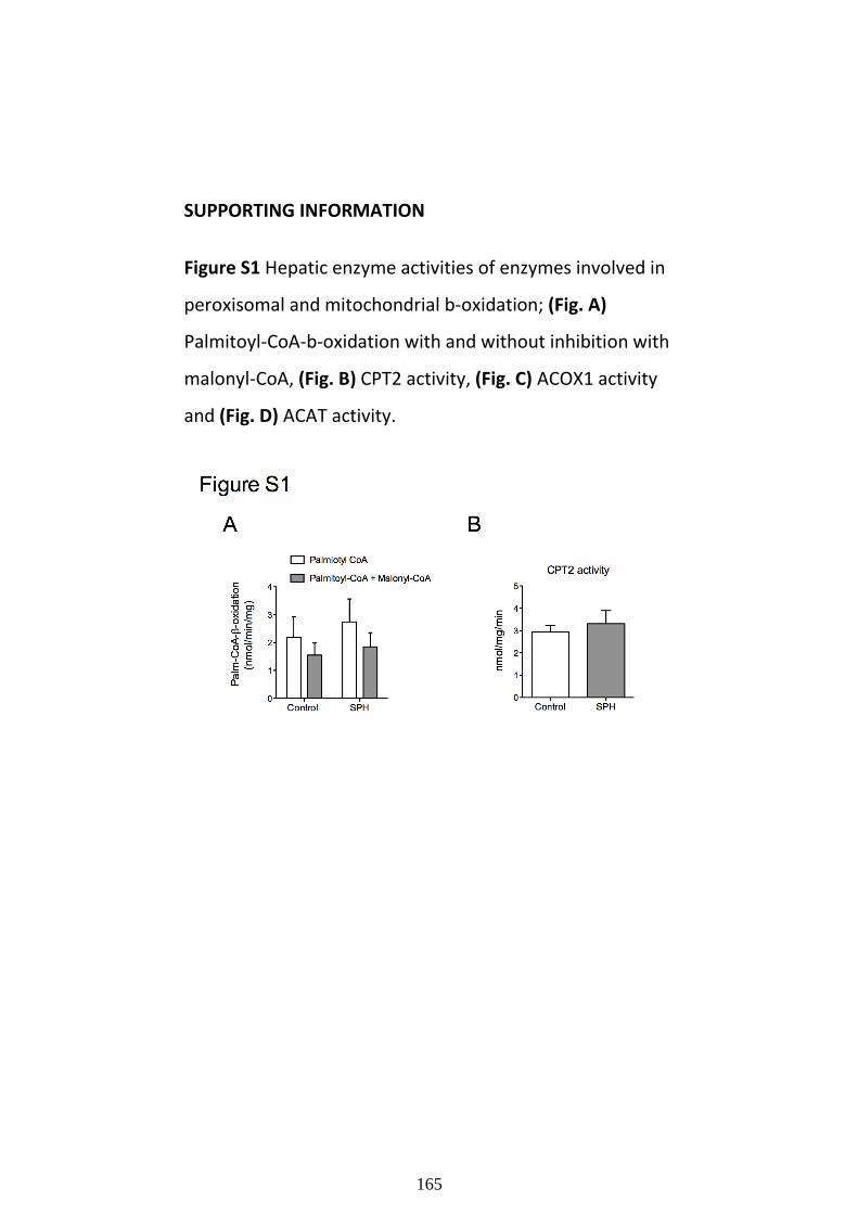
165
SUPPORTING INFORMATION
Figure S1 Hepatic enzyme activities of enzymes involved in
peroxisomal and mitochondrial b-oxidation; (Fig. A)
Palmitoyl-CoA-b-oxidation with and without inhibition with
malonyl-CoA, (Fig. B) CPT2 activity, (Fig. C) ACOX1 activity
and (Fig. D) ACAT activity.

166

167
Table S1
1Composition of the diets
2Ingredients 3Control 4SPH
Protein source
Casein 250 200
Salmon protein hydrolysate 0 50
Fat source
Soy oil 24 24
Lard 213 213
5Amino acid composition
Hydroxyproline 0 0.4
Histidine 6.4 5.4
Taurine 0 0.8
Serine 13 11
Arginine 7.5 7.1
Glycine 4.2 5.4
Aspartic acid 17 15
Glutamic acid 51 44
Threonine 9.4 8.3
Alanine 6.8 7.3
Proline 24 20
Lysine 18 17
Tyrosine 9.8 7.4
Methionine 6.0 5.3
Valine 14 13
Isoleucine 11 9.7
Leucine 21 19

168
Phenylalanine 12 9.0
SPH, salmon protein hydrolysate
1The diets were isonitrogenous, and contained 21 g protein per 100 g
diet
2Ingredients (g/kg diet)
3Amino acid composition (mg/g protein)
4Casein consisted of 84% protein and 0.2% fat
5SPH consisted of 91% protein
Other ingredients (g/kg diet): Cornstarch (105); Dyetrose (154);
Sucrose (117); Fiber (58); Dextrin/Cellulose (20); AIN-93G-MX
mineral
mix (41); AIN-93G-VX vitamin mix (12); L-Cysteine (3.5); Choline
bitartrate (17); tert-Butyl-hydroquinone (0.016)
Table S1 Composition and amino acid contents of the
diets.

169
Table S2 Overview of analysed genes.
Table S2
Gene names of analysed genes, primer/probe sets and analysed tissues
Gene Full name 1AoD no. Liver Heart Aorta
Acaca Acetyl-Coenzyme A carboxylase alpha Mm01304277_m1 x
Scd1 Stearoyl-Coenzyme A desaturase 1 Mm00772290_m1 x
Cat Catalase Mm00437992_m1 x
Mcp1 Monocyte chemoattractant protein Mm00441242 x x
Icam1 Intracellular adhesion molecule Mm00516023_m1 x x
Nos2 Nitric oxide synthase 2 Mm00440502_m1 x x
Sod1 Superoxide dismutase 1, soluble Mm01344233_g1 x
Sod2 Superoxide dismutase 2, mitochondrial Mm01313000_m1 x
Tnfa Tumor necrosis factor alpha Mm00443260_g1 x
Vcam1 Vascular cell adhesion molecule 1 Mm00443281 x x 1 Catalog number of custom TaqMan gene expression primer/probe sets from Applied Biosystems.

170
ACKNOWLEDGMENTS
We thank Kari Williams, Liv Kristine Øysæd, Randi Sandvik
and Svein Krüger for excellent technical assistance, and
Eline Milde Nevdal for assisting with the animal
experiment.
AUTHOR CONTRIBUTION
Conceived and designed the experiments: CP BB TB JS RKB
GC. Performed the experiments: RV MB SH TB SM GSG BH.
Analyzed the data: RV BB FD. Contributed
reagents/materials/analysis tools: CRS JEN. Wrote the
paper: CP RV BB PA RKB.
REFERENCES
1. Lloyd-Jones DM (2010) Cardiovascular risk prediction:
basic concepts, current status, and future
directions. Circulation 121: 1768–1777
2. Woollard KJ (2013) Immunological aspects of
atherosclerosis. Clinical Science 125: 221–235
3. Hansson GK, Robertson A-KL, Söderberg-Nauclér C
(2006) Inflammation and atherosclerosis annual review of

171
pathology: Mechanisms of disease. Annual Reviews 1 297–
329
4. van der Wal AC, Becker AE, van der Loos CM, Das PK
(1994) Site of intimal rupture or erosion of thrombosed
coronary atherosclerotic plaques is characterized by an
inflammatory process irrespective of the dominant plaque
morphology. Circulation 89: 36–44
5. Sabesin SM (1981) Lipid and lipoprotein abnormalities in
alcoholic liver disease. Circulation 64: III 72–84
6. Kleemann R, Kooistra T (2005) HMG-CoA reductase
inhibitors: effects on chronic subacute inflammation and
onset of atherosclerosis induced by dietary
cholesterol. Curr Drug Targets Cardiovasc Haematol
Disord5: 441–453
7. Rein D, Schijlen E, Kooistra T, Herbers K, Verschuren L, et
al. (2006) Transgenic flavonoid tomato intake reduces C-
reactive protein in human C-reactive protein transgenic
mice more than wild-type tomato. J Nutr136: 2331–2337
8. Kleemann R, Verschuren L, van Erk MJ, Nikolsky Y,
Cnubben NH, et al. . (2007) Atherosclerosis and liver
inflammation induced by increased dietary cholesterol

172
intake: a combined transcriptomics and metabolomics
analysis. Genome Biol 8 R200.
9. Singh U, Jialal I (2006) Oxidative stress and
atherosclerosis. Pathophysiology 13: 129–142
10. Ridker PM, Rifai N, Cook NR, Bradwin G, Buring JE
(2005) Non-HDL cholesterol, apolipoproteins A-I and B100,
standard lipid measures, lipid ratios, and CRP as risk factors
for cardiovascular disease in women. JAMA 294: 326–333
11. Harnedy PA, FitzGerald RJ (2012) Bioactive peptides
from marine processing waste and shellfish: A
review. Journal of Functional Foods 4: 6–24
12. Chalamaiah M, Dinesh Kumar B, Hemalatha R,
Jyothirmayi T (2012) Fish protein hydrolysates: proximate
composition, amino acid composition, antioxidant activities
and applications: a review. Food Chem 135: 3020–3038
13. Elvevoll EO, Eilertsen KE, Brox J, Dragnes BT, Falkenberg
P, et al. (2008) Seafood diets: hypolipidemic and
antiatherogenic effects of taurine and n-3 fatty
acids. Atherosclerosis 200: 396–402
14. Liaset B, Madsen L, Hao Q, Criaeles G, Mellgren G
(2009) Fish protein hydrolysate elevates plasma bile acids

173
and reduces visceral adipose tissue mass in rats. Biochim
Biophys Acta 1791: 254–262
15. Jacques H, Gascon A, Bergeron N, Lavigne C, Hurley C,
et al. . (1995) Role of dietary fish protein in the regulation
of plasma lipids. Can J Cardiol 11: Suppl G 63G–71G.
16. Zhang X, Beynen AC (1993) Influence of dietary fish
proteins on plasma and liver cholesterol concentrations in
rats. Br J Nutr 69: 767–777
17. Wergedahl H, Liaset B, Gudbrandsen OA, Lied E, Espe
M, et al. (2004) Fish protein hydrolysate reduces plasma
total cholesterol, increases the proportion of HDL
cholesterol, and lowers acyl- CoA:cholesterol
acyltransferase activity in liver of Zucker rats. Journal of
Nutrition 134: 1320–1327
18. Grimstad T, Bjorndal B, Cacabelos D, Aasprong OG,
Omdal R, et al. (2012) A salmon peptide diet alleviates
experimental colitis as compared with fish oil. Journal of
Nutritional Science 1: 1–8
19. Palinski W, Ord VA, Plump AS, Breslow JL, Steinberg D,
et al. (1994) ApoE-deficient mice are a model of lipoprotein
oxidation in atherogenesis. Demonstration of oxidation-
specific epitopes in lesions and high titers of autoantibodies

174
to malondialdehyde-lysine in serum. Arterioscler
Thromb 14: 605–616
20. Bjorndal B, Vik R, Brattelid T, Vigerust NF, Burri L, et al.
(2012) Krill powder increases liver lipid catabolism and
reduces glucose mobilization in tumor necrosis factor-alpha
transgenic mice fed a high-fat diet. Metabolism 61: 1461–
1472
21. Vigerust NF, Bjorndal B, Bohov P, Brattelid T, Svardal A,
et al. (2012) Krill oil versus fish oil in modulation of
inflammation and lipid metabolism in mice transgenic for
TNF-alpha. Eur J Nutr 52: 1315–1325
22. Strand E, Bjorndal B, Nygard O, Burrin L, Berge C, et al.
(2012) Long-term treatment with the pan-PPAR agonist
tetradecylthioacetic acid or fish oil is associated with
increased cardiac content of n-3 fatty acids in rat. Lipids in
Health and Disease 11: 82.
23. Berge RK, Flatmark T, Osmundsen H
(1984) Enhancement of long-chain acyl-CoA hydrolase
activity in peroxisomes and mitochondria of rat liver by
peroxisomal proliferators. Eur J Biochem 141: 637–644

175
24. Bremer J (1981) The effect of fasting on the activity of
liver carnitine palmitoyltransferase and its inhibition by
malonyl-CoA. Biochim Biophys Acta 665: 628–631
25. Skorve J, al-Shurbaji A, Asiedu D, Bjorkhem I, Berglund
L, et al. (1993) On the mechanism of the hypolipidemic
effect of sulfur-substituted hexadecanedioic acid (3-
thiadicarboxylic acid) in normolipidemic rats. J Lipid Res 34:
1177–1185
26. Madsen L, Rustan AC, Vaagenes H, Berge K, Dyroy E, et
al. (1999) Eicosapentaenoic and docosahexaenoic acid
affect mitochondrial and peroxisomal fatty acid oxidation in
relation to substrate preference. Lipids 34: 951–963
27. Field FJ, Albright E, Mathur S (1991) Inhibition of
acylcoenzyme A: cholesterol acyltransferase activity by
PD128O42: effect on cholesterol metabolism and secretion
in CaCo-2 cells. Lipids 26: 1–8
28. Cinq-Mars DC, Hu C, Kitts DD, Li-Chan EC
(2008) Investigations into inhibitor type and mode,
simulated gastrointestinal digestion, and cell transport of
the angiotensin i-converting enzyme-inhibitory peptides in
pacific hake (merluccius productus) fillet hydrolysate. J
Agric Food Chem 56: 410–419

176
29. Raatz SK, Silverstein JT, Jahns L, Picklo MJ (2013) Issues
of fish consumption for cardiovascular disease risk
reduction,. Nutrients 5: 1081–1097
30. Jung UJ, Torrejon C, Tighe AP, Deckelbaum RJ (2008) n-
3 Fatty acids and cardiovascular disease: mechanisms
underlying beneficial effects. Am J Clin Nutr 87: 2003S–
2009S
31. Duarte J, Vinderola G, Ritz B, Perdigon G, Matar C
(2006) Immunomodulating capacity of commercial fish
protein hydrolysate for diet
supplementation. Immunobiology 211: 341–350
32. Murakami S, Kondo-Otha Y (1998) Tomisawa
(1998) Improvement in cholesterol metabolism in mice
given chronic treatment of taurine and fed a high-fat
diet. Life Sciences 64: 83–91
33. Chen W, Matuda K, Nishimura N, Yokogoshi H
(2004) The effect of taurine on cholesterol degradation in
mice fed a high-cholesterol diet. Life Sciences. 74: 1889–
1898
34. Dembinska-Kiec A, Gryglewski RJ (1986) Contribution of
arachidonic acid metabolites to atherosclerosis. Wien Klin
Wochenschr 98: 198–206

177
35. Bjorndal B, Berge C, Ramsvik MS, Svardal A, Bohov P, et
al. (2013) A fish protein hydrolysate alters fatty acid
composition in liver and adipose tissue and increases
plasma carnitine levels in a mouse model of chronic
inflammation. Lipids Health Dis 12: 143.
36. Muoio DM, Way JM, Tanner CJ, Winegar DA, Kliewer
SA, et al. (2002) Peroxisome proliferator-activated
receptor-alpha regulates fatty acid utilization in primary
human skeletal muscle cells. Diabetes 51: 901–909
37. Kersten S, Mandard S, Escher P, Gonzalez FJ, Tafuri S, et
al. (2001) The peroxisome proliferator-activated receptor
alpha regulates amino acid metabolism. FASEB J 15: 1971–
1978
38. Delerive P, Fruchart JC, Staels B (2001) Peroxisome
proliferator-activated receptors in inflammation control. J
Endocrinol 169: 453–459
39. Zhu CF, Li GZ, Peng HB, Zhang F, Chen Y, et al.
(2010) Treatment with marine collagen peptides modulates
glucose and lipid metabolism in Chinese patients with type
2 diabetes mellitus. Appl Physiol Nutr Metab. 36: 797–804

178
Chapter 4
MicroRNA expression profiles of tissues
involved in cholesterol metabolism and
atherosclerosis from athero-prone and
athero-resistant mouse models
Presented at meetings:
(1) Manzini S., Busnelli M., Chiara M., Parolini C., Dellera
F., Ganzetti GS, Horner DS and Chiesa G. MiRNA
expression profiling in normolipidemic wild-type,
hyperlipidemic LDLR knockout and hypolipidemic
PCSK9 knockout mice. 84th
EAS Congress, May 29 -
June 1, 2016; Innsbruck (Austria).
(2) Dellera F., Manzini S., Busnelli M., Chiara M., Parolini
C., Ganzetti GS, Horner DS, Chiesa G. MicroRNA
expression profiles of tissues involved in cholesterol
metabolism from athero-prone and athero-resistant
mouse models. ABCD National Ph.D. Meeting, April 7
– 9, 2016; Salerno (Italy).
(3) Manzini S., Busnelli M., Chiara M., Parolini C., Dellera
F., Ganzetti GS, Horner DS, Chiesa G. Genome-wide
identification of microRNAs involved in the regulation
of cholesterol metabolism and atherosclerosis
development. 17th
ISA Congress, May 23 - 26, 2015;
Amsterdam (Netherlands).

179

180
INTRODUCTION AND AIM
MiRNAs are small non-coding RNAs which act as potent
post-transcriptional regulators of gene expression. MiRNAs
typically control the expression of their target genes by
imperfect base-pairing to 3’ UTR regions of mRNAs, thereby
inducing repression of the target mRNA (1, 2). In recent
years, several miRNAs have been directly linked to the
regulation of key genes involved in cholesterol metabolism
and atherosclerosis development, as extensively discussed
in the introduction of this thesis.
Despite these recent advances, what is lacking is a global
approach to the comprehension of the interplay between
genes, miRNA-dependent regulation of those genes and
the phenotypic effects.
The current study was aimed to identify miRNA expression
profiles of tissues involved in cholesterol intake,
processing, production, metabolism and in atherosclerosis
development harvested from LDLR -/-
(LDLR KO), PCSK9 -/-
(PCSK9 KO) and wild-type C57BL/6 mice after chow
(standard) or a high fat feeding (3, 4). MiRNAs showing
differential expression between genotypes/dietary

181
regimens/tissues have been subjected to detailed in silico
target prediction analysis, with particular attention being
paid to identify members of transcriptional regulatory
circuits potentially involved in cholesterol metabolism and
regulated by miRNAs.
This project is ongoing and we are mainly validating the
results of target prediction analysis by performing in vitro
experiments of cell co-transfection with appropriate
reporter vectors and synthetic microRNA mimics. The
identification of targets for dysregulated microRNAs is
expected to provide insights into transcriptional circuits
subject to changes in perturbations of cholesterol
metabolism and could represent a crucial step towards the
identification of candidate targets for personalized
therapeutic strategies.

182
MATERIALS AND METHODS
Experimental design: animals and diet
The study was conducted according to national (D.L. 116,
G.U. Suppl. 40, February 18, 1992, Circolare No. 8, G.U July
1994) and international laws and policies (EEC Council
Directive 2010/63, September 22, 2010: Guide for the Care
and Use of Laboratory Animals, United States National
Research Council, 2011). The Italian Ministry of Health
approved the protocol.
Wild-type C57BL/6, LDLR -/-
(prone to atherosclerosis
development) and PCSK9 -/-
mice (resistant to
atherosclerosis development) (3, 4) were purchased from
Charles River (Calco, Italy) and housed in the animal facility
for the whole duration of the treatment (16 weeks).
Starting from the sixth week from birth, animals have been
fed with either standard (Chow) or Western-type diet (21%
fat, 0.2% cholesterol). The latter has a high fat and
cholesterol content and is commonly used to induce the
development of dyslipidemia and/or plaque development
in susceptible genotypes. Each experimental group was
made up of 10 animals. (Fig. 1).

183
Fig. 1 Experimental design of the study
Blood and tissue harvesting
Blood samples have been collected before the start of
dietary regimens and at sacrifice (16 weeks) from the
mouse retro-orbital plexus into tubes containing 0.1% (w/v)
EDTA after an overnight fast (Fig. 1) Blood samples were

184
chilled on ice for at least 15 minutes and stored at -80°C
until analyses.
At sacrifice, heart, aorta, liver, small intestine (duodenum,
jejunum, ileum), white adipose tissue and brain have been
harvested and properly stored for the subsequent
molecular and biochemical analyses.
Plasma lipid profile
A general characterization of the lipid profile of mice has
been performed on plasma collected at sacrifice by
enzymatically measuring total cholesterol and triglycerides
(ABX Diagnostics, France).
Atherosclerosis development
The area covered by atherosclerotic plaques has been
quantified at the aortic sinus. Serial cryosections (7 mm
thick) of the aortic sinus were cut. Approximately 25 slides
with 3 cryosections/slide were obtained, spanning the
three cusps of the aortic valves. Every fifth slide was fixed
and stained with hematoxylin and eosin (Bio-Optica,
Milano, Italy) to detect plaque area. Plaque area was
calculated as the mean area of those sections showing the

185
three cusps of the aortic valves. To acquire and process
digital images, an Aperio ScanScope GL Slide Scanner
(Aperio Technologies, Vista, CA, USA), equipped with a
Nikon 206/0.75 Plan Apochromat objective producing a
0.25 mm/pixel scanning resolution with a 406
magnification, and the Aperio ImageScope software
(version 8.2.5.1263) was used.

186
Total RNA extraction and construction of cDNA libraries
for high throughput sequencing
Total RNA has been extracted from the tissues and organs
collected (NucleoSpin® miRNA columns, Mackerey-Nagel,
Germany) and libraries suitable for use with Illumina
Genome Analyzer IIx (Illumina, San Diego, CA, USA) have
been constructed. During the generation of cDNA libraries,
techniques have been applied to enrich in low molecular
weight cDNA species, maximising the miRNA component of
libraries. All samples have been then sequenced with the
high throughput sequencer Illumina Genome Analyzer IIx
(analysis performed by IGA Technology Services S.r.l.,
Udine, Italy) (5).
Analysis of miRNA expression profiles and target
prediction for differentially expressed miRNAs
For each experimental condition (diet/genotype/tissue)
small RNA sequence reads have been mapped to known
mature and precursor miRNA sequences from miRBase
using the Short Oligonucleotide Analysis Package SOAP and
outputs have been parsed using custom scripts prepared in
the Python/PERL programming languages to obtain digital

187
expression values for both canonical mature miRNA and
similar, incorrectly processed sequences. After exclusion of
reads corresponding to RNA and rRNA degradation
products, evaluation of differential expression has been
performed within a statistical framework based on the
negative binomial distribution and implemented in the
Bioconductor packages edgeR and DEseq for the R
language. Further statistical tests have been performed
using the same instruments to confirm consistency of
expression pattern changes between individual animals.
Subsequently, all reads have been mapped to the reference
murine genome using SOAP and preliminary candidate
miRNA/miRNA* pairs have been identified using a simple,
but effective, algorithm previously developed by Prof.
David Horner (Università degli Studi di Milano, Milano,
Italy). Genomic sequences corresponding to loci defined by
these preliminary candidate pairs have been extracted and
their predicted secondary structures modeled using RNA
fold software and custom scripts to confirm the expected
juxtaposition of the candidate miRNA/miRNA* sequences.
Cases fulfilling these conditions have been further
evaluated using a Support Vector machine trained to

188
recognize vertebrate miRNA precursors and simple
statistical tests to demonstrate miRNA-like behaviour of
small RNA mapping patterns in candidate precursor loci
(strand specificity, clustering of the majority of reads in the
regions of the canonical miRNA and miRNA* sequences).
Candidates passing these tests have been evaluated for
differential expression among experimental conditions.
Two on-line databases of established target predictions,
miRDB (http://www.mirdb.org/miRDB) and TargetScan
(http://www.targetscan.org), have been downloaded and
used to identify all predicted targets for differentially
expressed known miRNAs. Sets of predictions made by the
two methods have been then used to perform KEGG
pathway enrichment analyses
(http://www.genome.jp/kegg/pathway) in order to identify
physiological processes and pathways enriched among
target predictions and thus probable physiological target
according to the accepted model of multiple miRNA target
associations.

189
Validation of candidate miRNA targets identified by high-
throughput sequencing and bioinformatics analyses: in
vitro experiments
In silico prediction of target mRNAs needed to be
empirically confirmed. From the group of candidate targets
of the miRNAs resulted DE, we concentrated our attention
on those playing a role in cholesterol metabolism and
atherosclerosis development (Tab. 1).
The 3' UTR of each of these genes containing the miRNA
response element has been cloned into pmirGLO Dual-
Luciferase miRNA Target Expression Vector (Promega Italia,
Milano, Italy) (Fig.2, upper figure). In parallel, as a control,
a vector has been constructed with the same miRNA
responsive element inserted, but containing a sequence
mismatch.
PmiRGLO vector is designed to quantitatively evaluate
miRNA activity by the insertion of miRNA target sites
downstream or 3´ of the firefly luciferase gene (luc2) (Fig.
2, upper figure). Firefly luciferase is the primary reporter
gene. Reduced firefly luciferase expression indicates the
binding of endogenous or introduced miRNAs to the cloned
miRNA target sequence (Fig. 2, lower figure). This vector is

190
based on dual-luciferase technology, with luc2 used as the
primary reporter to monitor mRNA regulation
and Renilla luciferase (hRluc-neo) (Fig. 2, upper figure)
acting as a control reporter for normalization and selection.

191
Fig. 2 PmirGLO Dual-Luciferase miRNA Target Expression Vector
(upper figure) and mechanism of action for in vitro miRNA target
validation (lower figure).
Main abbreviations: luc2, Firefly luciferase; hRluc-neo, Renilla
luciferase; Ampr, β−lactamase coding region; MCS, multiple cloning
site.

192
For in vitro target validation, cell line NIH 3T3 (batch
number 61062005, ATCC, USA) has been chosen. NIH 3T3
cells were cultured in DMEM high glucose with stable L-
glutamine (Lonza, USA), supplemented with 10% fetal calf
serum (Euroclone, Milano, Italy) and 1X
penicillin/streptomycin (Euroclone, Milano, Italy). Cell
cultures were incubated at 37°C in a humidified
atmosphere with 5% CO2.
The experiments needed to investigate the effect of
selected DE miRNAs (Tab. 1) on their candidate target
mRNAs are currently ongoing in our laboratory. In detail,
NIH 3T3 cells will be seeded in 24-well plates. After 24
hours, at 80-90% of confluence, cells will be transiently co-
transfected with the selected miRNA mimic (Exiqon,
Denmark) and the vector pmiRGLO containing the
candidate responsive sequence or with the same miRNA
mimic and the vector containing the mismatched target
sequence, used as a control. Co-transfection will be
performed by using Lipofectamine 3000 diluted in Opti-
MEM (both purchased from Invitrogen, Life Technologies
Italia, Monza, Italy). Hsa-miR-21 and its known responsive
sequence cloned into the vector will be used as a positive

193
control. After 4 h of incubation, the transfection medium
will be replaced with fresh complete growth medium. At 48
hours after transfection, cells will be collected in order to
perform Luciferase reporter assay using Dual-Luciferase®
Reporter (DLR™) Assay System (Promega Italia, Milano,
Italy) according to standard protocols.

194
RESULTS
Atherosclerosis evaluation: only LDLR-/-
mice showed
increased atherosclerotic plaque development
Plaque area has been quantified at the aortic sinus. As
expected, only LDLR-/-
mice showed atherosclerosis
development. Atherosclerotic lesions were exacerbated by
Western-type diet, as shown in figure 3 (Fig. 3).
Fig. 3 Atherosclerotic plaque development in C57BL/6, PCSK9 -/-
(PCSK9 KO) and LDLR -/-
(LDLR KO) mice fed chow or Western diet
for 16 weeks.
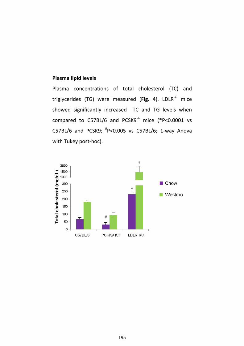
195
Plasma lipid levels
Plasma concentrations of total cholesterol (TC) and
triglycerides (TG) were measured (Fig. 4). LDLR-/-
mice
showed significantly increased TC and TG levels when
compared to C57BL/6 and PCSK9-/-
mice (*P<0.0001 vs
C57BL/6 and PCSK9; #P<0.005 vs C57BL/6; 1-way Anova
with Tukey post-hoc).

196
Fig. 4 Plasma total cholesterol and triglycerides in C57BL/6, PCSK9 -
/- (PCSK9 KO) and LDLR
-/- (LDLR KO) mice fed chow or Western diet
for 16 weeks (*P<0.0001 vs C57BL/6 and PCSK9; #P<0.005 vs
C57BL/6; 1-way Anova with Tukey post-hoc).

197
MiRNA expression profiles in tissues involved in
cholesterol metabolism
At sacrifice, mouse tissues and organs involved in
cholesterol metabolism such as aorta, liver, small intestine
(duodenum, jejunum, ileum), white adipose tissue and
brain have been collected and stored for the analysis of
miRNA expression profiles and validation experiments.
Figure 5 (Fig. 5) shows miRNAs resulted as DE by the high
throughput sequencing performed and the following
bioinformatic analyses. A high number of DE miRNAs was
observed among genotypes in several tissues with both
dietary regimens, although after Western diet the overall
DE number was lower than after Chow diet. The most
relevant DE number has been observed at both Chow and
Western diet in the comparison between C57BL/6 and
LDLR-/-
mouse aortas, which are sites of atherosclerosis
development.

198
Fig. 5 Number of miRNAs resulted as DE among genotypes in the
considered tissues from animals fed both Chow and Western diet
Abbreviations: Abd. WAT, abdominal white adipose tissue.
In silico target prediction analysis of DE miRNAs
MiRNAs which resulted DE in all the comparisons
underwent detailed in silico target prediction analyses.
Target mRNAs were predicted by miRDB/Targetscan
softwares, then functional enrichment analysis was run for
each comparison in order to provide insights into the
biological interpretation of the obtained pattern of
candidate genes.
In particular, focusing on the comparison between the two
diets, aggregated KEGG pathways showed overlapping and

199
recurring enriched pathways, that are the chemokine
signaling pathway, the cytokine-cytokine receptor
interaction and the insulin signaling pathway, as shown in
figure 6 (Fig. 6).
Among the genes annotated in these pathways, we
identified targets known for their role in lipid
metabolism/cardiovascular disease as well as novel
potential genes of interest, such as Leukemia inhibitory
factor receptor (Lifr) and Braf transforming gene (Braf) (Fig.
7).

200
Fig. 6 Schematic summary showing the recurring pathways resulted
from KEGG pathway mapping performed on our target genes in the
comparison between chow - fed and Western–fed animals.

201
Fig. 7 Detail of target genes annotated in the shared KEGG
pathways
Furthermore, as a complementary approach, we selected
genes that are known to be involved in cholesterol and lipid
metabolism and investigated if they were predicted as
targets of DE miRNAs. Table 1 (Tab. 1) shows some of them
and their associated DE miRNAs. The comparisons/tissues
in which these miRNAs resulted DE are indicated as well.

202
Tab. 1 Differentially expressed miRNAs and candidate targets
involved in cholesterol metabolism and atherosclerosis
development
Abbreviations: WAT, white adipose tissue

203
In vitro validation of candidate miRNA targets
In vitro validation of the interactions between miRNA-
candidate target listed in Tab.1 are currently ongoing. To
experimentally test whether selected miRNAs directly
target the 3’UTR of their candidate gene in vitro, NIH 3T3
cells will be co-transfected with reporter constructs along
with a synthetic mimic of miRNAs, as explained in detail in
the section “Material and Methods”. For successful
predictions, the expected result is a diminished luciferase
expression by cells transfected with miRNA mimics and
pmirGLO vector containing the miRNA-responsive element,
compared to controls.
Future steps
The future perspectives of this study include:
- the conclusion of in vitro validation experiments, in order
to experimentally confirm the interaction between DE
miRNAs and their candidate targets involved in cholesterol
metabolism.

204
- in depth examination of pathway enrichment analyses,
with a particular attention paid to signaling pathways
linked to upregulated or downregulated DE miRNAs.
The aim is the identification of members of regulatory
pathways subject to changes in perturbations of
cholesterol metabolism. These targets could be crucial for
the development of panels of biomarkers for disease
development.

205
REFERENCES
1. Ambros V (2004). The functions of animal
microRNAs. Nature 431:350–355.
2. Filipowicz W, Bhattacharyya SN, Sonenberg N (2008).
Mechanisms of post-transcriptional regulation by
microRNAs: are the answers in sight?. Nat Rev Genet
9:102–114.
3. Knowles JW, Maeda N (2000). Genetic modifiers of
atherosclerosis in mice. Arterioscler Thromb Vasc
Biol 20:2336 –2345.
4. Denis M, Marcinkiewicz J, Zaid A, et al (2012). Gene
inactivation of proprotein convertase subtilisin/kexin
type 9 reduces atherosclerosis in mice. Circulation
125:894–901.
5. Harismendy O, Ng PC, Strausberg RL, Wang X,
Stockwell TB, Beeson KY, Schork NJ, Murray SS, Topol
EJ,Levy S, Frazer KA (2009). Evaluation of next
generation sequencing platforms for population
targeted sequencing studies. Genome Biol 10(3):R32.

206
Chapter 5
Conclusions and
Future perspectives

207

208
During my PhD program I have contributed to the
realization of different research projects, all related to the
study of atherosclerosis and cholesterol metabolism
through the use of animal models.
Among these, I have focused my PhD thesis mainly on two
of the studies I co-authored (Chapter 2 and 3), one as first
author. Moreover, a third study, still ongoing and whose
results have been presented at National and International
conferences, has been presented as well, even though
further investigations are needed to complete it (Chapter
4).
The first project discussed (Chapter 2) is focused on the
effect of L-homoarginine, an L-arginine homologue derived
from lysine, on neointimal hyperplasia, a pathological
condition which can occur at sites of subclinical
atherosclerosis or after angioplasty interventions. For the
first time, we have demonstrated that in vivo
administration of homoarginine in a rat model of carotid
balloon injury was able to inhibit neointimal formation, at
least in part, by inhibiting vascular cell proliferation and by
increasing both arginine availability and nitric oxide

209
production, suggesting homoarginine as a potential
therapeutical option for cardiovascular protection.
The second study presented (Chapter 3) is instead focused
on the study of the effect of a salmon protein hydrolysate
on atherosclerosis development in apo E-deficient mice, on
the basis of previous evidences indicating that fish protein
peptides and hydrolysates are able to exert beneficial
effects on cardiovascular risk factors. Mice fed a high-fat
diet with 5% salmon protein hydrolysate for 12 weeks
showed a significantly reduced atherosclerotic plaque area
in the aortic arch and aortic sinus versus controls. The
effect of this dietary supplementation was shown to be
mediated by the inhibition of systemic inflammation
activation and it seemed to be independent of changes in
plasma lipid levels.
Finally, the fourth chapter of this thesis presents the results
obtained to date regarding the characterization of
microRNA expression patterns in tissues involved in
cholesterol metabolism and atherosclerosis, harvested
from LDLR-deficient, PCSK9-deficient and wild-type
C57BL/6 mice after chow or a high fat feeding. Briefly, we
have identified the microRNAs which resulted DE in our

210
experimental conditions (different genotypes/tissues/diet)
by high-throughput sequencing and bioinformatics
analyses. A detailed in silico target prediction study was
then performed to identify the genes regulated by DE
microRNAs. We are currently validating the in silico analysis
results by cell transfection with appropriate reporter
vectors and microRNA mimics.
The identification of targets for dysregulated microRNAs is
aimed at highlighting transcriptional regulatory circuits
subject to changes in atherosclerosis and in conditions of
perturbation of cholesterol metabolism.
The prevention and management of atherosclerosis and
disturbs of cholesterol metabolism are still an open
challenge.
In order to gain more insight into the disease, approaches
at different levels have been taken and this thesis
presented some of them.
Through the use of animal models, we demonstrated that
natural compounds or treatments based on novel
cardiovascular risk factors may exert a protective role in

211
atherosclerosis development and in its clinical
complications, such as neointimal hyperplasia.
In particular, our studies suggest L-homoarginine and
protein sources derived from salmon as
therapeutic/nutraceutical options for cardiovascular
protection, even though further investigations are needed
in order to assess the possibility to translate the obtained
results in humans.
Finally, miRNAs have been demonstrated to play an
important, although still vaguely characterized, role on
atherosclerosis formation and cholesterol metabolism
regulation (1). Recent evidences and our preliminary data
suggest that the identification of a miRNA profile could
probably represent an approach to detect vulnerable
patients and disease states (2).

212
Targeting miRNAs may represent a new pharmacological
tool to modulate the atherosclerotic process. Clearly,
further studies are encouraged to help us understand the
role of miRNAs in atherosclerosis and dyslipidemias.
REFERENCES:
1. Andreou I, Sun X, Stone PH, Edelman ER, Feinberg MW
(2015). MiRNAs in atherosclerotic plaque initiation,
progression, and rupture. Trends Mol Med 21: 307–318.
2. Ren J, Zhang J, Xu N, Han G, Geng Q, Song J, et al (2013).
Signature of circulating microRNAs as potential biomarkers
in vulnerable coronary artery disease. PLoS One 8: e80738.

213

214
Publications

215
216
1. Dellera F, Ganzetti GS, Froio A, Manzini S, Busnelli
M, Meinitzer A, Sirtori CR, Chiesa G, Parolini C (2016). L-
homoarginine administration reduces neointimal
hyperplasia in balloon-injured rat carotids. Thromb.
Haemost 116(2).
2. Arnaboldi F, Busnelli M, Cornaghi L, Manzini S,
Parolini C, Dellera F, Ganzetti GS, Sirtori CR, Donetti E,
Chiesa G (2015). High-density lipoprotein deficiency in
genetically modified mice deeply affects skin morphology:
A structural and ultrastructural study. Exp Cell Res
338(1):105-12.
3. Parolini C, Busnelli M, Ganzetti GS, Dellera F, Manzini
S, Scanziani E, Johnson JL, Sirtori CR, Chiesa G (2014).
Magnetic resonance imaging visualization of vulnerable
atherosclerotic plaques at the brachiocephalic artery of
apolipoprotein E knockout mice by the blood-pool contrast
agent B22956/1. Mol Imaging 13.
4. Parolini C, Vik R, Busnelli M, Bjørndal B, Holm S,
Brattelid T, Manzini S, Ganzetti GS, Dellera F, Halvorsen B,
Aukrust P, Sirtori CR, Nordrehaug JE, Skorve J, Berge RK,
Chiesa G (2014). A salmon protein hydrolysate exerts lipid-

217
independent anti-atherosclerotic activity in ApoE-deficient
mice. PLoS One 9(5).
5. Vik R, Busnelli M, Parolini C, Bjørndal B, Holm S,
Bohov P, Halvorsen B, Brattelid T, Manzini S, Ganzetti GS,
Dellera F, Nygård OK, Aukrust P, Sirtori CR, Chiesa G, Berge
RK (2013). An immunomodulating fatty acid analogue
targeting mitochondria exerts anti-atherosclerotic effect
beyond plasma cholesterol-lowering activity in apoe(-/-)
mice. PLoS One 8(12):e81963.