role of transverse abdominis plane block in laparoscopic … · his guidance it would have been...
TRANSCRIPT
Role of Transverse Abdominis Plane
Block in Laparoscopic Donor
Nephrectomy
Thesis to be submitted for the degree of
Doctor of Medicine
at the University of Leicester
by
Umasankar Mathuram Thiyagarajan MBBS, M.S (Gen
Surgery), MRCSEd.
Division of Transplant Surgery
Department of Infection, Immunity and Inflammation
University of Leicester
2016

1
The candidate confirms that the work submitted is his own and that appropriate credit has
been given where reference has been made to the work of others.
This copy has been supplied on the understanding that it is copyright material and that no
quotation from the thesis may be published without proper acknowledgement.
© 2016 The University of Leicester and Umasankar Mathuram Thiyagarajan

2
ABSTRACT
Post-operative wound pain is a disincentive to potential live kidney donors. The
transverse abdominis plane (TAP) block is a technique where the local anaesthetic
agent is given to block the afferent nerves of the abdominal wall. The safety and
efficacy of this technique is well established for other surgical procedures.
The TAP block technique is shown to be beneficial and reduced the postoperative
morphine requirement in lower abdominal surgeries such as hysterectomy,
appendicectomy, and caesarean section. A similar incision is being used for retrieval of
kidney after laparoscopic donor nephrectomy. This technique has never been tested
before in laparoscopic donor nephrectomy patients; hence the aim of thesis was to
establish the efficacy of TAP block in reducing the pain and assessing its impact on
reducing total morphine requirement.
An initial retrospective study showed a potential of TAP block to reduce postoperative
pain and cumulative postoperative morphine requirements. Thereafter based on these
results, a double blinded randomised placebo controlled trial was setup. The
randomised trial has demonstrated that a TAP block with bupivacaine reduced early
morphine requirement at 6 hours. But, there was no difference seen in the total
postoperative morphine usage.
The visual analogue pain score in TAP block group with bupivacaine was significantly
lower on day 1 and 2 after surgery. Further cytokine assay in this randomised study
showed no difference in the plasma cytokine levels at 6, 24 and 48 hours after donor
nephrectomy.

3
Hence the TAP block technique appears to be a safe and effective method of
postoperative pain in laparoscopic donor nephrectomy patients. The study thus
established the contributory role of TAP block in multimodal analgesia. This can be an
alternative to post-operative wound infiltration with local anaesthetic agents.

4
ACKOWLEDGEMENTS
Firstly I would like to thank my supervisors Professor Michael Nicholson and Mr. Atul
Bagul, without whom this work would not have been possible. Professor Nicholson
gave me an opportunity to conduct this research with necessary supports. I sincerely
thank Mr. Bagul for his continuous and invaluable scientific, technical support; without
his guidance it would have been harder to complete my project successfully. I also
would like to thank Ms. Sarah Hosgood for her assistance on conducting this trial.
I would like to thank all members of Professor Nicholson’s lab for their support and
assistance. I also would like to thank my colleagues Mr. Ismail Mohammed, Mr.
Wisam Ismail for their help during the project.
Finally and most importantly I would like to thank my wife Amirthavarshini for her
kindness, cooperation and loving support throughout without which I could have not
completed this thesis successfully.

5
PUBLICATIONS AND PRESENTATIONS
Publications:
1) Pain Management in Laparoscopic Donor Nephrectomy- A Review- Pain
Research and Treatment. 2012-doi:10.1155/2012/201852.
2) Randomized clinical trial of transversus abdominis plane block versus placebo
control in live-donor nephrectomy. Transplantation. 2012; 94(5):520-5.
Published Abstracts:
1) Efficacy of transversus abdominis plane block in laparoscopic live donor
nephrectomy – A single centre experience. International Journal of
Surgery; 2012:10(8) S84.
2) Double blinded randomised controlled trial on TAP block in laparoscopic
live donor nephrectomy patients. British Journal of Surgery 2012; 99
(S4): 17–18.
Presentations:
1) Efficacy of transverse abdominis plane block in laparoscopic live donor
nephrectomy- a single centre experience. Association of Surgeons of Great
Britain and Ireland (ASGBI), Liverpool -2012.
2) Double blinded randomised controlled trial on TAP block in laparoscopic live
donor nephrectomy patients. Society of Academic and Research Surgery
(SARS), Nottingham- 2012.

6
ABBREVIATIONS
ACTH adrenocorticotropic hormone
ADEs adverse effects
BMI body mass index
CIT cold ischemia time
CPSP chronic post surgical pain
COX cyclooxygenase
CNS central nervous system
CXCL C-X-C-motif ligand
DBD donation after brain death
DCD donation after cardiac death
DOP delta opioid receptor
ESRF end stage renal failure
FDA food and drug administration
HALN hand assisted laparoscopic nephrectomy
HARN hand assisted retroperitoneal nephrectomy
HLA human leukocyte antigen
IASP international association for study of pain
IL interleukin
KOP kappa opioid receptor
LA local anaesthetics
LDN live donor nephrectomy
LTDN laparoscopic transperitoneal donor nephrectomy
LLDN laparoscopic live donor nephrectomy
MAC membrane attacking complex

7
MODN mini open donor nephrectomy
MOP mu opioid receptor
NHS national health service
NHSBT national health service blood and transfusion
NRM nucleus raphe magnus
NRPG nucleus reticularis paragigantocellularis
NSAIDS non-steroidal anti-inflammatory drugs
ODN open donor nephrectomy
PAG periaqueductal grey
PCA patient controlled analgesia
PCAS patient controlled analgesia system
PG prostaglandin
PNL percutaneous nephrolithotomy
PO per oral
PONV postoperative nausea and vomiting
QDS quarter die sumendus
RDN robotic donor nephrectomy
RADN robotic assisted donor nephrectomy
TAP transversus abdominis plane
TNF tumour necrosis factor
8
Contents
CHAPTER 1 Aims and Objectives ............................................................................................................... 12
1 Aims: .......................................................................................................................................... 13
1.1 Objectives: .................................................................................................................................. 13
CHAPTER 2 Introduction: Renal Transplantation and Live Kidney donation ........................ 16
2 Introduction - Renal transplantation ........................................................................................... 17
2.1 History of early transplantation models: .................................................................................... 17
2.1.1 History of kidney transplantation: .............................................................................................. 18
2.1.2 Immunosuppression and the modern era of kidney transplantation: .......................................... 19
2.1.3 Transplant activity in the UK: .................................................................................................... 21
2.1.4 Cost effectiveness of renal transplantation: ................................................................................ 22
2.2 Why live kidney donation: ......................................................................................................... 23
2.2.1 Advantages of live donation: ...................................................................................................... 25
2.2.2 Live Donor Nephrectomy (LDN): .............................................................................................. 26
2.2.3 The technique of open donor nephrectomy: ............................................................................... 27
2.2.4 Mini Open Donor Nephrectomy (MODN): ................................................................................ 28
2.2.5 Hand Assisted Laparoscopic Nephrectomy (HALN): ................................................................ 29
2.2.6 Hand Assisted Retroperitoneal Nephrectomy (HARN): ............................................................ 30
2.2.7 Robotic Donor Nephrectomy (RDN): ........................................................................................ 31
2.3 Laparoscopic Transperitoneal Donor Nephrectomy (LTDN): ................................................... 32
2.3.1 The technique of laparoscopic donor nephrectomy: ................................................................... 33
2.4 Figure 2.1: Port sites in right laparoscopic donor nephrectomy ................................................ 35
2.4.1 Benefits of laparoscopic donor nephrectomy: ............................................................................ 36
2.4.2 Disadvantages of laparoscopic technique: .................................................................................. 38
2.5 Conclusion: ................................................................................................................................. 39
CHAPTER 3 Review of Literature on Pain Management after Laparoscopic Donor
Nephrectomy ........................................................................................................................................................ 40
3 Introduction: ............................................................................................................................... 41
3.1 Pain after LLDN: ........................................................................................................................ 42
3.1.1 Postoperative pain and its implications: ..................................................................................... 44
3.2 Multimodal approach in LLDN: ................................................................................................. 46
3.2.1 Non-steroidal anti-inflammatory drugs (NSAIDS): ................................................................... 46
3.2.2 Opioids: ...................................................................................................................................... 49

9
3.2.2.1 Opioids analgesia as patient controlled analgesia: ..................................................................... 50
3.2.3 Epidural analgesia: ..................................................................................................................... 52
3.2.4 Neuroaxial techniques: ............................................................................................................... 54
3.2.5 Continuous infusion of local anesthetic agents: ......................................................................... 55
3.2.6 Long acting local anesthetics agents: ......................................................................................... 57
3.2.7 Liposomal bupivacaine prolonged-release formulation in the surgical setting .......................... 59
3.2.8 Other methods: ........................................................................................................................... 60
3.3 Transversus abdominis plane (TAP) block: ............................................................................... 61
3.3.1 TAP Block technique: ................................................................................................................ 62
3.4 Conclusion: ................................................................................................................................. 63
CHAPTER 4 Retrospective Study on Transversus Abdominis Plane Block in
Laparoscopic Live Donor Nephrectomy ................................................................................................. 64
4 Introduction ................................................................................................................................ 65
4.1 Study population and methods: .................................................................................................. 66
4.2 Table 4.1: Demography of the study population ........................................................................ 67
4.3 Results: ....................................................................................................................................... 67
4.4 Table 4.2: Patient controlled analgesia scoring (PCAS) ............................................................ 68
4.5 Figure 4.1: Cumulative usage of morphine between TAP and No TAP group .......................... 70
4.6 Table 4.3: Pain and sedation scoring between the TAP block versus No TAP group ................ 72
4.7 Figure 4.2: Pain scores between TAP and No TAP group ......................................................... 73
4.8 Figure 4.3: Postoperative sedation score .................................................................................... 74
4.9 Figure 4.4: PCAS usage.............................................................................................................. 76
4.10 Figure 4.5: Hospital stay ............................................................................................................. 77
4.11 Discussion: ................................................................................................................................. 78
4.12 Conclusion .................................................................................................................................. 82
CHAPTER 5 A Randomised Clinical Trial of TAP Block in Laparoscopic Live Donor
Nephrectomy ........................................................................................................................................................ 83
5 Randomised clinical trial of TAP block versus placebo control in laparoscopic live
donor nephrectomy ................................................................................................................................. 84
5.1 Introduction ................................................................................................................................ 84
5.2 Recent studies: ............................................................................................................................ 85
5.3 The trial: ..................................................................................................................................... 85
5.3.1 Methods ...................................................................................................................................... 86
5.3.2 CONSORT Diagram .................................................................................................................. 87
5.3.3 Table 5.1 Demography ............................................................................................................... 87
5.3.4 Anaesthetic Protocol ................................................................................................................... 89

10
5.4 TAP block procedure .................................................................................................................. 89
5.5 Surgical technique ...................................................................................................................... 90
5.6 Peri-operative protocol ............................................................................................................... 90
5.6.1 Post-operative care ..................................................................................................................... 91
5.7 Outcome measures ...................................................................................................................... 91
5.7.1 Primary end points: ..................................................................................................................... 91
5.7.2 Secondary end points: ................................................................................................................. 91
5.8 Statistical analysis ...................................................................................................................... 93
5.9 Results: ....................................................................................................................................... 94
5.9.1 Table 5.2 - Visual analogue score .............................................................................................. 95
5.9.2 Table 5.3 -Verbal response scores for pain, sedation and nausea & vomiting ........................... 96
5.10 Timed up & go ............................................................................................................................ 97
5.10.1 Table 5.4 - Time up & go assessments: ...................................................................................... 97
5.11 Grip strength ............................................................................................................................... 98
5.11.1 Table 5.5: Grip strength assessment ........................................................................................... 98
5.11.2 Figure 5.1: Time up & go ........................................................................................................... 99
5.11.3 Grip strength scoring ................................................................................................................ 100
5.12 Discussion ................................................................................................................................. 101
5.13 Cytokines: ................................................................................................................................. 104
5.13.1 Interaction between the immune system and the neuroendocrine system: ............................... 106
5.13.2 Hypothesis for cytokines estimation: ....................................................................................... 106
5.13.3 Cytokines assay: ....................................................................................................................... 107
5.13.4 Interleukin-1 Beta (IL-1β): ....................................................................................................... 108
5.13.5 Results of IL-1β assay: ............................................................................................................. 108
5.13.6 Interleukin-6 (IL-6): ................................................................................................................. 111
5.13.7 Results of IL-6 assay: ............................................................................................................... 111
5.13.8 Interleukin-8 (IL-8): ................................................................................................................. 114
5.13.9 Results of IL-8 assay ................................................................................................................ 114
5.13.10 Tumour Necrosis Factor (TNFα): ............................................................................................ 117
5.13.11 Result of TNFα assay : ............................................................................................................ 118
5.14 Conclusion: ............................................................................................................................... 121
CHAPTER 6 Discussion - Summary and Future Work .................................................................................. 122
6 Summary of key findings ......................................................................................................... 123
6.1 Cytokines after donor nephrectomy: ........................................................................................ 125
6.2 Strengths and weaknesses. ........................................................................................................ 126

11
6.3 Future work: ............................................................................................................................. 127
6.4 Conclusion ................................................................................................................................ 129
References ........................................................................................................................................... 129
Appendix 1 Research Protocol – A randominsed controlled trial of tranversus abdominis
plane block after laparoscopic live donor nephrectomy ....................................................................... 129
Appendix 2 - Patient information sheet ................................................................................................ 129

12
CHAPTER 1
Aims and Objectives

13
1 Aims:
The first aim of this thesis was to revisit the history and evolution of transplantation,
more specific to renal transplantation models followed by advancement in immunology
and immunosuppression; without these new developments, the idea of transplantation
itself could still be a dream.
Secondly we reviewed the evolution of different surgical techniques of donor
nephrectomy and their place in current transplantation practice. As the laparoscopic
donor nephrectomy is well established and widely practiced, the thesis also reviewed
the literature on pain management after laparoscopic donor nephrectomy.
Thirdly this thesis examined the role of transversus abdominis plane (TAP) block in
patients undergoing laparoscopic donor nephrectomy. Finally we have also assessed the
role of TAP block and its effect of cytokines expression in the donor nephrectomy
patients.
1.1 Objectives:
Firstly a literature review was performed to assess spectrum of pain management
techniques and their role in laparoscopic donor nephrectomy patients. The search
strategy included MEDLINE (PubMed 1966-2015), The Cochrane Central Register of
Controlled Trials, EMBASE (1974-2015), the database of Abstracts of Reviews of
Effects (DARE), the Health Technology Assessment (HTA) database, and to identify
relevant studies, randomised trials, meta-analysis and case series, and all related
reference articles in English literature were included and reviewed.

14
Secondly a retrospective study was set up to assess the effect TAP block on cumulative
morphine requirement, pain score, sedation score and postoperative nausea & vomiting
score (PONV) in laparoscopic donor nephrectomy patients. The patients who had the
TAP block were compared to the historical controls who did not receive TAP block and
this study has included consecutive donors in both groups to minimise bias.
Thirdly a randomised placebo controlled trial was performed to assess the role of
transversus abdominis plane (TAP) in laparoscopic donor nephrectomy patients.
Sample size was estimated on the basis of 24-hour post-operative morphine
requirements in a previous series of patients (from pilot data) undergoing laparoscopic
donor nephrectomy. For the purposes of sample size calculation, we considered that a
clinically important reduction in morphine consumption would be a 50% absolute
reduction.
The randomised study also assessed the impact of TAP block and its effect on
cytokines expression in a donor nephrectomy setting. Although there is some evidence
to support that the cytokine expression/response was significantly high following donor
nephrectomy models, the effect of TAP block on neuroendocrine response and cytokine
expression is yet to be answered an hence the reason for this investigation.

15
Finally we summarised our study findings with definitive answers to our questions.
Some of the unanswered questions paved the way for future work as the main aim of
this thesis is to assess the role of TAP block in laparoscopic donor nephrectomy
patients.

16
CHAPTER 2
Introduction: Renal Transplantation and Live Kidney
Donation

17
2 Introduction - Renal transplantation
2.1 History of early transplantation models:
The history of renal transplantation starts from 1902; an experimental renal
transplantation was performed in an animal model by Emerich Ullmann. Ullmann’s
autotransplant dog kidney model was anastomosed to neck vessels and did initially
produce some urine. This inspired Dacastello who carried out a dog-to-dog kidney
transplant at the Vienna institute of experimental pathology in the same year
(Dacastello A, 1902).
This was followed by Ullmann’s dog to goat renal transplant which also produced a
small volume of urine. Neither Ullman nor Dacastello progressed any further with
transplant models and further advances depended on advancements in vascular
suturing. Later, Mathieu Jaboulay’s assistants Carrel, Briau and Villard researched
further into techniques of vascular anastomosis. Carrel developed the modern method
of vascular anastomosis which eventually led to his Nobel Prize in 1912 (Carrel A,
1902).
Carrel had committed himself more on organ grafting and successfully carried out
transplantation in cats and dogs but showed that graft failure sets in after a brief period
of function.

18
2.1.1 History of kidney transplantation:
Mathieu Jaboulay carried out the first human renal transplantation in 1906 (Jaboulay
M, 1906).The donors were pig and a goat, transplanting organs to thighs of the
recipients. Both the kidneys did work for an hour and then failed. Ernst Unger, who
had trained by performing more than one hundred animal transplants, performed a
kidney transplant from a still born child’s into a baboon. The animal died shortly after
operation and a postmortem examination showed that the anastomoses were intact. In
the same month, taking advantage of the serological similarities between humans and
monkeys, Unger performed a monkey to human renal transplantation. There was no
urine production. This eventually led the medical world to think of other potential
biochemical barriers. Recurrent failures, an inability to identify the biochemical barrier
and the world wars resulted in a loss of interest in transplantation until the 1950s and
60s.
In the early 1950s Dempster and Simonsen revealed that an immunological mechanism
was responsible for graft rejection (Simonsen M, 1953; Dempster WJ, 1953) and
concluded that it was likely to be due to a humoral mechanism. Further technical
lessons learned in the 1950s allowed improved confidence in surgical methods; Murray
et al. had performed the first successful renal transplantation between identical twins
without immunosuppression in 1954 (Murray JE et al, 1958). From then on, many such
transplantations were performed successfully in Boston (Murray JE et al, 1958).

19
Although sometimes seen now merely as a technical triumph, valuable new findings
emerged from this series. Some workers had predicted that, in the short term, the
inactivity of the bladder could not be restored, and that in the long term, human kidney
grafts would decline in vitality as a result of denervation or ureteric reflux (Hamilton D
et al, 2014). Other workers were convinced that a single kidney graft could not restore
biochemical normality in an adult, and that the existing changes caused by the chronic
renal failure were irreversible (Hamilton D et al, 2014).
All of these gloomy predictions were neutralized by the success of the twin kidney
transplants, and the greatest triumph came when one such recipient became pregnant
and had a normal infant, delivered cautiously by Caesarean section, with the anxious
transplanters in attendance.
2.1.2 Immunosuppression and the modern era of kidney
transplantation:
The discovery of the immunological basis of graft rejection has opened a window to the
modern era of transplantation (Medawar PB et al, 1948; Billingham RE et al, 1953;
Billingham RE et al, 1951). Careful studies by Medawar’s group in the early 1950s
suggested a modest immunosuppressive effect of cortisone, but when Medwar shortly
afterward showed a profound, specific, long lasting graft acceptance via development
of immunological tolerance, the weak steroid effect was understandably sidelined and
thought to be of no clinical interest (Hamilton D, 2014).
In 1962, Azathioprine had been shown to be an immunosuppression for renal transplant
recipients. The immunosuppressive effects of prednisolone and its synergistic effect

20
with Azathioprine attracted Starzl’s group (Starzl TE et al, 1963) who used this
combination as a regular immunosuppressive regimen in transplantation.
In the search for better immunosuppression, there was great excitement when
laboratory studies by Woodruff and Medawar produced a powerful immunosuppressive
antilymphocyte serum, and a production of versions suitable for human use started
(Woltenholme GEW, 1967).
Initial studies were favourable, but the whole antilymphocyte serum had an
unspectacular role thereafter, added to from 1975 onward by the use of monoclonal
antibody versions. Jean Dausset first described an antigen MAC, later known as HL-A2
to be a part of the major histocompatibility complex. Successful clinical application of
HLA-DR matching was introduced into clinical practice by Ting and Morris (Ting A,
1978).
In 1980, the fungal metabolite cyclosporine was shown to be useful in preventing
organ rejection in kidney transplants by Calne et al. in Cambridge (R Y Calne et al,
1981). Cyclosporine replaced the earlier immunosuppressive regimens and was the
dominant agent until the 1990s. Transplantation had grown to a sufficiently large
clinical service that it was worth the attention of pharmaceutical companies, and in the
1990s steady production of new agents occurred, including tacrolimus, mycophenolate
mofetil, rapamycin, FTY720, brequinar and others (Hamilton D, 2014).

21
2.1.3 Transplant activity in the UK:
The first live donor renal transplantation, between identical twins, was done in 1960 by
Sir Michael Woodruff in Edinburgh. Currently each year in the UK about 2100 renal
transplantations are being performed. During the last decade in UK, there has been
significant growth in living donor kidney transplantation. The demand for renal
transplantation has increased due to the growing prevalence of end stage renal failure
(ESRF) and extension of the criteria for accepting patients on to the transplant waiting
list. In response to increasing demand for organs, deceased donor programs [donation
after circulatory death (DCD) and donation after brain death (DBD)] are being
optimised and living kidney donation expanded in several countries to include both
related and unrelated donation.
There has been a significant growth in living donor kidney transplantation with 485
transplants in 2005, increasing to 1,114 in 2012-2013 (Organ Donation and
Transplantation Activity Data: UNITED KINGDOM 2013).
But in spite of this the number of patients on the waiting list is increasing
progressively and every year about 3000 new patients are added to the waiting list.
Despite the slight drop in the last 5 years, the number of patients registered on the
active kidney transplant list at 2014 has risen by 8% since 2005 (Organ Donation and
Transplantation Activity Data: UNITED KINGDOM 2013).

22
It is also worth noting that about 6-9% patients on the waiting list either died or were
removed. This demand for more organs subsequently improved live donation and now
half of kidneys in the UK are coming from a live donors (Organ Donation and
Transplantation Activity Data: UNITED KINGDOM 2013).
2.1.4 Cost effectiveness of renal transplantation:
Epidemiological data from the past decade suggest that the global burden of patients
with renal failure who receive renal replacement therapy exceeds 1.4 million and that
this figure is growing by about 8% a year (Moeller S et al, 2002) (Schieppati A, 2005).
Kidney transplantation is highly cost-effective, particularly in relation to NHS
spending, and is the treatment of choice for many patients with end-stage renal failure.
The indicative cost of maintaining a patient with end-stage renal failure on renal
replacement therapy (dialysis) is £17,500 per patient per year for a patient on peritoneal
dialysis and £35,000 per year if the patient is receiving hospital haemodialysis (Cost-
effectiveness of transplantation-http://www.organdonation.nhs.uk/newsroom/factsheets
/cost_effectiveness_of_transplantation.asp).
There are over 37,800 patients with end-stage renal failure in the UK. Nearly 21,000
are on dialysis, whilst the remainders have a transplant. Of those on dialysis, 76% are
on haemodialysis and 24% on peritoneal dialysis (Cost-effectiveness of transplantation-
http://www.organdonation.nhs.uk/newsroom/factsheets/cost_effectiveness_of_transplantati
on.asp).

23
It is also worth noting that 3% of the National Health Service (NHS) budget is spent on
kidney failure services in the UK. The indicative cost of a kidney transplant [including
induction therapy but excluding National Health Service Blood and Transplant
(NHSBT) cost] is £17,000 per patient per transplant for the first year. Thereafter the
immunosuppression after renal transplantation costs only £5,000 per patient per year
which leads to a cost benefit in the second and subsequent years of £25,800 per year
(Cost-effectiveness of transplantation http://www.organdonation.nhs.uk/ newsroom/
factsheets/cost_effectiveness_of_transplantation.asp).
Hence the cost benefit of renal transplantation compared to dialysis over a period of ten
years (the median transplant survival time) is £241,000 or £24,100 per year for each
year that the patient has a functioning transplanted kidney.
2.2 Why live kidney donation:
Since 1960 live donor renal transplantation in the UK has grown steadily; initially it
was performed in identical twins to avoid an immunological barrier. With
advancements in transplant immunology this has reached a fast pace in the 1990s. In
2004, live donor renal transplantation accounted for 25% of total renal transplantation;
this has increased further in 2009 to 37% and in 2013-14 it is 52% of all renal
transplants (Organ Donation and Transplantation Activity Data: UNITED KINGDOM
2013).
In live donors who are close relatives there can be an excellent tissue-type match and
this is an added bonus for the recipient. In contrast, unrelated donors, such as spouses,
are unlikely to be well matched to their recipient. However, in all but the perfectly

24
matched situation, the success rates of these transplants are equal to those of related
donors (Santori G et al, 2012).
Living kidney donation also allows the transplant operation to be planned at a time that
is convenient for the recipient. Another advantage is the reduced cold ischemia time
(CIT); every additional hour of CIT affects the graft and patient survival (Debout A et
al, 2015).
Living donation provides a better patient and allograft survival when compared with
deceased-donor transplantation, especially when the live donor transplant is performed
before the onset of dialysis (Meier-Kriesche HU, 2002; Mange KC et al, 2001).
There are several possible reasons why the outcome of live donor renal transplantation
is superior to deceased donor transplantation. Live donor kidneys are, by virtue of the
rigorous pre-donation assessment and selection procedure, from healthy individuals
with good renal function. They are not exposed to major cardiovascular instability,
sepsis, or nephrotoxic agents that may occur during the period of hospitalization
preceding diagnosis of brain death (or cardiac deaths in the case of non-heart beating
donors). Nor are they subjected to detrimental systemic effects of brain death itself and
finally kidneys from live donor experience only a short period of ischemia before
implantation (Roodnat JI et al, 2003) (Bos EM et al, 2007).
In the past, only family and close friends were allowed donate their kidney because this
could lead to an exploitative or coercive relationship between recipient and donor. But
since 2006, altruistic non-directed kidney donation has been legalized and there has
been significant rise in this type of kidney donation (Organ Donation and
Transplantation Activity Data: UNITED KINGDOM 2013).

25
There were 118 altruistic non-directed kidney transplants performed in 2012-13, which
is about 10% of total live donations in the UK (Organ Donation and Transplantation
Activity Data: UNITED KINGDOM 2013).
2.2.1 Advantages of live donation:
Live donor renal transplantation is clearly superior to deceased donor transplantation.
The advantages in live donation includes shorter waiting time, less cold ischemic time
and greater chance of better HLA mismatch. As live donors are in good health, the
organs were perfect and functionally better than from a deceased donor. Because of
these factors, usually less immunosuppression is required and with better graft function
(Rettowski O et al, 2007).
UK transplant follow up data showed that live donor renal transplants have one-year
graft survival of 90-95% compared with 80-90% in organs from deceased donors. This
difference in graft survival increases more at five and ten years after transplantation.
Other than these, the donor enjoys an immense satisfaction after donating their kidney
as a life gift, which improves not only the quality of life in recipients but also increases
their life expectancy.
Although there are advantages in live donation, it needs the donor to undergo a major
surgical operation entirely to benefit another individual. In the early days after open
nephrectomy donors experienced a prolonged hospitalisation and of course
postoperative pain as well. This has caused loss of wages and cosmetic issues with a
long scar. These are obvious disincentives to potential donors who have to think
carefully before making their final decision.

26
2.2.2 Live Donor Nephrectomy (LDN):
Since the first renal transplantation, the live donor nephrectomy was carried out by an
open technique. This was usually performed through a flank incision with rib-resection
(Marchioro et al, 1964) or by a supracostal approach (Barry and Hodges et al, 1974) or
by an anterior extraperitoneal incision (Jones et al, 1999). The technique had not
changed much until the arrival of minimally invasive surgery in the 1990s.
Types of minimally invasive donor nephrectomy are:
1. Mini Open Donor Nephrectomy (MODN)
2. Laparoscopic Transperitoneal Donor Nephrectomy (LTDN)
3. Hand Assisted Laparoscopic Nephrectomy (HALN)
4. Hand Assisted Retroperitoneal Nephrectomy (HARN)
5. Robotic Donor Nephrectomy (RDN)

27
2.2.3 The technique of open donor nephrectomy:
After induction of general anaethesia, skin preparation and draping is done from
inferior rib margin to the superior iliac spine. The technique was most commonly
performed through a loin incision with the patient positioned fully lateral and a break
on the table.
Alternatively, an oblique incision can be performed inferior to the 12th
rib with the
separation or division of oblique and transverse musculature. A segmental resection of
the inferior rib can be done to improve the access to the upper pole of the kidney (Barth
R, 2014).
A combination of diathermy and manual dissection are usually performed around the
Gerota’s fascia to permit retractor position; this is followed by a gentle anteromedial
sweep of peritoneum to create a plane towards the renal vessels (Barth R, 2014).
Retroperitoneal dissection is continued around the kidney and inferiorly to identify and
isolate the ureter and gonadal vessels. Normally the ureter is traced down until the iliac
vessels are encountered in order to ensure an adequate length. Complete mobilization
of the kidney is performed and the artery and vein are isolated proximal to the insertion
of the aorta and inferior vena cava. The adrenal gland can be separated from the
parenchyma of the kidney lateral to medial and the adrenal vein on the left side can be
divided between the clips or ligatures. The lumbar vein posterior to the renal vein is
divided to maximize the length of the renal vein (Barth R, 2014).
After complete isolation of the vascular pedicle, division of the renal vessels and ureter
proceeds. The ureter and the gonadal vessels are divided; the renal artery is divided
after ligation or using vascular staples. Finally the renal vein can be divided in a similar

28
technique. After securing good haemostasis, the abdominal wall closure is performed
layer by layer with preference for absorbable suture material.
2.2.4 Mini Open Donor Nephrectomy (MODN):
Conventional open living donor nephrectomy is associated with disincentives including
long hospital stay, prolonged postoperative pain, cosmetic problems and slow
convalescence (Kok NF et al, 2006).
Mini open donor nephrectomy usually requires a similar position to a conventional
open donor nephrectomy. But this is being done through a small incision with no rib
resection (Kessaris N, 2010) and avoidance of muscle tissue destruction particularly
latissimus dorsi muscle. Here, tissue retraction is achieved by using 2.5 cm hand held
wound retractors and the surgeon’s index and the middle finger. The lead surgeon also
uses a headlight and magnifying loupes (Kessaris N, 2010).
Gerota’s fascia is then opened, followed by dissection between the perinephric fat and
the kidney. Dissection continues in the same way as the open procedure but
incorporates a linear articulated stapling device (ETS – FLEX, Ethicon Endo-Surgery,
Inc, Cincinnati, OH) for dividing the ureter, artery and vein (Kessaris N, 2010).
A meta-analysis has shown that operative and warm ischemia times were significantly
shorter for the MODN compared to fully laparoscopic donor nephrectomy (Antcliffe D
et al, 2009).

29
Nonetheless, analgesic requirements were greater for the MODN procedure. There
were no significant differences in blood loss, hospital stay, donor complications or
ureteric complications (Antcliffe D et al, 2009).
However further detailed long term cost analysis study has shown that the cost-
effectiveness is better with laparoscopic live donor nephrectomy (LLDN) compare the
MODN (Kok NF et al, 2007). More importantly, the LLDN rewards both employer and
employee because total productivity losses are lower. The donor’s experience was also
found to be better in LLDN as well as the quality of life (QOL) (Kok NF et al, 2007).
2.2.5 Hand Assisted Laparoscopic Nephrectomy (HALN):
Hand-assisted laparoscopic donor nephrectomy (HALN) was originally described in
2001(Tokuda N et al, 2001). Hand-assisted laparoscopy nephrectomy (HALN)
combines the safety of hand-guided surgery with the benefits of endoscopic techniques
and retroperitoneal access (Dols LF et al, 2014).
A pneumoperitoneum is created to insufflate the abdomen, increasing the working
space. A laparoscope is introduced to provide magnified visualization of the operative
field, and laparoscopic instruments are utilized to perform the surgery.
The only difference between standard laparoscopy and HALN is that the surgeons are
also able to introduce their hand into the operative field (Stifelman MD et al, 2001).
Surgery is performed with the assistance of a hand port device placed either a left upper
abdominal transverse incision or supraumbilical midline incision. Two further
laparoscopic ports are inserted in ipsilateral iliac fossa. It is necessary to use a relatively
30
high pressure (15 mm of Hg) to create enough space for the operative field (Graetz KP
et al, 2010). Two further laparoscopic ports are inserted in the left iliac fossa. The
surgical techniques are otherwise similar to full laparoscopic donor nephrectomy. The
operative hand, via the hand port wound, retrieves the kidney.
Proponents of HALN have justified that the use of this technique provides good hand
retraction while allowing rapid control of intraoperative bleeding by direct pressure if
needed (Graetz KP et al, 2010).
2.2.6 Hand Assisted Retroperitoneal Nephrectomy (HARN):
Hand Assisted Retroperitoneal Nephrectomy is performed with the donor placed in
lateral decubitus position as in HALN. Balloon dilatation or digital creation of the
retroperitoneal space is performed to create a working space (Dols LF et al, 2014).
Three or more trocars are introduced, and the retroperitoneum is insufflated with
carbon dioxide at a pressure of 12 mmHg.
But the remaining steps are as mentioned in open nephrectomy. This approach has been
used more commonly for right donor nephrectomy procedures but is practiced by very
few centres (Graetz KP et al, 2010).The advantages are related to the avoidance of the
peritoneal cavity, hence minimizing the potential for intraoperative viscus injury and
postoperative adhesions.
A recent randomised trial of HARN versus transperitoneal laparoscopic donor
nephrectomy demonstrated that HARN could be a valuable alternative to the
laparoscopic approach for left-sided donor nephrectomy. Though HARN resulted in
significantly shorter skin-to-skin time, and shorter warm ischemia, the length of

31
hospital stay and postoperative complications were not significantly different (Dols LF
et al, 2014).
2.2.7 Robotic Donor Nephrectomy (RDN):
The use of the da Vinci surgical system to assist in donor nephrectomy was first
reported in 2002 (Horgan S et al, 2002). Robotic-assisted donor nephrectomy (RADN)
can be performed as either a ‘pure’ laparoscopic procedure (Hubert J et al, 2007) or
with hand-assistance (Gorodner V et al, 2006).
Robotic assistance provides additional freedom of movement of instruments, three-
dimensional vision and elimination of tremor (Hubert J et al, 2007).The great potential
for robotic approaches to overcome limitations of single port surgery may allow for
wider application of both techniques; however, current instrumentation does not allow
articulation or use of energy devices (Barth R, 2014).
Robotic approaches are usually performed with multiple laparoscopic ports and require
bedside assistance ports for use of energy devices, staplers, and eventually extraction.
While the feasibility of the robotic assisted laparoscopic nephrectomy has been
demonstrated, it is not clear that there are significant advantages over the total
laparoscopic nephrectomy and HALN and there is an associated increase in cost (Boger
M et al, 2010).
A recent study of robotic versus laparoscopic donor nephrectomy has shown that the
blood loss, operative time, warm ischemia time and recipient estimated glomerular
filtration rates were similar. In this study, robotic-assistance did not improve the
outcomes associated with LDN (Liu XS et al, 2012).

32
To date, no high quality evidence is available to compare robotic assisted laparoscopic
nephrectomy with other widely practiced techniques for live donor nephrectomy.
2.3 Laparoscopic Transperitoneal Donor Nephrectomy (LTDN):
As donor nephrectomy is being carried out to benefit another individual, clearly there
are only disincentives to the donor. By going through an open operation, prolonged
hospitalisation and a large scar all discouraged potential donors for a long time
(Nicholson ML et al, 2010).This has stimulated the surgeons to come out with
alternative approaches for donor nephrectomy.
Laparoscopic nephrectomy was first performed for neoplasm in 1990 (Clayman RV et
al, 1991) and subsequently this technique was applied to live donor nephrectomy. The
first laparoscopic live donor nephrectomy was performed at the Johns Hopkins Bay
View Medical Center at Baltimore in the USA (Ratner LE et al, 1995).
The donor was discharged on the first postoperative day and returned to work 2 weeks
later. This technique has revolutionised donor nephrectomy and also removed many of
the disincentives of open donor operation. Improved cosmesis and faster recovery times
are the main advantages to the donor patient. Hospital stays have been reduced by
several days, with discharge being possible as early as the second postoperative day
(Ratner LE et al, 1995). Meta-analysis of laparoscopic versus open donor nephrectomy
showed that although the open technique may be associated with shorter operative time
and warm ischaemic time, the laparoscopic technique shortens hospital stay and allows
early return to work, without compromising allograft function in the recipient (Wilson
CH et al, 2011).

33
In the United States, almost all living donor nephrectomies are performed by a
laparoscopic approach. The minority of cases are performed via an open technique this
has continued to decrease over the last 5 years with less than 5% of donated kidneys
removed by open operation (Rockville MD: Department of Health and Human
Services, Health Resources and Services Administration, Healthcare Systems Bureau,
Division of Transplantation, 2011).
In the United Kingdom, living donor kidney transplants have increased by 4% to 1114
in 2013-2014, representing 34% of the total kidney transplant programme (Available at:
http://www.organdonation.nhs.uk/statistics/downloads/united_kingdom_april13.pdf).
More importantly, the number of non-directed altruistic living kidney donations has
increased to comprise 10% of live donor procedures. This clearly reflects the positive
perception of the public towards live kidney donation and this could not have been
possible without the development of laparoscopic donor nephrectomy (Schweitzer EJ et
al, 2000).
2.3.1 The technique of laparoscopic donor nephrectomy:
The operation is performed under general anaesthetic using a transperitoneal approach
to the kidney. The donor is placed in a modified lateral decubitus position with a slight
break in the table. A pneumoperitoneum is established by using a Veress needle and in
general four laparoscopic ports are used. The laparoscope is introduced through a 12-
mm infra-umbilical port and two further 12 mm ports, in the epigastrium and the left
iliac fossa, are used for the main dissecting instruments. One or two 5mm port can be
placed in the mid axillary line or in an appropriate location in order to introduce an
instrument for retraction of the colon or spleen (see figure 2.2).

34
The operation begins with mobilization of the colon by incising the lateral peritoneal
reflection (white line of Toldt) from the splenic flexure to the pelvic inlet. The kidney is
then identified by opening the overlying Gerota's fascia. The renal vein is dissected to
display its adrenal and gonadal tributaries, which are divided between metal clips. The
renal artery is then gently dissected free to demonstrate its origin from the aorta.
The ureter is mobilised with a generous amount of meso-ureteric tissue down to the
level of the pelvic inlet. The ureter is clipped and divided at this point and then the
remaining lateral, posterior and superior fascial attachments of the kidney are divided
to leave the kidney attached only by its vascular pedicle.
At this stage a 5–6 cm kidney retrieval incision is made either in the midline below the
umbilicus or transversely just above the pubis. A purse string suture is placed in the
peritoneum, which is then incised to allow the introduction of a plastic kidney retrieval
bag without loss of the pnemoperitoneum.
The renal artery and vein are divided with an endovascular stapler. The kidney is then
placed in the retrieval bag and removed the midline or Pfannenstiel incision.

35
2.4 Figure 2.1: Port sites in right laparoscopic donor
nephrectomy
A 12-mm infra-umbilical port (camera), two further 12 mm ports (main
instruments) in the epigastrium and the left iliac fossa; one or two 5mm port at
mid axillary line or in an appropriate location to retract colon/spleen.
Preoperative donor’s position
36
2.4.1 Benefits of laparoscopic donor nephrectomy:
When the laparoscopic technique of donor nephrectomy was introduced, it was
associated with long operation times but this has been addressed with increasing
experience. Initial report of laparoscopic donor nephrectomy had showed rapid
convalescence, less postoperative pain, shorter inpatient stay and quicker return to
normal activities (Ratner LE et al, 1995).
The advantages of laparoscopic surgery come from minimizing the trauma of access in
to the abdomen. By avoiding a long incision through the muscles, many post-operative
problems are eliminated and pain is markedly reduced. This enables the donor to
breathe and cough better and a previous study from our centre demonstrated improved
respiratory function and decreased donor related complications (Nicholson ML et al,
2010).
Meta-analysis of 44 studies showed an overall complication rate of 13.7% for fully
laparoscopic and HALN combined, compared with 16.4% for the open nephrectomy
group (Fehrman-Ekholm I et al, 1997).
Although LDN was associated with prolonged first warm ischemia time in early
studies, this has not increased the risk of delayed graft function and recipient
complication rates (Nicholson ML et al, 2010).
This was possibly because of the effect of the learning curve on early cases of this
technique. Later it was shown that with experience operation time was reduced
(Nanidis TG et al, 2008; Muthu C et al, 2003) and so were the warm ischemic time
(Berends FJ et al, 2002) and blood loss (Rawlins MC et al, 2002).
37
Complication rates were also lower in the LDN group compared with open donor
nephrectomy (ODN) (Nanidis TG et al, 2008). The incidence of prolonged wound pain
was significantly higher after ODN compared to LDN.
Results from several meta-analyses (Antcliffe D et al, 2009; Nanidis TG et al, 2008)
compared the open versus laparoscopic donor nephrectomy and clearly showed that
laparoscopic technique is associated with short hospital stay, less analgesic
requirement, good aesthetic results and early return to work. A Randomised clinical
study in our centre has also confirmed these findings without increasing complication
rate in the laparoscopic group (Nicholson ML et al, 2010).
38
2.4.2 Disadvantages of laparoscopic technique:
While this technique offered benefits, it does have disadvantages too. Learning these
deceptive skills takes a steep, long learning curve before mastery of minimally invasive
techniques is attained. In support of this complications have been reported to occur in
the first 30 cases, with none occurring in the next 50 cases in a single centre experience
(Leventhal JR et al, 2000).
Su et al. have also reported 5 cases of bowel injuries (4 small bowel and 1 large bowel)
during laparoscopic donor nephrectomy (Su LM et al, 2004).
There are rare incidences of internal hernia or hernia through the port sites and
adhesion formation (Øyen O et al, 2005).
Vascular injuries involving lumbar vessels, the renal artery, the aorta and adrenal
arteries, along with retroperitoneal haematomata have all been reported during the
laparoscopic donor nephrectomy (Leventhal JR et al, 2004).

39
2.5 Conclusion:
The arrival of laparoscopic donor nephrectomy has encouraged live kidney donation
and significantly increased the number of donor nephrectomies in recent years. The
recent trends in live kidney donation, suggests continued growth in the coming years
(Organ Donation and Transplantation Activity Data: UNITED KINGDOM 2013).

40
CHAPTER 3
Review of Literature on Pain Management after Laparoscopic
Donor Nephrectomy

41
3 Introduction:
Donor nephrectomy is a procedure carried out to benefit another individual but it
carries with it a number of important potential disincentives for the donor. Subjecting a
patient to an open operation leads to increased hospital inpatient stay and a much more
painful larger scar, thus open surgery not only discourages the potential donors but it
may also lead to increased morbidity in the longterm. This has stimulated surgeons to
come up with an alternative, thus the birth of laparoscopic donor nephrectomy.
The first laparoscopic live donor nephrectomy (LLDN) was performed by Dr Louis
Kavoussi and Dr Lloyd Ratner at the Johns Hopkins Bay View Medical Center,
Baltimore, USA in February 1995 (Ratner LE et al, 1995).
The donor was discharged on the first postoperative day and returned to work 2 weeks
later. This technique thus revolutionized donor nephrectomy and also removed the
added disincentives of open operation.
The first laparoscopic donor nephrectomy in the UK was carried out in our centre by
Professor Michael Nicholson and Mr Peter Veitch in 1998. LLDN is now the preferred
method and gold standard operation for kidney donation. Although LLDN is associated
with a longer operation time, it has reduced morphine requirement, hospital stay and
postoperative complications with an earlier return to work. Randomized controlled
trials and systematic reviews confirmed that LLDN is a safe technique; it has also
shown to be associated with reduced morbidity following the operation (Nicholson ML
et al, 2010; Greco F et al, 2010; Wilson CH et al, 2011).

42
Post-operative recovery is largely determined by the consequences of post-operative
pain and the concomitant need for opioids. Therefore, adequate assessment and
management of post-operative pain is an important clue to the optimisation of recovery
after laparoscopic donor nephrectomy. (Ergün M et al, 2014).
3.1 Pain after LLDN:
Postoperative pain is a common problem. In open LDN, the subcostal wound is often
long (10–12 cm in length), making breathing and coughing extremely painful. But pain
following LLDN is multifactoral. Pain after laparoscopic surgery can be divided into
three components: incisional or superficial wound pain, deep intra-abdominal pain and
referred shoulder pain. In addition, urinary catheter discomfort adds up and contributes
to the total pain experience (Bisgaard T et al, 2001; Alexander JI et al, 1997;
Steinhauser MM et al, 2003; Ekstein P et al, 2006).
Superficial wound pain is mainly nociceptive, although evidence exists that a
neuropathic component is also involved. Considering that after every surgical
intervention there is a certain degree of nerve injury, there are neuropathic pain features
within the post-operative pain itself (Ceyhan D, 2010).
The second component of post-operative pain is deep intra-abdominal pain. In
laparoscopic surgery, mechanical stimuli such as bowel handling, stretch of the
abdominal wall and compression of organs would be the main cause of deep intra-
abdominal pain. The deep intra-abdominal pain component consists of both visceral
and parietal stimuli. Visceral stimuli are transmitted through autonomic nerve bundles
often leading to a sensation of pain that is described as diffuse and dull, whereas
43
parietal stimuli are sent directly through local spinal nerves resulting in a more severe
and localised pain sensation (Ergün M et al, 2014).
The last component of pain after laparoscopic surgery is referred pain, which is often
attributed to a direct effect of carbon dioxide and/or mechanical stretch of the muscle
fibers within the diaphragm during the pnemoperitoneum phase (Sarli L et al, 2000).
Several studies have confirmed that laparoscopic and hand assisted nephrectomies
produce less pain when compared with an open operation ( Dillenburg W et al, 2006;
Bachmann A et al, 2006; Andersen MH et al, 2006; Perry KT et al, 2003).
Nonetheless, some patients undergoing laparoscopic live donor nephrectomy still suffer
significant post-operative pain, to the point where they require parenteral opioids.
Based on the assumption that minimally invasive approaches are less traumatic, some
units avoid opioids and neuraxial techniques. Nevertheless, laparoscopic nephrectomy
can cause severe neuropathic pain possibly by nerve lesions caused by trocars (Oefelein
MG, 2003).
The aim of this chapter is to create an evidence based document reviewing the current
literature with a view to address the best possible pain relief methods for laparoscopic
donor nephrectomy patients.

44
3.1.1 Postoperative pain and its implications:
Pain has a wide spectrum of effects on the body. An inadequately controlled
postoperative pain may have harmful physiologic and psychological consequences
which potentially increases morbidity and mortality (Joshi GP et al, 2005; Liu SS et al,
1995).
Inadequate postsurgical pain control may also lead to delayed hospital discharge,
unanticipated readmissions, delayed convalescence, and increased health care costs
(Pavlin DJ et al, 2002).
Risks associated with pain management include opiate overdose, medication adverse
effects, and required administration by nursing staff. Non-narcotic pain medications
may decrease patient morbidity, expedite discharge, and help contain cost (Knight MK
et al, 2002).
All these factors are especially important for elective operations, such as laparoscopic
donor nephrectomy, which are often performed on healthy individuals who desire an
uncomplicated recovery and a short convalescence.
Studies have confirmed that inadequately treated postoperative pain may lead to
chronic pain which is often misdiagnosed and neglected (Williams M, 2003; Nikolajsen
L et al, 2004).
45
The significance of this association has been confirmed in other studies on healthy
patients undergoing Caesarean section (Nikolajsen L et al, 2004) and in patients after
inguinal hernia repair (Bay-Nielsen M et al, 2001).
The International Association for Study of Pain (IASP) defines chronic post surgical
pain (CPSP) as pain lasting more than 6 months after non-tumour cause and more 3
months in cases of malignancy (Merskey H, 1994; Dillenburg W et al, 2006).
It was reported that 20% of patients reported CPSP 6 months after nephrectomy.
Similarly high incidences of CPSP have been shown after open donor nephrectomy in
other studies (Owen M et al, 2010; Chatterjee S et al, 2004). In our centre, we have
reported 5% chronic pain in patients undergoing LLDN (Waller JR et al, 2002).
Chronic persistent pain after surgery can be caused by many factors but most notably
the severity of postoperative pain and psychologic vulnerability. Patients with a higher
severity of postoperative pain (particularly movement evoked pain - dynamic pain) are
more likely to have chronic pain (Katz J et al, 1996; Tasmuth T et al, 1997; Callesen B
et al, 1999), (Bisgaard T et al, 2001; Aasvang E, 2005; Poleshuck EL et al, 2006;
Gerbershagen HJ et al, 2009). Hence an adequate dynamic pain relief protocol may
reduce the development of chronic pain after surgery.
Multimodal analgesic methods have been shown to control the dynamic pain. Opioids
are potent analgesics but unfortunately they are mostly inadequate to treat such
dynamic pain (Kehlet H, 1994; Wilder-Smith CH et al, 1999).

46
The local anaesthetic methods, NSAIDS, α2 agonists and NMDA receptor antagonists
may be important for controlling the dynamic type of pain and also in preventing
central sensitization (Bisgaard T et al, 2001; Aasvang E, 2005; Poleshuck EL et al,
2006; Gerbershagen HJ et al, 2009).
3.2 Multimodal approach in LLDN:
3.2.1 Non-steroidal anti-inflammatory drugs (NSAIDS):
NSAIDs and acetaminophen (paracetamol) are commonly used in the management of
moderate to severe pain alone or in combination with opioids (Moller PL et al, 2005).
Paracetamol is an inhibitor of the synthesis of prostaglandins (PGs) and has some
effects similar to those of the selective cyclooxygenase-2 (COX-2) inhibitors, in vivo
(Graham GG, 2005).
NSAID-mediated inhibition of cyclooxygenases inhibits vasodilatory prostanoid
production, thus reducing the diameter of the afferent arteriole and contributing to a
decrease in the glomerular filtration rate (Brenner B.M., 2007).
Patients with underlying volume depletion, which is common in the postoperative
setting, are at risk for this phenomenon. Non-steroidal anti-inflammatory drugs
(NSAIDS) are generally avoided because of their potential nephrotoxicity and other
adverse effects. NSAIDS are found to have little effect on the surgical stress response
and organ dysfunction (Kehlet H., 1997; Kehlet H, 1998).
NSAIDs are also associated with increased risk of a range of adverse effects (ADEs),
including peptic ulcers, gastritis, bleeding, renal dysfunction, bronchospasm,
hypertension, and pedal oedema (Dahl JB, 1991; White PF et al, 2002).

47
Although NSAIDs as a class are associated with an increased risk of cardiovascular
events, this risk is primarily attributable to higher doses and/or long-term use and may
not affect patients without established cardiovascular disease who are receiving
NSAIDs for short-term analgesia (Graham DJ et al, 2005) (Young D., 2005).
Nevertheless, prescribing information for all NSAIDs includes a black box warning
about potential increased risk of cardiovascular and GI adverse events and they should
be avoided in patients with established cardiovascular disease (Golembiewski J, 2015).
Important ADEs associated with paracetamol include hepatotoxicity and skin reactions
such as Stevens-Johnson syndrome or toxic epidermal necrolysis. Patients with
underlying risk factors for acetaminophen-associated hepatotoxicity, such as liver
steatosis, obesity, starvation, malnutrition, concomitant use of antiepileptic drugs, and
alcohol use, should avoid the use of acetaminophen for postsurgical pain (Forget P et
al, 2009).
Concerns about hepatotoxicity associated with paracetamol use have prompted the US
Food and Drug Administration (FDA) to limit amounts of paracetamol in prescription
drug products (eg, combinations of paracetamol and opioids) to less than 325 mg (
Questions and Answers about Oral Prescription Acetaminophen Products to be Limited
to 325 mg Per Dosage Unit.2011).

48
The intravenous formulation of acetaminophen, approved for use in the United States
since 2010, was found to be an effective analgesic for management of postsurgical pain
with a favorable safety profile (Pasero C, 2012) (Jahr JS, 2010).
But on the other hand, it has been shown that NSAIDS provide moderate postoperative
analgesia and thereby an opioid sparing effect in about 20-30% (Chatterjee S et al,
2004).
Hence they can reduce the incidence of opioid related ADEs like nausea, vomiting,
respiratory depression, ileus and bladder disturbances. If NSAIDS are used for less than
5 days with adequate hydration, they can provide a potential alternative to opioids.
A recent prospective, double-blind, randomized, placebo-controlled trial of a
continuous infusion of ketorolac on LDN/ percutaneous nephrolithotomy (PNL) has
shown that the patients receiving ketorolac infusion had a numerical, but not
statistically significant, decrease in mean pain score compared with that in patients
receiving placebo (1.1 vs. 0.6 points, respectively; P=0.10) (Grimsby GM et al, 2012).
No statistically significant differences were found between the treatment and placebo
groups in time to oral intake of fluids, flatus, or change from preoperative to
postoperative weight either overall or by LDN. A statistically significant improvement
was noted in time to ambulation in the Ketorolac LDN group (11 vs 13.5 hours;
P=0.04). In another study, Freeland et al. used Ketorolac in LLDN patients and noted
no differences in renal function (Freedland SJ et al, 2002).
49
Patients who underwent surgery after introduction of Ketorolac-based analgesia had a
significantly shorter postoperative stay. But Ketorolac has been associated with serious
side effects including gastrointestinal bleeding, postsurgical bleeding, and impairment
of renal function, particularly when used for more than 5 days (Gillis JC, 1997; Strom
BL et al, 1996; Feldman HI et al, 1997).
This suggests that transplant units should consider the wide use of NSAIDS in donor
nephrectomy patients.
3.2.2 Opioids:
Morphine is commonly considered to be the archetypal opioid analgesic and the agent
to which all other painkillers are compared (Pathan H, 2012).
There is evidence to suggest that as long ago as 3000 BC the opium poppy, Papaver
somniferum, was cultivated for its active ingredients. However, it was not until
morphine was isolated from opium in 1806 by Sertürner that modern opioid
pharmacology was truly born (Pathan H, 2012).
In 1847 the chemical formula for morphine was deduced and this, coupled with the
invention of the hypodermic needle in 1853, led to the more precise and widespread
clinical use of morphine (Blakemore PR, 2002) (Charlton JE, 2005).
Within the central nervous system, activation of mu opioid (MOP) receptors in the
midbrain is thought to be a major mechanism of opioid-induced analgesia (Pathan H,
2012).

50
Here, MOP agonists act by indirectly stimulating descending inhibitory pathways
which act upon the periaqueductal grey matter (PAG) and nucleus reticularis
paragigantocellularis (NRPG) with the net effect of an activation of descending
inhibitory neurons. This leads to greater neuronal traffic through the nucleus raphe
magnus (NRM), increasing stimulation of 5-hydroxytryptamine and enkephalin-
containing neurons which connect directly with the substantia gelatinosa of the dorsal
horn. This in turn results in a reduction of nociceptive transmission from the periphery
to the thalamus (Pathan H, 2012).
All opioids used in clinical practice today exert their action, at least in part, at the MOP
receptor, with some having additional activity at one or more further opioid receptors or
receptors distinct from the opioid family (Pathan H, 2012).
Of those drugs used in clinical practice, morphine, though generally considered to be
the archetypal MOP agonist to which all other analgesics are compared, also displays
some degree of activity at additional receptors, acting as an agonist at MOP receptors,
but also having activity at both DOP (delta) and KOP (kappa) receptors (Pathan H,
2012).
In clinical practice, morphine is frequently administered via oral or intravenous routes,
although subcutaneous, transdermal, sublingual, intramuscular, epidural, intrathecal and
intra-articular routes are also commonly utilised depending upon the clinical setting
(Pathan H, 2012).
3.2.2.1 Opioids analgesia as patient controlled analgesia:
The use of morphine in the postoperative period is a standard practice in United
Kingdom. Morphine can be given either as an intramuscular/intravenous bolus or

51
through a patient controlled analgesia system (PCAS). Although PCAS systems are
widely used, we do not know the best way of morphine administration.
Some studies found PCAS as the preferred method (Chang AM et al, 2004) but others
could not replicate similar results (Rosen DM et al, 1998; Gorevski E et al, 2011).
Patient satisfaction was found to be more with the use of PCAS (Ballantyne JC et al,
1993) with less nursing time (Boulanger A et al, 1993).
A recent literature review found that patients receiving intramuscular morphine were
found to have higher rates of inadequate analgesia exposure/experience (Dolin S. J et
al, 2002). PCAS does not appear to provide optimal dynamic pain relief after major
surgery (Dolin S. J et al, 2002).
Meta-analysis (Ballantyne JC et al, 1993) and randomised control trials (Boulanger A
et al, 1993) (Chan VW et al, 1995; Egbert AM et al, 1990; Gust R et al,1999) have also
shown that postoperative morbidity is not reduced by PCAS compared to intermittent
morphine opioids. These findings are consistent with the lack of effect on PCAS on
surgical stress response and organ dysfunction (Kehlet H., 1997; Kehlet H, 1998).
In addition, high incidences of postoperative nausea/vomiting (PONV), respiratory
depression and sedation are noted in morphine use when compared to epidural
analgesia (Dolin S. J et al, 2002).
Hence the use of opioids is far from being the ideal postoperative analgesic of choice
following major surgery like LLDN.

52
3.2.3 Epidural analgesia:
Epidural analgesia is a well-established technique for managing postoperative pain and
this has been in use for decades. Although epidural analgesia is invasive, labour-
intensive, and expensive, the costs and potential risks have been justified because of the
assumed benefits (Rawal N., 2012).
Some studies have shown a shorter length of hospital stay when the technique is a
component of fast-track rehabilitation routines after major abdominal surgery (Kehlet
H, 2008; Park WY et al, 2001) thus adding cost-effectiveness to its list of advantages.
Usually this technique is used as a substitute for PCAS in LLDN patients. Epidural
opioids and local anaesthetics infiltrations are known to provide more effective
dynamic pain relief (Kehlet H, 2001). But it is also worthwhile noting that epidural
opioids are less effective on the stress response (Kehlet H, 2001).

53
Epidural anaesthesia can also block sympathetic responses and may reduce cardiac
morbidity (Liu SS et al, 1995). Epidural analgesia is found to be associated with a
lower incidence of PONV, sedation and postoperative bowel dysfunction when
compared with opioids (Dolin S. J et al, 2002).
But epidural analgesia has its own problems including urinary retention and risk of
infection at the catheter site. Urinary retention is a common problem and 23% patients
undergoing an epidural needed a urinary catheterization (Dolin SJ, 2005).
Although it is one of the best modalities of analgesia, its efficacy in major abdominal
procedures is somewhat smaller because of the insufficient afferent neural blockade
(Kehlet H, 2001).
A retrospective series using a cohort of open donor nephrectomies has showed that
thoracic epidural analgesia is better than a lumbar (Suarez-Sanchez L et al, 2006).
Though epidural analgesia provides good pain relief, it is associated with a high
incidence of complications including nausea (47%), vomiting (22%), hypotension
(11%), lower extremity motor blockade (8%), pruritus (5.5%), and somnolence (5%)
(Suarez-Sanchez L et al, 2006). Although epidural analgesia provides good pain relief,
a thorough literature review cannot confirm a single study in LLDN patients.

54
3.2.4 Neuroaxial techniques:
Blockade of afferent neural stimulus by local anaesthetic agents is very effective in
reducing the classical catabolic responses to surgery (Dolin S. J et al, 2002; Ashcraft
EE et al, 2001; Kehlet H., 1993). Thus the unusual increase of cortisol, catecholamines
and glucose concentration can be prevented, insulin resistance reduced and glucose and
nitrogen economy improved (Dolin S. J et al, 2002; Ashcraft EE et al, 2001; Kehlet H.,
1993).
There are also unfavourable changes in the coagulation and fibrinolytic systems, which
are modified in favour of less thrombosis formation. In contrast, most changes in
immune function and markers of inflammation remain unaltered by a neural block and
concomitant hormonal inhibition (Dolin S. J et al, 2002; Ashcraft EE et al, 2001;
Kehlet H., 1993).
It is also worth noting that pain relief by other techniques such as epidural analgesia,
systemic opioids or NSAIDS are less effective than a normal block with local
anaesthetic ( Ashcraft EE et al, 2001; Kehlet H., 1993).Opioids are effective at the
transmission stage, whereas pre-emptive local anaesthetic and peripheral nerve
blockade take effect by preventing conduction of the nociceptive stimulus as well as
preventing central sensitization (Katz J et al, 1996) (Yndgaard S et al, 1994).

55
The sub-fascial administration of local anaesthetic has been shown to be much more
effective than subcutaneous injection in reducing the pain (Yndgaard S et al, 1994).
Sub-fascial administration of bupivacaine (0.5%) at the trocar and incision sites not
only reduced pain, but they also shortened hospital stay in patients undergoing a
laparoscopic donor nephrectomy (Ashcraft EE et al, 2001).
3.2.5 Continuous infusion of local anesthetic agents:
We know that local anaesthetic agents can provide a number of benefits without any
systemic adverse effects. The trend towards increased use of multimodal analgesia has
led to increased use of local anaesthetics in the management of postsurgical pain. Local
anaesthetics represent important therapeutic options for incorporation into multimodal
analgesic approaches to postsurgical pain management because their mechanism of
action involves suppression of pain neurotransmission at its origin (Beydoun A, 2003)
(Thomas JM, 1999).
Local anaesthetics have a well-characterized safety profile that includes the risk of CNS
and cardiovascular toxicity and other adverse events such as motor weakness,
peripheral nerve irritation, and chondrolysis (White PF., 2002) (Neal JM et al, 2010).
The rate and extent of systemic absorption of local anaesthetics vary widely when
administered peripherally, depending on the dose and vascularity at the administration
site (Heavner JE., 2007).

56
Numerous approaches for extending the duration of action of local anaesthetics have
been developed, including alteration of molecular structures, combining of local
anaesthetic compounds with different pharmacodynamic profiles, and the addition of
drugs such as epinephrine or clonidine (Lipfert P et al, 1987) (C.J. McCartney et al,
2007) (B.G. Covino, 1998).
One of the most popular approaches for prolonging the duration of analgesia in the
postsurgical setting was to use a continuous infusion of local anaesthetics via an
elastomeric pump (Ilfeld BM, 2005). Potential drawbacks of delivery via continuous
infusion devices include expense, lack of staff education/knowledge of how to use the
devices, the cumbersome nature of the pump apparatus, inadvertent dislodgment of the
catheter, unpredictable or inaccurate delivery rate, and technical failure (Ilfeld BM,
2005) (Dowling R et al, 2003) (Pepin JL et al, 2011) (Birrer KL et al, 2011).
Recently Biglarnia and colleagues showed the benefit of a continuous infusion of
ropivacaine (0.5%) as a tool for postoperative pain relief (Biglarnia AR et al, 2011).
But in this study, the donor nephrectomy was done through a hand-assisted
retroperitoneoscopic technique (HARS). Two catheters were placed, one in the
retroperitoneal space and another in the rectus sheath followed by continuous infusion
of ropivacaine into both these spaces. This technique has dramatically reduced pain
scoring and cumulative consumption of morphine equivalent.
Panaro et al. have shown that continuous infusion of ropivacaine is a good pain relief
technique following laparoscopic donor nephrectomy (Panaro F et al, 2011).

57
In this study, a retroperitoneal approach was used during the donor nephrectomy. Two
catheters were used; one in between parietal peritoneum and muscle layers and the
second catheter was placed on subcutaneous tissue. This study also showed a reduction
in the pain score, morphine consumption and hospital stay compared to the counterpart
controls.
Unfortunately a similar technique has not been used in transperitoneal LLDN patients.
The use of similar technique in the transperitoneal approach would be difficult due to a
large peritoneal cavity; leaving the catheter in the peritoneal cavity for a long period
also carries the risk of introducing infection and migration.
3.2.6 Long acting local anesthetics agents:
The long acting local anaesthetic agents do play an important role in postoperative pain
relief. The prospect of long acting local anaesthetic agents that last for a prolonged
period is ideal and attractive but still under evaluation. But preemptive port-site
infiltration can reduce central sensitization, facilitates recovery by enabling earlier
ambulation and may reduce the postoperative analgesics requirement (Vloka JD et al,
1997; Song D et al, 2000; Li S et al, 2000). However, it is most effective for superficial
procedures and analgesia lasts for only 6–8 h.
Simple administration of local anaesthetic after cholecystectomy also reduces right
upper quadrant and shoulder pain (Michaloliakou C et al, 2000). Although preincisional
infiltration has been shown to reduce postoperative pain after cholecystectomy,
(Bisgaard et al, 1999; Fong SY et al, 2001; Hasaniya NW et al, 2001) other
investigators reported better pain relief when local anesthetic was infiltrated at the end
of surgery (Sarac AM et al, 1996).
58
Hence the use of local anesthetic infiltration at the trocar site is still controversial
(Moiniche S et al, 2000).
The pain from suprapubic Pfannenstiel incision during LLDN could be relieved by
local anaesthetic wound infiltration. Recent studies showed
local anaesthetic infiltration before and/or after abdominal hysterectomy did not reduce
the intensity of postoperative pain and analgesic requirements (Hariharan S et al, 2009;
Klein JR et al, 2000).
But another randomized study proved the benefit of preemptive administration of
lidocaine as an efficient mode to reduce pain in the first eight hours after hysterectomy
(Lowenstein L et al, 2008).
To date, no study has been done in the LLDN setting to assess the effect of long acting
local anaesthetic agents for postoperative pain relief.

59
3.2.7 Liposomal bupivacaine prolonged-release formulation in the
surgical setting
Liposomal bupivacaine (Exparel®, Pacira Pharmaceuticals Inc, Parsippany, NJ) is an
extended-release formulation of bupivacaine designed to allow drug diffusion to occur
for up to 72 hours following a single administration at the end of surgery. It is a
multivesicular liposomal formulation of bupivacaine approved by the FDA in 2011.
This is indicated for single-dose administration of up to a total of 266 mg into the
surgical site to produce postsurgical analgesia; it can be diluted up to 0.89 mg/mL (ie,
1:14 dilution by volume) with preservative-free normal (0.9%) sterile saline for
injection, if needed for coverage over larger surgical areas. Liposome bupivacaine
contains approximately 3% extra-liposomal bupivacaine, allowing for a fast onset of
analgesia.
Its pharmacokinetics are characterized by a bimodal release profile with an initial peak
serum concentration approximately 1 hour after administration, followed by a later
peak that occurs within 12 to 36 hours of administration (Hu D et al, 2013).
In a prospective sequential-cohort study, liposomal bupivacaine has been shown to
reduce opioid consumption, lower hospital costs, and shorten length of stay when
compared with a standard opioid-based analgesic regimen for postsurgical pain in
patients undergoing open colectomy (Cohen SM., 2012).
The efficacy and safety profile of single-dose administration of liposome bupivacaine
at the surgical site were evaluated in 10 randomized, double-blind, controlled phase 2
and phase 3 studies in patients undergoing inguinal hernia repair, total knee

60
arthroplasty, hemorrhoidectomy, breast augmentation, and bunionectomy (Bergese SD
et al, 2012).
In a pooled analysis across these studies, in which 823 patients received liposomal
bupivacaine (545 received liposomal bupivacaine doses ≤266 mg, the highest FDA-
approved dose), the drug was found to provide postsurgical analgesia for up to 72 hours
and to extend the median time to first postsurgical use of opioid analgesics compared
with bupivacaine and placebo (Bergese SD et al, 2012).
A recent prospective randomized controlled trial of liposomal bupivacaine versus
0.25% bupivacaine in laparoscopic, robotic urologic surgery showed no significant
difference in median total opioid use in the postoperative period (Knight RB et al,
2015).
Furthermore, pain scores, length of hospital stay, and time to first opioid use did not
differ between groups. Subgroup analysis of laparoscopic renal surgery revealed no
difference between liposomal bupivacaine and 0.25% plain bupivacaine.
3.2.8 Other methods:
Acetazolamide can decrease the formation of H+ ions and thereby retard peritoneal
acidification which is probably responsible for visceral and referred pain (Ives HIE.,
2004). Woehlck et al (Woehlck HJ et al, 2003), have
shown that the intravenous use of
acetazolamide reduces referred pain following laparoscopic cholecystectomy.
Singh R et al. reported the beneficial use of acetazolamide as a part of multimodal
analgesic approach in laparoscopic live donor nephrectomy. In this randomised double-
blind controlled trial, nasogastric administration of acetazolamide in combination with

61
bupivacaine (0.5%) installation into the renal fossa and (0.25%) infiltration at the port
and retrieval wounds was shown to be effective (Singh R et al, 2009). The patients who
received this multimodal therapy experienced less shoulder tip pain, had lower pain
scores at 12 hrs, lower total analgesic requirements and less nausea compared to
controls. A limitation of the study was that it was not powered to detect drug-related
side effects. It was, the first documented study to test acetazolamide role in multimodal
analgesia and this strategy requires further research.
3.3 Transversus abdominis plane (TAP) block:
Since the arrival of the TAP block technique in 2001, (Rafi AN, 2001) it has been
widely used for postoperative pain relief. Previous randomised controlled trials have
shown that TAP blocks can reduce post-operative pain and morphine requirement after
abdominal surgery for large bowel resection (McDonnell JG et al, 2007) and Caesarean
section ( McDonnell JG et al, 2008).
The latter study is particularly relevant as caesarean section is performed through a
suprapubic Pfannenstiel incision, which is similar to the approach used for retrieving
kidneys that have been dissected laparoscopically.
A preemptive TAP block can potentially reduce metabolic responses during surgery
and also avoid central sensitization. The principle behind pre-emptive TAP blocking is
that local anaesthetic is injected into the neuro-fascial plane where it may act on the
afferent sensory nerves of the lower 6 thoracic and upper lumbar nerves as they course
through the plane before they pierce the musculature to innervate the abdominal wall.

62
This plane is poorly vascularized and it has been suggested that a prolonged analgesic
effect can be observed in TAP blocking, due to slow drug clearance (McDonnell JG et
al, 2007; McDonnell JG et al 2008).
3.3.1 TAP block technique:
With the patient in the supine position, a 22 gauge 50mm blunted regional anaesthesia
needle is introduced laterally and posterior to the mid-axillary line between the iliac
crest and the inferior extent of the rib cage. An ultrasound probe is placed transverse to
the long axis of the abdomen and the needle introduced perpendicular to the linear array
beam of ultrasound.
The presence of fascial extensions of the abdominal wall muscles can also used to
correctly place the needle tip using the loss of resistance technique. The needle is held
perpendicular to the coronal plane and advanced until resistance is encountered and a
first “pop” sensation is felt. This indicates that the needle has entered the plane
between the external and internal oblique layers. The needle is then further advanced
until a second “pop” sensation is encountered, indicating that the needle tip has
traversed the fascial extensions of the internal oblique and is thus within the transversus
abdominis plane.

63
3.4 Conclusion:
The journey of finding the ideal method for pain relief in LLDN patients is yet far from
over. The arrival of new techniques and multimodal approaches appears to be safe and
potentially effective in providing postoperative analgesia. It is important that the
donors get the best possible postoperative pain relief which could shorten hospital stay
and minimally impact on their day to day lives. The TAP block technique has not been
tested in LLDN patients thus warrants a randomised control trial. As such, the aim of
this thesis is to investigate the role of transverse abdominis plane block in laparoscopic
live donor nephrectomy patients.

64
CHAPTER 4
Retrospective Study on Transversus Abdominis Plane Block in
Laparoscopic Live Donor Nephrectomy.

65
4 Introduction
Laparoscopic live donor nephrectomy (LLDN) is gaining popularity and has a number
of advantages over open surgery including a significant reduction in post-operative pain
and early recovery. Nonetheless, the suprapubic incision used for kidney retrieval
during LLDN still causes significant pain in some patients, to the point where they
require parenteral opiates. Dynamic pain relief may be a powerful technique in
modifying the major surgical stress response; it is a pre-requisite to an early post-
operative recovery and reduction in post-operative morbidity (Tasmuth T et al, 1997)
(Katz J et al, 1996).
Different stages in the pain pathway are susceptible to various analgesic techniques.
Opiates are effective at the transmission stage, whereas pre-emptive local anaesthetic
and peripheral nerve blockade take effect by preventing conduction of the nociceptive
stimulus as well as preventing central sensitisation (Coderre TJ et al 1990).
Despite the analgesic efficacy exerted by morphine at the mu (μ) opioid receptor,
troublesome opiate side effects, including respiratory depression, nausea, vomiting, and
sedation justify an exploration of alternative methods of pain relief (Calvey TN, 1997).
Regional anaesthetic techniques are thought to produce a substantial reduction in the
surgical stress response whilst possessing the benefit of an opioid sparing effect
(Tverskoy M et al, 1990) (Kehlet H, 1998).

66
The transversus abdominis plane (TAP) block has been shown to have good efficacy in
other studies (McDonnell JG et al, 2007; McDonell JG et al, 2008). There is, however,
a paucity of published data relating to the use of TAP blocks in laparoscopic live donor
nephrectomy.
The aim of this pilot study was to examine the effects of TAP blocks on post-operative
pain, morphine consumption and the morphine side effect profile following
laparoscopic live donor nephrectomy.
4.1 Study population and methods:
A consecutive series of 50 patients receiving TAP block prior to LLDN were compared
to a historical control group of 50 patients who had no TAP block. Patients in both
groups were offered post-operative morphine delivered by a patient controlled
analgesia system (PCAS). The parameters of age, body mass index (BMI), gender, side
of the kidney were comparable within the study population (Table 4.1).
Data were recorded in an Excel spread sheet (Microsoft, Redmond, Washington –
United States) and GraphPad Instat version 3.06 (GraphPad Software, Inc, San Diego,
California, USA). Descriptive statistics for continuous variables were recorded as
mean SD or median (range) according to whether or not they were normally
distributed. Normality testing was performed using the Kolmogorov-Smirnov test.
Comparison between groups was performed with unpaired t-test with Welch correction
for parametric variables and the Mann-Whitney test for non parametric variables.
Categorical variables were analysed using the Chi-squared test. Statistical significance
was defined as P<0.05.

67
4.2 Table 4.1: Demography of the study population (Values are
mean ± SD )
4.3 Results:
The 100 consecutive patients studied here underwent laparoscopic live donor
nephrectomy between January 2008 and December 2009. The demographics and
clinical characteristics of patients in the TAP block and control (No TAP) groups are
shown in Table 4.1. Both groups within the study were well matched without any
significant differences.
Parameter No TAP Block
(n = 50)
TAP Block
(n = 50)
P Value
Age (years) 47±13 47±10 P = 0.67
BMI (kg/m2) 26±4 25±3 P = 0.26
Gender (M:F) 21:29 22:28 P = 0.58
Kidney (R:L) 8:42 8:42 P = 1.21

68
4.4 Table 4.2: Patient Controlled Analgesia Scoring (PCAS)
Grade Pain PONV Sedation
0 No pain at rest or movement No PONV None- alert
1 No pain at rest,
slight at movement
No N&V at rest
N&V on slight
movement
Mild, awake
but drowsy
2 Intermittent pain at rest,
moderate on movement
Intermittent N&V
at rest, moderate
on movement
Moderate, asleep
but rousable
3
Continuous pain at rest and
severe on movement
Continuous N&V at
rest
Severe, unrousable

69
Post-operative morphine requirement at 6, 24, 48 hours and the cumulative dose of
morphine are shown in figure 4.1. Overall, the patients in the TAP group required
significantly less post-operative morphine 22.8± 29.2 mg versus 57.4±31.7 mg; P
<0.0001 (figure 4.1). The differences in cumulative morphine doses between the two
groups were significant at all time points up to 48 hours post-operatively.
Post-operative sedation scores were significantly higher in the control group at 6, 24,
48 hrs after operation.

70
4.5 Figure 4.1: Cumulative usage of morphine between TAP and
No TAP group

71
A further analysis showed lower oral analgesic usage in TAP group (paracetamol
13.35±3.6 grams versus 17±4.8 grams; P <0.0002) and (tramadol 353±283mg versus
820±521mg; P <0.0001) compared with No TAP group. Similar results were noted for
anti-emetics (Cyclizine 60±82.1mg versus 159±212 mg; P <0.0017) and ondansetran
3.2±3.8mg versus 5.5±5.5mg; P <0.035) requirements as well.
Few patients in either group recorded continuous pain at rest and severe pain on
movement at any post-operative time point but grade 1 and 2 post-operative pain were
recorded more in patients in the control (No TAP) group, compared to TAP patients
(table 4.3, figure 4.2). Post-operative sedation scores were significantly higher in the
control group at 6, 24, 48 hrs after operation (table 4.3, figure 4.3). No difference was
noted in PONV scoring between TAP and No TAP group and this might be related to
adequate use of anti-emetics in both arms.

72
4.6 Table 4.3: Pain and sedation scoring between the TAP block
versus No TAP group
Pain and Sedation scoring
6hr 24hrs 48hrs
TAP No TAP TAP No TAP TAP No TAP
Pain
0 45a 20
a 48
a 16
a 50
a 20
a
1 5 30 2 31 0 29
2 0 0 0 3 0 1
3 0 0 0 0 0 0
Sedation
0 49 a
26 a
49 b
39 b
50 c 40
c
1 1 14 1 11 0 10
2 0 10 0 0 0 0
3 0 0 0 0 0 0
a p<0.0001
b p<0.03
c p<0.0012

73
4.7 Figure 4.2: Pain scores between TAP and No TAP group

74
4.8 Figure 4.3: Postoperative sedation score

75
The TAP block group discontinued their PCAS significantly quicker than patients in
the control group 1.3±0.6 days versus 1.9±0.7 days; P <0.0001 (figure 4.4). Similarly,
there was a significant difference in the length of hospital stay between the TAP and
control group 4.3±1.1 versus 5.1±1.1 days; P < 0.0034. (Figure 4.5)
There were no episodes of bleeding, bruising or other adverse events in the TAP block
group.

76
4.9 Figure 4.4: PCAS usage

77
4.10 Figure 4.5: Hospital stay
78
4.11 Discussion:
This study shows that pre-operative TAP blocks significantly reduced post-operative
pain and morphine requirements in patients undergoing laparoscopic live donor
nephrectomy. The median post-operative morphine dose in the TAP block was 22.8
mg compared to 57 mg in controls, demonstrating a significant effect. The low
morphine requirement in the TAP group was associated with a reduction in
postoperative sedation.
In the non-TAP block group, PCAS morphine requirements increased steeply 12-hour
postoperatively, presumably due to the effects of local anaesthetic wound infiltration
wearing off at this time point. However, this effect was not seen in the TAP block
group, where good analgesia was recorded up to 48 hours postoperatively. This
prolonged action of TAP blocking has previously been described in patients following
Caesarean section and found to be effective up to 36 hours (McDonnell JG et al 2008).
The principle behind pre-emptive TAP blocking is that local anaesthetic agent is
injected into the neuro-fascial plane where it may act on the afferent sensory nerves of
the lower 6 thoracic and upper lumbar nerves as they course through the plane before
they pierce the musculature to innervate the abdominal wall. This plane is poorly
vascularised and it has been suggested that the prolonged analgesic effect observed in
TAP blocking is due to slow drug clearance (McDonnell JG et al 2008).

79
The pathophysiological basis of postoperative abdominal wound pain is related to the
input of afferent neuronal activity, enhancing sensitisation of the dorsal column to
nociception both during and after surgery as the inflammatory process continues at the
site of injury (Nicholson ML et al 2010).
Surgical stress is also thought to be due to a combination of endocrine and metabolic
responses, which are differentially affected by various pain relieving techniques.
Minimally invasive surgery helps to reduce the inflammatory response and morbidity
(Andersen MH et al, 2006) (Bachmann A et al, 2006; Perry KT et al, 2003) (Jackobs S
et al, 2005).
However opiates have minimal effects on the endocrine and metabolic responses in
contrast to regional anaesthetic techniques, which lead to a substantial reduction in
surgical stress (Kehlet H, 1998).
Thus opiates should be used sparingly post-operatively as this class of drugs only
suppresses intra-operative responses but is much less effective in post-operative
responses (Kehlet H, 1998).
In addition, clinically relevant tolerance can occur within hours of intra-operative
opioid administration (Guignard B et al, 2000), thus reducing their post-operative
efficacy and perpetuating a dose-dependent increase in opioid related side effects
(Marret E et al, 2005) (Roberts GW et al, 2005; Zhao SZ et al, 2004), (Wheeler M et al,
2005) (Dolin SJ, 2005).

80
Pre-emptive analgesia is believed to prevent the establishment of peripheral and central
neural sensitisation that is caused by the surgical incision and the subsequent
inflammatory injury (Zhao SZ et al, 2004). This prevents the consequent amplification
and continuation of pain. Pre-emptive analgesia also allows a reduction in intra-
operative opioid requirements. Furthermore, the peak effects of pre-emptive analgesia
occur prior to emergence from anaesthesia thus reducing post-operative opioid
requirements.
This is the first study of the effects of TAP blocks after laparoscopic live donor
nephrectomy. The pilot study was large enough to provide a relatively robust
evaluation of the technique and although the study was performed retrospectively,
extensive data was available from standardised forms used to record PCAS outcomes.
Although the potential benefits of regional anaesthetic techniques are well known, these
have tended to be under-utilised. The present study has shown that when the correct
anatomical area is targeted with local anaesthetic then efficacious and long-lasting
analgesia is possible.
The sensory level achieved by TAP blocks in this study was not formally tested since
the patients were under anaesthesia at the time of administration. One of the main
targets of the TAP block is the ilioinguinal nerve.

81
However, its path is known to be subject to considerable variability (Jamieson RW et
al, 1952). The point at which the ilioinguinal nerve penetrates through the transversus
abdominis muscle to run in the neurovascular plane varies from 20-60% of the distance
along the iliac crest from the anterior to the posterior superior iliac spines (Jamieson
RW et al, 1952).
The area covered by TAP blocking in patients undergoing laparoscopic nephrectomy is
likely to include the suprapubic retrieval incision and perhaps the lower two ports sites
but the upper two port sites are unlikely to have been reached by the block. When dye
was injected into the transversus abdominis plane, it had been shown to spread
cephalad to the T10 dermatome in only 50% of cases (Tran TM et al, 2009).
A recent study has shown that continuous local infusion of ropivacaine administration
after donor nephrectomy provides effective postoperative pain relief and reduces the
need for opioid treatment (Biglarnia AR et al, 2011).
Similar results were found also in patients undergoing colorectal surgery (Beaussier M
et al, 2007).

82
4.12 Conclusion
This retrospective analysis suggests that the use of TAP blocks in laparoscopic donor
nephrectomy is effective and provides potential long lasting analgesia. The evidence
generated from this pilot retrospective analysis supported the need for a randomised
controlled trial.
83
CHAPTER 5
A Randomised Clinical Trial of TAP Block in Laparoscopic Live
Donor Nephrectomy

84
5 Randomised clinical trial of transversus
abdominal plane block versus placebo control
in laparoscopic live donor nephrectomy
5.1 Introduction
Laparoscopic surgery has many advantages over open techniques including reduced
analgesic requirements and faster patient recovery (Nanidis TG et al, 2008) (Nicholson
ML et al, 2010; Ratner LE et al, 2000; Wilson CH el al, 2005). It is now a preferred and
popular method of surgery for live donor nephrectomy throughout the UK.
Nonetheless, donors still suffer pain following surgery, resulting in a need for
parenteral opiates. The side effects of opiates are numerous and include nausea,
vomiting, pruritus and respiratory depression.
The sensory nerve supply to the anterior abdominal wall is largely derived from the
anterior divisions of the lower thoracic nerves. Studies have shown that the introduction
of a local anaesthetic agent into the anatomical plane between the internal oblique and
transversus abdominis muscle on either side of the incision site can block the sensory
nerves that supply the lower abdominal wall from a level of T10 – L1(Tran TM et al,
2009). This technique is known as a TAP block and can be used to complement the oral
and parental analgesia regimen, targeting the somatic component of pain after surgery.

85
5.2 Recent studies:
TAP blocks have been successfully used in reducing post-operative pain and the overall
requirement for morphine after abdominal surgeries such as hysterectomy (Carney J et
al, 2008), appendicectomy (Niraj G et al, 2009) and Caesarean section (Belavy D et al,
2009).
Nonetheless, several recent studies in patients undergoing Caesarean section, which
employs a similar incision to the one used for kidney retrieval during laparoscopic
donor nephrectomy (Costello JF et al, 2009) have found no benefit in the
administration of a TAP block. This questions the suitability of the technique for all
types of lower abdominal surgery.
The aim of this study was to determine the safety and efficacy of TAP block in patients
undergoing LLDN in a single centre, double-blind, randomised placebo controlled trial.
5.3 The trial:
A consecutive series of 51 patients were invited to take part in the trial (see CONSORT
diagram 5.3.2). One patient declined to participate. A total of 50 patients were
randomised, 25 underwent the TAP block with 0.375% bupivacaine or 25 TAP block
with 0.9% Saline (placebo control). In the bupivacaine group one lady had their donor
nephrectomy operation cancelled as she had not stopped taking the oral contraceptive
pill. Two patients in the control group had their operations cancelled due to ill health.
A further patient was excluded from the placebo arm due to the trial drug not being
available. In three cases (2 bupivacaine, 1 Control group) it was unknown which drug
was administered due to an error in dispensing by the Pharmacy Department. These
86
three patients were analysed in their respective groups on an intention to treat basis.
Thus a total of 24 patients receiving bupivacaine and 22 in the control group were
analysed in the study (Figure 5.3.2).
5.3.1 Methods
Local ethics committee and R&D approval were obtained before the start of the trial
and the study was registered (Clinical trial registration is SRCTN14709684). A
computer-generated sequence of random numbers was used to create a sealed envelope
system for a consecutive series of 50 patients randomised in a 1:1 ratio to either TAP
blocking with bupivacaine or the saline placebo (control). There was no significant
difference in the demography between two groups. We assume that there will be no
difference on cumulative morphine use and other parameters between the placebo and
bupivacaine group (null hypothesis).Written consent was obtained the day before
surgery.
The trial pharmacist opened the randomization envelope and labelled the assigned pre-
filled syringes (bupivacaine 0.375% or normal saline 0.9% sodium chloride) with the
trial number and a patient name/number. These syringes were delivered to the
consultant anaesthetist who was blinded to the treatment group. All members of the
nursing, medical and surgical team apart from the trial administrator/coordinator and
the pharmacist were also blinded to the treatment allocation.

87
5.3.2 CONSORT Diagram
Enrollment Assessed for eligibility (n =51)
Allocation
Excluded (n=0) Did not meeting inclusion criteria (n=0) Decline to participate (n =1) Other reason (n=0)
Randomised (n= 50)
Allocated to TAP block with Bupivacaine (n=25) Received Allocated intervention (n=22) Operation cancelled due to patient medication (oral contraception) (n=1) Unknown drug received but included as intention to treat (n=2)
Allocated to TAP block with saline (n=25) Received Allocated intervention (n=21) Operation cancelled due to patient ill health (n=2) Patient excluded due to the trial drug not available (n=1) Unknown drug received but included as intention to treat (n=1)
Follow up
Lost follow up (n=0) Discontinued intervention (n=0)
Lost follow up (n=0) Discontinued intervention (n=0)
Analysis
Analysed (n=24) Excluded from the analysis (n=0)
Analysed (n=22) Excluded from the analysis (n=0)

88
5.3.3 Table 5.1 Demography
Bupivacaine Control p value
Male: Female 10:14 9:13 1.00
Age (years) 52 (10) 47 (9) 0.19
Weight (Kg) 79.8 (21.5) 74.3 (13.7) 0.31
Body mass index,
kg/m2
26.2 (4.4) 25.6 (3.0) 0.55
Length of operation
(min)
186.4 (32.1) 190.5 (13.4) 0.32
Side of operation (R:L) 2:22 3:20 0.32
Values are mean (SD) or ratios using raw numbers
Both groups were comparable in terms of gender, age, weight, BMI and length of
operation time.

89
5.3.4 Anaesthetic protocol
All general anaesthetics were performed by a member of the consultant anaesthetic
staff using a standardised technique. In brief, anaesthesia was induced with propofol
2.5-3 mg/kg and Fentanyl 1-2µg/kg and maintained with isofluorane and 50% oxygen
in air. Muscle relaxation was achieved with atracurium 1 mg/kg. Intravenous fluids
were administered to maintain the systolic blood pressure above 100 mmHg.
5.4 TAP block procedure
All TAP blocks were performed by a consultant anaesthetist experienced in
anaesthetising donor nephrectomy patients. All TAP block needles were inserted under
ultrasound control. After skin preparation with antiseptic, a blunt regional anaesthesia
needle (22 gauge) was introduced through the skin just cephalad to the anterior superior
iliac spine of the pelvis. The needle was introduced until resistance was encountered,
indicating that the needle tip was at the external oblique muscle. Gentle advancement of
the needle resulted in a ‘pop’ sensation as the needle entered the plane between the
external and internal oblique fascial layers. Further gentle advancement of the needle
resulted in a second ‘pop’, indicating that the needle tip entered the transversus
abdominis fascial plane. These manoeuvres were performed under direct ultrasound
control which allowed careful delineation of the three lateral muscle layers and ensured
that the anaesthesia needle was in the correct plane. After careful aspiration to exclude
vascular puncture, 20ml of solution (either 0.375% Bupivacaine or 0.9% saline) was
injected through the needle into the TAP plane. The same procedure was then repeated
on the contralateral side, again using 20ml of test solution.

90
5.5 Surgical technique
All operations were performed by the transplant surgical team using a standardised
technique. In brief, four laparoscopic ports were used, two 12 mm ports placed near to
the umbilicus and in an iliac fossa and two 5mm ports placed in the epigastrium and
flank. The ureter and renal vessels were dissected and then all of the lateral and
posterior attachments of the kidney divided until the kidney was free on its vascular
pedicle. A 6cm suprapubic retrieval incision was made through a transverse skin
incision with division of the abdominal muscles in the midline. An Endocatch II
(Covidien, Mansfield, MA, USA) retrieval system was introduced through this retrieval
incision. The ureter and renal vessels were secured with clips or staples and then
divided. The kidney was captured in the Endocatch bag and removed through the
suprapubic retrieval incision. The lengths of wound incisions were recorded.
5.6 Peri-operative protocol
As prophylaxis against venous thromboembolism all donors wore TED stockings and
subcutaneous dalteparin 5000 IU administered once per day until fully mobile. Donors
were allowed to begin mobilisation on the first post-operative day and were allowed to
start eating and drinking at their own discretion.

91
5.6.1 Post-operative Care
All patients received post-operative pain relief using a patient controlled analgesia
system (PCAS) delivering 1mg boluses of morphine with a 5-minute lock-out period.
Opiate analgesia was discontinued at the discretion of the patient and then replaced by
oral analgesia with tramadol (50-100mg PO up to QDS) and paracetamol (1g PO up to
QDS).
5.7 Outcome measures
5.7.1 Primary end points:
The primary endpoint was the total post-operative morphine requirement. Post-
operative analgesic use was recorded by the nursing staff and pain team using the
standard PCAS form.
Daily post-operative pain levels were recorded using visual analogue (a continuous
scale comprised of a horizontal 10 centimetres (100 mm) in length used to document
the pain at the time points and verbal response scales as follows.
0 - no pain at rest or movement
1 - no pain at rest, slight at movement
2 - intermittent pain at rest, moderate on movement
3 - continuous pain at rest and severe on movement.
5.7.2 Secondary end points:
1. Daily post-operative nausea and vomiting (PONV) was recorded by using visual
analogue and verbal response scales. (0 - No nausea and vomiting at rest or
movement; 1- no nausea and vomiting at rest, slight at movement; 2 -

92
Intermittent nausea and vomiting at rest, moderate on movement; 3 - continuous
nausea and vomiting at rest and severe on movement).
2. Daily post-operative sedation was recorded and scored as following: (0 - None,
patient is alert; 1- Mild, awake but drowsy; 2 - Moderate, asleep but rousable; 3 -
Severe, unrousable).
3. Adverse events caused by the TAP block procedure were also recorded
prospectively. This includes evidence of inflammation or infection, viscus
damage at the administration sites or adverse effects of the local anaesthetic
agent or saline.
4. Duration of post-operative stay: Patients were encouraged to make their own
decision about fitness for discharge from hospital. This decision was not
influenced by the views of the medical and nursing team, except in the event of
complications.
5. Timed up & go: Patients were timed as they rise from a chair, walk 3 meters,
turn, walk back and sit. This was measured before surgery and on post-operative
days 1 and 3.
6. Grip strength: A hydraulic Hand Dynamometer was used to measuring grip
strength (Kg)

93
The scores of 3 successive trials using the right and left hand was measure before
surgery and on each post-operative day until day 7 or at discharge, whichever
was sooner.
7. Estimation of inflammatory cytokines (IL-1, IL-6, IL-8 and TNFα): Blood
samples were taken a day before surgery and post operatively at 6, 24 and 48
hours in addition to routine daily blood samples. These blood samples were
centrifuged immediately and the plasma was stored at -80ºC until the completion
of clinical trial for further analysis.
5.8 Statistical analysis:
Sample size was estimated on the basis of 24-hour post-operative morphine
requirements in a previous series of patients undergoing LLDN. This pilot data showed
that the normal 24-hour morphine requirement was 37 ± 11mg (mean ± standard
deviation). For the purposes of sample size calculation, we considered that a clinically
important reduction in morphine consumption would be a 50% absolute reduction.
Using this data, it was calculated that 20 patients per group will be required for an
experimental design incorporating two equal sized groups using α=0.05 and β=0.1, thus
giving a power of 90%. To minimise any effect of data loss, 25 patients per
group were recruited into the study.
The data was collected prospectively and stored on a dedicated departmental
computerised database. Comparisons of outcome were made on an intention-to-treat
basis. Data are presented as mean (S.D.). Continuous variables were compared using
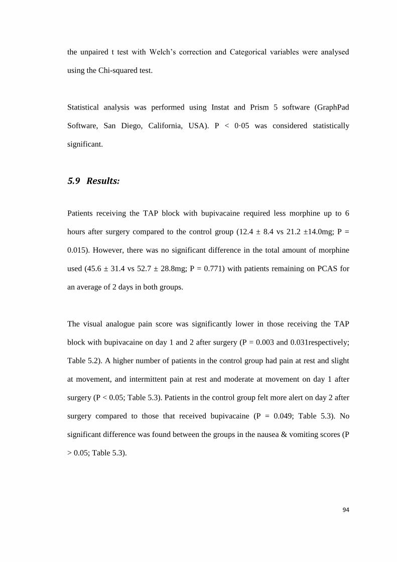
94
the unpaired t test with Welch’s correction and Categorical variables were analysed
using the Chi-squared test.
Statistical analysis was performed using Instat and Prism 5 software (GraphPad
Software, San Diego, California, USA). P < 0·05 was considered statistically
significant.
5.9 Results:
Patients receiving the TAP block with bupivacaine required less morphine up to 6
hours after surgery compared to the control group (12.4 ± 8.4 vs 21.2 ±14.0mg; P =
0.015). However, there was no significant difference in the total amount of morphine
used (45.6 ± 31.4 vs 52.7 ± 28.8mg; P = 0.771) with patients remaining on PCAS for
an average of 2 days in both groups.
The visual analogue pain score was significantly lower in those receiving the TAP
block with bupivacaine on day 1 and 2 after surgery (P = 0.003 and 0.031respectively;
Table 5.2). A higher number of patients in the control group had pain at rest and slight
at movement, and intermittent pain at rest and moderate at movement on day 1 after
surgery (P < 0.05; Table 5.3). Patients in the control group felt more alert on day 2 after
surgery compared to those that received bupivacaine (P = 0.049; Table 5.3). No
significant difference was found between the groups in the nausea & vomiting scores (P
> 0.05; Table 5.3).

95
5.9.1 Table 5.2 - Visual analogue Score
The visual analogue scale comprised of a horizontal 10 centimetres in length used to
document the pain at the time points. This methodology of scoring the pain is well
established and being widely used in the clinical research.
Day Bupivacaine Control p value
1 19(15) 37(20) 0.03
2 11(10) 19(13) 0.031
3 13(18) 14(13) 0.523
Table 5.2 showed a significantly low pain score in bupivacaine group compared to
controls in day 1 and 2. The values showed above are in mean ± SD.
Patients receiving the TAP block with bupivacaine required less tramadol 156.5 ±
185.4 vs 471.4 ± 372.2mg (P = 0.001) and ondansetran 3.7 ± 4.2 vs 7.1 ± 4.6mg
compared to the control group during their hospital stay. There were no differences in
the amount of paracetamol, codeine or cyclizine required in either of the groups (P >
0.05). The average length of post operative stay was 3.7 ± 1.8 vs 3.1 ± 1.1 days in the
bupivacaine and control groups (P = 0.242).

96
5.9.2 Table 5.3 -Verbal response scores for pain, sedation and nausea
& vomiting
Day 1 Day 2 Day 3
Bupivacaine Control Bupivacaine Control Bupivacaine Control
Verbal response pain scale a
0 2 0 3 5 6 5
1 17* 9 11 11 9 8
2 4 11** 9 5 4 1
3 0 2 0 0 0 0
Sedation score b
0 13 12 13 18©
17 11
1 8 8 8 3 2 3
2 2 2 2 0 0 0
3 0 0 0 0 0 0
Nausea and vomiting score c
0 13 13 15 15 18 12
1 5 4 5 4 0 1
2 3 5 2 2 1 1
3 2 0 0 0 0 0
a Verbal Response Pain Scale, (0 no pain at rest or movement; 1 no pain at rest, slight on movement; 2 intermittent pain at rest, moderate on movement; 3 continuous pain at rest and severe on movement). a Sedation Score, (0 none, patient is alert; 1 mild, awake but drowsy; 2 moderate, asleep but rousable; 3 Severe,
unrousable).
b Nausea & Vomiting Score (0 no nausea and vomiting at rest or on movement; 1 no nausea and vomiting at rest, slight on movement; 2 intermittent nausea and vomiting at rest, moderate on movement; 3 continuous nausea and vomiting at rest and severe on movement) in patients receiving a TAP block with either Bupivacaine or saline control on days 1, 2 and 3 after laparoscopic donor nephrectomy.
c *P=0.003, **P=0.031 and
©P=0.049.

97
5.10 Timed up & go
Pre assessment levels of the timed up & go were similar in both groups (bupivacaine
7.3 ± 1.0 vs control 7.5 ± 1.2 seconds; P = 0.524, Table 5.4). On day 1 post surgery the
time taken to complete the task was significantly longer in both groups (bupivacaine
17.6 ± 6.3 vs control 19.1 ± 8.5 seconds; P <0.0001). 7 out of 24 patients receiving
bupivacaine and 8 out of 22 in the control group felt unable to perform the assessment
on day 1 (P = 0.837). On day 3, patients in both groups performed the task quicker
compared to day 1 but the time was significantly prolonged compared to the pre
operative duration (bupivacaine 11.4 ± 4.8 vs control 11.4 ± 4.0 seconds; P= 0.002,
0.003). One patient in the bupivacaine group was unable to complete the assessment on
day 3. However, all patients in the control group were able to perform the task.
5.10.1 Table 5.4 - Time up & go assessments:
Although the time up & go is a validated technique on assessing the mobility in
postoperative scenario, there is a possibility for bias due to the connected intravenous
line and postoperative complications.
Bupivacaine Control
Preop 7.5 seconds 7.3 seconds
Day 1 17.6 seconds 19.1 seconds
Day 3 11.4 seconds 11.5 seconds

98
5.11 Grip Strength
The pre operative grip strength assessed using the dominant hand was similar between
groups (bupivacaine 81.5 ± 28.2 vs control 74.8 ± 27.5 Kg; P = 0.417, Table 5.5).
Levels fell significantly in both groups on day 1 after surgery and to a numerically
lower level in the control group compared to those receiving bupivacaine (bupivacaine
76.4 ± 27.1 vs control 61.2 ± 25.4 Kg; P = 0.058). Levels recovered on day 3 post
surgery and were comparable to pre operative levels in both groups (bupivacaine 74.6 ±
30.0 vs control 67.9 ± 19.1 Kg; P>0.05). All patients were able to perform the
assessment on the scheduled days.
5.11.1 Table 5.5: Grip Strength Assessment
Bupivacaine Control
Preop 81.5 Kg 74.8 Kg
Day 1 76.4 Kg 61.2 Kg
Day 3 74.6 Kg 67.9 Kg
Though grip strength assessment is a standardised technique on assessing the
postoperative recovery of muscle function, any condition which interferes with the
hand movement could bias the values. A simple condition like thrombophebitis or
presence of cannula in forearm/hand could potentially interfere with grip strength
testing and hence it is the limitation of this test.

99
5.11.2 Figure 5.1: Time up & go
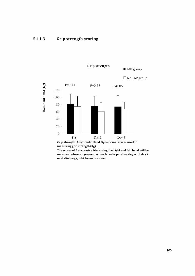
100
5.11.3 Grip strength scoring

101
5.12 Discussion
This randomised double-blind placebo controlled trial in patients undergoing
laparoscopic nephrectomy demonstrated that TAP blocks using bupivacaine reduced
early morphine requirements. Interestingly, there was no difference in the total amount
of morphine required but patients reported less post-operative pain and required less
oral analgesics during their hospital stay.
There is a growing body of evidence in favour of TAP blocks in reducing post-
operative pain for various types of surgery (Carney J et al, 2008; Niraj G et al, 2009;
Conaghan P et al, 2010; Sandeman DJ et al, 2011).
Nonetheless, the results are inconsistent. A randomised controlled trial by McDonnell
et al. found that TAP blocks reduced morphine requirements by more than 70% for up
to 36 hours after Caesarean section (McDonnell JG et al, 2008). Patients also had lower
visual analogue pain scores for 48 hours after surgery.
In another study of Caesarean section, Belavy et al. also found that TAP blocks reduced
post-operative analgesic requirements however, the results were less pronounced with
no differences in the post-operative pain scores (Belavy D et al, 2009).
In contrast, Costello et al. found that TAP blocks did not have any beneficial effect
after Caesarean sections (Costello JF et al, 2009).

102
However, TAP blocks were used in conjunction with long acting intrathecal opioids,
which are extremely effective and likely to reduce the need for morphine after surgery
(Factor D, 2010) (McMorrow RC et al, 2011).
The posterior TAP block procedure used in this study involved a single injection of
local anaesthetic which may have potential disadvantages. Bupivacaine has a relatively
short duration of action, lasting 6 to 8 hours and its effectiveness can be dependent on
the type of surgery (Charlton S et al, 2010).
The results from this trial showed that patients receiving a TAP block with bupivacaine
required 42% less morphine in the first 6 hours after surgery compared to patients
receiving the placebo control. Thereafter, morphine requirements were similar.
Nonetheless, the duration of action using this technique is unknown and the poor
vascularisation in this region may prolong the effect, which may also be dependent on
the amount and concentration of local anaesthetic agent used.
Higher concentrations have been used (McDonnell JG et al, 2008), however there is a
risk of toxicity, as the local anaesthetic may spill over into the adjacent muscle
resulting in high systemic concentrations and therefore some caution must be taken
(Kato N et al, 2009).
Bupivacaine TAP blocks showed no advantage for patients in terms of nausea and
vomiting scores, post-operative recovery or in the length of hospital stay. Surprisingly,
sedation scores were lower in the control group with more patients feeling more alert
on post-operative day 2 compared to the patients receiving a TAP block with

103
bupivacaine. Nonetheless, and perhaps most importantly, patients receiving a
bupivacaine TAP block had lower visual analogue pain scores for 2 days after surgery
and experienced less pain at rest and on movement. Furthermore, patients required less
tramadol and anti-emetics during their hospital stay. Although, there was no reduction
in the total morphine requirement, the perceived reduction in pain is an important
outcome and likely to be of benefit. Pain is influenced by many factors and studies have
shown that a higher use of PCA can be influenced by anxiety and the anticipation of
pain (Logan DE, 2005) (Crombez G et al, 1996).
Pre-emptive analgesia is an effective and standard way of targeting post-operative pain.
A recent study in patients undergoing a hysterectomy found that TAP blocks
administered before surgery reduced pain and analgesic requirements compared to TAP
blocks placed at the end of the surgery (Amr YM, 2011).
A potential disadvantage of pre-emptive TAP blocks is that some of their post-
operative effect may be lost if surgery is prolonged. The effects of TAP blocks can be
prolonged by continuous infusion of the local anaesthetic agent via an implanted TAP
catheter and this may be more beneficial.
A recent study demonstrated that patients undergoing open donor nephrectomy with a
flank incision required significantly less morphine and had lower pain scores up to 48
hours after surgery when local anaesthetic was continually infused through catheters
placed in the transversus abdominis plane for 48 hours after surgery (Harish R., 2009).

104
Continuous administration of local anaesthetic has also been beneficial in kidney
transplant recipients (Philip A., 2010).
Nonetheless, more evidence is needed to establish the efficacy of this approach. There
have also been reports of using an intra-abdominal approach administering the TAP
blocks under direct vision during surgery (Owen DJ et al, 2011). This has been
described as a more simple method that improves accuracy and reduces the risk of
damaging the viscera.
The numbers of patients included in this study were informed by a realistic power
calculation. Furthermore, it must be taken into consideration that although the TAP
blocks were administered under ultrasound guidance by an experienced consultant
anaesthetist, in some cases they may not have been effective. Pin-prick sensation can be
used to assess sensory blockage and the effectiveness of the TAP bock. However, this
was avoided in the present trial as blinding could not have been maintained.
Complications are also possible, including infection or haematoma at the administration
site, nerve damage, transient femoral palsy and even the potential to damage underlying
organs (Jankovic ZB et al, 2009). Nonetheless, there were no complications associated
with the TAP block procedure in this present study.
5.13 Cytokines:
Cytokines are proteins produced by haematopoetic and non-haematopoetic cells which
play a role in innate and adaptive immune responses (Longo D et al, 2012). They are
produced from activated leucocytes, fibroblasts and endothelial cells as an early

105
response to tissue injury and have a major role in mediating immunity and
inflammation (Longo D et al, 2012) (Sheeran P, 1997). Cytokines act on surface
receptors on many different target cells and their effects are produced ultimately by
influencing protein synthesis within these cells (Desborough JP., 2000).
Cytokines have a major role in the inflammatory response to surgery and trauma. They
have local effects of mediating and maintaining the inflammatory response to tissue
injury, and also initiate some of the systemic changes which occur (Desborough JP.,
2000). They can act in an autocrine manner, affecting the behavior of the cell that
releases the cytokine, or in a paracrine manner, affecting the behavior of adjacent cells.
Some cytokines can also act in an endocrine manner, affecting the behavior of distant
cells, although this depends on their ability to enter the circulation and on their half-life.
After major surgery, the main cytokines released are interleukin-1 (IL-1), tumour
necrosis factor α (TNF-α) and IL-6. The initial reaction is the release of IL-1 and TNF
α from activated macrophages and monocytes in the damaged tissues. This stimulates
the production and release of more cytokines, in particular, IL-6, the main cytokine
responsible for inducing the systemic changes known as the acute phase response
(Sheeran P, 1997).
Cytokines and growth factors may have multiple functions that can initiate and
influence the wound healing process (Bennett NT, 1993).
Previous studies have shown that the inflammatory cytokines are released following
donor nephrectomy though their significance is still debatable (Yap S et al, 2012).

106
5.13.1 Interaction between the immune system and the
neuroendocrine system:
The cytokines IL-1and IL-6 can stimulate secretion of ACTH from isolated pituitary
cells in vitro. In patients after surgery, cytokines may augment pituitary ACTH
secretion and subsequently increase the release of cortisol (Desborough JP., 2000).
A negative feedback system exists, so that glucocorticoids inhibit cytokine production.
The cortisol response to surgery is sufficient to depress IL-6 concentrations (Jameson P
et al, 1997).
5.13.2 Hypothesis for cytokines estimation:
In a mouse model, nephrectomy resulted in early onset of inflammation, apoptosis, and
tissue damage in hepatocytes with increased tumour necrosis factor-(TNF) and
interleukin (IL-6) (Golab F et al, 2009). Bilateral nephrectomy in animal models has
shown an increased TNF-and IL-6 levels, which lead to uncontrolled systemic
inflammation, neutrophil infiltration, and impaired vascular permeability resulting in
pulmonary oedema (Klein CL et al, 2008).
Patients who underwent donor nephrectomy demonstrated increased serum IL-6 and
serum TNF which was possibly secondary to acute deterioration in renal function (Yap
S et al, 2012).

107
Although Yap S et al, showed significantly high levels of cytokines and other
inflammatory markers following laparoscopic donor nephrectomy the significance of
this elevation whether merely due to significant tissue trauma or due to nephrectomy is
not established yet.
Secondly there is also evidence available to support that pain relief techniques does
interfere with expression of cytokines and this could potentially be blocked (Stefano
GB et al, 1994, Xu YJ et al, 2014). Hence in our study we reassess the cytokine
expression following laparoscopic donor nephrectomy and also evaluate the role of
TAP block on blocking the cytokine expression in 24, 48 hrs after surgery. The
cytokine expression can be spuriously low or high by the use of isoflurane, sevoflurane
and dexamathasone and potentially this could bias the values.
5.13.3 Cytokines assay:
Venous blood samples were taken preoperatively then at 6, 24 and 48 hours
postoperatively to measure levels of inflammatory cytokines. After collection samples
were centrifuged at 1000g, 4°C for 15 minutes and the plasma stored at -80ºC until
analysed. The Aushon Search Light® Human Cytokine Array 2 (Aushon Biosystems,
Billerica, MA. USA) a multiplex sandwich enzyme-linked immunosorbent assay
(ELISA) was used to measure levels of interleukin -1ß (IL-1ß), interleukin-6 (IL-6),
interleukin-8 (IL-8) and tumour necrosis factor alpha (TNFα).
Samples and standards were diluted 2 fold and added to the pre coated plate in
duplicate. Unbound proteins were washed away and a biotinylated detecting antibody
added to bind to a second site on the target proteins. After washing the excess detecting

108
antibody, streptavidin-horseradish peroxidase (SA-HRP) was added to produce a
luminescent signal which was detected by a cooled CCD camera.
5.13.4 Interleukin-1 Beta (IL-1β):
Interleukin-1 (IL-1) is the prototypic pro-inflammatory cytokine. There are two forms
of IL-1, IL-1alpha and IL-1beta and their biological activities are indistinguishable
(Dinarello CA., 1997). Although IL-1 can upregulate host defences and function as an
immunoadjuvant, IL-1 is a highly inflammatory cytokine (Dinarello CA., 1997).
When IL-1 gains access to the circulation, it acts like a hormone and induces a broad
spectrum of systemic changes in neurological, metabolic, hematologic, and
endocrinologic systems. Usually an early and short-lived IL-1 beta response to major
surgery was reported followed by surge of other cytokines (Baigrie RJ et al, 1992).
Recent study has shown that a longer operative time is associated with significantly
high level of IL-1 (Beyza Özçınar et al, 2014).
5.13.5 Results of IL-1β assay:
IL-1 β was detected in patients in both groups throughout the study period. In the
bupivacaine group preop, postop at 6th
hours, 24 hours, and 48 hours serum levels were
6.51, 8.16, 10.1 and 67.16 picograms/ml respectively. Similarly serum levels of IL-1 in
the control group were 5.3, 6.43, 9.45 and 62.38 picograms/ml at preop, 6, 24 and 48
hours respectively. Hence there was no difference between the bupivacaine and control
groups at any time point (P=NS; figure 5.2, 5.3). More importantly, in contrast to
previous evidence serum IL-1 levels were significantly higher only at 48 hours, not as
documented as an early surge following a surgical stimulus (Baigrie RJ et al, 1992).

109
Figure 5.2: Results of IL-1 β Assay between Bupivacaine and
Control

110
Figure 5.3: Results of IL-1 β Assay between Bupivacaine and
Control

111
5.13.6 Interleukin-6 (IL-6):
IL-6 is a polypeptide with a molecular weight of 21.5-30 kDa (Bauer J, 1991). The
concentrations of circulating cytokines are normally low and may be undetectable. The
molecular weight heterogeneity of IL-6 results from post-translational modifications
such as N- and O-glycosylation and phosphorylation (Bauer J, 1991).
Within 30–60 min after the start of surgery, IL-6 concentration increases, the change in
concentration becoming significant after 2–4 h. Cytokine production reflects the degree
of tissue trauma, so cytokine release is lowest with the least invasive and traumatic
procedures, for example, laparoscopic surgery (Desborough JP., 2000).
The largest increases in IL-6 occur after major procedures such as joint replacement,
major vascular and colorectal surgery. After these operations, cytokine concentrations
are maximal at about 24 h and remain elevated for 48–72 h postoperatively.
5.13.7 Results of IL-6 Assay:
IL-6 was detected amongst all patients in both groups throughout the study period. In
the bupivacaine group preop, postop at 6th hours, 24 hours, and 48 hours serum levels
were 19.3, 60.5, 85.9 and 91.2 picograms/ml respectively. Similarly serum levels of IL-
6 in the control group were 20.6, 58.7, 89.8 and 76.7 picograms/ml at preop, 6, 24 and
48 hours respectively (figure 5.4 and 5.5). Hence there was no difference between the
bupivacaine and control groups at any time point (P=NS).

112
Figure 5.4: Results of IL-6 Assay between Bupivacaine and
Control

113
Figure 5.5: Results of IL-6 assay between Bupivacaine and Control

114
5.13.8 Interleukin-8 (IL-8):
Interleukin-8 (IL-8), also known as CXCL-8, a member of the CXC chemokine family
that was originally classified as a potent neutrophil chemoattractant, has been shown to
regulate biological processes through interactions with relative receptors (Roebuck
KA., 1999).
In leukocytes, IL-8 has been shown to stimulate the activation of G proteins and several
downstream serine/threonine kinases that are responsible for chemotaxis,
degranulation, and production of superoxide anions by phagocytes (Jones SA et al,
1996, Kupper RW et al, 1992). IL-8 is generated as a precursor of 99 amino acids and
is secreted after cleavage of a signal sequence of 20 residues (Baggionlini, 1992). N-
terminal extracellular processing of the mature form yields several biologically active
variants. The predominant variant consists of 72 amino acids and has a molecular
weight of 8,383kDa (Baggionlini, 1992).
5.13.9 Results of IL-8 Assay
IL-8 was detected in all patients in both groups throughout the study period. In the
control group preop, postop at 6 hours, 24 hours, and 48 hours serum levels were 28.7,
16.3, 26.8 and 36.5 picograms/ml respectively. Similarly serum levels of IL-8 in the
bupivacaine group were 33.9, 30.1, 36.5 and 35 picograms per ml at preop, 6, 24 and
48 hours respectively. Hence there was no difference between the control group and the
bupivacaine group at any time point (figure 5.6 and 5.7) (P=NS). This analysis did not
show any significant increase in serum IL-8 level in the postoperative period when
compared with preop levels.

115
Figure 5.6: Results of IL-8 assay between Bupivacaine and
Control

116
Figure 5.7: Results of IL-8 assay between Bupivacaine and
Control

117
5.13.10 Tumour Necrosis Factor (TNFα):
TNFα is a cytokine with effects that include a contribution to initiating and
orchestrating the complex events involved in inflammation and immune response
(Sethi G et al 2008). TNF-alpha is the prototypic member of a gene superfamily that
regulates essential biologic functions such as immune response, cell proliferation,
survival, differentiation and apoptosis (Bagul A., 2012; Esposito E, 2009).
When microorganisms activate proinflammatory signals, TNFα is produced by
macrophages, dendritic cells, B cells, and T cells; as well as by other types of somatic
cells such as endothelial cells, mast cells, neuronal tissue cells, and tumour cells
(Vassalli P., 1992) (Sethi G et al 2008).
TNF-alpha is a transmembrane protein with a molecular mass of 26 kDa that was
originally found to be expressed in macrophages and has now been found to be
expressed by a wide variety of cells (Sethi G et al, 2008).
The pro-inflammatory effects of TNF-alpha are primarily due to its ability to activate
NF-kappaB. Almost all cell types, when exposed to TNF-alpha, activate NF-kappaB,
leading to the expression of inflammatory genes (Sethi G et al, 2008). Over 400 genes
have been identified that are regulated by NF-kappaB activation (Sethi G et al, 2008).

118
5.13.11 Result of TNFα Assay :
TNFα was detected in all patients in both groups throughout the study period. In the
control group preop, postop at 6th hours, 24 hours, and 48 hours serum levels were
68.9, 59.7, 49.3 and 5.7 picograms/ml respectively. Similarly serum levels of TNFα in
Bupivacaine group were 75.5, 88, 75.8 and 7.4 picograms/ml at preop, 6, 24 and 48
hours respectively (figure 5.8 and 5.9).. Hence, there was no difference between the
control group and the bupivacaine group at any time point. (P=NS). This analysis did
not show any significant increase in serum TNFα levels in the postoperative period
when compared with preop levels. In fact, there was a high titre of TNFα noted preop
which has progressively fallen over time following donor nephrectomy. These results
seem puzzling, although previous studies have reported similar findings in patients
following major surgery (Høgevold HE et al, 2000) (Shimada M et al, 1993).
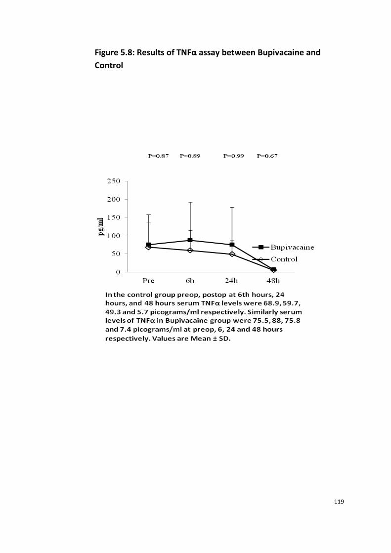
119
Figure 5.8: Results of TNFα assay between Bupivacaine and
Control

120
Figure 5.9: Results of TNFα assay between Bupivacaine and
Control

121
5.14 Conclusion:
In conclusion, the addition of a pre-emptive bupivacaine based TAP block in
laparoscopic live donor nephrectomy is of benefit in reducing post-operative pain and
early post-operative morphine requirements. But TAP block did not show any
significant effect on cytokine response following laparoscopic donor nephrectomy.
Future studies should aim to extend the effects the TAP blocks by investigating the use
of TAP catheters to provide continual infusions to optimise the delivery of local
anaesthetic and prolong the effect.

122
CHAPTER 6
Discussion - Summary and Future Work

123
6 Summary of key findings
This work, to the best of my knowledge, is the first investigation to assess the role of
transversus abdominis plane block in laparoscopic donor nephrectomy patients. The
initial retrospective study showed the effect of TAP on reducing the cumulative use of
morphine when compared to the control group. More importantly, the oral analgesic
requirement was also significantly reduced in the TAP group; paracetamol 13.35±3.6
grams vs 17±4.8 grams (P <0.0002) and tramadol 353±283 mg versus 820±521 mg (P
<0.0001) compared with control group. Similar results were noted for anti-emetics
(cyclizine 60±82.1mg versus 159±212 mg; P <0.0017 and ondansetran 3.2±3.8 mg
versus 5.5±5.5 mg; P <0.035) requirements as well.
The retrospective study showed that TAP block group discontinued their PCAS
significantly earlier than patients in the control group (1.3±0.6 days versus 1.9±0.7
days; P <0.0001). Similarly, there was a significant difference noted in the length of
hospital stay between the TAP and control group (4.3±1.1 versus 5.1±1.1 days; P <
0.0034).
Similar results have been shown in other studies where TAP blocks were successful in
reducing post-operative pain and the overall morphine requirement after abdominal
surgeries such as hysterectomy (Carney J et al, 2008), appendicectomy (Niraj G et al,
2009) and Caesarean section (Belavy et al, 2009). Results from this retrospective study
and the above study findings formed the back-bone to perform a randomised placebo
controlled clinical trial.

124
The randomised clinical trial of TAP block in laparoscopic donor nephrectomy patients
has given interesting and supporting results. The study has demonstrated that a TAP
block with bupivacaine reduced early morphine requirement at 6 hours with no
difference in cumulative morphine usage. More importantly, the bupivacaine group had
significantly less post-operative pain and required less oral analgesics during their
hospital stay. The visual analogue pain score in the TAP block group with bupivacaine
was significantly lower on day 1 and 2 after surgery (P = 0.003; 0.031). A significantly
higher number of patients in the control group had pain at rest and on movement, and
intermittent pain at rest and moderate at movement on day 1 after surgery (P <0.05).
Recent studies on patients undergoing caesarean section (Costello JF et al 2008) and
laparoscopic hysterectomy (Calle GA et al, 2014) demonstrated that patients did not
benefit from the administration of a TAP block and therefore this raises questions about
the suitability of this technique for all types lower abdominal surgery. The results from
our study are supported by a recent study (Waits SA et al, 2015) showing overall
reduction of narcotic by 50% through similar pain scores.

125
6.1 Cytokines after donor nephrectomy:
The initial cytokine reaction is the release of IL-1 and TNF α from activated
macrophages and monocytes after tissue damage including surgery. This stimulates the
production and release of more cytokines, in particular, IL-6, the main cytokine
responsible for inducing the systemic changes known as the acute phase response
(Sheeran P et al, 1997).
Cytokine analysis has revealed valuable and important findings from our study. A
recent study of laparoscopic donor nephrectomy has shown that cytokines levels
including interleukin-1 (IL-1), tumour necrosis factor α (TNF-α), IL-6 and IL-18
(markers of acute kidney injury) were high after nephrectomy (Yap S et al, 2012).
Our study has shown a significantly rise in IL-6 level 6 hours after donor nephrectomy
and this continued to be high at 48 hours. Similar results were reported in another study
where the IL-6 level was raised significantly 1 hour after nephrectomy and was
persistently high a few days after donor nephrectomy (Kielstein JT el al, 2011).
Interestingly, IL-8 and TNF-α levels fell significantly compared with preoperative
levels. But in comparison with the previous studies, our sample size is significantly
larger and hence the results are likely to be more reliable. This signifies the need for
further investigation on IL-8 and TNF-α in donor nephrectomy patients.
Finally, a more enigmatic twist was observed in terms of an IL-1ß level raised at 48
hours after donor nephrectomy. IL-1ß is known to be an initial cytokine in response to

126
the inflammatory stimulus, but our result does not support this view. Hence further
research is also required to address whether the cytokine response is atypical in this
setting.
6.2 Strengths and weaknesses.
The strengths of this thesis are the retrospective analysis results which helped to set up
a successful double blinded randomised placebo controlled trial. The randomised study
has given robust and high quality evidence on TAP block in laparoscopic donor
nephrectomy patients. Though reduced pain scores and lower oral tramadol doses were
noted in the TAP group, cumulative morphine usages between the groups were not
different. As we have utilised a lateral approach for TAP block in our patients, the role
of an alternative posterior TAP block approach in laparoscopic donor nephrectomy
remains to be answered. There is a growing body of evidence to support the hypothesis
that the posterior approach could result in better pain relief.
Furthermore cytokine assays up to 48 hours after donor nephrectomy were similar
between the TAP block and control groups. Although this may appear as a negative
result, further research is needed to focus on the cytokine response following donor
nephrectomy. To date, this randomised study is the only study with a large sample size
that can be extrapolated as robust evidence on cytokine responses in after donor
nephrectomy. In the randomised study, we have analysed cytokine samples up to 48
hours; perhaps further delayed samples may have been beneficial in assessing the
cytokine response following donor a nephrectomy. But, one could argue that this means
unnecessary donor visit to hospital and potential inconvenience.

127
6.3 Future work:
For the reasons discussed above, it was essential to confirm the role of TAP block in
the setting of laparoscopic live donor nephrectomy. This thesis includes a literature
review on pain relief methods, a retrospective pilot study and finally a double blinded
randomised placebo controlled trial of TAP block in laparoscopic donor nephrectomy
patients. Though it may appear that role of TAP block in donor nephrectomy patients
has been established here, the role of the posterior technique of TAP blocking in similar
settings is still unknown.
Secondly, in our study we have used the TAP block after general anaesthesia was
established; If the TAP block was administered before anaesthesia, the effectiveness of
TAP blocks could have been evaluated further.
Thirdly, the role of continuous infusion of local anaesthetics in transversus abdominis
plane needs further investigation as this provides constant availability of drug, which in
principle could offer a better postoperative pain relief.
Fourthly, the liposomal bupivacaine prolonged-release formulation as a part of
multimodal analgesia in laparoscopic donor nephrectomy patients has never been tested
before; this area needs early attention and further evaluation.

128
Finally a cytokine assay in donor nephrectomy with delayed blood samples up to 10
days following operation could shed a light on their role and cytokine response after the
donor surgery. To date, there are only conflicting findings available in this setting;
hence it is hard to establish a reasonable conclusion.

129
6.4 Conclusion
The research presented here has established that the TAP block is a useful and
beneficial method of analgesia in patients undergoing laparoscopic donor nephrectomy.
The study has demonstrated that a TAP block with bupivacaine reduced early morphine
requirement at 6 hours. The visual analogue pain score in the TAP block group with
bupivacaine was significantly lower on day 1 and 2 after surgery. There was also a
reduced oral tramadol usage in the bupivacaine group. But the total usage of morphine
has not been reduced by the use of TAP blocks. As a part of multimodal analgesia,
TAP blocks could play an important role in conjunction with PCAS in postoperative
pain management after laparoscopic donor nephrectomy.
In contrast to the expected result, preoperative local anaesthetic administration did not
reduce the cytokine response. The physiological response to surgery can be very
complex and clearly the TAP block has not been found to be interfering with this
mechanism.
Despite the improvements in the postoperative analgesic modalities in laparoscopic
donor nephrectomy patients, the ideal method of analgesia remains to be established.

130
References
Could I be a living kidney donor?. 2015 [online]. Available at: http://www. organdonation.
nhs.uk/ how_to_become_a_donor/living_kidney_donation/ [accessed 07/20 2015].
Organ Donation and Transplantation Activity Data: UNITED KINGDOM.2013 [online]. Available
at: http://www.organdonation.nhs.uk/statistics/downloads/united_kingdom_april13.pdf
[accessed 07/20 2015].
Questions and Answers about Oral Prescription Acetaminophen Products to be Limited to
325mg Per Dosage Unit. 2011 [online]. Available at: http://www.fda.gov/Drugs/ Drug
Safety / Information by Drug Class/ ucm239871.htm [accessed 04/22 2015].
Cost-effectiveness of transplantation. [online]. Available at:http://www.organdonation.nhs.uk
/newsroom/fact_sheets/cost_effectiveness_of_transplantation.asp [accessed 07/13
2013].
Aasvang E, K. H. (2005). Chronic postoperative pain: The case of inguinal herniorrhaphy. Br J
Anaesth, 95, 69-76.
Addison NV, Brear FA, Budd K, Whitaker M. (1974). Epidural analgesia following
cholecystectomy. Br J Surgery.1974; 61:850-52.
Alexander JI. (1997). Pain after laparoscopy. Br J Anaesth. 79(3): 369-378.
Amr YM, A. S. (2011). Comparative study between the effect of pre-versus post-incisional
transversus abdominis plane block on acute and chronic post-hysterectomy pain.
Anesthesis Essays and Researches, 5(1), 77-82.
131
Andersen MH, Mathisen L, Oyen O, Edwin B, Digernes R, Kvarstein G, Tønnessen TI, Wahl AK,
Hanestad BR, Fosse E. (2006). Postoperative pain and convalescence in living kidney
donors laparoscopic versus open donor nephrectomy: A randomized study. Am J
Transplant, 6(6), 1438-1443.
Antcliffe D, Nanidis TG, Darzi AW, Tekkis PP, Papalois VE. (2009). A meta-analysis of mini-open
versus standard open and laparoscopic living donor nephrectomy. Transpl Int, 22(4), 463-
474.
Ashcraft EE, Baillie GM, Shafizadeh SF, McEvoy JR, Mohamed HK, Lin A, Baliga PK, Rogers J,
Rajagopalan PR, Chavin KD. (2001). Further improvements in laparoscopic donor
nephrectomy: Decreased pain and accelerated recovery. Clin Transplant, 15(Suppl 6), 59-
61.
B.G. Covino, J. A. W. (1998). Clinical pharmacology of local anesthetic agents. In P. O. B. M.J.
Cousins (Ed.), Neural blockade in clinical anaesthesia and management of pain (3rd ed.,
pp. 97-128). Philadelphia,: Lippincott-Raven,.
Baaj JM, Alsatli RA, Majaj HA, Babay ZA, Thallaj AK. (2010). Efficacy of ultrasound-guided
transversus abdominis plane (TAP) block for postcesarean section delivery analgesia-a
double-blind, placebo-controlled, randomized study. Middle East J Anesthesiol, 20(6),
821-826.
Bachmann A, Wolff T, Giannini O, Dickenman M, Ruszat R, Gürke L, Kaufmann M, Gasser TC,
Steiger J, Stief CG, Sulser T. (2006a). How painful is donor nephrectomy? Retrospective
analysis of early pain and pain management in open versus laparoscopic versus
retroperitoneoscopic nephrectomy. Transplantation, 81(12), 1735-1738.

132
Baggiolini M, C. I. (1992). Interleukin-8, a chemotactic and inflammatory cytokine. FEBS Lett,
307(1), 97-101.
Bagul A. (2012). Ischaemic/reperfusion injury: Role of infliximab. World J Transplant., 2(3), 35-
40.
Baigrie RJ, Lamont PM, Kwiatkowski D, Dallman MJ, Morris PJ. (1992). Systemic cytokine
response after major surgery. Br J Surg., 79(8), 757-760.
Ballantyne JC, Carr DB, Chalmers TC, Dear KB, Angelillo IF, Mosteller F. (1993). Postoperative
patient controlled analgesia: Meta-analyses of initial randomized control trials. J Clin
Anesth, 5(3), 182-193.
Barth. R. (2014). Donor nephrectomy. In K. S. Morris P (Ed.), Kidney transplantation -
principles and practice, 7th edition (7th ed., pp. 122-128). USA: Elsevier Saunders.
Bauer J, H. F. (1991). Interleukin-6 in clinical medicine. Ann Hematol, 62(6), 203-210.
Bay-Nielsen M, Perkins FM, Kehlet H. (2001). Pain and functional impairment 1 year after
inguinal herniorrhaphy: A nationwide questionnaire study. Ann Surg, 233, 1-7.
Beaussier M, El'Ayoubi H, Schiffer Rollin M, Parc Y, Mazoit JX, Azizi L, Gervaz P, Rohr S,
Biermann C, Lienhart A, Eledjam JJ. (2007). Continuous preperitoneal infusion of
ropivacaine provides effective analgesia and accelerates recovery after colorectal surgery:
A randomized, double-blind, placebo-controlled study. Anesthesiology, 107(3), 461-468.
Belavy D, Cowlishaw PJ, Howes M, Phillips F. (2009). Ultrasound-guided transversus abdominis
plane block for analgesia after caesarean delivery. Br J Anaesth, 103(5), 726-730.
133
Bennett NT, S. G. (1993). Growth factors and wound healing: Part II. role in normal and
chronic wound healing. Am J Surg, 166(1), 74-81.
Berends FJ, den Hoed PT, Bonjer HJ, Kazemier G, van Riemsdijk I, Weimar W, IJzermans JN.
(2002). Technical considerations and pitfalls in laparoscopic live donor nephrectomy. Surg
Endoscopy, 16(6), 893-898.
Bergese SD, Ramamoorthy S, Patou G, Bramlett K, Gorfine SR, Candiotti KA. (2012). Efficacy
profile of liposome bupivacaine, a novel formulation of bupivacaine for postsurgical
analgesia. J Pain Res., 5, 107-116.
Beydoun A, B. M. (2003). Mechanistic stratification of antineuralgic agents. J Pain Symptom
Manage., 25(5 Sippl), S18-30.
Beyza Özçınar, Nihat Aksakal, Fatih Yanar, Orhan Agcaoglu, Kıvanç Derya Peker, Ümit
Türkoglu, Selçuk Mercan, Selçuk Özarmagan, Yeşim Erbil. (2014). Increased interleukin 1
and tumor necrosis factor alpha levels after thyroid surgery. Turkish Journal of Surgery,
30(2), 080-084.
Biglarnia AR, Tufveson G, Lorant T, Lennmyr F, Wadström J. (2011). Efficacy and safety of
continuous local infusion of ropivacaine after retroperitoneoscopic live donor
nephrectomy. Am J Transplant, 11(1), 93-100.
Billingham RE, Brent L, Medawar PB. (1953). Actively acquired tolerance of foreign cells.
Nature, 172(4379), 603-606.
Billingham, R., Medawar PB. (1951). The technique of free skin grafting in mammals. J. Exp.
Biol, 28, 385-402.

134
Birrer KL, Anderson RL, Liu-DeRyke X, Patel KR. (2011). Measures to improve safety of an
elastomeric infusion system for pain management. Am J Health Syst Pharm., 68(13), 1251-
1255.
Bisgaard T, Klarskov B, Rosenberg J, Kehlet H. (2001). Characteristics and prediction of early
pain after laparoscopic cholecystectomy. Pain, 90, 261-269.
Bisgaard, T., Klarskov, B., Kristiansen, V. B., Callesen, T., Schulze, S., Kehlet, H., & Rosenberg, J.
(1999). Multi-regional local anesthetic infiltration during laparoscopic cholecystectomy in
patients receiving prophylactic multimodal analgesia: A randomized, double-blinded,
placebo-controlled study. Anesth Analg, 89(4), 1017-1024.
Biton J, Boissier MC, Bessis N. (2012). TNFα: Activator or inhibitor of regulatory T cells? Joint
Bone Spine., 79(2), 119-123.
Blakemore PR, W. J. (2002). Morphine, the proteus of organic molecules. Chem Commun
(Camb), 7(11), 1159-1168.
Boger M, Lucas SM, Popp SC, Gardner TA, Sundaram CP. (2010). Comparison of robot-assisted
nephrectomy with laparoscopic and hand-assisted laparoscopic nephrectomy. JSLS., 14(3),
374-380.
Bos EM, Leuvenink HG, van Goor H, Ploeg RJ. (2007). Kidney grafts from brain dead donors:
inferior quality or opportunity for improvement?. Kidney Int, 72(7), 797-805.
Boulanger A, Choiniere M, Roy D, Boure B, Chartrand D, Choquette R, Rousseau P. (1993).
Comparison between patient controlled analgesia and intramuscular meperidine after
thoracotomy. Can J Anaesth, 40(5 Pt 1), 409-415.

135
Brenner B.M. (2007). Renal complications of NSAIDs. In L. S. A. Brenner B.M. (Ed.), Brenner
and rector's the kidney. (8th ed.,). Philadelphia, PA: Elsevier Saunders.
C.J. McCartney, E. Duggan, E. Apatu. (2007). Should we add clonidine to local anesthetic for
peripheral nerve blockade? A qualitative systematic review of the literature. Reg Anesth
Pain Med, 32(4), 330-338.
Calle GA, López CC, Sánchez E, De Los Ríos JF, Vásquez EM, Serna E, Arango AM, Castañeda JD,
Vásquez RA, González A, Escobar A, Almanza LA. (2014). Transversus abdominis plane
block after ambulatory total laparoscopic hysterectomy: Randomized controlled trial. Acta
Obstet Gynecol Scand, 93(4), 345-350.
Callesen B, K. H. (1999). Prospective study of chronic pain after hernia repair. Br J Surg, 86,
1528-1531.
Calvey TN, W. N. (1997). Principles and practice of pharmacology for anaesthetists.
Cambridge: Blackwell Science.
Carney J, McDonnell JG, Ochana A, Bhinder R, Laffey JG. (2008). The transversus abdominis
plane block provides effective post-operative analgesia in patients undergoing total
abdominal hysterectomy. Anesth Analg, 107(6), 2056-2060.
Carrel A. (1902). La technique operatoire des anastomosis vasculaires et la transplantation des
visceres. [null] Lyon Med, 98, 859.
Ceyhan D, G. M. (2010). Is postoperative pain only a nociceptive pain? Agri, 22(2), 47-52.

136
Chan VW, Chung F, McQuestion M, Gomez M. (1995). Impact of patient controlled analgesia
on required nursing time and duration of postoperative recovery. Reg Anesth, 20(6), 506-
514.
Chang AM, Ip Wy, Cheung TH. (2004). Patient-controlled analgesia versus conventional
intramuscular injection: A cost effectiveness analysis. J Adv Nurs, 46(5), 531-541.
Charlton JE. (2005). Opioids: Core curriculum for professional education in pain. Seattle: IASP
Press.
Charlton S, Cyna AM, Middleton P, Griffiths JD. (2010). Perioperative transversus abdominis
plane (TAP) blocks for analgesia after abdominal surgery. Cochrane Database Syst Rev,
8(12), CD007705.
Chatterjee S, Nam R, Fleshner N,Klotz L. (2004). Permanent flank bulge is a consequence of
flank incision for radical nephrectomy in one half of the patient. Urol Oncol, 22, 36-39.
Chien CH, Wang HH, Chiang YJ, Chu SH, Liu HE, Liu KL. (2010). Quality of life after laparoscopic
donor nephrectomy. [null] Transplant Proc, 42(3), 696-698.
Clayman RV, Kavoussi LR, Soper NJ, Dierks SM, Meretyk S, Darcy MD, Roemer FD, Pingleton
ED, Thomson PG, Long SR. (1991). Laparoscopic nephrectomy: Initial case report. J Urol,
146, 278-282.
Coderre TJ, Katz J, Vaccarino A.L,and Melzack R. (1993). Contribution of central neuroplasticity
to pathological pain: Review of clinical and experimental evidence. Pain, 52(3), 259-285.

137
Cohen SM. (2012). Extended pain relief trial utilizing infiltration of exparel(®), a long-acting
multivesicular liposome formulation of bupivacaine: A phase IV health economic trial in
adult patients undergoing open colectomy. J Pain Res., 5, 567-572.
Colwell CW Jr, Morris BA. (1995). Patient controlled analgesia compared with intramuscular
injection of analgesics for the management of pain after an orthopaedic procedure. J
Bone Joint Surg Am.1995; 77(5), 726-733.
Conaghan P, Maxwell-Armstrong C, Bedforth N, Gornall C, Baxendale B, Hong LL, Carty HM,
Acheson AG. (2010). Efficacy of transversus abdominis plane blocks in laparoscopic
colorectal resections. Surg Endosc, 24(10), 2480-2484.
Costello JF, Moore AR, Wieczorek PM, Macarthur AJ, Balki M, Carvalho JC. (2009).
The transversus abdominis plane block, when used as part of a multimodal regimen
inclusive of intrathecal morphine, does not improve analgesia after cesarean delivery Reg
Anesth Pain Med, 34(6), 586-589.
Crombez, G, Vervaet, L, Baeyens, F, Lysens, R, & Eelen, P. (1996). Do pain expectancies cause
pain in chronic low back patients? A clinical investigation. Behaviour Research & Therapy,
34(11-12), 919-925.
Dacastello A. (1902). Experimentelle nierentransplantation. Wien Kil Wochenschr 15:317.
Dahl JB, K. H. (1991). Non-steroidal anti-inflammatory drugs: Rationale for use in severe
postoperative pain. Br J Anaesth., 66(6), 703-712.
Dempster WJ. (1953). The homotransplantation of kidneys in dogs. Br J Surg, 40, 447.

138
Desborough JP. (2000). The stress response to trauma and surgery. Br J Anaesth., 85(1), 109-
117.
Debout A, Foucher Y, Trébern-Launay K, Legendre C, Kreis H, Mourad G, Garrigue V, Morelon
E, Buron F, Rostaing L, Kamar N, Kessler M, Ladrière M, Poignas A, Blidi A, Soulillou JP,
Giral M, Dantan E. Each additional hour of cold ischemia time significantly increases the
risk of graft failure and mortality following renal transplantation. Kidney Int. 2015;
87(2):343-9.
Dillenburg W Poulakis V, Skriapas K, de Vries R, Ferakis N, Witzsch U, Melekos M, Becht E.
(2006). Retroperitoneoscopic versus open surgical radical nephrectomy for large renal cell
carcinoma in clinical stage cT2 or cT3a: Quality of life, pain and reconvalescence. Eur Urol.
2006; 49:314-22., 49, 314-322.
Dinarello CA. (1997). Interleukin-1. Cytokine Growth Factor Rev, 8(4), 253-265.
Dolin S. J, Cashman J. N, Bland J. M. (2002). Effectiveness of acute postoperative pain
management: Evidence from published data. British Journal of Anaesthesia, 89(3), 409-
423.
Dolin SJ, C. J. (2005). Tolerability of acute postoperative pain management: Nausea, vomiting,
sedation, pruritus and urinary retention. Evidence from published data. British Journal of
Anaesthesia, 95(5), 584-591.
Dols LF, Kok NF, d'Ancona FC, Klop KW, Tran TC, Langenhuijsen JF, Terkivatan T, Dor FJ,
Weimar W, Dooper IM, Ijzermans JN. (2014). Randomised controlled trial comparing the
hand-assisted retroperitoneal versus standard laparoscopic donor nephrectomy.
Transplantation, 97(2), 161-167.

139
Dowling R, Thielmeier K, Ghaly A, Barber D, Boice T, Dine A. (2003). Improved pain control
after cardiac surgery: Results of a randomized, double-blind, clinical trial. J Thorac
Cardiovasc Surg, 126(5), 1271-1278.
Egbert AM, Parks LH, Short LM, Burnett ML. (1990). Randomized trial of postoperative patient
controlled analgesia vs intramuscular narcotics in frail elderly men. Arch Intern Med,
150(9), 1897-1903.
Ekstein P, Szold A, Sagie B, Werbin N, Klausner JM, Weinbroum AA. (2006). Laparoscopic
surgery may be associated with severe pain and high analgesia requirements in the
immediate postoperative period. Ann Surg, 243(1), 41-46.
Ergün M, Berkers AW, van der Jagt MF, Langenhuijsen JF, van Özdemir-Brunschot D, van der
Vliet JA, D'Ancona FC, Warlé MC. (2014). Components of pain assessment after
laparoscopic donor nephrectomy. Acta Anaesthesiol Scand, 58(2), 219-222.
Esposito E, C. S. (2009). TNF-alpha as a therapeutic target in inflammatory diseases, ischemia-
reperfusion injury and trauma. Curr Med Chem, 16(24), 3152-3167.
Factor D, C. K. (2010). Transversus abdominis plane block in lower segment caesarean section:
A question of block failure or lack of efficacy? Reg Anesth Pain Med, 35(4), 404-405.
Farooq M, C. M. (2008). A case of liver trauma with a blunt regional anesthesia needle while
performing transversus abdominis plane block. Reg Anesth Pain Med, 33(3), 274-275.
Fehrman-Ekholm I, Elinder CG, Stenbeck M, Tydén G, Groth CG. (1997). Kidney donors live
longer. Transplantation, 64(7), 976-978.

140
Feldman HI, Kinman JL, Berlin JA, Hennessy S, Kimmel SE, Farrar J, Carson JL, Strom BL. (1997).
Parenteral ketorolac: The risk for acute renal failure. Ann Intern Med, 126(3), 193-199.
Fong SY, Pavy TJ, Yeo ST, Paech MJ, Gurrin LC. (2001). Assessment of wound infiltration with
bupivacaine in women undergoing day-case gynecological laparoscopy. Reg Anesth Pain
Med, 26(2), 131-136.
Forget P, Wittebole X, Laterre PF. (2009). Therapeutic dose of acetaminophen may induce
fulminant hepatitis in the presence of risk factors: A report of two cases. Br J Anaesth.,
103(6), 899-900.
Freedland SJ, Blanco-Yarosh M, Sun JC, Hale SJ, Elashoff DA, Litwin MS, Smith RB, Rajfer J,
Gritsch HA. (2002). Ketorolac-based analgesia improves outcomes for living kidney
donors. Transplantation, 73(5), 741-745.
Gerbershagen HJ, Dagtekin O, Rothe T, Heidenreich A, Gerbershagen K, Sabatowski R, Petzke
F, Ozgür E. (2009). Risk factors for acute and chronic postoperative pain in patients with
benign and malignant renal disease after nephrectomy. Eur J Pain, 13(8), 853-860.
Gharaibeh KI, A. T. (2000). Bupivacaine instillation into gallbladder bed after laparoscopic
cholecystectomy: Does it decrease shoulder pain? J Laparoendosc Adv Surg Tech A, 10,
137-141.
Gillis JC, B. R. (1997). Ketorolac. A reappraisal of its pharmacodynamic and pharmacokinetic
properties and therapeutic use in pain management. Drugs. 1997; 53 (1): 139., 53(1), 139-
88.

141
Golab F, Kadkhodaee M, Zahmatkesh M, Hedayati M, Arab H, Schuster R, Zahedi K, Lentsch
AB, Soleimani M. (2009). Ischemic and non-ischemic acute kidney injury cause hepatic
damage. Kidney Int., 75(8), 783-792.
Golembiewski J, D. J. (2015). Evolving role of local anesthetics in managing postsurgical
analgesia. Clin Ther., S0149-2918(00155), 1.
Gorevski E, Wead S, Tevar A, Succop P, Volek P, Martin-Boone J. (2011). Retrospective
evaluation of donor pain and pain management after laparoscopic nephrectomy.
Transplant Proc, 43(7), 2487-2491.
Gorodner V, Horgan S, Galvani C, Manzelli A, Oberholzer J, Sankary H, Testa G, Benedetti E.
(2006). Routine left robotic-assisted laparoscopic donor nephrectomy is safe and effective
regardless of the presence of vascular anomalies. Transpl Int., 19(8), 636-640.
Graetz KP, Inston N, Rigg KM. (2010). The donor procedure. In FORSYTHE JL (Ed.), A
companion to specialist surgical practice-transplantation. [null] (4TH ed., pp. 117-117).
London: Saunders Elsevier.
Graham DJ, Campen D, Hui R, Spence M, Cheetham C, Levy G, Shoor S, Ray WA. (2005). Risk of
acute myocardial infarction and sudden cardiac death in patients treated with cyclo-
oxygenase 2 selective and non-selective non-steroidal anti-inflammatory drugs: Nested
case-control study. Lancet, 365(9458), 475-481.
Graham GG, S. K. (2005). Mechanism of action of paracetamol. Am J Ther., 12(1), 46-55.
Greco F, Hoda MR, Alcaraz A, Bachmann A, Hakenberg OW, Fornara P. (2010). Laparoscopic
living-donor nephrectomy: Analysis of the existing literature. Eur Urol, 58(4), 498-509.
142
Grimsby GM, Conley SP, Trentman TL, Castle EP, Andrews PE, Mihalik LA, Hentz JG,
Humphreys MR. (2012). A double-blind randomized controlled trial of continuous
intravenous ketorolac vs placebo for adjuvant pain control after renal surgery. Mayo Clin
Proc, 87(11), 1089-1097.
Guignard B, Bossard AE, Coste C, Sessler DI, Lebrault C, Alfonsi P, Fletcher D, Chauvin M.
(2000). Acute opioid tolerance: Intraoperative remifentanil increases postoperative pain
and morphine requirement. Anesthesiology, 93(2), 409-417.
Gust R, Pecher S, Gust A, Hoffmann V, Bohrer H, Martin E. (1999). Effect of patient controlled
analgesia on pulmonary complications after coronary artery bypass grafting. Crit Care
Med, 27(10), 2218-2223.
Hamilton D. (2014). Kidney transplantation: A history. In K. S. Morris PJ (Ed.), Kidney
transplantation: Principles and practice. (7th ed., pp. 1-5). Philadelphia: Elsevier Saunders.
Hariharan S, Moseley H, Kumar A, Raju S. (2009). The effect of preemptive analgesia in
postoperative pain relief--a prospective double-blind randomized study. Pain Med, 10(1),
49-53.
Harish R. (2009). Low-dose infusion with 'surgical transverse abdominis plane (TAP) block in
open 1nephrectomy. Br J Anaesth, 102(6), author reply 889-890.
Hasaniya NW, Zayed FF, Faiz H, Severino R. (2001). Preinsertion local anesthesia at the trocar
site improves perioperative pain and decreases costs of laparoscopic cholecystectomy.
Surg Endosc, 15(9), 962-964.
Heavner JE. (2007). Local anesthetics. Curr Opin Anaesthesiol., 20(4), 336-342.
143
Høgevold HE, Lyberg T, Kähler H, Haug E, Reikerås O. (2000). Changes in plasma IL-1beta, TNF-
alpha and IL-6 after total hip replacement surgery in general or regional anaesthesia.
Cytokine, 12(7), 1156-1159.
Horgan S, Vanuno D, Sileri P, Cicalese L, Benedetti E. (2002). Robotic-assisted laparoscopic
donor nephrectomy for kidney transplantation. Transplantation, 73(9), 1474-1479.
Hu D, Onel E, Singla N, Kramer WG, Hadzic A. (2013). Pharmacokinetic profile of liposome
bupivacaine injection following a single administration at the surgical site. Clin Drug
Investig., 33(2), 109-115.
Hubert J, Renoult E, Mourey E, Frimat L, Cormier L, Kessler M. (2007). Complete robotic-
assistance during laparoscopic living donor nephrectomies: An evaluation of 38
procedures at a single site. Int J Urol, 14(11), 986-989.
Ives HIE. (2004). Diuretic agents. In Katzung BG. (Ed.), Basic and clinical pharmacology (9th ed.,
pp. 241-258). New York: Lange.
J.R Waller, P.S. Veitch and ML Nicholson. (2001). Laparoscopic live donor nephrectomy: A
comparison with the open operation. Transplant Proc, 33 (7-8), 3787-3788.
Jaboulay M. (1906). Greffe de reins au pli du coude par soudure arte. Bull Lyon Med, 107, 575.
Jackobs S, Becker T, Lück R, Jäger MD, Nashan B, Gwinner W, Schwarz A, Klempnauer J, Neipp
M. (2005). Quality of life following living donor nephrectomy comparing classical flank
incision and anterior vertical mini-incision. World J Urol, 23, 343-348.
Jahr JS, L. V. (2010). Intravenous acetaminophen. Anesthesiol Clin., 28(4), 619-645.

144
Jameson P, Desborough JP, Bryant AE, Hall GM. (1997). The effect of cortisol suppression on
interleukin-6 and white blood cell responses to surgery. Acta Anaesthesiol Scand, 41(2),
304-308.
Jamieson RW, Swigart LL, Anson BJ. (1952). Points of parietal perforation of the ilioinguinal
and iliohypogastric nerves in relation to optimal sites for local anaesthesia. Quarterly
Bulletin Northwestern University (Evanston, Ill), 26(1), 22-26.
Jankovic ZB, du Feu FM, McConnell P. (2009). An anatomical study of the transversus
abdominis plane block: Location of the lumbar triangle of petit and adjacent nerves.
Anesth Analg, 109(3), 981-985.
Jones SA, Wolf M, Qin S, Mackay CR, Baggiolini M. (1996). Different functions for the
interleukin 8 receptors (IL-8R) of human neutrophil leukocytes: NADPH oxidase and
phospholipase D are activated through IL-8R1 but not IL-8R2. Proc Natl Acad Sci U S A.,
93(13), 6682-6686.
Joshi GP. (2005). Multimodal analgesia techniques and postoperative rehabilitation.
Anesthesiology Clinics of North America, 23, 185-202.
Kato N, Fujiwara Y, Harato M, Kurokawa S, Shibata Y, Harada J, Komatsu T. (2009). Serum
concentration of lidocaine after transversus abdominis plane block. J Anesth, 23(2), 298-
300.
Katz J, Jackson M, Kavanagh BP, Sandler AN. (1996). Acute pain after thoracic surgery persists:
Long-term post-thoracotomy pain. Clin J Pain, 12, 50-55.
Kehlet H. (1994). Postoperative pain relief--what is the issue?. Br J Anaesth, 72(4), 375-378.

145
Kehlet H. (1998). Modification of responses to surgery by neural blockade: Clinical
implications. In B. Cousins MJ (Ed.), Neural blockade in clinical aneathesia and
management of pain. (3rd Edn ed., pp. 129-131). Philadelphia: Lippincott-Raven.
Kehlet H, D. J. (2008). Epidural analgesia and post-operative outcomes: A need for a different
approach. Acta Anaesthesiol Scand., 52(10), 1311-1312.
Kehlet H, H. H. (2001). Effect of postoperative analgesia on surgical outcome. Br J Anaesth,
87(1), 62-67.
Kehlet H. (1993). General versus regional anaesthesia. In Rogers M, Tinker J, Covino B,
Longnecker DE (Ed.), Principles and practice of anesthesiology (pp. 1218-1234). St Louis:
CV Mosby.
Kehlet H. (1997). Multimodal approach to control postoperative pathophysiology and
rehabilitation. Br J Anaesth, 78, 606-617.
Kessaris N. (2010). In Hakim N, Canelo R, Papalois V. (Ed.), Living related transplantation (1st
ed., pp. 48-50). London: Imperial College Press.
Kielstein JT, Veldink H, Martens-Lobenhoffer J, Haller H, Perthel R, Lovric S, Lichtinghagen R,
Kliem V, Bode-Böger SM. (2011). Unilateral nephrectomy causes an abrupt increase in
inflammatory mediators and a simultaneous decrease in plasma ADMA: A study in living
kidney donors. Am J Physiol Renal Physiol, 301(5), F1042-F1046.
Klein CL, Hoke TS, Fang WF, Altmann CJ, Douglas IS, Faubel S. (2008). Interleukin-6 mediates
lung injury following ischemic acute kidney injury or bilateral nephrectomy. Kidney Int,
74(7), 901-909.

146
Klein JR, Heaton JP, Thompson JP, Cotton BR, Davidson AC, Smith G. (2000). Infiltration of the
abdominal wall with local anaesthetic after total abdominal hysterectomy has no opioid-
sparing effect. Br J Anaesth, 84(2), 248-249.
Knight MK, DiMarco DS, Myers RP, Gettman MT, Baghai M, Engen D, Segura JW. (2002).
Subjective and objective comparison of critical care pathways for open donor
nephrectomy. J Urol, 167(6), 2368-2371.
Knight RB, Walker P, Keegan K, Overholser S, Baumgartner T, Ebertowski J, Aden J, White M.
(2015). A randomized controlled trial for pain control in laparoscopic urologic surgery:
0.25% bupivacaine versus long-acting liposomal Bupivacaine. J Endourol, Apr 21. [Epub
ahead of print]
Kok NF, Alwayn IP, Schouten O, Tran KT, Weimar W, Ijzermans JN. (2006). Mini-incision open
donor nephrectomy as an alternative to classic lumbotomy: Evolution of the open
approach. Transpl Int, 19(6), 500-505.
Kok NF, Ijzermans JN, Alwayn IP. (2008). Re: Laparoscopic versus open live donor
nephrectomy in renal transplantation: A meta-analysis. Ann Surg, 248(4), 691-692.
Kok NF, Adang EM, Hansson BM, Dooper IM, Weimar W, van der Wilt GJ, Ijzermans JN. (2007).
Cost effectiveness of laparoscopic versus mini-incision open donor nephrectomy: A
randomized study. Transplantation, 83(12), 1582-1587.
Kupper RW, Dewald B, Jakobs KH, Baggiolini M, Gierschik P. (1992). G-protein activation by
interleukin 8 and related cytokines in human neutrophil plasma membranes. Biochem J,
282(Pt 2), 429-434.

147
Lange MP, Dahn MS, Jacobs LA. (1988). Patient-controlled analgesia versus intermittent
analgesia dosing. Heart Lung, 17(5), 495-498.
Leventhal JR, Deeik RK, Joehl RJ, Rege RV, Herman CH, Fryer JP, Kaufman D, Abecassis M,
Stuart FP. (2000). Laparoscopic live donor nephrectomy--is it safe? Transplantation, 70(4),
602-606.
Leventhal JR, Kocak B, Salvalaggio PR, Koffron AJ, Baker TB, Kaufman DB, Fryer JP, Abecassis
MM, Stuart FP. (2004). Laparoscopic donor nephrectomy 1997 to 2003: Lessons learned
with 500 cases at a single institution. Surgery, 136(4), 881-890.
Li S, Coloma M, White PF, Watcha MF, Chiu JW, Li H, Huber PJ Jr. (2000). Comparison of the
costs and recovery profiles of three anesthetic techniques for ambulatory anorectal
surgery. Anesthesiology, 93(5), 1225-1230.
Lipfert P, Seitz RJ, Arndt JO. (1987). Ultralong-lasting nerve block: Triethyldodecyl ammonium
bromide is probably a neurotoxin rather than a local anesthetic. Anesthesiology., 67(6),
896-904.
Liu SS, Carpenter RL, Mackey DC, Thirlby RC, Rupp SM, Shine TS, Feinglass NG, Metzger PP,
Fulmer JT, Smith SL. (1995). Effects of perioperative analgesic technique on rate of
recovery after colon surgery. Anesthesiology, 83, 757-765.
Liu XS, Narins HW, Maley WR, Frank AM, Lallas CD. (2012). Robotic-assistance does not
enhance standard laparoscopic technique for right-sided donor nephrectomy. JSLS, 16(2),
202-207.
148
Logan DE, R. J. (2005). Is post-operative pain a self-fulfilling prophecy? expectancy effects
on post-operative pain and patient-controlled analgesia use among adolescent surgical
patients. J Pediatr Psychol., 30(2), 187-196.
Longo D, Kasper D, Larry Jamieson J, Fauci A, Hauser S, Loscalzo J. (2012). Harrison's principles
of internal medicine. (18th ed.). London: McGraw Hill.
Lowenstein L, Zimmer EZ, Deutsch M, Paz Y, Yaniv D, Jakobi P. (2008). Preoperative analgesia
with local lidocaine infiltration for abdominal hysterectomy pain management. Eur J
Obstet Gynecol Reprod Biol, 136(2), 239-242.
Mange KC, Joffe MM, Feldman HI. (2001). Effects the use or nonuse of long-term dialysis on
the subsequent survival of renal transplants from living donors. N Engl J Med, 344(10),
726-731.
Marret E, Kurdi O, Zufferey P, Bonnet F. (2005). Effects of nonsteroidal anti-inflammatory
drugs on patient-controlled analgesia morphine side effects: Meta-analysis of randomized
controlled trials. Anesthesiology, 102(6), 1249-1260.
McDonnell JG, Curley G, Carney J, Benton A, Costello J, Maharaj CH, Laffey JG. (2008). The
analgesic efficacy of transversus abdominis plane block after cesarean delivery: A
randomized controlled trial. Anaesthesia and Analgesia, 106(1), 186-191.
McDonnell JG, O’Donnell B, Curley G, Heffernan A, Power C, Laffrey JG. (2007). The analgesic
efficacy of transversus abdominis plane block after abdominal surgery: A prospective
randomised controlled trial. Anaesthesia and Analgesia, 104(1), 193-197.
McMorrow RC, Ni Mhuircheartaigh RJ, Ahmed KA, Aslani A, Ng SC, Conrick-Martin I, Dowling
JJ, Gaffney A, Loughrey JP, McCaul CL. (2011). Comparison of transversus abdominis plane

149
block vs spinal morphine for pain relief after caesarean section. Br J Anaesth, 106(5), 706-
712.
Medawar PB. (1948). [null] Br J Exp Pathol, 29, 58-69.
Meier-Kriesche HU, K. B. (2002). Waiting time on dialysis as the strongest modifiable risk
factor for renal transplant outcomes: A paired donor kidney analysis. Transplantation,
74(10), 1377-1381.
Merskey H, B. N. (1994). Description of chronic pain syndromes and definitions of pain terms.
Classification of chronic pain. (pp. 20-27). Seattle: IASP Press.
Michaloliakou C, Chung F, Sharma S. (1996). Preoperative multimodal analgesia facilitates
recovery after ambulatory laparoscopic cholecystectomy. Anesth Analg, 82, 44-51.
Moeller S, Gioberge S, Brown G. (2002). ESRD patients in 2001: global overview of patients,
treatment modalities and development trends. Nephrol Dial Transplant, 17(12), 2071-
2076.
Moiniche S, Jorgensen H, Wetterslev J, Dahl JB. (2000). Local anesthetic infiltration for
postoperative pain relief after laparoscopy: A qualitative and quantitative systematic
review of intraperitoneal, port-site infiltration and mesosalpinx block. Anesth Analg,
90(4), 899-912.
Moller PL, Juhl GI, Payen-Champenois C, Skoglund LA. (2005). Intravenous acetaminophen
(paracetamol): Comparable analgesic efficacy, but better local safety than its prodrug,
propacetamol, for postoperative pain after third molar surgery. Anesth Analg, 101(1), 90-
96.

150
Murray JE, Merrill JP, Harrison JH. (1958). Kidney transplantation between seven pairs of
identical twins. Ann Surg, 148(3), 343-359.
Muthu C, McCall J, Windsor J, Harman R, Dittmer I, Smith P, Munn S. (2003). The Auckland
experience with laparoscopic donor nephrectomy. NZ Med J, 116(1178), U516.
Nanidis TG, Antcliffe D, Kokkinos C, Borysiewicz CA, Darzi AW, Tekkis PP, Papalois VE. (2008).
Laparoscopic versus open live donor nephrectomy in renal transplantation- A meta-
analysis. Annals of Surgery, 241(1), 58-70.
Neal JM, Bernards CM, Butterworth JF 4th, Di Gregorio G, Drasner K, Hejtmanek MR, Mulroy
MF, Rosenquist RW, Weinberg GL. (2010). ASRA practice advisory on local anesthetic
systemic toxicity. Reg Anesth Pain Med, 35(2), 152-161.
Nicholson ML, Kaushik M, Lewis GRR, Brook NR, Bagul A, Kay MD, Harper SJ, Elwell R, Veitch
PS. (2010). Randomised clinical trial of laparoscopic versus open donor nephrectomy. Br J
Surg, 97(1), 21-28.
Nicholson ML, Veitch P. (2000). Laparoscopic live-donor nephrectomy. [null] Nephrol Dial
Transplant, 15(8), 1124-1126.
Nikolajsen L, Sorensen HC, Jensen TS, Kehlet H. (2004). Chronic pain following caesarean
section. Acta Anaesthesiol Scand, 48, 111-116.
Niraj G, Searle A, Mathews M, Misra V, Baban M, Kiani S, Wong M. (2009). Analgesic efficacy
of ultrasound-guided transversus abdominis plane block in patients undergoing open
appendicectomy. Br J Anaesth, 103(4), 601-606.

151
Oefelein MG, B. Y. (2003). Chronic pain syndrome after laparoscopic radical nephrectomy.
J Urol, 170, 1939-1940.
Owen DJ, Harrod I, Ford J, Luckas M, Gudimetla V. (2011). The surgical transversus abdominis
plane block--a novel approach for performing an established technique. BJOG, 118(1), 24-
27.
Owen M, Lorgelly P, Serpell M. (2010). Chronic pain following donor nephrectomy-a study of
the incidence, nature and impact of chronic post-nephrectomy pain. Eur J Pain, 14(7),
732-734.
Øyen O, Andersen M, Mathisen L, Kvarstein G, Edwin B, Line PD, Scholz T, Pfeffer PF. (2005).
Laparoscopic versus open living donor nephrectomy: Experience from prospective,
randomised, single center study focusing on donor safety. Transplantation, 79(9), 1236-
1240.
Panaro F, Gheza F, Piardi T, Woehl Jaegle ML, Audet M, Cantù M, Cinqualbre J, Wolf P. (2011).
Continuous infusion of local anesthesia after living donor nephrectomy: A comparative
analysis. Transplant Proc, 43(4), 985-987.
Park WY, Thompson JS, Lee KK. (2001). Effect of epidural anesthesia and analgesia on
perioperative outcome: A randomized, controlled veterans affairs cooperative study. Ann
Surg, 234(4), 560-569.
Pasero C, S. D. (2012). The role of intravenous acetaminophen in acute pain management: A
case-illustrated review. Pain Manag Nurs., 13(2), 107-124.
Pathan H, W. J. (2012). Basic opioid pharmacology: An update. British Journal of Pain, 6(1), 11-
16.

152
Pavlin DJ, Chen C, Penaloza DA, Polissar NL, Buckley FP. (2002). Pain as a factor complicating
recovery and discharge after ambulatory surgery. Anesth Analg, 95(3), 627-634.
Pepin JL, Dasta JF, New M. (2011). Ensuring safe and economical use of elastomeric infusion
devices. Am J Health Syst Pharm., 68(24), 2330.
Perry KT, Freedland SJ, Hu JC, Phelan MW, Kristo B, Gritsch AH, Rajfer J, Schulam PG. (2003).
Quality of life, pain and return to normal activities following laparoscopic donor
nephrectomy versus open mini-incision donor nephrectomy. J Urol, 169, 2018-2021.
Philip A. (2010). Transversus abdominis plane block for renal transplant recipients. Br J
Anaesth, 105(2), 238-author reply 238.
Poleshuck EL, Katz J, Andrus CH, Hogan LA, Jung BF, Kulick DI, Dworkin RH. (2006). Risk factors
for chronic pain following breast cancer surgery: A prospective study. J Pain, 7, 626-634.
R Y Calne, D J White, D B Evans, S Thiru, R G Henderson, D V Hamilton, K Rolles, P McMaster, T
J Duffy, B R MacDougall, R Williams. (1981). Cyclosporin A in cadaveric organ
transplantation. Br Med J (Clin Res Ed), 282(6268), 934-936.
Rachmawati H, Beljaars L, Reker-Smit C, Van Loenen-Weemaes AM, Hagens WI, Meijer DK,
Poelstra K. (2004). Pharmacokinetic and biodistribution profile of recombinant human
interleukin-10 following intravenous administration in rats with extensive liver fibrosis.
Pharm Res., 21(11), 2072-2078.
Rafi AN. (2001). Abdominal field block: A new approach via the lumbar triangle. Anaesthesia,
56(10), 1024-1026.
153
Ratner LE, Ciseck LJ, Moore RG, Cigarroa FG, Kaufman HS, Kavoussi LR. (1995b). Laparoscopic
live donor nephrectomy. Transplantation., 60, 1047-1049.
Ratner LE, Montgomery RA, Maley WR, Cohen C, Burdick J, Chavin KD, Kittur DS, Colombani P,
Klein A, Kraus ES, Kavoussi LR. (2000). Laparoscopic live donor nephrectomy: The
recipient. Transplantation, 69(11), 2319-2323.
Rawal N. (2012). Epidural technique for postoperative pain: Gold standard no more?. Reg
Anesth Pain Med, 37(3), 310-317.
Rawlins MC, Hefty TL, Brown SL, Biehl TR. (2002). Learning laparoscopic donor nephrectomy
safely. Arch Surg, 2002; 137(5):531-535., 137(5), 531-535.
Remy C, Marret E, Bonnet F. (2005). Effects of acetaminophen on morphine side-effects and
consumption after major surgery: Meta-analysis of randomized controlled trials. British
Journal of Anaesthesia, 94(4), 505-513.
Rettowski O, Hamza A, Markau S, Osten B, Fornara P. (2007). Ten year of laparoscopic living
donor nephrectomy: Retrospect and prospect from the nephrologist’s point of view.
Transplant Proc, 39, 30-33.
Roberts GW, Becker TB, Carlsen HH, Moffatt CH, Slattery PJ, McClure AF. (2005).
Postoperative nausea and vomiting are strongly influenced by postoperative opioid use in
a dose-related manner. Anesthesia and Analgesia, 101(5), 1343-1348.
Rockville MD: Department of Health and Human Services, Health Resources and Services
Administration, Healthcare Systems Bureau, Division of Transplantation. (2011). Organ
procurement and transplantation network (OPTN) and scientific registry of transplant
recipients (SRTR).

154
Roebuck KA. (1999). Regulation of interleukin-8 gene expression. J Interferon Cytokine Res.,
19(5), 429-438.
Roodnat JI, van Riemsdijk IC, Mulder PG, Doxiadis I, Claas FH, IJzermans JN, van Gelder T,
Weimar W. (2003). The superior results of living-donor renal transplantation are not
completely caused by selection or short cold ischemia time: A single-center, multivariate
analysis. Transplantation, 75(12), 2014-2018.
Rosen DM, Lam AM, Carlton MA, Cario GM, McBride L. (1998). Analgesia following major
gynecological laparoscopic surgery-PCA versus intermittent intramuscular injection. JSLS,
2(1), 25-29.
Sandeman DJ, Bennett M, Dilley AV, Perczuk A, Lim S, Kelly KJ. (2011). Ultrasound-guided
transversus abdominis plane blocks for laparoscopic appendicectomy in children: A
prospective randomized trial. Br J Anaesth, 106(6), 882-886.
Santori G, Barocci S, Fontana I, Bertocchi M, Tagliamacco A, Biticchi R, Valente U, Nocera A.,
2012. Kidney transplantation from living donors genetically related or unrelated to the
recipients: a single-center analysis. Transplant Proc; 44(7):1892-6.
Sarac AM, Aktan AO, Baykan N, Yegen C, Yalin R. (1996). The effect of timing of local
anesthesia in laparoscopic cholecystectomy. Surg Laparosc Endosc, 6(5), 362-366.
Sarli L, Costi R, Sansebastiano G, Trivelli M, Roncoroni L. (2000). Prospective randomized trial
of low-pressure pneumoperitoneum for reduction of shoulder-tip pain following
laparoscopy. Br J Surg, 87(9), 1161-1165.
Schieppati A, R. G. (2005). Chronic renal diseases as a public health problem: epidemiology,
social, and economic implications. Kidney Int Suppl. 2005 Sep ;( 98):S7-S10, (98), S7-S10.

155
Schweitzer EJ, Wilson J, Jacobs S, Machan CH, Philosophe B, Farney A, Colonna J, Jarrell BE,
Bartlett ST., 2000. Increased rates of donation with laparoscopic donor nephrectomy.
Ann Surg. 2000;232(3):392-400.
Sethi G, Sung B, Aggarwal BB. (2008). TNF: A master switch for inflammation to cancer. Front
Biosci., 13, 5094-5110.
Sheeran P, H. G. (1997). Cytokines in anaesthesia. Br J Anaesth., 78(2), 201-219.
Shimada M, Winchurch RA, Beloucif S, Robotham JL. (1993). Effect of anesthesia and surgery
on plasma cytokine levels. J Crit Care., 8(2), 109-116.
Simonsen M. (1953). Biological incompatibility in kidney transplantation in dogs: Serological
investigations. Acta Pathol Microbiol Scand, 32(1)
Singh R, Sen I, Wig J, Minz M, Sharma A, Bala I. (2009). An acetazolamide based multimodal
analgesic approach versus conventional pain management in patients undergoing
laparoscopic living donor nephrectomy. Indian J Anaesth, 53(4), 434-441.
Song D, Greilich NB, White PF, Watcha MF, Tongier WK. (2000). Recovery profiles and costs of
anesthesia for outpatient unilateral inguinal herniorrhaphy. Anesth Analg, 91, 876-881.
Starzl TE, Machioro TL, Waddel WR. (1963). The reversal of rejection in human renal
homografts with subsequent development of homograft tolerance. Surg Gynaecol
Obstetrics, 117, 385.
Steinhauser MM, Dawson PB, Barshick RM, Janecek JL. (2003). Pain Experienced by
laparoscopic donor nephrectomy patients in an academic medical setting. Prog
Transplant, 13(2), 117-122.

156
Stefano GB, Kushnerik V, Rodriguez M, Bilfinger TV - Inhibitory effect of morphine on
granulocyte stimulation by tumor necrosis factor and substance P. Int J
Immunopharmacol, 1994;16:329-334
Stifelman MD, Sosa RE, Shichman SJ. (2001). Hand-assisted laparoscopy in urology. Rev Urol.,
3(2), 63-71.
Strom BL, Berlin JA, Kinman JL, Spitz PW, Hennessy S, Feldman H, Kimmel S, Carson JL. (1996).
Parenteral ketorolac and risk of gastrointestinal and operative site bleeding. A
postmarketing surveillance study. JAMA, 275(5), 376-382.
Su LM, Ratner LE, Montgomery RA, Jarrett TW, Trock BJ, Sinkov V, Bluebond-Langner R,
Kavoussi LR. (2004). Laparoscopic live donor nephrectomy: Trends in donor and recipient
morbidity following 381 consecutive cases. Ann Surg, 240(2), 358-363.
Suarez-Sanchez L, Perales-Caldera E, Pelaez-Luna MC, Bernal-Flores R.(2006). Postoperative
outcome of open donor nephrectomy under epidural analgesia: A descriptive analysis.
Transplant Proc, 38(3), 877-881.
Tasmuth T, Kataja M, Blomqvist C, von Smitten K, Kalso E. (1997). Treatment-related factors
predisposing to chronic pain in patients with breast cancer-a multivariate approach. Acta
Oncol (Madr), 36, 625-630.
Thomas JM, S. S. (1999). Recent advances in the pharmacokinetics of local anaesthetics. long-
acting amide enantiomers and continuous infusions. Clin Pharmacokinet., 36(1), 67-83.
Ting A, M. P. (1978). Matching for B-cell antigens of the HLA-DR (D-related) series of cadaver
renal transplantation. Lancet, 1, 575.

157
Tokuda N, Nakamura M, Tanaka M, Naito S. (2001). Hand-assisted laparoscopic live donor
nephrectomy using newly produced LAP DISC: Initial three cases J Endourol, 15(6), 571-
574.
Tran TM, Ivanusic JJ, Hebbard P, Barrington MJ. (2009). Determination of spread of injectate
after ultrasound-guided transversus abdominis plane block: A cadaveric study. Br J
Anaesth, 102(1), 123-127.
Tverskoy M, Cozacov C, Ayache M, Bradley EL, Kissin I. (1990). Postoperative pain after
inguinal herniorrhaphy with different types of anesthesia. Anesth Analg, 70(1), 29-35.
Tverskoy M, Oren M, Dahkovsky I, Kissin I. (1996). Alfentanil dose–response relationships for
relief of postoperative pain. Anesth Analg, 83, 387-393.
Vassalli P. (1992). The pathophysiology of tumor necrosis factors. Annu Rev Immunol, 10(411),
452.
Vloka JD, Hadzic A, Mulcare R, Lesser JB, Kitain E, Thys DM. (1997). Femoral and genitofemoral
nerve blocks versus spinal anesthesia for outpatients undergoing long saphenous vein
stripping surgery. Anesth Analg, 84, 749-752.
Volz J, Köster S, Weiss M, Schmidt R, Urbaschek R, Melchert F, Albrecht M. (1996).
Pathophysiologic features of a pneumoperitoneum at laparoscopy: A swine model. Am J
Obstet Gynecol, 174(1 Pt 1), 132-140.
Waits SA, Hilliard P, Sheetz KH, Sung RS, Englesbe MJ. Building the case for enhanced recovery
protocols in living kidney donors. Transplantation. 2015 Feb;99(2):405-8.

158
Waller JR, Hiley AL, Mullin EJ, Veitch PS, Nicholson ML. (2002). Living kidney donation: A
comparison of laparoscopic and conventional open operations. Postgrad Med J, 78(917),
153-157.
Wheeler M, Odera GM, Ashburn MA, Lipman AG. (2002). Adverse events associated with
postoperative opioid analgesia: A systematic review. Journal of Pain, 3(3), 159-180.
White PF. (2002). The role of non-opioid analgesic techniques in the management of pain
after ambulatory surgery. Anesth Analg., 94(3), 577-585.
Wilder-Smith CH, Hill L, Wilkins J, Denny L. (1999). Effects of morphine and tramadol on
somatic and visceral sensory function and gastrointestinal motility after abdominal
surgery. Anesthesiology, 91, 639-647.
Williams M, M. Q. (2003). Postoperative analgesia following renal transplantation-current
practice in the UK. Anaesthesia, 58(7), 712-713.
Wilson CH, Bhatti AA, Rix DA, Soomro NA. (2005). Comparison of laparoscopic and open donor
nephrectomy: UK experience. BJU Int, 95(1), 131-135.
Wilson CH, Sanni A, Rix DA, Soomro NA. (2011). Laparoscopic versus open nephrectomy for
live kidney donors. Cochrane Database Syst Rev, 9(11), CD006124.
Wilson CH, Sanni A, Rix DA, Soomro NA. (2011). Laparoscopic versus open nephrectomy for
live kidney donors. Cochrane Database of Systematic Reviews., (11), 12/12-Art. No.:
CD006124. doi:DOI: 10.1002/14651858.CD006124.pub2.

159
Woehlck HJ, Otterson M, Yun H, Connolly LA, Eastwood D, Colpaert K. (2003). Acetazolamide
reduces referred postoperative pain after laparoscopic surgery with carbon dioxide
insufflation. Anesthesiology, 99(4), 924-928.
Woltenholme GEW, O. M. (1967). Antilymphocytic serum. London: J&A Churchill.
Xu YJ ,Chen WK ,Zhu Y1,Wang SL1,Miao CH. Effect of thoracic epidural anaesthesia on serum
vascular endothelial growth factor C and cytokines in patients undergoing anaesthesia
and surgery for colon cancer. Br J Anaesth. 2014;113 Suppl 1:i49-55.doi:
10.1093/bja/aeu148.
Yap S, Park SW, Egan B, Lee HT. (2012). Cytokine elevation and transaminitis after laparoscopic
donor nephrectomy. Am J Physiol Renal Physiol., 302(9), F1104-F1111.
Yndgaard S, Holst P, Bjerre-Jepsen K, Thomsen CB, Struck-mann J, Mogensen T. (1994).
Subcutaneously versus subfascially administered lidocaine in pain treatment after inguinal
herniotomy. Anesth Analg, (79), 324-327.
Young D. (2005). FDA labors over NSAID decisions: Panel suggests COX-2 inhibitors stay
available. Am J Health Syst Pharm., 62(7), 668-672.
Zhao SZ, Chung F, Hanna DB Raymundo AL, Cheung RY, Chen C. (2004). Dose-response
relationship between opioid use and adverse effects after ambulatory surgery. Journal of
Pain and Symptom Management, 28(1), 35-46.
160
Appendix 1 Research Protocol - A randomized controlled trial of transversus abdominis plane
block after laparoscopic live donor nephrectomy

161
Summary
Laparoscopic (Keyhole) live donor nephrectomy is now a well established operation in the UK.
The laparoscopic technique reduces post operative pain and analgesic requirements
compared to the traditional open technique. Nonetheless, some patients still suffer
significant post operative pain to the point where they require parenteral opiates. A local
anaesthetic agent can be used to block the sensory nerves in the abdominal wall at the
incision site. This procedure is called a transversus abdominis plane (TAP) block. Previous
randomized controlled trials have shown that TAP blocking can reduce post-operative
pain after abdominal surgery for large bowel resection and after caesarean section. The
aim of this study is to determine the safety and efficacy of TAP blocking in patients
undergoing laparoscopic live donor nephrectomy.
This study is a randomized placebo controlled double-blind trial. Eligible patients will be
identified, written informed consent will be obtained and patients will be randomized in a
1:1 ratio into one of two groups. Group 1: TAP block performed by injecting local
anaesthetic into the transversus abdominis plane pre-operatively, after induction of
anaesthesia. Group 2: An equal amount of saline will be injected into the transversus
abdominis plane pre-operatively, after induction of anaesthesia.
The primary end points will be the amount of post-operative pain relief that the patients
requires and the level of pain that the patient experiences. Secondary end points include
post-operative nausea, vomiting and sedation, duration of post-operative stay, time to
the introduction of free oral fluids and first solid food. Other secondary measures also
include the inflammatory response to surgery and recovery of the patient assessed by the
‘Timed Up & Go’ and grip strength test. Any adverse events caused by the TAP block

162
procedure will be recorded and reported. Patients will be required to remain in the trial
for 7 days or until they are discharged from hospital, whichever is sooner.
Introduction
Laparoscopic live donor nephrectomy (LDN) is now a well-established operation in the UK.
Leicester was the first transplant unit in the UK to perform laparoscopic live donor
nephrectomy and the Unit has now completed more than 260 of these operations. We
have also performed a randomised controlled trial comparing laparoscopic donor
nephrectomy with minimal incision open donor nephrectomy and have demonstrated
that the laparoscopic operation removes some of the disincentives to donation including
a reduction in post-operative pain and analgesic requirements. Nonetheless, some
patients undergoing laparoscopic live donor nephrectomy still suffer significant post-
operative pain, to the point where they require parenteral opiates. A substantial
component of the post-operative pain in these patients is derived from the suprapubic
kidney retrieval incision. Opiates are an effective analgesic however they do have side
effects, including respiratory depression, nausea, vomiting, sedation and pruritis.
Therefore alternative methods of pain relief should be explored 1.
During live donor nephrectomy, the peritoneum is accessed through the suprapubic approach
by dividing the linea alba in the midline and separating the rectus abdominis muscles. The
muscular wall of the abdomen in this region is innervated by afferent nerves that course
through the more lateral oblique muscles of the abdomen. These lateral muscles are in
three layers: the external oblique, the internal oblique and the transversus abdominis.
The afferent nerves run in the plane between the internal oblique and the transversus
abdominis muscle and this is known as the transversus abdominis neurofascial plane. It is
possible to block the sensory nerves supplying the lower abdominal wall by introducing

163
local anaesthetics into this plane. This is known as the transversus abdominis plane (TAP)
block. Previous randomised controlled trials have shown that TAP blocking can reduce
post-operative pain after abdominal surgery for large bowel resection 2 and after
caesarean section 3. The latter study is particularly relevant as caesarean section is
performed through a suprapubic Pfannenstiel incision, which is similar to the approach
used for retrieving kidneys that have been dissected laparoscopically. The aim of this
study is to determine the safety and efficacy of TAP blocking in patients undergoing
laparoscopic live donor nephrectomy.
Hypothesis
The hypothesis to be tested in this study is that the use of a transversus abdominis plane
(TAP) block will reduce post-operative pain and analgesic requirements in patients
undergoing laparoscopic live donor nephrectomy.
Inclusion Criteria
Patients will be eligible for the trial if ALL of the following criteria are met:
Age ≥ 18
American Society of Anaesthesiology (ASA) grade 1 or 2
Individuals who have completed the full work-up for laparoscopic donor nephrectomy,
including an assessment by the Human Tissue Authority
Written, signed informed consent to the trial
Exclusion Criteria
164
Patients will NOT be eligible for the trial if ANY of the following criteria apply:
A history of relevant drug allergy
Patients receiving medical therapies considered to result in tolerance to opioids
Any condition which, in the opinion of the investigator, makes the patient unsuitable for entry
into the study
Study Monitoring
The results will be prospectively reviewed at regular intervals to assess patient safety and any
requirement to modify the protocol. Any adverse events will be recorded by a qualified
clinician and assessed accordingly.
Methods
This study is a randomised placebo controlled double-blind trial to study the safety and
efficacy of TAP blocking in patients undergoing laparoscopic live donor nephrectomy.
All patients meeting the inclusion criteria will be given a patient information sheet and the
trial explained to them during a pre-assessment appointment approximately 1 – 2 weeks
before surgery. Written informed consent will be obtained on admission of the patient
the day before surgery. Each patient will be randomly assigned in a 1:1 ratio to one of two
treatments groups as follows:
The Trial Administrator/coordinator will use a computer-generated sequence of random
numbers to create a sealed envelope system for a consecutive series of 50 patients
randomised in a 1:1 ratio to either TAP blocking with Bupivacaine local anaesthetic or the
saline placebo control group. Pharmacy will open the randomization envelopes, supply

165
the drugs and label the syringes with the trial number and code. These syringes will be
delivered to the Consultant Anaesthetist who will be blinded to the treatment group. All
members of the nursing, medical and surgical team will also be blinded to the treatment
allocation apart from the trial administrator/coordinator and the Pharmacy trial staff.
Group One – TAP blocking
TAP blocks will be performed by injecting Bupivacaine local anaesthetic into the transversus
abdominis plane pre-operatively, after the induction of anaesthesia.
Group Two – Placebo Control Group
Donors in this group will have an equal volume of normal saline injected into the transversus
abdominis plane pre-operatively, after the induction of anaesthesia.
General Anaesthesia
All general anaesthetics will be performed by a member of the consultant anaesthetic staff
using a standardised technique as follows:
Anaesthesia will be induced with propofol 2.5-3 mg/kg and fentanyl 1-2g/kg and maintained
with isofluorane and 50% oxygen in air. Muscle relaxation will be achieved with
atracurium 1 mg/kg. Intravenous fluids will be administered to maintain the CVP in the
range 8-10 mmHg and systolic blood pressure above 100 mmHg.
166
Technique for TAP Block
All TAP blocks will be performed by Consultant Anaesthetists who are experienced in
anaesthetising donor nephrectomy patients. All TAP block needles will be placed under
ultrasound control using an ultrasound machine. After skin preparation with antiseptic a
blunt regional anaesthesia needle (22 gauge) will be introduced through the skin just
cephalad to the anterior superior iliac spine of the pelvis. The needle will be introduced
until resistance is encountered, indicating that the needle tip is at the external oblique
muscle. Gentle advancement of the needle will result in a ‘pop’ sensation as the needle
enters the plane between the external and internal oblique fascial layers. Further gentle
advancement of the needle will result in a second ‘pop’, indicating that the needle tip has
entered the transversus abdominis fascial plane. These manoeuvres will be performed
under direct ultrasound control which will allow careful delineation of the three lateral
muscle layers and ensure that the anaesthesia needle is in the correct plane. After careful
aspiration to exclude vascular puncture, 20ml of solution (either 0.375% Bupivacaine or
0.9% saline) will be injected through the needle into the TAP plane. The same procedure
will then be repeated on the contralateral side, again using 20ml of test solution.
Laparoscopic Donor Nephrectomy
All operations will be performed by consultant transplant surgical staff using a standardised
technique. In brief, four laparoscopic ports will be used, two 12mm ports placed near to
the umbilicus and in an iliac fossa and two 5mm ports placed in the epigastrium and flank.
The ureter and renal vessels will be dissected and then all of the lateral and posterior
attachments of the kidney will be divided until the kidney is free on its vascular pedical. A
6cm suprapubic retrieval incision will be performed through a tranverse skin incision with
division of the abdominal muscles in the midline. An Endocatch II (Covidien, Mansfield,

167
MA, USA) retrieval system will be introduced through this retrieval incision. The ureter
and renal vessels will be secured with clips or staples and then divided. The kidney will be
captured in the Endocatch bag and removed through the suprapubic retrieval incision.
The length of wound incisions shall be recorded.
Post-operative Analgesia
All patients will receive post-operative pain relief using a patient controlled analgesia system
(PCAS) delivering 1mg boluses of morphine with a 5-minute lock-out period. The
hospital’s pain control team, who are independent of the transplant surgical team, will
manage the PCAS in all cases. Opiate analgesia will be discontinued as the discretion of
the patient and then replaced by oral analgesia with Tramadol (50-100mg po up to qds) or
Paracetamol (1g po up to qds).
Peri-operative Protocol
As prophylaxis against venous thromboembolism all donors will wear TED stockings and be
administered subcutaneous Dalteparin 2500iu once per day until fully mobile. Donors will
be allowed to begin mobilisation on the first post-operative day and will be allowed to
start eating and drinking at their own discretion. Donors will make their own decision
about fitness for discharge from hospital. These decisions will not be affected by the
views of the medical and nursing team, except in the event of complications.
Patient Assessment & Outcomes

168
The trial has been designed to test the safety and efficacy of TAP blocking in patients
undergoing laparoscopic live donor nephrectomy.
Primary End Points (Assessment of post-operative and analgesic requirement)
Total post-operative morphine requirement. Post-operative analgesic use will be recorded by
the nursing staff and pain team using the standard PCAS form. This will include the total
dose of morphine used in mg
Daily post-operative pain levels recorded using visual analogue and verbal response scales.
[100mm line with ‘no pain at all’ written at the left hand (zero) end and ‘worst pain
imaginable’ written at the right hand (100) end]. [0 No pain at rest or movement; 1 no
pain at rest, slight at movement; 2 Intermittent pain at rest, moderate on movement; 3
continuous pain at rest and severe on movement).
Total duration of PCAS use. Post-operative analgesic use will be recorded by the nursing staff
and pain team using the standard PCAS from. This will include the duration of PCAS use in
hours.

169
Secondary End Points (Recovery from surgery)
Daily post-operative nausea and vomiting recorded using visual analogue and verbal response
scales. (0 No nausea and vomiting at rest or movement; 1 no nausea and vomiting at rest,
slight at movement; 2 Intermittent nausea and vomiting at rest, moderate on movement;
3 continuous nausea and vomiting at rest and severe on movement).
Daily post-operative sedation recorded and scored as following: (0 None, patient is alert; 1
Mild, awake but drowsy; 2 Moderate, asleep but rousable; 3 Severe, unrousable).
Adverse events caused by the TAP block procedure. These will include evidence of
inflammation or infection at the administration sites or adverse effects of the local
anaesthetic agent or saline.
Duration of post-operative stay. Patients will make their own decision about fitness for
discharge from hospital. This decision will not be affected by the views of the medical and
nursing team, except in the event of complications.
Time to the introduction of free oral fluids and the first solid food. Patients will make their
own decision about the intake of fluids and solids. This decision will not be affected by the
views of the medical and nursing team, except in the event of complications.

170
Timed up & go. Patients are timed as they rise from a chair, walk 3 metres, turn, walk back
and sit. This will be measured before surgery and on post-operative days 1 and 3.
Grip strength. A hydraulic Hand Dynamometer is used to measuring grip strength (Kg)
The scores of 3 successive trials using the right and left hand will be measure before surgery
and on each post-operative day until day 7 or at discharge, whichever is sooner.
Inflammation, cytokines (IL-1, IL-6 and TNFα). A blood sample will be taken pre operatively
and post operatively at 6, 24 and 48 hours in addition to routine daily blood samples. The
blood will be centrifuged and the plasma stored at -80ºC until analysed.
Information & Consent
Patients will be given a patient information sheet explaining the trial and written consent will
be obtained by a member of the surgical team.
Data Collection and Analysis
The Trial Administrator/coordinator will collect and record all data prospectively, entering this
onto a computerised database that has been specifically designed for this study. Data will
be presented as mean ± SD and statistical analysis performed using Instat software for
MacIntosh (Graphpad, San Diego, USA, www.Graphapad.com). Normality testing of data
will be performed using the Kolmogorov-Smirnov test. Continuous variables will be
compared using the students’ T-test or the Mann-Whitney U-test as appropriate.

171
Categorical variables will be compared using Fisher’s exact test. P<0.05 will be considered
statistically significant.
Power Calculation
Sample size has been estimated on the basis of 24-hour post-operative morphine
requirements in a previous series of patients undergoing laparoscopic live donor
nephrectomy. This pilot data showed that the normal 24-hour morphine requirement
was 37±11mg (mean ± standard deviation). For the purposes of sample size calculation,
we considered that a clinically important reduction in morphine consumption would be a
50% absolute reduction. Using this data, we calculate that 20 patients per group will be
required for an experimental design incorporating two equal sized groups using α=0.05
and β=0.1, thus giving a power of 90%. To minimise any effect of data loss, we will recruit
25 patients per group into the study.
References
Calvey TN, Williams NE. Principles and practice of pharmacology for anaesthetists. Blackwell
Science: Cambridge, 1997
McDonnell JG, O’Donnell B, Curley G, Heffernan A, Power C, Laffrey JG. The analgesic efficacy
of transversus abdominis plane block after abdominal surgery: a prospective randomised
controlled trial. Anaesthesia and Analgesia 2007 104 (1); 193-197
McDonnell JG, Curley G, Carney J, et al. The analgesic efficacy of transversus abdominis plane
block after cesarean delivery: a randomized controlled trial. Anaesthesia and Analgesia
2008;106(1); 186-191

172
Appendix 2 - PATIENT INFORMATION SHEET
You are being invited to take part in a research study.
Before you decide it is important for you to understand why the research is being done and
what it will involve. Please take time to read the following information carefully and
discuss it with others if you wish. Ask us if there is anything that is not clear or if you
would like more information. Take time to decide whether or not you wish to take part.
About the study
Laparoscopic (Keyhole) live donor nephrectomy is now a well established operation in the UK
and Leicester was the first unit in the UK to perform this operation and has now carried
out more than 270 of these procedures. The laparoscopic technique reduces post-
operative pain and the need for pain relieving medication (analgesics) compared to the

173
traditional open operation to remove kidneys, which was performed through quite a large
incision. Nonetheless, some patients still require a reasonable amount of pain relieving
medication, including intravenous injections of morphine after laparoscopic nephrectomy.
We wish to study methods of decreasing the amount of post-operative pain even further.
We would like to investigate the use of a local anaesthetic technique called the
transversus abdominis plane (TAP) block. This is a method of anaesthetising or numbing
the nerve endings in the abdominal wall at the site of the operation incisions. It involves
injecting a local anaesthetic agent into this region after the patient has gone to sleep
under general anaesthetic and before the surgery begins. Previous studies have shown
that this technique can reduce pain after operations such as caesarean section but there
are no studies, so far, in patients undergoing kidney donation.
In this study, patients will be randomly allocated to have either the TAP block, with local
anaesthetic, or a similar procedure with an inert salt solution that does not contain any
anaesthetic (the control group). The study will be a so-called ‘blinded’ one in which the
patient, the surgeon and the anaesthetist are unaware whether local anaesthetic has
been used or not in a particular patient. However, this information will be known by an
independent Trial administrator/coordinator, who will only release the information if
there are any problems or at the end of the trial when the results need to be analysed.
This is the most accurate and impartial way to assess whether the TAP block can reduce
post-operative pain.
Outcomes of the study will be judged primarily on the amount of pain relief that you require
and the level of pain you experience after surgery. Your recovery will also be assessed and
any nausea, vomiting and drowsiness recorded. The duration of your hospital stay and

174
time to the introduction of free oral fluids and first solid food will also be recorded. A
blood sample will also be taken to assess the effects of surgery.
Each participant will remain in the study for the duration of their hospital stay only. Patients
will be recruited over a period of 2 years.
Why have I been chosen?
Every patient over 18 years that has completed the full work-up for laparoscopic donor
nephrectomy and is eligible will be approached for the trial. A total of 50 patients, 25 in
each group will be required for the study.
Do I have to take part?
No - It is up to you to decide whether or not to take part. If you do decide to take part you will
be asked to sign a consent form. You are still free to withdraw at anytime and without
giving a reason. A decision to withdraw at anytime, or a decision not to take part, will not
affect the standard of care you receive.
What will happen to me if I take part?
We need to establish if the TAP block can reduce the amount of pain after live nephrectomy.
Whether you receive TAP anaesthetic or are in the saline control group, will be decided by
chance using a sequence of random numbers generated by a computer. Therefore, you
will have a one in two chance of receiving either the TAP block with anaesthetic or the
same procedure with a simple saline solution. This is called a randomisation procedure
and is rather like deciding which treatment you get by tossing a coin. We know that this
sort of trial is the best way to determine how well a particular treatment works as it
removes biases that may be there if you or your doctors know that you have received a

175
particular treatment. Neither yourself, nor the surgeon nor the anaesthetist will be told
which treatment you have received and so this will have no influence on your subsequent
treatment and recovery. The TAP block procedure is performed after you have gone to
sleep under general anaesthetic. The anaesthetist uses a very fine needle to inject 20ml
of solution into both side of the abdominal wall, just above the groin. These injections are
performed using an ultrasound scanner to guide the needle tip into the correct muscle
layer of the abdominal wall.
If you decide to take part you will only be participating during your hospital stay.
The following procedures will take place to assess the outcome of the TAP block
a. After surgery you will receive pain relief through a self-administering system. This is called
a patient controlled analgesia system (PCAS). In brief, an intravenous morphine drip is
attached to you and you can administer a small dose of morphine to yourself by pressing
a button. The system is computerised so that it is impossible for you to overdose
yourself. In this way, the button can be pressed successfully only once every 5 minutes,
so that this limits the amount of drug you receive. We know that this is a very good
method of treating post-operative pain and this is our current best practice. The TAP
block, with or without anaesthetic, is being given as an addition to this standard
treatment.
b. The discomfort/pain that you feel will also be assessed daily after surgery by asking you to
score the level of pain that you feel.
c. The amount of nausea, vomiting or drowsiness after surgery will also be assessed daily.

176
d. You will be asked to perform a handgrip before, then once daily after your surgery to assess
your recovery. This test will only take a minute or two to complete.
e. Your ability to rise from a chair, walk a few metres, turn, then sit down again will be timed
before your operation, and on day 1 and 3 after surgery.
f. You will have routine blood samples taken before your operation then daily until you leave
hospital as part of normal practice. In addition, a small amount of blood will be taken to
analysis the effects of surgery.
g. The time that you stay in hospital and time until you start eating and drinking will be
recorded.
What do I have to do?
You will experience no additional restrictions from entering into this trial other than the
normal limitations after a surgery.
What is the drug or procedure that is being tested?
We are testing the use of the TAP block local anaesthetic injections in the abdomen. The local
anaesthetic agent being used is called Bupivacaine (Marcaine). This is a drug which has
been used as a local anaesthetic for many years and has a very good safety record. In the
control group of patients, the TAP block will be performed without anaesthetic, simply
injecting a salt solution, which should not cause any harm as it has the same composition
as salts already present in your body.
What are the alternatives for diagnosis or treatment?

177
Our current practice is to give pain relief after kidney donation using a patient controlled
analgesia system. This delivers small doses of intravenous morphine and the system is
controlled by the patient themselves. We know that this system is very good at treating
post-operative pain and discomfort after kidney donation. This system will be given to all
patients in the trial, whether they receive a TAP block containing anaesthetics or a TAP
block consisting of an inert salt solution. In this way, all patients should be comfortable
but we will be able to determine whether the addition of some local anaesthetic in a TAP
block improves post-operative relief even more than the morphine system alone.
What are the side effects of any treatment received when taking part?
Both Bupivacaine anaesthetic and normal saline are routinely used in clinical practice and
should cause no adverse effects. The TAP block procedure will be performed by a
Consultant Anaesthetist who is experienced in the technique and giving anaesthetics for
kidney donation. As the TAP block involves injections into the skin, it is possible that this
will cause a small amount of bruising in the region but this is likely to be very minor. Local
anaesthetics do have some adverse effects if they are injected directly into blood vessels.
In the unlikely event of this happening, it is possible that there could be effects on the
heart and on the nervous system. Precautions are taken to prevent injection of
anaesthetics into blood vessels and the injections will also be performed using ultrasound
guidance which, again, makes the procedure safer.
What are the possible disadvantages and risks of taking part?
The short term follow up allows an assessment to be made of your recovery from surgery in
order to establish whether TAP reduces post-operative pain. There will be no long term
risks from taking part in this study and there will be no additional affects on your lifestyle.

178
What are the possible benefits of taking part?
You will receive an appropriate level of pain relief after surgery according to your needs. You
will not know what treatment you have received, however if you are in the treatment
group you may need less pain relief which may speed your recovery. This study will help
to determine if the TAP block procedure is beneficial to patients undergoing laparoscopic
live donor nephrectomy.
What if new information becomes available?
Once you have undergone the surgery you will be treated in the normal post operative way.
The additional outcome measures that we ask of you as part of this trial will not interfere
with post operative care that you need.
What happens when the research study stops?
Your care will not be affected by the research study stopping. At the end of the study, the
treatment groups will be ‘unblinded’. At this point, we will be able to know which
patients were treated with local anaesthetic and which patients were treated with the
inert salt solution. The levels of pain and the requirement for pain relieving medication
will be analysed in the two groups to see if TAP blocking with local anaesthetic decreased
overall pain levels after the operation.
What if something goes wrong?
If you are harmed by taking part in this research project, there are no special compensation
arrangements. If you are harmed due to someone’s negligence, then you may have

179
grounds for a legal action but you may have to pay for it. Regardless of this, if you wish to
complain, or have any concerns about any aspect of the way you have been approached
or treated during the course of this study, the normal National Health Service complaints
mechanisms would be available to you.
Will my taking part in this study be kept confidential?
All information which is collected about you during the course of the research will be kept
strictly confidential. Your GP will be notified of your participation in the trial.
What will happen to the results of the research study?
You will not be told which group you are in after you give consent to participant in the trial.
The results of the trial will be analysed and written up as a research paper to be published
in a medical journal. The results may also be presented to national and internal meetings
of transplant surgeons as an educational activity. A copy of the results can be obtained by
contacting a member of the surgical team. You will not be identified in any report or
publication.
Who is organising and funding the research?
The funding for the trial will be provided by the Renal and Urology Department, University
Hospitals of Leicester NHS trust and sponsored by The University of Leicester. No
members of the surgical or research team will receive any additional payment for
including you in this study.
Who has reviewed the study?

180
All research that involves NHS patients or staff, information from NHS medical records or uses
NHS premises or facilities must be approved by an NHS Research Ethics Committee before
it goes ahead. Approval does not guarantee that you will not come to any harm if you take
part. However, approval means that the committee is satisfied that your rights will be
respected, that any risks have been reduced to a minimum and balanced against possible
benefits and that you have been given sufficient information on which to make an
informal decision.
Contact for further information
If you require any further information you can contact Professor Michael Nicholson, Ward 17
Leicester General Hospital.
Thank you for taking the time to read this patient information sheet and agreeing to take part
in the study.
This information sheet and a copy of the consent form are to be kept for your information.