role of the cytochrome be. f complex in the redox-controlled
TRANSCRIPT

THE JOURNAL 0 1988 by The American Society for Biochemistry
OF BIOLOGICAL CHEMISTRY and Molecular Biology, Inc.
Vol. 263, No. 16, Issue of June 5, pp. 7785-7791,1988 Printed in U. S A .
Role of the Cytochrome be. f Complex in the Redox-controlled Activity of Acetabularia Thylakoid Protein Kinase*
(Received for publication, November 30, 1987)
Alma Gal, Gadi Schuster, Debora Frid, Ora CanaaniS, Hans-Georg Schwiegerp, and Itzhak Ohadll From the Department of Biological Chemistry, The Hebrew University of Jerusalem, 91904 Jerusalem, Israel, the $Department of Biochemistry, The Weizmann Institute, Rehovot, 76100 Israel, and §The Max-Plamk-Institute of Cell Biology, Rosenhoff, D 6802 Ladenburg, West Germany
The regulation of the protein kinase activity respon- sible for the phosphorylation of the light-harvesting complex of photosystem I1 (LHCII) 27-kDa polypeptide involved in the State I-State I1 transitions in Acetabu- laria thylakoids was investigated. The LHCII kinase of isolated thylakoids retains its activity in absence of light-driven electron flow or reductants added in the dark. However, the kinase is reversibly inactivated by addition of oxidants in vitro or by far red (710 nm) light in vivo. Inhibitors of the quinol oxidase site of the cytochrome be. f complex inactivate the LHCII kinase in the dark, and also in the light, or in presence of duroquinol when the plastoquinone pool is reduced. Inhibitors of the quinone reductase site of the be.f complex have practically no effect in the dark and stimulate the kinase activity in the light. Based on these data and on our previous report, showing specific loss of LHCII kinase activity in a Lemna mutant lack- ing the cytochrome be. f complex (Gal, A., Shahak, Y., Schuster, G., and Ohad, I. (1987) FEBS Lett. 221, 205-210), we propose that the activity of the LHCII kinase is regulated by the redox state of a cytochrome ba.f complex component(s) which responds to the bal- ance of electron flow from photosystem I1 via the plas- toquinone pool to photosystem I.
The regulation of energy distribution between the two photosystems of oxygenic chloroplasts at limiting light inten- sity involves the phosphorylation of the chlorophyll alb bind- ing polypeptides forming the light-harvesting complex anten- nae of photosystem I1 (LHCII)’ (1-3). The LHCII k’ lnase responsible for this phosphorylation is membrane bound, and it is activated when the plastoquinone pool is reduced either by light-dependent electron flow via PSII or in the dark, upon addition of duroquinol or dithionite (2, 4). Although the different aspects of the regulatory role of LHCII kinase have been extensively investigated (1-9), the exact mechanism of
This work was supported by a grant awarded (to I. 0.) by the BIRD Foundation in cooperation with FMC Agricultural Group, Princeton, NJ, USA, and Luxembourg Chemicals, Tel-Aviv, Israel. The costs of publication of this article were defrayed in part by the payment of page charges. This article must therefore be hereby marked “aduertisement” in accordance with 18 U.S.C. Section 1734 solely to indicate this fact.
ll To whom correspondence should be addressed. ’ The abbreviations used are: LHCII, light-harvesting chlorophyll
a/b complex of photosystem 11; DCMU, 3-(3’,4’-dichloropheny1)-1,l- dimethylurea; DBMIB, 2,5,-dibromo-3-methyl-6-isopropyl-p-benzo- quinone; NQNO, 2-nonyl-4-hydroxyquinoline N-oxide; DNPINT, 2- iodo-2’,4,4’-trinitro-3-methyl-6-isopropyl-diphenylether; Me2S0, di- methyl sulfoxide; PQ, PQH,, oxidized or reduced plastoquinone; PS, photosystem.
its activation by the reduced quinones is not yet completely understood.
I t was previously suggested that a quinone-binding site might be involved in the process of activation of the LHCII kinase (10, 11). One of the basic tools for the elucidation of the mechanism of the LHCII kinase activation was the use of different quinone analogs as potential activators or inhibitors (1, 2, 10, 11). However, due to the fact that LHCII kinase activity cannot be elicited in vitro without concomitant re- duction of the PQ pool either by light-driven electron flow or by the addition of an appropriate reductant in the dark, the interpretation of the data obtained so far was rather difficult.
The observation that quinone analogs known to inhibit electron flow via the quinol-oxidizing site of the cytochrome b,.fcomplex inhibited the LHCII kinase activity (10, ll), and our recent finding that the cytochrome b6.f less mutant of Lemna perpusilla does not phosphorylate LHCII but never- theless exhibits redox-controlled kinase(s) activity of other PSII polypeptides (ll), has pointed out the possibility that the cytochrome b6.f complex might be involved in the acti- vation of the LHCII kinase.
We have previously reported that the LHCII kinase of Prochloron, a prokaryote symbiont with certain types of As- cidans of tropical seas, retains its activity in the dark incu- bated thylakoids in absence of electron flow or addition of reductants (12). However, the difficulty in obtaining sufficient biological material has prevented us from pursuing the study of this LHCII kinase system.
We have now found a similar LHCII kinase activity in the thylakoids of the green alga Acetabularia mediterranea. The ability of this kinase system to phosphorylate the LHCII polypeptides in the dark offers a good experimental model for studying the activation mechanism of the LHCII kinase in general. In this work we describe the phenomenon of “dark” phosphorylation of LHCII polypeptides in A. mediterrunea and the effect of different herbicides and quinone analogs on this activity. The results of this work suggest that the LHCII kinase activity is regulated by the redox state of a cytochrome 6, .f complex component(s).
MATERIALS AND METHODS
Cell Growth and Preparation of Thylakoid Membranes-A. medi- terrama cells were cultivated at the Max-Planck-Institute for Cell Biology in Ladenburg (Federal Republic of Germany (F. R. G.)). The cells were grown in artificial sea water at 22 T , using a 12:12 h light- dark regime (13). Batches of -2000 cells in 2 liters of medium were sent to the Jerusalem laboratory by air carrier. The cella were gently handled throughout the transport (total delivery time -7 h) and further kept under similar growth conditions for up to 2 weeks. Chloroplast and subsequent thylakoid membrane preparations were carried out according to Ref. 14. The membrane pellet was suspended in 50 mM sodium Tricine, pH 8.0, containing 5 mM MgCl, (TM buffer), to 0.3-0.5 mg.chlorophyll/ml, and stored on ice until use.
7785

7786 Regulation of the LHCII Kinase Activity by Cytochrome b6. f Chlamydomonas reinhardtii y-1 cells were grown and thylakoid
membranes prepared as previously described (15). When the isolated membranes of both types of cells were not used
immediately, they were frozen in liquid nitrogen, and the frozen aliquots were kept a t -80 'C for up to 2 months without loss of activity.
I n Vitro Thylakoid Protein Phosphorylation-In vitro LHCII ki- nase activity was routinely assayed by incubating the thylakoid membrane (1-2 pg of chlorophyll/assay) in a reaction medium (final volume 100 pl) containing 50 mM Tris-CI, pH 8.0, 10 mM MgCI,, 5 mM NaF, and 100 p~ [y-"'PIATP (3 Ci/mmol). Incubation was continued for 15 min a t 25 "C, either in the light (100 W/m') or in the dark. When indicated, 1 mM duroquinol was added to the dark incubations assay. The effect of the different inhibitors used was assayed by adding them to the phosphorylation incubation mixture, such that the final solvent concentration (either methanol or Me,SO) did not exceed 2% (v/v). Phosphorylation was stopped by rapid centrifugation of the assay mixture in a microfuge (Beckman Instru- ments, Inc.), and the membrane pellet was resuspended in electro- phoresis sample buffer, and heated for 5 min a t 80 "C prior to resolution of the thylakoid polypeptides by sodium dodecyl sulfate- polyacrylamide gel electrophoresis, as described by Laemmli (16). Unless otherwise specified 14% polyacrylamide gels were run, using the midget electrophoresis unit LKB2050. The gels were stained with Coomassie Brilliant Blue R, dried, and exposed to x-ray film. For estimation of the LHCII phosphorylation, the stained LHCII poly- peptide bands were excised from the dried gels, and Cerenkov radio- activity was counted. Estimation of total thylakoid polypeptide phos- phorylation was carried out by drawing 50-pl samples from the phosphorylation incubation mixture onto Whatman 3MM paper discs, and the radioactivity of the 5% trichloroacetic acid-insoluble material was measured according to Bollum (17).
I n Vivo Phosphorylation of Acetabularia Cells and Measurements of State Transitions-The cells were incubated for 24 h with ["'PI orthophosphate (1.2 pCi/nmol) in the growth medium under normal light regime, prior to 30 min exposure to various illumination treat- ments as described below. White light was provided by cool fluores- cent lamps (20 W/m'). Red or far red light was provided by a quartz halogen fiber light (Dolan Jenner Industries, Inc., Woburn, MA) passing through a 650-nm or 715-nm interference filter (Baird Atomic half-band width, 40 nm) giving incident light intensities of 30 and 20 W/m', respectively.
State I-State I1 transitions monitored by the change in modulated chlorophyll fluorescence were measured according to Ref. 18. The algae were kept in growth medium throughout the measurement and retained their photosynthetic activity for several hours.
Fluorescence kinetics measurements were carried out as described before (12). For the estimation of the plastoquinone pool size, the area above the fluorescence induction curve of isolated membranes recorded in the absence of DCMU was compared to that of the same membranes recorded in the presence of DCMU.
Other Measurements-Chlorophyll was measured according to Ar- non. (19), and protein concentration was determined by the method of Peterson (20).
The halogenated derivatives of 1,4-benzoquinones, tetrabromo-1,4- benzoquinone, 2,3-diido-5-t-butyl-1,4-benzoquinone, and 2,3-di- bromo-5-t-butyl-l,4-benzoquinone, were the generous gift of Dr. W. Oettmeier from the Ruhr University, Bochum (F. R. G.) . Stigmatellin was kindly supplied by Dr. G. Hofle from Braunschweig University (F. R. G . ) . [y-"ZP]ATP was purchased from Amersham Corp. (Eng- land).
RESULTS
I n Vitro Phosphorylation of Isolated Thylakoids-Fig. 1 shows the phosphorylation pattern of the thylakoid mem- branes from A. mediterranea as compared to that of the green algae Chlamydomonas reinhardtii y-1 under different incu- bation conditions such as light, dark, or dark in the presence of duroquinol. As opposed to the phosphorylation pattern of Chlamydomonas thylakoids, the in vitro phosphorylation of Acetabularia LHCII polypeptides is light independent. Only the upper LHCII band (27 kDa) is phosphorylated when isolated Acetabularia thylakoids are incubated with [Y-:'~P] ATP i n vitro.
Similarly to higher plants and green algae (21-23) some
L D DO1 L D DQ
FIG. 1. In uitro phosphorylation pattern of thylakoid mem- brane polypeptides of Acetabularia and Chlamydomonas. Thylakoid membranes were included with [y-:"P]ATP for 15 min a t 25 'C, as described under "Materials and Methods." Panels A, Chlamydomonas, and panel B, Acetabularia thylakoids, respectively. G, stained gel; Ar, autoradiogram; L, light (100 W/m'); D, dark; DQ, dark with addition of duroquinol; LHCII, the light-harvesting poly- peptides.
500 c
400
\ E l /
0
I
2 3 4 5 8 12 ( m i d
Time, min FIG. 2. Kinetics of in uitro '*P incorporation into Acetabu-
laria thylakoid polypeptides. Thylakoid membranes (20 pg of chlorophyll/ml) were incubated with [y-:"P]ATP a t 25 "C, either in the light (100 W/m') or in the dark. At the indicated time points, 50- pl aliquots were withdrawn and absorbed onto Whatman 3MM paper discs. Total R2P incorporation was estimated according to Ref. 17; concomitantly 50-pl samples were withdrawn into tubes containing 10 mM cold ATP. The membranes were pelleted, and LHCII phos- phorylation was detected by autoradiography following sodium do- decyl sulfate-polyacrylamide gel electrophoresis analysis (inset).
additional Acetabularia polypeptides besides the LHCII are phosphorylated as well. These include small molecular mass (-12-14 kDa) polypeptides, heavily labeled polypeptides in the 32-35-kDa region, and a 43-kDa polypeptide (Fig. 1). Like LHCII these polypeptides are also phosphorylated in the dark.
The kinetics of "'P incorporation into the thylakoid poly- peptides are presented in Fig. 2. The inset (Fig. 2) shows the time course of phosphorylation of the specific LHCII poly-

Regulation of the LHCII Kinase Activity by Cytochrome bs f 7787
peptide. As can be observed, while in the light-incubated system the LHCII kinase is active from the onset of illumi- nation, there is an apparent lag time of circa 3 min in the dark system until LHCII polypeptide phosphorylation can be detected. The kinetics of in uitro LHCII phosphorylation were also assayed in the dark in the presence of duroquinol. The phosphorylation kinetics in this case were similar to that observed for the light-incubated thylakoids and did not ex- hibit a lag period (data not shown).
In Viuo Phosphorylation of Acetabularia Thylakoids-To find out whether LHCII phosphorylation in the dark in ab- sence of electron flow occurs also in uiuo, the cells were incubated with ['"Plorthophosphate as described under "Ma- terials and Methods." As can be seen in Fig. 3, unlike the results obtained in uitro (Fig. l), both LHCII polypeptides (27 and 26 kDa) were phosphorylated in uiuo in the light and in the dark. The activation of the LHCII kinase in vivo in the dark could also be due to chlororespiration (24), and thus one cannot conclude that the same mechanisms operate in the dark in viuo and in uitro.
It has been demonstrated before that the oxidation of the plastoquinone pool following exposure of intact cells or plants to far red light (FR, 710 nm) inactivates the LHCII kinase, while illumination with red light ( R , 650 nm) which causes the reduction of the plastoquinone pool, activates the LHCII kinase in uiuo (25, 26). This process is associated with the phenomenon of state transition. When Acetabularia cells preincubated in the presence of ['"Plorthophosphate in white light were exposed to red light (R, 650 nm) prior to exposure to far red light (FR, 710 nm), the phosphorylated 27-kDa LHCII band was dephosphorylated while the lower 26-kDa polypeptide band was not (Fig. 3). To ascertain that the red/ far red light treatment induced state transitions, measure- ments of modulated fluorescence under similar light regime were carried out on whole cells. The results are presented in Fig. 4.
The fluorescence emitted by dark-adapted cells with onset of modulated red light (650 nm) decreases following continu- ous illumination for 10 min with added excess nonmodulated far red light I (710 nm, Fig. 4, I). This decrease in fluorescence is ascribed to the increase in the linear electron flow due to the excitation of PSI by far red light (Emerson effect (27, 28)). The excess excitation of PSI causes a partial oxidation
W L D R FR FIG. 3. In vivo phosphorylation pattern of Acetabularia
thylakoids. Cells were incubated under growth conditions with addition of [R'P]orthophosphate (1.2 pCi/nmol) for 24 h, prior to 30 min of illumination as indicated below. Thylakoid membranes were then prepared and analyzed by sodium dodecyl sulfate-polyacrylamide gel electrophoresis as described under "Materials and Methods." Only the autoradiogram is presented. Arrows indicate the LHCII polypep- tides. WL, white light 20 W/m2; D, darkness; R, red 650 nm, 30 W/ m'; FR, far red 715 nm, 20 W/m2.
L >
I 650 710
710 1 s TIME
FIG. 4. State I-State I1 transitions in Acetabularia cells. State transitions were monitored by changes in the far red-induced quenching of modulated fluorescence. f , modulated light, 650 nm, 5 W/m2; 1: , continuous far red light, 710 nm, 15 W/m". Modulation frequency, 22 hZ. F, fluorescence intensity; A F , change in fluorescence intensity; numbers (1-4) represent order of events in experimental conditions; downward black arrows indicate turning off the continuous light.
of the PQH, pool and hence the inactivation of the kinase followed by the dephosphorylation of LHCII which can asso- ciate with PSII and increase its cross-section antenna (State I, (8, 18)). This is demonstrated by the increase in the fluo- rescence emission elicited by modulated red light upon re- moval of the far red light (Fig. 4,Z). The extent of the rise in fluorescence (AF) normalized to the total fluorescence (AF/ F, Fig. 4) was 0.28. Continuation of illumination with red light (650 nm) for an additional 10 min (Fig. 4, 3) causes a decrease in the fluorescence due to the reduction of linear electron flow in absence of far red light, increase in the PQH, pool and subsequent reactivation of the LHCII kinase. The phosphorylation of LHCII causes its dissociation from PSII correlated with a reduction in the cross-section of PSII anten- nae relative to PSI (State 11, (8, 18)). Addition of excess nonmodulated far red light in this condition (Fig. 4, 4 ) does not significantly reduce the fluorescence emitted by PSII nor does the fluorescence increase when this light I is removed. The ratio AF/F in this case was only 0.073. The State I-State I1 transitions in Acetabularia have kinetics similar to those reported for intact leaves (8).
The Effect of Fe(CN)6 and Duroquinone on LHCII Phos- phorylation-Since all steps of thylakoid preparation were carried out in room light prior to freezing and storage in the dark, one could consider that light exposure of the isolated thylakoids was sufficient to activate the kinase, thus enabling the enzyme to retain its activity during the subsequent dark incubation. The presence of a large plastoquinone pool which remains reduced in the dark could contribute to the persist- ence of the kinase activation in this case. To test this possi- bility, isolated thylakoids were preincubated in the dark at 25 "C for up to 1 h prior to the assay of LHCII kinase in the dark, without apparent loss of activity (data not shown).
The size of the plastoquinone pool of isolated Acetabularia thylakoids estimated from fluorescence kinetics measure- ments was similar to that of thylakoids from higher plants (Lemma) or green algae (Chlamydomonas). The ratio of the area above the fluorescence induction curve in absence of DCMU to that in the presence of DCMU was about 8, in all cases. The light-reduced plastoquinone pool of isolated thy- lakoids was rapidly oxidized in the dark under aerobic condi- tions (toh about 15 s).
The persistence of LHCII kinase activity in isolated thyla- koids in the dark, in the absence of electron flow-dependent reduction of the PQ pool, or without addition of reduced PQ

7788 Regulation of the LHCII Kinase Actiuity by Cytochrome bs - f analogs could be explained if one assumes that reduced PQ might be bound to the Acetabularia LHCII kinase-activating system. In order to test this possibility, thylakoid membranes were incubated in presence of Fe(CN), under kinase assay conditions in the light or in the dark. It is clearly seen from Fig. 5B that in the presence of this oxidizing agent, LHCII phosphorylation in the light or in the dark is completely inhibited. The inhibition of LHCII kinase activity in the dark, following Fe(CN), treatment, was not reversed upon washing out the Fe(CN),. However, LHCII polypeptides could be phosphorylated after removal of Fe(CN), when the thylakoids were incubated in the light, or in the dark in presence of duroquinol (Fig. 5B, lanes L, D, DQH2, FeCY washed). When a similar experiment was carried out in the presence of oxidized duroquinone the dark activation was abolished, while light-activated LHCII kinase was not affected. On the other hand, after attempted removal of duroquinone by extensive washings, the dark activation was significantly restored (Fig. 5A).
Inhibition of LHCII Kinase Activity by Various Quinone Analogs-To test the possibility that the secondary quinone- binding site of photosystem 11, the QR site, might be involved in the regulation of LHCII kinase activity in the dark, the effect of the QR sitespecific inhibitors Atrazine and DCMU in the light, dark, or dark with addition of reduced duroquinone was assayed. The results of these experiments (Fig. 6) show that Atrazine and DCMU inhibit the activity of the LHCII kinase in the light a t concentrations similar to those required to inhibit light-dependent electron flow in Acetabularia (data not shown). However at least a 10-fold increase in the con- centration of these herbicides was required to cause the in- activation of LHCII kinase activity in the dark. This activity could not be inhibited in the dark when tested in the presence of duroquinol unless DCMU and Atrazine concentration were further raised to the millimolar range.
Thus, the inhibitors of the QR site of PSII do not appear to specifically inhibit the Acetabularia LHCII kinase system. The effect of DCMU or Atrazine in the light might simply arise from the light-dependent oxidation of the electron chain due to the activity of PSI while its reduction by PSII is inhibited.
The quinone analog DBMIB is known to inhibit photosyn-
thetic electron flow at two different sites. At 0.1-0.5 p~ DBMIB interacts directly with the Rieske protein of the cytochrome b,-f complex, thus blocking electron flow from PQH, to PSI (29). Indeed, addition of DBMIB at 2-5 X M did not inhibit the light-dependent reduction of the plas- toquinone pool in isolated Acetabularia thylakoids, whereas it inhibited electron flow from duroquinol to methyl viologen over 90%. At higher concentrations (5-10 p ~ ) , DBMIB blocked electron flow via PSII as well (30). Thus, it was of interest to assess the effect of DBMIB on LHCII phospho- rylation under the various assay conditions.
Fig. 7A shows that indeed DBMIB inhibits LHCII kinase activity. Its inhibitory effect in the light (apparent KIsO, 5 p ~ ) corresponds with data previously reported for higher plants (2). However, the inhibition of LHCII kinase by DBMIB in the dark, with an apparent KIso, 0.1 p ~ , resembled its inhib- itory effect on the cytochrome b,.f complex (29). In the presence of 1 mM duroquinol, the inhibition curve is shifted toward an apparent KIso of 10 p~ (Fig. 7A). This shift could be due to a competition on the same quinone-binding site. Since DBMIB can be reduced by electron flow or by addition of duroquinol, the effect of chemically reduced DBMIB was also assayed. No difference could be detected in the inhibition curve of LHCII kinase when reduced DBMIB was used (data not shown).
The nonreducible quinone analog DNPINT, an inhibitor of cytochrome b,. f complex at the quinol oxidase site (29,30) which interacts with the Rieske protein in a way apparently different from DBMIB (31) was tested as well. As opposed to DBMIB, DNPINT inhibited LHCII kinase activity under all experimental conditions with a similar apparent KIso of 8 p ~ .
The data so far presented indicated a similarity between the site(s) involved in the regulation of the LHCII kinase and the cytochrome bs. f complex. For further characterization of the activity of Acetabularia LHCII kinase system, the effect of other inhibitors known to interact with specific components of the cytochrome bs.f complex, as well as that of additional halogenated quinone analogs reported to inhibit electron flow via this complex to PSI (32) was also tested. The inhibition of LHCII kinase by all the above compounds was assessed at an inhibitor concentration of 1 p ~ . The results (Table I) show a strong inhibition of the kinase activity in the dark by the
A B
L D L D DQH2 L D L D DQH2 FIG. 5. I n vitro phosphorylation
pattern of Acetabularia thylakoids following preincubation with Fe(CN)B or duroquinone. Thylakoid membranes (20 pg chlorophyll/ml) were incubated with 1 mM duroquinone (DQ, A) or Fe(CN)s (FeCy) (B), for 15 min, on ice, and the kinase assay was per- formed as described under “Materials and Methods” before (control) or after extensive washings of the thylakoids in TM buffer. Only the autoradiogram is shown. L, light (100 W/m‘); I), dark; LHClI- DQH,, dark with addition of 1 mM dur- oquinol.
“ - -
I 1 / . _ I _ , - . .
I
+DQ + DQ(washed1 + FeCy +FeCy(washed)

Regulation of the LHCII Kinase Activity by Cytochrome bs f 7789
FIG. 6. Inhibition of LHCII phosphorylation in Acetabu- laria thylakoids by Atrazine and DCMU. Thylakoid membranes (20 pg chlorophyll/ml) were assayed for kinase activity for 15 min at 25 “C as described under “Materials and Methods,” in the presence of the indicated concentration of Atrazine (A) or DCMU (B) in the light (100 W/m’) (O), in the dark (O), or in the dark in the presence of 1 mM DQH, ((3). The dried, stained LHCII polypeptide band (27 kDa) was excised from the gel, and Cerenkov radioactivity was counted 100% phosphorylation was equal to 400-500 cpm incorpo- rated into the LHCII band.
DSMlS [ M I DNPINT [MI
FIG. 7. Inhibition of LHCII phosphorylation in Acetabu- laria thylakoids by DBMIB and DNPINT. Thylakoid mem- branes (20 pg chlorophyll/ml) were incubated under kinase assay conditions for 15 min, at 25 ‘C, as described under “Materials and Methods,” in the presence of the indicated concentrations of DBMIB (A) and DNPINT (B) , in the light (100 W/m’) (O), dark (O), or in the dark in the presence of 1 mM DQH, ((3). The dried, stained LHCII polypeptide band (27 kDa) was excised, and Cerenkov radio- activity was counted; 100% phosphorylation was 400-500 cpm incor- porated per LHCII band.
halogenated quinone analogs 2,3-diiodo-5-t-butyl-1,4-benzo- quinone and 2,3-dibromo-5-t-butyl-1,4-benzoquinone, as well as by tetrabromo-l,4-benzoquinone, an inhibitor known to bind covalently to the Rieske protein of the cytochrome b6-f complex (32). As opposed to that, Antimycin A and NQNO, interacting with cytochrome be at the quinone reduc- tase site of the complex (33, 34) had only a low inhibitory effect on LHCII kinase activity in the dark and significantly stimulated its activity in the light. Stigmatellin reported to bind to the Rieske protein in a way similar to DNPINT (31, 35) had also a low inhibitory activity at 1 WM concentration (Table I, cf. also Fig. 7B).
DISCUSSION
The experimental results obtained in this work demonstrate the presence of an LHCII kinase system in Acetabularia thylakoids which, unlike other LHCII kinase systems of higher plants and algal thylakoids, retains its activity in vitro in the dark in absence of added reductants. However, the activity of this kinase like that of other LHCII kinases de- scribed can be inhibited if the electron transfer chain is oxidized in vitro by addition of relatively strong oxidants
TABLE I Inhibition of Acetabularia LHCII phosphorylation by various
cytochrome be. f complex inhibitors Thylakoids (20 pg chlorophyll/ml) were incubated under kinase
assay conditions with the various inhibitors, at a concentration of 1 p~ for 15 min at 25 “C, under white light illumination (100 W/m2) or in the dark. Following sodium dodecyl sulfate-polyacrylamide gel electrophoresis, the dried LHCII 27-kDa band was excised and Cer- enkov radioactivity was counted; 100% phosphorylation corresponded to 400-500 cpm incorporated into the 27-kDa LHCII band. The abbreviations used are: Bromanil, tetrabromo-1,4-benzoquinone; DIBB, 2,3-diiodo-5-t-butyl-1,4-benzoquinone; DBBB, 2,3-dibromo-5- t-butyl-1,4-benzoquinone.
Activity, % of Inhibitor control Reported site of Refs,
interaction Light Dark
Antimycin A 165 100 Quinone reductase (33,37)
NQNO 140 70 Quinone reductase (34,37)
Bromanil 40 40 Rieske Fe-S (32)
DIBB 34 5 Not identified, DBBB 40 10 possibly the
Stigmatellin 100 75 Rieske Fe-S (31)
site
site (low conc.)
protein (32)
Qz site
protein
(Fe(CN)6) or in vivo by far red light. Furthermore, the Ace- tabularia kinase system is inactivated in the dark by inhibitors of the quinol oxidase (QJ but not by those of the quinone reductase (Q.) sites of the cytochrome b6.f complex. Taken together, the above data and our recent findings showing that the LHCII kinase of L. perpusilla wild type thylakoids reacts in a similar way to cytochrome b6.f inhibitors while it is inactive in thylakoids of a mutant lacking the cytochrome b6.
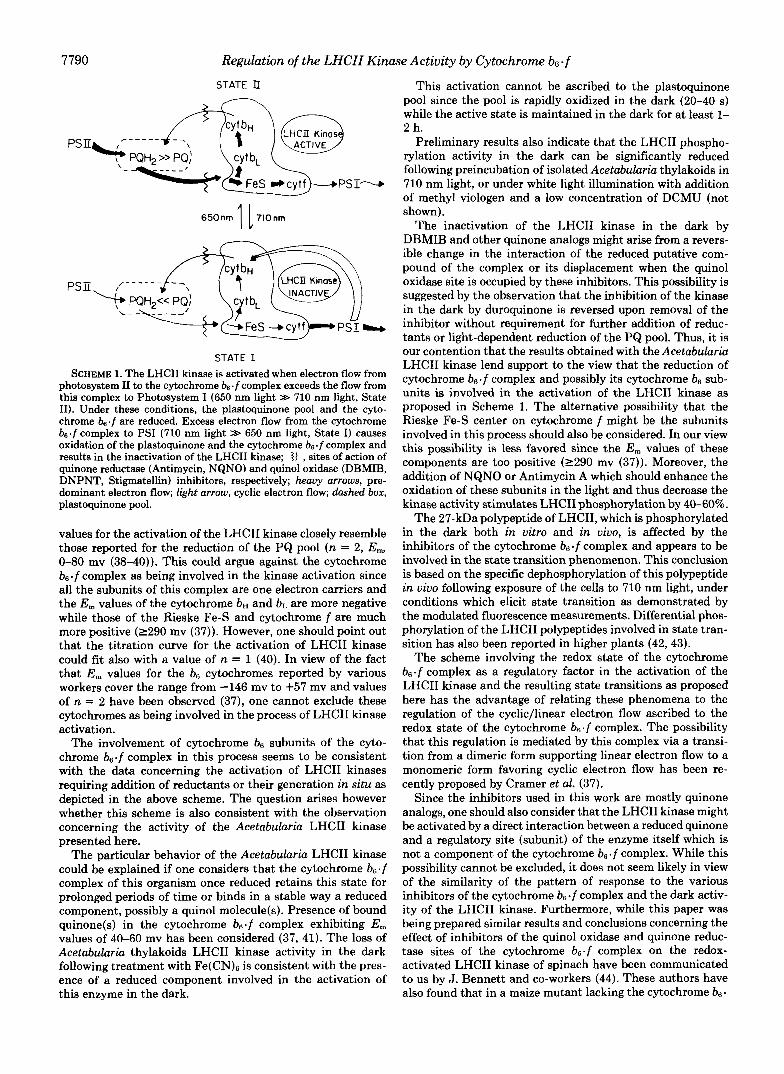
f complex (11) prompted us to reconsider the accepted schemes ascribing a direct role of activating the LHCII kinase to the reduced plastoquinone pool. We propose here as a working hypothesis that the activation of this kinase system is mediated by the redox state of a cytochrome b6.f complex component which in turn responds to the oxidation/reduction state of the plastoquinone pool (Scheme 1).
The inhibition of LHCII kinase by DBMIB while the plastoquinone pool is reduced by light-driven electron flow or in the presence of duroquinol is not consistent with a direct interaction between the PQH2 pool and the LHCII kinase which, however, might be mediated via the cytochrome b6.f complex. The K150 value for DBMIB under the above condi- tions was higher (5-10 WM) than that reported for the binding of this inhibitor to the quinol oxidase site of the cytochrome b6.f complex (0.1-1 pM (29)). This could be due to the com- petition between DBMIB and the reduced quinones for the same binding site. Duroquinol was reported to interact di- rectly with the quinol oxidase site of the cytochrome bs.f complex (36). The activity of the Acetabularia kinase in the dark in absence of added reductants is indeed inhibited by DBMIB and other halogenated quinone analogs with an KIso of 0.1-1 WM. Thus, the light-driven reduction of the PQ pool required for the activation of LHCII kinase may be needed to reduce the cytochrome bs . f complex. Inhibition of electron flow via the quinone reductase site by NQNO or Antimycin A (37) enhanced the activation of LHCII kinase in the light (Table I), or in the dark with addition of DQH, (not shown). Under these conditions both NQNO and Antimycin A can promote the reduction of the cytochrome b6.f by blocking the quinone-reducing site and thus its reoxidation (Scheme 1).
It has been reported before that the mid-point potential
7790 Regulation of the LHCII Kinase Activity by Cytochrome bs.f
STATE II -
650nm 1 1710nrn
PS Ii
STATE I SCHEME 1. The LHCII kinase is activated when electron flow from
photosystem I1 to the cytochrome bs.fcomplex exceeds the flow from this complex to Photosystem I (650 nm light >> 710 nm light, State 11). Under these conditions, the plastoquinone pool and the cyto- chrome b6.f are reduced. Excess electron flow from the cytochrome b, .f complex to PSI (710 nm light >> 650 nm light, State I) causes oxidation of the plastoquinone and the cytochrome be.f complex and results in the inactivation of the LHCII kinase; <I , sites of action of quinone reductase (Antimycin, NQNO) and quinol oxidase (DBMIB, DNPNT, Stigmatellin) inhibitors, respectively; heavy arrows, pre- dominant electron flow; light arrow, cyclic electron flow; dashed box, plastoquinone pool.
values for the activation of the LHCII kinase closely resemble those reported for the reduction of the PQ pool (n = 2, E,,,, 0-80 mv (38-40)). This could argue against the cytochrome b6 .f complex as being involved in the kinase activation since all the subunits of this complex are one electron carriers and the E , values of the cytochrome bH and bL are more negative while those of the Rieske Fe-S and cytochrome f are much more positive (2290 mv (37)). However, one should point out that the titration curve for the activation of LHCII kinase could fit also with a value of n = 1 (40). In view of the fact that E , values for the b6 cytochromes reported by various workers cover the range from -146 mv to +57 mv and values of n = 2 have been observed (37), one cannot exclude these cytochromes as being involved in the process of LHCII kinase activation.
The involvement of cytochrome be subunits of the cyto- chrome b6.f complex in this process seems to be consistent with the data concerning the activation of LHCII kinases requiring addition of reductants or their generation in situ as depicted in the above scheme. The question arises however whether this scheme is also consistent with the observation concerning the activity of the Acetabularia LHCII kinase presented here.
The particular behavior of the Acetabularia LHCII kinase could be explained if one considers that the cytochrome b6.f complex of this organism once reduced retains this state for prolonged periods of time or binds in a stable way a reduced component, possibly a quinol molecule(s). Presence of bound quinone(s) in the cytochrome b6.f complex exhibiting E, values of 40-60 mv has been considered (37, 41). The loss of Acetabularia thylakoids LHCII kinase activity in the dark following treatment with Fe(CN)6 is consistent with the pres- ence of a reduced component involved in the activation of this enzyme in the dark.
This activation cannot be ascribed to the plastoquinone pool since the pool is rapidly oxidized in the dark (20-40 s) while the active state is maintained in the dark for at least 1- 2 h.
Preliminary results also indicate that the LHCII phospho- rylation activity in the dark can be significantly reduced following preincubation of isolated Acetabularia thylakoids in 710 nm light, or under white light illumination with addition of methyl viologen and a low concentration of DCMU (not shown).
The inactivation of the LHCII kinase in the dark by DBMIB and other quinone analogs might arise from a revers- ible change in the interaction of the reduced putative com- pound of the complex or its displacement when the quinol oxidase site is occupied by these inhibitors. This possibility is suggested by the observation that the inhibition of the kinase in the dark by duroquinone is reversed upon removal of the inhibitor without requirement for further addition of reduc- tants or light-dependent reduction of the PQ pool. Thus, it is our contention that the results obtained with the Acetabularia LHCII kinase lend support to the view that the reduction of cytochrome b6 .f complex and possibly its cytochrome be sub- units is involved in the activation of the LHCII kinase as proposed in Scheme 1. The alternative possibility that the Rieske Fe-S center on cytochrome f might be the subunits involved in this process should also be considered. In our view this possibility is less favored since the E , values of these components are too positive (2290 mv (37)). Moreover, the addition of NQNO or Antimycin A which should enhance the oxidation of these subunits in the light and thus decrease the kinase activity stimulates LHCII phosphorylation by 40-60%.
The 27-kDa polypeptide of LHCII, which is phosphorylated in the dark both in vitro and in vivo, is affected by the inhibitors of the cytochrome b6 .f complex and appears to be involved in the state transition phenomenon. This conclusion is based on the specific dephosphorylation of this polypeptide in vivo following exposure of the cells to 710 nm light, under conditions which elicit state transition as demonstrated by the modulated fluorescence measurements. Differential phos- phorylation of the LHCII polypeptides involved in state tran- sition has also been reported in higher plants (42, 43).
The scheme involving the redox state of the cytochrome b6.f complex as a regulatory factor in the activation of the LHCII kinase and the resulting state transitions as proposed here has the advantage of relating these phenomena to the regulation of the cyclic/linear electron flow ascribed to the redox state of the cytochrome b6.f complex. The possibility that this regulation is mediated by this complex via a transi- tion from a dimeric form supporting linear electron flow to a monomeric form favoring cyclic electron flow has been re- cently proposed by Cramer et al. (37).
Since the inhibitors used in this work are mostly quinone analogs, one should also consider that the LHCII kinase might be activated by a direct interaction between a reduced quinone and a regulatory site (subunit) of the enzyme itself which is not a component of the cytochrome b6.f complex. While this possibility cannot be excluded, it does not seem likely in view of the similarity of the pattern of response to the various inhibitors of the cytochrome b6.f complex and the dark activ- ity of the LHCII kinase. Furthermore, while this paper was being prepared similar results and conclusions concerning the effect of inhibitors of the quinol oxidase and quinone reduc- tase sites of the cytochrome b6.f complex on the redox- activated LHCII kinase of spinach have been communicated to us by J. Bennett and co-workers (44). These authors have also found that in a maize mutant lacking the cytochrome b6.

Regulation of the LHCII Kinase Activity by Cytochrome b6 f 7791
f complex the LHCII kinase is inactive as previously reported by us for a Lemna mutant (11). Involvement of the cyto- chrome b6.f complex in the activation of LHCII kinase of Chlamydomom was also reported by Wolman and co-workers (45). The finding of a permanently active LHCII kinase system which, however, responds to the alteration of the cytochrome bs-f complex redox state adds support to the concept that the regulation of LHCII kinase activity is me- diated via the redox state of a cytochrome b6- f complex component, possibly one of the cytochrome b6 subunits.
Acknowledgments-We wish to thank Professor Peter Traub and Dr. Siegrid Berger of the Max-Planck Institute of Cell Biology, Rosenhoff, Ladenburg, F. R. G., for their invaluable help in providing us with the Acetabularia cultures throughout this work.
REFERENCES
1. Bennett, J., Steinback, K. E., and Arntzen, C. J. (1980) Proc.
2. Allen, J. F., Bennett, J., Steinback, K. E., and Arntzen, C. J.
3. Staehelin, L. A., and Arntzen, C. J. (1983) J. Cell Biol. 9 7 , 1327-
4. Allen, J. F., and Horton, P. (1981) Biochim. Biophys. Acta 6 3 8 ,
5. Kyle, D. J., Haworth, P., and Artzen, C. J. (1982) Biochim.
6. Haworth, P., Kyle, D. J., and Arntzen, C. J. (1982) Biochim.
7. Black, M. T., and Horton, P. (1984) Biochim. Biophys. Acta 7 6 7 ,
8. Canaani, O., Barber, J., and Malkin, S. (1984) Proc. Natl. Acad.
9. Barber, J. (1986) Photosynth. Res. 10 , 243-253
Natl. Acad. Sei. U. S. A. 77,5253-5257
(1981) Nature 2 9 1 , 21-25
1337
290-295
Biophys. Acta 680,336-342
Biophys. Acta 680,343-351
568-573
S C ~ . U. S. A. 8 1 , 1614-1618
10. Bennett, J., Shaw, E. K., and Bakr, S. (1987) FEBS Lett. 2 1 0 ,
11. Gal, A., Shahak, Y., Schuster, G., and Ohad, I. (1987) FEBS Lett. 221,205-210
12. Schuster, G., Owens, G. C., Cohen, Y., and Ohad, I. (1984) Biochim. Biophys. Acta 767,596-605
13. Schweiger, H.-G., Dehm, S., and Berger, S. (1977) in Progress in Acetabularia Research (Woodcock, C. L. F., ed) pp. 319-330, Academic Press, New York
14. Kloppstech, K., Ohad, I., and Schweiger, H.-G. (1986) Eur. J. Cell Biol. 4 2 , 239-245
15. Schuster, G., Dewit, M., Staehelin, L. A., and Ohad, I. (1986) J. Cell Biol. 103,71-80
22-26
16. Laemmli, U. K. (1970) Nature 227 , 680-685 17. Bollum, F. (1965) in Procedures in Nucleic Acid Research (Can-
toni, G. L., and Davis, D. R., eds) pp. 296-300, Harper and Row, New York
18. Canaani, O., and Malkin, S. (1984) Biochim. Biophys Acta 7 6 6 ,
19. Arnon, D. I. (1949) Plant Physiol. 2 4 , 1-15 20. Peterson, G. L. (1977) Anal. Biochem. 8 3 , 346-356 21. Steinback, K. E., Bose, S., and Kyle, D. J. (1982) Arch. Biochim.
22. Millner, P. A., Marder, J. B., Gounaris, K., and Barber, J. (1986)
23. Owens, G. C., and Ohad, I. (1983) Biochim. Biophys. Acta 7 2 2 ,
24. Bennoun, P. (1982) Proc. Natl. Acad. Sci. U. S. A. 79,4352-4356 25. Wollman, F-A., and Delepelaire, P. (1984) J. Cell Biol. 9 8 , 1-7 26. Moll, B. A., and Steinback, K. E. (1986) Plant Physiol. 80,420-
27. Emerson, R. (1958) Annu. Reu. Plant Physiol. 9, 1-24 28. Myers, J. (1971) Annu. Reu. Plant Physiol. 22,289-312 29. Malkin, R. (1982) Biochemistry 2 1 , 2945-2950 30. Trebst, A. (1980) Methods Enzymol. 69,675-715 31. Malkin, R. (1986) FEBS Lett. 208,317-320 32. Oettmeier, W., Masson, K., and Dostatni, R. (1987) Biochim.
33. Hartung, A., and Trebst, A. (1985) Physiol. Veg. 2 3 , 635-648 34. Jones, R. W., and Whitmarsh, J. (1985) Photobiochem. Photobio-
35. Oettmeier, W., Godde, D., Kunze, B., and Hofle, G. (1985)
36. Nanba, M., and Katoh, S. (1986) Biochim. Biophys. Acta 8 5 1 ,
37. Cramer, W. A., Black, M. T., Widger, W. R., and Givrin, M. F. (1987) in The Light Reactions (Barber, J., ed) pp. 447-493, Elsevier Scientific Publishing Co., New York
38. Horton, P., Allen, J. F., Black, M. T., and Bennett, J. (1981)
39. Horton, P., and Black, M. T. (1980) FEBS Lett. 119 , 141-144 40. Millner, P. A., Widger, W. R., Abbott, M. S., Cramer, W. A., and
41. Rich, P. R. (1984) Biochim. Biophys. Acta 768,53-79 42. Larsson, U. K., and Andersson, B. (1985) Biochim. Biophys. Acta
43. Larsson, U. K., Sundby, C., and Andersson, B. (1987) Biochim. Biophys. Acta 894,59-68
44. Bennett, J., Shaw, E. K., and Michel, H. (1988) Eur. J. Biochem.,
45. Lemaire, C., Girard-Bascou, J., and Wollman, F.-A. (1986) in in press
Progress in Photosynthesis Research (Biggins, J., ed) Vol. IV, pp. 655-658, Nijhoff, Dordrecht
513-524
Biophys. 216,356-361
Biochim. Biophys. Acta 852, 30-37
234-241
423
Biophys. Acta 890,260-269
phys. 9, 119-127
Biochim. Biophys. Acta 807,216-219
484-490
FEBS Lett., 125,193-196
Dilley, R. A. (1982) J. Biol. Chem. 257 , 1736-1742
809,396-402