role of stat3 signaling pathway in breast cancer
TRANSCRIPT
REVIEW Open Access
Role of STAT3 signaling pathway in breastcancerJia-hui Ma1†, Li Qin2,3† and Xia Li1,4*
Abstract
Breast cancer has grown to be the second leading cause of cancer-related deaths in women. Only a few treatmentoptions are available for breast cancer due to the widespread occurrence of chemoresistance, which emphasizesthe need to discover and develop new methods to treat this disease. Signal transducer and activator of transcription 3(STAT3) is an early tumor diagnostic marker and is known to promote breast cancer malignancy. Recent clinical andpreclinical data indicate the involvement of overexpressed and constitutively activated STAT3 in the progression,proliferation, metastasis and chemoresistance of breast cancer. Moreover, new pathways comprised of upstreamregulators and downstream targets of STAT3 have been discovered. In addition, small molecule inhibitors targetingSTAT3 activation have been found to be efficient for therapeutic treatment of breast cancer. This systematic reviewdiscusses the advances in the discovery of the STAT3 pathways and drugs targeting STAT3 in breast cancer.
Keywords: STAT3, Breast cancer, Oncogene, Small molecule inhibitors
BackgroundTranscription factors (TFs) are proteins possessing do-mains that bind to the DNA of promoter or enhancerregions of specific genes. Several TFs are directly in-volved in the development and progression of breastcancer. One of the most prominent TF families in breastcancer is the signal transducers and activators of tran-scription (STAT) family, which is comprised of sevenstructurally similar and highly conserved members,namely, STAT1, STAT2, STAT3, STAT4, STAT5a,STAT5b and STAT6 [1, 2]. In general, these familymembers contain six common functional domains: anN-terminal domain (NH2) which is called STAT_intnow, a coiled-coil domain (CCD), a DNA-binding do-main (DBD), a linker domain, an SRC homology 2 do-main (SH2) and a transactivation domain (TAD) [3].
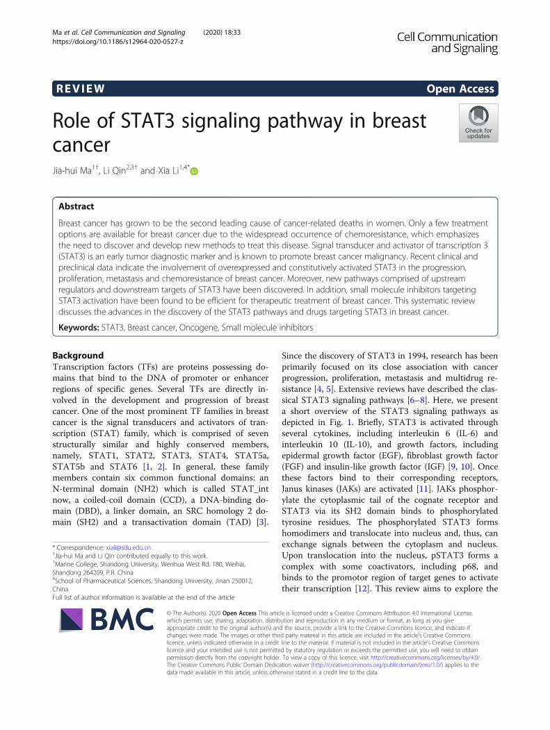
Since the discovery of STAT3 in 1994, research has beenprimarily focused on its close association with cancerprogression, proliferation, metastasis and multidrug re-sistance [4, 5]. Extensive reviews have described the clas-sical STAT3 signaling pathways [6–8]. Here, we presenta short overview of the STAT3 signaling pathways asdepicted in Fig. 1. Briefly, STAT3 is activated throughseveral cytokines, including interleukin 6 (IL-6) andinterleukin 10 (IL-10), and growth factors, includingepidermal growth factor (EGF), fibroblast growth factor(FGF) and insulin-like growth factor (IGF) [9, 10]. Oncethese factors bind to their corresponding receptors,Janus kinases (JAKs) are activated [11]. JAKs phosphor-ylate the cytoplasmic tail of the cognate receptor andSTAT3 via its SH2 domain binds to phosphorylatedtyrosine residues. The phosphorylated STAT3 formshomodimers and translocate into nucleus and, thus, canexchange signals between the cytoplasm and nucleus.Upon translocation into the nucleus, pSTAT3 forms acomplex with some coactivators, including p68, andbinds to the promotor region of target genes to activatetheir transcription [12]. This review aims to explore the
© The Author(s). 2020 Open Access This article is licensed under a Creative Commons Attribution 4.0 International License,which permits use, sharing, adaptation, distribution and reproduction in any medium or format, as long as you giveappropriate credit to the original author(s) and the source, provide a link to the Creative Commons licence, and indicate ifchanges were made. The images or other third party material in this article are included in the article's Creative Commonslicence, unless indicated otherwise in a credit line to the material. If material is not included in the article's Creative Commonslicence and your intended use is not permitted by statutory regulation or exceeds the permitted use, you will need to obtainpermission directly from the copyright holder. To view a copy of this licence, visit http://creativecommons.org/licenses/by/4.0/.The Creative Commons Public Domain Dedication waiver (http://creativecommons.org/publicdomain/zero/1.0/) applies to thedata made available in this article, unless otherwise stated in a credit line to the data.
* Correspondence: [email protected]†Jia-hui Ma and Li Qin contributed equally to this work.1Marine College, Shandong University, Wenhua West Rd. 180, Weihai,Shandong 264209, P.R. China4School of Pharmaceutical Sciences, Shandong University, Jinan 250012,ChinaFull list of author information is available at the end of the article
Ma et al. Cell Communication and Signaling (2020) 18:33 https://doi.org/10.1186/s12964-020-0527-z
mechanism of STAT3 in breast cancer development andsummarize the latest advancements made.
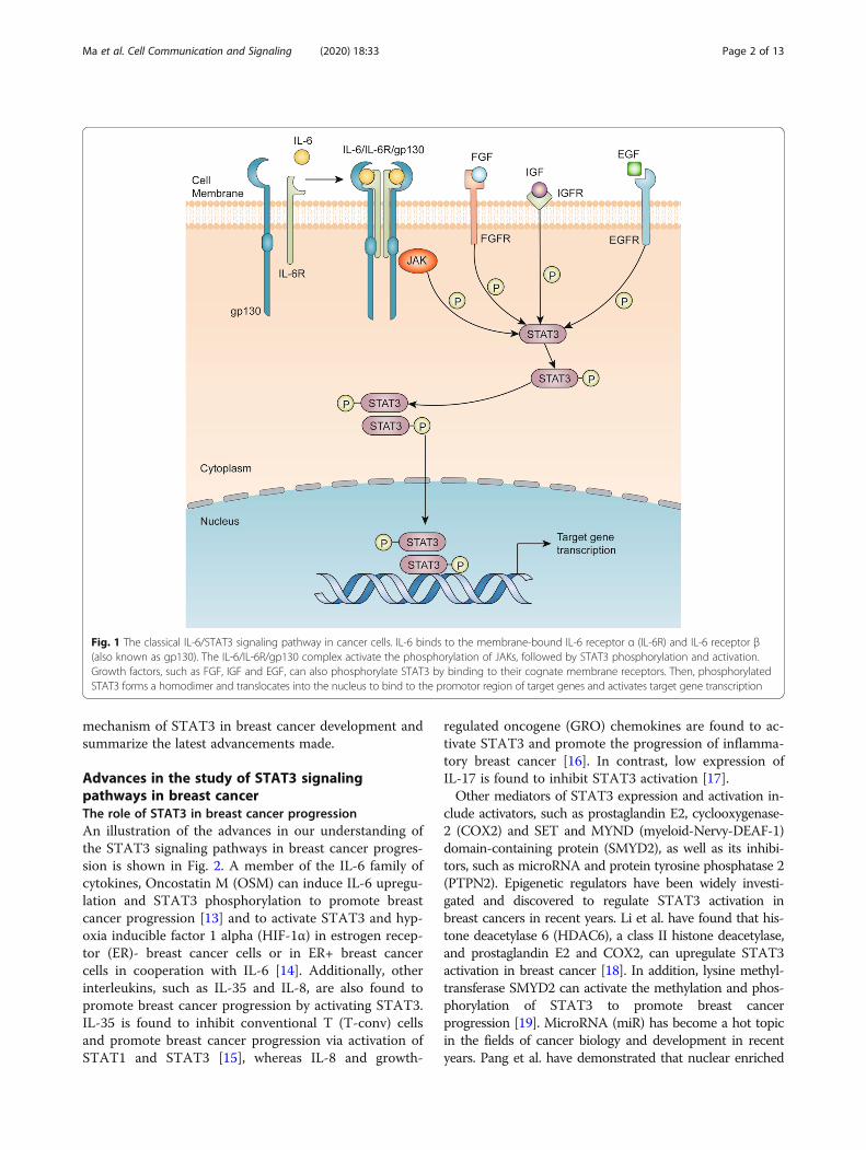
Advances in the study of STAT3 signalingpathways in breast cancerThe role of STAT3 in breast cancer progressionAn illustration of the advances in our understanding ofthe STAT3 signaling pathways in breast cancer progres-sion is shown in Fig. 2. A member of the IL-6 family ofcytokines, Oncostatin M (OSM) can induce IL-6 upregu-lation and STAT3 phosphorylation to promote breastcancer progression [13] and to activate STAT3 and hyp-oxia inducible factor 1 alpha (HIF-1α) in estrogen recep-tor (ER)- breast cancer cells or in ER+ breast cancercells in cooperation with IL-6 [14]. Additionally, otherinterleukins, such as IL-35 and IL-8, are also found topromote breast cancer progression by activating STAT3.IL-35 is found to inhibit conventional T (T-conv) cellsand promote breast cancer progression via activation ofSTAT1 and STAT3 [15], whereas IL-8 and growth-
regulated oncogene (GRO) chemokines are found to ac-tivate STAT3 and promote the progression of inflamma-tory breast cancer [16]. In contrast, low expression ofIL-17 is found to inhibit STAT3 activation [17].Other mediators of STAT3 expression and activation in-
clude activators, such as prostaglandin E2, cyclooxygenase-2 (COX2) and SET and MYND (myeloid-Nervy-DEAF-1)domain-containing protein (SMYD2), as well as its inhibi-tors, such as microRNA and protein tyrosine phosphatase 2(PTPN2). Epigenetic regulators have been widely investi-gated and discovered to regulate STAT3 activation inbreast cancers in recent years. Li et al. have found that his-tone deacetylase 6 (HDAC6), a class II histone deacetylase,and prostaglandin E2 and COX2, can upregulate STAT3activation in breast cancer [18]. In addition, lysine methyl-transferase SMYD2 can activate the methylation and phos-phorylation of STAT3 to promote breast cancerprogression [19]. MicroRNA (miR) has become a hot topicin the fields of cancer biology and development in recentyears. Pang et al. have demonstrated that nuclear enriched
Fig. 1 The classical IL-6/STAT3 signaling pathway in cancer cells. IL-6 binds to the membrane-bound IL-6 receptor α (IL-6R) and IL-6 receptor β(also known as gp130). The IL-6/IL-6R/gp130 complex activate the phosphorylation of JAKs, followed by STAT3 phosphorylation and activation.Growth factors, such as FGF, IGF and EGF, can also phosphorylate STAT3 by binding to their cognate membrane receptors. Then, phosphorylatedSTAT3 forms a homodimer and translocates into the nucleus to bind to the promotor region of target genes and activates target gene transcription
Ma et al. Cell Communication and Signaling (2020) 18:33 Page 2 of 13
abundant transcript 1 (NEAT1) forms a feedback loop withSTAT3 to promote breast cancer progression. However,NEAT1 is suppressed by miR-124 [20]. Interestingly, glu-cosamine is found to suppress the activation of STAT3 anddecrease breast cancer stemness and progression [21]. Add-itionally, knockdown of PTPN2 leads to EGF-mediatedSTAT3 activation [22]. The association of chronic inflam-mation with breast cancer progression is widely recognized,but it can be inhibited by blocking STAT3 [23]. Other me-diators of STAT3 signaling pathways are also extensivelystudied. Kim et al. have found that the IL-6/STAT3/ROSpathway can not only promote breast cancer progressionand inflammation but also increase the formation of breastcancer stem cells [24]. Moreover, TGFβ-regulated FAM3C/Interleukin-like EMT Inducer (ILEI), an oncogenic memberof the FAM3 cytokine family, can mediate STAT3 signalingpathway to drive breast cancer stem cell formation and
promote breast cancer progression [25]. In addition,TNFRSF1A, a gene encoding a transmembrane receptorfor TNF-α, can be modulated by STAT3 and promote NF-κB signaling in breast cancer [26].There were also some STAT3 co-factors influenced
the proliferation and progress of breast cancer. Progranu-lin (PGRN), was seen to associate with chemoresistanceand worse prognosis in breast cancer [27, 28], and the useof a specific progranulin antisense oligonucleotide was re-cently seen to hamper STAT3 oncogenic functions inCRC cells [29], suggesting a similar effect also in breastcancer cells. The cyclin dependent kinase 5 (CDK5) regu-latory subunit-associated protein 3 (CDK5RAP3, alsocalled C53/LZAP) was originally regarded as a p53 co-activator [30]. A recent research reported that CDK5RAP3was associated with primary breast cancer progressionand proliferation, and also enhanced the expression of
Fig. 2 Advances of the STAT3 signaling pathways involved in breast cancer progression. Interleukins, including IL-6, IL-8 and IL-35, can bind totheir receptors and activate the phosphorylation of JAKs and STAT3, OSM can increase IL-6-mediated activation, and IL-17 binding to its receptorleads to inhibition of STAT3 phosphorylation. STAT3 phosphorylated by EGF can be inhibited by PTPN2. COX2 and prostaglandin E2 upregulatedby HDAC6 can activate STAT3 phosphorylation, and SMYD2 has a similar effect. Additionally, STAT3 and NEAT1 can form a loop to activate thephosphorylation of STAT3, which is inhibited by miR-124. The activated and phosphorylated STAT3 dimers translocate into the nucleus andactivate the transcription of target genes involved in breast cancer progression
Ma et al. Cell Communication and Signaling (2020) 18:33 Page 3 of 13
STAT3-dependent genes [31]. Thus, targeting the co-factor of STAT3 maybe a potential therapeutic approachin breast cancer management.
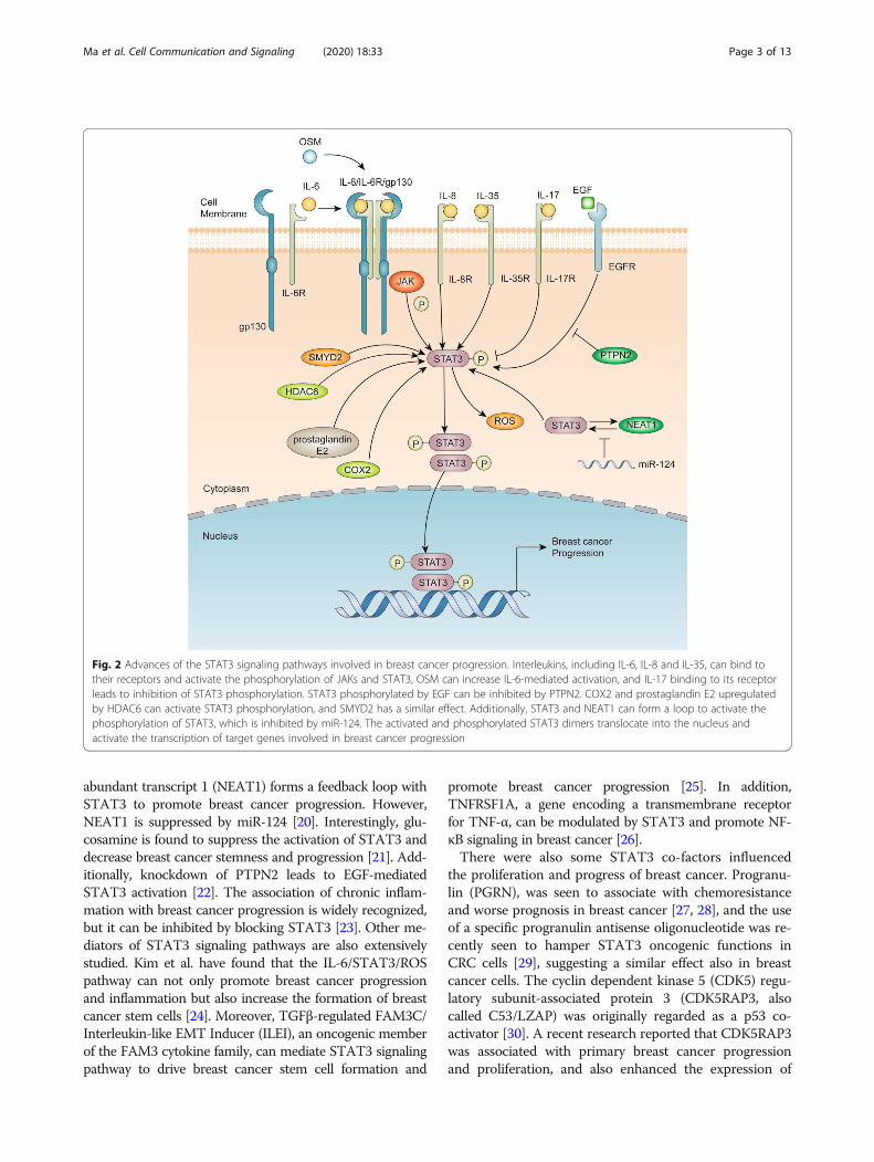
The role of STAT3 in breast cancer proliferation andapoptosisThe illustration with advances of STAT3 signaling path-ways in breast cancer proliferation and apoptosis isshown in Fig. 3. A recent research has reported thatdownregulation of zinc-finger gene DPF3 (also known asCERD4) promotes proliferation and motility of breastcancer via activating JAK2/STAT3 pathway [32]. It hasbeen reported earlier that STAT3 can upregulate cyclinD-1, c-myc, and bcl-2 to suppress the apoptosis of breastcancer cells, indicating a potential involvement of STAT3
in cell cycle and survival [33]. Moreover, STAT3 activatedby IL-6/JAK2 pathway can inhibit Bax/Bcl-2-relatedcaspase-dependent apoptosis [34]. However, overexpres-sion of WW domain-containing oxidoreductase (Wwox)blocks the combination of STAT3 and IL-6R, resulting ininhibition of proliferation [35]. Another research showsthat IL-32θ targets chemokine ligand (CCL)18/STAT3pathway to suppress macrophage-promoted breast cancerprogression [36]. In addition, miRNAs are also widely in-vestigated in breast cancer proliferation and invasion. Parket al. have found that miR-125a and let-7e could inhibitIL-6/STAT3 pathway to mediate the breast cancer prolif-eration and vasculogenic mimicry formation [37], and Shiet al. have found that miR-124 could suppress the mRNAand protein levels of STAT3 and inhibit the proliferation
Fig. 3 Advances of the STAT3 signaling pathways involving breast cancer proliferation and apoptosis. Classical IL-6 /JAK/STAT3 pathways canactivate the transcription of cyclin D-1, c-myc, bcl-2 and Bax to promote the proliferation and inhibit the apoptosis of breast cancer. miR-125a,miR-25-3p and p16 can promote the binding of IL-6 to its receptors, whereas Wwox has the opposite effect. CCL-18 binding to its receptor canactivate the phosphorylation of STAT3, which can be inhibited by IL-32θ. The circuit loop of phosphorylated STAT3, TMEM16A and EGF leads tocontinuous activation of STAT3. miR-93-5p, SMYD2, TRIM14 and PKT-M2 induce the activation of STAT3, whereas miR-124 and miR-9 inhibit theactivation of STAT3 and breast cancer proliferation. Let-7a-5p, hnRN-A and phosphorylated STAT3 dimers form a circuit loop to upregulate PKM2and promote the proliferation and inhibit the apoptosis of breast cancer cells. DPF3 suppressed by phosphorylated STAT3 can promote breastcancer proliferation. Additionally, transcription factor EB (TFEB) can combine with phosphorylated STAT3 dimers to promote the transcription oftarget genes involved in breast cancer proliferation
Ma et al. Cell Communication and Signaling (2020) 18:33 Page 4 of 13
and invasion of breast cancer [38]. Similarly, miR-9 isreported to inhibit STAT3 activation and breast cancerproliferation [39]. In contrast, miR-93-5p and miR-25-3pare found to mediate STAT3 and promote breast cancerproliferation [40, 41]. Since the discovery of Warburgeffects, metabolism is strongly linked with proliferation ofcancer cells. It has been suggested that let-7a-5p, Stat3,and hnRNP-A1 form a feedback loop to regulate PKM2expression and modulate glucose metabolism in breastcancer cells, suggesting that inhibiting STAT3-related me-tabolism may inhibit breast cancer proliferation [42].There are several new pathways associated with STAT3
and breast cancer that have been minimally studied to date.It has been revealed that the Ca2+ activated chloride chan-nel TMEM16A forms an activation loop with EGFR/STAT3 to promote breast cancer proliferation [43]. More-over, tripartite motif-containing 14 (TRIM14) is found toincrease the expression of p-STAT3 to promote breast can-cer proliferation [44]. In addition, it is reported that pyru-vate kinase type M2 (PKT-M2) regulates phosphorylationof STAT3 in breast cancer [45], whereas cystathionine-lyase (CSE) suppresses the expression of STAT3/matrixmetallopeptidases-2 (MMP2), MMP9, p-protein kinase Band B-cell lymphoma 2 [46].
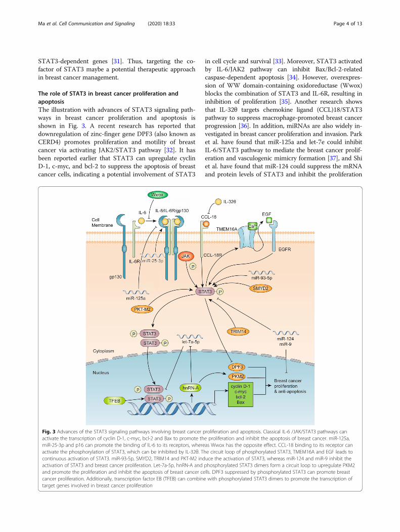
The role of STAT3 in breast cancer metastasisAn illustration of the advances of the STAT3 signalingpathways in breast cancer metastasis is shown in Fig. 4.Matrix metallopeptidases (MMPs) are known to playimportant roles in breast cancer metastasis. A well-studied mechanism of STAT3-mediated cell metastasisis through upregulating MMP2, MMP9, Twist, Snail,Slug and vimentin [47–49]. Ma et al. have reported thatinhibition of STAT3 phosphorylation could reduce theexpression of vasodilator-stimulated phosphoprotein(VASP), MMP2 and MMP9 in breast cancer [50]. Asmentioned previously, STAT3 signaling is usually acti-vated upon binding of cytokines and growth factors totheir cognate receptors on the plasma membrane. Thepreviously mentioned Wwox can inhibit breast cancermetastasis by preventing receptor binding [35]. Further-more, Kim et al. have demonstrated that Mesoderm-specific transcript (MEST) induces Twist expression byactivating the JAK/STAT3 signaling pathway [51],whereas Khanna et al. have shown the inhibition ofGRAM domain-containing protein 1B (GRAMD1B) inbreast cancer migration via the suppression of the JAK/STAT3 and protein kinase B (Akt) pathway [52]. Insteadof classical ligand/receptor binding in the plasma mem-brane for STAT3 activation, a new pathway is found inwhich OSM/SMAD3 could also activate STAT3 andmediate Snail expression and promote epithelial-mesenchymal transition (EMT) in breast cancer, indicat-ing a distinct route of STAT3 activation through
cytoplasmic molecules and endogenous signaling [53].Other signaling molecules, including miRNA, proto-oncogene serine/threonine-protein kinase (PIM1),Mucin-1-C (MUC1-C), natriuretic peptide receptor A(NPRA) and RhoU, were also discovered to participatein STAT3-mediated breast cancer metastasis. miR-30dis found to mediate migration and invasion in breastcancer cells by regulating Krüppel-like factor 11(KLF-11), a new exogenous signaling pathway thatcan activate STAT3 by binding to its transmembranereceptor KLF-11R [54]. In addition, IL-11 is alsofound to regulate the JAK/STAT3 pathway in breastcancer-bone metastasis [55]. PIM1, a proto-oncogeneresponsible for promoting cell invasion and upregulat-ing EMT expression in breast cancer, is found to beregulated by the IL-6/STAT3 signaling pathway [56].MUC1-C, an oncogenic protein, can activate STAT3and induce Twist transactivation to promote EMT[57]. Moreover, NPRA, one of the natriuretic peptidereceptors, is found to increase the expression ofSTAT3 and MMP9 to promote the migration andinvasion of breast cancer cells [58]. STAT3, by co-operating with Specificity Protein 1 (SP1), is found toinduce high Ras Homolog Family Member U (RhoU)expression and breast cancer cell migration [59]. Add-itionally, some enzymes are also found to participatein breast cancer metastasis by the posttranscriptionalmodification of STAT3. ARHGAP24, a Rac-specificRho GTPase-activating protein (Rho GAP), is foundto promote phosphorylation of STAT3 and to in-crease the expression of MMP2 and MMP9 in breastcancer cells [60]. GCN5, a histone acetyltransferase, isfound to upregulate the expression of p-STAT3, p-AKT, MMP9 and E2F1 and promote breast cancermigration and invasion [61].Hypoxia is a stressed state that is extensively studied in
cancers. Abyaneh et al. have found that hypoxia cansignificantly induce the activation of STAT3 to promotebreast cancer stemness and metastasis [62]. Thisphenomenon provides us with a new direction for STAT3research and targeted STAT3 therapy in breast cancer.Moreover, our recent research has found that estrogen re-lated receptor alpha could promote the metastasis of triplenegative breast cancer as a target gene of STAT3 [63].
The role of STAT3 in breast cancer chemoresistanceAn illustration of the advances of the STAT3 signalingpathways in breast cancer chemoresistance is shown inFig. 5. Tzeng et al. have indicated that the Src/STAT3signaling pathway is involved in multidrug resistance intriple negative breast cancer cells [64]. It is also foundthat crosstalk between breast cancer cells and macro-phages can induce tamoxifen and ICI 182,780 resistancethrough the NF-κB/STAT3/ERK pathways [65].
Ma et al. Cell Communication and Signaling (2020) 18:33 Page 5 of 13
The newly discovered downstream targets of STAT3-mediated chemoresistance include fatty acid beta-oxidation (FAO), carnitine palmitoyltransferase 1B(CPT1B), mitogen-activated protein kinase (MAPK)/AKT, HIF-1 and octamer-binding transcription factor-4(Oct-4). It has been found that the JAK2/STAT3 signal-ing pathway increases CPT1B and FAO to increase che-moresistance in breast cancer [66]. Wang et al. foundthat IL-22 can promote JAK-STAT3/MAPKs/AKT path-way activation to induce breast cancer migration andpaclitaxel resistance [67]. Moreover, miR-124 has beenidentified to reverse doxorubicin (DOX) resistance ofbreast cancer cells through targeting the STAT3/hyp-oxia-inducible factor 1 (HIF-1) pathway [68]. A recentstudy shows that Oct-4 and c-myc can form a signal
circuit to increase Adriamycin resistance in breast can-cer [69]. Meanwhile, Kim et al. have discovered thatOct-4 confers radiation resistance via STAT3 and NF-B-mediated IL-24 production in breast cancer cells [70]. Inaddition, paclitaxel is widely used as a clinical drug ofbreast cancer treatment, and phosphorylated STAT3could mediate Survivin to promote paclitaxel resistance[71].There are several upstream regulators of STAT3-
mediated chemoresistance that have been identified inrecent years. The COOH-terminal proline-rich region of78-kDa glucose-regulated protein (GRP78), by regulatingSTAT3, is found to play a crucial role in the develop-ment of tamoxifen-resistant breast cancer cells [72].Wang et al. have found that leukemia inhibitory factor
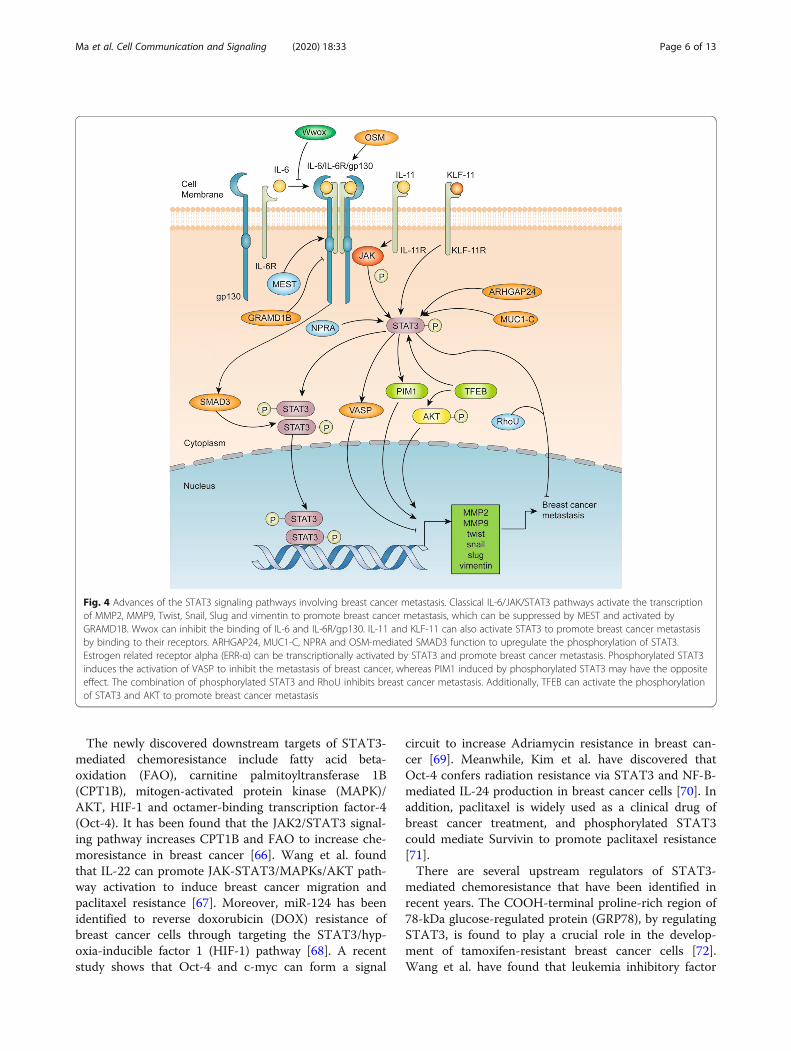
Fig. 4 Advances of the STAT3 signaling pathways involving breast cancer metastasis. Classical IL-6/JAK/STAT3 pathways activate the transcriptionof MMP2, MMP9, Twist, Snail, Slug and vimentin to promote breast cancer metastasis, which can be suppressed by MEST and activated byGRAMD1B. Wwox can inhibit the binding of IL-6 and IL-6R/gp130. IL-11 and KLF-11 can also activate STAT3 to promote breast cancer metastasisby binding to their receptors. ARHGAP24, MUC1-C, NPRA and OSM-mediated SMAD3 function to upregulate the phosphorylation of STAT3.Estrogen related receptor alpha (ERR-α) can be transcriptionally activated by STAT3 and promote breast cancer metastasis. Phosphorylated STAT3induces the activation of VASP to inhibit the metastasis of breast cancer, whereas PIM1 induced by phosphorylated STAT3 may have the oppositeeffect. The combination of phosphorylated STAT3 and RhoU inhibits breast cancer metastasis. Additionally, TFEB can activate the phosphorylationof STAT3 and AKT to promote breast cancer metastasis
Ma et al. Cell Communication and Signaling (2020) 18:33 Page 6 of 13
receptor (LIFR) could promote STAT3 activation andcontribute to breast cancer resistance to Trastuzumab-emtansine (T-DM1) [73]. Furthermore, miR-4532 isfound to suppress hypermethylated in cancer-1 (HIC-1)and IL-6/STAT3 to promote Adriamycin resistance inbreast cancer [74].Some small molecules have also been found to con-
tribute to chemoresistance mediated by STAT3. Piper-longumine combined with DOX is also found to induceapoptosis and inhibit DOX resistance of breast cancercells via the JAK/STAT3 pathway [75]. In addition, tar-geting IL6/STAT3 activity using STAT3 inhibitor com-bined with a poly ADP-ribose polymerase (PARP)inhibitor could effectively treat palbociclib resistance inbreast cancer cells [76].
Advances in the study of compounds targetingSTAT3 in breast cancerCompounds inhibiting the upstream of STAT3 in breastcancerSeveral compounds are found to inhibit the upstreammediators of STAT3 in breast cancer since 2018(Table 1). Many of these compounds target the IL-6/STAT3 signaling pathway. Ilamycin C is found to induceapoptosis and inhibit migration and invasion by sup-pressing the IL-6/STAT3 pathway [34]. A small mol-ecule, bazedoxifene, is a novel IL-6/GP130 inhibitor thatreduces breast cancer proliferation and migration [77].Moreover, Esparza-Lopez et al. have discovered the in-hibitory effect of metformin in IL-6-induced prolifera-tion and EMT through the STAT3/NF-κB pathway in
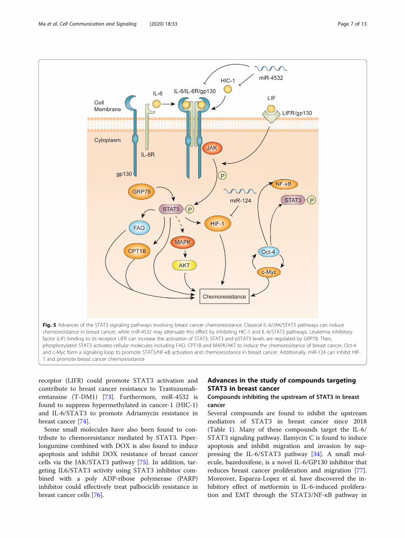
Fig. 5 Advances of the STAT3 signaling pathways involving breast cancer chemoresistance. Classical IL-6/JAK/STAT3 pathways can inducechemoresistance in breast cancer, while miR-4532 may attenuate this effect by inhibiting HIC-1 and IL-6/STAT3 pathways. Leukemia inhibitoryfactor (LIF) binding to its receptor LIFR can increase the activation of STAT3. STAT3 and pSTAT3 levels are regulated by GRP78. Then,phosphorylated STAT3 activates cellular molecules including FAO, CPT1B and MAPK/AKT to induce the chemoresistance of breast cancer. Oct-4and c-Myc form a signaling loop to promote STAT3/NF-κB activation and chemoresistance in breast cancer. Additionally, miR-124 can inhibit HIF-1 and promote breast cancer chemoresistance
Ma et al. Cell Communication and Signaling (2020) 18:33 Page 7 of 13
breast cancer [89]. DT-13, the saponin monomer 13 ofthe Dwarf lilyturf tuber, has been identified as a suppres-sor of breast cancer metastasis that acts by inhibitingboth JAK/STAT3 and PI3K/AKT signaling pathways[81]. Furthermore, a natural compound called esculento-side A, a triterpene saponin derived from the root of
Phytolacca esculenta, can also inhibit the IL-6/STAT3pathway [78]. Meanwhile, another nature compoundcalled catechol, which is derived from Aronia juice,shows similar effects in breast cancer cells [79]. Inaddition, scorpion venom can decrease IL-6, RhoC, ERK(1/2), and STAT3 and inhibit breast cancer proliferation
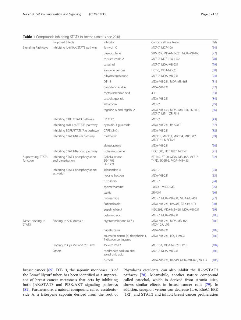
Table 1 Compounds inhibiting STAT3 in breast cancer since 2018
Proposed Effects Inhibitor Cancer cell line tested Refs
Signaling Pathways Inhibiting IL-6/JAK/STAT3 pathway Ilamycin C MCF-7, MCF-10A [34]
bazedoxifene SUM159, MDA-MB-231, MDA-MB-468 [77]
esculentoside A MCF-7, MCF-10A, LO2 [78]
catechol MCF-7, MDA-MB-231 [79]
scorpion venom HCT-8, MDA-MB-231 [80]
dihydrotanshinone MCF-7, MDA-MB-231 [24]
DT-13 MDA-MB-231, MDA-MB-468 [81]
ganoderic acid A MDA-MB-231 [82]
methylseleninic acid 4 T1 [83]
sesquiterpenoid MDA-MB-231 [84]
sabutoclax MCF-7 [85]
tagalide A and tagalol A MDA-MB-453, MDA- MB-231, SK-BR-3,MCF-7, MT-1, ZR-75-1
[86]
Inhibiting SIRT1/STAT3 pathway I157172 MCF-7 [43]
Inhibiting miR-124/STAT3 pathway cyanidin-3-glucoside MDA-MB-231, Hs-578 T [87]
Inhibiting EGFR/STAT3/Akt pathway CAPE-pNO2 MDA-MB-231 [88]
Inhibiting STAT3/NF-κB pathway metformin MBCDF, MBCD3, MBCD4, MBCD17,MBCD23, MBCD25
[89]
alantolactone MDA-MB-231 [90]
Inhibiting STAT3/Nanong pathway isoharringtonine HCC1806, HCC1937, MCF-7 [91]
Suppressing STAT3function
Inhibiting STAT3 phosphorylationand dimerization
GaliellalactoneSG-1709SG-1721
BT-549, BT-20, MDA–MB-468, MCF-7,T47D, SK-BR-3, MDA–MB-453
[92]
Inhibiting STAT3 phosphorylation/activation
schisandrin A MCF-7 [93]
hexane fraction MDA-MB-231 [33]
ruxolitinib MCF-7 [94]
pyrimethamine TUBO, TM40D-MB [95]
stattic ZR-75-1 [96]
niclosamide MCF-7, MDA-MB-231, MDA-MB-468 [97]
flubendazole MDA-MB-231, Hs578T, BT-549, 4 T1 [98]
eupalinolide J HEK 293, MDA-MB-468, MDA-MB-231 [99]
betulinic acid MCF-7, MDA-MB-231 [100]
Direct binding toSTAT3
Binding to SH2 domain cryptotanshinone KYZ3 MDA-MB-231, MDA-MB-468,MCF-10A, L02
[101]
napabucasin MDA-MB-231 [102]
coumarin-benzo [b] thiophene 1,1-dioxide conjugates
MDA-MB-231, LO2, HepG2 [103]
Binding to Cys 259 and 251 sites 15-keto PGE2 MCF10A, MDA-MB-231, PC3 [104]
Others risedronate sodium andzoledronic acid
MCF-7, MDA-MB-231 [105]
osthole MDA-MB-231, BT-549, MDA-MB-468, MCF-7 [106]
Ma et al. Cell Communication and Signaling (2020) 18:33 Page 8 of 13
[80]. As discussed previously, dihydrotanshinone inhibitsbreast cancer cells progression and stem cell formationthrough the IL-6/STAT3 pathway [24].Other compounds target different signaling pathways,
including the JAK2/STAT3 and Akt pathways. Bothganoderic acid A, which is isolated from ganoderma, andmethylseleninic acid are found to suppress breast cancerproliferation via the JAK2/STAT3 pathway [82, 83]. Acompound called caffeic acid p-nitro-phenethyl ester(CAPE-pNO2) is found to inhibit the EGFR/STAT3/Aktpathway and suppress breast cancer proliferation andmetastasis [88]. Moreover, I157172, a novel inhibitor ofcystathionine-lyase, is found to inhibit the proliferationand migration of breast cancer cells via upregulation ofSIRT1 and inhibition of STAT3 signaling pathway [46].Other compounds target the regulation of STAT3 ex-
pression. Alantolactone, a sesquiterpene lactone, can sig-nificantly decrease the expression of STAT3 and NF-κBin breast cancer [90]. Similarly, cyanidin-3-glucoside(C3G) can increase miR-124 expression and attenuatebreast cancer proliferation by downregulating STAT3expression [87].
Compounds inhibiting the activation of STAT3 in breastcancerIn recent years, various novel compounds have beenfound to inhibit the phosphorylation and activation ofSTAT3. A sesquiterpenoid from Farfarae Flos (ECN) isfound to inhibit the phosphorylation and dimerization ofSTAT3 in the JAK/STAT3 pathway [84]. Moreover,(−)-galiellalactone and its novel analogues, SG-1709 andSG-1721, are found to inhibit STAT3 phosphorylationand suppress the dimerization and DNA-binding ofSTAT3 in breast cancer [92]. Similarly, schisandrin A isfound to reverse doxorubicin resistance via inhibition ofSTAT3 phosphorylation in breast cancer [93]. Chun et al.have found that the hexane fraction from I. helenium(HFIH) can inhibit STAT3 phosphorylation at tyrosine705 [33]. Niclosamide, that was reported to be a potentSTAT3 inhibitor in TNBC cells, was found to overcomethe radioresistance in TNBC cells via inhibition of STAT3and Bcl-2 activation and induction of reactive oxygen spe-cies (ROS) [97]. In addition, flubendazole (FLU), a widelyused anthelmintic agent, eupalinolide J, a Michael-reaction acceptor extracted from Eupatorium lindleya-num, and betulinic acid are found to inhibit STAT3 acti-vation in breast cancer cells [98–100]. As an upstreamactivator of STAT3, inhibition of JAK2 can undoubtedlysuppress STAT3 activation. The classical JAK2 inhibitor isknown as AG490. Recently, ruxolitinib is found to have apotential to be a new selective JAK2 inhibitor and to blockSTAT3 activation [94]. Furthermore, tagalide A and taga-lol A are also found to inhibit the phosphorylation ofSTAT3 and JAK2 in breast cancer [86]. Additionally,
sabutoclax, a pan-active BCL-2 protein family antagonist,is found to inhibit the IL-6/STAT3 pathway and therebyovercome multidrug resistance in breast cancer [85],whereas isoharringtonine (IHT) is found to suppress theSTAT3/Nanong pathway to inhibit breast cancer prolifer-ation [91].Notably, some STAT3 inhibitors are found to function
in many biological processes. Sravanthi et al. havescreened 29,388 ligands docking with STAT3 and foundthat Risedronate Sodium (RES) and Zoledronic acid(ZOL) could tightly combine with STAT3 and show sig-nificant cytotoxicity in breast cancer cells [105]. More-over, a new synthetic derivative of cryptotanshinoneKYZ3 is found to directly bind to the SH2 domain ofSTAT3 and act as a new STAT3 inhibitor [101]. Napa-bucasin and its angularly anellated isomer could alsocombine with SH2 domain of STAT3 [102]. One ofcoumarin-benzo [b] thiophene 1, 1-dioxide conjugates,compound 7a, could also combine with SH2 domain ofSTAT3 [103]. 15-Keto prostaglandin E-2 could bind tothe Cys 251 and Cys 259 sites of STAT3 protein to in-hibit the migration and proliferation of breast cancer[104]. Furthermore, pyrimethamine, a classic anti-microbial drug, is found to be a new STAT3 inhibitorand shows strong anti-cancer effects [95]. In addition,osthole, via binding to STAT3 protein, is found to sup-press STAT3 activity and inhibit breast cancer cellsapoptosis [106], whereas another STAT3 inhibitor, stat-tic, is found to promote the Bax/Bcl-2-mediated apop-tosis in breast cancer and to increase the therapeuticeffects of doxorubicin [96].
ConclusionsIn summary, evidence discussed in this review highlightsthe potential value of discovering new biological andphysiological mechanisms in breast cancer. STAT3 actsas a transcriptional activator in breast cancer, which reg-ulates several target oncogenes and affects breast cancerprogression, proliferation, apoptosis, metastasis and che-moresistance. It is intriguing that various upstream regu-lators and downstream target genes have been newlydiscovered, suggesting potential targets that can be usedfor breast cancer therapy. Among these pathways, circuitloops and network crosstalk are notable. Together withthe development of neural networks, these phenomenaremind us that signaling pathways may not be regulatedonly in sequential order, suggesting that findings regard-ing the feedback-loops and networks still need ourcontinuous attention. Using Bayesian inference, a mathe-matic framework, researchers have found that combin-ation therapy targeting mTOR and STAT3 may be thebest therapeutic target in breast cancer [107]. Therewere also several efficient and available clinical trials tar-geting STAT3, which was recently reported by Qin et al.
Ma et al. Cell Communication and Signaling (2020) 18:33 Page 9 of 13
[108]. Notably, several new specific STAT3 inhibitorshave been found in recent years. Structure optimizationof these inhibitors for reduced cytotoxicity to normal tis-sues and higher stability may be an interesting directionfor researchers. Treatment with STAT3 inhibitors aloneor combined with other clinical therapeutic drugs mayprovide more promising effects on suppressing or re-versing chemoresistance in breast cancer. Especially forbreast cancer patients suffering from doxorubicin orcapecitabine resistance, STAT3 inhibitors instead of ex-pensive monoclonal antibodies may be more beneficial.Therefore, STAT3 remains to be a strong clinical targetfor breast cancer prevention and therapy, which is worthcontinuous research.
AbbreviationsAKT: Protein kinase B; CCL: Chemokine ligand; COX: Cyclooxygenase;CPT: Carnitine palmitoyltransferase; EGF: Epidermal growth factor; FAO: Fattyacid beta-oxidation; FGF: Fibroblast growth factor; GRAMD: GRAM domain-containing protein; GRP: Glucose-regulated protein; HIF: Hypoxia induciblefactor; IGF: Insulin-like growth factor; IL: Interleukin; JAK: Janus kinase;KLF: Krüppel-like factor; LIF: Leukemia inhibitory factor; MAPK: Mitogen-activated protein kinase; MEST: Mesoderm-specific transcript; MMP: Matrixmetallopeptidases; MUC: Mucin-1-C; NEAT: Nuclear enriched abundanttranscript; NPRA: Natriuretic peptide receptor A; OSM: Oncostatin M;PTPN: Protein tyrosine phosphatase; ROS: Reactive oxygen species;SMYD: SET and MYND ( myeloid-Nervy-DEAF-1) domain-containing protein;STAT: Signal transducer and activator of transcription; TFEB: Transcriptionfactor EB; VASP: Vasodilator-stimulated phosphoprotein
AcknowledgementsNot applicable
Authors’ contributionsDr. Jia-hui Ma and Dr. Qin Li did literature jobs and wrote this paper. Prof.Xia Li received research supports. The authors declare no conflict of interest.All authors read and approved the final manuscript.
FundingThis work was supported in part by grants from Shandong Provincial NaturalScience Foundation (No. ZR2019MH001), the National Natural ScienceFoundation of China (No.81803562 and No. 81273532), the FundamentalResearch Funds for the Central Universities (No.2019ZRJC004).
Availability of data and materialsNot applicable
Ethics approval and consent to participateNot applicable
Consent for publicationAll authors agreed to consent for publication.
Competing interestsThe authors declare that they have no competing interests.
Author details1Marine College, Shandong University, Wenhua West Rd. 180, Weihai,Shandong 264209, P.R. China. 2Department of Pathology and Lab Medicine,Institute of Hematology and Blood Diseases Hospital, Chinese Academy ofMedical Sciences, Tianjin, China. 3Tianjin Sino-US Diagnostics Co., Ltd., Tianjin,PR China. 4School of Pharmaceutical Sciences, Shandong University, Jinan250012, China.
Received: 13 December 2019 Accepted: 29 January 2020
References1. Levy DE, Lee CK. What does Stat3 do? J Clin Investig. 2002;109:1143–8.2. Furtek SL, Backos DS, Matheson CJ, Reigan P. Strategies and approaches of
targeting STAT3 for cancer treatment. ACS Chem Biol. 2016;11:308–18.3. Mitchell TJ, John S. Signal transducer and activator of transcription (STAT)
signalling and T-cell lymphomas. Immunology. 2005;114:301–12.4. Darnell JE, Kerr IM, Stark GR. JAK-stat pathways and transcriptional activation
in response to IFNS and other extracellular signaling proteins. Science. 1994;264:1415–21.
5. Siveen KS, Sikka S, Surana R, Dai XY, Zhang JW, Kumar AP, Tan BKH, Sethi G,Bishayee A. Targeting the STAT3 signaling pathway in cancer: role ofsynthetic and natural inhibitors. Biochim Biophys Acta. 2014;1845:136–54.
6. Bromberg JF, Wrzeszczynska MH, Devgan G, Zhao YX, Pestell RG, AlbaneseC, Darnell JE. Stat3 as an oncogene. Cell. 1999;98:295–303.
7. Yu H, Lee H, Herrmann A, Buettner R, Jove R. Revisiting STAT3 signalling incancer: new and unexpected biological functions. Nat Rev Cancer. 2014;14:736–46.
8. Akira S. Roles of STAT3 defined by tissue-specific gene targeting. Oncogene.2000;19:2607–11.
9. Mitsuyama K, Matsumot S, Masuda J, Yamasaki H, Kuwaki K, Takedatsu H,Sata M. Therapeutic strategies for targeting the IL-6/STAT3 cytokinesignaling pathway in inflammatory bowel disease. Anticancer Res. 2007;27:3749–56.
10. Lo HW, Hsu SC, Ali-Seyed M, Gunduz M, Xia WY, Wei YK, Bartholomeusz G,Shih JY, Hung MC. Nuclear interaction of EGFR and STAT3 in the activationof the iNOS/NO pathway. Cancer Cell. 2005;7:575–89.
11. Garbers C, Aparicio-Siegmund S, Rose-John S. The IL-6/gp130/STAT3signaling axis: recent advances towards specific inhibition. Curr OpinImmunol. 2015;34:75–82.
12. Hashemi V, Masjedi A, Hazhir-karzar B, Tanomand A, Shotorbani SS, Hojjat-Farsangi M, Ghalamfarsa G, Azizi G, Anvari E, Baradaran B, Jadidi-Niaragh F.The role of DEAD-box RNA helicase p68 (DDX5) in the development andtreatment of breast cancer. J Cell Physiol. 2019;234:5478–87.
13. Tawara K, Scott H, Emathinger J, Wolf C, LaJoie D, Hedeen D, Bond L,Montgomery P, Jorcyk C. HIGH expression of OSM and IL-6 are associatedwith decreased breast cancer survival: synergistic induction of IL-6 secretionby OSM and IL-1beta. Oncotarget. 2019;10:2068–85.
14. Tawara K, Scott H, Emathinger J, Ide A, Fox R, Greiner D, LaJoie D, HedeenD, Nandakumar M, Oler AJ, et al. Co-expression of VEGF and IL-6 familycytokines is associated with decreased survival in HER2 negative breastcancer patients: subtype-specific IL-6 family cytokine-mediated VEGFsecretion. Transl Oncol. 2019;12:245–55.
15. Hao SN, Chen X, Wang F, Shao QQ, Liu J, Zhao H, Yuan C, Ren HX, Mao HT.Breast cancer cell-derived IL-35 promotes tumor progression via induction ofIL-35-producing induced regulatory T cells. Carcinogenesis. 2018;39:1488–96.
16. Valeta-Magara A, Gadi A, Volta V, Walters B, Arju R, Giashuddin S, Zhong H,Schneider RJ. Inflammatory breast cancer promotes development of M2tumor-associated macrophages and cancer mesenchymal cells through acomplex chemokine network. Cancer Res. 2019;79:3360–71.
17. Ma M, Huang W, Kong DH. IL-17 inhibits the accumulation of myeloid-derived suppressor cells in breast cancer via activating STAT3. IntImmunopharmacol. 2018;59:148–56.
18. Li A, Chen P, Leng Y, Kang JH. Histone deacetylase 6 regulates theimmunosuppressive properties of cancer-associated fibroblasts in breastcancer through the STAT3-COX2-dependent pathway. Oncogene. 2018;37:5952–66.
19. Li L, Zhou JX, Calvet JP, Godwin AK, Jensen RA, Li XG. Lysinemethyltransferase SMYD2 promotes triple negative breast cancerprogression. Cell Death Dis. 2018;9:17.
20. Pang Y, Wu J, Li X, Wang C, Wang M, Liu J, Yang G. NEAT1/miR-124/STAT3feedback loop promotes breast cancer progression. Int J Oncol. 2019;55(3):745–54.
21. Hosea R, Hardiany NS, Ohneda O, Wanandi SI. Glucosamine decreases thestemness of human ALDH(+) breast cancer stem cells by inactivating STAT3.Oncol Lett. 2018;16:4737–44.
22. Veenstra C, Karlsson E, Mirwani SM, Nordenskjold B, Fornander T, Perez-Tenorio G, Stal O. The effects of PTPN2 loss on cell signalling and clinical
Ma et al. Cell Communication and Signaling (2020) 18:33 Page 10 of 13

outcome in relation to breast cancer subtype. J Cancer Res Clin Oncol.2019;145:1845–56.
23. Sun YQ, Li XQ, Zhang LL, Liu X, Jiang BH, Long ZG, Jiang YY. Cell permeableNBD peptide-modified liposomes by hyaluronic acid coating for thesynergistic targeted therapy of metastatic inflammatory breast cancer. MolPharm. 2019;16:1140–55.
24. Kim SL, Choi HS, Kim JH, Jeong DK, Kim KS, Lee DS. Dihydrotanshinone-induced NOX5 activation inhibits breast cancer stem cell through the ROS/Stat3 signaling pathway. Oxidative Med Cell Longev. 2019;16:9296439.https://doi.org/10.1155/2019/9296439.
25. Woosley AN, Dalton AC, Hussey GS, Howley BV, Mohanty BK, Grelet S,Dincman T, Bloos S, Olsen SK, Howe PH. TGF beta promotes breast cancerstem cell self-renewal through an ILEI/LIFR signaling axis. Oncogene. 2019;38:3794–811.
26. Egusquiaguirre SP, Yeh JE, Walker SR, Liu SH, Frank DA. The STAT3 targetgene TNFRSF1A modulates the NF-kappa B pathway in breast cancer cells.Neoplasia. 2018;20:489–98.
27. Abrhale T, Brodie A, Sabnis G, Macedo L, Tian CS, Yue BB, Serrero G. GP88(PC-cell derived growth factor, progranulin) stimulates proliferation andconfers letrozole resistance to aromatase overexpressing breast cancer cells.BMC Cancer. 2011;11:10.
28. Wang WG, Hayashi J, Serrero G. PC cell-derived growth factor confersresistance to dexamethasone and promotes tumorigenesis in humanmultiple myeloma. Clin Cancer Res. 2006;12:49–56.
29. Laudisi F, Cherubini F, Di Grazia A, Dinallo V, Di Fusco D, Frenz E, Ortenzi A,Salvatori I, Scaricamazza S, Monteleone I, et al. Progranulin sustains STAT3hyper-activation and oncogenic function in colorectal cancer cells. MolOncol. 2019;13:2142–59.
30. Wang JB, Wang LW, Li Y, Huang CQ, Zheng CH, Li P, Xie JW, Lin JX, Lu J,Chen QY, et al. CDK5RAP3 acts as a tumor suppressor in gastric cancerthrough inhibition of beta-catenin signaling. Cancer Lett. 2017;385:188–97.
31. Egusquiaguirre SP, Liu SH, Tosic I, Jiang K, Walker SR, Nicolais M, Saw TY,Xiang M, Bartel K, Nelson EA, Frank DA. CDK5RAP3 is a co-factor for theoncogenic transcription factor STAT3. Neoplasia. 2020;22:47–59.
32. Lin W-h, Dai W-g, Xu X-d, Yu Q-h. Zhang B, Li J, Li H-p: Downregulation ofDPF3 promotes the proliferation and motility of breast cancer cells throughactivating JAK2/STAT3 signaling. Biochem Biophys Res Commun. 2019;514:639–44.
33. Chun J, Song K, Kim YS. Sesquiterpene lactones-enriched fraction of Inulahelenium L. induces apoptosis through inhibition of signal transducers andactivators of transcription 3 signaling pathway in MDA-MB-231 breastcancer cells. Phytother Res. 2018;32:2501–9.
34. Xie Q, Yang ZJ, Huang XM, Zhang ZK, Li JB, Ju JH, Zhang H, Ma JY. Ilamycin Cinduces apoptosis and inhibits migration and invasion in triple-negative breastcancer by suppressing IL-6/STAT3 pathway. J Hematol Oncol. 2019;12:14.
35. Chang RX, Song LL, Xu Y, Wu YJ, Dai C, Wang XY, Sun X, Hou YY, Li W,Zhan XB, Zhan LX. Loss of Wwox drives metastasis in triple-negative breastcancer by JAK2/STAT3 axis. Nat Commun. 2018;9:12.
36. Pham TH, Bak Y, Kwon T, Kwon SB, Oh JW, Park JH, Choi YK, Hong JT, YoonDY. Interleukin-32 theta inhibits tumor-promoting effects of macrophage-secreted CCL18 in breast cancer. Cell Commun Signal. 2019;17:14.
37. Park Y, Kim J. Regulation of IL-6 signaling by miR-125a and let-7e inendothelial cells controls vasculogenic mimicry formation of breast cancercells. BMB Rep. 2019;52:214–9.
38. Shi PF, Chen C, Li X, Wei ZJ, Liu ZM, Liu YJ. MicroRNA-124 suppresses cellproliferation and invasion of triple negative breast cancer cells by targetingSTAT3. Mol Med Rep. 2019;19:3667–75.
39. Zhang Y, Dai GQ. Overexpression of MicroRNA-9 inhibits proliferation ofhuman breast cancer cells by targeting STAT3. Trop J Pharm Res. 2018;17:1753–8.
40. Li JP, Xiang Y, Fan LJ, Yao A, Li H, Liao XH. Long noncoding RNA H19competitively binds miR-93-5p to regulate STAT3 expression in breastcancer. J Cell Biochem. 2019;120:3137–48.
41. Li ZM, He F, Yang ZJ, Cao XM, Dai SY, Zou J, Xu PS, Zhou Z. Exosomal miR-25-3p derived from hypoxia tumor mediates IL-6 secretion and stimulatescell viability and migration in breast cancer. RSC Adv. 2019;9:1451–9.
42. Yao A, Xiang Y, Si YR, Fan LJ, Li JP, Li H, Guo W, He HX, Liang XJ, Tan Y,et al. PKM2 promotes glucose metabolism through a let-7a-5p/Stat3/hnRNP-A1 regulatory feedback loop in breast cancer cells. J Cell Biochem.2019;120:6542–54.
43. Wang H, Yao F, Luo SY, Ma K, Liu M, Bai LC, Chen S, Song C, Wang TY, DuQ, et al. A mutual activation loop between the Ca2+−activated chloridechannel TMEM16A and EGFR/STAT3 signaling promotes breast cancertumorigenesis. Cancer Lett. 2019;455:48–59.
44. Hu GW, Pen W, Wang M. TRIM14 promotes breast cancer cell proliferationby inhibiting apoptosis. Oncol Res. 2019;27:439–47.
45. Guan MX, Tong YN, Guan MH, Liu XB, Wang M, Niu RF, Zhang F, Dong D,Shao J, Zhou YL. Lapatinib inhibits breast cancer cell proliferation byinfluencing PKM2 expression. Technol Cancer Res Treat. 2018;17:12.
46. Wang LP, Shi HM, Zhang XY, Zhang XL, Liu Y, Kang WY, Shi XY, Wang TX.I157172, a novel inhibitor of cystathionine -lyase, inhibits growth andmigration of breast cancer cells via SIRT1-mediated deacetylation of STAT3.Oncol Rep. 2019;41:427–36.
47. Kamran MZ, Patil P, Gude RP. Role of STAT3 in cancer metastasis andtranslational advances. Biomed Res Int. 2013;15:421821. https://doi.org/10.1155/2013/421821.
48. Zhang F, Yin GD, Han XF, Jiang XQ, Bao ZS. Chlorogenic acid inhibitsosteosarcoma carcinogenesis via suppressing the STAT3/Snail pathway. JCell Biochem. 2019;120:10342–50.
49. Li ZZ, Chen YD, An TT, Liu PF, Zhu JY, Yang HC, Zhang W, Dong TX, Jiang J,Zhang Y, et al. Nuciferine inhibits the progression of glioblastoma bysuppressing the SOX2-AKT/STAT3-Slug signaling pathway. J Exp Clin CancerRes. 2019;38:15.
50. Ma Q, Gao F-F, He X, Li K, Gao Y, Xu X-L, Jiang N-H, Ding L, Song W-J, He Y-Q, et al. Antitumor effects of saikosaponin b2 on breast cancer cellproliferation and migration. Mol Med Rep. 2019;20:1943–51.
51. Kim MS, Lee HS, Kim YJ, Lee DY, Kang SG, Jin W. MEST induces Twist-1-mediated EMT through STAT3 activation in breast cancers. Cell Death Differ.2019;26(12):2594–606.
52. Khanna P, Lee JS, Sereemaspun A, Lee H, Baeg GH. GRAMD1B regulates cellmigration in breast cancer cells through JAK/STAT and Akt signaling. SciRep. 2018;8:10.
53. Doherty MR, Parvani JG, Tamagno I, Junk DJ, Bryson BL, Cheon HJ, Stark GR,Jackson MW. The opposing effects of interferon-beta and oncostatin-M asregulators of cancer stem cell plasticity in triple-negative breast cancer.Breast Cancer Res. 2019;21:12.
54. Han ML, Wang YM, Guo GC, Li L, Dou DW, Ge X, Lv PW, Wang F. Gu YT:microRNA-30d mediated breast cancer invasion, migration, and EMT bytargeting KLF11 and activating STAT3 pathway. J Cell Biochem. 2018;119:8138–45.
55. Liang MM, Ma QY, Ding N, Luo F, Bai Y, Kang F, Gong XS, Dong R, Dai JJ,Dai QJ, et al. IL-11 is essential in promoting osteolysis in breast cancer bonemetastasis via RANKL-independent activation of osteoclastogenesis. CellDeath Dis. 2019;10:12.
56. Gao X, Liu X, Lu Y, Wang Y, Cao W, Liu X, Hu H, Wang H. PIM1 isresponsible for IL-6-induced breast cancer cell EMT and stemness via c-mycactivation. Breast Cancer (Tokyo, Japan). 2019;26(5):663–71.
57. Hata T, Rajabi H, Yamamoto M, Jin C, Ahmad R, Zhang Y, Kui L, Li W,Yasumitzu Y, Hong D, et al. Targeting MUC1-C inhibits TWIST1 signaling intriple-negative breast cancer. Mol Cancer Ther. 2019;18(10):1744–54. https://doi.org/10.1158/1535-7163.MCT-19-0156.
58. Qu JK, Zhao XX, Liu X, Sung YC, Wang JZ, Liu L, Wang JS, Zhang J.Natriuretic peptide receptor a promotes breast cancer development byupregulating MMP9. Am J Cancer Res. 2019;9:1415–28.
59. Monteleone E, Orecchia V, Corrieri P, Schiavone D, Avalle L, Moiso E, SavinoA, Molineris I, Provero P, Poli V. SP1 and STAT3 functionally synergize toinduce the RhoU small GTPase and a subclass of non-canonical WNTresponsive genes correlating with poor prognosis in breast cancer. Cancers.2019;11:17.
60. Dai XP, Geng F, Dai JL, Li MS, Liu M. Rho GTPase activating protein 24(ARHGAP24) regulates the anti-cancer activity of Sorafenib against breastcancer MDA-MB-231 cells via the signal transducer and activator oftranscription 3 (STAT3) signaling pathway. Med Sci Monit. 2018;24:8669–77.
61. Zhao LM, Pang AX, Li YC. Function of GCN5 in the TGF-1-induced epithelial-to-mesenchymal transition in breast cancer. Oncol Lett. 2018;16:3955–63.
62. Abyaneh HS, Gupta N, Alshareef A, Gopal K, Lavasanifar A, Lai R. Hypoxiainduces the acquisition of cancer stem-like phenotype via upregulation andactivation of signal transducer and activator of transcription-3 (STAT3) inMDA-MB-231, a triple negative breast cancer cell line. Cancer Microenviron.2018;11:141–52.
Ma et al. Cell Communication and Signaling (2020) 18:33 Page 11 of 13
63. Ma J-H, Qi J, Lin S-Q, Zhang C-Y, Liu F-Y, Xie W-D, Li X. STAT3 targets ERR-alpha to promote epithelial-mesenchymal transition, migration and invasionin triple negative breast cancer cells. Mol Cancer Res. 2019;17(11):2184–95.
64. Tzeng YDT, Liu PF, Li JY, Liu LF, Kuo SY, Hsieh CW, Lee CH, Wu CH, Hsiao M,Chang HT, Shu CW. Kinome-wide siRNA screening identifies Src-enhancedresistance of chemotherapeutic drugs in triple-negative breast cancer cells.Front Pharmacol. 2018;9:11.
65. Castellaro AM, Rodriguez-Baili MC, Di Tada CE, Gil GA. Tumor-associatedmacrophages induce endocrine therapy resistance in ER plus breast cancercells. Cancers. 2019;11:29.
66. Wang TY, Fahrmann JF, Lee H, Li YJ, Tripathi SC, Yue CY, Zhang CY, LifshitzV, Song J, Yuan Y, et al. JAK/STAT3-regulated fatty acid beta-oxidation iscritical for breast cancer stem cell self-renewal and chemoresistance. CellMetab. 2018;27:136–+.
67. Wang SQ, Yao YN, Yao MY, Fu PF, Wang WL. Interleukin-22 promotes triplenegative breast cancer cells migration and paclitaxel resistance throughJAK-STAT3/MAPKs/AKT signaling pathways. Biochem Biophys Res Commun.2018;503:1605–9.
68. Liu C, Xing H, Guo C, Yang Z, Wang Y, Wang Y. MiR-124 reversed thedoxorubicin resistance of breast cancer stem cells through STAT3/HIF-1signaling pathways. Cell Cycle (Georgetown, Tex). 2019;18(18):2215–27.
69. Cheng CC, Shi LH, Wang XJ, Wang SX, Wan XQ, Liu SR, Wang YF, Lu Z,Wang LH, Ding Y. Stat3/Oct-4/c-Myc signal circuit for regulating stemness-mediated doxorubicin resistance of triple-negative breast cancer cells andinhibitory effects of WP1066. Int J Oncol. 2018;53:339–48.
70. Kim JY, Kim JC, Lee JY, Park MJ. Oct4 suppresses IR-induced prematuresenescence in breast cancer cells through STAT3-and NF-B-mediated IL-24production. Int J Oncol. 2018;53:47–58.
71. Xiang S, Dauchy RT, Hoffman AE, Pointer D, Frasch T, Blask DE, Hill SM.Epigenetic inhibition of the tumor suppressor ARHI by light at night-induced circadian melatonin disruption mediates STAT3-driven paclitaxelresistance in breast cancer. J Pineal Res. 2019;67(2):e12586.
72. Tseng C-C, Zhang P, Lee AS. The COOH-terminal proline-rich region ofGRP78 is a key regulator of its cell surface expression and viability oftamoxifen-resistant breast cancer cells. Neoplasia (New York, NY). 2019;21:837–48.
73. Wang L, Wang QR, Gao MZ, Fu L, Li Y, Quan HT, Lou LG. STAT3 activationconfers trastuzumab-emtansine (T-DM1) resistance in HER2-positive breastcancer. Cancer Sci. 2018;109:3305–15.
74. Feng F, Zhu XL, Wang CY, Chen L, Cao WP, Liu YQ, Chen Q, Xu WL.Downregulation of hypermethylated in cancer-1 by miR-4532 promotesadriamycin resistance in breast cancer cells. Cancer Cell Int. 2018;18:12.
75. Chen D, Ma YM, Li PQ, Liu M, Fang Y, Zhang JJ, Zhang BL, Hui YY, Yin Y.Piperlongumine induces apoptosis and synergizes with doxorubicin byinhibiting the JAK2-STAT3 pathway in triple-negative breast cancer.Molecules. 2019;24:15.
76. Kettner NM, Vijayaraghavan S, Durak MG, Bui TY, Kohansal M, Ha MJ, Liu B,Rao XY, Wang J, Yi M, et al. Combined inhibition of STAT3 and DNA repairin palbociclib-resistant ER-positive breast cancer. Clin Cancer Res. 2019;25:3996–4013.
77. Tian JL, Chen X, Fu SL, Zhang RJ, Pan L, Cao Y, Wu XJ, Xiao H, Lin HJ, LoHW, et al. Bazedoxifene is a novel IL-6/GP130 inhibitor for treating triple-negative breast cancer. Breast Cancer Res Treat. 2019;175:553–66.
78. Liu CL, Dong LH, Sun Z, Wang L, Wang QP, Li HY, Zhang J, Wang XJ.Esculentoside A suppresses breast cancer stem cell growth throughstemness attenuation and apoptosis induction by blocking IL-6/STAT3signaling pathway. Phytother Res. 2018;32:2299–311.
79. Choi HS, Kim JH, Kim SL, Deng HY, Lee D, Kim CS, Yun BS, Lee DS. Catecholderived from aronia juice through lactic acid bacteria fermentation inhibitsbreast cancer stem cell formation via modulation Stat3/IL-6 signalingpathway. Mol Carcinog. 2018;57:1467–79.
80. Al-Asmari AK, Riyasdeen A, Islam M. Scorpion venom causes upregulation ofp53 and downregulation of Bcl-x(L) and BID protein expression bymodulating signaling proteins Erk(1/2) and STAT3, and DNA damage inbreast and colorectal cancer cell lines. Integr Cancer Ther. 2018;17:271–81.
81. He JY, Wei XH, Li SJ, Quan XP, Li RM, Du HZ, Yuan ST, Sun L. DT-13suppresses breast cancer metastasis by modulating PLOD2 in theadipocytes microenvironment. Phytomedicine. 2019;59:9.
82. Yang YG, Zhou HF, Liu WM, Wu J, Yue XL, Wang JC, Quan LN, Liu H, Guo L,Wang ZP, et al. Ganoderic acid A exerts antitumor activity against MDA-MB-231 human breast cancer cells by inhibiting the Janus kinase 2/signal
transducer and activator of transcription 3 signaling pathway. Oncol Lett.2018;16:6515–21.
83. Qiu CW, Zhang T, Zhu XY, Qiu JX, Jiang KF, Zhao G, Wu HC, Deng GZ.Methylseleninic acid suppresses breast cancer growth via the JAK2/STAT3pathway. Reprod Sci. 2019;26:829–38.
84. Jang H, Ko H, Song K, Kim YS. A Sesquiterpenoid from Farfarae Flos inducesapoptosis of MDA-MB-231 human breast cancer cells through inhibition ofJAK-STAT3 signaling. Biomolecules. 2019;9(7):278. https://doi.org/10.3390/biom9070278.
85. Hu YH, Yague E, Zhao J, Wang LY, Bai JC, Yang QX, Pan T, Zhao H, Liu JJ,Zhang J. Sabutoclax, pan-active BCL-2 protein family antagonist, overcomesdrug resistance and eliminates cancer stem cells in breast cancer. CancerLett. 2018;423:47–59.
86. Zhang XH, Yang Y, Liu JJ, Shen L, Shi Z, Wu J. Tagalide A and tagalol A,naturally occurring 5/6/6/6-and 5/6/6-fused cyclic dolabrane-typediterpenes: a new insight into the anti-breast cancer activity of thedolabrane scaffold. Org Chem Front. 2018;5:1176–83.
87. Ma X, Ning SL. Cyanidin-3-glucoside attenuates the angiogenesis of breastcancer via inhibiting STAT3/VEGF pathway. Phytother Res. 2019;33:81–9.
88. Huang Q, Li S, Zhang LW, Qiao XF, Zhang YY, Zhao XY, Xiao GJ, Li ZB.CAPE-pNO(2) inhibited the growth and metastasis of triple-negativebreast cancer via the EGFR/STAT3/Akt/E-cadherin signaling pathway.Front Oncol. 2019;9:14.
89. Esparza-Lopez J, Alvarado-Munoz JF, Escobar-Arriaga E, Ulloa-Aguirre A, deJesus I-SM. Metformin reverses mesenchymal phenotype of primary breastcancer cells through STAT3/NF-kappaB pathways. BMC Cancer. 2019;19:728.
90. Cui L, Bu WQ, Song J, Feng L, Xu TT, Liu D, Ding WB, Wang JH, Li CY, Ma BE,et al. Apoptosis induction by alantolactone in breast cancer MDA-MB-231cells through reactive oxygen species-mediated mitochondrion-dependentpathway. Arch Pharm Res. 2018;41:299–313.
91. Chen W, Wang H, Cheng M, Ni L, Zou L, Yang Q, Cai XH, Jiao BW.Isoharringtonine inhibits breast cancer stem-like properties and STAT3signaling. Biomed Pharmacother. 2018;103:435–42.
92. Ko H, Lee JH, Kim HS, Kim T, Han YT, Suh YG, Chun J, Kim YS, Ahn KS. NovelGaliellalactone analogues can target STAT3 phosphorylation and causeapoptosis in triple-negative breast cancer. Biomolecules. 2019;9:16.
93. Zhang ZL, Jiang QC, Wang SR. Schisandrin A reverses doxorubicin-resistanthuman breast cancer cell line by the inhibition of P65 and Stat3phosphorylation. Breast Cancer. 2018;25:233–42.
94. Kim JW, Gautam J, Kim JE, Kim JA, Kang KW. Inhibition of tumor growthand angiogenesis of tamoxifen-resistant breast cancer cells by ruxolitinib, aselective JAK2 inhibitor. Oncol Lett. 2019;17:3981–9.
95. Khan MW, Saadalla A, Ewida AH, Al-Katranji K, Al-Saoudi G, Giaccone ZT,Gounari F, Zhang M, Frank DA, Khazaie K. The STAT3 inhibitor pyrimethaminedisplays anti-cancer and immune stimulatory effects in murine models ofbreast cancer. Cancer Immunol Immunother. 2018;67:13–23.
96. Khaki-khatib F, Ghorbani M, Sabzichi M, Ramezani F, Mohammadian J.Adjuvant therapy with stattic enriches the anti-proliferative effect ofdoxorubicin in human ZR-75-1 breast cancer cells via arresting cell cycleand inducing apoptosis. Biomed Pharmacother. 2019;109:1240–8.
97. Lu L, Dong JL, Wang LL, Xia Q, Zhang D, Kim HJ, Yin T, Fan SJ, Shen Q.Activation of STAT3 and Bcl-2 and reduction of reactive oxygen species(ROS) promote radioresistance in breast cancer and overcome ofradioresistance with niclosamide. Oncogene. 2018;37:5292–304.
98. Oh E, Kim YJ, An H, Sung D, Cho TM, Farrand L, Jang S, Seo JH, Kim JY.Flubendazole elicits anti-metastatic effects in triple-negative breast cancervia STAT3 inhibition. Int J Cancer. 2018;143:1978–93.
99. Yang B, Shen JW, Zhou DH, Zhao YP, Wang WQ, Zhu Y, Zhao HJ. Precisediscovery of a STAT3 inhibitor from Eupatorium lindleyanum and evaluation ofits activity of anti-triple-negative breast cancer. Nat Prod Res. 2019;33:477–85.
100. Zeng AQ, Yu Y, Yao YQ, Yang FF, Liao M, Song LJ, Li YL, Yu Y, Li YJ, DengYL, et al. Betulinic acid impairs metastasis and reduces immunosuppressivecells in breast cancer models. Oncotarget. 2018;9:3794–804.
101. Zhang WD, Yu WY, Cai GP, Zhu JW, Zhang C, Li SS, Guo JP, Yin GP, Chen C,Kong LY. A new synthetic derivative of cryptotanshinone KYZ3 as STAT3inhibitor for triple-negative breast cancer therapy. Cell Death Dis. 2018;9:11.
102. Locken H, Clamor C, Muller K. Napabucasin and related Heterocycle-fusedNaphthoquinones as STAT3 inhibitors with Antiproliferative activity againstcancer cells. J Nat Prod. 2018;81:1636–44.
103. Cai GP, Yu WY, Song DM, Zhang WD, Guo JP, Zhu JW, Ren YH, Kong LY.Discovery of fluorescent coumarin-benzo b thiophene 1, 1-dioxide
Ma et al. Cell Communication and Signaling (2020) 18:33 Page 12 of 13
conjugates as mitochondria-targeting antitumor STAT3 inhibitors. Eur J MedChem. 2019;174:236–51.
104. Lee EJ, Kim SJ, Hahn YI, Yoon HJ, Han B, Kim K, Lee S, Kim KP, Suh YG, NaHK, Surh YJ. 15-Keto prostaglandin E-2 suppresses STAT3 signaling andinhibits breast cancer cell growth and progression. Redox Biol. 2019;23:17.
105. Sravanthi VS, Palaka BK, Vel G, Venkatesan R, Ampasala DR, Periyasamy L.Identification of novel inhibitors of signal transducer and activator oftranscription 3 over signal transducer and activator of transcription 1 for thetreatment of breast cancer by in-silico and in-vitro approach. ProcessBiochem. 2019;82:153–66.
106. Dai XX, Yin CT, Zhang Y, Guo GL, Zhao CG, Wang OC, Xiang YQ, Zhang XH,Liang G. Osthole inhibits triple negative breast cancer cells by suppressingSTAT3. J Exp Clin Cancer Res. 2018;37:11.
107. Vundavilli H, Datta A, Sima C, Hua J, Lopes R, Bittner ML. Bayesian inferenceidentifies combination therapeutic targets in breast cancer. IEEE TransBiomed Eng. 2019;66(9):2684–92.
108. Qin JJ, Yan L, Zhang J, Zhang WD. STAT3 as a potential therapeutic targetin triple negative breast cancer: a systematic review. J Exp Clin Cancer Res.2019;38:16.
Publisher’s NoteSpringer Nature remains neutral with regard to jurisdictional claims inpublished maps and institutional affiliations.
Ma et al. Cell Communication and Signaling (2020) 18:33 Page 13 of 13