"role of receptors in bacillus thuringiensis crystal toxin...
TRANSCRIPT

MICROBIOLOGY AND MOLECULAR BIOLOGY REVIEWS, June 2007, p. 255–281 Vol. 71, No. 21092-2172/07/$08.00�0 doi:10.1128/MMBR.00034-06Copyright © 2007, American Society for Microbiology. All Rights Reserved.
Role of Receptors in Bacillus thuringiensis Crystal Toxin ActivityCraig R. Pigott and David J. Ellar*
Department of Biochemistry, University of Cambridge, 80 Tennis Court Road, Cambridge CB2 1GA, United Kingdom
INTRODUCTION .......................................................................................................................................................256Modes of Action ......................................................................................................................................................256Cry Toxins as Biopesticides ..................................................................................................................................256Toxin Diversity ........................................................................................................................................................256Toxin Structure and Function...............................................................................................................................257
Domain I ..............................................................................................................................................................258Domain II.............................................................................................................................................................258Domain III ...........................................................................................................................................................258
IDENTIFICATION AND VALIDATION OF RECEPTORS.................................................................................259APN...........................................................................................................................................................................261
APN as a Cry-binding protein ..........................................................................................................................261(i) Class 1 ........................................................................................................................................................261(ii) Class 2 .......................................................................................................................................................263(iii) Class 3 ......................................................................................................................................................263(iv) Class 4.......................................................................................................................................................264(v) Class 5........................................................................................................................................................264(vi) Other APNs ..............................................................................................................................................264(vii) Summary..................................................................................................................................................265
APN as a mediator of Cry toxin susceptibility ...............................................................................................265(i) Permeability ...............................................................................................................................................265(ii) In vitro toxicity .........................................................................................................................................265(iii) In vivo toxicity .........................................................................................................................................265(iv) Summary...................................................................................................................................................267
Cadherin...................................................................................................................................................................267BT-R1 (Manduca sexta).......................................................................................................................................267BtR175 (Bombyx mori)........................................................................................................................................267HevCaLP (Heliothis virescens)............................................................................................................................268Cadherin-like proteins in other species...........................................................................................................268Summary ..............................................................................................................................................................269
ALP ...........................................................................................................................................................................269Glycolipid .................................................................................................................................................................270Other Receptors ......................................................................................................................................................270
DETERMINANTS OF BINDING.............................................................................................................................271APN...........................................................................................................................................................................271
Receptor determinants .......................................................................................................................................271Toxin determinants.............................................................................................................................................271
(i) Domain III..................................................................................................................................................271(ii) Domain II..................................................................................................................................................272(iii) Domain II/III interface ..........................................................................................................................272
Cadherin...................................................................................................................................................................272Receptor determinants .......................................................................................................................................272Toxin determinants ..........................................................................................................................................................273
TOXIN MOLECULAR MECHANISM OF ACTION ............................................................................................273The Bravo Model ....................................................................................................................................................274The Zhang Model ...................................................................................................................................................275The Jurat-Fuentes Model ......................................................................................................................................276
CONCLUDING REMARKS......................................................................................................................................276REFERENCES ............................................................................................................................................................276
* Corresponding author. Mailing address: Department of Biochem-istry, University of Cambridge, 80 Tennis Court Road, Cambridge CB21GA, United Kingdom. Phone: 44 (0) 1223333651. Fax: 44 (0)1223766043. E-mail: [email protected].
255
on May 22, 2018 by guest
http://mm
br.asm.org/
Dow
nloaded from

INTRODUCTION
Bacillus thuringiensis is a member of the Bacillaceae familyand belongs to the Bacillus cereus group, which contains B.cereus, B. thuringiensis, B. anthracis, B. mycoides, B. pseudo-mycoides, and B. weihenstephanensis (146). B. thuringiensis iso-lates have been found worldwide, and 82 different serovarshave been reported (102).
B. thuringiensis is pathogenic to insects and can be readilydistinguished from other members of the B. cereus group by theproduction of large crystalline inclusions that consist of entomo-cidal protein protoxins. When activated upon ingestion,these toxins, in addition to other virulence factors, weaken orkill insects and allow B. thuringiensis spores to germinate in theinsect. The type and number of different protoxins in the crys-talline inclusions of B. thuringiensis determine a particularstrain’s toxicity profile. Cry proteins are highly diverse andprimarily target insects in the orders Lepidoptera (butterfliesand moths), Diptera (flies and mosquitoes), and Coleoptera(beetles and weevils) (152); however, some Cry toxins havebeen reported to kill hymenopterans (wasps and bees) (46) andnematodes (118, 186).
Modes of Action
The transformation of Cry proteins from a relatively inertcrystalline protoxin form to a cytotoxic form is a multistepprocess (152). First, inclusions must be ingested by a suscep-tible larva. The environment of the midgut promotes crystalsolubilization and the consequential release of protoxin. Cleav-age sites on the protoxin are recognized and cut by host pro-teases to produce active toxin that subsequently binds to spe-cific receptors on the midgut epithelium. It is then generallyaccepted that toxin subunits oligomerize to form pore struc-tures capable of inserting into the membrane. These poresallow ions and water to pass freely into the cells, resulting inswelling, lysis, and the eventual death of the host (96). Re-cently, an alternative hypothesis has been proposed that sug-gests Cry toxicity is independent of toxin oligomerization (195,196). Both of these models will be discussed in more detail inthe sections that follow.
Cry Toxins as Biopesticides
The insecticidal properties of B. thuringiensis toxins havebeen exploited commercially, and preparations of spores andcrystals have been used to control insects in the orders Lepi-doptera, Diptera, and Coleoptera. Such biopesticides havebeen used for almost 60 years in areas such as forestry man-agement, agriculture, and vector-borne disease control (37,152). Recently, the use of Cry toxins has increased dramaticallyfollowing the introduction of cry genes into plants (156, 178).These “Bt crops” have thus far proved to be an effective con-trol strategy, and in 2004 Bt maize and Bt cotton were grownon 22.4 million hectares worldwide (79). Such widespread use,however, has led to concerns about the effect Bt crops mayhave on the environment and on human health (156). Theseissues—particularly the effect of Bt crops on nontarget organ-isms (148), food safety (156), and the selection of resistant
insect populations (9, 39)—are currently being actively re-searched.
Toxin Diversity
The remarkable variety of known Cry proteins is the resultof a continuing international effort to isolate and characterizenew strains of B. thuringiensis with the hope of finding toxinswith novel properties particularly suited for the control ofagronomically or medically important pests. Thousands ofstrains have been screened and there are currently 143unique Cry toxins, according to the B. thuringiensis ToxinNomenclature webpage (http://www.lifesci.sussex.ac.uk/home/Neil_Crickmore/Bt/).
The extraordinary diversity of Cry toxins is believed to bedue to a high degree of genetic plasticity. Many cry genes areassociated with transposable elements that may facilitate geneamplification, leading to the evolution of new toxins (29). Inaddition, most cry genes are found on plasmids, and horizontaltransfer by conjugation may result in the creation of newstrains with a novel complement of cry genes (166, 167).
The large number of known Cry proteins has permittedcomparative sequence analyses and has helped to elucidateelements important for both basic toxin function and insectspecificity. In 1989, Hofte and Whiteley (70) carried out thefirst detailed analysis of Cry protein sequence. At that time, 13holotype Cry proteins were known and assigned to one of fourgroups based on their insect specificity. Sequence alignmentshowed a high degree of diversity among the Cry proteins;however, five blocks of conserved amino acids were identifiedthat were found in most sequences. The discovery of new Cryproteins prompted further analysis: first by Bravo in 1997 (17)and then by de Maagd et al. in 2001 (29). In the more recentwork, the sequences of proteins from Cry1 to Cry31 wereanalyzed. Most toxins had some or all of the five conservedblocks identified by Hofte and Whiteley (70), suggesting thatthese regions may be important for some aspects of toxinstability or function. It was also evident that Cry toxins weregenerally one of two lengths: either 130 to 140 kDa or approx-imately 70 kDa. The conserved blocks were present in theN-terminal half of the longer toxins, whereas the C-terminalhalf constituted a protoxin domain not found in the smallertoxins.
Using domain information derived from the crystal struc-tures of active Cry1Aa, Cry2Aa, and Cry3Aa (described in thenext section), de Maagd et al. (29) aligned each of three toxindomains separately and created phylogenetic trees to assessthe individual contribution of each domain to insect specificity.The different trees showed that in general, there was a corre-lation between sequence similarity and insect order specificitybut that relatively unrelated clusters could sometimes havesimilar activities. This suggested that insect specificity mayhave developed along multiple evolutionary paths. A compar-ison of the different trees showed that domains I and II hadrelatively similar tree architectures, suggesting coevolution inmany cases. In contrast, the topology of the domain III treewas quite different, and thus it was hypothesized that shufflingin this domain may have contributed to Cry toxin diversity.
256 PIGOTT AND ELLAR MICROBIOL. MOL. BIOL. REV.
on May 22, 2018 by guest
http://mm
br.asm.org/
Dow
nloaded from
Toxin Structure and Function
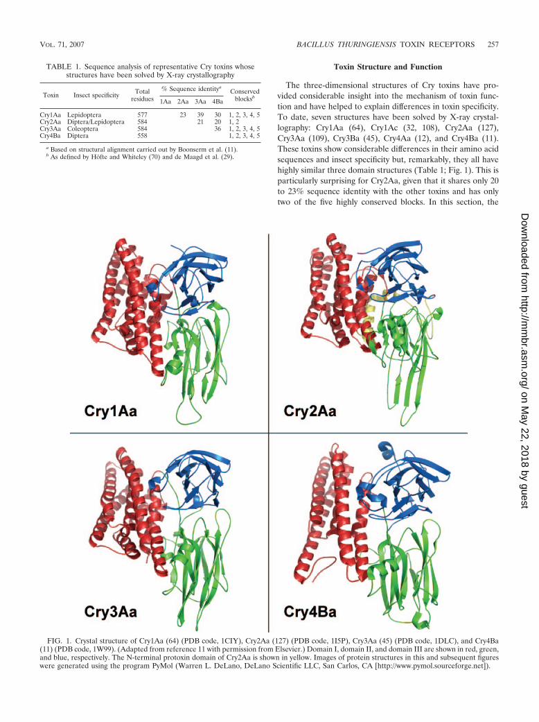
The three-dimensional structures of Cry toxins have pro-vided considerable insight into the mechanism of toxin func-tion and have helped to explain differences in toxin specificity.To date, seven structures have been solved by X-ray crystal-lography: Cry1Aa (64), Cry1Ac (32, 108), Cry2Aa (127),Cry3Aa (109), Cry3Ba (45), Cry4Aa (12), and Cry4Ba (11).These toxins show considerable differences in their amino acidsequences and insect specificity but, remarkably, they all havehighly similar three domain structures (Table 1; Fig. 1). This isparticularly surprising for Cry2Aa, given that it shares only 20to 23% sequence identity with the other toxins and has onlytwo of the five highly conserved blocks. In this section, the
TABLE 1. Sequence analysis of representative Cry toxins whosestructures have been solved by X-ray crystallography
Toxin Insect specificity Totalresidues
% Sequence identityaConserved
blocksb1Aa 2Aa 3Aa 4Ba
Cry1Aa Lepidoptera 577 23 39 30 1, 2, 3, 4, 5Cry2Aa Diptera/Lepidoptera 584 21 20 1, 2Cry3Aa Coleoptera 584 36 1, 2, 3, 4, 5Cry4Ba Diptera 558 1, 2, 3, 4, 5
a Based on structural alignment carried out by Boonserm et al. (11).b As defined by Hofte and Whiteley (70) and de Maagd et al. (29).
FIG. 1. Crystal structure of Cry1Aa (64) (PDB code, 1CIY), Cry2Aa (127) (PDB code, 1I5P), Cry3Aa (45) (PDB code, 1DLC), and Cry4Ba(11) (PDB code, 1W99). (Adapted from reference 11 with permission from Elsevier.) Domain I, domain II, and domain III are shown in red, green,and blue, respectively. The N-terminal protoxin domain of Cry2Aa is shown in yellow. Images of protein structures in this and subsequent figureswere generated using the program PyMol (Warren L. DeLano, DeLano Scientific LLC, San Carlos, CA [http://www.pymol.sourceforge.net]).
VOL. 71, 2007 BACILLUS THURINGIENSIS TOXIN RECEPTORS 257
on May 22, 2018 by guest
http://mm
br.asm.org/
Dow
nloaded from

general structure of each Cry toxin domain will be describedand related to its proposed function.
Domain I. Domain I was first described in Cry3Aa by Li etal. (109). It consists of an alpha-helical bundle in which sixhelices surround a central helix. Each of the outer helices isamphipathic in nature; polar or charged residues are generallysolvent exposed and hydrophobic residues, typically aromaticin nature, project towards the central helix. Polar groups arepresent in the interhelical space, but all are either hydrogenbonded or involved in salt bridges. Most of the helices arelonger than 30 Å and would thus be capable of spanning ahydrophobic membrane. These properties, and an overallstructural similarity to the pore-forming domain of colicin(137) (Fig. 2), led to the hypothesis that domain I was themajor determinant of pore formation in Cry toxins (109). Forthis theory to be correct, a major conformational change would
be necessary to transform domain I from a water-soluble formto a structure capable of membrane insertion. How this trans-formation occurs is a focus of ongoing research (11, 135, 145).
Domain II. Domain II is formed by three antiparallel�-sheets packed together to form a �-prism with pseudo three-fold symmetry (109). Two of the sheets are composed of fourstrands in a Greek key motif and are solvent exposed. Thethird sheet packs against domain I and is arranged in a Greek-key-like motif with three strands and a short alpha-helix. Struc-turally, domain II is the most variable of the toxin domains(11). This is especially true for the apex loops, which differconsiderably in length, conformation, and sequence. Thelengths of the �-strands are also highly variable, with Cry2Aaand Cry4Ba being the extreme examples. Given this variability,domain II is believed to be an important determinant of toxinspecificity. Similarities between the domain II apex and thecomplementarity-determining region of immunoglobulins sug-gested a role in receptor binding (109), and extensive mutagen-esis studies have provided evidence for this hypothesis (145).
The structure of domain II has been compared to those ofother �-prism proteins (28), including vitelline (158) and theplant lectins jacalin (151) and Maclura pomifera agglutinin(107). Other proteins with a �-prism fold were identified in theProtein Data Bank (PDB) (http://www.rcsb.org/pdb/Welcome.do) and include Helianthus tuberosus lectin (15), artocarpin(141), Calystegia sepium lectin (14), and banana lectin (123).Vitelline is found in the vitelline membrane of hen eggs andalthough its biological function is unknown, it is believed tobind the carbohydrate N-acetylglucosamine pentasaccharide atits apex (157). The structure of vitelline is much more sym-metrical than that of domain II; each four-�-strand sheet isrelated by sequence, unlike the �-sheets that comprise domainII of Cry toxins. The protein is also characterized by long,flexible loops at its apex (158), similar to what is observed forsome Cry toxins. The plant lectins are part of the jacalin-related superfamily of lectins (139) and are either mannose orgalactose specific. Several of these lectins have been cocrystal-lized with their ligands, and the binding site is invariably at theapex. Banana lectin is unique in that two carbohydrate bindingsites have been identified at the apex (123) (Fig. 3). The struc-tural similarity between domain II and lectin domains has ledto speculation that domain II may bind to carbohydrates, butthis has not yet been demonstrated.
Domain III. Domain III forms a �-sandwich (109). In thisarrangement, two antiparallel �-sheets pack together with a“jelly roll” topology. Both sheets are composed of five strands,with the outer sheet facing the solvent and the inner sheetpacking against domain II. Two long loops extend from oneend of the domain and interact with domain I (64). Domain IIIshows less structural variability than domain II, and the maindifferences are found in the lengths, orientations, and se-quences of the loops (11). The importance of these differencesis particularly evident with Cry1Aa and Cry1Ac, where a loopextension in Cry1Ac creates a unique N-acetylgalactosamine(GalNAc) binding pocket implicated in receptor binding (21,32, 108).
Domain III has been compared to a number of differentproteins (28), but its similarity to carbohydrate-binding mod-ules (CBMs) found in microbial glycoside hydrolases, lyases,and esterases is particularly striking. These enzymes generally
FIG. 2. Crystal structure of colicin N (137) (PDB code, 1A87). Thehelical bundle with structural similarity to Cry toxin domain I is shownin red.
258 PIGOTT AND ELLAR MICROBIOL. MOL. BIOL. REV.
on May 22, 2018 by guest
http://mm
br.asm.org/
Dow
nloaded from

consist of a catalytic domain linked to one or more CBMs. Thefunction of the CBM is to target the catalytic domain to itspolysaccharide substrate (172). This enhances the enzyme’scatalytic efficiency by increasing its effective concentration atthe substrate surface. The structure of several CBMs in com-plex with carbohydrate ligands has now been solved, and twobinding sites have been identified (140). One site (cleft A) isfound at the loops connecting the two �-sheets, and the other(cleft B) is located on the concave surface of one of the�-sheets (Fig. 4). Aromatic residues are important componentsof each binding site, and in general they are the best-charac-terized mediators of carbohydrate-protein interactions inCBMs (13). Figure 4 shows an overlay of domain III fromCry1Aa and CmCBM6-2 (the family 6 CBM from Cellvibriomixtus endoglucanase 5a) in complex with two cellotriose mol-ecules (140). As shown, there is significant structural similaritybetween these domains, suggesting that some Cry toxins mayalso bind to carbohydrates in these regions. It should be noted,however, that the aromatic residues important for carbohy-
drate binding in CBMs are generally not conserved in Crytoxins.
IDENTIFICATION AND VALIDATION OF RECEPTORS
Cry toxin binding to insect midgut epithelial receptors is animportant determinant of specificity. The correlation betweenbinding and toxicity was first demonstrated using brush bordermembrane vesicles (BBMV) prepared from microvilli by use ofa technique developed by Wolfersberger (187). Early studiesshowed that a Cry toxin (Cry1Ba) lethal to Pieris brassicaebound specifically to the insect’s BBMV but not to BBMVprepared from rat intestine (68). It was later shown thatCry1Ab and Cry1Ba bound specifically and saturably to P.brassicae BBMV, whereas only Cry1Ab bound to BBMV pre-pared from Manduca sexta (69). Since both toxins killed P.brassicae, but only Cry1Ab killed M. sexta, there was a goodcorrelation between binding and toxicity data. With other toxin-insect combinations, the correlation has not always been as
FIG. 3. Crystal structure of banana lectin (yellow/green) bound to laminaribiose (red) at two sites. PDB code, 2BMZ (123).
FIG. 4. Crystal structure overlay of the CBM CmCBM6-2 (blue) in complex with two molecules of cellotriose (yellow), and domain III ofCry1Aa (green). Aromatic residues important for carbohydrate binding are shown in magenta. The PDB codes are 1UYY (140) (CmCBM6-2) and1CIY (64) (Cry1Aa). Clefts involved in CBM carbohydrate binding are indicated (140).
VOL. 71, 2007 BACILLUS THURINGIENSIS TOXIN RECEPTORS 259
on May 22, 2018 by guest
http://mm
br.asm.org/
Dow
nloaded from

strong (47, 180). For example, Wolfersberger (188) reportedthat Cry1Ac was less toxic to Lymantria dispar than wasCry1Ab, despite having a relatively stronger binding affinity.This same binding interaction was later studied by Liang et al.(110), who used a two-step interaction scheme to analyze sep-arately the kinetics of reversible and irreversible binding. Bythis method, it was demonstrated that the rate constant ofirreversible binding, rather than the maximum extent of bind-ing, correlated better with toxicity. The general view has beenthat reversible binding correlates with toxin binding to recep-tor while irreversible binding equates with the membrane in-sertion step.
After it was demonstrated that specific high-affinity toxinbinding sites were present in the insect midgut, efforts to iden-tify and clone toxin receptors were intensified. Many putativeCry toxin receptors have since been reported, of which the bestcharacterized are the aminopeptidase N (APN) receptors (51,93, 142, 150) and the cadherin-like receptors (44, 130, 131, 174,175) identified in lepidopterans. In nematodes, glycolipids arebelieved to be an important class of Cry toxin receptors (60).Other putative receptors include alkaline phosphatases (ALPs)(38, 85, 86), a 270-kDa glycoconjugate (176), and a 252-kDaprotein (73). In the following sections, each receptor class willbe discussed with a particular focus on toxin-receptor binding
FIG. 5. Phylogenetic analysis of lepidopteran midgut APN sequences. (A) Phylogenetic tree of representative lepidopteran midgut APNsequences, created using the programs CLUSTALX and DRAWTREE (PHYLIP package). The species name and GenBank accession numberare shown for each protein. APNs boxed in purple indicated those reported to interact with Cry toxins. Classes are as proposed by Herrero et al.(67). Species names abbreviations are as follows: Se, Spodoptera exigua; Ms, Manduca sexta; Ld, Lymantria dispar; Hv, Heliothis virescens; Ha,Helicoverpa armigera; Hp, Helicoverpa punctigera; Bm, Bombyx mori; Sl, Spodoptera litura; Px, Plutella xylostella; Pi, Plodia interpunctella; Ep,Epiphyas postvittana; and Tn, Trichoplusia ni. References for binding studies are as indicated in the relevant section of the text. (B) Average aminoacid sequence identity within and among the different APN classes.
260 PIGOTT AND ELLAR MICROBIOL. MOL. BIOL. REV.
on May 22, 2018 by guest
http://mm
br.asm.org/
Dow
nloaded from

interactions and the ability of receptors to confer toxin suscep-tibility.
APN
The APN family is a class of enzymes that cleaves neutralamino acids from the N terminus of polypeptides. They servea variety of functions in a wide range of species, but in thelepidopteran larval midgut, they work in cooperation with en-dopeptidases and carboxypeptidases to digest proteins derivedfrom the insect’s diet (185). The proteins belong to the zinc-binding metalloprotease/peptidase superfamily and to a sub-family called the gluczincins (72). Members of this family arecharacterized by the short zincin motif HEXXH, where Xstands for any amino acid, followed by a conserved glutamicacid residue 24 amino acids downstream from the first histi-dine. The histidines and the last glutamic acid residue serve aszinc ligands, while the first glutamic acid residue is importantfor enzyme catalysis. A highly conserved GAMEN motif is alsobelieved to form part of the active site (101).
In addition to being studied for their role in digestion, APNshave been extensively studied as putative Cry toxin receptors.Since it was first shown that Cry toxins can bind to APN (93,150), many different forms have been isolated and character-ized. Figure 5 shows the phylogenetic relationship betweenrepresentative lepidopteran APNs and indicates those thathave been reported to interact with Cry toxins. As shown, theAPNs have been divided into five different classes (67). Theaverage sequence identity within a class varies from 56% (class5) to 67% (class 4). Among the different classes, class 2 is theleast like the others, with an average sequence identity of only25 to 26% relative to the other classes, whereas class 1 andclass 3 are the most similar, with an average sequence identityof 38%. To date, all known APNs within a particular specieshave been found to cluster into different classes. In fact, someAPNs share higher sequence identity with those in nonlepi-dopterans than with other APNs in the same species. Forexample, class 2 APN from M. sexta is more similar to APNs inchicken and frog (GenBank accession numbers NP_990192and AAH85055, respectively) than to class 1 M. sexta APN, andyet both M. sexta APNs are reported to bind to Cry1Ab (31,120).
Of the many different APNs that have been studied, severalcommon features have emerged (Fig. 6). The genes encodeproteins of approximately 1,000 amino acids that undergo var-ious forms of posttranslational modification to produce mature
proteins of between 90 and 170 kDa in size. The proteins havea cleavable N-terminal signal peptide that directs nascentpolypeptides to the outer surface of the cytoplasmic mem-brane. There, they are attached to the membrane by a glyco-sylphosphatidylinositol (GPI) anchor (2, 31, 94, 113, 164), incontrast to what is seen for vertebrates, where a hydrophobicN-terminal stalk is used for attachment (155). As will be dis-cussed, glycosylation is important for some Cry toxin-APNinteractions, and in many cases the presence of N- or O-linkedcarbohydrates has been shown biochemically or predicted bysequence analysis (84) (http://www.cbs.dtu.dk/services/). Asshown in Fig. 7, the predicted number of O-linked glycosyla-tion sites differs considerably among the different classes ofAPN, whereas differences in predicted N-linked glycosyla-tion sites are less distinct. Carbohydrate structures includingGalNAc are believed to be particularly important for someinteractions between Cry1Ac and APN (93).
APN as a Cry-binding protein. Cry1 proteins are toxic tolepidopterans, and several different toxins, including Cry1Aa,Cry1Ab, Cry1Ac, Cry1Ba, Cry1C,a and Cry1Fa, have beenshown to bind to APNs (references in the following sections).Based on the experiments carried out so far, APNs and toxinswithin these families show different patterns of binding. SomeAPNs bind to multiple Cry toxins and some Cry toxins bind tomultiple APNs, and in other cases, unique toxin-APN pairshave been reported. While many toxin-APN binding combina-tions have yet to be tested, preliminary data are providingsome insight into the determinants of receptor binding. In thissection, a class-by-class account of APNs and their interactionswith Cry toxins will be presented.
(i) Class 1. Class 1 APNs have been identified in nine dif-ferent lepidopterans. In addition to the features already dis-cussed, class 1 APNs generally have a threonine-rich sequencedownstream of the C-terminal GPI signal sequence. This re-gion is believed to have extensive O-linked glycosylation and isthought to form a rigid stalk that elevates the active site of theenzyme above the membrane surface (92, 93). By use of theNetOGlyc 3.1 server (84) (http://www.cbs.dtu.dk/services/NetOGlyc), the number of predicted O-linked glycosylationsites in class 1 APNs has been shown to vary from 6 in Bombyxmori to 39in Helicoverpa armigera (Fig. 7). In species wherenative APN has been isolated from the midgut, the correlationbetween observed molecular mass and the number of pre-dicted O-linked glycosylation sites is strong. For example,APNs from B. mori, M. sexta, and Heliothis virescens are re-ported to have observed molecular masses of 120 kDa (192),
FIG. 6. Schematic representation of a typical lepidopteran APN protein. The proregion and the threonine-rich region, shown with broken lines,have been reported only in some APNs.
VOL. 71, 2007 BACILLUS THURINGIENSIS TOXIN RECEPTORS 261
on May 22, 2018 by guest
http://mm
br.asm.org/
Dow
nloaded from
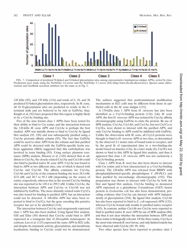
120 kDa (93), and 170 kDa (116) and totals of 6, 10, and 36predicted O-linked glycosylation sites, respectively. In M. sexta,all 10 O-glycosylation sites are predicted to reside in the C-terminal stalk and are believed to be rich in GalNAc; thus,Knight et al. (92) have proposed that this region is highly likelyto be a Cry1Ac binding site.
Five of the nine known class 1 APNs have been tested fortheir ability to bind to Cry toxins, and the interaction betweenthe 120-kDa M. sexta APN and Cry1Ac is perhaps the beststudied. APN was initially shown to bind to Cry1Ac by ligandblot analysis (93, 150) and was subsequently purified using aCry1Ac protoxin affinity column (93). The fact that GalNAccould be used to elute APN from the column and that purifiedAPN could be detected with the GalNAc-specific lectin soy-bean agglutinin (SBA) suggested that this carbohydrate wasinvolved in toxin binding (93). Using surface plasmon reso-nance (SPR) analysis, Masson et al. (120) showed that in ad-dition to Cry1Ac, the closely related Cry1Aa and Cry1Ab couldalso bind to purified native M. sexta APN. Cry1Ac was found tobind to APN at two different sites, one of which it shared withCry1Aa and Cry1Ab. The affinity constants for Cry1Aa,Cry1Ab and Cry1Ac at the common binding site were 28.4 nM,42.8 nM, and 40.7 to 95.3 nM (depending on the source oftoxin), respectively, whereas that at the second Cry1Ac bindingsite was reported to be 149.4 to 299.3 nM. Unlike Cry1Ac, theinteraction between APN and Cry1Aa or Cry1Ab was notinhibited by GalNAc. The more distantly related toxin Cry1Cawas also tested for binding to purified APN, but an interactioncould not be detected. A 106-kDa APN has since been re-ported to bind to Cry1Ca, but the gene encoding this putativereceptor has yet to be identified (114).
The interaction between Cry1Ac and exogenously expressedclass 1 APN has also been studied. Using ligand blot analysis,Gill and Ellar (50) showed that Cry1Ac could bind to APNexpressed in a transgenic line of Drosophila melanogaster. Incontrast, Luo et al. (115) expressed the same APN in Sf21 cells,and despite its enzymatic activity, glycosylation, and membranelocalization, binding to Cry1Ac could not be demonstrated.
The authors suggested that posttranslational modificationmechanisms in Sf21 cells may be different from those in epi-thelial cells in the M. sexta midgut (115).
A 170-kDa class 1 APN from H. virescens has also beenidentified as a Cry1A-binding protein (116). Like M. sextaAPN, the first H. virescens APN was isolated by Cry1Ac affinitychromatography using GalNAc to elute the protein. By use ofSPR analysis, Cry1Aa, Cry1Ab, and Cry1Ac, but not Cry1Ca orCry1Ea, were shown to interact with the purified APN, andonly Cry1Ac binding to APN could be inhibited with GalNAc.Unlike the observation with M. sexta, all Cry1A proteins werethought to bind to H. virescens APN at two sites, as determinedby the observed 2:1 molar ratio of bound toxin to receptor, andby the good fit of experimental data to a two-binding-sitemodel based on kinetics (116). In a later study (8), Cry1Fa wasshown to bind to this APN by ligand blot analysis, and thus itappeared that class 1 H. virescens APN was not exclusively aCry1A-binding protein.
Class 1 APN from B. mori has also been shown to interactwith Cry toxins and is best characterized as a Cry1Aa-bindingprotein. The 120-kDa APN was released from BBMV withphosphatidylinositol-specific phospholipase C (PI-PLC) andthen purified by ion-exchange chromatography (192). Thispreparation was shown to interact with Cry1Aa by both dotblot and ligand blot analysis. Cry1Aa binding to denaturedAPN expressed as a glutathione S-transferase (GST) fusionprotein in Escherichia coli has also been demonstrated, pro-viding evidence that Cry1Aa can interact with APN indepen-dently of glycosylation (194). By ligand blot analysis, Cry1Abhas also been reported to bind to E. coli-expressed APN (132),whereas Cry1Ac bound only weakly to purified native receptor(193). In contrast, neither Cry1Ab nor Cry1Ac were reported tobind to purified native B. mori APN by use of SPR analysis (80),and thus it not clear whether the interaction between APN andthese toxins is biologically relevant. Of the three toxins, Cry1Aa isthe most toxic towards B. mori larvae, and only limited toxicity hasbeen observed with Cry1Ac (49, 99, 104).
Two other species have been reported to produce class 1
FIG. 7. Comparison of predicted N-linked and O-linked glycosylation sites among representative lepidopteran midgut APNs, sorted by class.Predictions were made using the NetNGlyc 1.0 server and the NetOGlyc 3.1 server (84) (http://www.cbs.dtu.dk/services/). Species name abbre-viations and GenBank accession numbers are the same as in Fig. 5.
262 PIGOTT AND ELLAR MICROBIOL. MOL. BIOL. REV.
on May 22, 2018 by guest
http://mm
br.asm.org/
Dow
nloaded from

APN capable of interacting with Cry toxins: H. armigera (142)and Plutella xylostella (132). In both cases, binding was studiedusing exogenously expressed protein. Rajagopal et al. (142)expressed H. armigera APN in Trichoplusia ni cells by use of abaculovirus expression vector. The expressed protein wasfound to be membrane associated, catalytically active, and gly-cosylated and by ligand blot analysis could bind to Cry1Aa,Cry1Ab, and Cry1Ac. To study the interaction of Cry toxinswith P. xylostella APN, a truncation mutant was expressed in E.coli as a GST fusion protein (132). This mutant was based ona previously identified toxin binding region found in a homol-ogous region of B. mori APN (193). By ligand blot analysis,both Cry1Aa and Cry1Ab were reported to bind to this trun-cated mutant (132).
(ii) Class 2. Lepidopteran APNs in class 2 share the leastsequence identity with the other classes (Fig. 5). Each memberis predicted to be N glycosylated, but in stark contrast to class1, there are no O-linked glycosylation sites predicted for any ofthe APNs, and the threonine-rich C-terminal stalk region re-ported for class 1 APN is completely absent. Interestingly,Cry1Ac has not been reported to bind to any member of class2, supporting the theory that Cry1Ac binds to APN at theO-glycosylated C-terminal stalk (92). Cry1Aa and Cry1Abhave been reported to bind to class 2 APNs under certainconditions (31, 132), but it remains to be seen whether theseinteractions are biologically relevant.
Cry1Ab was first shown to bind to class 2 M. sexta APN byDenolf et al. (31). This interaction was demonstrated whenAPN was partially purified using a Cry1Ab affinity column.Using a high-pH carbonate buffer, proteins of many differentmolecular weights were eluted, in contrast to the Cry1Ac pro-toxin affinity purification of class 1 APN from M. sexta in whicha single band was observed following GalNAc elution (93).Nevertheless, by ligand blot analysis Cry1Ab was shown to bindto a 120-kDa band in the purified fraction, and internal aminoacid sequence data facilitated cloning of the encoding gene(31). Attempts to express the protein in Sf9 cells were unsuc-cessful, and thus it was not possible to confirm that the clonedgene actually encoded a Cry1Ab-binding protein.
Using the sequence information derived from class 2 M.sexta APN, Denolf et al. (31) were also able to study thecorresponding APN in P. xylostella. In this case, expression inSf9 cells was possible, and a 105-kDa glycoprotein with enzy-matic activity was produced. Homologous competition bindingassays using Cry1Ab, Cry1Ac, Cry1Ba, Cry1Ca, or Cry9Ca andintact Sf9 cells expressing the APN did not reveal any specifictoxin binding, and similar results were obtained with cell-de-rived membrane preparations. In addition, Cry1Ab binding toAPN could not be demonstrated by ligand blot analysis. SinceCry1Ab had previously been shown by ligand blot analysis tobind to a 120-kDa protein in P. xylostella BBMV, it was notclear whether the cloned APN was a different protein orwhether differences in glycosylation were responsible for thelack of binding. A study by Nakanishi et al. (132) furthercomplicated the matter. This group expressed a putative Crytoxin binding region from class 2 P. xylostella APN as a GSTfusion protein in E. coli and by ligand blotting showed bindingto both Cry1Aa and Cry1Ab. In addition, they showed thatCry1Aa and Cry1Ab could bind to the same region in a class 2APN from B. mori. Thus, it seems that further work must be
carried out to clarify whether class 2 APNs are genuine Cry1Areceptors, and if so, to what extent glycosylation plays a role intoxin binding.
(iii) Class 3. Class 3 is made up of the largest group ofknown lepidopteran APNs, with members from 11 differentspecies. This class is most closely related to class 1 (Fig. 5), andsimilarly has a threonine-rich C terminus predicted to be highlyglycosylated by the NetOGlyc 3.1 server (84) (http://www.cbs.dtu.dk/services/NetOGlyc/). Class 3 APNs generally havefewer predicted O-linked glycosylation sites than class 1 APNs(Fig. 7) and, of those isolated from BBMV, all have had amolecular mass near 120 kDa. Within this class, binding toCry1Aa, Cry1Ab, Cry1Ac, Cry1B,a and Cry1Fa has been re-ported, as will be discussed.
APN from L. dispar is perhaps the best-studied member ofclass 3. It was cloned by Garner et al. (48), and based onsequence identity was believed to be the APN1 previouslydescribed by Valaitis et al. (177). In these earlier experiments,APN was released from BBMV by use of PI-PLC and wassubsequently purified using a series of chromatographic steps.By ligand blot analysis, Cry1Ac was shown to bind to purifieddenatured APN. Binding was also tested by SPR analysis andunder these conditions Cry1Ac, but not Cry1Aa or Cry1Ab,bound to native APN and Cry1Ac binding could be completelyblocked with competing GalNAc (177). The binding of Cry1Acto APN was reported to occur in a 1:1 ratio, in contrast to the2:1 ratio reported for the interaction between Cry1Ac and class1 M. sexta APN (120) and class 1 H. virescens APN (116).Cloned, exogenously expressed L. dispar APN has also beenstudied (48). Sf9 cells were transformed with a baculovirusvector encoding this APN, and the expressed protein was rec-ognized by an APN-specific antibody; however, ligand blotanalysis revealed only weak binding to Cry1Ac. As suggested inother studies, differences in posttranslational modification mayaccount for the differences observed in toxin binding to nativeand recombinant forms of APN.
The binding of Cry1Ac to class 3 APN purified from H.virescens has also been reported (51). In this case, Cry1Ac wasshown to bind to purified protein by ligand blot analysis, andthe interaction could be blocked with competing GalNAc. Itwas also reported that Cry1Ac failed to bind to APN preparedby in vitro translation, providing additional evidence that gly-cosylation, or at least some form of posttranslational modifi-cation, was important for binding. Additional characterizationwas reported by Banks et al. (8), who partially purified a 120-kDa APN believed to be the same as that reported by Gill etal. (51) but whose identity was not confirmed by amino acidsequencing. The study showed that APN could bind to Cry1Acand Cry1Fa by both affinity chromatography and ligand blot-ting (8). APN was also shown to react with SBA, a lectinspecific for GalNAc, and chemical deglycosylation with periodateeliminated toxin binding.
Another APN from class 3 was identified in H. armigera(142). In this study, toxin binding to APN expressed exog-enously in T. ni cells was studied. The expressed protein was120 kDa in size, glycosylated, and enzymatically active and wasshown by ligand blot analysis to react with Cry1Ac but not withCry1Ab or Cry1Aa. Although the involvement of GalNAc inbinding was not reported, a study by Wang et al. (183) suggests
VOL. 71, 2007 BACILLUS THURINGIENSIS TOXIN RECEPTORS 263
on May 22, 2018 by guest
http://mm
br.asm.org/
Dow
nloaded from

that glycosylation may not be required for this interaction. Thisgroup expressed APN in E. coli and showed binding to Cry1Acwith ligand blot analysis.
Class 3 APN from Epiphyas postvittana has been studied inboth its native and exogenously expressed forms (160). Thenative protein was purified from detergent-solubilized BBMVproteins by use of a combination of gel filtration and ion-exchange chromatography. APN expressed in Sf9 cells waspurified using a similar method. Both Cry1Ac and Cry1Bacould bind to either form of APN by ligand blotting, but incompetitive binding assays, neither toxin bound specifically toSf9 cells expressing APN. To ensure that APN was being ex-pressed on the cell surface, the researchers measured APNactivity in cells before and after lysis and found the values to becomparable. Thus, concerns about the relevance of bindingdemonstrated by ligand blotting were raised (160).
Finally, there is some evidence that class 3 APNs from B.mori and P. xylostella can bind to Cry1Aa and Cry1Ab, basedon reports by Nakanishi et al. (132), where binding to toxinbinding regions expressed as GST fusion proteins in E. coli wasshown by ligand blot analysis.
(iv) Class 4. Like class 2, class 4 lacks the C-terminal threo-nine-rich tract found in class 1 and class 3. There are currentlynine members in this class, and three have been reported tointeract with Cry toxins (2, 8, 132).
Class 4 APN from H. virescens has been reported to be aCry1Ac-binding protein. This has been demonstrated usingseveral different methods and was first shown by affinity chro-matography, where CHAPS {3-[(3-cholamidopropyl)-dimeth-ylammonio]-1-propanesulfonate}-solubilized BBMV proteinswere passed over a Cry1Ac affinity column and bound proteinseluted with 2 M NaSCN (8). Under these conditions, threemajor binding proteins were eluted: a 170-kDa protein (class 1APN), a 120-kDa protein (class 3 APN), and a 110-kDa pro-tein (class 4 APN). Similar results were obtained with Cry1Fa,but no proteins were isolated using Cry1Ea, a toxin previouslyshown not to bind to H. virescens BBMV. That Cry1Ac couldbind to class 4 APN following periodate treatment was evi-dence that glycosylation was not important for binding (8).In addition, SBA did not bind to this APN, suggesting thatGalNAc moieties were not present in its glycans. It thus appearedthat the interaction between Cry1Ac and class 4 APN differedfrom the interaction of Cry1Ac with class 1 and class 3 APN,where GalNAc was believed to be an important determinant ofbinding (93, 116, 177). Cry toxin binding to exogenously ex-pressed class 4 H. virescens APN was also reported (7) and hereit was shown by fluorescence microscopy that Cry1Ac couldbind to S2 cells expressing APN but not to control cells. At-tempts were also made to express APN in E. coli, but recom-binant protein was not produced and thus the importance ofglycosylation could not be tested by this method.
Class 4 also includes an APN isolated from Spodoptera litura.This species has been reported to be susceptible to Cry1Ca butresistant to Cry1Ac (2). Agrawal et al. (2) examined the bind-ing of Cry1Ca and Cry1Ac to this APN expressed in Sf21 cellsand showed that the protein was glycosylated, enzymaticallyactive, and present on the cell surface. Ligand blot analysisshowed that Cry1Ca but not Cry1Ac could bind to the dena-tured form of the protein. Toxin binding to APN was alsostudied under nondenaturing conditions (2). CHAPS-solubi-
lized APN was incubated with Cry1Ca and immunoprecipi-tated with anti-Cry1Ca antibodies attached to protein A-Sepharose beads. Using negative control Sf21 cells or omittingCry1Ca failed to precipitate the APN. A binding assay was alsocarried out with intact cells, and immunofluorescence showedthat Cry1Ca but not Cry1Ac could bind to cells expressingAPN. Attempts to express this protein in E. coli were unsuc-cessful, however, and thus the importance of glycosylation intoxin binding was not determined.
There is also some evidence that class 4 B. mori APN canbind to Cry1Aa and Cry1Ab, in particular to the proposedtoxin binding region described by Nakanishi et al. (132).
(v) Class 5. Class 5 makes up the smallest group of APNsand consists of only two members identified in P. xylostella(132) and H. armigera. These APNs have an altered form of thehighly conserved GAMEN motif, where methionine has beenchanged to threonine. The effect of mutations in this motif onenzymatic activity have been studied in the related proteininsulin-regulated aminopeptidase (101). Here it was shownthat mutating methionine to isoleucine, lysine, or glutamic aciddecreased activity by 16-fold, decreased activity by 30-fold, orcompletely abolished activity, respectively. Although the enzy-matic activity of the class 5 APNs has not been reported, itseems likely that at least some decrease in activity would beexpected.
A comparison of the amino acid sequences of the P. xylos-tella and H. armigera APNs shows that there are marked dif-ferences in the threonine-rich C-terminal region. H. armigeraAPN has the longest reported open reading frame of any of thelepidopteran APNs and has many threonine residues at the Cterminal, 32 of which are predicted to be O glycosylated ac-cording to the NetOGlyc 3.1 server (84) (http://www.cbs.dtu.dk/services/NetOGlyc/). In contrast, class 5 APN from P.xylostella completely lacks this threonine-rich region and hasno predicted O-linked glycosylation sites.
Studies on the binding of class 5 APNs to Cry toxins arelimited. The only report is from Nakanishi et al. (132), whoshowed by ligand blot analysis that a region of P. xylostella APNexpressed in E. coli as a GST fusion protein could bind toCry1Aa and Cry1Ab. Additional research is needed to deter-mine the significance of these interactions and the generalimportance of class 5 APNs in mediating Cry toxin suscepti-bility.
(vi) Other APNs. In addition to the APNs already discussed,three other variants have been reported to bind to Cry toxinsbut have yet to be cloned and sequenced: a 106-kDa proteinfrom M. sexta (114), a 100-kDa protein from the dipteranAnopheles quadrimaculatus (1), and a 96-kDa protein from B.mori (159). Based on immunoprecipitation experiments, the106-kDa protein from M. sexta appeared to be a Cry1Ca-binding protein, although weaker binding to Cry1Ac was alsodetected. The N-terminal sequence was nearly identical to thatof class 1 M. sexta APN (93, 113, 150), whereas an internalsequence—as it was later discovered—was identical to a regionof class 2 M. sexta APN (31). Whether the preparation of106-kDa APN used for sequencing contained a single novelfusion of class 1 and class 2 APN or two separate APNs derivedfrom each class was never reported. The 100-kDa protein iso-lated from A. quadrimaculatus was purified from solubilizedBBMV and tested for binding to mosquitocidal Cry toxins. By
264 PIGOTT AND ELLAR MICROBIOL. MOL. BIOL. REV.
on May 22, 2018 by guest
http://mm
br.asm.org/
Dow
nloaded from

SPR analysis (1), Cry11Ba, but not Cry2Aa, Cry4Ba, orCry11Aa, was found to bind to the purified protein. Databasesearches with the N-terminal sequence of the 100-kDa proteinled to its classification as an APN. The 96-kDa protein from B.mori was shown to bind to Cry1Ac by ligand blot analysis, andthis interaction could be blocked with competing GalNAc(159). The protein was recognized by an antibody with speci-ficity for class 3 B. mori APN, but by peptide mass fingerprint-ing, only 54% of the peptides could be matched. It was thusproposed that the 96-kDa protein was a novel isoform of class3 APN.
(vii) Summary. The data on Cry toxin binding to APNs arecomplex, and several factors make this interaction difficult tostudy. First, several different APNs are believed to be simul-taneously expressed in the larval gut. These proteins sharesequence identity and can have similarities in properties suchas molecular weight, enzymatic activity, and glycosylation. Thiscan make it difficult to purify a particular APN to homogeneityand equally difficult to prove that the protein is pure. Indeed,mass spectrometry analysis of purified class 1 APN from M.sexta revealed the presence of contaminating class 3 and class4 APNs (163). Although exogenous expression of cloned APNsis a possible solution, this can sometimes be difficult (7, 31, 48)and even when successful, tissue- or organism-specific differ-ences in posttranslational modification may eliminate the toxinbinding site (48, 115). To further complicate matters, differentmethods of studying toxin-APN interactions can sometimesgive conflicting results (31, 132, 160). This was studied in detailby Daniel et al. (26), who showed that denaturing M. sextaAPN or Cry1A toxins exposes binding epitopes hidden undernondenaturing conditions. Studies on Cry toxin binding toAPN may also be complicated by the presence of cadherins, asecond class of Cry toxin receptor particularly sensitive toproteolytic degradation (23, 119, 174). Cadherins have beenshown to form approximately 120-kDa degradation productsthat could possibly be misinterpreted as APN in ligand blotassays (119).
Figure 8 presents a summary of the reported binding be-tween Cry toxins and exogenous or endogenous nondenaturedor denatured APN. While it is clear from this figure thatseveral toxin-APN combinations have yet to be explored, somegeneral conclusions can be drawn. Cry1Aa and Cry1Ab arebest characterized as class 1 APN-binding proteins. Binding toboth endogenous and exogenous forms of APN has been ob-served for several species and, as shown in Fig. 8, a lack ofbinding to any class 1 member has been reported in one caseonly (80). Binding to other APN classes under nondenaturingconditions either has not been reported or has not been ob-served. Cry1Ac has specificity broader than those of Cry1Aaand Cry1Ab and seems to be primarily a class 1 and class 3APN-binding protein. Binding to class 1 APNs appears tooccur at two sites, one of which it shares with Cry1Aa andCry1Ab (120). Binding to the other site seems to be GalNAcdependent, and given that both class 1 and class 3 have athreonine-rich region predicted to be highly glycosylated, it istempting to speculate that Cry1Ac mediates contact to bothAPNs in this region. As for the remaining toxins, it is difficultto make generalizations about their binding specificity basedon the limited data available, and additional studies must becarried out to better characterize these proteins.
APN as a mediator of Cry toxin susceptibility. Since it wasdemonstrated that Cry toxins can bind to APN, additionalstudies have been carried out to distinguish between Cry-bind-ing proteins and proteins that confer Cry toxin susceptibility.The following sections describe the various methods that havebeen used to make this distinction, along with the major find-ings of these studies.
(i) Permeability. The insecticidal nature of Cry toxins isgenerally believed to be due to their ability to form pores in themidgut of susceptible organisms (96), and assays have beendeveloped to assess whether putative toxin receptors can en-hance pore formation. The 86Rb� efflux assay has been usedfor this purpose, where pore formation is indicated by therelease of 86Rb� from phospholipid vesicles containing puta-tive receptor. Sangadala et al. (150) used this technique todemonstrate that a mixture of class 1 APN and phosphatasefrom M. sexta could enhance Cry1Ac pore formation. Whenreconstituted into phospholipid vesicles, these proteins werereported to increase toxin binding by 35% and to enhancetoxin induced 86Rb� release 1,000-fold relative to protein-freevesicles. Similar results were obtained by Luo et al. (116), whoshowed that class 1 APN purified from H. virescens couldenhance Cry1Aa-, Cry1Ab-, or Cry1Ac-induced release of86Rb� but had no effect on Cry1Ca-induced release; Cry1a is atoxin shown not to bind to this class of APN (116). Poreformation has also been studied by measuring toxin channelactivity in planar lipid bilayers. Schwartz et al. (153) showedthat the inclusion of a purified M. sexta receptor complex inphospholipid bilayers caused Cry1Aa, Cry1Ac, and Cry1Ca toform channels at concentrations much lower than that in re-ceptor free membranes. Analysis of this receptor complex byligand blotting suggested that class 1 APN was the major Cry-binding protein.
(ii) In vitro toxicity. While assays that measure membranepermeability are good indicators of pore formation, they donot necessarily predict whether a receptor will confer toxinsusceptibility to an organism. A more direct approach is to testwhether Cry toxin-resistant cell lines can be made susceptibleby expressing putative toxin receptors. So far, testing APNs bythis method has been relatively unsuccessful. Garner et al. (48)expressed class 3 APN from L. dispar in Sf9 cells but did notobserve cytotoxicity at Cry1Ac concentrations between 0.2 and50 �g/ml. Because the Sf9 cells expressed APN with a bindingaffinity for Cry1Ac much lower than that of the native protein,the experiment was somewhat inconclusive. Class 4 APN fromH. virescens was also tested for its ability to confer toxin sus-ceptibility and was expressed in S2 cells (7). While it wasdemonstrated that Cry1Ac could bind to APN on the surfaceof intact cells, cytotoxicity was not observed at a toxin concen-tration of 30 �g/ml. The limited number of in vitro cytotoxicity-based studies is likely due to difficulties in correctly expressingAPN, and obtaining proper glycosylation seems to be the mainobstacle (48, 115). If these problems could be resolved, thismethod of receptor validation may become more useful.
(iii) In vivo toxicity. In vivo methods have also been used totest APN receptors for functionality. Gill and Ellar (50) fedCry1Ac to transgenic Drosophila larvae expressing class 1 APNfrom M. sexta and showed 100% toxicity at a toxin concentra-tion of 50 �g/ml. In comparison, control larvae were resistantto Cry1Ac at concentrations up to 1 mg/ml, the highest con-
VOL. 71, 2007 BACILLUS THURINGIENSIS TOXIN RECEPTORS 265
on May 22, 2018 by guest
http://mm
br.asm.org/
Dow
nloaded from

centration tested. The expression of M. sexta APN was con-firmed by ligand blotting with Cry1Ac, and although expressionlevels were low, the receptor binding determinants were ap-parently intact. These results suggested that APN expressed inthe Drosophila midgut may be properly glycosylated and that invivo expression systems may be more suitable for evaluatingtoxin-receptor interactions than those based on cell lines.
Gene silencing has also been used to determine whether
APN can confer toxin susceptibility. Rajagopal et al. (143)injected S. litura larvae with double-stranded RNA corre-sponding to a region of the class 4 APN gene and showed a95% reduction in transcript levels over what was shown forcontrol larvae. In addition, an 80% reduction in APN expres-sion was observed, as determined by immunoblot analysis ofBBMV proteins. When treated with Cry1Ca, a 75% reductionin mortality was observed in larvae previously injected with
FIG. 8. Summary of reported binding between Cry toxins and endogenous (En) or exogenous (Ex) nondenatured (N) or denatured (D) APNsas discussed and referenced in the preceding sections. Binding, no binding, conflicting reports, and absence of data are indicated by green, red,yellow, or white/gray boxes, respectively. APN was expressed exogenously by E. coli (A) in vitro translation (B), S2 cells (C), Sf9 cells (D), Sf21cells (E), T. ni cells (F), Drosophila or Sf21 cells (G), E. coli or T. ni cells (H), or E. coli or Sf9 cells (I). In cases where the conditions of binding(denaturing or nondenaturing) were not reported, boxes are merged. Species names are abbreviated as in Fig. 5. Species and class are abbreviated“Sp” and “Cl,” respectively.
266 PIGOTT AND ELLAR MICROBIOL. MOL. BIOL. REV.
on May 22, 2018 by guest
http://mm
br.asm.org/
Dow
nloaded from
double-stranded RNA. These results suggest that class 4 APNcan confer Cry1Ca susceptibility to S. litura and demonstratethat gene silencing may be an effective way to study the bio-logical significance of toxin-receptor interactions.
(iv) Summary. The biological relevance of the Cry toxin-APN interaction has yet to be studied extensively. To date, 17different APNs have been reported to bind to Cry toxins, andyet only 2 have been shown to mediate toxin susceptibility.Twelve of the 17 APNs have not been studied for functionalityby any method. In vivo methods of testing APN functionalityhave shown considerable promise, and using these methods tostudy the remaining APNs may lead to a better understandingof the overall importance of this class of Cry toxin receptor.
Cadherin
The cadherin superfamily of proteins is highly diverse andserves a variety of functions, including cell adhesion, migration,cytoskeletal organization, and morphogenesis (4, 65). The ex-pression of cadherins is highly regulated, both spatially andtemporally, and is often unique to a particular cell type. Theproteins are defined by the presence of repeating calcium-binding domains or cadherin repeats of approximately 110amino acids in length. Classical cadherins have 5 cadherinrepeats (4, 134) but as many as 34 repeats have been reported(34). Some cadherins also have mucin (53), laminin, or epider-mal growth factor-like repeats (133). The proteins are glyco-sylated and are usually anchored to the membrane by a singletransmembrane domain, although seven-transmembrane (173)or GPI-anchored variants have also been identified (181).
In 1993, a novel cadherin-like protein was isolated from themidgut epithelium of M. sexta by virtue of its binding affinityfor Cry1Ab (174). The protein was cloned in 1995 and se-quence analysis predicted a signal peptide, 12 cadherin re-peats, a membrane proximal extracellular domain, a trans-membrane domain, and a small cytoplasmic domain (33, 175).Since then, additional lepidopteran cadherins have been iden-tified, and all have been shown to have a similar domain or-ganization (40, 44, 126, 130, 184). In M. sexta cadherin, addi-tional features, such as the cell adhesion sequence HAV (10)and the integrin-binding sequences RGD (149) and LDV (98,170), have been identified in the ectodomain; however, thefunctional role of these sequences has not yet been confirmed(33). In contrast, an analysis of the cytoplasmic domain did notreveal sequences known to interact with intracellular proteinssuch as catenins (33). While classical cadherins are locatedprimarily within adherens junctions involved in cell-cell adhe-sion (4), lepidopteran cadherin-like proteins have been iden-tified on the apical membrane of midgut columnar epithelialcells (3, 24, 66, 124), the target site of Cry toxins (16, 19, 20,24). The expression of cadherin has been shown to vary withdevelopmental stage and increases progressively from the firstto the fifth instar in M. sexta larvae (124). In eggs and adults,however, cadherin expression has not been detected. Althoughthe exact physiological function of midgut cadherins is notknown, the tight control of cadherin levels during larval devel-opment has been proposed to indicate their importance inmaintaining midgut epithelial organization (124).
Lepidopteran cadherin-like proteins have been extensivelystudied as Cry1A receptors, and there is good evidence to
suggest they play a critical role in mediating toxin susceptibil-ity. The following sections describe the best-characterized cad-herin-like proteins and their interactions with toxins of theCry1A family.
BT-R1 (Manduca sexta). The first cadherin-like proteinshown to interact with Cry toxins, BT-R1, was a 210-kDa gly-coprotein identified in M. sexta BBMV (174). The protein waspurified by immunoprecipitation with Cry1Ab followed by two-dimensional gel electrophoresis. Partial sequence informationderived from the purified receptor facilitated cloning, and theidentified gene was 30 to 60% similar and 20 to 40% identicalto other members of the cadherin superfamily (175).
To confirm that BT-R1 was a genuine Cry1Ab receptor, itwas expressed in cultured cells. The protein was first expressedin mammalian COS-7 and HEK-293 cells and by ligand blotanalysis was detected as a 195-kDa band by probing withCry1Ab (175). Cry1Ab binding to intact cells expressing BT-R1
was also demonstrated, and the measured dissociation con-stant of 1 nM was similar to that of the native receptor. BT-R1
was subsequently expressed in insect-derived Sf21 cells and wasshown to bind to Cry1Aa and Cry1Ac but not to Cry3Aa andCry11Aa, which are not toxic to M. sexta (91). In addition,competition binding studies showed that Cry1Aa and Cry1Accould block Cry1Ab binding to membranes prepared fromSf21 cells expressing BT-R1, suggesting that the toxins bind toa common epitope. Taken together, these results showed thatCry1A toxins could bind to endogenously or exogenously ex-pressed BT-R1 under both denaturing and nondenaturingconditions.
BT-R1 was also tested for its ability to confer toxin suscep-tibility. Initially, HEK-293, COS-7, or Sf21 cells transfectedwith BT-R1 failed to show any phenotypic changes when ex-posed to activated Cry1Ab, even at concentrations as high as100 �g/ml (91). This unexpected result was explained when aframeshift mutation was discovered in the original cDNA clone(33). A revised analysis of the protein sequence showed thatthe frameshift mutation occurred upstream of the transmem-brane domain, thus explaining why the protein was not em-bedded in the cytoplasmic membrane in earlier experimentsand why the observed molecular weight of exogenously ex-pressed BT-R1 was less than that of the native protein. Subse-quently, the error-free protein localized to the cell surface andrendered COS-7 cells sensitive to Cry1Ab at 0.6 �g/ml (33). S2cells expressing BT-R1 were also susceptible to Cry1A toxins,and 12 to 14% of cells were killed by Cry1Aa, Cry1Ab, orCry1Ac at 20 �g/ml (75). The toxicity of Cry1Ab towards H5cells expressing BT-R1 was determined at a range of concen-trations, and the 50% lethal concentration was reported to be65 nM (about 4 �g/ml) (195). These results strongly suggestthat BT-R1 is an important determinant of Cry1A toxin spec-ificity.
BtR175 (Bombyx mori). A cadherin-like protein was alsoidentified as a Cry toxin receptor in B. mori. In this case, a175-kDa glycoprotein, BtR175, was identified as a Cry1Aareceptor by immunoprecipitation (130, 131). Partial N-termi-nal sequencing of the purified receptor led to the cloning of agene that shared significant homology to the cadherin super-family of proteins and 69.5% identity to M. sexta BT-R1. Thepredicted molecular mass of the encoded protein (193.3 kDa)was larger than that of the 175-kDa natural protein, and it was
VOL. 71, 2007 BACILLUS THURINGIENSIS TOXIN RECEPTORS 267
on May 22, 2018 by guest
http://mm
br.asm.org/
Dow
nloaded from
believed that BtR175 was expressed as a proprotein. This wasconfirmed when the gene was expressed in Sf9 cells and a175-kDa band comigrated with BtR175 isolated from BBMV.It was postulated that the sequence 288RPPRWV292 may bean endoproteolytic cleavage signal that when cut gives rise toa mature BtR175 with only nine cadherin repeats (130).Interestingly, the proposed cleavage signal is also present inM. sexta BT-R1, where there is no apparent cleavage at thissite.
A second group has independently purified and partiallysequenced a Cry1Aa receptor with a reported molecular massof 180 kDa (77). The 103-amino-acid sequence obtained bythis group was identical to a region within the sequence ofBtR175 previously reported by Nagamatsu et al. (130). A laterpublication by the same group reported the sequence of threeallelic BtR175 variants that differed from BtR175 by one, five,or six amino acids (78). All three receptors bound to Cry1Aawith a similar binding affinity (3.6 to 6.4 nM), although tran-sient expression levels in COS7 cells varied considerably.
To further demonstrate the importance of BtR175 as a toxinreceptor, various cell types expressing the gene were tested forcytotoxicity. Nagamatsu et al. (129) showed that exposure to 8�g/ml Cry1Aa caused BtR175-expressing Sf9 cells, but notcontrol cells, to swell within 15 min, and the number of swollencells increased for 45 min after the addition of the toxin. Thesechanges were quite similar to those of midgut columnar cells inB. mori fed with Cry1Aa and to those of epithelial cells isolatedfrom the midgut and treated with toxin ex vivo (66). In anotherstudy, Cry1Aa caused cell swelling and cytotoxicity in mamma-lian cells expressing BtR175b (an allelic variant of BtR175)(171). Cry1Ab and Cry1Ac had similar albeit weaker effects, incorrelation with their lower binding affinities for BtR175 (80,171). This work demonstrated that BtR175 could confer Cry1Asusceptibility outside of an insect system and made it possibleto rule out the requirement for other insect specific factors incytotoxicity.
To determine whether Cry1Aa-induced cell swelling was dueto changes in ionic permeability, the membrane currents of Sf9cells expressing BtR175 with or without the toxin binding re-gion (discussed later) were compared (129). The toxin-inducedcurrents of cells expressing the toxin binding region increaseddramatically upon the addition of Cry1Aa, whereas no appre-ciable difference was observed with the control cells. Theseresults suggested that pore formation leading to aberrations inosmoregulation was responsible for the observed morpholog-ical changes in the cells.
HevCaLP (Heliothis virescens). In H. virescens, genetics pre-ceded biochemistry in identifying a cadherin-like Cry toxinreceptor. This was accomplished by Gahan et al. (44), whostudied a laboratory strain of H. virescens, YHD2, with a highlevel of recessive resistance to Cry1Ac (resistance ratio,10,128�). Genetic studies revealed that a single major genewas responsible for 40 to 80% of resistance. With the knowl-edge that in some insects, resistance is accompanied by a lossin toxin binding, the researchers tested the genes of known Crytoxin-binding proteins to see whether they mapped to the re-gion that conferred resistance. Two genes encoding APNs(class 1 and class 3) were tested for linkage, but they mappedto different regions of the genome. It was known that cadherin-like proteins bound to Cry toxins, but they had yet to be
isolated from H. virescens. For this reason, the researcherssearched for and found a BtR175 homologue in a susceptiblestrain. The gene was 70% identical to BtR175 and was namedHevCaLP. Subsequently, the gene was mapped in resistantinsects and found to reside in the resistance locus (44). Theallele in resistant strains (r1) differed from the allele in sus-ceptible strains (s1) by the presence of a 2.3-kb insert withhallmarks of a long terminal repeat-type retrotransposon. Theinsertion introduced a stop codon that truncated the encodedprotein prior to the predicted transmembrane domain; thus, anexplanation for why the mutation may have conferred resis-tance was provided.
The importance of HevCaLP as a toxin receptor was furtherstudied by looking at the correlation between expression, bind-ing, and toxin susceptibility in strains believed to have differentmechanisms of Cry toxin resistance (89). Initially, it was con-firmed that only s1 homozygotes or heterozygotes expressedfull-length HevCaLP. It was subsequently shown that HevCaLPexpression was necessary for Cry1Aa, but not Cry1Ab or Cry1Ac,binding to BBMV. This was in agreement with earlier studiesshowing that Cry1Ab and Cry1Ac bind to multiple sites on H.virescens BBMV, whereas Cry1Aa binds to a single site (88,179). Confirmation that Cry1Ab and Cry1Ac could also bind toH. virescens cadherin was later provided by Xie et al., whoexpressed the receptor recombinantly in E. coli (190).
More recently, HevCaLP has been expressed in cell lines todetermine whether the receptor can confer toxin susceptibility.Drosophila S2 cells expressing HevCaLP were sensitive toCry1Aa, Cry1Ab, and Cry1Ac but, unexpectedly, not to Cry1Fa(87). Human embryonic kidney (HEK) cells expressing H.virescens cadherin were also treated with Cry1Ab or Cry1Ac, andalthough membrane blebbing was observed in some cells, cy-totoxicity could not be demonstrated (3). Based on these re-sults, it was proposed that other receptors, such as ALP oraminopeptidase, may be necessary for full toxicity (3, 87).
Cadherin-like proteins in other species. A link between cad-herin-like proteins and Cry toxin susceptibility has been dem-onstrated for several other lepidopteran species. In 2005, twogroups independently reported the sequence of a cadherin-likeprotein in Ostrinia nubilalis (25, 40). Flannagan et al. (40)cloned and expressed the putative Cry1Ab receptor in Sf9 cellsand showed toxin susceptibility at concentrations as low as 0.1�g/ml. Morin et al. (126) reported the sequence of a cadherin-like gene in Pectinophora gossypiella and identified three mu-tant alleles linked with resistance to Cry1Ac. In 2005, Xu et al.(191) published the sequence of a cadherin-like gene in H.armigera and found that disruption of the gene by a prematurestop codon was linked to Cry1Ac resistance. Another group(184) demonstrated Cry1Ac binding to a recombinant H.armigera cadherin-like protein and using semiquantitative re-verse transcription-PCR showed reduced gene expression in astrain resistant to Cry1Ac. A gene encoding a cadherin-likeprotein was also cloned from L. dispar, and the E. coli-ex-pressed protein was reported to bind to Cry1A toxins (89a). Inaddition, insect cells expressing the gene were rendered toxinsusceptible. Finally, the sequences of several other lepidop-teran cadherin-like proteins have been deposited in GenBank(http://www.ncbi.nlm.nih.gov/GenBank/index.html), includingthose of P. xylostella, Chilo suppressalis, Helicoverpa zea, Agrotis
268 PIGOTT AND ELLAR MICROBIOL. MOL. BIOL. REV.
on May 22, 2018 by guest
http://mm
br.asm.org/
Dow
nloaded from
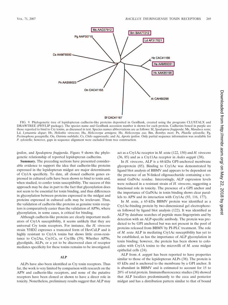
ipsilon, and Spodoptera frugiperda. Figure 9 shows the phylo-genetic relationship of reported lepidopteran cadherins.
Summary. The preceding sections have presented consider-able evidence to support the idea that cadherin-like proteinsexpressed in the lepidopteran midgut are major determinantsof Cry1A specificity. To date, all cloned cadherin genes ex-pressed in cultured cells have been shown to bind to toxin and,when studied, to confer toxin susceptibility. The success of thisapproach may be due in part to the fact that glycosylation doesnot seem to be essential for toxin binding, and thus differencesin glycosylation between proteins expressed in the midgut andproteins expressed in cultured cells may be irrelevant. Thus,the validation of cadherin-like proteins as genuine toxin recep-tors is comparatively easier than the validation of APNs, whereglycosylation, in some cases, is critical for binding.
Although cadherin-like proteins are clearly important medi-ators of Cry1A susceptibility, it seems unlikely that they areuniversal Cry toxin receptors. For instance, the H. virescensstrain YHD2 expresses a truncated form of HevCaLP and ishighly resistant to Cry1A toxins but shows little cross-resis-tance to Cry2Aa, Cry1Ca, or Cry1Ba (59). Whether APNs,glycolipids, ALPs, or a yet to be discovered class of receptormediates specificity for these toxins remains to be investigated.
ALP
ALPs have also been identified as Cry toxin receptors. Thusfar, the work is very limited by comparison with research on theAPN and cadherin-like receptors, and none of the putativereceptors have been cloned or shown to have a direct role intoxicity. Nonetheless, preliminary results suggest that ALP may
act as a Cry1Ac receptor in M. sexta (122, 150) and H. virescens(36, 85) and as a Cry11Aa receptor in Aedes aegypti (38).
In H. virescens, ALP is a 68-kDa GPI-anchored membraneglycoprotein (85). Binding to Cry1Ac was demonstrated byligand blot analysis of BBMV and appears to be dependent onthe presence of an N-linked oligosaccharide containing a ter-minal GalNAc residue. Interestingly, ALP expression levelswere reduced in a resistant strain of H. virescens, suggesting afunctional role in toxicity. The presence of a GPI anchor andthe importance of GalNAc in toxin binding shows clear paral-lels to APN and its interaction with Cry1Ac (93, 116, 177).
In M. sexta, a 65-kDa BBMV protein was identified as aCry1Ac-binding protein by two-dimensional gel electrophore-sis followed by ligand blot analysis (122). It was identified asALP by database searches of peptide mass fingerprints and bydetection with an ALP-specific antibody. The protein was pre-dicted to be GPI anchored but was not present in the pool ofproteins released from BBMV by PI-PLC treatment. The roleof M. sexta ALP in mediating Cry1Ac susceptibility has yet tobe established, as has the importance of ALP glycosylation intoxin binding; however, the protein has been shown to colo-calize with Cry1A toxins to the microvilli of M. sexta midgutepithelial cells (24).
ALP from A. aegypti has been reported to have propertiessimilar to those of the lepidopteran ALPs (38). The protein is65 kDa and is anchored to the membrane by a GPI anchor. Itis abundant in BBMV and is estimated to account for 15 to20% of total protein. Immunofluorescence studies (38) showedthat ALP localizes predominantly to the ceca and posteriormidgut and has a distribution pattern similar to that of bound
FIG. 9. Phylogenetic tree of lepidopteran cadherin-like proteins deposited in GenBank, created using the programs CLUSTALX andDRAWTREE (PHYLIP package). The species name and GenBank accession number is shown for each protein. Cadherins boxed in purple arethose reported to bind to Cry toxins, as discussed in text. Species names abbreviations are as follows: Sf, Spodoptera frugiperda; Ms, Manduca sexta;Ld, Lymantria dispar; Hv, Heliothis virescens; Ha, Helicoverpa armigera; Hz, Helicoverpa zea; Bm, Bombyx mori; Px, Plutella xylostella; Pg,Pectinophora gossypiella; On, Ostrinia nubilalis; Cs, Chilo suppressalis; and Ai, Agrotis ipsilon. Only partial sequence information was available forP. xylostella; however, gaps in sequence alignment were excluded from tree construction.
VOL. 71, 2007 BACILLUS THURINGIENSIS TOXIN RECEPTORS 269
on May 22, 2018 by guest
http://mm
br.asm.org/
Dow
nloaded from
Cry11Aa. Binding to ALP has been demonstrated by ligandblot analysis and by Cry11Aa affinity chromatography (38).Phage displaying ALP-specific peptides blocked Cry11Aabinding to ALP and decreased toxicity, suggesting a functionalrole for ALP in mediating Cry toxin susceptibility (38).
Based on their similar properties, A. aegypti ALP and thesame Cry-binding protein reported by Krieger et al. (100) andBuzdin et al. (22) are believed to be the same. In these studies,both Cry11Aa and Cry4Ba were shown to bind to ALP andcompete for a common epitope. Interestingly, Cry9Aa alsobound to ALP but did not compete with the other toxins andwas not toxic to A. aegypti (22). Incubation with several carbo-hydrates, including GalNAc, failed to disrupt the interactionbetween toxin and ALP, and neither N-acetylglucosamine(GlcNAc) nor GalNAc could be used to elute the receptorfrom Cry11Aa- or Cry4Ba-Sepharose. As such, a role for car-bohydrates in binding could not be demonstrated, in contrastto the GalNAc-dependent binding observed between Cry1Acand H. virescens ALP (85).
Glycolipid
Glycolipids are another important class of putative Cry toxinreceptor. While an interaction between glycosphingolipidsand Cry toxins was first reported in 1986 (30), the impor-tance of this receptor class was demonstrated only recentlyusing the nematode Caenorhabditis elegans (60). A numberof genetic, genomic, and cell biological tools have beendeveloped for the study of C. elegans—tools not availablefor the study of insects controlled by Cry toxins—that makethis organism particularly suitable for elucidating toxin re-ceptors (118). Since nematocidal and insecticidal Cry toxinsshare sequence similarity (Cry6Aa being the exception), in-sight gained from C. elegans models may lead to advances inthe study of Cry toxins active against other more economi-cally important organisms.
The identification of glycolipids as Cry toxin receptors wasmade possible by the characterization of chemically mu-tagenized C. elegans strains selected for their resistance toCry5Ba (61, 118). In total, 200 toxin-resistant lines were iso-lated, of which 45 were further characterized and found to bemutated at one of five loci (61, 118). Using forward genetics,four genes were identified that could restore toxin susceptibil-ity and were named the bre genes for B. thuringiensis toxinresistant (61, 62). The first gene to be characterized, bre-5, wasfound to be a member of the �1,3-galactosyltransferase familyand encoded a protein most similar to the Drosophila proteinBRAINIAC (62). Subsequently, bre-2, bre-3, and bre-4 werecharacterized (61). bre-2 encoded a putative �1-3-glycosyl-transferase that shared approximately 25% amino acid identityin its enzymatic domain with other family members. bre-4 waspredicted to encode the previously characterized enzyme UDP-GalNAc:GlcNAc �1-4-N-acetylgalactosaminyltransferase (90),and bre-3 was shown to encode a putative glycosyltransferasehomologous to the Drosophila protein EGGHEAD. Thus, allresistance genes appeared to encode glycosyltransferases thatshowed no similarity to putative toxin receptors previously iden-tified in insects.
Several experiments were conducted to determine how theinactivation of glycosyltransferases could lead to the observed
toxin-resistant phenotype. Experiments showing that fluores-cently labeled Cry5Ba was readily endocytosed by wild-typeanimals but not by bre mutants suggested that the glycosyl-transferases synthesized a component necessary for the toxinto interact with intestinal cells (61, 62). Dose-response assaysfound no difference in the susceptibilities of single and doubleC. elegans mutants to Cry14Aa, providing evidence that the bregenes acted in a common pathway (61). Finally, the findingthat bre mutants failed to produce certain ceramide-based gly-colipids that specifically bound to Cry5Ba demonstrated thatthe bre genes function to produce a glycosphingolipid toxinreceptor (60).
Because the Drosophila proteins EGGHEAD and BRAINIACwere predicted to catalyze consecutive glycosylation reactionsinvolved in glycosphingolipid synthesis (128, 154, 182), it waspostulated that glycolipids may also serve as Cry toxin recep-tors in insects (60). Preliminary results suggested that thismight be the case, as glycolipids extracted from the midguts ofM. sexta were shown to bind to Cry1Aa, Cry1Ab, and Cry1Ac.The importance of this interaction in mediating toxin sus-ceptibility was not reported, however, and remains to beexplored.
Other Receptors
Preliminary reports suggest that Cry toxins may bind to twoadditional types of receptors. The first, a 270-kDa glycoconju-gate isolated from L. dispar called BTR-270, was shown to bindstrongly to Cry1Aa, Cry1Ab, and Cry1Ba, weakly to Cry1Ac,and not at all to Cry1Ca, Cry2Aa, Cry2Ba, and Cry3Aa (176).The receptor has thus far been difficult to characterize; how-ever, it appears to be a highly glycosylated anionic protein thatmay be a component of the brush border membrane glyco-calyx. The second receptor, named P252, was isolated from aTriton X-100-soluble fraction derived from B. mori BBMV(73). By sodium dodecyl sulfate-polyacrylamide gel electro-phoresis, nsrsid1200748\delrsid1200748 the protein was esti-mated to have a molecular mass of 252 kDa; however, a985-kDa homo-oligomer was detected by gel filtrationchromatography. The purified receptor bound to Cry1Aa,Cry1Ab, and Cry1Ac under nondenaturing conditions. Lectinbinding suggested the presence of N- and O-linked glycans, butGalNAc did not inhibit toxin binding. To characterize theprotein further, two internal peptides were sequenced but didnot match any proteins in the available databases. Interest-ingly, however, reanalysis of these peptides for the presentreview generated significant matches (12/15 and 15/15 identicalresidues) with a recently described protein called chlorophyllidA-binding protein (ChBP) (121). This protein was identified inthe midgut of B. mori and was characterized by its ability tobind to a derivative of chlorophyll. The mass of ChBP wasestimated to be between 302 and 312 kDa, but unlike for P252,glycosylation could not be detected. Sequence analysis re-vealed that ChBP was an unusual member of the lipocalinfamily (41, 42) and contained 15 prototypic lipocalin domains.Whether ChBP and P252 are the same protein remains to beconfirmed.
270 PIGOTT AND ELLAR MICROBIOL. MOL. BIOL. REV.
on May 22, 2018 by guest
http://mm
br.asm.org/
Dow
nloaded from

DETERMINANTS OF BINDING
APN
Receptor determinants. While many reports have identifiedAPNs as Cry toxin-binding proteins, little is known about howthe molecules interact. As discussed earlier, GalNAc appearsto be an important determinant of the Cry1Ac-APN interac-tion; however, the nature and position of the glycan structureinvolved in binding have not yet been determined. A recentreport on the glycans of class 1 APN from M. sexta has pro-vided some insight into the possible determinants of binding.Stephens et al. (163) carried out an extensive characterizationof the N-linked oligosaccharides by mass spectrometry. Threeof the four glycosylation sites (N295, N623, N752) were linkedto highly fucosylated glycans with unusual difucosylated cores.The remaining site (N609) was glycosylated with the paucim-annosidic glycan common to insect glycoproteins (168). Inter-estingly, GalNAc was not identified as a component of any ofthe glycans, suggesting that N-linked carbohydrates are un-likely to be involved in toxin binding. This finding supports thetheory that Cry1Ac interacts with APN at its threonine-richC-terminal stalk predicted to be O glycosylated with mucin-type (GalNAc-�-O-Ser/Thr) glycans (92, 93).
Attempts have also been made to locate putative toxin bind-ing sites in APNs believed to interact with toxin in a glycan-independent manner. In one study, class 1 APN purified fromB. mori was proteolytically digested and probed with Cry1Aa ina ligand blot assay (193). The toxin bound to two digestionproducts, the smaller a 30-kDa fragment believed to consist ofamino acids Asp40 to Lys313. Binding was also assessed usingfragments of APN expressed as GST fusion proteins in E. coli.The smallest fragment that retained toxin binding affinity cor-responded to APN amino acids residues Ile135 to Pro198.More recently, Cry1Aa binding to the same APN fragment wasdemonstrated under nondenaturing conditions by enzyme-linked immunosorbent assay (6).
Cry1Aa and Cry1Ab have also been tested for their abilitiesto bind to the equivalent toxin-binding fragments in class 1,class 2, class 3, and class 4 APN derived from B. mori and P.xylostella (132). By ligand blot analysis, both toxins bound to allrecombinant APNs, suggesting a conserved, common receptorbinding site; however, other results questioned the biologicalrelevance of this finding. It was shown by ligand blot analysisthat neither Cry1Aa nor Cry1Ab bound to intact class 2 (90-kDa), class 3 (110-kDa), or class 4 (100-kDa) APN isolatedfrom B. mori BBMV (132). This was in agreement with resultsreported by Jenkins and Dean (80), who showed that Cry1Aafailed to bind to 100-kDa and 110-kDa forms of B. mori APNunder nondenaturing conditions. It was also shown thatCry1Ab did not bind to class 1 APN purified from B. moriBBMV by SPR analysis (80). Since it has been demonstratedthat denaturing APN can reveal binding epitopes hidden in thefolded protein (26), studies under nondenaturing conditionsmust be conducted to determine whether the Cry1Aa bindingsite identified in class 1 B. mori APN also binds to Cry1Ab andwhether this site is truly conserved among the other APNs.
Toxin determinants. Of the different Cry toxins that havebeen reported to bind to APN, only Cry1A toxins have beenexamined further for their determinants of binding. From
these studies, three APN binding sites have been proposed.The best characterized site is unique to Cry1Ac and is found onthe outer sheet of domain III. At this site, Cry1Ac is believedto bind to APN via a GalNAc-containing glycan. A secondbinding site shared by both Cry1Ab and Cry1Ac was identifiedat the apex of domain II and is believed to be formed bysurface-exposed loops. The remaining site was identified onCry1Aa and Cry1Ab and has been proposed to occur near theinterface between domain II and domain III.
(i) Domain III. The importance of Cry1Ac domain III as adeterminant of APN binding was first shown by Lee et al.(106), who constructed hybrids from Cry1Aa and Cry1Ac andanalyzed their binding properties. Under nondenaturing con-ditions, it was shown that only toxins with domain III derivedfrom Cry1Ac bound strongly to purified L. dispar APN. Sub-sequently, it was shown that GalNAc could completely disruptthis interaction (82). Similar results were obtained by DeMaagd et al. (27), who showed that only Cry1Ac/Cry1Ca hy-brids constructed with Cry1Ac domain III could bind to puri-fied M. sexta APN and that binding could be inhibited withGalNAc.
To understand how Cry1Ac domain III might interact withAPN, Burton et al. (21) mutated several residues on the outersheet of domain III. This region was proposed to form a GalNAcbinding pocket based on the structural similarity betweendomain III and the N-terminal cellulose-binding domain ofCellulomonas fimi 1,4-�-glucanase CenC (CBDN1) (83). Resi-dues within and around �-strand 16 were of particular interestbased on a preliminary analysis of the Cry1Ac crystal structurethat showed a unique conformation in this region (21). In total,18 single mutants, 1 double mutant, and 1 triple mutant wereconstructed and tested for binding to M. sexta BBMV. Severalmutants showed reduced binding, but the greatest decreasewas observed with the triple N506D Q509E Y513A mutant.Ligand blot analysis confirmed that this mutation decreasedbinding to APN. That incubation with GalNAc did not furtherdecrease triple mutant binding to BBMV provided evidencethat the interaction between Cry1Ac and M. sexta APN at thissite was indeed GalNAc dependent. Similar results were ob-tained in other studies. Jenkins et al. showed that mutants withalanine substitutions at Q509, R511, Y513, or 509QNR511
failed to bind to purified M. sexta APN (81) and bound topurified L. dispar APN with lower affinity (82).
The crystal structure of Cry1Ac, solved in the presence andabsence of GalNAc, has helped to further characterize thedomain III APN binding site and has provided a structuralexplanation for why Cry1Ac, but not Cry1Aa, binds to APN ina manner dependent on GalNAc (32, 108). Analysis of thestructure revealed a unique 6-residue insertion (505GNNIQN510) in domain III that was not observed in Cry1Aa andwas not predicted by an automatically generated model ofCry1Ac (81). This insertion was shown to project from theouter sheet of domain III and curve back to form a shallowGalNAc-binding cavity. The structure of Cry1Ac with boundGalNAc was very similar to that without bound GalNAc;however, residues in contact with GalNAc were moreordered. In addition, it was observed that the temperaturefactor of residues in domain I increased upon GalNAcbinding. This led to the hypothesis that ligand binding atdomain III may increase the mobility of the pore-forming
VOL. 71, 2007 BACILLUS THURINGIENSIS TOXIN RECEPTORS 271
on May 22, 2018 by guest
http://mm
br.asm.org/
Dow
nloaded from

domain, possibly triggering a conformational change leadingto membrane insertion (108). Consistent with this hypothesis,Pardo-Lopez et al. (136) recently reported that GalNAcbinding to an oligomeric form of Cry1Ac enhanced toxinmembrane insertion.
(ii) Domain II. The loops at the apex of domain II have alsobeen implicated in APN binding. Lee et al. (105) mutated twoarginine residues (368 and 369) in loop 2 of Cry1Ab andCry1Ac that had previously been shown to be important fortoxicity and BBMV binding in M. sexta and L. dispar (144).Consistent with previous results, SPR studies suggested thatCry1Ab bound to M. sexta APN at a single site, whereasCry1Ac bound to both M. sexta and L. dispar APN at two sites(82, 120). Cry1Ab mutants with alanine or glutamic acid sub-stitutions showed a nearly 10-fold decrease in binding to M.sexta APN. The corresponding Cry1Ac mutants also showedreduced binding and kinetic data suggesting that only one ofthe two proposed binding sites was affected, consistent with thepresence of a distinct APN binding site in domain III (seeabove). Similar results were obtained when the Cry1Ac alanineand glutamic acid mutants were tested for binding to purifiedL. dispar APN. In another study, additional Cry1Ac derivativeswith mutations at loop 2, loop 3, and loop �8 were tested fortheir abilities to bind to purified L. dispar APN, and eachshowed decreased binding to only one of the two proposedAPN binding sites (82). A subsequent study showed that loop�8 mutants also decreased binding to purified M. sexta APN(103).
In contrast to the results reported above with folded albeitmutated whole toxin, work carried out in the Soberon labora-tory found that synthetic peptides derived from the domain IIapex loops had no effect on APN binding. Gomez et al. (55, 56)used ligand blot analysis to examine the binding of Cry1Ab todenatured M. sexta APN in the presence or absence of syn-thetic peptides corresponding to sequences from loop 1, loop2, loop 3, or loop �8. Even in the presence of a 500- to1,000-fold excess of synthetic peptide, no decrease in bindingwas observed. Since Cry1Ab detection of APN by ligand blotanalysis varies among different laboratories (27, 124, 174), thesignificance of these results is unclear and requires furtherinvestigation.
(iii) Domain II/III interface. A third region that has beenproposed to act as an APN binding site was first characterizedfor Cry1Aa (6). This region was identified at the interfacebetween domain II and III and was shown to interact with the64-amino-acid toxin-binding fragment of class 1 B. mori APN(193). The putative binding site was identified by mapping theepitopes of two monoclonal antibodies that bound to Cry1Aaand blocked its interaction with APN. Based on the results ofseveral binding studies, both antibodies appeared to bind tothe outer sheet of domain III at distinct but overlapping sitesthat included residues 508STLRVN513 and 582VFTLSAHV589.Additional studies showed that the APN binding site wasclose to, but did not include, this region. The interfacebetween domain II and domain III was proposed as acandidate APN binding site based on its proximity to theantibody binding sites and on the presence of conservedresidues in this region that are common to Cry toxins withspecificity for class 1 B. mori APN. Recently, Gomez et al.
used synthetic peptides to show that a similar region inCry1Ab may interact with APN from M. sexta (54).
Cadherin
Receptor determinants. To learn more about the involve-ment of cadherin-like proteins in facilitating Cry toxin suscep-tibility, several groups have tried to define a toxin bindingregion. Nagamatsu et al. (129) studied the B. mori receptorBtR175. The group expressed truncated variants of BtR175 inSf9 cells and under nondenaturing conditions identified a re-gion within amino acids 1245 to 1464 necessary for Cry1Aabinding (129). That a variant consisting of amino acids 1108 to1715 conferred Cry1Aa susceptibility to Sf9 cells but a variantlacking amino acids 1246 to 1390 did not confirmed the func-tional importance of the toxin binding region. That a syntheticpeptide corresponding to BtR175 residues 1296 to 1301 par-tially blocked the interaction between Cry1Aa and Cry1Ab andBtR175 provided additional support (57).
Toxin binding sites have been studied more extensively inthe M. sexta cadherin receptor; however, there are some dis-crepancies over the regions involved in binding. Gomez et al.(55–57) used competition studies to show that synthetic pep-tides derived from BT-R1 at 869 to 876 and 1331 to 1342 couldinterfere with Cry1A binding to denatured cadherin and couldmitigate Cry1Ab toxicity to neonates. They also showed thatrecombinant fragments corresponding to amino acids 831 to900 and 1291 to 1360 could bind to both nondenatured anddenatured forms of Cry1Ab (55). In contrast, Dorsch et al. (33)identified a single toxin binding site. The group expressedtruncated BT-R1 peptides by use of in vitro translation andunder denaturing conditions showed that a fragment corre-sponding to amino acids 1296 to 1465 was sufficient for Cry1Abbinding. Since Cry1Ab did not bind to a fragment containingresidues 1 to 1296, there was no evidence for a binding sitebetween residues 831 and 900. Subsequent work by Hua et al.(74) examined Cry1Ab binding to various truncated forms ofBT-R1a and under nondenaturing conditions, a fragment con-sisting of residues 1363 to 1464 was found to be sufficient forbinding. This fragment did not include any of the binding sitesproposed by Gomez et al. (55–57), but when expressed in S2cells, it could confer susceptibility to Cry1Ab. It was also foundthat fragments containing residues 869 to 876 or 1331 to 1342but lacking residues 1363 to 1464 failed to bind to Cry1Abunder nondenaturing conditions.
Toxin binding sites have also been mapped for cadherinsfrom two other species. Cry1Ac was shown to bind to H.armigera cadherin residues 1217 to 1461 expressed as a GSTfusion protein in E. coli (184), and in H. virescens cadherin, acombination of truncation analysis and site-directed mutagen-esis was used to identify a Cry1A toxin binding region withinamino acids 1422 to 1440 (190).
Since the majority of toxin binding sites appear to be locatedat or near the most membrane-proximal cadherin repeats (Fig.10), it is tempting to speculate that binding in this region isimportant for the toxin’s mechanism of action. To test thishypothesis, the toxin susceptibility of cells expressing wild-typecadherin could be compared to cells expressing a recombinantform of cadherin modified to position the toxin binding siteaway from the membrane.
272 PIGOTT AND ELLAR MICROBIOL. MOL. BIOL. REV.
on May 22, 2018 by guest
http://mm
br.asm.org/
Dow
nloaded from

Toxin determinants. Thus far, studies on the toxin determi-nants of binding to cadherin are limited. In most cases, putativebinding sites have been assessed by testing whether syntheticpeptides can disrupt the interaction between toxin and denaturedreceptor. By this method, Gomez et al. (55, 56) showed thatsynthetic peptides derived from loop �8 and loop 2 of Cry1Aband that loop 2 and loop 3 of Cry1Aa were involved in binding tothe M. sexta cadherin receptor BT-R1. Additional studies withCry1Ab suggested that loop �8 and loop 2 bound to two distinctsites on BT-R1 (55), but as discussed in the previous section, thevalidity of these toxin binding sites is currently in dispute (33, 74).Similar methods have been used to identify the regions of Cry1Aband Cry1Ac responsible for binding to denatured CAD3, a re-combinant toxin-binding fragment derived from the H. virescenscadherin-like protein (190). In the presence of a synthetic peptide
corresponding to loop 3, Cry1Ab and Cry1Ac binding to CAD3was diminished, whereas a loop �8 peptide had no effect onbinding. In addition, it was found that a synthetic peptide corre-sponding to loop 2 decreased Cry1Ab, but not Cry1Ac, binding toCAD3. These apparent differences in CAD3 binding are surpris-ing given that the sequences of Cry1Ab and Cry1Ac in domain IIare nearly identical. It would be interesting to see whether addi-tional studies with correctly folded proteins or with domain IImutants support the findings of these preliminary studies.
TOXIN MOLECULAR MECHANISM OF ACTION
Given the number of Cry toxins, putative toxin receptors,and insects currently under investigation, it is perhaps notsurprising that the Cry toxin mode of action is controversial.
FIG. 10. Domain structure and putative Cry1A toxin binding sites in lepidopteran cadherin-like proteins. The proteins are labeled as follows:PRO, proprotein region; SP, signal peptide; EC, ectodomain; MPED, membrane-proximal extracellular domain; TM, transmembrane domain;CYTO, cytoplasmic domain. Domains are as defined by Nagamatsu et al. (130) (B. mori), Wang et al. (184) (H. armigera), Gahan et al. (44) (H.virescens), and Dorsch et al. (33) (M. sexta). Putative toxin binding sites are as reported by Nagamatsu et al. (129) (B. mori), Wang et al. (184) (H.armigera), Xie et al. (190) (H. virescens), and Dorsch et al. (33), Gomez et al. (55–57), and Hua et al. (74) (M. sexta). Proteins are illustrated suchthat homologous regions are horizontally adjacent. Features not expected in the mature form of the protein are outlined with a dashed line.
VOL. 71, 2007 BACILLUS THURINGIENSIS TOXIN RECEPTORS 273
on May 22, 2018 by guest
http://mm
br.asm.org/
Dow
nloaded from

Even the general view that toxin monomers bind to midgutreceptors, oligomerize, and insert into the membrane to formlytic pores has recently been challenged (195, 196). In thissection, three contrasting models of Cry1A toxin mode of ac-tion will be presented and discussed. The first two modelsdiffer considerably in terms of which receptors are importantfor toxicity and how these receptors facilitate toxin function.The third model combines elements of the first two models andalso proposes a functional role for an additional receptor class.
The Bravo Model
The Bravo model is an updated version of the colloid-os-motic lysis model of Knowles and Ellar (96) and proposes thatboth cadherin and APN receptors are required for full Cry1Atoxicity towards M. sexta (18). The model is based primarily onexperiments involving Cry1Ab interactions with BBMV (18,58, 197), and suggests that receptor binding is sequential: ac-tivated toxin monomer binds initially to BT-R1 and then toclass 1 APN. After binding to BT-R1, it is proposed that thetoxin undergoes a conformational change that facilitates cleav-age of helix �-1 by membrane-bound proteases (58). This formof the toxin oligomerizes to form a tetrameric prepore (58)that preferentially binds to APN as a result of an increase inbinding affinity (18). APN then directs the prepore to deter-gent-resistant membranes (DRMs), or lipid rafts, that facilitatemembrane insertion to form a lytic pore (18). The generationof as few as 200 copies of this pore, with a reported radius of0.5 to 1.0 nm, in the columnar cell apical membrane leads tovery rapid changes in membrane potential, equilibration ofions across the membrane, influx of water, cell swelling, andeventual lysis (95). In the following sections, experimental ev-idence for and against this model will be presented.
The theory that solubilized, proteolytically activated Crytoxin monomer binds to BT-R1 before APN is based primarilyon immunoprecipitation experiments with Cry1Ab protoxinand BBMV that show differences in the recoveries of BT-R1
and APN over time (18). While both BT-R1 and APN weredetected at all time points, the amount of BT-R1 that copre-cipitated with Cry1Ab decreased slightly with time, whereasAPN recovery increased by fourfold over the course of theexperiment. This, in conjunction with earlier data showing thatCry toxin binds to BT-R1 with an affinity 100-fold greater thanthat to APN (150, 174), was taken to indicate that Cry1Abbinding to BT-R1 occurs before APN binding; however, otherexplanations of the data are possible. For instance, differencesin receptor abundance could explain why maximal levels ofBT-R1 are coprecipitated at all time points whereas APN re-covery increases progressively. In B. mori, APN was estimatedto be 100-fold more abundant than BtR175 on midgut mi-crovilli (66, 131). Similarly, disruption of the gene encodingHevCaLP did not decrease Cry1Ab and Cry1Ac binding toBBMV (89), suggesting that in H. virescens, cadherins may alsobe of low abundance relative to other receptors. If this differ-ence in receptor concentration is also present in M. sexta, itwould seem likely that high-affinity (1 nM [174]), low-abun-dance BT-R1 binding sites are quickly saturated with toxin,whereas low-affinity (100 nM [150]), high-abundance APNbinding sites become saturated more slowly. In such a situa-tion, APN may actually function to concentrate Cry1Ab at the
surface of the membrane before the toxin is relayed to lessabundant cadherin receptors. A similar model has been used toexplain how the channel-forming toxin aerolysin might pene-trate the glycocalyx to reach receptors on the surface of targetepithelial cells: the toxin initially binds to low-affinity carbohy-drate structures before being shuttled to high-affinity bindingsites on the glycan core of GPI-anchored proteins (117).
After monomeric Cry1Ab binds to BT-R1, it is believed toundergo a conformational change that facilitates proteolyticcleavage between helices �-1 and �-2. This event exposes ahydrophobic surface that is important for toxin oligomeriza-tion (58). While a direct role for native BT-R1 in facilitatingtoxin cleavage has not yet been demonstrated, experimentswith Cry1Ab and recombinant toxin-binding fragments (55) orthe single-chain antibody scFv73 (58) suggest that this may bethe case. scFv73 is reported to act as a BT-R1 mimic. It hasbeen shown to bind to domain II of Cry1Ab and block itsinteraction with denatured BT-R1 and to reduce the toxicity ofCry1Ab towards M. sexta larvae (57). When scFv73 was incu-bated with solubilized Cry1Ab protoxin prior to digestion withM. sexta midgut juice, the resulting toxin preparation had ahigh in vitro pore-forming activity relative to toxin prepared inthe absence of scFv73. Examination of each preparation byimmunoblotting revealed a 250-kDa toxin oligomer found onlyin the sample preincubated with scFv73. The separation of theoligomer from monomer by gel filtration allowed each speciesto be tested for pore-forming activity independently, and it wasshown that one-fifth the molar concentration of oligomer wasalmost 2.6-fold more active than monomer. A later studyshowed that unlike monomeric Cry1Ab, the oligomer formedstable channels with a high probability of being open in a lipidbilayer (147). Analysis of each sample by N-terminal sequenc-ing revealed differences in the sites of proteolytic cleavage: theoligomer between helices �-1 and �-2 (before residue Val-51)and the monomer at the beginning of helix �-1 (before Ile-29).It was thus concluded that binding to BT-R1 facilitated cleav-age of helix �-1, leading to toxin oligomerization and poreformation.
While the results presented above are intriguing, not all datasupport the hypothesis that Cry1Ab binding to BT-R1 facili-tates cleavage of helix �-1 or that cleavage in this region pro-motes toxin oligomerization. For example, Miranda et al. (125)treated Cry1Ab with various dilutions of M. sexta midgut juiceand identified toxin products migrating at 60, 58, and 30 kDa.N-terminal sequencing of the 60-kDa product revealed that inthe absence of receptor, cleavage occurred between residuesLeu-57 and Gly-58. It was also shown that Cry1Ab protoxinactivated either in vitro with M. sexta midgut juice or in vivo inM. sexta larvae failed to form oligomeric toxin, as determinedby immunoblotting. Other studies have questioned whetherBT-R1 has any involvement in toxin oligomerization and evenwhether the oligomeric species detected by immunoblottinghas any role in cytotoxicity. Aronson et al. (5) tested wild-typeCry1Ac and domain I mutants with or without toxicity towardsM. sexta larvae for their abilities to oligomerize when incubatedwith BBMV. While a correlation between toxicity and oligo-mer formation was observed in most cases, the mutantH168R was two- to threefold more toxic than the wild type andyet failed to oligomerize, according to immunoblot analysis.More evidence against the biological relevance of the oligo-
274 PIGOTT AND ELLAR MICROBIOL. MOL. BIOL. REV.
on May 22, 2018 by guest
http://mm
br.asm.org/
Dow
nloaded from

meric Cry1Ab species has come from a study by Zhang et al.(195), who examined Cry1Ab binding to toxin-susceptible cellsexpressing BT-R1 (S5) and toxin-resistant control cells (H5).After treatment with Cry1Ab, each cell type was analyzed byimmunoblotting. Both monomeric and oligomeric Cry1Ab as-sociated with S5 cells but, surprisingly, the Cry1Ab oligomeralso associated with control cells. This result showed not onlythat the Cry1Ab oligomer could be detected in the absence ofBT-R1 but also that the presence of this species in the cellmembrane did not correlate with cytotoxicity, despite its highin vitro pore-forming activity (58, 147). Although these dataargue against the biological relevance of the oligomeric speciesdetected by immunoblotting, they do not necessarily rule outthe importance of oligomerization in the toxin’s mode of ac-tion, since the true oligomeric form of the toxin may not bedetectable by immunoblotting.
The next step in the Bravo model of toxin mode of actionproposes that oligomeric toxin attached to BT-R1 binds toAPN. This is based on experiments showing that relative to themonomer, oligomeric Cry1Ab immunoprecipitates more APNfrom solubilized BBMV and has a higher affinity for an en-riched preparation of APN (18). Once bound to APN, thetoxin-receptor complex is then believed to localize to DRMmicrodomains, where the prepore inserts into the membrane.This hypothesis is based on several experiments. First, an anal-ysis of receptor localization in BBMV prior to toxin exposurerevealed that APN and BT-R1 associate with different mem-brane fractions: BT-R1 with the soluble membrane fractionand the majority of APN with the DRM fraction (18). AfterBBMV was incubated with Cry1Ab protoxin, BT-R1 was foundexclusively in the DRM fraction, along with the majority ofCry1Ab (monomer and oligomer) and APN. It thus appearedthat BT-R1 was recruited to DRMs due to its association witholigomeric Cry1Ab. To confirm the importance of APN in thisprocess, BBMV were pretreated with PI-PLC to reduce APNlevels and then incubated with Cry1Ab protoxin (18). As aresult of this treatment, a drastic reduction in the amount ofCry1Ab bound to BBMV was observed and Bt-R1 no longerassociated with the DRM fraction.
A functional role for DRMs has been suggested by experi-ments examining the effect of DRM integrity on pore forma-tion (197). DRM vesicles isolated from M. sexta or H. virescensBBMV were incubated with methyl-�-cyclodextrin, a reagentthat destroys DRMs by extracting cholesterol from the mem-brane, and compared to control vesicles in an assay measuringpore-forming activity. Methyl-�-cyclodextrin was found to sub-stantially reduce Cry1Ab pore formation in vesicles from bothspecies.
The Zhang Model
In a recent study, the theory that Cry1Ab kills cells exclu-sively by osmotic lysis has been challenged. Zhang et al. (195)argued that the correlation between pore formation and cyto-toxicity has not been adequately demonstrated and proposedan alternative mode of action in which monomeric Cry1Abbinds to BT-R1 and initiates an Mg2�-dependent signalingcascade that promotes cell death. Additional work suggestedthat receptor binding activates a signaling pathway involvingstimulation of G protein, adenylyl cyclase (AC), increased cy-
clic AMP (cAMP) levels, and activation of protein kinase A(PKA), leading to destabilization of the cytoskeleton and ionchannels and subsequent cell death (196). This section willdiscuss the experimental evidence upon which the Zhangmodel is based.
To study the mechanism of Cry1Ab cytotoxicity, Zhang et al.(195, 196) carried out various treatments on toxin-susceptiblecells heterologously expressing BT-R1 (S5) and toxin-resistantcontrol cells (H5). In the presence of Cry1Ab, S5 cells under-went membrane blebbing and ruffling that led to swelling andeventually to cell death. Since calcium had been previouslyshown to be important for the structural integrity of BT-R1
(23) and was proposed to mediate BT-R1-dependent adhesionof M. sexta BBMV (63), the effect of calcium on receptorbinding and cell death was examined using the divalent cationchelators EDTA and EGTA. Neither chelator affected toxin-receptor binding; however, EDTA (chelator of Ca2� andMg2�), but not EGTA (Ca2�-specific chelator), completelyabolished Cry1Ab-induced cell death (195, 196). It was alsoshown that the addition of Mg2� to S5 cells pretreated withEDTA restored Cry1Ab-mediated cytotoxicity. Since neitherion was shown to effect Cry1Ab binding to BT-R1 by immu-noblotting, it was proposed that Cry1Ab binding to BT-R1 waslinked to an Mg2�-dependent signaling pathway associatedwith cell death.
Evidence to suggest that Cry1Ab induces an AC/PKA sig-naling pathway was based on experiments examining the effectof Cry1Ab and various inhibitors on cAMP production andcytotoxicity in S5 cells (196). This pathway was initially hypoth-esized to mediate Cry1Ab activity, because many pathwaymembers are Mg2� dependent (162, 165, 198) and the secondmessenger cAMP had previously been implicated in signalingrelated to cell death (161, 169). First, it was demonstrated thatCry1Ab treatment stimulated cAMP production in S5 cells, butnot in H5 control cells, and that this effect could be reversed bypreincubation with EDTA. To test whether G protein activitywas involved in Cry1Ab-induced cAMP production, S5 cellswere incubated with the G protein �-subunit (G�s) antagonistNF449 (71). This compound reduced levels of cytotoxicity by50%, whereas the inhibitory G protein �-subunit (G�i) antag-onist NF449 (43) had no effect. The importance of AC intoxin-induced cell death was tested with the inhibitor ddADP,a reagent that blocks substrate utilization. ddADP decreasedlevels of cytotoxicity, suggesting that the stimulation of AC wasalso important for Cry1Ab-mediated cell death. The inhibitorsH-89 (35) and myristoylated amide 14-22 (PKAI 14-22-amide)(52) were used to test whether PKA was involved in the toxin’smechanism of action. Both inhibitors completely preventedmembrane blebbing, cell swelling, and cytotoxicity, and thusPKA appeared to play a critical role in Cry1Ab-induced sig-naling. Although Cry1Ab stimulation correlated with the pro-duction of cAMP, the AC activator forskolin and the cAMPanalog pCPT-cAMP were not sufficient to cause cell death.This may indicate that other signaling events originating di-rectly from BT-R1 or from the interaction of G�s with effectorsother than AC work in conjunction with PKA signaling to bringabout Cry1Ab-mediated cytotoxicity (196). This has not yetbeen proven, however, and would be a necessary step towardsthe validation of this model.
Some caution may be advisable in assessing the Zhang
VOL. 71, 2007 BACILLUS THURINGIENSIS TOXIN RECEPTORS 275
on May 22, 2018 by guest
http://mm
br.asm.org/
Dow
nloaded from
model, however (196). In a previous study (97), several Crytoxins including Cry1Ab were found to cause an increase inintracellular cAMP in a Mamestra brassicae cell line, but theauthors concluded that the effects on AC were a secondaryeffect of the interaction of the toxin with the membrane ratherthan a direct cause of the cytolytic mechanism. Further work toestablish an unambiguous causal connection between cytotox-icity and the rise in cAMP may therefore be necessary tostrengthen the Zhang model. Of particular importance wouldbe an examination of how PKA mediates Cry toxicity in vivo,since all work to date has been carried out using cell lines.
The Jurat-Fuentes Model
A third model has recently been proposed to explain themode of action of Cry1Ac in H. virescens (86). The modelsuggests that cytotoxicity is due to the combined effects ofosmotic lysis and cell signaling, and thus elements of both theBravo model and the Zhang model of toxin mode of action areincorporated. First, activated monomeric Cry1Ac binds to thecadherin-like protein HevCaLP. This results in the activationof an intracellular signaling pathway regulated by phospha-tases. Evidence to support the involvement of phosphatases inCry1Ac-mediated signaling was provided by a proteomic anal-ysis of BBMV that showed that resistant and susceptible strainsof H. virescens have different levels of intracellular phospha-tases (86). Signaling may also be dependent on a direct interac-tion between Cry1Ac and actin (86, 122), a cytoskeletal proteinthat interacts with the cytosolic domain of cadherins throughtyrosine phosphatases, catenin, and actinin (111). After bind-ing to HevCaLP, monomeric Cry1Ac oligomerizes and thenbinds to the GPI-anchored proteins APN and—in an extensionof the Bravo model—the ALP HvALP (85). Either proteinthen drives Cry1Ac oligomers into DRMs, where pore forma-tion results in osmotic shock and the activation of signalingpathways leading to cell death. While this model is intriguing,it should be noted that many steps are at present speculativeand remain to be confirmed experimentally.
CONCLUDING REMARKS
Since the first Cry toxin receptors were cloned in 1995 (94,175), much progress has been made in our understanding oftoxin specificity and mode of action. By far the best-studiedreceptors are from lepidopterans, and their interactions withCry1A toxins are particularly well characterized. Receptors inthe APN family have been identified in several lepidopteranspecies, and 38 members in five different classes have beendescribed. While many studies suggest that APNs can serve asCry-binding proteins and their ability to mediate pore-forma-tion in vitro is well documented, a direct role in cytotoxicity hasbeen yet to be firmly established. In contrast, a great deal ofprogress has been made in the functional characterization ofcadherin receptors. To date, almost all cloned cadherin genesexpressed in cultured cells encode proteins that bind to toxinand, when studied, have been found to confer toxin suscepti-bility. While it is clear that cadherins play a pivotal role in theCry1A toxin mode of action, there is currently uncertainty as towhether other putative receptors, known (Table 2) or yet to bediscovered, are required for full toxicity.
The recent report that Cry toxin activity can be modulatedby perturbing intracellular signaling pathways may open up anew avenue of research. In addition to studies showing thatinappropriate activation of the AC/PKA pathway can mediatetoxin-induced cell death (196), inactivation of the p38 pathwayin C. elegans apparently increases toxin sensitivity (76). In thisway, different signaling pathways may potentiate or mitigateCry toxicity. At present, the importance of internal signaling asa mechanism of insect resistance is largely unexplored. Re-search in this area may help to explain why some insects de-velop resistance without a loss in toxin binding (112, 189).
Important questions about how midgut receptors confer tox-icity remain. For Cry1A toxins, binding to cadherin appears tobe critical, but the details of this interaction remain elusive.Some studies suggest that cadherins function to convert mo-nomeric toxin into an insertion-competent prepore; however,an oligomeric species has been identified for resistant cells thatdo not express cadherin. If pore formation is not responsiblefor cytotoxicity, as has been recently suggested (195, 196), howmight loss-of-function mutations that do not affect receptorbinding be explained? A better understanding of the interac-tion between Cry1A toxins and cadherin receptors at a struc-tural level will likely improve our ability to answer such ques-tions.
While progress is being made in our understanding of Cry1Atoxicity, comparatively little is known about the remaining Crytoxin families. Based on sequence identity, it seems likely thatmost Cry toxins share a common three-domain structure; how-ever, binding and genetic studies have shown that cadherins donot mediate cell death in all cases (59, 89). Indeed, nematode-specific toxins apparently utilize a glycolipid receptor (60), andit was recently reported that the B. thuringiensis toxin Cyt1Aasynergizes Cry11Aa in mosquitoes by functioning as a mem-brane-bound receptor (138). As more toxin receptors are dis-covered and as our understanding of toxin-receptor interac-tions increases, it will be interesting to see the extent to whichCry toxins utilize a common mode of action.
REFERENCES
1. Abdullah, M. A., A. P. Valaitis, and D. H. Dean. 2006. Identification of aBacillus thuringiensis Cry11Ba toxin-binding aminopeptidase from the mos-quito, Anopheles quadrimaculatus. BMC Biochem. 7:16.
2. Agrawal, N., P. Malhotra, and R. K. Bhatnagar. 2002. Interaction of gene-
TABLE 2. Cry toxins for which a putative receptorhas been identifieda
ToxinPresence of receptor type
APN Cadherin ALP Glycolipid BTR-270 P252
Cry1Aa � � � � �Cry1Ab � � � � �Cry1Ac � � � � � �Cry1Ba � �Cry1Ca �Cry1Fa �Cry4Ba �Cry5Ba �Cry9Aa �Cry11Aa �Cry11Ba �
a See the text for references. �, a putative receptor for the indicated toxin hasbeen identified.
276 PIGOTT AND ELLAR MICROBIOL. MOL. BIOL. REV.
on May 22, 2018 by guest
http://mm
br.asm.org/
Dow
nloaded from

cloned and insect cell-expressed aminopeptidase N of Spodoptera litura withinsecticidal crystal protein Cry1C. Appl. Environ. Microbiol. 68:4583–4592.
3. Aimanova, K. G., M. Zhuang, and S. S. Gill. 2006. Expression of Cry1Accadherin receptors in insect midgut and cell lines. J. Invertebr. Pathol.92:178–187.
4. Angst, B. D., C. Marcozzi, and A. I. Magee. 2001. The cadherin superfamily:diversity in form and function. J. Cell Sci. 114:629–641.
5. Aronson, A. I., C. Geng, and L. Wu. 1999. Aggregation of Bacillus thurin-giensis Cry1A toxins upon binding to target insect larval midgut vesicles.Appl. Environ. Microbiol. 65:2503–2507.
6. Atsumi, S., E. Mizuno, H. Hara, K. Nakanishi, M. Kitami, N. Miura, H.Tabunoki, A. Watanabe, and R. Sato. 2005. Location of the Bombyx moriaminopeptidase N type 1 binding site on Bacillus thuringiensis Cry1Aa toxin.Appl. Environ. Microbiol. 71:3966–3977.
7. Banks, D. J., G. Hua, and M. J. Adang. 2003. Cloning of a Heliothis virescens110 kDa aminopeptidase N and expression in Drosophila S2 cells. InsectBiochem. Mol. Biol. 33:499–508.
8. Banks, D. J., J. L. Jurat-Fuentes, D. H. Dean, and M. J. Adang. 2001.Bacillus thuringiensis Cry1Ac and Cry1Fa �-endotoxin binding to a novel110 kDa aminopeptidase in Heliothis virescens is not N-acetylgalactosaminemediated. Insect Biochem. Mol. Biol. 31:909–918.
9. Bates, S. L., J. Z. Zhao, R. T. Roush, and A. M. Shelton. 2005. Insectresistance management in GM crops: past, present and future. Nat. Bio-technol. 23:57–62.
10. Blaschuk, O. W., R. Sullivan, S. David, and Y. Pouliot. 1990. Identificationof a cadherin cell adhesion recognition sequence. Dev. Biol. 139:227–229.
11. Boonserm, P., P. Davis, D. J. Ellar, and J. Li. 2005. Crystal structure of themosquito-larvicidal toxin Cry4Ba and its biological implications. J. Mol.Biol. 348:363–382.
12. Boonserm, P., M. Mo, C. Angsuthanasombat, and J. Lescar. 2006. Struc-ture of the functional form of the mosquito larvicidal Cry4Aa toxin fromBacillus thuringiensis at a 2.8-angstrom resolution. J. Bacteriol. 188:3391–3401.
13. Boraston, A. B., D. Nurizzo, V. Notenboom, V. Ducros, D. R. Rose, D. G.Kilburn, and G. J. Davies. 2002. Differential oligosaccharide recognition byevolutionarily-related �-1,4 and �-1,3 glucan-binding modules. J. Mol. Biol.319:1143–1156.
14. Bourne, Y., V. Roig-Zamboni, A. Barre, W. J. Peumans, C. H. Astoul, E. J.Van Damme, and P. Rouge. 2004. The crystal structure of the Calystegiasepium agglutinin reveals a novel quaternary arrangement of lectin subunitswith a b-prism fold. J. Biol. Chem. 279:527–533.
15. Bourne, Y., V. Zamboni, A. Barre, W. J. Peumans, E. J. Van Damme, andP. Rouge. 1999. Helianthus tuberosus lectin reveals a widespread scaffold formannose-binding lectins. Structure 7:1473–1482.
16. Braun, L., and B. A. Keddie. 1997. A new tissue technique for evaluatingeffects of Bacillus thuringiensis toxins on insect midgut epithelium. J. Inver-tebr. Pathol. 69:92–104.
17. Bravo, A. 1997. Phylogenetic relationships of Bacillus thuringiensis delta-endotoxin family proteins and their functional domains. J. Bacteriol. 179:2793–2801.
18. Bravo, A., I. Gomez, J. Conde, C. Munoz-Garay, J. Sanchez, R. Miranda,M. Zhuang, S. S. Gill, and M. Soberon. 2004. Oligomerization triggersbinding of a Bacillus thuringiensis Cry1Ab pore-forming toxin to aminopep-tidase N receptor leading to insertion into membrane microdomains. Bio-chim. Biophys. Acta 1667:38–46.
19. Bravo, A., S. Hendrickx, S. Jansens, and M. Peferoen. 1992. Immunocyto-chemical analysis of specific binding of Bacillus thuringiensis insecticidalcrystal proteins to lepidopteran and coleopteran midgut membranes. J. In-vertebr. Pathol. 60:247–254.
20. Bravo, A., S. Jansens, and M. Peferoen. 1992. Immunocytochemical local-ization of Bacillus thuringiensis insecticidal crystal proteins in intoxicatedinsects. J. Invertebr. Pathol. 60:237–246.
21. Burton, S. L., D. J. Ellar, J. Li, and D. J. Derbyshire. 1999. N-Acetylgalac-tosamine on the putative insect receptor aminopeptidase N is recognised bya site on the domain III lectin-like fold of a Bacillus thuringiensis insecticidaltoxin. J. Mol. Biol. 287:1011–1022.
22. Buzdin, A. A., L. P. Revina, L. I. Kostina, I. A. Zalunin, and G. G. Chestukhina.2002. Interaction of 65- and 62-kD proteins from the apical membranes of theAedes aegypti larvae midgut epithelium with Cry4B and Cry11A endotoxins ofBacillus thuringiensis. Biochemistry (Moscow) 67:540–546.
23. Candas, M., B. R. Francis, N. B. Griko, E. G. Midboe, and L. A. Bulla, Jr.2002. Proteolytic cleavage of the developmentally important cadherinBT-R1 in the midgut epithelium of Manduca sexta. Biochemistry 41:13717–13724.
24. Chen, J., M. R. Brown, G. Hua, and M. J. Adang. 2005. Comparison of thelocalization of Bacillus thuringiensis Cry1A delta-endotoxins and their bind-ing proteins in larval midgut of tobacco hornworm, Manduca sexta. CellTissue Res. 321:123–129.
25. Coates, B. S., D. V. Sumerford, R. L. Hellmich, and L. C. Lewis. 2005.Sequence variation in the cadherin gene of Ostrinia nubilalis: a tool for fieldmonitoring. Insect Biochem. Mol. Biol. 35:129–139.
26. Daniel, A., S. Sangadala, D. H. Dean, and M. J. Adang. 2002. Denaturation
of either Manduca sexta aminopeptidase N or Bacillus thuringiensis Cry1Atoxins exposes binding epitopes hidden under nondenaturing conditions.Appl. Environ. Microbiol. 68:2106–2112.
27. de Maagd, R. A., P. L. Bakker, L. Masson, M. J. Adang, S. Sangadala, W.Stiekema, and D. Bosch. 1999. Domain III of the Bacillus thuringiensis�-endotoxin Cry1Ac is involved in binding to Manduca sexta brush bordermembranes and to its purified aminopeptidase N. Mol. Microbiol. 31:463–471.
28. de Maagd, R. A., A. Bravo, C. Berry, N. Crickmore, and H. E. Schnepf.2003. Structure, diversity, and evolution of protein toxins from spore-form-ing entomopathogenic bacteria. Annu. Rev. Genet. 37:409–433.
29. de Maagd, R. A., A. Bravo, and N. Crickmore. 2001. How Bacillus thurin-giensis has evolved specific toxins to colonize the insect world. TrendsGenet. 17:193–199.
30. Dennis, R. D., H. Wiegandt, D. Haustein, B. H. Knowles, and D. J. Ellar.1986. Thin layer chromatography overlay technique in the analysis of thebinding of the solubilized protoxin of Bacillus thuringiensis var. kurstaki toan insect glycosphingolipid of known structure. Biomed. Chromatogr. 1:31–37.
31. Denolf, P., K. Hendrickx, J. Van Damme, S. Jansens, M. Peferoen, D.Degheele, and J. Van Rie. 1997. Cloning and characterization of Manducasexta and Plutella xylostella midgut aminopeptidase N enzymes related toBacillus thuringiensis toxin-binding proteins. Eur. J. Biochem. 248:748–761.
32. Derbyshire, D. J., D. J. Ellar, and J. Li. 2001. Crystallization of the Bacillusthuringiensis toxin Cry1Ac and its complex with the receptor ligand N-acetyl-D-galactosamine. Acta Crystallogr. Sect. D 57:1938–1944.
33. Dorsch, J. A., M. Candas, N. B. Griko, W. S. Maaty, E. G. Midboe, R. K.Vadlamudi, and L. A. Bulla, Jr. 2002. Cry1A toxins of Bacillus thuringiensisbind specifically to a region adjacent to the membrane-proximal extracel-lular domain of BT-R(1) in Manduca sexta: involvement of a cadherin in theentomopathogenicity of Bacillus thuringiensis. Insect Biochem. Mol. Biol.32:1025–1036.
34. Dunne, J., A. M. Hanby, R. Poulsom, T. A. Jones, D. Sheer, W. G. Chin,S. M. Da, Q. Zhao, P. C. Beverley, and M. J. Owen. 1995. Molecular cloningand tissue expression of FAT, the human homologue of the Drosophila fatgene that is located on chromosome 4q34-q35 and encodes a putativeadhesion molecule. Genomics 30:207–223.
35. Engh, R. A., A. Girod, V. Kinzel, R. Huber, and D. Bossemeyer. 1996.Crystal structures of catalytic subunit of cAMP-dependent protein kinase incomplex with isoquinolinesulfonyl protein kinase inhibitors H7, H8, andH89. Structural implications for selectivity. J. Biol. Chem. 271:26157–26164.
36. English, L. H., and T. L. Readdy. 1989. �-Endotoxin inhibits a phosphatasein midgut epithelial membranes of Heliothis virescens. Insect Biochem.19:145–152.
37. Federici, B. A. 2005. Insecticidal bacteria: an overwhelming success forinvertebrate pathology. J. Invertebr. Pathol. 89:30–38.
38. Fernandez, L. E., K. G. Aimanova, S. S. Gill, A. Bravo, and M. Soberon.2006. A GPI-anchored alkaline phosphatase is a functional midgut receptorof Cry11Aa toxin in Aedes aegypti larvae. Biochem. J. 394:77–84.
39. Ferre, J., and J. Van Rie. 2002. Biochemistry and genetics of insect resis-tance to Bacillus thuringiensis. Annu. Rev. Entomol. 47:501–533.
40. Flannagan, R. D., C. G. Yu, J. P. Mathis, T. E. Meyer, X. Shi, H. A.Siqueira, and B. D. Siegfried. 2005. Identification, cloning and expres-sion of a Cry1Ab cadherin receptor from European corn borer, Ostrinianubilalis (Hubner) (Lepidoptera: Crambidae). Insect Biochem. Mol.Biol. 35:33–40.
41. Flower, D. R. 1996. The lipocalin protein family: structure and function.Biochem. J. 318:1–14.
42. Flower, D. R., A. C. North, and C. E. Sansom. 2000. The lipocalin proteinfamily: structural and sequence overview. Biochim. Biophys. Acta 1482:9–24.
43. Freissmuth, M., S. Boehm, W. Beindl, P. Nickel, A. P. Ijzerman, M.Hohenegger, and C. Nanoff. 1996. Suramin analogues as subtype-selectiveG protein inhibitors. Mol. Pharmacol. 49:602–611.
44. Gahan, L. J., F. Gould, and D. G. Heckel. 2001. Identification of a geneassociated with Bt resistance in Heliothis virescens. Science 293:857–860.
45. Galitsky, N., V. Cody, A. Wojtczak, D. Ghosh, J. R. Luft, W. Pangborn, andL. English. 2001. Structure of the insecticidal bacterial �-endotoxinCry3Bb1 of Bacillus thuringiensis. Acta Crystallogr. Sect. D 57:1101–1109.
46. Garcia-Robles, I., J. Sanchez, A. Gruppe, A. C. Martinez-Ramirez, C.Rausell, M. D. Real, and A. Bravo. 2001. Mode of action of Bacillusthuringiensis PS86Q3 strain in hymenopteran forest pests. Insect Biochem.Mol. Biol. 31:849–856.
47. Garczynski, S. F., J. W. Crim, and M. J. Adang. 1991. Identification ofputative insect brush border membrane-binding molecules specific to Ba-cillus thuringiensis �-endotoxin by protein blot analysis. Appl. Environ. Mi-crobiol. 57:2816–2820.
48. Garner, K. J., S. Hiremath, K. Lehtoma, and A. P. Valaitis. 1999. Cloningand complete sequence characterization of two gypsy moth aminopepti-dase-N cDNAs, including the receptor for Bacillus thuringiensis Cry1Actoxin. Insect Biochem. Mol. Biol. 29:527–535.
49. Ge, A. Z., N. I. Shivarova, and D. H. Dean. 1989. Location of the Bombyx
VOL. 71, 2007 BACILLUS THURINGIENSIS TOXIN RECEPTORS 277
on May 22, 2018 by guest
http://mm
br.asm.org/
Dow
nloaded from
mori specificity domain on a Bacillus thuringiensis delta-endotoxin protein.Proc. Natl. Acad. Sci. USA 86:4037–4041.
50. Gill, M., and D. Ellar. 2002. Transgenic Drosophila reveals a functional invivo receptor for the Bacillus thuringiensis toxin Cry1Ac1. Insect Mol. Biol.11:619–625.
51. Gill, S. S., E. A. Cowles, and V. Francis. 1995. Identification, isolation, andcloning of a Bacillus thuringiensis CryIAc toxin-binding protein from themidgut of the lepidopteran insect Heliothis virescens. J. Biol. Chem. 270:27277–27282.
52. Glass, D. B., L. J. Lundquist, B. M. Katz, and D. A. Walsh. 1989. Protein kinaseinhibitor-(6-22)-amide peptide analogs with standard and nonstandard aminoacid substitutions for phenylalanine 10. Inhibition of cAMP-dependent proteinkinase. J. Biol. Chem. 264:14579–14584.
53. Goldberg, M., C. Peshkovsky, A. Shifteh, and Q. Al-Awqati. 2000. m-Proto-cadherin, a novel developmentally regulated protocadherin with mucin-likedomains. J. Biol. Chem. 275:24622–24629.
54. Gomez, I., I. Arenas, I. Benitez, J. Miranda-Rios, B. Becerril, R. Grande,J. C. Almagro, A. Bravo, and M. Soberon. 2006. Specific epitopes of do-mains II and III of Bacillus thuringiensis Cry1Ab toxin involved in thesequential interaction with cadherin and aminopeptidase-N receptors inManduca sexta. J. Biol. Chem. 281:34032–34039.
55. Gomez, I., D. H. Dean, A. Bravo, and M. Soberon. 2003. Molecular basis forBacillus thuringiensis Cry1Ab toxin specificity: two structural determinantsin the Manduca sexta Bt-R1 receptor interact with loops a-8 and 2 in domainII of Cy1Ab toxin. Biochemistry 42:10482–10489.
56. Gomez, I., J. Miranda-Rios, E. Rudino-Pinera, D. I. Oltean, S. S. Gill, A.Bravo, and M. Soberon. 2002. Hydropathic complementarity determinesinteraction of epitope 869HITDTNNK876 in Manduca sexta Bt-R1 receptorwith loop 2 of domain II of Bacillus thuringiensis Cry1A toxins. J. Biol.Chem. 277:30137–30143.
57. Gomez, I., D. I. Oltean, S. S. Gill, A. Bravo, and M. Soberon. 2001. Mappingthe epitope in cadherin-like receptors involved in Bacillus thuringiensisCry1A toxin interaction using phage display. J. Biol. Chem. 276:28906–28912.
58. Gomez, I., J. Sanchez, R. Miranda, A. Bravo, and M. Soberon. 2002.Cadherin-like receptor binding facilitates proteolytic cleavage of helix a-1 indomain I and oligomer pre-pore formation of Bacillus thuringiensis Cry1Abtoxin. FEBS Lett. 513:242–246.
59. Gould, F., A. Anderson, A. Reynolds, L. Bumgarner, and W. J. Moar. 1995.Selection and genetic analysis of a Heliothis virescens (Lepidoptera: Noctuidae)strain with high levels of resistance to Bacillus thuringiensis toxins. J. Econ.Entomol. 88:1545–1559.
60. Griffitts, J. S., S. M. Haslam, T. Yang, S. F. Garczynski, B. Mulloy, H.Morris, P. S. Cremer, A. Dell, M. J. Adang, and R. V. Aroian. 2005.Glycolipids as receptors for Bacillus thuringiensis crystal toxin. Science 307:922–925.
61. Griffitts, J. S., D. L. Huffman, J. L. Whitacre, B. D. Barrows, L. D. Mar-roquin, R. Muller, J. R. Brown, T. Hennet, J. D. Esko, and R. V. Aroian.2003. Resistance to a bacterial toxin is mediated by removal of a conservedglycosylation pathway required for toxin-host interactions. J. Biol. Chem.278:45594–45602.
62. Griffitts, J. S., J. L. Whitacre, D. E. Stevens, and R. V. Aroian. 2001. Bttoxin resistance from loss of a putative carbohydrate-modifying enzyme.Science 293:860–864.
63. Griko, N., M. Candas, X. Zhang, M. Junker, and L. A. Bulla, Jr. 2004.Selective antagonism to the cadherin BT-R1 interferes with calcium-in-duced adhesion of epithelial membrane vesicles. Biochemistry 43:1393–1400.
64. Grochulski, P., L. Masson, S. Borisova, M. Pusztai-Carey, J. L. Schwartz,R. Brousseau, and M. Cygler. 1995. Bacillus thuringiensis CryIA(a) insecti-cidal toxin: crystal structure and channel formation. J. Mol. Biol. 254:447–464.
65. Gumbiner, B. M. 1996. Cell adhesion: the molecular basis of tissue archi-tecture and morphogenesis. Cell 84:345–357.
66. Hara, H., S. Atsumi, K. Yaoi, K. Nakanishi, S. Higurashi, N. Miura, H.Tabunoki, and R. Sato. 2003. A cadherin-like protein functions as a recep-tor for Bacillus thuringiensis Cry1Aa and Cry1Ac toxins on midgut epithelialcells of Bombyx mori larvae. FEBS Lett. 538:29–34.
67. Herrero, S., T. Gechev, P. L. Bakker, W. J. Moar, and R. A. de Maagd. 2005.Bacillus thuringiensis Cry1Ca-resistant Spodoptera exigua lacks expression ofone of four aminopeptidase N genes. BMC Genomics 6:96.
68. Hofmann, C., P. Luthy, R. Hutter, and V. Pliska. 1988. Binding of the dendotoxin from Bacillus thuringiensis to brush-border membrane vesicles ofthe cabbage butterfly (Pieris brassicae). Eur. J. Biochem. 173:85–91.
69. Hofmann, C., H. Vanderbruggen, H. Hofte, J. Van Rie, S. Jansens, and H.Van Mellaert. 1988. Specificity of Bacillus thuringiensis �-endotoxins is cor-related with the presence of high-affinity binding sites in the brush bordermembrane of target insect midguts. Proc. Natl. Acad. Sci. USA 85:7844–7848.
70. Hofte, H., and H. R. Whiteley. 1989. Insecticidal crystal proteins of Bacillusthuringiensis. Microbiol. Rev. 53:242–255.
71. Hohenegger, M., M. Waldhoer, W. Beindl, B. Boing, A. Kreimeyer, P.
Nickel, C. Nanoff, and M. Freissmuth. 1998. Gsa-selective G protein an-tagonists. Proc. Natl. Acad. Sci. USA 95:346–351.
72. Hooper, N. M. 1994. Families of zinc metalloproteases. FEBS Lett. 354:1–6.73. Hossain, D. M., Y. Shitomi, K. Moriyama, M. Higuchi, T. Hayakawa, T.
Mitsui, R. Sato, and H. Hori. 2004. Characterization of a novel plasmamembrane protein, expressed in the midgut epithelia of Bombyx mori, thatbinds to Cry1A toxins. Appl. Environ. Microbiol. 70:4604–4612.
74. Hua, G., J. L. Jurat-Fuentes, and M. J. Adang. 2004. Bt-R1a extracellularcadherin repeat 12 mediates Bacillus thuringiensis Cry1Ab binding andcytotoxicity. J. Biol. Chem. 279:28051–28056.
75. Hua, G., J. L. Jurat-Fuentes, and M. J. Adang. 2004. Fluorescent-basedassays establish Manduca sexta Bt-R1a cadherin as a receptor for multipleBacillus thuringiensis Cry1A toxins in Drosophila S2 cells. Insect Biochem.Mol. Biol. 34:193–202.
76. Huffman, D. L., L. Abrami, R. Sasik, J. Corbeil, F. G. Van Der Goot, andR. V. Aroian. 2004. Mitogen-activated protein kinase pathways defendagainst bacterial pore-forming toxins. Proc. Natl. Acad. Sci. USA 101:10995–11000.
77. Ihara, H., T. Uemura, M. Masuhara, S. Ikawa, K. Sugimoto, A. Wadano,and M. Himeno. 1998. Purification and partial amino acid sequences of thebinding protein from Bombyx mori for CryIAa �-endotoxin of Bacillusthuringiensis. Comp. Biochem. Physiol. B 120:197–204.
78. Ikawa, S., Y. Tsuda, T. Fukada, K. Sugimoto, and M. Himeno. 2000. cDNAcloning of the Cry1Aa receptor variants from Bombyx mori and their ex-pression in mammalian cells. Biosci. Biotechnol. Biochem. 64:2682–2685.
79. James, C. 2004. Preview: global status of commercialized biotech/GMcrops: 2004. ISAAA brief no. 32. International Service for the Acquisitionof Agri-Biotech Applications, Ithaca, NY.
80. Jenkins, J. L., and D. H. Dean. 2001. Binding specificity of Bacillus thurin-giensis Cry1Aa for purified, native Bombyx mori aminopeptidase N andcadherin-like receptors. BMC Biochem. 2:12.
81. Jenkins, J. L., M. K. Lee, S. Sangadala, M. J. Adang, and D. H. Dean. 1999.Binding of Bacillus thuringiensis Cry1Ac toxin to Manduca sexta aminopep-tidase-N receptor is not directly related to toxicity. FEBS Lett. 462:373–376.
82. Jenkins, J. L., M. K. Lee, A. P. Valaitis, A. Curtiss, and D. H. Dean. 2000.Bivalent sequential binding model of a Bacillus thuringiensis toxin to gypsymoth aminopeptidase N receptor. J. Biol. Chem. 275:14423–14431.
83. Johnson, P. E., M. D. Joshi, P. Tomme, D. G. Kilburn, and L. P. McIntosh.1996. Structure of the N-terminal cellulose-binding domain of Cellulomonasfimi CenC determined by nuclear magnetic resonance spectroscopy. Bio-chemistry 35:14381–14394.
84. Julenius, K., A. Molgaard, R. Gupta, and S. Brunak. 2005. Prediction,conservation analysis, and structural characterization of mammalian mucin-type O-glycosylation sites. Glycobiology 15:153–164.
85. Jurat-Fuentes, J. L., and M. J. Adang. 2004. Characterization of a Cry1Ac-receptor alkaline phosphatase in susceptible and resistant Heliothis virescenslarvae. Eur. J. Biochem. 271:3127–3135.
86. Jurat-Fuentes, J. L., and M. J. Adang. 2006. Cry toxin mode of action insusceptible and resistant Heliothis virescens larvae. J. Invertebr. Pathol.92:166–171.
87. Jurat-Fuentes, J. L., and M. J. Adang. 2006. The Heliothis virescens cad-herin protein expressed in Drosophila S2 cells functions as a receptor forBacillus thuringiensis Cry1A but not Cry1Fa toxins. Biochemistry 45:9688–9695.
88. Jurat-Fuentes, J. L., and M. J. Adang. 2001. Importance of Cry1 �-endo-toxin domain II loops for binding specificity in Heliothis virescens (L.). Appl.Environ. Microbiol. 67:323–329.
89. Jurat-Fuentes, J. L., L. J. Gahan, F. L. Gould, D. G. Heckel, and M. J.Adang. 2004. The HevCaLP protein mediates binding specificity of theCry1A class of Bacillus thuringiensis toxins in Heliothis virescens. Biochem-istry 43:14299–14305.
89a.Jurat-Fuentes, J. L., A. P. Valaitis, and M. J. Adang. 2005. Abstr. 38thAnnu. Meet. Soc. Invertebr. Pathol., abstr. 17.
90. Kawar, Z. S., I. Van Die, and R. D. Cummings. 2002. Molecular cloning andenzymatic characterization of a UDP-GalNAc:GlcNAcb-R �1,4-N-acetyl-galactosaminyltransferase from Caenorhabditis elegans. J. Biol. Chem. 277:34924–34932.
91. Keeton, T. P., and L. A. Bulla, Jr. 1997. Ligand specificity and affinity ofBT-R1, the Bacillus thuringiensis Cry1A toxin receptor from Manduca sexta,expressed in mammalian and insect cell cultures. Appl. Environ. Microbiol.63:3419–3425.
92. Knight, P. J., J. Carroll, and D. J. Ellar. 2004. Analysis of glycan structureson the 120 kDa aminopeptidase N of Manduca sexta and their interactionswith Bacillus thuringiensis Cry1Ac toxin. Insect Biochem. Mol. Biol. 34:101–112.
93. Knight, P. J., N. Crickmore, and D. J. Ellar. 1994. The receptor for Bacillusthuringiensis CrylA (c) �-endotoxin in the brush border membrane of thelepidopteran Manduca sexta is aminopeptidase N. Mol. Microbiol. 11:429–436.
94. Knight, P. J., B. H. Knowles, and D. J. Ellar. 1995. Molecular cloning of aninsect aminopeptidase N that serves as a receptor for Bacillus thuringiensisCryIA(c) toxin. J. Biol. Chem. 270:17765–17770.
278 PIGOTT AND ELLAR MICROBIOL. MOL. BIOL. REV.
on May 22, 2018 by guest
http://mm
br.asm.org/
Dow
nloaded from

95. Knowles, B. H., and J. A. Dow. 1993. The crystal �-endotoxins of Bacillusthuringiensis: models for their mechanism of action in the insect gut. Bioes-says 15:469–475.
96. Knowles, B. H., and D. J. Ellar. 1987. Colloid-osmotic lysis is a generalfeature of the mechanism of action of Bacillus thuringiensis �-endotoxinswith different insect specificity. Biochim. Biophys. Acta 924:507–518.
97. Knowles, B. H., and R. W. Farndale. 1988. Activation of insect cell adenyl-ate cyclase by Bacillus thuringiensis �-endotoxins and melittin. Toxicity isindependent of cyclic AMP. Biochem. J. 253:235–241.
98. Komoriya, A., L. J. Green, M. Mervic, S. S. Yamada, K. M. Yamada, andM. J. Humphries. 1991. The minimal essential sequence for a major celltype-specific adhesion site (CS1) within the alternatively spliced type IIIconnecting segment domain of fibronectin is leucine-aspartic acid-valine.J. Biol. Chem. 266:15075–15079.
99. Kondo, S., N. Tamura, A. Kunitata, M. Hattori, A. Akashi, and I. Ohmori.1987. Cloning and nucleotide sequencing of two insecticidal �-endotoxingenes from Bacillus thuringiensis var. kurstaki HD-1 DNA. Agric. Biol.Chem. 51:455–463.
100. Krieger, I. V., L. P. Revina, L. I. Kostina, A. A. Buzdin, I. A. Zalunin, G. G.Chestukhina, and V. M. Stepanov. 1999. Membrane proteins of Aedesaegypti larvae bind toxins Cry4B and Cry11A of Bacillus thuringiensis ssp.israelensis. Biochemistry (Moscow) 64:1163–1168.
101. Laustsen, P. G., S. Vang, and T. Kristensen. 2001. Mutational analysis ofthe active site of human insulin-regulated aminopeptidase. Eur. J. Biochem.268:98–104.
102. Lecadet, M. M., E. Frachon, V. C. Dumanoir, H. Ripouteau, S. Hamon, P.Laurent, and I. Thiery. 1999. Updating the H-antigen classification ofBacillus thuringiensis. J. Appl. Microbiol. 86:660–672.
103. Lee, M. K., J. L. Jenkins, T. H. You, A. Curtiss, J. J. Son, M. J. Adang, andD. H. Dean. 2001. Mutations at the arginine residues in �8 loop of Bacillusthuringiensis �-endotoxin Cry1Ac affect toxicity and binding to Manducasexta and Lymantria dispar aminopeptidase N. FEBS Lett. 497:108–112.
104. Lee, M. K., R. E. Milne, A. Z. Ge, and D. H. Dean. 1992. Location of aBombyx mori receptor binding region on a Bacillus thuringiensis delta-endotoxin. J. Biol. Chem. 267:3115–3121.
105. Lee, M. K., F. Rajamohan, J. L. Jenkins, A. S. Curtiss, and D. H. Dean.2000. Role of two arginine residues in domain II, loop 2 of Cry1Ab andCry1Ac Bacillus thuringiensis �-endotoxin in toxicity and binding toManduca sexta and Lymantria dispar aminopeptidase N. Mol. Microbiol.38:289–298.
106. Lee, M. K., B. A. Young, and D. H. Dean. 1995. Domain III exchanges ofBacillus thuringiensis CryIA toxins affect binding to different gypsy mothmidgut receptors. Biochem. Biophys. Res. Commun. 216:306–312.
107. Lee, X., A. Thompson, Z. Zhang, H. Ton-that, J. Biesterfeldt, C. Ogata, L. Xu,R. A. Johnston, and N. M. Young. 1998. Structure of the complex of Maclurapomifera agglutinin and the T-antigen disaccharide, Galb1,3GalNAc. J. Biol.Chem. 273:6312–6318.
108. Li, J., D. J. Derbyshire, B. Promdonkoy, and D. J. Ellar. 2001. Structuralimplications for the transformation of the Bacillus thuringiensis �-endotoxinsfrom water-soluble to membrane-inserted forms. Biochem. Soc. Trans. 29:571–577.
109. Li, J. D., J. Carroll, and D. J. Ellar. 1991. Crystal structure of insecticidal�-endotoxin from Bacillus thuringiensis at 2.5 Å resolution. Nature 353:815–821.
110. Liang, Y., S. S. Patel, and D. H. Dean. 1995. Irreversible binding kinetics ofBacillus thuringiensis CryIA �-endotoxins to gypsy moth brush border mem-brane vesicles is directly correlated to toxicity. J. Biol. Chem. 270:24719–24724.
111. Lilien, J., and J. Balsamo. 2005. The regulation of cadherin-mediatedadhesion by tyrosine phosphorylation/dephosphorylation of �-catenin.Curr. Opin. Cell Biol. 17:459–465.
112. Liu, Y. B., B. E. Tabashnik, L. Masson, B. Escriche, and J. Ferre. 2000.Binding and toxicity of Bacillus thuringiensis protein Cry1C to susceptibleand resistant diamondback moth (Lepidoptera: Plutellidae). J. Econ. En-tomol. 93:1–6.
113. Lu, Y. J., and M. J. Adang. 1996. Conversion of Bacillus thuringiensisCry1Ac-binding aminopeptidase to a soluble form by endogenous phospha-tidylinositol phospholipase C. Insect Biochem. Mol. Biol. 26:33–40.
114. Luo, K., Y. J. Lu, and M. J. Adang. 1996. A 106 kDa form of aminopep-tidase is a receptor for Bacillus thuringiensis CryIC �-endotoxin in the brushborder membrane of Manduca sexta. Insect Biochem. Mol. Biol. 26:783–791.
115. Luo, K., J. R. McLachlin, M. R. Brown, and M. J. Adang. 1999. Expressionof a glycosylphosphatidylinositol-linked Manduca sexta aminopeptidase Nin insect cells. Protein Expr. Purif. 17:113–122.
116. Luo, K., S. Sangadala, L. Masson, A. Mazza, R. Brousseau, and M. J.Adang. 1997. The Heliothis virescens 170 kDa aminopeptidase functions as“receptor A” by mediating specific Bacillus thuringiensis Cry1A �-endotoxinbinding and pore formation. Insect Biochem. Mol. Biol. 27:735–743.
117. MacKenzie, C. R., T. Hirama, and J. T. Buckley. 1999. Analysis of receptorbinding by the channel-forming toxin aerolysin using surface plasmon res-onance. J. Biol. Chem. 274:22604–22609.
118. Marroquin, L. D., D. Elyassnia, J. S. Griffitts, J. S. Feitelson, and R. V.Aroian. 2000. Bacillus thuringiensis (Bt) toxin susceptibility and isolation ofresistance mutants in the nematode Caenorhabditis elegans. Genetics 155:1693–1699.
119. Martınez-Ramırez, A. C., S. Gonzalez-Nebauer, B. Escriche, and M. D.Real. 1994. Ligand blot identification of a Manduca sexta midgut bindingprotein specific to three Bacillus thuringiensis CryIA-type ICPs. Biochem.Biophys. Res. Commun. 201:782–787.
120. Masson, L., Y. J. Lu, A. Mazza, R. Brousseau, and M. J. Adang. 1995. TheCryIA(c) receptor purified from Manduca sexta displays multiple specific-ities. J. Biol. Chem. 270:20309–20315.
121. Mauchamp, B., C. Royer, A. Garel, A. Jalabert, M. Da Rocha, A. M.Grenier, V. Labas, J. Vinh, K. Mita, K. Kadono, and G. Chavancy. 2006.Polycalin (chlorophyllid A binding protein): a novel, very large fluorescentlipocalin from the midgut of the domestic silkworm Bombyx mori L. InsectBiochem. Mol. Biol. 36:623–633.
122. McNall, R. J., and M. J. Adang. 2003. Identification of novel Bacillusthuringiensis Cry1Ac binding proteins in Manduca sexta midgut throughproteomic analysis. Insect Biochem. Mol. Biol. 33:999–1010.
123. Meagher, J. L., H. C. Winter, P. Ezell, I. J. Goldstein, and J. A. Stuckey.2005. Crystal structure of banana lectin reveals a novel second sugar bind-ing site. Glycobiology 15:1033–1042.
124. Midboe, E. G., M. Candas, and L. A. Bulla, Jr. 2003. Expression of amidgut-specific cadherin BT-R1 during the development of Manduca sextalarva. Comp. Biochem. Physiol. B 135:125–137.
125. Miranda, R., F. Z. Zamudio, and A. Bravo. 2001. Processing of Cry1Ab�-endotoxin from Bacillus thuringiensis by Manduca sexta and Spodopterafrugiperda midgut proteases: role in protoxin activation and toxin inactiva-tion. Insect Biochem. Mol. Biol. 31:1155–1163.
126. Morin, S., R. W. Biggs, M. S. Sisterson, L. Shriver, C. Ellers-Kirk, D.Higginson, D. Holley, L. J. Gahan, D. G. Heckel, Y. Carriere, T. J. Dennehy,J. K. Brown, and B. E. Tabashnik. 2003. Three cadherin alleles associatedwith resistance to Bacillus thuringiensis in pink bollworm. Proc. Natl. Acad.Sci. USA 100:5004–5009.
127. Morse, R. J., T. Yamamoto, and R. M. Stroud. 2001. Structure of Cry2Aasuggests an unexpected receptor binding epitope. Structure (Cambridge)9:409–417.
128. Muller, R., F. Altmann, D. Zhou, and T. Hennet. 2002. The Drosophilamelanogaster brainiac protein is a glycolipid-specific �1,3N-acetylglucosaminyl-transferase. J. Biol. Chem. 277:32417–32420.
129. Nagamatsu, Y., T. Koike, K. Sasaki, A. Yoshimoto, and Y. Furukawa. 1999.The cadherin-like protein is essential to specificity determination and cy-totoxic action of the Bacillus thuringiensis insecticidal CryIAa toxin. FEBSLett. 460:385–390.
130. Nagamatsu, Y., S. Toda, T. Koike, Y. Miyoshi, S. Shigematsu, and M.Kogure. 1998. Cloning, sequencing, and expression of the Bombyx morireceptor for Bacillus thuringiensis insecticidal CryIA(a) toxin. Biosci. Bio-technol. Biochem. 62:727–734.
131. Nagamatsu, Y., S. Toda, F. Yamaguchi, M. Ogo, M. Kogure, M. Nakamura,Y. Shibata, and T. Katsumoto. 1998. Identification of Bombyx mori midgutreceptor for Bacillus thuringiensis insecticidal CryIA (a) toxin. Biosci. Bio-technol. Biochem. 62:718–726.
132. Nakanishi, K., K. Yaoi, Y. Nagino, H. Hara, M. Kitami, S. Atsumi, N.Miura, and R. Sato. 2002. Aminopeptidase N isoforms from the midgut ofBombyx mori and Plutella xylostella-–their classification and the factors thatdetermine their binding specificity to Bacillus thuringiensis Cry1A toxin.FEBS. Lett. 519:215–220.
133. Nakayama, M., D. Nakajima, T. Nagase, N. Nomura, N. Seki, and O.Ohara. 1998. Identification of high-molecular-weight proteins with multipleEGF-like motifs by motif-trap screening. Genomics 51:27–34.
134. Nollet, F., P. Kools, and F. van Roy. 2000. Phylogenetic analysis of thecadherin superfamily allows identification of six major subfamilies besidesseveral solitary members. J. Mol. Biol. 299:551–572.
135. Pardo-Lopez, L., I. Gomez, C. Munoz-Garay, N. Jimenez-Juarez, M.Soberon, and A. Bravo. 2006. Structural and functional analysis of thepre-pore and membrane-inserted pore of Cry1Ab toxin. J. Invertebr.Pathol. 92:172–177.
136. Pardo-Lopez, L., I. Gomez, C. Rausell, J. Sanchez, M. Soberon, and A.Bravo. 2006. Structural changes of the Cry1Ac oligomeric pre-pore fromBacillus thuringiensis induced by N-acetylgalactosamine facilitates toxinmembrane insertion. Biochemistry 45:10329–10336.
137. Parker, M. W., F. Pattus, A. D. Tucker, and D. Tsernoglou. 1989. Structureof the membrane-pore-forming fragment of colicin A. Nature 337:93–96.
138. Perez, C., L. E. Fernandez, J. Sun, J. L. Folch, S. S. Gill, M. Soberon, andA. Bravo. 2005. Bacillus thuringiensis subsp. israelensis Cyt1Aa synergizesCry11Aa toxin by functioning as a membrane-bound receptor. Proc. Natl.Acad. Sci. USA 102:18303–18308.
139. Peumans, W. J., E. J. Van Damme, A. Barre, and P. Rouge. 2001. Classi-fication of plant lectins in families of structurally and evolutionary relatedproteins. Adv. Exp. Med. Biol. 491:27–54.
140. Pires, V. M., J. L. Henshaw, J. A. Prates, D. N. Bolam, L. M. Ferreira, C. M.Fontes, B. Henrissat, A. Planas, H. J. Gilbert, and M. Czjzek. 2004. The
VOL. 71, 2007 BACILLUS THURINGIENSIS TOXIN RECEPTORS 279
on May 22, 2018 by guest
http://mm
br.asm.org/
Dow
nloaded from

crystal structure of the family 6 carbohydrate binding module fromCellvibrio mixtus endoglucanase 5a in complex with oligosaccharides revealstwo distinct binding sites with different ligand specificities. J. Biol. Chem.279:21560–21568.
141. Pratap, J. V., A. A. Jeyaprakash, P. G. Rani, K. Sekar, A. Surolia, and M.Vijayan. 2002. Crystal structures of artocarpin, a Moraceae lectin withmannose specificity, and its complex with methyl-�-D-mannose: implica-tions to the generation of carbohydrate specificity. J. Mol. Biol. 317:237–247.
142. Rajagopal, R., N. Agrawal, A. Selvapandiyan, S. Sivakumar, S. Ahmad, andR. K. Bhatnagar. 2003. Recombinantly expressed isozymic aminopeptidasesfrom Helicoverpa armigera midgut display differential interaction withclosely related Cry proteins. Biochem. J. 370:971–978.
143. Rajagopal, R., S. Sivakumar, N. Agrawal, P. Malhotra, and R. K. Bhatnagar.2002. Silencing of midgut aminopeptidase N of Spodoptera litura by double-stranded RNA establishes its role as Bacillus thuringiensis toxin receptor.J. Biol. Chem. 277:46849–46851.
144. Rajamohan, F., J. A. Cotrill, F. Gould, and D. H. Dean. 1996. Role ofdomain II, loop 2 residues of Bacillus thuringiensis CryIAb �-endotoxin inreversible and irreversible binding to Manduca sexta and Heliothis virescens.J. Biol. Chem. 271:2390–2396.
145. Rajamohan, F., M. K. Lee, and D. H. Dean. 1998. Bacillus thuringiensisinsecticidal proteins: molecular mode of action. Prog. Nucleic Acid Res.Mol. Biol. 60:1–27.
146. Rasko, D. A., M. R. Altherr, C. S. Han, and J. Ravel. 2005. Genomics of theBacillus cereus group of organisms. FEMS Microbiol. Rev. 29:303–329.
147. Rausell, C., C. Munoz-Garay, R. Miranda-CassoLuengo, I. Gomez, E.Rudino-Pinera, M. Soberon, and A. Bravo. 2004. Tryptophan spectroscopystudies and black lipid bilayer analysis indicate that the oligomeric structureof Cry1Ab toxin from Bacillus thuringiensis is the membrane-insertion in-termediate. Biochemistry 43:166–174.
148. Romeis, J., M. Meissle, and F. Bigler. 2006. Transgenic crops expressingBacillus thuringiensis toxins and biological control. Nat. Biotechnol. 24:63–71.
149. Ruoslahti, E. 1996. RGD and other recognition sequences for integrins.Annu. Rev. Cell Dev. Biol. 12:697–715.
150. Sangadala, S., F. S. Walters, L. H. English, and M. J. Adang. 1994. Amixture of Manduca sexta aminopeptidase and phosphatase enhances Ba-cillus thuringiensis insecticidal CryIA(c) toxin binding and 86Rb�-K� effluxin vitro. J. Biol. Chem. 269:10088–10092.
151. Sankaranarayanan, R., K. Sekar, R. Banerjee, V. Sharma, A. Surolia, andM. Vijayan. 1996. A novel mode of carbohydrate recognition in jacalin, aMoraceae plant lectin with a �-prism fold. Nat. Struct. Biol. 3:596–603.
152. Schnepf, E., N. Crickmore, J. Van Rie, D. Lereclus, J. Baum, J. Feitelson,D. R. Zeigler, and D. H. Dean. 1998. Bacillus thuringiensis and its pesticidalcrystal proteins. Microbiol. Mol. Biol. Rev. 62:775–806.
153. Schwartz, J. L., Y. J. Lu, P. Sohnlein, R. Brousseau, R. Laprade, L. Masson,and M. J. Adang. 1997. Ion channels formed in planar lipid bilayers byBacillus thuringiensis toxins in the presence of Manduca sexta midgut re-ceptors. FEBS Lett. 412:270–276.
154. Schwientek, T., B. Keck, S. B. Levery, M. A. Jensen, J. W. Pedersen, H. H.Wandall, M. Stroud, S. M. Cohen, M. Amado, and H. Clausen. 2002. TheDrosophila gene brainiac encodes a glycosyltransferase putatively involvedin glycosphingolipid synthesis. J. Biol. Chem. 277:32421–32429.
155. Semenza, G. 1986. Anchoring and biosynthesis of stalked brush bordermembrane proteins: glycosidases and peptidases of enterocytes and renaltubuli. Annu. Rev. Cell Biol. 2:255–313.
156. Shelton, A. M., J. Z. Zhao, and R. T. Roush. 2002. Economic, ecological,food safety, and social consequences of the deployment of Bt transgenicplants. Annu. Rev. Entomol. 47:845–881.
157. Shimizu, T., and K. Morikawa. 1996. The �-prism: a new folding motif.Trends Biochem. Sci. 21:3–6.
158. Shimizu, T., D. G. Vassylyev, S. Kido, Y. Doi, and K. Morikawa. 1994.Crystal structure of vitelline membrane outer layer protein I (VMO-I): afolding motif with homologous Greek key structures related by an internalthree-fold symmetry. EMBO J. 13:1003–1010.
159. Shitomi, Y., T. Hayakawa, D. M. Hossain, M. Higuchi, K. Miyamoto, K.Nakanishi, R. Sato, and H. Hori. 2006. A novel 96-kDa aminopeptidaselocalized on epithelial cell membranes of Bombyx mori midgut, which bindsto Cry1Ac toxin of Bacillus thuringiensis. J. Biochem. (Tokyo) 139:223–233.
160. Simpson, R. M., and R. D. Newcomb. 2000. Binding of Bacillus thuringiensis�-endotoxins Cry1Ac and Cry1Ba to a 120-kDa aminopeptidase-N ofEpiphyas postvittana purified from both brush border membrane vesiclesand baculovirus-infected Sf9 cells. Insect Biochem. Mol. Biol. 30:1069–1078.
161. Skalhegg, B. S., and K. Tasken. 2000. Specificity in the cAMP/PKA signal-ing pathway. Differential expression, regulation, and subcellular localiza-tion of subunits of PKA. Front. Biosci. 5:678–693.
162. Sprang, S. R. 1997. G protein mechanisms: insights from structural analysis.Annu. Rev. Biochem. 66:639–678.
163. Stephens, E., J. Sugars, S. L. Maslen, D. H. Williams, L. C. Packman, andD. J. Ellar. 2004. The N-linked oligosaccharides of aminopeptidase N from
Manduca sexta: site localization and identification of novel N-glycan struc-tures. Eur. J. Biochem. 271:4241–4258.
164. Takesue, S., K. Yokota, S. Miyajima, R. Taguchi, H. Ikezawa, and Y.Takesue. 1992. Partial release of aminopeptidase N from larval midgut cellmembranes of the silkworm, Bombyx mori, by phosphatidylinositol-specificphospholipase C. Comp. Biochem. Physiol. B 102:7–11.
165. Tesmer, J. J., R. K. Sunahara, R. A. Johnson, G. Gosselin, A. G. Gilman,and S. R. Sprang. 1999. Two-metal-ion catalysis in adenylyl cyclase. Science285:756–760.
166. Thomas, D. J., J. A. Morgan, J. M. Whipps, and J. R. Saunders. 2001.Plasmid transfer between Bacillus thuringiensis subsp. israelensis strains inlaboratory culture, river water, and dipteran larvae. Appl. Environ. Micro-biol. 67:330–338.
167. Thomas, D. J., J. A. Morgan, J. M. Whipps, and J. R. Saunders. 2000.Plasmid transfer between the Bacillus thuringiensis subspecies kurstaki andtenebrionis in laboratory culture and soil and in lepidopteran and coleop-teran larvae. Appl. Environ. Microbiol. 66:118–124.
168. Tomiya, N., S. Narang, Y. C. Lee, and M. J. Betenbaugh. 2004. ComparingN-glycan processing in mammalian cell lines to native and engineeredlepidopteran insect cell lines. Glycoconj. J. 21:343–360.
169. Tortora, G., and F. Ciardiello. 2002. Protein kinase A as target for novelintegrated strategies of cancer therapy. Ann. N. Y. Acad. Sci. 968:139–147.
170. Tselepis, V. H., L. J. Green, and M. J. Humphries. 1997. An RGD to LDVmotif conversion within the disintegrin kistrin generates an integrin antag-onist that retains potency but exhibits altered receptor specificity. Evidencefor a functional equivalence of acidic integrin-binding motifs. J. Biol. Chem.272:21341–21348.
171. Tsuda, Y., F. Nakatani, K. Hashimoto, S. Ikawa, C. Matsuura, T. Fukada,K. Sugimoto, and M. Himeno. 2003. Cytotoxic activity of Bacillus thurin-giensis Cry proteins on mammalian cells transfected with cadherin-like Cryreceptor gene of Bombyx mori (silkworm). Biochem. J. 369:697–703.
172. Tunnicliffe, R. B., D. N. Bolam, G. Pell, H. J. Gilbert, and M. P. Williamson.2005. Structure of a mannan-specific family 35 carbohydrate-binding mod-ule: evidence for significant conformational changes upon ligand binding. J.Mol. Biol. 347:287–296.
173. Usui, T., Y. Shima, Y. Shimada, S. Hirano, R. W. Burgess, T. L. Schwarz,M. Takeichi, and T. Uemura. 1999. Flamingo, a seven-pass transmembranecadherin, regulates planar cell polarity under the control of Frizzled. Cell98:585–595.
174. Vadlamudi, R. K., T. H. Ji, and L. A. Bulla, Jr. 1993. A specific bindingprotein from Manduca sexta for the insecticidal toxin of Bacillus thuringiensissubsp. berliner. J. Biol. Chem. 268:12334–12340.
175. Vadlamudi, R. K., E. Weber, I. Ji, T. H. Ji, and L. A. Bulla, Jr. 1995.Cloning and expression of a receptor for an insecticidal toxin of Bacillusthuringiensis. J. Biol. Chem. 270:5490–5494.
176. Valaitis, A. P., J. L. Jenkins, M. K. Lee, D. H. Dean, and K. J. Garner. 2001.Isolation and partial characterization of gypsy moth BTR-270, an anionicbrush border membrane glycoconjugate that binds Bacillus thuringiensisCry1A toxins with high affinity. Arch. Insect Biochem. Physiol. 46:186–200.
177. Valaitis, A. P., A. Mazza, R. Brousseau, and L. Masson. 1997. Interactionanalyses of Bacillus thuringiensis Cry1A toxins with two aminopeptidasesfrom gypsy moth midgut brush border membranes. Insect Biochem. Mol.Biol. 27:529–539.
178. Van Rie, J. 2000. Bacillus thuringiensis and its use in transgenic insectcontrol technologies. Int. J. Med. Microbiol. 290:463–469.
179. Van Rie, J., S. Jansens, H. Hofte, D. Degheele, and H. Van Mellaert. 1989.Specificity of Bacillus thuringiensis �-endotoxins. Importance of specificreceptors on the brush border membrane of the mid-gut of target insects.Eur. J. Biochem. 186:239–247.
180. Van Rie, J., W. H. McGaughey, D. E. Johnson, B. D. Barnett, and H. VanMellaert. 1990. Mechanism of insect resistance to the microbial insecticideBacillus thuringiensis. Science 247:72–74.
181. Vestal, D. J., and B. Ranscht. 1992. Glycosyl phosphatidylinositol-anchoredT-cadherin mediates calcium-dependent, homophilic cell adhesion. J. CellBiol. 119:451–461.
182. Wandall, H. H., J. W. Pedersen, C. Park, S. B. Levery, S. Pizette, S. M.Cohen, T. Schwientek, and H. Clausen. 2003. Drosophila egghead encodes ab1,4-mannosyltransferase predicted to form the immediate precursor gly-cosphingolipid substrate for brainiac. J. Biol. Chem. 278:1411–1414.
183. Wang, G. R., G. M. Liang, K. M. Wu, and Y. Y. Guo. 2005. Gene cloningand sequencing of aminopeptidase N3, a putative receptor for Bacillusthuringiensis insecticidal Cry1Ac toxin in Helicoverpa armigera (Lepidop-tera: Noctuidae). Eur. J. Entomol. 102:13–19.
184. Wang, G. R., K. M. Wu, G. M. Liang, and Y. Y. Guo. 2005. Gene cloningand expression of cadherin in midgut of Helicoverpa armigera and its Cry1Abinding region. Sci. China Ser. C 48:346–356.
185. Wang, P., X. Zhang, and J. Zhang. 2005. Molecular characterization of fourmidgut aminopeptidase N isozymes from the cabbage looper, Trichoplusiani. Insect Biochem. Mol. Biol. 35:611–620.
186. Wei, J. Z., K. Hale, L. Carta, E. Platzer, C. Wong, S. C. Fang, and R. V.Aroian. 2003. Bacillus thuringiensis crystal proteins that target nematodes.Proc. Natl. Acad. Sci. USA 100:2760–2765.
280 PIGOTT AND ELLAR MICROBIOL. MOL. BIOL. REV.
on May 22, 2018 by guest
http://mm
br.asm.org/
Dow
nloaded from

187. Wolfersberger, M. G. 1984. Enzymology of plamsa membranes of insectintestinal cells. Am. Zool. 24:187–197.
188. Wolfersberger, M. G. 1990. The toxicity of two Bacillus thuringiensis �-en-dotoxins to gypsy moth larvae is inversely related to the affinity of bindingsites on midgut brush border membranes for the toxins. Experientia 46:475–477.
189. Wright, D. J., M. Iqbal, F. Granero, and J. Ferre. 1997. A change in a singlemidgut receptor in the diamondback moth (Plutella xylostella) is only in partresponsible for field resistance to Bacillus thuringiensis subsp. kurstaki andB. thuringiensis subsp. aizawai. Appl. Environ. Microbiol. 63:1814–1819.
190. Xie, R., M. Zhuang, L. S. Ross, I. Gomez, D. I. Oltean, A. Bravo, M.Soberon, and S. S. Gill. 2005. Single amino acid mutations in the cadherinreceptor from Heliothis virescens affect its toxin binding ability to Cry1Atoxins. J. Biol. Chem. 280:8416–8425.
191. Xu, X., L. Yu, and Y. Wu. 2005. Disruption of a cadherin gene associatedwith resistance to Cry1Ac �-endotoxin of Bacillus thuringiensis in Helicoverpaarmigera. Appl. Environ. Microbiol. 71:948–954.
192. Yaoi, K., T. Kadotani, H. Kuwana, A. Shinkawa, T. Takahashi, H. Iwahana,and R. Sato. 1997. Aminopeptidase N from Bombyx mori as a candidate forthe receptor of Bacillus thuringiensis Cry1Aa toxin. Eur. J. Biochem. 246:652–657.
193. Yaoi, K., K. Nakanishi, T. Kadotani, M. Imamura, N. Koizumi, H. Iwa-
hana, and R. Sato. 1999. Bacillus thuringiensis Cry1Aa toxin-binding regionof Bombyx mori aminopeptidase N. FEBS Lett. 463:221–224.
194. Yaoi, K., K. Nakanishi, T. Kadotani, M. Imamura, N. Koizumi, H. Iwa-hana, and R. Sato. 1999. cDNA cloning and expression of Bacillus thurin-giensis Cry1Aa toxin binding 120 kDa aminopeptidase N from Bombyx mori.Biochim. Biophys. Acta 1444:131–137.
195. Zhang, X., M. Candas, N. B. Griko, L. Rose-Young, and L. A. Bulla, Jr.2005. Cytotoxicity of Bacillus thuringiensis Cry1Ab toxin depends on specificbinding of the toxin to the cadherin receptor BT-R1 expressed in insectcells. Cell Death Differ. 12:1407–1416.
196. Zhang, X., M. Candas, N. B. Griko, R. Taussig, and L. A. Bulla, Jr. 2006.A mechanism of cell death involving an adenylyl cyclase/PKA signalingpathway is induced by the Cry1Ab toxin of Bacillus thuringiensis. Proc. Natl.Acad. Sci. USA 103:9897–9902.
197. Zhuang, M., D. I. Oltean, I. Gomez, A. K. Pullikuth, M. Soberon, A. Bravo,and S. S. Gill. 2002. Heliothis virescens and Manduca sexta lipid rafts areinvolved in Cry1A toxin binding to the midgut epithelium and subsequentpore formation. J. Biol. Chem. 277:13863–13872.
198. Zimmermann, G., D. Zhou, and R. Taussig. 1998. Mutations uncover a rolefor two magnesium ions in the catalytic mechanism of adenylyl cyclase.J. Biol. Chem. 273:19650–19655.
VOL. 71, 2007 BACILLUS THURINGIENSIS TOXIN RECEPTORS 281
on May 22, 2018 by guest
http://mm
br.asm.org/
Dow
nloaded from