role of mkl1 in megakaryocytopoiesis and in t(1;22
TRANSCRIPT

Yale UniversityEliScholar – A Digital Platform for Scholarly Publishing at Yale
Yale Medicine Thesis Digital Library School of Medicine
2008
Role of MKL1 in megakaryocytopoiesis and int(1;22)-associated Acute MegakaryoblasticLeukemiaJames TroyYale University
Follow this and additional works at: http://elischolar.library.yale.edu/ymtdl
Part of the Medicine and Health Sciences Commons
This Open Access Thesis is brought to you for free and open access by the School of Medicine at EliScholar – A Digital Platform for ScholarlyPublishing at Yale. It has been accepted for inclusion in Yale Medicine Thesis Digital Library by an authorized administrator of EliScholar – A DigitalPlatform for Scholarly Publishing at Yale. For more information, please contact [email protected].
Recommended CitationTroy, James, "Role of MKL1 in megakaryocytopoiesis and in t(1;22)-associated Acute Megakaryoblastic Leukemia" (2008). YaleMedicine Thesis Digital Library. 465.http://elischolar.library.yale.edu/ymtdl/465
Role of MKL1 in megakaryocytopoiesis and in t(1;22)-associated Acute
Megakaryoblastic Leukemia
A Thesis Submitted to the
Yale University School of Medicine
in Partial Fulfillment of the Requirements for the Joint
Degree of Doctor of Medicine and Master of Health Science
by
James A. Troy
2008

Abstract
ROLE OF MKL1 IN MEGAKARYOCYTOPOIESIS AND IN t(1;22)-ASSOCIATED ACUTE
MEGAKARYOBLASTIC LEUKEMIA. James A. Troy (Sponsored by Diane S. Krause).
Department of Laboratory Medicine, Yale University, School of Medicine, New Haven, CT.
The t(1;22)(p13;q13) translocation creates a fusion of the genes RBM15 and MKL1 and is
exclusively associated with the infantile form of Acute Megakaryoblastic Leukemia, or AML
variant M7. Although MKL1 is a known coactivator of the Serum Response Factor (SRF)
pathway, it is not known whether MKL1 plays an important role in megakaryocytic commitment
or differentiation. This project investigated the role of MKL1 in normal megakaryocytopoiesis by
using a human CD34+ hematopoietic stem cell (HSC) model. Human CD34+ HSCs were
transduced with lentiviral vectors expressing either MKL1 or dominant negative MKL1 (DN-
MKL1) and were cultured in conditions promoting megakaryocytopoiesis. After nine days, cells
transduced with MKL1-expressing virus had increased expression of the megakaryocyte-
associated markers CD41a (52±10% vs. 28±15%), CD61 (56±15% vs. 26±13%) and CD42b
(46±17% vs. 14±5%) when compared to cells transduced with the vector alone (all p < 0.05).
Transduction with the DN-MKL1 lentivirus was unexpectedly also associated with an increase in
these same markers, though this experiment was only performed once and will require further
investigation. In separate megakaryocyte progenitor assays, MKL1 was associated with an
increased frequency of megakaryocyte colony forming units (CFU-Mk) (263±111 vs. 164±61
CFU-Mk per 10,000 cells), though these data did not reach statistical significance. These findings
suggest that MKL1 may be an important regulator of megakaryocytopoiesis, and that abnormal
MKL1 function may be involved in megakaryoblastic leukemogenesis.

Acknowledgment
I am extremely grateful to many people for the support and guidance that made this work
possible. First and foremost, I would like to express my deep gratitude to my mentor, Dr.
Diane S. Krause, for her invaluable encouragement and advice; to Dr. Matt Renda, who
was always willing to share his wealth of knowledge and to lend a helping hand; and to
the other members of the Krause lab who have contributed to the success of this project
in innumerable ways. I would also like to thank the Yale University School of Medicine
Office of Student Research and the NIH for arranging and providing the funding for this
research fellowship.

Table of Contents
Introduction....................................................................................................................1
Statement of purpose....................................................................................................12
Materials and methods.................................................................................................14
Results………………………………………………………………………………..25
Discussion……………………………………………………………………………37
References……………………………………………………………………………45

1
Introduction
Acute Megakaryoblastic Leukemia
Acute megakaryoblastic leukemia (AMKL), also referred to as acute myeloid
leukemia (AML) variant M7, is characterized by infiltration of the bone marrow by 20%
or more megakaryoblasts and circulation of these blasts in the peripheral blood. [1]
Patients with AMKL typically exhibit the signs and symptoms of pancytopenia (anemia,
thrombocytopenia and neutropenia), including fatigue, weakness, petechiae, purpura and
impaired immune function. AMKL is also generally associated with extensive
myelofibrosis and reticulin fiber deposition in the marrow, which can further exacerbate
the symptoms related to bone marrow failure. AMKL most commonly occurs in children
with one of two cytogenetic abnormalities: trisomy 21 (Down syndrome) or
t(1;22)(p13;q13), a translocation involving chromosomes 1 and 22. In adults, AMKL
occurs at a lower frequency and is associated with a wide range of cytogenetic changes
and complex karyotypes that are different than those most commonly seen in children.
Down syndrome associated AMKL (DS-AMKL)
DS-AMKL is the most common form of AMKL. Approximately 10% of children
with Down syndrome are born with a condition called Transient Myeloproliferative
Disorder (TMD), in which immature megakaryoblasts are found in the liver, bone
marrow and blood. [2] Although most cases of TMD resolve spontaneously, as many as

2
30% of these cases progress to AMKL and require medical intervention. Overall, the
incidence of AMKL is approximately 500 times greater in children with Down syndrome
than in the general pediatric population, and AMKL accounts for approximately 62-86%
of all AML in these children. [3, 4]
Both AMKL and TMD in patients with Down syndrome are associated with
various mutations of the X-linked GATA-1 gene that lead to the production of a truncated
GATA-1 protein referred to as GATA-1 short (GATA-1s). [5] Investigations into the
function of GATA-1 using GATA-1-/- embryonic stem (ES) cells and GATA-1-/- mice
have demonstrated that the loss of GATA-1 is associated with hyperproliferation and
failed differentiation of erythro-megakaryocytic precursors. [6, 7] However, DS-AMKL
and non-DS-AMKL have been shown to have very different gene expression profiles, [8,
9] suggesting that these forms of AMKL may lead to megakaryoblastic leukemogenesis
by drastically different mechanisms. This is further supported by the vastly improved
response to therapy in DS-AMKL versus non-DS-AMKL. Down syndrome patients with
AMKL have a three year survival as high as 91%, [10] a characteristic that appears to be
related to a greater susceptibility of leukemic clones to conventional chemotherapeutic
agents such as cytarabine. [11]
AMKL caused by the t(1;22)(p13;q13) translocation
The second most common cytogenetic abnormality in children with AMKL is the
t(1;22)(p13;q13) translocation. [12, 13, 14] This form of AMKL occurs predominantly in

3
very young children, with a median age at diagnosis of only 6 months. [15] The t(1;22)
translocation brings together the RBM15 gene on chromosome 1 and MKL1 on
chromosome 22, and results in an in-frame fusion that includes all known functional
domains of each constituent. [16, 17] The reciprocal MKL1-RBM15 fusion transcript is
also expressed, but is only 17 or 25 amino acids in length, depending on which RBM15
isoform in involved, and is not believed to be associated with megakaryoblastic
leukemogenesis.
Figure 1: A depiction of RBM15, MKL1, and the corresponding fusion products formed by the t(1;22)(p13;q13) translocation.
Image from Ma, Z., Morris, SW., Valentine, V. et al. (2001). Fusion of two novel genes, rbm15 and mkl1, in the t(1;22)(p13;q13) of acute megakaryoblastic leukemia. Nat Genet 28, 220-221. [17]

4
RBM15
Though this project was specifically focused on the role of MKL1 in
megakaryocytopoiesis, it is also worthwhile to briefly discuss its partner in the
t(1;22)(p13;q13) translocation, RBM15. RBM15, also referred to as OTT (for its
involvement in the one-twenty two translocation) is a member of the spen (split ends)
family that is conserved from Drosophila. In Drosophila, spen is involved in the
regulation of EGFR, Ras and Notch-mediated signaling pathways that all can play a role
in cell-fate decisions. Notch, in particular, has been shown to have a role in the
development of hematopoietic cells. When the Notch receptor is activated, its
intracellular domain (NICD) is cleaved, translocates to the nucleus, and interacts with
CBF1/RBP-Jĸ and an associated corepressor complex. It is thought that NICD activates
transcription by causing dissociation of inhibitory cofactors such as HDAC-1 (histone
deacetylase) from CBF1/RBP-Jĸ and, possibly, by helping to recruit other activators. [18]
Notch1, one of four mammalian Notch receptors, activates transcription of HES1, which
suppresses the activity of the GATA-1 complex. [19] It has been shown that two
members of the spen family, SHARP (SMRT/HDAC1 Associated Repressor Protein) and
MINT (which are found in humans and mice, respectively), inhibit Notch signaling by
binding to RBP-Jĸ. [20, 21] A study of the crystalline structure of SHARP demonstrated
that the SPOC domain, which is conserved amongst all spen family genes, mediates this
interaction. [22] Based on the homology between RBM15 and SHARP, it is surmised that
RBM15 may also be able to inhibit the Notch signaling pathway.

5
Indeed, experiments conducted in the Krause lab have demonstrated a role for
RBM15 in myeloid differentiation. A preliminary experiment showed that RBM15-GFP
binds to RBP-Jĸ and that there is no interaction between RBP-Jĸ and GFP alone. Another
experiment, using a HES1 driven luciferase assay, showed that 3T3 and HEL (human
erythroleukemia) cells transfected with either RBM15 or RBM15-MKL1 expression
cassettes exhibited increased Notch-mediated activation of the HES1 promoter.
Interestingly, it was found that RBM15-MKL1 increased HES1 activity (and thus, it is
presumed, GATA-1 suppression) to a greater degree than RBM15 alone. Finally, shRNA-
mediated knockdown of RBM15 caused increased myeloid differentiation of 32DWT18
cell (a myeloid precursor cell line), while expression of RBM15 inhibited myeloid
differentiation.[23] Taken together with the finding that GATA-1 is essential for normal
megakaryocytic differentiation, this suggests that increased activity of the RBM15-
MKL1 product may be involved in megakaryoblastic leukemogenesis.
MKL1
MKL1, also referred to as MAL, BSAC or MRTF-A, is a member of the
myocardin family of transcription factors, which also includes myocardin and MKL2.
Members of this family have 35% homology, including several highly conserved
domains: RPEL repeats involved in actin binding (important for cytoplasmic versus
nuclear localization, as discussed below); a basic "B box" and a glutamine-rich domain
which bind Serum Response Factor (SRF); SAP (SAF-A, acinus, PIAS), which is thought

6
to bind matrix attachment regions (MARs) of active chromatin, though deletion has no
effect on transcriptional activity in vitro; [24] a leucine zipper-like domain that may
facilitate dimerization; and a C-terminal transcriptional activation domain. [25]
Investigations into the functions of the myocardin family members have largely
been focused on the role of myocardin in cardiac and smooth muscle differentiation.
Myocardin family members can bind Serum Response Factor (SRF) and facilitate the
expression of genes that have Serum Response Elements (SRE) in their promoters. The
Serum Response Element in the 5' region of these genes binds SRF. The SRE typically
consist of one or more CArG (CC-AT-rich-GG) box, though SRE may also be associated
with a ternary complex factor (TCF) binding site. It has been shown that p62TCF and
MKL1 exhibit mutually exclusive binding to SRF, suggesting that these factors may have
complementary roles in the regulation of the SRF pathway.
MKL1, MKL2 and myocardin are all regulated by a RhoA-actin pathway, which
controls their cytoplasmic-nuclear translocation. In the presence of G-actin, MKL1 is
retained within the cytoplasm via its actin-binding RPEL repeat domain. Activation of
the RhoA pathway induces polymerization of actin into stress fibers, and it is thought that
the resultant depletion of G-actin allows translocation of MKL1 into the nucleus. [26]
There is evidence that phosphorylation or sumoylation of these proteins also plays a role
in the regulation of this movement. [27] Studies in skeletal muscle cells have shown that
nuclear MKL1 binds to SRF and activates SRE (Serum Response Element) promoters,
thereby activating transcription of Immediate-Early genes (IEGs) involved in growth (e.g.

7
c-fos) and other genes involved in cellular differentiation and regulation of the actin
cytoskeleton. [28, 29] In non-hematopoietic cells, transient transfection data show that
RBM15-MKL1 product increases activation of SRE responsive genes when compared to
MKL1 alone. [24]
A separate study found that BSAC, a murine homolog of MKL1, can inhibit TNF-
α mediated cell death in embryonic fibroblasts. [30] This, too, could represent a
mechanism by which abnormal activity of MKL1 could contribute to the development of
leukemia.
Surprisingly, a study that examined the effects of MKL -/- knockout mice did not
identify any significant hematopoietic effects, though several of these mice died in utero
due to cardiac abnormalities. Additionally, all surviving MKL -/- female mice exhibited
impaired mammary gland function due to defective myoepithelial cell development,
resulting in the death of pups born to MKL-/- mothers. [31]
Previous findings suggest that MKL1 may have an important role in
megakaryocytopoiesis in both human and murine cells. Levels of MKL1 mRNA increase
in E12.5-14.5 murine fetal liver cells that are induced to differentiate along the
megakaryocytic lineage, with the highest levels found in mature megakaryocytes. [32] In
the human erythroleukemia (HEL) cell line, which can be stimulated to undergo
megakaryocytopoiesis with 12-O-tetradecanoylphorbol 13-acetate (TPA), stable
transduction of a tet-inducible promoter driving expression of MKL1 cDNA and the
addition of doxycycline leads to a significant increase in the number of mature

8
megakaryocytes (94% vs. 42%) and an increase in the number of cells with ploidy greater
than 4N. Gene expression assays using these cells identified those genes that were
significantly up- or down-regulated in response to the overexpression of MKL1. These
included several genes involved in cytoskeletal structure, as well as cdc2, an important
regulator of megakaryocytic endomitosis. Further experimentation showed that siRNA-
mediated knockdown of SRF completely abrogated the effects of MKL1. Thus, it has
been demonstrated that MKL1 plays an important role in effecting
megakaryocytopoiesis, and that these effects are mediated by the SRF pathway.
Hematopoietic stem cells
Hematopoietic stem cells (HSCs) are pluripotent cells that are capable of
differentiation into all cell types of the hematopoietic system. In mammalian embryos,
the earliest stage of hematopoiesis occurs in the yolk sac and is termed primitive
hematopoiesis. At approximately six weeks of gestation, definitive hematopoiesis arises
from a region of the dorsal aorta, which is thought to “seed” the liver and spleen, organs
that are responsible for fetal hematopoiesis, and the bone marrow, which is the site of
hematopoiesis throughout adulthood. Under disease conditions, the liver and spleen can
regain some of their fetal hematopoietic functionality. HSCs are quite rare, even in the
bone marrow, in which they may represent as few as 1 in 20 thousand nucleated cells.
However, they can be isolated by selecting for and against various criteria. For example,
HSCs are small in size, are negative for markers of lineage commitment (Lin-), and

9
positive for CD34. Thus, a sample of bone marrow can be dramatically enriched for a
population of hematopoietic precursors. These cells can likewise be isolated from
umbilical cord blood or from peripheral blood. A common practice for collection of
CD34+ HSCs from peripheral blood is to administer several doses of G-CSF, which
stimulates the mobilization of HSCs from within the bone marrow into the bloodstream.
These cells are consequently called mobilized peripheral blood stem cells (PBSCs).
HSCs are most commonly isolated for use in stem cell transplants, an important
therapeutic option for patients with leukemias, lymphomas, and multiple myeloma. There
has also been investigation into transplant as a potentially curative treatment for patients
with various non-malignant disorders including metabolic disorders, β-thalassemia, sickle
cell disease, or serious autoimmune conditions. Studies have also investigated the
potential role of marrow-derived stem cells in repair of ischemic damage, such as
following a myocardial infarction, though it is not clear whether HSCs are the cells that
are responsible for promoting repair.
HSCs also represent a highly valuable investigational tool for the study of
hematopoiesis. In this project, HSCs are an ideal model to study the effects of the gene of
interest, MKL1, on the process of megakaryocytopoiesis.
Megakaryocytopoiesis
Megakaryocytopoiesis is the process by which pluripotent hematopoietic cells
differentiate and mature into megakaryocytes, culminating in the release of platelets. This

10
is a complex and tightly regulated process, essential for the maintenance of the
thrombopoietic system. In general, megakaryocytopoiesis occurs in the bone marrow,
However, as a few CD34+ hematopoietic precursors circulate in the peripheral blood, it is
possible for some of these cells to engraft in organs such as the spleen and undergo
extramedullary megakaryocytopoiesis. This is thought to represent a minor percentage of
megakaryocytopoiesis.
One of the earliest markers of megakaryocytic progenitors is the platelet
glycoprotein complex IIa/IIIb (GP IIa/IIIb). These markers are also known as CD41a
(GPIIb) and CD61 (GPIIIa). CD41 is present on approximately 2% of bone marrow
CD34+ cells. The earliest identified megakaryocytic progenitor is called the
megakaryocyte high-proliferative-potential colony forming cell (Mk-HPP-CFC), which
has a remarkably high proliferative capacity, forming large colonies that are visible
without a microscope. [33] The Mk-HPP-CFC successively give rise to megakaryocyte
burst forming units (BFU-MK) and megakaryocyte colony forming units (CFU-MK),
which have progressively decreasing capacity for proliferation.
The process of megakaryocytopoiesis continues with the development of
promegakaryoblasts. These cells can not yet be identified by morphologic criteria, but
begin expressing additional megakaryocyte-associated markers, such as platelet
peroxidase, von Willebrand factor and platelet glycoprotein Ib (also referred to as
CD42b).
As the cells continue to mature, there is a progressive increase in the volume of

11
cytoplasm and increased numbers of platelet specific granules. Mature megakaryocytes
are capable of undergoing DNA replication without cell division, a process termed
endomitosis. Thus, mature megakaryocytes can acquire DNA content with a ploidy of
64N or greater. These cells are also remarkable for a very high degree of expansion of the
plasma membrane, which, due to complex cytoskeletal rearrangements, forms a network
called the demarcation membrane system. The cell finally forms long appendages called
proplatelets, and the cell undergoes apoptosis, releasing as many as 5,000 platelets per
cell. In total, this process is responsible for the production of 2x10¹¹ platelets per day.

12
Statement of purpose
Acute Megakaryoblastic Leukemia with the t(1;22)(p13;q13) chromosomal
translocation is a form of myeloid leukemia that affects infants and very young children.
It is poorly understood how the t(1;22) translocation and its resultant fusion of RBM15-
MKL1 promotes megakaryoblastic leukemogenesis. It is likely that the RBM15-MKL1
fusion product has a different activity than wildtype MKL1, possibly acting on pathways
affecting megakaryoblastic differentiation and proliferation. The purpose of this project
was to investigate the effects of MKL1 on normal megakaryocytopoiesis. Determining
whether MKL1 plays a role in megakaryocytopoiesis is a crucial first step toward
understanding the fusion protein, which could lead to the design of novel, targeted
therapies for infants with AMKL.
Human CD34+ hematopoietic progenitor cells were used as a model to study the
effects of MKL1 on megakaryocytopoiesis. These cells were transduced with viral
vectors expressing MKL1 or dominant negative MKL1 and were cultured in conditions
that favor megakaryocytopoiesis. Cells were assessed for morphologic evidence of
differentiation, expression of megakaryocyte-associated antigens and the presence of
committed megakaryocytic progenitors.

13
Figure 2: This schematic illustrates the overall experimental plan, in which CD34-selected human mobilized peripheral blood hematopoietic stem cells are transduced with lentiviral vectors expressing MKL1 or dominant negative MKL1, and then cultured in conditions that favor megakaryocytopoiesis. The cells are then assessed for morphologic and phenotypic evidence of megakaryocytic differentiation and maturation.

14
Materials and methods
Construction of retroviral and lentiviral plasmids (James Troy, Matthew Renda and
Lin Wang)
Human and murine pcDNA3-MKL1 plasmids were generously provided by
Stephan W. Morris, St. Jude's Children's Research Hospital.
A human dominant negative MKL1 plasmid, pCMVx3-FLAG7.1-C630, was a
generous gift from Dr. Ron Prywes, Columbia University. This plasmid includes the first
630 amino acids of the hMKL1 coding sequence, and acts as a dominant negative MKL1
by suppressing MKL1 activity on SRE-containing reporters. [24] Dr. Prywes also
donated pm18, a Luciferase reporter with a modified c-fos promoter containing a CArG
box (a SRF binding site) but without a binding site for ternary complex factor (TCF).
pm18 is one of the reporters that has been used to test dominant negative MKL1 function,
and was similarly used in this work.
The lentiviral plasmid, pCCL-C-MND, was generously provided by Dr. Carolyn
Lutzco, Children's Hospital of Los Angeles. [34] pCCL-C-MND includes a
phosphoglycerate kinase (PGK) promoter driving expression of GFP, and a second
promoter, MND, which can be used to express an inserted coding sequence. This MND-
driven site was used to create MKL1 and dominant negative MKL1 expressing lentiviral
vectors.
A retroviral MKL1 vector, MigR1-human C630, was made by digesting

15
pCMVx3-FLAG7.1-C630 with Ssp I and Hinc III, digesting MigR1 (MSCV-IRES-GFP,
a vector derived from murine stem cell virus) with Hpa I, and ligating the appropriate
fragments with T4 DNA ligase. Subsequent sequencing identified a known 150 bp
deletion within the C630 coding region, present on the parent vector.
pCCL-human C630 was prepared by digesting both pCCL-C-MND and MigR1-
human C630 with EcoRI and ligating the appropriate fragments.
A murine dominant negative MKL1 variant was designed by performing two
successive mutagenesis reactions on MigR1-murine MKL1, which includes an in-frame
C-terminal His tag. First, a Bsi WI site was created at approximately position 630. A
second Bsi WI site was added at the end of the MKL1 coding sequence, but prior to the
His tag coding sequence. This plasmid was digested with Bsi WI to excise the region
between the two Bsi Wi restriction sites and religated with T4 DNA ligase. This brought
about a truncated MKL1 coding sequence but kept the His tag sequence in frame.
MigR1-human MKL1 was initially difficult to clone directly, so it was made by a
complicated strategy that started with MigR1-FLAG-C630 plasmid, a dominant negative
construct that includes a truncated MKL1 sequence. MigR1-FLAG-C630 and pcDNA-
FLAG-human MKL1 were digested with Sfi I and Bam HI in order to append the latter
portion of the MKL1 coding sequence onto the dominant negative construct. However,
this has a 150 bp deletion within the MKL1 sequence, as noted above. Thus, a multiple
cloning site (MCS) was inserted upstream of MKL1, and Mlu I (within the MCS) and Sfi
I were used to excise the first portion of the MKL1 coding sequence, which included the
16
deletion, and this was replaced using a different fragment from pcDNA-FLAG-human
MKL1.
To prepare the MKL1-expressing lentivirus, pcDNA-FLAG-human MKL1 was
digested with Bam HI, and Klenow fragments were used to create blunt ends. These blunt
ends were ligated with an Eco RI linker. The resulting plasmid was digested with Eco RI
and ligated to an Eco RI digested fragment from the pCCL-C-MND lentiviral vector.
A murine MKL1 lentivirus was prepared in a similar manner as the human
version described above. The pCCL-C-MND lentiviral vector was digested with Eco RI
and Klenow fragments were used to create blunt ends. The lentiviral vector backbone was
ligated to a Pme I digested insert from the pcDNA-murine MKL1 plasmid.
Luciferase assays
Several dominant negative MKL1 plasmids were obtained or produced in order to
assess the effects associated with blocking MKL1 activity. The pcDNA-hMKL1 C630
plasmid was generously provided by Dr. Ron Prywes, Columbia University. C630
includes the first 630 bases of the human MKL1 coding sequence. As described above,
the MKL1 C630 coding sequence was cloned into MigR1 to create a retroviral vector. A
murine dominant negative vector was also designed by truncating the MigR1-mMKL1
coding sequence at the equivalent position in the murine MKL1 coding sequence.
HeLa cells were cultured in Dulbecco's modified Eagle's medium (DMEM) plus
10% fetal bovine serum (FBS), 2 mM L-glutamine (Gibco) and penicillin/streptomycin

17
(Gibco). When the cells reached greater than 90% confluence, the media was changed to
antibiotic-free DMEM plus 10% FBS and 2mM L-glutamine. Cells were transfected with
the pm18 Luciferase reporter, the PRL-TK Renilla control, and varying combinations of
the pcDNA-hMKL1 and dominant negative MKL1 constructs described above. The
pcDNA backbone was added to normalize the total amount of DNA in each sample. The
transfection was performed in the presence of Lipofectamine 2000 and Opti-MEM for a
total incubation of 5 hours. After transfection, the cells were allowed to recover overnight
in DMEM plus FBS and L-glutamine. This was followed by a period of serum starvation,
in which the cells were incubated in DMEM plus 0.2% FBS and L-glutamine for 24
hours, to block early serum-stimulation of the RhoA actin pathway. Finally, cells were
placed in DMEM plus 20% FBS for three hours.
To prepare the samples for the Luciferase assay, cells were lysed with the
Luciferase assay lysis buffer and briefly frozen at -80º C. The assay was performed on an
automated luminometer, which adds 100 mcL successively of each of the Firefly
Luciferase and Renilla substrates (Biotium) and measures the light output at the
appropriate intervals. These experiments were performed in triplicate and p values were
calculated by performing unpaired t tests on the relative Luciferase/Renilla values.
Production and purification of retroviral and lentiviral vectors
Retroviruses and lentiviruses were prepared as vectors for the delivery of MKL1
and dominant negative MKL1 expressing cDNA. To make Mig-MKL1, Mig-DN-MKL1,

18
and the Mig R1 control, these plasmids were cotransfected with the PCL Ampho
packaging vector into 293FT cells in Opti-MEM media supplemented with
Lipofectamine 2000. The cells were incubated for 5 hours, after which the media was
changed, and the cells were subsequently incubated in DMEM plus 10% FBS, L-
glutamine and penicillin/streptomycin. Starting on the day after transfection, the virus-
containing supernatant was collected for 3 days. The supernatant was filtered through a
0.45 um syringe filter (to remove cellular debris) and concentrated in a centrifugal filter
unit (Amicon Ultra-15) at 4000 rpm in a Sorvall RT7 centrifuge for approximately 20
minutes at 4º C. This concentrated viral supernatant was frozen at -80º C. A sample of the
virus was titrated by a titered transduction of HeLa cells, assessed for GFP expression by
FACS.
Lentiviral concentrates were formed in a similar manner to make pCCL-MKL1,
pCCL-DN-MKL1, and the pCCL vector control. These lentiviral plasmids were
cotransfected into 293FT cells with the packaging, rev, and vsv-g plasmids in Opti-MEM
and Lipofectamine 2000, as described above. Supernatant was likewise collected each
day for the following three days. However, the lentiviral supernatant was filtered through
a large-volume 0.45 um bottle-top flask (Sigma-Aldrich) and concentrated by
ultracentrifugation in a Beckman SW41 rotor at 25,000 rpm for 2 hours at 4º C. After
forming a viral pellet, the supernatant was removed, the virus was resuspended and
frozen as described above. The lentiviral preparations were also titered in HeLa cells.

19
CD34+ HSC collection (Wendy Haskell)
Healthy human donors were injected with G-CSF to mobilize CD34+ HSCs.
Peripheral blood mononuclear cells were collected by apheresis for the primary purpose
of HSC transplantation. CD34+ cells were isolated by immunomagnetic selection, using
the Baxter Isolex Cell Separation Device (Miltenyi Biotec). These cells were
cryopreserved in 10% DMSO. The use of human subjects was approved by the Yale
Institutional Review Board (IRB). Surplus cells from these collections were used in the
following experiments.
Thawing CD34+ HSCs (adapted from Stem Cell Technologies protocol)
Frozen vials of CD34+ HSCs were thawed quickly in a 37º C water bath. Cells
were transferred to a 50 mL conical tube (Falcon). 15 mL of Iscove's modified
Dulbecco's medium (IMDM) plus 2% FBS and penicillin/streptomycin (Gibco) were
added dropwise. The tube was subsequently filled to 50 mL with the same media and
gently mixed. The cells were pelleted by centrifugation at 1000 rpm for 10 minutes.
Supernatant was removed and the cells were resuspended at a concentration of 100,000
cells/mL in serum free expansion media (SFEM, Stem Cell Technologies) plus
penicillin/streptomycin and various cytokines.

20
Determination of optimal cytokines to induce human mobilized peripheral blood
CD34+ cells to undergo megakaryocytopoiesis
CD34+ cells, as described above, were cultured in various cytokine combinations
in order to determine the ideal conditions for megakaryocytopoiesis. The following
cytokine combinations were tested: 100 ng/mL thrombopoietin alone; 100 ng/mL
thrombopoietin plus 50 ng/mL stem cell factor; and 100 ng/mL thrombopoietin, 50
ng/mL stem cell factor (SCF) and 0.01 ng/mL IL-3 (all cytokines purchased from
Peprotech).
Proliferation of these cells was assessed every 2-3 days by staining non-viable
cells with Trypan Blue and counting the remaining cells on a hematocytometer.
Cytospins were performed to assess the degree of megakaryocytic differentiation in
response to culture with various cytokines. CD34+ cells were adjusted to a concentration
of 10,000-20,000 cells / 100-200 mcL in SFEM media. These samples were placed onto
glass microscopic slide by centrifugation in the Cytospin apparatus at 500 rpm for 7
minutes. Slides were air dried, then stained with Wright-Giemsa. The cytospins were
examined using an Olympus BX51 microscope, and images were collected with a Cooke
Sensicam QE camera. Uniform segments of each slide were counted for the total number
of cells and the total number of megakaryocytes under each culture condition.
FACS experiments were also performed to assess the expression of
megakaryocyte-associated markers. Samples composed of 100,000-200,000 cells were
washed with FACS buffer plus 10% FBS, and distributed into a V-well plate. Samples
21
were incubated with anti-CD32 to block non-specific binding, washed again, and then
incubated with various combinations of differentiation-specific antibodies, including anti-
CD41a, anti-CD42b, anti-CD61, anti-CD13 and anti-CD33. The corresponding
immunoglobulin isotypes were used as controls. Cells were analyzed by FACS using a
FACSCalibur and the FlowJo flow cytometry software.
Comparison of retroviral and lentiviral transduction efficiency in human CD34+ cells
Human mobilized peripheral blood CD34+ cells were thawed as previously
described and cultured in SFEM (StemCell Technologies) plus 100 ng/mL TPO
(Peprotech), 50 ng/mL SCF (Peprotech), 2 mM L-glutamine (Gibco) and
penicillin/streptomycin (Gibco). On days 1 and 2, cells were transduced with the GFP-
expressing MigR1 virus, GFP-expressing pCCL lentivirus, or underwent a mock
transduction, by spinfection in a Sorvall RT centrifuge at 1000 rpm for 2 hours at 32º C
in the presence of 8 ng/mL polybrene. After spinfection, the media was changed and the
cells were allowed to recover overnight. Cells were subsequently analyzed by FACS for
the expression of GFP, to determine whether the retroviral or lentiviral vectors were
optimal for transduction of these cells.
Comparison of polybrene and Lipofectamine 2000 as transduction enhancing agents
There is evidence in the literature that Lipofectamine 2000 may serve as a better
transduction-enhancing agent than polybrene for human CD34+ cells. [35] Thus, we

22
performed a direct comparison of polybrene and Lipofectamine 2000, using the pCCL-
dominant negative MKL1 virus. Two-day differentiated human mobilized peripheral
blood CD34+ cells were distributed evenly (approximately 230,000 cells/well) into four
separate wells of a 12-well plate. Three wells received aliquots of pCCL-DN-MKL1
virus. The final well received an equivalent volume of DMEM plus 10% FBS, to serve as
a mock transduction control. All wells were adjusted to include 100 ng/mL TPO, 50
ng/mL SCF, 2 mM L-glutamine and penicillin/streptomycin. Separate wells received
either polybrene (at a final concentration of 8 ng/mL) or Lipofectamine 2000 (either 1
mcg or 10 mcg in a total volume of 1 mL). Samples were centrifuged at 2000 rpm for 2
hours at 37º C. After spinfection, the media was replaced with fresh SFEM plus TPO,
SCF, L-glutamine, and penicillin/streptomycin at the concentrations listed above. After 2
days, cells were assessed by FACS for the expression of GFP, and data was analyzed
with the FlowJo flow cytometry software.
FACS Analysis of MKL1 and dominant negative MKL1 transduced cells (James Troy
and Matthew Renda)
Human mobilized peripheral blood CD34+ cells were thawed as previously
described and cultured in SFEM (StemCell Technologies) plus 100 ng/mL TPO
(Peprotech), 50 ng/mL SCF (Peprotech), 2 mM L-glutamine (Gibco) and
penicillin/streptomycin (Gibco). On day 2, the cells were pelleted, resuspended in a total
volume of 1.5 mL, and distributed into 3 wells of a 12-well plate. Each well received

23
either pCCL, pCCL-MKL1, or pCCL-DN-MKL1 viral supernatant, or an equivalent
volume of DMEM. Transductions were performed at a multiplicity of infection (MOI) of
approximately 50-100, except for pCCL-DN-MKL1, which had MOI of only
approximately 5 due to lower viral yields. Cytokines, L-glutamine and
penicillin/streptomycin were present in the same concentrations as listed above.
Polybrene was added at a concentration of 8 ng/mL, and plates were “spinfected” in a
tabletop centrifuge at 1000 RPM, 32° C, for 2 hours. An additional 2 mL of complete
medium was added and the cells were incubated for at least 10 hours to recover. A
second spinfection was then performed as described above, after which the cells were
placed in fresh medium at a concentration of approximately 100,000 cells/mL.
On day 9, the cells were prepared for FACS analysis, to determine the expression
of megakaryocyte-associated markers and the expression of other lineage defining
markers. Cells were incubated with anti-CD32 to block non-specific binding, and then
incubated with anti-CD41a, anti-CD42b, anti-CD61, anti-CD13 and anti-CD33. Cells
were counted on a BD FACSCalibur and analysis was performed using the FlowJo flow
cytometry software.
Cells from the same donor were used for all repeats of this experiment, to avoid
potential differences associated with different individuals.

24
Assessment of the effect of Retronectin on the efficiency of lentiviral transduction into
CD34+ cells
In one experiment, a direct comparison was made to determine the potential
benefit of using Retronectin-coated plates for lentiviral transduction into CD34+ cells. A
50 mcg/mL solution of Retronectin was prepared and added to one well of a non-tissue
culture treated plate. After incubation for 2 hours at 25º C, the Retronectin solution was
replaced with a 2% BSA for another 30 minutes. The plate was then washed with PBS
and stored at 4º C until use. The transduction protocol was otherwise performed as
described above.
Megakaryocyte progenitor assays (James Troy and Matthew Renda)
CD34+ cells were thawed, cultured and “spinfected” as described above with
pCCL and pCCL-MKL1, except that on day 4, cells were underwent FACS sorting to
isolate GFP+ expressing cells.
Cells were pelleted and resuspended in 5 mL PBS plus 0.5% FBS, filtered through
a coarse filter to remove cell clumps, pelleted in a FACS tube, and resuspended in
approximately 500 mcL PBS plus 0.5% FBS. The samples were subsequently kept on ice,
and sorted for GFP+ cells by the flow cytometry technician using a Dako MoFlo cell
sorter. The cells were resuspended in the complete medium described above.
On day 5, sorted cells were used to perform a MegaCult-C assay. Cell
concentrations were adjusted to give either 5,000 or 20,000 cells per chamber. The

25
medium was IMDM plus 50 ng/mL rhTPO, 10 ng/mL rhIL-6 and 10 ng/mL rhIL-3.
Slides were incubated at 37° C for 12 days.

26
Results
Assessment of dominant negative MKL1 suppression of SRE activation
Three different dominant negative MKL1 constructs, pcDNA-hMKL1 C630,
MigR1-hMKL1 C630, and MigR1-mMKL1 C630 were assessed for ability to suppress
activity of MKL1. pm18, a modified c-fos promoter was used as the reporter in
Luciferase assays because the c-fos promoter contains a Serum Response Factor binding
site. As shown in Figure 3, transient transfections of three dominant negative constructs
had the ability to significantly suppress the activity of both exogenous and endogenous
MKL1. In particular, the pcDNA-hMKL1 C630 construct reduced the activity of
exogenously added MKL1 by 96% (p < 0.0001). The human and murine MigR1
dominant negative constructs reduced activity by 20% (p < 0.05) and 36% (p < 0.005),
respectively. In the absence of exogenous MKL1, I also assessed the effect of dominant
negative MKL1 on endogenous MKL1 activity. pcDNA-hMKL1 C630 reduced the
activity of endogenous MKL1 by 87% (p < 0.005), compared to 76% (p < 0.005) and
86% (p < 0.005) seen with the human and murine MigR1 samples.
A similar experiment was conducted to test the effects of these three dominant
negative constructs on the activity of murine MKL1. As shown in Figure 4, pcDNA
mMKL C630 was highly effective at suppressing the activity of exogenous mMKL1 on
the pm18 reporter. However, in this setting, the MigR1 dominant negative constructs
(including the murine MKL1 C630) did not significantly block the activity of mMKL1.

27
Figure 3: Comparison of the ability of three human dominant negative constructs to suppress the activity of exogenous (columns 1-4) and endogenous (columns 5-8) human MKL1 on a Serum Response Element reporter. The Firefly luciferase readings were normalized against a Renilla control.
Figure 4: Luciferase assays were also performed to compare the ability of the three dominant negative MKL1 constructs to suppress the activity of exogenous murine MKL1. The Firefly luciferase readings were normalized against a Renilla control.
pcDNA dominant negative hMKL suppresses the activity of exogenous mMKL1 on a Serum Response
Element reporter
0
100
200
300
400
500
600
700
800
900
pcDNA3 pcDNA3 humanDN-MKL1
MigR1 humanDN-MKL1
MigR1 mouseDN-MKL1
pcDNA3 mMKL1 pcDNA3 mMKL1 pcDNA3 mMKL1 pcDNA3 mMKL1
Luci
fera
se/R
enill
a
p < 0.0001
Dominant negative MKL1 constructs suppress the activity of exogenous and endogenous human MKL1 activity on a Serum Response Element reporter
0
500
1000
1500
2000
pcDNA3 pcDNA3 humanDN-MKL1
MigR1 humanDN-MKL1
MigR1 murineDN-MKL1
pcDNA3 pcDNA3 humanDN-MKL1
MigR1 humanDN-MKL1
MigR1 murineDN-MKL1
pcDNA3 hMKL1 pcDNA3 hMKL1 pcDNA3 hMKL1 pcDNA3 hMKL1 No MKL1 No MKL1 No MKL1 No MKL1
1 2 3 4 5 6 7 8
Lu
cife
rase
/Ren
illa
p < 0.005
p < 0.005p < 0.005
p < 0.005
p < 0.05p < 0.0001

28
HSC culture and differentiation
Human CD34+ hematopoietic progenitor cells were cultured in media containing
various combinations of thrombopoietin (TPO), stem cell factor (SCF) and interleukin-3
(IL-3) in order to determine the ideal conditions in which to induce these cells to undergo
megakaryocytopoiesis. The use of TPO alone yielded the highest proportion of
megakaryocytes after 9 days, as measured by the expression of megakaryocyte-associated
markers (such as CD41/61, found on immature and mature megakaryocytes) and by
morphology, which assesses only mature cells. For example, in one experiment, 50% of
cells cultured in thrombopoietin alone were CD41+, compared to only 28% of cells
cultured with TPO plus SCF, or TPO, SCF and IL-3. However, the cells cultured in TPO
alone had the lowest degree of expansion. The addition of SCF yielded a lower
percentage of megakaryocytes but increased numbers of cells, due to either increased
proliferation or survival. Cells cultured with TPO plus both SCF and IL-3 had the
greatest degree of expansion and the lowest proportion of megakaryocytic differentiation.
FACS results, cell counts, and representative images from day 9 cytospins are shown in
Figure 5. Results of these studies are further summarized in Table 1.

29
Figure 5: Proliferation and megakaryocytic differentiation of human CD34+ cells in various cytokines. A. Cells were counted at regular intervals to assess the degree of proliferation. B. Flow cytometry was performed to determine the expression of megakaryocyte-associated markers CD41a, CD61 and CD42. CD13 was used to assess myeloid cell differentiation. C. Cytospins were prepared and examined microscopically to assess the degree of megakaryocytic maturation. Tpo = thrombopoietin, SCF = stem cell factor, IL3 = interleukin-3.
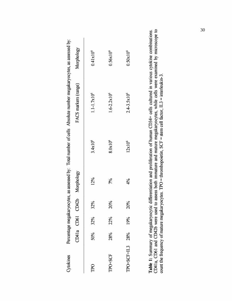
30

31
Lentivirus is more effective than retrovirus for the transduction of human CD34+ HSC
Human CD34+ peripheral blood stem cells were transduced with GFP-expressing
lentivirus (pCCL) or retrovirus (MigR1) to determine which vector is more effective in
these cells. Cells were assessed by FACS to determine the percentage of GFP+ cells,
using cells which underwent a mock transduction as the control. Transduction with pCCL
was associated with GFP expression in 27% of cells, while MigR1 only transduced 6% of
cells.
Figure 6: Comparison of the transduction efficiencies of MigR1 and pCCL viral vectors into human CD34+ hematopoietic stem cells. Cells were analyzed by flow cytometry for the expression of GFP, illustrating a superior transduction efficiency of the pCCL lentivirus.

32
Retronectin does not increase the lentiviral transduction efficiency of CD34+ cells
Retronectin is thought to improve viral transduction efficiency by binding both
viruses and cells, and bringing these into close proximity. [36] A direct comparison was
performed to determine if Retronectin is beneficial for the transduction of VSV-G
pseudotyped lentivirus into peripheral blood CD34+ cells. CD34+ cells were transduced
with pCCL-MKL1 using identical protocols, except some cells were placed in a
Retronectin-coated well. As shown in Figure 7, the transduction efficiency in the
presence of a Retronectin-coated well was approximately 12%, compared to greater than
15% without the addition of Retronectin.
Figure 7: Human CD34+ cells were transduced with lentivirus, with and without the presence of Retronectin. Flow cytometry was performed to assess the expression of GFP, and to determine whether Retronectin was beneficial in enhancing the transduction efficiency in these experiments. Retronectin was not found to be beneficial for lentiviral transduction of human PBSCs.

33
Polybrene enhances lentiviral transduction of human CD34+ cells
Human CD34+ cells were transduced with the pCCL-dominant negative MKL1
virus using either polybrene or Lipofectamine 2000 as a transduction-enhancing agent.
Additionally, Lipofectamine 2000 was tested at two different concentrations. In Figure 8,
it is shown that the highest transduction efficiency, 12%, was obtained with the use of
polybrene. The Lipofectamine 2000 samples had 9% and 7% efficiencies at the low and
high doses, respectively.
Figure 8: Two polycations, polybrene and Lipofectamine 2000, were compared as “transduction-enhancing agents” for the lentiviral transduction of human CD34+ cells. Polybrene was associated with a higher transduction efficiency than Lipofectamine 2000. Note that higher concentrations of Lipofectamine 2000 were also inversely correlated with lower rates of transduction.

34
FACS Assays
Human peripheral blood CD34+ cells were transduced with either pCCL, pCCL-
MKL1, pCCL-DN-MKL1 or underwent a mock transduction. On day 9, FACS was
performed to assess the expression of megakaryocyte-associated antigens such as CD41a,
CD42b and CD61. As shown in Figure 9, transduction with the MKL1-expressing virus
was associated with an increase in expression of each of these megakaryocytic markers
when compared to the pCCL control. For example, the pCCL-MKL1 transduced cells
were, when averaged across four experiments, 52±10% CD41a+, 46±17% CD42b+ and
56±15% CD61+. In contrast, the pCCL cells were 28±15% CD41a+, 26±13% CD61+ and
14±5% CD42b+. P values for each of these markers were < 0.05, using paired t tests for
the four pairs of parallel samples. In one experiment, dual staining with both CD41a and
CD61 also demonstrated that these cells were almost entirely dually positive, consistent
with the heterodimeric fibrinogen receptor that these factors are known to form.
Surprisingly, cells transduced with pCCL-DN-MKL1 had a similar, if not greater,
increase in megakaryocyte antigen expression, though due to poor transduction
efficiencies, this experiment was only performed once. In this single experiment, the
GFP+ fraction was also 65% CD41a+, 68% CD42b+, and 92% CD61+ (data not shown),
greater than pCCL or pCCL-MKL1 transduced cells that were incubated and prepared in
parallel. As this experiment was only performed once, it will require further
investigation.

35
Figure 9: Several experiments were performed to determine the effects of MKL1 and dominant negative MKL1 on megakaryocytopoiesis in human CD34+ hematopoietic stem cells. Cells transduced with either pCCL-MKL1 lentivirus were compared to both a “mock” control and to cells transduced with the lentiviral vector alone. The pCCL-MKL1 transduced cells consistently had greater expression of megakaryocyte-associated markers than the pCCL control.
Megakaryocyte progenitor assays
We assessed the effect of MKL1 on differentiation of megakaryocytic progenitors
from human CD34+ peripheral blood stem cells. CD34+ cells were transduced with
lentiviral vectors expressing either GFP or both GFP and MKL1. In four different
experiments, transduction with pCCL-MKL1 was associated with an increase in the
number of megakaryocytic colony forming units (CFU-Mk), compared to cells
transduced with pCCL alone, though this finding did not reach statistical significance.
pCCL-MKL1 was also associated with a trend decrease in the number of non-
MKL1 increases the expression of megakaryocyte-associated markers
0
10
20
30
40
50
60
70
80
CD41a CD42b CD61
Per
cen
tag
e o
f po
sitiv
e ce
lls b
y F
AC
S a
nal
ysis
Mock (n=1)
pCCL (n=4)
pCCL-MKL1 (n=4)
* *
*
* p < 0.05

36
megakaryocytic CFU. Surprisingly, both pCCL and pCCL-MKL1 were associated with
greater numbers of CFU-Mk in comparison to cells which underwent a mock
transduction. As an additional control in some experiments, the GFP- fraction from the
sorts were also used in some of the CFU-Mk assays. Although transduction with pCCL-
MKL1 was associated with significant increase in CFU-Mk versus the GFP- cells, the
same effect was seen with the cells transduced with pCCL only. Some unsorted (“pre-
sort”) cells were also tested, to determine the effects of sorting on the survival and/or
differentiation of megakaryocytic progenitors. In general, the process of sorting was
associated with a decrease in the total numbers of CFU-Mk.
Figure 10: Human CD34+ hematopoietic stem cells were transduced with pCCL-MKL1 or pCCL, and sorted for GFP-expressing cells as indicated. These cells were placed into megakaryocyte progenitor assays and cultured for 12 days. Slides were then stained and read for the presence of megakaryocyte colony forming units (CFU-Mk) and other colonies. In four different experiments, the MKL1-expressing cells yielded greater numbers of megakaryocytic progenitors compared to cells transduced with pCCL alone.
Transfection with pCCL-MKL1 increases the number of megakaryocyte progenitors
050
100150200250300350400
MKL1 pCCL mock MKL1 pCCL MKL1 pCCL mock
GFP+ GFP+ GFP- GFP- Pre-sort Pre-sort Pre-sort
(n=4) (n=4) (n=4) (n=3) (n=3) (n=1) (n=1) (n=1)
Co
lon
y co
un
t (C
FU
-Mk
/ 10,
000
cells
)
CFU-Mk
Other CFU

37
Discussion
The goal of this project was to determine the role of MKL1 in the process of
megakaryocytic differentiation, which is an important step toward understanding the
mechanism by which RBM15-MKL1 is involved in megakaryocytic leukemogenesis in
t(1;22)(p13;q13)-associated AMKL. To do so is essential for the development of novel
targeted therapies for infants with this particularly aggressive form of leukemia. A human
CD34+ hematopoietic progenitor cell model was developed and used to demonstrate that
MKL1 indeed promotes megakaryocytopoiesis.
Much of the initial effort in this project was focused on the design and
construction of various viral vectors. Several of the plasmids used in this work, such as
human and murine pcDNA3-MKL1, pCMVx3-FLAG7.1-C630 (dominant negative
MKL1), and the pCCL-C-MND lentiviral vector, were donated from other laboratories.
These plasmids were used to construct a complete set of vectors including all
permutations of human and murine, retroviral and lentiviral, and wild-type and dominant
negative negative MKL1. Luciferase assays demonstrated that the pcDNA3 and MigR1
DN-MKL1 constructs suppress activity of exogenous and endogenous MKL1 on the
pm18 reporter. One surprising finding was that the pcDNA3-DN-MKL1 was much more
effective at suppressing exogenously-added pcDNA3-hMKL1 than either the human or
murine MigR1 DN-MKL1 constructs. This difference may have to do with different
levels of cDNA expression between the pcDNA3 or MigR1 plasmids.

38
Another major component of this work was development of the human CD34+
hematopoietic progenitor cell model. There are several published studies which have
sought to determine the optimal conditions for inducing CD34+ cells to undergo
megakaryocytopoiesis, adjusting factors such as cytokines and temperature. For example,
some researchers have attempted to use combinatorial techniques to evaluate the roles of
numerous cytokines, such as thrombopoietin, stem cell factor, Flt-3 ligand, and many
others. [37] However, these studies have not always agreed on the ideal combination of
cytokines. Furthermore, different investigators have used different sources of
hematopoietic stem cells, which may have different responses to equivalent cytokines.
For example, there are several very significant differences between HSCs derived from
bone marrow versus peripheral blood or umbilical cord blood, and these differences may
impact the cells' ability to differentiate along the megakaryocytic lineage. Though some
reports indicate the ability to create a highly pure population of megakaryocytes using
thrombopoietin alone, [38] this sort of result has not been published using mobilized
peripheral blood HSCs. Our findings were that thrombopoietin alone yielded the highest
proportion of megakaryocytes, but even so, the total percentage of cells which resembled
megakaryocytes by morphology or expressed megakaryocyte-specific markers was
significantly below some published results using other sources of hematopoietic
progenitor cells. Moreover, the majority of cells under our culture conditions were
positive for CD13 and CD33, which are markers of myeloid cells, generally not seen on
mature megakaryocytes. This is consistent with the finding that CD34+ cells from

39
peripheral blood are much more often CD13+ and CD33+ than CD34+ cells obtained from
bone marrow. [39] This may be related to the method of mobilization in which donors are
given several doses of G-CSF.
One of the challenges with the CD34+ cells was achieving a satisfactory
transduction efficiency. A major decision was whether to use a Moloney Murine
Leukemia Virus (MMLV), such as MigR1, or lentivirus as the vector. MMLV, also
referred to as oncoretroviruses, were the first form of viruses used for transduction of
hematopoietic cells. However, these viruses have a major limitation: they require that a
cell be undergoing cell division for nuclear import and integration of the transgene. This
can be overcome by stimulating cell division with various cytokines, but this may have
an unwanted effect on the pluripotency of experimental cells. For this reason, lentiviral
vectors may be preferred for transgene delivery into HSCs, because they have been
shown to be able to transduce quiescent HSCs. [40] In a direct comparison, we also found
that the pCCL lentivirus had a greater transduction efficiency than the MigR1
oncoretrovirus in the transduction of human mobilized peripheral blood CD34+ cells.
There are many other factors that can affect the transduction efficiency of viral
vectors into hematopoietic cells. For example, the presence of cytokines can play an
important role. A survey done by Zielske et al. looked at the evidence favoring cytokines
in the lentiviral transduction of HSCs. The majority of studies included in this survey
found that prestimulation with various combinations of cytokines resulted in higher
transduction rates than without prestimulation. [41]

40
Under some conditions, the use of Retronectin (a recombinant fibronectin
fragment) can improve transduction of hematopoietic cells. The rationale behind this is
that the Retronectin fragment has domains which are independently capable of binding
both the viral envelope as well as the membrane of the target cell, thus bringing these two
elements into close proximity. A benefit has been shown with CD34+ cells and some
types of lentiviral vectors, [36] but this benefit was not seen in the above experiments
when using a VSV-G pseudotyped lentivirus.
Other factors that have been shown to improve transduction include the use of
cationic polymers, such as polybrene or Lipofectamine 2000, [35] and the use of a
“spinfection” or “spinoculation” protocol. [42] Our experiments demonstrated that
polybrene was associated with superior transduction efficiency compared to
Lipofectamine 2000. Thus, polybrene was used in all subsequent transductions, though
transduction efficiencies were still somewhat disappointing. There are few other options
for increasing the level of transduction for future experiments. One possibility would be
to attempt to generate a much higher lentiviral titer by dramatically scaling up the virus
production process. However, this was already being performed at the upper limits of our
capabilities, with regard to the equipment and materials involved.
After optimization of the above factors, CD34+ cells were transduced with the
lentiviral vectors expressing GFP alone, GFP and MKL1, or GFP and dominant negative
MKL1. The transduction efficiencies for pCCL-MKL1 and pCCL-DN-MKL1 were quite
low in comparison to the pCCL virus alone. It is possible that the smaller size of the

41
empty viral vector accounts for this difference. In future studies, it may be wise to create
a control vector of equal size as the pCCL-MKL1 or pCCL-DN-MKL1 plasmids, perhaps
by inverting the insert coding sequence or by inserting stop codons that prevent
expression of a full length transcript. Despite these low transduction efficiencies, it is
possible to analyze such experimental populations by selecting for GFP+ cells in a FACS
analysis or a FACS sort. When examining only the GFP+ cells, transduction with pCCL-
MKL1 was associated with a clear and consistent increase in the expression of
megakaryocyte-specific markers such as CD41a, CD42b and CD61 in four different
experiments compared to cells transduced with pCCL alone. However, there was also a
large percentage of CD13+ and CD33+ cells in this experiment, suggesting some degree
of granulocytic differentiation. Moreover, the percentage of cells positive for
megakaryocytic markers plus the percentage of cells positive for granulocytic markers
exceeded one hundred percent, which means there was some overlap between these two
populations.
Perhaps the most surprising result found during this experiment was that cells
transduced with dominant negative MKL1 lentivirus had a similar trend as those
transduced with normal MKL1. That is, both MKL1 and dominant negative MKL1 were
associated with an increase in cells positive for CD41a, CD42b and CD61. However, the
pCCL-DN-MKL1 experiment was performed only once. Given that previous experiments
using HEL cells have shown that MKL1 induces megakaryocytopoiesis via the Serum
Response Factor pathway, it was expected that the C630 dominant negative MKL1 would

42
inhibit megakaryocytopoiesis by blocking this activity. The mechanism for the increase
that we observed is completely unclear, though it is possible that dominant negative
MKL1 can exert some activity via pathways unrelated to SRF, or that it has different
effects on the SRF pathway at earlier versus later stages of megakaryocytopoiesis. It
should also be noted that pCCL-DN-MKL1 was not tested for dominant negative activity,
even though the other DN-MKL1 constructs were shown to suppress MKL1 function.
However, it is difficult to imagine a mechanism by which a different vector could reverse
the function of the dominant negative expression product. These interesting DN-MKL1
results will require further experimentation.
The megakaryocyte colony forming unit assays performed in this project provide
preliminary evidence that transduction with the MKL1 expressing lentivirus increases the
formation of megakaryocyte progenitors. In four experiments, cells transduced with
pCCL-MKL1 generated a higher proportion of CFU-Mk than transduction with pCCL
alone. These experiments were limited by low transduction efficiencies and thus having
very few positively transduced cells, though, fortunately, these assays require a very low
number of cells. It is somewhat concerning that transduction with pCCL appeared to
promote megakaryocyte colony formation versus cells put through a “mock”
transduction. The mechanism for how the empty virus causes such a trend is unclear and
is worthy of further investigation.

43
Implications of this work for the understanding and treatment of Acute
Megakaryoblastic Leukemia
There are several forms of leukemia that are known to be associated with various
chromosomal translocations. In the past, chemotherapeutic regimens consisted entirely of
drugs which non-specifically impair the cell cycle or are toxic to rapidly dividing cells.
Though these regimens have proven to be effective in some populations, treatments are
generally associated with many chemotherapy-related side effects due to toxic effects on
other cells types in the body. However, the recent decade has been notable for greater
understanding of the mechanisms by which several chromosomal translocations are
involved in leukemogenesis, and the development of novel, targeted treatment options for
some of these diseases. For example, imatinib mesylate (Gleevec) was found to be a
potent inhibitor of the bcr-abl fusion product that is formed by the Philadelphia
chromosome translocation, t(9;22), in chronic myelogenous leukemia (CML), some cases
of acute lymphoblastic leukemia (ALL), and rare cases of AML. [43] Gleevec has
consequently become an important part of therapy for these types of leukemia.
However, it is not yet clear how the RBM15-MKL1 fusion product is involved in
the pathogenesis of t(1;22)(p13;q13)-associated Acute Megakaryoblastic Leukemia, or
what roles its component genes play in normal megakaryocytopoiesis. In this project, we
have used a human hematopoietic stem cell model to show that MKL1 is an important
factor in the process of megakaryocytic differentiation. CD34+ HSCs transduced with
MKL1 had both increased expression of megakaryocyte-associated markers CD41a,

44
CD42b and CD61, and a trend suggestive of increased numbers of committed
megakaryocyte progenitors. Thus, it appears probable that one of the mechanisms by
which RBM15-MKL1 causes enhanced commitment to the megakaryocytic lineage is by
increased activity of the pathways which MKL1 acts upon in normal
megakaryocytopoiesis. It is possible that targeted therapies directed at MKL1 and,
perhaps, other factors in the Serum Response Factor pathway could one day play an
important role in the treatment of infants with t(1;22)(p13;q13)-associated AMKL.

45
References
1 Bennett JM, Catovsky D, Daniel MT, Flandrin G, Galton DA, Gralnick HR,
Sultan C: Criteria for the diagnosis of acute leukemia of megakaryocyte lineage (m7). a
report of the trench-American-British cooperative group. Ann Intern Med 1985, 103:460-
462. 2 Crispino JD: Gata1 mutations in down syndrome: implications for biology and
diagnosis of children with transient myeloproliferative disorder and acute
megakaryoblastic leukemia. Pediatr Blood Cancer 2005, 44:40-44. 3 Creutzig U, Reinhardt D, Diekamp S, Dworzak M, Stary J, Zimmermann M: Aml
patients with down syndrome have a high cure rate with aml-bfm therapy with reduced
dose intensity. Leukemia 2005, 19:1355-1360. 4 Lange BJ, Kobrinsky N, Barnard DR, Arthur DC, Buckley JD, Howells WB,
Gold S, Sanders J, Neudorf S, Smith FO, Woods WG: Distinctive demography, biology,
and outcome of acute myeloid leukemia and myelodysplastic syndrome in children with
down syndrome: children's cancer group studies 2861 and 2891. Blood 1998, 91:608-
615. 5 Wechsler J, Greene M, McDevitt MA, Anastasi J, Karp JE, Le Beau MM,
Crispino JD: Acquired mutations in gata1 in the megakaryoblastic leukemia of down
syndrome. Nat Genet 2002, 32:148-152. 6 Stachura DL, Chou ST, Weiss MJ: Early block to erythromegakaryocytic
development conferred by loss of transcription factor gata-1. Blood 2006, 107:87-97. 7 Li Z, Godinho FJ, Klusmann J, Garriga-Canut M, Yu C, Orkin SH:
Developmental stage-selective effect of somatically mutated leukemogenic transcription
factor gata1. Nat Genet 2005, 37:613-619. 8 Lightfoot J, Hitzler JK, Zipursky A, Albert M, Macgregor PF: Distinct gene
signatures of transient and acute megakaryoblastic leukemia in down syndrome.

46
Leukemia 2004, 18:1617-1623. 9 McElwaine S, Mulligan C, Groet J, Spinelli M, Rinaldi A, Denyer G, Mensah A,
Cavani S, Baldo C, Dagna-Bricarelli F, Hann I, Basso G, Cotter FE, Nizetic D:
Microarray transcript profiling distinguishes the transient from the acute type of
megakaryoblastic leukaemia (m7) in down's syndrome, revealing prame as a specific
discriminating marker. Br J Haematol 2004, 125:729-742. 10 Creutzig U, Reinhardt D, Diekamp S, Dworzak M, Stary J, Zimmermann M: Aml
patients with down syndrome have a high cure rate with aml-bfm therapy with reduced
dose intensity. Leukemia 2005, 19:1355-1360. 11 Taub JW, Matherly LH, Stout ML, Buck SA, Gurney JG, Ravindranath Y:
Enhanced metabolism of 1-beta-d-arabinofuranosylcytosine in down syndrome cells: a
contributing factor to the superior event free survival of down syndrome children with
acute myeloid leukemia. Blood 1996, 87:3395-3403. 12 Baruchel A, Daniel MT, Schaison G, Berger R: Nonrandom t(1;22)(p12-p13;q13)
in acute megakaryocytic malignant proliferation. Cancer Genet Cytogenet 1991, 54:239-
243. 13 Carroll A, Civin C, Schneider N, Dahl G, Pappo A, Bowman P, Emami A, Gross
S, Alvarado C, Phillips C: The t(1;22) (p13;q13) is nonrandom and restricted to infants
with acute megakaryoblastic leukemia: a pediatric oncology group study. Blood 1991,
78:748-752. 14 Lion T, Haas OA, Harbott J, Bannier E, Ritterbach J, Jankovic M, Fink FM,
Stojimirovic A, Herrmann J, Riehm HJ: The translocation t(1;22)(p13;q13) is a
nonrandom marker specifically associated with acute megakaryocytic leukemia in young
children. Blood 1992, 79:3325-3330. 15 Lu G, Altman AJ, Benn PA: Review of the cytogenetic changes in acute
megakaryoblastic leukemia: one disease or several?. Cancer Genet Cytogenet 1993,
67:81-89. 16 Mercher T, Coniat MB, Monni R, Mauchauffe M, Nguyen Khac F, Gressin L,

47
Mugneret F, Leblanc T, Dastugue N, Berger R, Bernard OA: Involvement of a human
gene related to the drosophila spen gene in the recurrent t(1;22) translocation of acute
megakaryocytic leukemia. Proc Natl Acad Sci U S A 2001, 98:5776-5779. 17 Ma Z, Morris SW, Valentine V, Li M, Herbrick JA, Cui X, Bouman D, Li Y,
Mehta PK, Nizetic D, Kaneko Y, Chan GC, Chan LC, Squire J, Scherer SW, Hitzler JK:
Fusion of two novel genes, rbm15 and mkl1, in the t(1;22)(p13;q13) of acute
megakaryoblastic leukemia. Nat Genet 2001, 28:220-221. 18 Kao HY, Ordentlich P, Koyano-Nakagawa N, Tang Z, Downes M, Kintner CR,
Evans RM, Kadesch T: A histone deacetylase corepressor complex regulates the notch
signal transduction pathway. Genes Dev 1998, 12:2269-2277. 19 Ishiko E, Matsumura I, Ezoe S, Gale K, Ishiko J, Satoh Y, Tanaka H, Shibayama
H, Mizuki M, Era T, Enver T, Kanakura Y. Notch signals inhibit the development of
erythroid/megakaryocytic cells by suppressing GATA-1 activity through the induction of
HES1. J Biol Chem. 2005 Feb 11;280(6):4929-39. 20 Shi Y, Downes M, Xie W, Kao HY, Ordentlich P, Tsai CC, Hon M, Evans RM.
Sharp, an inducible cofactor that integrates nuclear receptor repression and activation.
Genes Dev. 2001 May 1;15(9):1140-51. 21 Kuroda K, Han H, Tani S, Tanigaki K, Tun T, Furukawa T, Taniguchi Y,
Kurooka H, Hamada Y, Toyokuni S, Honjo T. Regulation of marginal zone B cell
development by MINT, a suppressor of Notch/RBP-J signaling pathway. Immunity. 2003
Feb;18(2):301-12. 22 Ariyoshi M, Schwabe JW. A conserved structural motif reveals the essential
transcriptional repression function of Spen proteins and their role in developmental
signaling. Genes Dev. 2003 Aug 1;17(15):1909-20. 23 Ma X, Renda MJ, Wang L, Cheng E, Niu C, Morris SW, Chi AS, Krause DS:
Rbm15 modulates notch-induced transcriptional activation and affects myeloid
differentiation. Mol Cell Biol 2007, 27:3056-3064. 24 Cen B, Selvaraj A, Burgess RC, Hitzler JK, Ma Z, Morris SW, Prywes R:

48
Megakaryoblastic leukemia 1, a potent transcriptional coactivator for serum response
factor (srf), is required for serum induction of srf target genes. Mol Cell Biol 2003,
23:6597-6608. 25 Cen B, Selvaraj A, Prywes R: Myocardin/mkl family of srf coactivators: key
regulators of immediate early and muscle specific gene expression. J Cell Biochem 2004,
93:74-82. 26 Miralles F, Posern G, Zaromytidou A, Treisman R: Actin dynamics control srf
activity by regulation of its coactivator mal. Cell 2003, 113:329-342. 27 Nakagawa K, Kuzumaki N: Transcriptional activity of megakaryoblastic
leukemia 1 (mkl1) is repressed by sumo modification. Genes Cells 2005, 10:835-850. 28 Hinson JS, Medlin MD, Lockman K, Taylor JM, Mack CP: Smooth muscle cell-
specific transcription is regulated by nuclear localization of the myocardin-related
transcription factors. Am J Physiol Heart Circ Physiol 2007, 292:H1170-80. 29 Miano JM, Long X, Fujiwara K: Serum response factor: master regulator of the
actin cytoskeleton and contractile apparatus. Am J Physiol Cell Physiol 2007, 292:C70-
81. 30 Sasazuki T, Sawada T, Sakon S, Kitamura T, Kishi T, Okazaki T, Katano M,
Tanaka M, Watanabe M, Yagita H, Okumura K, Nakano H: Identification of a novel
transcriptional activator, bsac, by a functional cloning to inhibit tumor necrosis factor-
induced cell death. J Biol Chem 2002, 277:28853-28860 31 Sun Y, Boyd K, Xu W, Ma J, Jackson CW, Fu A, Shillingford JM, Robinson
GW, Hennighausen L, Hitzler JK, Ma Z, Morris SW: Acute myeloid leukemia-associated
mkl1 (mrtf-a) is a key regulator of mammary gland function. Mol Cell Biol 2006,
26:5809-5826. 32 Cheng E, Renda M, Troy J, Hahn K, Wang L, Tuck D, Schulz V, Mane S, Krause
DS: MKL1 promotes megakaryocytopoiesis via serum response factor. In preparation for
publication. 33 Han ZC: Identification of a murine high-proliferative-potential colony-forming
49
cell (hpp-cfc) capable of producing a number of megakaryocytes and replating for
secondary hpp-cfcs in culture. J Lab Clin Med 1994, 123:610-616. 34 Lutzko C, Senadheera D, Skelton D, Petersen D, Kohn DB: Lentivirus vectors
incorporating the immunoglobulin heavy chain enhancer and matrix attachment regions
provide position-independent expression in b lymphocytes. J Virol 2003, 77:7341-7351. 35 Szyda A, Paprocka M, Krawczenko A, Lenart K, Heimrath J, Grabarczyk P,
Mackiewicz A, Dus D: Optimization of a retroviral vector for transduction of human
cd34 positive cells. Acta Biochim Pol 2006, 53:815-823. 36 Haas DL, Case SS, Crooks GM, Kohn DB: Critical factors influencing stable
transduction of human cd34(+) cells with hiv-1-derived lentiviral vectors. Mol Ther
2000, 2:71-80. 37 Cortin V, Garnier A, Pineault N, Lemieux R, Boyer L, Proulx C: Efficient in
vitro megakaryocyte maturation using cytokine cocktails optimized by statistical
experimental design. Exp Hematol 2005, 33:1182-1191. 38 Guerriero R, Testa U, Gabbianelli M, Mattia G, Montesoro E, Macioce G, Pace
A, Ziegler B, Hassan HJ, Peschle C: Unilineage megakaryocytic proliferation and
differentiation of purified hematopoietic progenitors in serum-free liquid culture. Blood
1995, 86:3725-3736. 39 Schmitz N, Barrett J: Optimizing engraftment--source and dose of stem cells.
Semin Hematol 2002, 39:3-14. 40 Uchida N, Sutton RE, Friera AM, He D, Reitsma MJ, Chang WC, Veres G,
Scollay R, Weissman IL: Hiv, but not murine leukemia virus, vectors mediate high
efficiency gene transfer into freshly isolated g0/g1 human hematopoietic stem cells. Proc
Natl Acad Sci U S A 1998, 95:11939-11944. 41 Zielske SP, Braun SE: Cytokines: value-added products in hematopoietic stem
cell gene therapy. Mol Ther 2004, 10:211-219. 42 Zielske SP, Gerson SL: Lentiviral transduction of p140k mgmt into human
cd34(+) hematopoietic progenitors at low multiplicity of infection confers significant

50
resistance to bg/bcnu and allows selection in vitro. Mol Ther 2002, 5:381-387. 43 Druker BJ, Tamura S, Buchdunger E, Ohno S, Segal GM, Fanning S,
Zimmermann J, Lydon NB: Effects of a selective inhibitor of the abl tyrosine kinase on
the growth of bcr-abl positive cells. Nat Med 1996, 2:561-566.