role of forkhead transcription factors in diabetes-induced
TRANSCRIPT
University of Pennsylvania University of Pennsylvania
ScholarlyCommons ScholarlyCommons
Departmental Papers (Dental) Penn Dental Medicine
2012
Role of Forkhead Transcription Factors in Diabetes-Induced Role of Forkhead Transcription Factors in Diabetes-Induced
Oxidative Stress Oxidative Stress
Bhaskar Ponugoti University of Pennsylvania
Guangyu Dong University of Pennsylvania
Dana T. Graves University of Pennsylvania, [email protected]
Follow this and additional works at: https://repository.upenn.edu/dental_papers
Part of the Endocrinology, Diabetes, and Metabolism Commons, and the Medical Cell Biology
Commons
Recommended Citation Recommended Citation Ponugoti, B., Dong, G., & Graves, D. T. (2012). Role of Forkhead Transcription Factors in Diabetes-Induced Oxidative Stress. Experimental Diabetes Research, 2012 Article ID 939751-. http://dx.doi.org/10.1155/2012/939751
This paper is posted at ScholarlyCommons. https://repository.upenn.edu/dental_papers/13 For more information, please contact [email protected].

Role of Forkhead Transcription Factors in Diabetes-Induced Oxidative Stress Role of Forkhead Transcription Factors in Diabetes-Induced Oxidative Stress
Abstract Abstract Diabetes is a chronic metabolic disorder, characterized by hyperglycemia resulting from insulin deficiency and/or insulin resistance. Recent evidence suggests that high levels of reactive oxygen species (ROS) and subsequent oxidative stress are key contributors in the development of diabetic complications. The FOXO family of forkhead transcription factors including FOXO1, FOXO3, FOXO4, and FOXO6 play important roles in the regulation of many cellular and biological processes and are critical regulators of cellular oxidative stress response pathways. FOXO1 transcription factors can affect a number of different tissues including liver, retina, bone, and cell types ranging from hepatocytes to microvascular endothelial cells and pericytes to osteoblasts. They are induced by oxidative stress and contribute to ROS-induced cell damage and apoptosis. In this paper, we discuss the role of FOXO transcription factors in mediating oxidative stress-induced cellular response.
Keywords Keywords Diabetes Mellitus, Forkhead Transcription Factors, Humans, Liver, Oxidative Stress, Reactive Oxygen Species
Disciplines Disciplines Endocrinology, Diabetes, and Metabolism | Medical Cell Biology
This journal article is available at ScholarlyCommons: https://repository.upenn.edu/dental_papers/13

Hindawi Publishing CorporationExperimental Diabetes ResearchVolume 2012, Article ID 939751, 7 pagesdoi:10.1155/2012/939751
Review Article
Role of Forkhead Transcription Factors in Diabetes-InducedOxidative Stress
Bhaskar Ponugoti, Guangyu Dong, and Dana T. Graves
School of Dental Medicine, University of Pennsylvania, Philadelphia, PA 19104-6030, USA
Correspondence should be addressed to Dana T. Graves, [email protected]
Received 6 September 2011; Revised 11 October 2011; Accepted 26 October 2011
Academic Editor: Robert A. Harris
Copyright © 2012 Bhaskar Ponugoti et al. This is an open access article distributed under the Creative Commons AttributionLicense, which permits unrestricted use, distribution, and reproduction in any medium, provided the original work is properlycited.
Diabetes is a chronic metabolic disorder, characterized by hyperglycemia resulting from insulin deficiency and/or insulin resistance.Recent evidence suggests that high levels of reactive oxygen species (ROS) and subsequent oxidative stress are key contributors inthe development of diabetic complications. The FOXO family of forkhead transcription factors including FOXO1, FOXO3, FOXO4,and FOXO6 play important roles in the regulation of many cellular and biological processes and are critical regulators of cellularoxidative stress response pathways. FOXO1 transcription factors can affect a number of different tissues including liver, retina,bone, and cell types ranging from hepatocytes to microvascular endothelial cells and pericytes to osteoblasts. They are induced byoxidative stress and contribute to ROS-induced cell damage and apoptosis. In this paper, we discuss the role of FOXO transcriptionfactors in mediating oxidative stress-induced cellular response.
1. Introduction
Diabetes mellitus is a chronic disease characterized by ele-vated blood sugar levels resulting from either lack of insulinproduction or resistance to insulin. In 2010, there were nearly230 million individuals with diabetes worldwide which isestimated to reach 430 million by 2030 [1]. Recently, astudy conducted by the U.S. Centers for Disease Control andPrevention (CDC) indicated that 25.8 million Americans or8.3% of its population were affected by diabetes in 2010 [2].Diabetes has severe health consequences associated with nu-merous diabetic complications including retinopathy, neu-ropathy, and nephropathy [3–5]. Accumulating evidencesuggests that hyperglycemia-induced production of free rad-icals and the subsequent oxidative stress contributes to thedevelopment and progression of diabetes and related com-plications [6–8].
Reactive oxygen species (ROS) are oxygen free radicalsthat are generated as by-products of mitochondrial metab-olism and function as signaling molecules in various intra-cellular processes including cell proliferation, migration, andapoptosis [9]. ROS produced during normal metabolic proc-esses are removed rapidly with the help of various endoge-
nous detoxifying enzymes. While normal cellular ROSconcentrations are necessary for proper functioning of cells,excessive, non-physiological concentrations of ROS result inoxidative stress. ROS such as superoxide (O2
−) and hydroxylradicals (HO•), and hydrogen peroxide (H2O2), are highlyreactive and can cause damage to biological macromoleculessuch as DNA, proteins, and lipids [9]. Major sources ofoxidative stress during diabetes include glucose autooxida-tion, overproduction of ROS by mitochondria, non-enzy-matic glycation, and the polyol pathway [6, 10]. In the polyolpathway, aldose reductase converts glucose into sorbitol withNADPH as a coenzyme. In diabetes, increased flux throughthe polyol pathway enhances oxidative stress because of in-creased consumption of NADPH by aldose reductase. SinceNADPH is required for generation of endogenous antiox-idant glutathione (GSH), reduced NADPH availability de-pletes GSH leading to greater oxidative stress [6]. Othermechanisms through which high glucose levels can lead toadvanced glycation endproducts are discussed below.
ROS leads to the generation of intracellular signals thatstimulate inflammation and cell death. They include proteinkinase C (PKC), c-Jun-N-terminal kinase (JNK), and p38mitogen-activated protein kinase (MAPK) [11–15]. In many

2 Experimental Diabetes Research
cell types, ROS lead to the activation of the forkhead box O(FOXO) transcription factors that include FOXO1, FOXO3,and FOXO4, which can mediate the effects of ROS throughregulation of gene transcription. These transcription factorshave been implicated in diverse cellular processes rangingfrom glucose metabolism to cell behavior including cell cycleand apoptosis [16, 17]. In addition to being activated byROS, FOXO proteins play a critical role in oxidative stress byupregulating expression of antioxidant genes [9]. However,FOXO proteins are involved in many other processes and canhave apparently contradictory effects in different cell types[18]. FOXO proteins are transcription factors but also haveimportant function as corepressors or coactivators so thatdirect DNA binding is not a prerequisite for modulating thetranscription of gene targets [19]. For simplicity, we will usethe term FOXO for all or any of the FOXO transcriptionfactors throughout this paper, unless otherwise specified.
2. Regulation of FOXO by Oxidative Stress
FOXO transcription factors are critical mediators of oxida-tive stress and are activated by various kinds of cellularstress stimulus. Oxidative stress regulates FOXO activitythrough various posttranslational modifications includingphosphorylation, acetylation, and ubiquitination, which inturn regulate the subcellular localization of FOXOs, protein-protein interactions, and transcriptional activity of FOXOproteins. While some of these modifications promote FOXOtranscriptional activity, others are inhibitory. For example,stress-activated kinase JNK directly phosphorylates FOXO4at residues Thr447 and Thr451, which leads to its nucleartranslocation and induces FOXO4 transcriptional activity[20]. Another kinase implicated in oxidative stress-inducedphosphorylation of FOXO is mammalian Ste20-like proteinkinase 1 (MST1). During oxidative stress, MST1 phospho-rylates FOXO3 at residue Ser207, which results in FOXO3release from binding protein, 14-3-3. This release allowsFOXO3 to translocate to the nucleus thereby modulatingtarget gene expression [21].
FOXO transcriptional activity is also regulated by acety-lation. The effects of oxidative stress-induced acetylation onFOXO function vary based upon the experimental condi-tions. Sirtuins (SIRTs), mammalian homologs of the yeastsilent information regulator 2 (sir2) deacetylase, are criticalregulators of FOXO transcriptional activity and are in-duced by oxidative stress [22, 23]. It has been reportedthat acetylation by cAMP-response-element-binding-protein(CREB-) binding protein (CBP)/P300 positively regulatesFOXO transcriptional activity during oxidative stress, whileSIRT1-mediated deacetylation represses the activity of FOXOtranscription factors (FOXO1, FOXO3, and FOXO4) [24].Other reports suggest that oxidative stress-induced FOXO4acetylation negatively regulates its transcriptional activity,and deacetylation by SIRT1 counteracts the acetylation-mediated FOXO4 inhibition [25]. Furthermore, studies fromBrunet et al. suggest that SIRT1 differentially affects FOXO3function in response to oxidative stress [23]. SIRT1 associateswith and deacetylates FOXO3 both in vitro and in vivo.
SIRT1 deacetylation of FOXO3 increases expression of itstarget genes involved in cell cycle arrest and DNA repairsuch as p27 and GADD45. In contrast, SIRT1 deacetylationreduces expression of FOXO3 proapoptotic target genes suchBim and Fas ligand. These results indicate that deacetylationcan both enhance and reduce FOXO3-induced activitydepending upon the target gene.
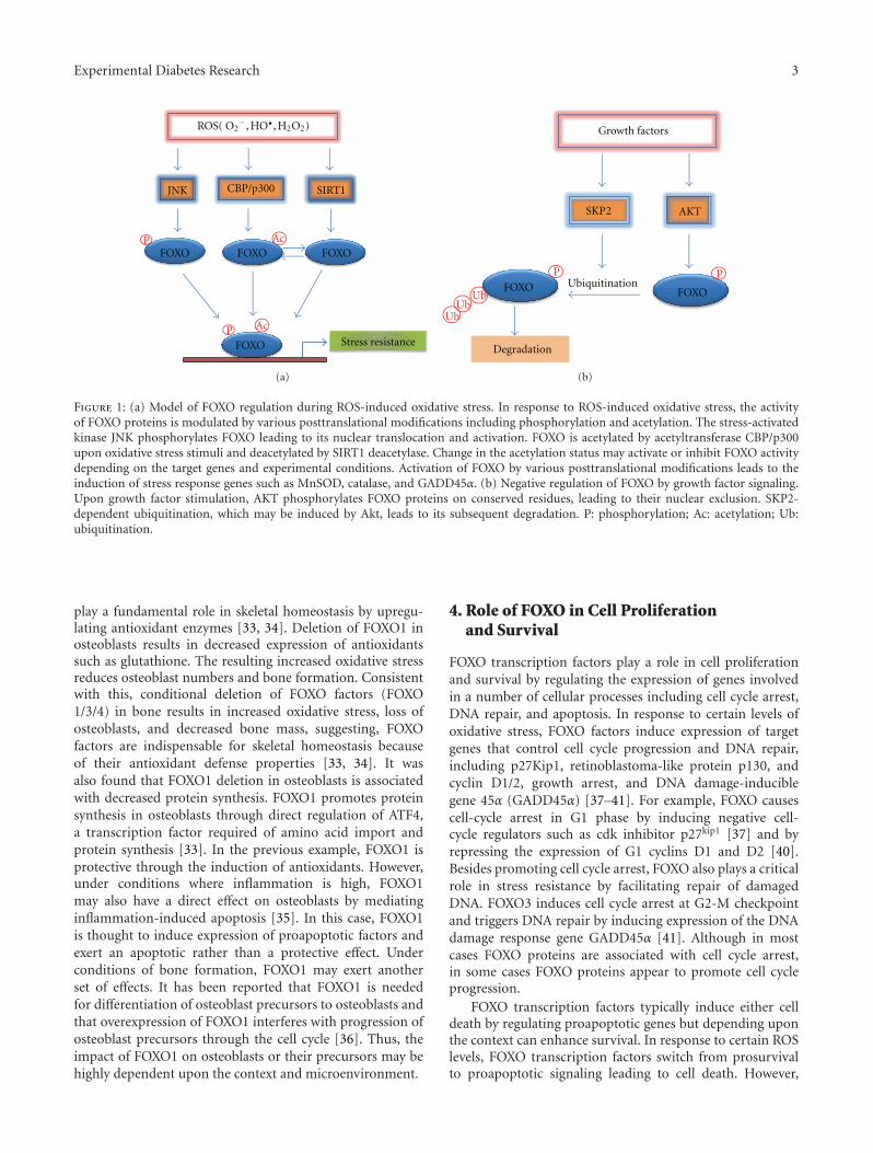
Besides phosphorylation and acetylation, FOXO proteinsare further regulated by ubiquitination during oxidativestress. In response to insulin or growth factor signaling,FOXO transcription factors are phosphorylated, polyubiqui-tinated, and degraded [26]. It has been reported that AKT-dependent phosphorylation is required as a prerequisitefor ubiquitin-mediated degradation of FOXO1 and FOXO3.FOXO ubiquitination is mediated by F-box protein Skp2, asubunit of the SCF (Skp1/Cul1/F-box) E3 ubiquitin ligaseprotein complex [27, 28]. In contrast to insulin/growthfactor signaling, upon oxidative stress, FOXO4 becomesmonoubiquitinated and translocated into the nucleus, result-ing in its increased transcriptional activity. Monoubiquitina-tion of FOXO4 is mediated by E3 ubiquitin ligase murinedouble minute 2 (MDM2) [28]. Figure 1(a) shows the effectof ROS-induced oxidative stress that regulates FOXO byaltering its phosphorylation or acetylation status. In contrast,growth factor-mediated induction of AKT which phospho-rylates FOXO at specific amino acids leads to its export fromthe nucleus and Skp2 which leads to its ubiquitination anddegradation (Figure 1(b)).
3. Role of FOXO in Oxidative Stress
FOXO proteins play an important role in protection ofcells against oxidative stress. Oxidative stress is caused byoverproduction of ROS or inefficient breakdown of ROS.Efficient detoxification of ROS by cellular detoxificationsystems protects cells against oxidative damage. The levelsand enzymatic activities of various antioxidant enzymes suchas manganese superoxide dismutase (MnSOD), catalase, andglutathione peroxidase are decreased during hyperglycemia-induced oxidative stress [11]. It is now well establishedthat cells activate FOXO transcription factors to reduce thelevel of oxidative stress by the induction of enzymes thatbreakdown ROS such as MnSOD and catalase [29, 30]. Forexample, FOXO3 directly binds to MnSOD promoter atFOXO binding elements to increase its expression. Activationof MnSOD in mitochondria protects cells from ROS-mediated injury by converting superoxide radicals to oxy-gen and hydrogen peroxide (H2O2). Enzymes catalase andglutathione peroxidase further breakdown H2O2 into waterand oxygen [30, 31]. The functional significance of FOXOin regulating oxidative stress is further revealed by genedeletion studies. Mice lacking FOXO factors (FOXO 1/3/4)in hematopoietic stem cells (HSCs) exhibit decreased self-renewal, leading to defective repopulating activity [32]. Con-sistent with this, FOXO-deficient HSCs showed increasedROS levels, decreased expression of antioxidant proteins,and increased apoptosis, suggesting critical role of FOXOs instress resistance. Recent evidence suggests that FOXO factors
Experimental Diabetes Research 3
FOXO
ROS( O2−, HO•, H2O2)
JNK CBP/p300
FOXO
SIRT1
FOXOP Ac
Ac
Stress resistance
FOXOP
(a)
Growth factors
AKTSKP2
UbiquitinationFOXO
PFOXO
UbUb
Degradation
Ub
P
(b)
Figure 1: (a) Model of FOXO regulation during ROS-induced oxidative stress. In response to ROS-induced oxidative stress, the activityof FOXO proteins is modulated by various posttranslational modifications including phosphorylation and acetylation. The stress-activatedkinase JNK phosphorylates FOXO leading to its nuclear translocation and activation. FOXO is acetylated by acetyltransferase CBP/p300upon oxidative stress stimuli and deacetylated by SIRT1 deacetylase. Change in the acetylation status may activate or inhibit FOXO activitydepending on the target genes and experimental conditions. Activation of FOXO by various posttranslational modifications leads to theinduction of stress response genes such as MnSOD, catalase, and GADD45α. (b) Negative regulation of FOXO by growth factor signaling.Upon growth factor stimulation, AKT phosphorylates FOXO proteins on conserved residues, leading to their nuclear exclusion. SKP2-dependent ubiquitination, which may be induced by Akt, leads to its subsequent degradation. P: phosphorylation; Ac: acetylation; Ub:ubiquitination.
play a fundamental role in skeletal homeostasis by upregu-lating antioxidant enzymes [33, 34]. Deletion of FOXO1 inosteoblasts results in decreased expression of antioxidantssuch as glutathione. The resulting increased oxidative stressreduces osteoblast numbers and bone formation. Consistentwith this, conditional deletion of FOXO factors (FOXO1/3/4) in bone results in increased oxidative stress, loss ofosteoblasts, and decreased bone mass, suggesting, FOXOfactors are indispensable for skeletal homeostasis becauseof their antioxidant defense properties [33, 34]. It wasalso found that FOXO1 deletion in osteoblasts is associatedwith decreased protein synthesis. FOXO1 promotes proteinsynthesis in osteoblasts through direct regulation of ATF4,a transcription factor required of amino acid import andprotein synthesis [33]. In the previous example, FOXO1 isprotective through the induction of antioxidants. However,under conditions where inflammation is high, FOXO1may also have a direct effect on osteoblasts by mediatinginflammation-induced apoptosis [35]. In this case, FOXO1is thought to induce expression of proapoptotic factors andexert an apoptotic rather than a protective effect. Underconditions of bone formation, FOXO1 may exert anotherset of effects. It has been reported that FOXO1 is neededfor differentiation of osteoblast precursors to osteoblasts andthat overexpression of FOXO1 interferes with progression ofosteoblast precursors through the cell cycle [36]. Thus, theimpact of FOXO1 on osteoblasts or their precursors may behighly dependent upon the context and microenvironment.
4. Role of FOXO in Cell Proliferationand Survival
FOXO transcription factors play a role in cell proliferationand survival by regulating the expression of genes involvedin a number of cellular processes including cell cycle arrest,DNA repair, and apoptosis. In response to certain levels ofoxidative stress, FOXO factors induce expression of targetgenes that control cell cycle progression and DNA repair,including p27Kip1, retinoblastoma-like protein p130, andcyclin D1/2, growth arrest, and DNA damage-induciblegene 45α (GADD45α) [37–41]. For example, FOXO causescell-cycle arrest in G1 phase by inducing negative cell-cycle regulators such as cdk inhibitor p27kip1 [37] and byrepressing the expression of G1 cyclins D1 and D2 [40].Besides promoting cell cycle arrest, FOXO also plays a criticalrole in stress resistance by facilitating repair of damagedDNA. FOXO3 induces cell cycle arrest at G2-M checkpointand triggers DNA repair by inducing expression of the DNAdamage response gene GADD45α [41]. Although in mostcases FOXO proteins are associated with cell cycle arrest,in some cases FOXO proteins appear to promote cell cycleprogression.
FOXO transcription factors typically induce either celldeath by regulating proapoptotic genes but depending uponthe context can enhance survival. In response to certain ROSlevels, FOXO transcription factors switch from prosurvivalto proapoptotic signaling leading to cell death. However,
4 Experimental Diabetes Research
the exact molecular mechanisms by which FOXO switchesfrom prosurvival to prodeath signaling remain unknown.In diabetes, chronic hyperglycemia-induced mitochondrialROS stimulate various signaling pathways leading to activa-tion of FOXO, which in turn activates several proapoptoticfactors. FOXO1 activation is elevated in diabetic connectivetissue and mediates advanced glycation endproduct andTNF-alpha-induced apoptosis both of which are elevatedin diabetic connective tissue [42–44]. It has been proposedthat diabetes-enhanced activation of FOXO1 limits woundhealing by enhancing fibroblast apoptosis and proliferation[43]. FOXO1 regulates genes of both the extrinsic and intrin-sic apoptotic pathways [42]. FOXO3 and FOXO4 induceapoptosis by directly binding Bcl-6 promoter and enhancingits expression and negatively regulate expression of anantiapoptotic protein BCL-XL [45]. It was further shownthat silencing endogenous FOXO3 or overexpression of adominant negative mutant of FOXO3 resulted in decreasedexpression of a variety of proapoptotic genes, includingBcl-6 and Bim, in response to hydrogen peroxide-inducedoxidative stress. We have recently shown that hyperglycemiaduring diabetes stimulates microvascular endothelial celland pericyte apoptosis leading to early stages of diabeticretinopathy [46]. High glucose leads to ROS generationthat enhances FOXO1 activation and induction of severalclasses of genes that regulate endothelial cell behaviorincluding proapoptotic and proinflammatory factors. Theseresults suggest that FOXO1 plays an important role in thedevelopment of diabetic retinopathy due to its effect oninflammatory and apoptotic gene expression in microvas-cular cells [46]. Moreover, high glucose and advancedglycation endproducts that are elevated in diabetes stimulateloss of microvascular retinal pericytes through a processthat involves activation of FOXO1 [46, 47]. In the latter,advanced glycation endproducts activate FOXO1 in pericytesthrough the MAP kinase pathway, and the loss of pericytes iscountered by activation of Akt and NF-kappaB [47].
5. FOXO in Diabetes-Induced Inflammation
Inflammation has long been considered as a major risk factorin diabetes and associated with development and progressionof diabetic complications. Hyperglycemia-induced oxidativestress promotes inflammation through increased endothe-lial cell damage, microvascular permeability, and increasedrelease of proinflammatory cytokines, including TNF-α,interlukin-1β (IL-1β), and interlukin-6 (IL-6), ultimatelyleading to decreased insulin sensitivity and diabetic compli-cations. Hyperglycemia-induced FOXO plays an importantrole in the induction of proinflammatory cytokines. It wasshown that FOXO1 directly binds to IL-1β promoter andincreases its expression in macrophages [48]. FOXO1 isinduced by inflammatory cytokines and may be involved in aforward amplification loop. For example, in microvascularendothelial cells, FOXO1 is induced in vivo by diabetes-enhanced TNF-α and also induces expression of TNF-α levelsin these cells [46]. Increased IL-1β and TNF-α productionhas been implicated in pathogenesis of obesity and diabetes.Hyperglycemia in diabetes also stimulates toll-like receptor
(TLR) signaling, which results in prolonged inflammationand tissue damage. Recent studies show that FOXO1 pro-motes inflammation during diabetes by enhancing TLR4-mediated signaling, suggesting FOXO1 as a key mediator ofinflammatory responses during obesity and diabetes [49].In diabetic fracture healing, there is enhanced upregulationof proinflammatory and proapoptotic factors [50, 51]. Ithas been shown that FOXO1 induces expression of bothproinflammatory and proapoptotic factors in chondrocytesand that FOXO1 directly binds to the TNF-α promoter.Moreover, diabetes-enhanced TNF-α activates FOXO1 inchondrocytes in vivo by enhancing its nuclear localization[50].
Another transcription factor that plays an importantrole in stimulating inflammation during hyperglycemiaand oxidative stress is NF-κB [52]. Activation of NF-κBpathway has been implicated in the development of diabeticcomplications, including retinopathy, and has been shownto regulate expression of various proinflammatory cytokines,including TNF-α and IL-1β [53]. Chronically elevated ROSlevels associated with diabetes may induce both NF-κBand FOXO leading to increased inflammation and cellulardamage. In most cell types, NF-κB is directly antiapoptotic,while FOXO1 is directly proapoptotic. Thus, in inflammatoryconditions when both NF-κB and FOXO1 are activated, theirrelative balance may determine whether a cell ultimatelysurvives or undergoes apoptosis [42, 47].
5.1. Mitochondria, ROS, and Diabetes. A mechanism throughwhich diabetes can increase oxidative stress involves electrontransport in mitochondria. It has been proposed that highintracellular glucose levels increase the follow of electronsthrough the electron transport chain in mitochondria duringoxidative respiration [6]. This can result in the transferof electrons to O2 leading to formation of O2
− and thegeneration of various reactive oxygen species in the mito-chondria. Furthermore, changes caused by diabetes alterthe redox balance and affect redox-sensitive proteins suchas protein kinase C-epsilon, which can result in enhancedmitochondrial ROS production. Advanced glycation endproducts (AGEs) generated under conditions of hyper-glycemia stimulate NADPH oxidase that in turn can induceproduction of ROS. In a surprising development, increasedWnt signaling stimulates mitochondrial biogenesis that canlead to enhanced ROS levels in mitochondria and greateroxidative damage [54]. The increased ROS in mitochondriais thought to be problematic due to a number of differentmechanisms. One is that ROS damages mitochondrialcomponents such as DNA, membrane proteins, and lipids.ROS can also induce the opening of the mitochondrialpermeability transition pore (MPTP) [55]. When this poreis opened, proapoptotic proteins are released from themitochondria such as cytochrome c that stimulate cell death.ROS generated in the mitochondrial respiratory chain havebeen proposed as secondary messengers for activation of NF-κB by TNF-α and IL-1 [6].
ROS may affect insulin signalling. Insulin signalling isreduced under conditions of oxidative stress, which maycontribute to insulin resistance. This may occur through

Experimental Diabetes Research 5
ROSPI3K/Akt
IRS
Insulin
FOXO
S
P
HMOX1
IRS
PI3K
Fatty acid oxidation
Mitochondria
Oxidative respirationATP
MST1/JNKFOXOP
14-3-3
14-3-3
14-3-3
PP2A
Oxidative stress
PROS
Figure 2: Oxidative stress and insulin signaling affect mitochondrial function via FOXO. ROS induce IRS serine phosphorylation whichinhibits IRS activation by insulin signalling. As a result of reduced IRS activity, Akt activity is reduced. Reduced Akt reduces negativesignalling of FOXO so that FOXO1 is left in an activated state since it is not exported out of the nucleus by 14-3-3. Meanwhile, ROSactivate MST1 and JNK which induce FOXO nuclear translocation by disrupting the complex of FOXO and 14-3-3. PP2A activates FOXOby dephosphorylation of FOXO and by Akt dephosphorylation. FOXO nuclear translocation will induce HMOX1 gene expression whichinhibits mitochondrial function by affecting like fatty acid oxidation, ATP, and oxidative respiration (arrow indicates stimulatory event; barindicates inhibitory event).
several mechanisms. In one scenario, ROS induces serinephosphorylation of insulin receptor substrate, decreasingtyrosine phosphorylation thereby interfering with insulinsignaling [56]. Similarly, ROS have been shown to partiallymediate the effect of Angiotensin II inhibition of insulinsignalling [57]. Methylglyoxal, a biologically active AGE pre-cursor formed under conditions of hyperglycemia, inhibitsphosphorylation of insulin receptor substrate and activationof the phosphatidylinositol 3-kinase (PI3K)/protein kinase B(PKB) pathway [58].
Insulin signalling inactivates FOXO1, which is mediatedby insulin receptor substrates-1 and -2 through AKT. Acharacteristic feature of insulin resistance is the elevatedproduction of glucose that contributes to hyperglycemia.FOXO1 regulates glucose production in the liver throughthe expression of genes that promote gluconeogenesis [59].Thus, a pathway exists whereby insulin resistance leadsto elevated FOXO1 activation, upregulation of genes thatpromote glucose production, and greater serum glucoselevels. Disruption of the insulin-Akt-FOXO1 balance alsoaffects the mitochondria. Activated FOXO1 induces hemeoxygenase-1 (HMOX1), which cleaves heme and disruptsthe mitochondrial electron transport chain [60]. Thus,when FOXO1 activity is elevated by insulin resistance,greater expression of heme oxygenase-1 ensues. Greaterheme oxygenase-1 levels interfere with mitochondria leadingto impaired oxidative respiration, negatively affecting fattyacid oxidation and the production of ATP. Furthermore,
enhanced activation of FOXO1 affects the expression mito-chondrial fusion and fission thereby affecting mitochondrialbiogenesis. Under conditions of insulin resistance, thereare insufficient mitochondria and abnormal mitochondrialmorphology, which is reversed when FOXO1 is deleted[60]. Figure 2 demonstrates the complex signalling pathwaysthrough which oxidative stress and insulin can modulateFOXO activity to affect mitochondria.
6. Conclusion and Perspective
Hyperglycemia-induced ROS and subsequent oxidative stressare major contributors to the development and progressionof diabetes and related complications. However, effectivetherapeutic strategies to prevent the generation of these freeradicals remain limited. It is now well established that FOXOtranscription factors are the critical regulators of cell fateand play a major role in diabetes-induced oxidative stressresistance and in diabetes-enhanced apoptosis. It seems thatFOXO transcription factors might function as molecularswitches that determine cell fate in response to various levelsof oxidative stress by either promoting antioxidants (pro-survival) responses or alternatively enhancing proapoptoticgene expression and cell death. However, the precise mech-anism by which FOXO mediates prosurvival/proapoptoticresponse remains unclear and elucidation of molecularmechanisms involved may provide new targets for therapy.Furthermore, multiple signaling pathways, including JNK

6 Experimental Diabetes Research
and MAPK, regulate the activity of FOXO transcriptionfactors in response to hyperglycemia in diabetes. Becauseour understanding of how these diverse signaling pathwayscoordinate their effects to regulate FOXO activity duringdiabetes remains limited, detailed understanding of thesepathways may provide insights into development of newtherapeutic strategies for treatment of diabetes.
Acknowledgments
The authors would like to thank Sunitha Batchu for help inpreparing this paper, which was supported by National Insti-tutes of Health Grants R01 DE019108 and R01 DE017732.
References
[1] IDF, “Diabetes and impaired glucose tolerance. Global bur-dan: prevalance and projections 2010 and 2030,” 2009, http://www.idf.org/diabetesatlas/diabetes-and-impaired-glucose-tol-erance.
[2] CDC, “National diabetes fact sheet: national estimates andgeneral information on diabetes and prediabetes in the UnitedStates,” http://www.cdc.gov/diabetes/pubs/factsheet11.htm.
[3] N. A. Calcutt, M. E. Cooper, T. S. Kern, and A. M. Schmidt,“Therapies for hyperglycaemia-induced diabetic complica-tions: from animal models to clinical trials,” Nature ReviewsDrug Discovery, vol. 8, no. 5, pp. 417–429, 2009.
[4] H. Shamoon, H. Duffy, N. Fleischer et al., “The effect ofintensive treatment of diabetes on the development andprogression of long-term complications in insulin-dependentdiabetes mellitus,” The New England Journal of Medicine, vol.329, no. 14, pp. 977–986, 1993.
[5] D. K. Singh, P. Winocour, and K. Farrington, “Oxidative stressin early diabetic nephropathy: fueling the fire,” Nature ReviewsEndocrinology, vol. 7, no. 3, pp. 176–184, 2011.
[6] F. Giacco and M. Brownlee, “Oxidative stress and diabeticcomplications,” Circulation Research, vol. 107, no. 9, pp. 1058–1070, 2010.
[7] J. L. Rains and S. K. Jain, “Oxidative stress, insulin signaling,and diabetes,” Free Radical Biology and Medicine, vol. 50, no.5, pp. 567–575, 2011.
[8] D. Pitocco, F. Zaccardi, E. di Stasio et al., “Oxidative stress,nitric oxide, and diabetes,” Review of Diabetic Studies, vol. 7,no. 1, pp. 15–25, 2010.
[9] P. Storz, “Forkhead homeobox type O transcription factorsin the responses to oxidative stress,” Antioxidants and RedoxSignaling, vol. 14, no. 4, pp. 593–605, 2011.
[10] S. A. Wohaieb and D. V. Godin, “Alterations in free radicaltissue-defense mechanisms in streptozocin-induced diabetesin rat. Effects of insulin treatment,” Diabetes, vol. 36, no. 9,pp. 1014–1018, 1987.
[11] T. Nishikawa, D. Kukidome, K. Sonoda et al., “Impact ofmitochondrial ROS production on diabetic vascular compli-cations,” Diabetes Research and Clinical Practice, vol. 77, no. 3,supplement 1, pp. S41–S45, 2007.
[12] W. I. Sivitz and M. A. Yorek, “Mitochondrial dysfunction indiabetes: from molecular mechanisms to functional signifi-cance and therapeutic opportunities,” Antioxidants and RedoxSignaling, vol. 12, no. 4, pp. 537–577, 2010.
[13] G. Basta, A. M. Schmidt, and R. de Caterina, “Advanced gly-cation end products and vascular inflammation: implications
for accelerated atherosclerosis in diabetes,” CardiovascularResearch, vol. 63, no. 4, pp. 582–592, 2004.
[14] S. R. Thomas, P. K. Witting, and G. R. Drummond, “Redoxcontrol of endothelial function and dysfunction: molecularmechanisms and therapeutic opportunities,” Antioxidants andRedox Signaling, vol. 10, no. 10, pp. 1713–1765, 2008.
[15] S. Marshall, W. T. Garvey, and R. R. Traxinger, “New insightsinto the metabolic regulation of insulin action and insulinresistance: role of glucose and amino acids,” FASEB Journal,vol. 5, no. 15, pp. 3031–3036, 1991.
[16] S. S. Myatt and E. W. F. Lam, “The emerging roles of forkheadbox (Fox) proteins in cancer,” Nature Reviews Cancer, vol. 7,no. 11, pp. 847–859, 2007.
[17] K. K. Ho, S. S. Myatt, and E. W. F. Lam, “Many forks in thepath: cycling with FoxO,” Oncogene, vol. 27, no. 16, pp. 2300–2311, 2008.
[18] J. H. Paik, R. Kollipara, G. Chu et al., “FoxOs are lineage-restricted redundant tumor suppressors and regulate endothe-lial cell homeostasis,” Cell, vol. 128, no. 2, pp. 309–323, 2007.
[19] K. E. van der Vos and P. J. Coffer, “FOXO-binding partners: ittakes two to tango,” Oncogene, vol. 27, no. 16, pp. 2289–2299,2008.
[20] M. A. G. Essers, S. Weijzen, A. M. M. de Vries-Smits etal., “FOXO transcription factor activation by oxidative stressmediated by the small GTPase Ral and JNK,” EMBO Journal,vol. 23, no. 24, pp. 4802–4812, 2004.
[21] M. K. Lehtinen, Z. Yuan, P. R. Boag et al., “A conserved MST-FOXO signaling pathway mediates oxidative-stress responsesand extends life span,” Cell, vol. 125, no. 5, pp. 987–1001, 2006.
[22] L. Guarente, “Sirtuins, aging, and medicine,” The New EnglandJournal of Medicine, vol. 364, no. 23, pp. 2235–2244, 2011.
[23] A. Brunet, L. B. Sweeney, J. F. Sturgill et al., “Stress-dependentregulation of FOXO transcription factors by the SIRT1deacetylase,” Science, vol. 303, no. 5666, pp. 2011–2015, 2004.
[24] M. C. Motta, N. Divecha, M. Lemieux et al., “MammalianSIRT1 represses forkhead transcription factors,” Cell, vol. 116,no. 4, pp. 551–563, 2004.
[25] A. van der Horst, L. G. J. Tertoolen, L. M. M. de Vries-Smits, R. A. Frye, R. H. Medema, and B. M. T. Burgering,“FOXO4 is acetylated upon peroxide stress and deacetylatedby the longevity protein hSir2(SIRT1),” Journal of BiologicalChemistry, vol. 279, no. 28, pp. 28873–28879, 2004.
[26] P. L. J. de Keizer, B. M. T. Burgering, and T. B. Dansen,“Forkhead box O as a sensor, mediator, and regulator of redoxsignaling,” Antioxidants and Redox Signaling, vol. 14, no. 6, pp.1093–1106, 2011.
[27] H. Huang, K. M. Regan, F. Wang et al., “Skp2 inhibitsFOXO1 in tumor suppression through ubiquitin-mediateddegradation,” Proceedings of the National Academy of Sciencesof the United States of America, vol. 102, no. 5, pp. 1649–1654,2005.
[28] H. Huang and D. J. Tindall, “Regulation of FOXO proteinstability via ubiquitination and proteasome degradation,”Biochimica et Biophysica Acta, vol. 1813, no. 11, pp. 1961–1964,2011.
[29] G. J. P. L. Kops, T. B. Dansen, P. E. Polderman et al., “Forkheadtranscription factor FOXO3a protects quiescent cells fromoxidative stress,” Nature, vol. 419, no. 6904, pp. 316–321, 2002.
[30] S. Nemoto and T. Finkel, “Redox regulation of forkheadproteins through a p66shc-dependent signaling pathway,”Science, vol. 295, no. 5564, pp. 2450–2452, 2002.
[31] A. van der Horst and B. M. T. Burgering, “Stressing the roleof FoxO proteins in lifespan and disease,” Nature ReviewsMolecular Cell Biology, vol. 8, no. 6, pp. 440–450, 2007.

Experimental Diabetes Research 7
[32] Z. Tothova, R. Kollipara, B. J. Huntly et al., “FoxOs are criticalmediators of hematopoietic stem cell resistance to physiologicoxidative stress,” Cell, vol. 128, no. 2, pp. 325–339, 2007.
[33] M. T. Rached, A. Kode, L. Xu et al., “FoxO1 is a positiveregulator of bone formation by favoring protein synthesis andresistance to oxidative stress in osteoblasts,” Cell Metabolism,vol. 11, no. 2, pp. 147–160, 2010.
[34] E. Ambrogini, M. Almeida, M. Martin-Millan et al., “FoxO-mediated defense against oxidative stress in osteoblastsis indispensable for skeletal homeostasis in mice,” CellMetabolism, vol. 11, no. 2, pp. 136–146, 2010.
[35] Y. Behl, M. Siqueira, J. Ortiz et al., “Activation of theacquired immune response reduces coupled bone formationin response to a periodontal pathogen,” Journal of Immunol-ogy, vol. 181, no. 12, pp. 8711–8718, 2008.
[36] M. F. Siqueira, S. Flowers, R. Bhattacharya et al., “FOXO1modulates osteoblast differentiation,” Bone, vol. 48, no. 5, pp.1043–1051, 2011.
[37] R. H. Medema, G. J. P. L. Kops, J. L. Bos, and B. M. T.Burgering, “AFX-like forkhead transcription factors mediatecell-cycle regulation by Ras and PKB through p27(kip1),”Nature, vol. 404, no. 6779, pp. 782–787, 2000.
[38] Y. Furukawa-Hibi, K. Yoshida-Araki, T. Ohta, K. Ikeda, andN. Motoyama, “FOXO forkhead transcription factors induceG2-M checkpoint in response to oxidative stress,” Journal ofBiological Chemistry, vol. 277, no. 30, pp. 26729–26732, 2002.
[39] N. Nakamura, S. Ramaswamy, F. Vazquez, S. Signoretti, M.Loda, and W. R. Sellers, “Forkhead transcription factors arecritical effectors of cell death and cell cycle arrest downstreamof PTEN,” Molecular and Cellular Biology, vol. 20, no. 23, pp.8969–8982, 2000.
[40] M. Schmidt, S. F. de Mattos, A. van der Horst et al., “Cell cycleinhibition by FoxO forkhead transcription factors involvesdownregulation of cyclin D,” Molecular and Cellular Biology,vol. 22, no. 22, pp. 7842–7852, 2002.
[41] H. Tran, A. Brunet, J. M. Grenier et al., “DNA repair pathwaystimulated by the forkhead transcription factor FOXO3athrough the Gadd45 protein,” Science, vol. 296, no. 5567, pp.530–534, 2002.
[42] M. Alikhani, Z. Alikhani, and D. T. Graves, “FOXO1 functionsas a master switch that regulates gene expression necessary fortumor necrosis factor-induced fibroblast apoptosis,” Journal ofBiological Chemistry, vol. 280, no. 13, pp. 12096–12102, 2005.
[43] M. F. Siqueira, J. Li, L. Chehab et al., “Impaired woundhealing in mouse models of diabetes is mediated by TNF-α dysregulation and associated with enhanced activation offorkhead box O1 (FOXO1),” Diabetologia, vol. 53, no. 2, pp.378–388, 2010.
[44] M. Alikhani, C. M. MacLellan, M. Raptis, S. Vora, P. C. Track-man, and D. T. Graves, “Advanced glycation end productsinduce apoptosis in fibroblasts through activation of ROS,MAP kinases, and the FOXO1 transcription factor,” AmericanJournal of Physiology—Cell Physiology, vol. 292, no. 2, pp.C850–C856, 2007.
[45] T. T. L. Tang, D. Dowbenko, A. Jackson et al., “The forkheadtranscription factor AFX activates apoptosis by inductionof the BCL-6 transcriptional repressor,” Journal of BiologicalChemistry, vol. 277, no. 16, pp. 14255–14265, 2002.
[46] Y. Behl, P. Krothapalli, T. Desta, S. Roy, and D. T. Graves,“FOXO1 plays an important role in enhanced microvascularcell apoptosis and microvascular cell loss in type 1 and type 2diabetic rats,” Diabetes, vol. 58, no. 4, pp. 917–925, 2009.
[47] M. Alikhani, S. Roy, and D. T. Graves, “FOXO1 plays anessential role in apoptosis of retinal pericytes,” MolecularVision, vol. 16, pp. 408–415, 2010.
[48] D. Su, G. M. Coudriet, H. K. Dae et al., “FoxO1 links insulinresistance to proinflammatory cytokine IL-1β production inmacrophages,” Diabetes, vol. 58, no. 11, pp. 2624–2633, 2009.
[49] W. Fan, H. Morinaga, J. J. Kim et al., “FoxO1 regulates Tlr4inflammatory pathway signalling in macrophages,” EMBOJournal, vol. 29, no. 24, pp. 4223–4236, 2010.
[50] J. Alblowi, R. A. Kayal, M. Siqueria et al., “High levels of tumornecrosis factor-α contribute to accelerated loss of cartilage indiabetic fracture healing,” American Journal of Pathology, vol.175, no. 4, pp. 1574–1585, 2009.
[51] R. A. Kayal, M. Siqueira, J. Alblowi et al., “TNF-α medi-ates diabetes-enhanced chondrocyte apoptosis during frac-ture healing and stimulates chondrocyte apoptosis throughFOXO1,” Journal of Bone and Mineral Research, vol. 25, no. 7,pp. 1604–1615, 2010.
[52] G. Romeo, W. H. Liu, V. Asnaghi, T. S. Kern, and M. Lorenzi,“Activation of nuclear factor-κB induced by diabetes and highglucose regulates a proapoptotic program in retinal pericytes,”Diabetes, vol. 51, no. 7, pp. 2241–2248, 2002.
[53] J. M. Griscavage, S. Wilk, and L. J. Ignarro, “Inhibitors of theproteasome pathway interfere with induction of nitric oxidesynthase in macrophages by blocking activation of transcrip-tion factor NF-κB,” Proceedings of the National Academy ofSciences of the United States of America, vol. 93, no. 8, pp. 3308–3312, 1996.
[54] J. C. Yoon, A. Ng, B. H. Kim, A. Bianco, R. J. Xavier, and S.J. Elledge, “Wnt signaling regulates mitochondrial physiologyand insulin sensitivity,” Genes and Development, vol. 24, no.14, pp. 1507–1518, 2010.
[55] C. Mantel, S. V. Messina-Graham, and H. E. Broxmeyer,“Superoxide flashes, reactive oxygen species, and the mito-chondrial permeability transition pore: potential implicationsfor hematopoietic stem cell function,” Current Opinion inHematology, vol. 18, no. 4, pp. 208–213, 2011.
[56] T. Nishikawa and E. Araki, “Impact of mitochondrial ROSproduction in the pathogenesis of diabetes mellitus and itscomplications,” Antioxidants and Redox Signaling, vol. 9, no.3, pp. 343–353, 2007.
[57] M. K. Diamond-Stanic and E. J. Henriksen, “Direct inhibitionby angiotensin II of insulin-dependent glucose transportactivity in mammalian skeletal muscle involves a ROS-dependent mechanism,” Archives of Physiology and Biochem-istry, vol. 116, no. 2, pp. 88–95, 2010.
[58] F. Fiory, A. Lombardi, C. Miele, J. Giudicelli, F. Beguinot, andE. van Obberghen, “Methylglyoxal impairs insulin signallingand insulin action on glucose-induced insulin secretion in thepancreatic beta cell line INS-1E,” Diabetologia, vol. 54, no. 11,pp. 2941–2952, 2011.
[59] M. Matsumoto, A. Pocai, L. Rossetti, R. A. DePinho, and D.Accili, “Impaired regulation of hepatic glucose production inmice lacking the forkhead transcription factor Foxo1 in liver,”Cell Metabolism, vol. 6, no. 3, pp. 208–216, 2007.
[60] Z. Cheng, S. Guo, K. Copps et al., “Foxo1 integrates insulinsignaling with mitochondrial function in the liver,” NatureMedicine, vol. 15, no. 11, pp. 1307–1311, 2009.

Submit your manuscripts athttp://www.hindawi.com
Stem CellsInternational
Hindawi Publishing Corporationhttp://www.hindawi.com Volume 2014
Hindawi Publishing Corporationhttp://www.hindawi.com Volume 2014
MEDIATORSINFLAMMATION
of
Hindawi Publishing Corporationhttp://www.hindawi.com Volume 2014
Behavioural Neurology
EndocrinologyInternational Journal of
Hindawi Publishing Corporationhttp://www.hindawi.com Volume 2014
Hindawi Publishing Corporationhttp://www.hindawi.com Volume 2014
Disease Markers
Hindawi Publishing Corporationhttp://www.hindawi.com Volume 2014
BioMed Research International
OncologyJournal of
Hindawi Publishing Corporationhttp://www.hindawi.com Volume 2014
Hindawi Publishing Corporationhttp://www.hindawi.com Volume 2014
Oxidative Medicine and Cellular Longevity
Hindawi Publishing Corporationhttp://www.hindawi.com Volume 2014
PPAR Research
The Scientific World JournalHindawi Publishing Corporation http://www.hindawi.com Volume 2014
Immunology ResearchHindawi Publishing Corporationhttp://www.hindawi.com Volume 2014
Journal of
ObesityJournal of
Hindawi Publishing Corporationhttp://www.hindawi.com Volume 2014
Hindawi Publishing Corporationhttp://www.hindawi.com Volume 2014
Computational and Mathematical Methods in Medicine
OphthalmologyJournal of
Hindawi Publishing Corporationhttp://www.hindawi.com Volume 2014
Diabetes ResearchJournal of
Hindawi Publishing Corporationhttp://www.hindawi.com Volume 2014
Hindawi Publishing Corporationhttp://www.hindawi.com Volume 2014
Research and TreatmentAIDS
Hindawi Publishing Corporationhttp://www.hindawi.com Volume 2014
Gastroenterology Research and Practice
Hindawi Publishing Corporationhttp://www.hindawi.com Volume 2014
Parkinson’s Disease
Evidence-Based Complementary and Alternative Medicine
Volume 2014Hindawi Publishing Corporationhttp://www.hindawi.com