role of adaptive plasticity in recovery of function after ... · provided substantial evidence that...
TRANSCRIPT
INVITED REVIEW ABSTRACT: Based upon neurophysiologic, neuroanatomic, and neuroim-aging studies conducted over the past two decades, the cerebral cortex cannow be viewed as functionally and structurally dynamic. More specifically,the functional topography of the motor cortex (commonly called the motorhomunculus or motor map), can be modified by a variety of experimentalmanipulations, including peripheral or central injury, electrical stimulation,pharmacologic treatment, and behavioral experience. The specific types ofbehavioral experiences that induce long-term plasticity in motor maps ap-pear to be limited to those that entail the development of new motor skills.Moreover, recent evidence demonstrates that functional alterations in motorcortex organization are accompanied by changes in dendritic and synapticstructure, as well as alterations in the regulation of cortical neurotransmittersystems. These findings have strong clinical relevance as it has recentlybeen shown that after injury to the motor cortex, as might occur in stroke,post-injury behavioral experience may play an adaptive role in modifying thefunctional organization of the remaining, intact cortical tissue.
© 2001 John Wiley & Sons, Inc. Muscle Nerve 24: 1000–1019, 2001
ROLE OF ADAPTIVE PLASTICITY INRECOVERY OF FUNCTION AFTER DAMAGE TOMOTOR CORTEX
RANDOLPH J. NUDO, PhD, 1 ERIK J. PLAUTZ, PhD, 2 and
SHAWN B. FROST, PhD 1
1 Center on Aging and Department of Molecular and Integrative Physiology,University of Kansas Medical Center, 5026 Wescoe Pavilion,3901 Rainbow Boulevard, Kansas City, Kansas 66160, USA2 Department of Neurobiology and Anatomy, University of Texas at Houston HealthScience Center, Houston, Texas, USA
Accepted 18 January 2001
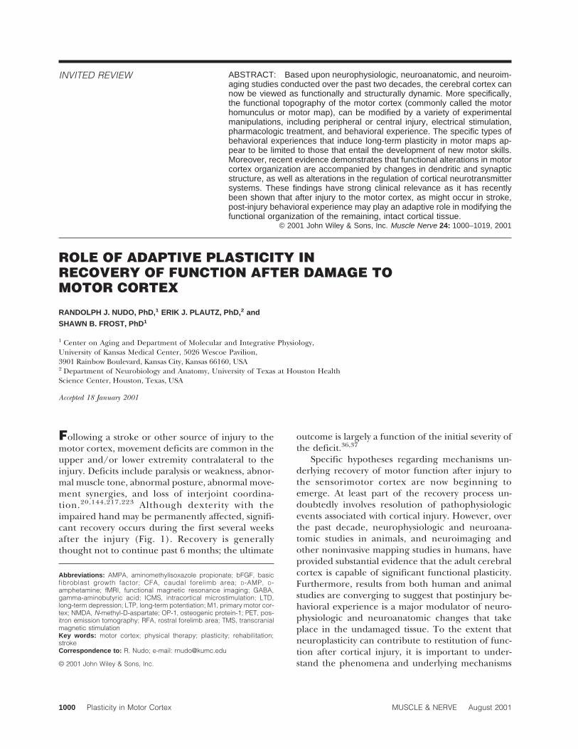
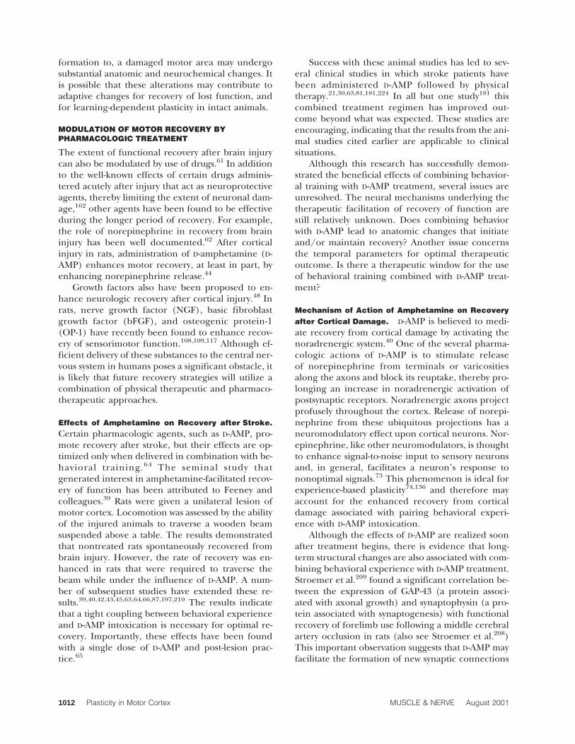
Following a stroke or other source of injury to themotor cortex, movement deficits are common in theupper and/or lower extremity contralateral to theinjury. Deficits include paralysis or weakness, abnor-mal muscle tone, abnormal posture, abnormal move-ment synergies, and loss of interjoint coordina-tion.20,144,217,223 Although dexterity with theimpaired hand may be permanently affected, signifi-cant recovery occurs during the first several weeksafter the injury (Fig. 1). Recovery is generallythought not to continue past 6 months; the ultimate
outcome is largely a function of the initial severity ofthe deficit.36,37
Specific hypotheses regarding mechanisms un-derlying recovery of motor function after injury tothe sensorimotor cortex are now beginning toemerge. At least part of the recovery process un-doubtedly involves resolution of pathophysiologicevents associated with cortical injury. However, overthe past decade, neurophysiologic and neuroana-tomic studies in animals, and neuroimaging andother noninvasive mapping studies in humans, haveprovided substantial evidence that the adult cerebralcortex is capable of significant functional plasticity.Furthermore, results from both human and animalstudies are converging to suggest that postinjury be-havioral experience is a major modulator of neuro-physiologic and neuroanatomic changes that takeplace in the undamaged tissue. To the extent thatneuroplasticity can contribute to restitution of func-tion after cortical injury, it is important to under-stand the phenomena and underlying mechanisms
Abbreviations: AMPA, aminomethylisoxazole propionate; bFGF, basicfibroblast growth factor; CFA, caudal forelimb area; D-AMP, D-amphetamine; fMRI, functional magnetic resonance imaging; GABA,gamma-aminobutyric acid; ICMS, intracortical microstimulation; LTD,long-term depression; LTP, long-term potentiation; M1, primary motor cor-tex; NMDA, N-methyl-D-aspartate; OP-1, osteogenic protein-1; PET, pos-itron emission tomography; RFA, rostral forelimb area; TMS, transcranialmagnetic stimulationKey words: motor cortex; physical therapy; plasticity; rehabilitation;strokeCorrespondence to: R. Nudo; e-mail: [email protected]
© 2001 John Wiley & Sons, Inc.
1000 Plasticity in Motor Cortex MUSCLE & NERVE August 2001

at the synaptic, cellular, and systems levels of orga-nization.
The goal of the present review is to summarizefindings to date on physiologic and neuroanatomicplasticity in the motor cortex that occur followingcortical injury. Although a large literature now existsregarding such plasticity in immature brains (e.g.,see review by Kolb and Whishaw118), the present re-view will focus primarily on adults. Because of itsimportance in normal motor control, and because itis often involved in stroke or other cortical injuries,this review is restricted primarily to neural plasticityfollowing injury to the primary motor cortex.
THEORIES OF RECOVERY
The theoretical framework for understanding recov-ery of function is still evolving after over a century ofstudy as reviewed elsewhere.2,132,203 According toone hypothesis described by von Monakow at thebeginning of the twentieth century,221 the functionof remote cortical tissue is temporarily suppressedafter focal cortical injury. This process is known asdiaschisis. Recovery is thought to result from thegradual reversal of diaschisis. Contemporary studiesof brain metabolism after cortical injury have largelyconfirmed that resolution of diaschisis is likely toplay a role in functional recovery.41,198 However, asmore specific injury-induced events at distant sitesare examined, it is becoming evident that: (a) dias-chisis may persist for long periods of time after in-
jury, that is, after significant recovery has occurred88;and (b) persistent remote effects of cortical injuryare more complex than previously thought, and in-clude disinhibition and hyperexcitability in additionto the well-known hypometabolism and inhibition.3
In addition to the resolution of diaschisis, motorrecovery after cortical injury occurs in large partthrough behavioral compensation, rather than via“true recovery” or restitution of “normal” motorstrategies.47,53,203,232 For example, in one recentstudy of stroke patients, severely to moderately im-paired subjects used more compensatory strategieswith the trunk to accomplish a pointing task.20 Thisis not unlike the response of rats after unilateralsensorimotor cortex lesions that employ posturalcompensation in retrieving food, rather than rees-tablishing normal motor strategies.232 It has alsobeen reported that original motor strategies can, insome cases, be regained after motor cortex injuriesin primates.53 However, this more complete recoveryoccurred after very small microinfarcts that resultedin mild and transient deficits in motor performance.
Although resolution of diaschisis (and otherpathologic sequelae) and behavioral compensationplay major roles in the phenomenon of motor recov-ery, it has also been suggested that other cortical (orsubcortical) structures, either adjacent to or remotefrom the damaged area can “take over” the functionof the damaged area. This theory, known as vicaria-tion of function,142 has gained considerable popu-larity over the past decade due to several recent ex-amples of functional plasticity after cortical injury.243
The degree to which the reorganization observed inspared tissue represents mechanisms related to res-titution of the original function, behavioral compen-sation, or both, is still not entirely clear. However, alarge number of recent studies are beginning toshed light on this issue and are reviewed in whatfollows.
FUNCTIONAL ORGANIZATION OF MOTOR CORTEX
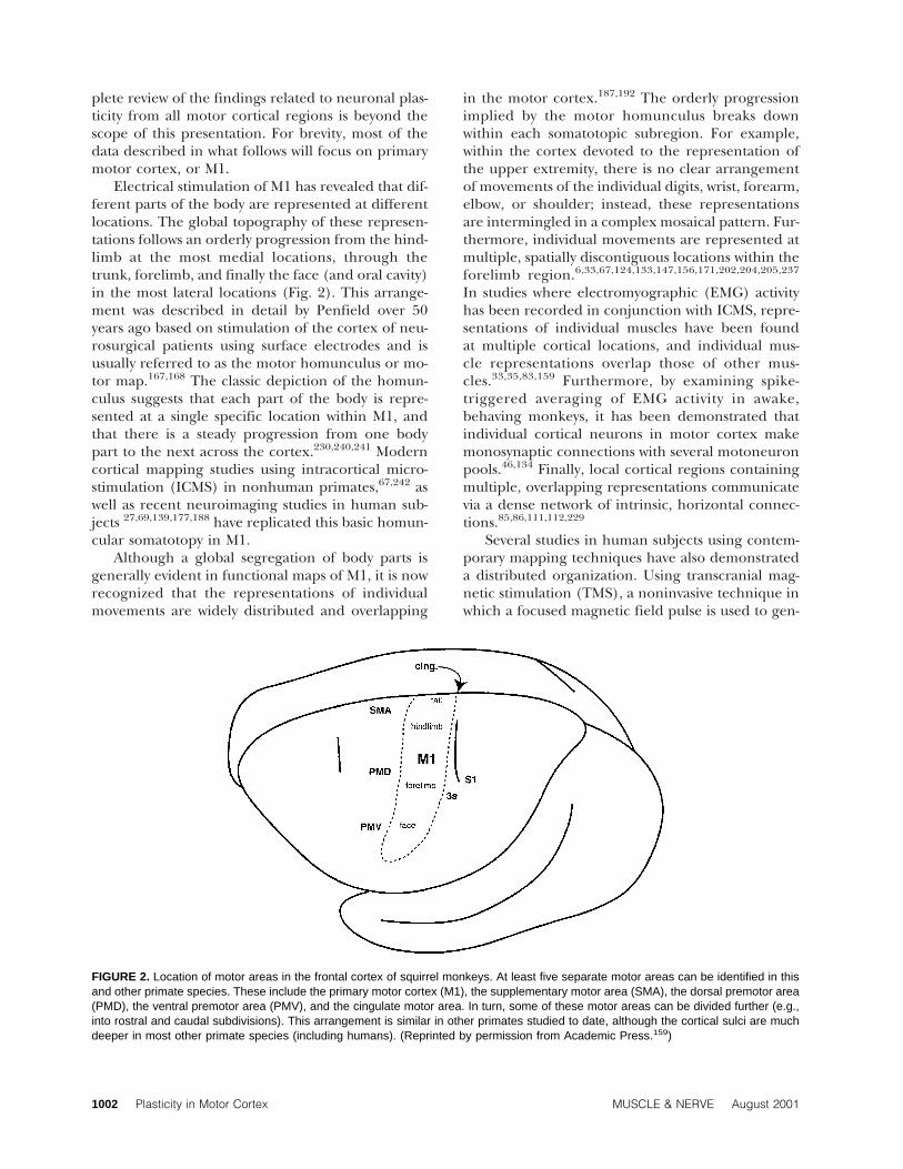
The primate motor cortex, located in the precentralgyrus, is classically defined as the portion of the ce-rebral cortex that requires the least amount of elec-trical stimulation to evoke movement of skeletalmuscles. In primate species, including humans, theso-called “motor cortex” is subdivided into severaldistinct regions based on anatomic, physiologic, orfunctional criteria. These regions include the pri-mary motor cortex, the premotor cortex, the supple-mentary motor area, and the cingulate motor area(Fig. 2).169,172,236,242 There is now evidence that atleast some of these motor areas can be subdividedinto even smaller components.58,172,173,212 A com-
FIGURE 1. Poststroke recovery profiles for three different levelsof stroke severity. Graph depicts means and 1 SD of Fugl–Meyerupper extermity scores after stroke in 459 individuals enrolled inthe Kansas City Stroke Study.37 Patients with different levels ofstroke severity show different probabilities of recovery. (Re-printed from Neuropharmacology, Vol 39, Duncan, PW, Lai, SM,Keighley, J, Defining post-stroke recovery: implications for designand interpretation of drug trials, pp 835–841. Copyright 2000,with permission from Elsevier Science.)
Plasticity in Motor Cortex MUSCLE & NERVE August 2001 1001
plete review of the findings related to neuronal plas-ticity from all motor cortical regions is beyond thescope of this presentation. For brevity, most of thedata described in what follows will focus on primarymotor cortex, or M1.
Electrical stimulation of M1 has revealed that dif-ferent parts of the body are represented at differentlocations. The global topography of these represen-tations follows an orderly progression from the hind-limb at the most medial locations, through thetrunk, forelimb, and finally the face (and oral cavity)in the most lateral locations (Fig. 2). This arrange-ment was described in detail by Penfield over 50years ago based on stimulation of the cortex of neu-rosurgical patients using surface electrodes and isusually referred to as the motor homunculus or mo-tor map.167,168 The classic depiction of the homun-culus suggests that each part of the body is repre-sented at a single specific location within M1, andthat there is a steady progression from one bodypart to the next across the cortex.230,240,241 Moderncortical mapping studies using intracortical micro-stimulation (ICMS) in nonhuman primates,67,242 aswell as recent neuroimaging studies in human sub-jects 27,69,139,177,188 have replicated this basic homun-cular somatotopy in M1.
Although a global segregation of body parts isgenerally evident in functional maps of M1, it is nowrecognized that the representations of individualmovements are widely distributed and overlapping
in the motor cortex.187,192 The orderly progressionimplied by the motor homunculus breaks downwithin each somatotopic subregion. For example,within the cortex devoted to the representation ofthe upper extremity, there is no clear arrangementof movements of the individual digits, wrist, forearm,elbow, or shoulder; instead, these representationsare intermingled in a complex mosaical pattern. Fur-thermore, individual movements are represented atmultiple, spatially discontiguous locations within theforelimb region.6,33,67,124,133,147,156,171,202,204,205,237
In studies where electromyographic (EMG) activityhas been recorded in conjunction with ICMS, repre-sentations of individual muscles have been foundat multiple cortical locations, and individual mus-cle representations overlap those of other mus-cles.33,35,83,159 Furthermore, by examining spike-triggered averaging of EMG activity in awake,behaving monkeys, it has been demonstrated thatindividual cortical neurons in motor cortex makemonosynaptic connections with several motoneuronpools.46,134 Finally, local cortical regions containingmultiple, overlapping representations communicatevia a dense network of intrinsic, horizontal connec-tions.85,86,111,112,229
Several studies in human subjects using contem-porary mapping techniques have also demonstrateda distributed organization. Using transcranial mag-netic stimulation (TMS), a noninvasive technique inwhich a focused magnetic field pulse is used to gen-
FIGURE 2. Location of motor areas in the frontal cortex of squirrel monkeys. At least five separate motor areas can be identified in thisand other primate species. These include the primary motor cortex (M1), the supplementary motor area (SMA), the dorsal premotor area(PMD), the ventral premotor area (PMV), and the cingulate motor area. In turn, some of these motor areas can be divided further (e.g.,into rostral and caudal subdivisions). This arrangement is similar in other primates studied to date, although the cortical sulci are muchdeeper in most other primate species (including humans). (Reprinted by permission from Academic Press.159)
1002 Plasticity in Motor Cortex MUSCLE & NERVE August 2001
erate an electrical discharge in cortical neurons,multiple and overlapping representations for move-ments of the arm and hand have been revealed.26
Functional neuroimaging techniques, such as posi-tron emission tomography (PET) and functionalmagnetic resonance imaging (fMRI), also suggestthat the representations of individual arm, hand,and finger movements are multiple and overlap-ping27,69,177,188 (also, see review by Schieber andHibbard193). For example, individual finger repre-sentations have been shown to overlap each other188
as well as the representations of the wrist and el-bow.177,188 It has been suggested that the somato-topic gradients superimposed across a largely distrib-uted representation may account for observedpatterns of separate and overlapping representa-tions.192
In summary, the functional organization of theprimate M1 is much more complex than has beenclassically described. Muscle representations overlapextensively; individual muscle and joint representa-tions are re-represented within the motor map; indi-vidual corticospinal neurons diverge to multiple mo-toneuron pools; horizontal fibers interconnectdistributed representations. This complex organiza-tion may provide the substrate for functional plastic-ity in motor cortex, at least within each local subre-gion.
ADAPTIVE PLASTICITY OF M1 AS A RESULTOF EXPERIENCE
Animal Studies. For nearly two decades, investiga-tions of cortical plasticity in adult animals have uti-lized neurophysiologic techniques to demonstratethe mutability of functional activity in sensory andmotor cortex. The invasive methods used in animalsubjects have permitted examination of plasticchanges at a variety of levels, including reorganiza-tion of representational maps, alterations in the ac-tivity of single or small groups of neurons, in vitrostudies of altered synaptic function, and anatomicchanges in neuronal structure. For example, in theearly 1980s, using microelectrode recording tech-niques to define receptive fields of neurons in thesomatosensory cortex, it was demonstrated that rep-resentations of the hand are altered by sensory ex-perience. After experimental amputation of a digitof the hand, the representation of that digit is re-placed by representations of adjacent digits.137,138
Behavioral training procedures can also result in al-terations in somatosensory maps. The representa-tions of skin surfaces that are stimulated during a
sensorimotor task are greatly expanded, and recep-tive field sizes are reduced.94,178
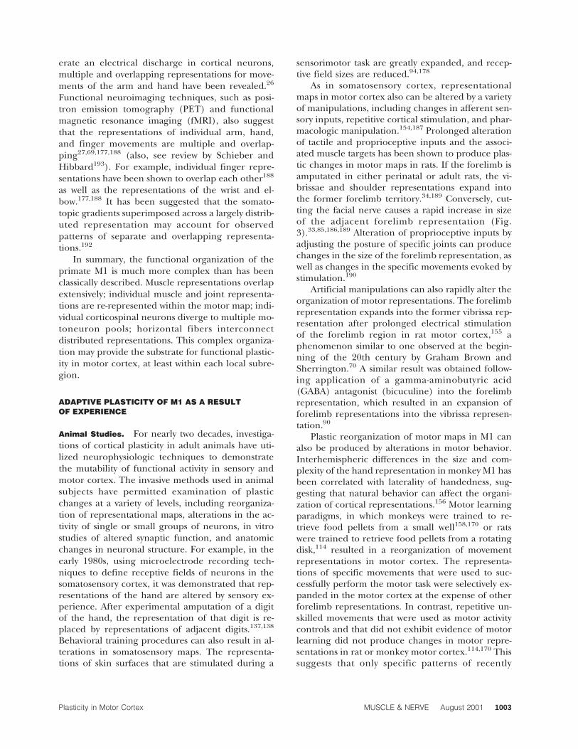
As in somatosensory cortex, representationalmaps in motor cortex also can be altered by a varietyof manipulations, including changes in afferent sen-sory inputs, repetitive cortical stimulation, and phar-macologic manipulation.154,187 Prolonged alterationof tactile and proprioceptive inputs and the associ-ated muscle targets has been shown to produce plas-tic changes in motor maps in rats. If the forelimb isamputated in either perinatal or adult rats, the vi-brissae and shoulder representations expand intothe former forelimb territory.34,189 Conversely, cut-ting the facial nerve causes a rapid increase in sizeof the adjacent forelimb representation (Fig.3).33,85,186,189 Alteration of proprioceptive inputs byadjusting the posture of specific joints can producechanges in the size of the forelimb representation, aswell as changes in the specific movements evoked bystimulation.190
Artificial manipulations can also rapidly alter theorganization of motor representations. The forelimbrepresentation expands into the former vibrissa rep-resentation after prolonged electrical stimulationof the forelimb region in rat motor cortex,155 aphenomenon similar to one observed at the begin-ning of the 20th century by Graham Brown andSherrington.70 A similar result was obtained follow-ing application of a gamma-aminobutyric acid(GABA) antagonist (bicuculine) into the forelimbrepresentation, which resulted in an expansion offorelimb representations into the vibrissa represen-tation.90
Plastic reorganization of motor maps in M1 canalso be produced by alterations in motor behavior.Interhemispheric differences in the size and com-plexity of the hand representation in monkey M1 hasbeen correlated with laterality of handedness, sug-gesting that natural behavior can affect the organi-zation of cortical representations.156 Motor learningparadigms, in which monkeys were trained to re-trieve food pellets from a small well158,170 or ratswere trained to retrieve food pellets from a rotatingdisk,114 resulted in a reorganization of movementrepresentations in motor cortex. The representa-tions of specific movements that were used to suc-cessfully perform the motor task were selectively ex-panded in the motor cortex at the expense of otherforelimb representations. In contrast, repetitive un-skilled movements that were used as motor activitycontrols and that did not exhibit evidence of motorlearning did not produce changes in motor repre-sentations in rat or monkey motor cortex.114,170 Thissuggests that only specific patterns of recently
Plasticity in Motor Cortex MUSCLE & NERVE August 2001 1003

learned motor behavior are capable of producingfunctional plasticity in motor cortex.
Human Studies. Recent advances in imaging tech-nology have permitted investigators to examine dy-namic changes in human brain function. Of particu-lar interest is whether this cortical activity ismodulated as a function of learning and experience,as has been demonstrated in animal models.
Several studies of sensorimotor cortex suggestthat functional activity can be altered in humans bychronic experience. In somatosensory cortex, therepresentation of the digits of the skilled hand isexpanded in string musicians and blind Braille read-ers compared to the unskilled hand.38,165 In motorcortex, the representation of the fingers of theskilled hand is reorganized in trained badmintonplayers compared to the unskilled hand and to therepresentations in untrained players.166 Together,these studies indicate that long-term practice of aparticular sensorimotor skill can produce functionalreorganization in relevant cortical representations.
There are several studies in humans that demon-strate functional reorganization associated with mo-tor learning over much briefer time periods. On thewhole, these studies indicate that motor cortex hasthe potential for rapid and large-scale functionalchanges in response to motor skill learning. Maps ofmotor outputs from M1 defined using transcranialmagnetic stimulation (TMS) have been shown tochange after brief periods of motor training. Re-peated paired movements of the thumb with move-ments of the shoulder,25 face,24 or foot130 produceda shift in the location of the thumb map toward therepresentation of the paired movement (i.e., a me-dial shift with shoulder or foot pairings, and a lateralshift with face pairings). Performance of unpairedmovements did not affect the location of the thumb
map. However, repeated practice of a single, specificmovement can affect its cortical representation.Thumb movements made in a direction opposite tothe movement direction evoked by TMS prior totraining produced a progressive shift in TMS-evokedthumb responses toward the trained direction.22
This effect manifested within 30 minutes, and wasreversed within 30 minutes after training was halted.
Motor sequence learning has been shown to pro-duce changes in M1 activity. Subjects either prac-t iced a known sequence of f inger move-ments,84,106,107,164 or performed cued movements ofindividual fingers in an initially unknown repeatedsequence.163,244 Repetitive practice of a knownmovement sequence caused a progressive expansionof finger representations in M1 within 30 minutes,84
over several days,164 and over several weeks107 as thesequence was learned. Map expansions paralleledimprovements in motor performance. Karni et al.found that the differential activation of M1 forlearned versus control sequences persisted for atleast 8 weeks after training had stopped.107 Repeti-tive, cued performance of an unknown movementsequence produced a decrease in reaction time tothe cue, suggesting an implicit learning process, anda concurrent expansion of finger representations inM1.163,244 Control sequences (i.e., without a re-peated pattern) did not affect the map. Once ex-plicit knowledge of the sequence was achieved, themap returned to its original size, presumably reflect-ing a difference in cognitive processing mechanismsfor implicit versus explicit motor performance.
Several studies using positron emission tomogra-phy in human subjects have demonstrated activitychanges in motor cortical structures, and in particu-lar primary motor cortex, during the acquisition ofnew motor skills.68,110,196,201,218,235 For example,Grafton et al.68 studied subjects as they learned to
FIGURE 3. Alterations in motor representations after facial nerve transection in rats. Representations were defined by microelectrodestimulation in the motor cortex of anesthetized rats. In normal rats (left), the forelimb representation was separated from the eyelidrepresentation by the vibrissa representation. Two weeks after a facial nerve transection (right), the forelimb and vibrissa representationswere contiguous. Redrawn from an article by Sanes et al.186 These experiments, and others like them, demonstrate that motor repre-sentations are modified by experience.
1004 Plasticity in Motor Cortex MUSCLE & NERVE August 2001
track a moving target with their hand. As accuracyincreased and smooth pursuit movements devel-oped, a parallel, progressive increase in M1 activa-tion was detected, beyond activity levels related tomovement performance. Learning-dependent in-creases in activation within M1 may occur in regionsdistinct from those activated by movement execu-tion.110
Some studies have failed to demonstrate substan-tial changes in M1 during learning.54,92 For ex-ample, Jenkins et al. found no difference in activityin sensorimotor cortex during the performance of aprelearned sequence versus learning of a novel se-quence.92 It should be noted, however, that subjectsin this study were attempting to deduce the correctsequence via a trial-and-error process. This kind ofsequence learning relies more on cognitive process-ing and less on strictly movement-related processing,such as correctly tapping the thumb to a particularfinger196,198 or smoothly tracking a moving targetwith the hand.68 Thus, the specific skills beinglearned or practiced may critically determine the de-gree of M1 functional plasticity.
Mechanisms of Learning-Dependent Plasticity in Mo-tor Cortex. One mechanism that has been sug-gested for mediating functional changes in the cere-bral cortex is modification of the synaptic strength ofhorizontal connections. The most widely studiedmodel of synaptic mechanisms underlying learningand memory comprises the phenomena of long-termpotentiation (LTP) and long-term depression (LTD)in the rat hippocampus or cerebral cortex. Recentstudies in slice preparations of rat motor cortex re-port that LTP and LTD can be induced in layer II/III horizontal connections,76–80 similar to extensivestudies in hippocampal slice preparations.9 More re-cently, it has been demonstrated that both LTP andLTD can be induced in the neocortex of the freelymoving rat, but multiple, spaced stimulation sessionsare required.55,214
Recent results show that LTP induction in neo-cortex is associated with alterations in dendrite mor-phology and increased spine density, similar to thosefound in rats exposed to complex environments.89
Furthermore, in postmortem slice preparations aftermotor learning, rats have larger amplitude field po-tentials in the motor cortex contralateral to thetrained forelimb.182 At the cellular level, severalstudies indicate that motor learning can producechanges in functional activity of neurons in motorcortex. Single-unit recording studies in awake, be-having animals have demonstrated altered neural ac-tivity in motor cortex in relation to skill acquisition
in monkeys1,56,57,140 and rats,126 as well as associativeconditioning in cats.4,5,135 Together, these studiessuggest that the synaptic strength of horizontal con-nections in the motor cortex are modifiable and mayprovide a substrate for altering the topography ofcortical motor maps during the acquisition of motorskills.
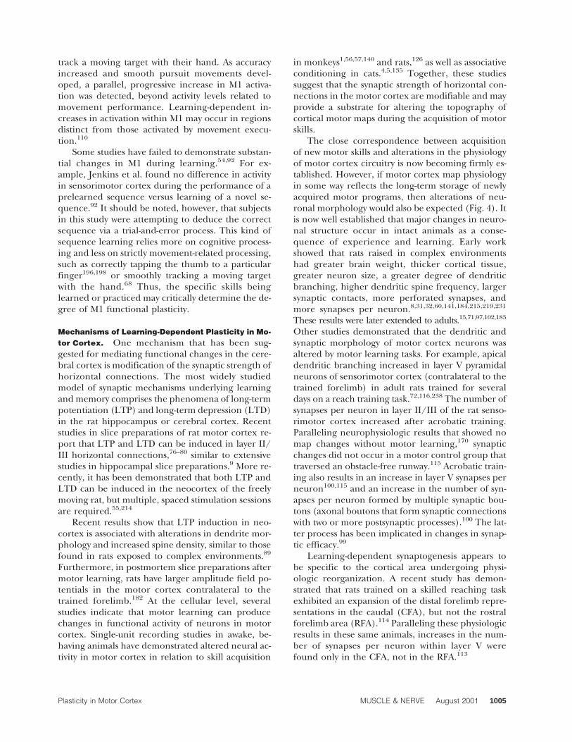
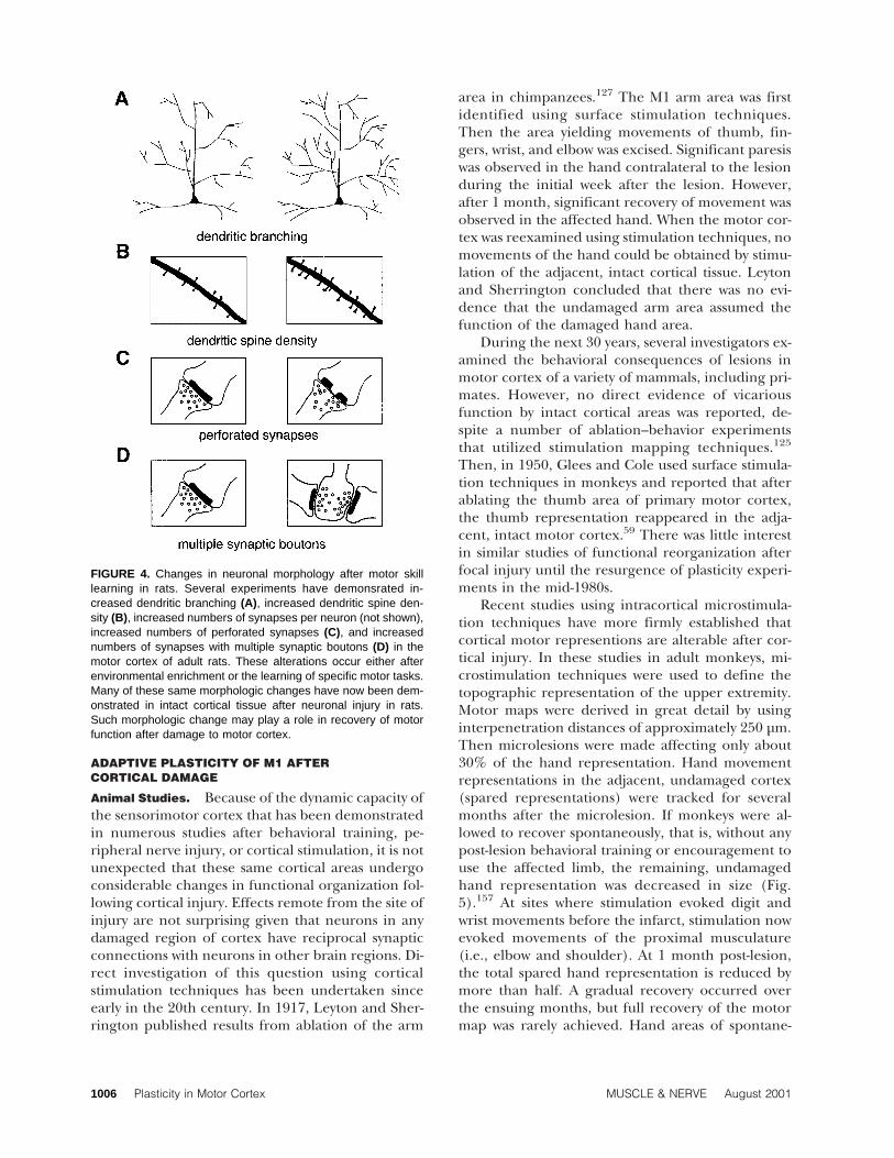
The close correspondence between acquisitionof new motor skills and alterations in the physiologyof motor cortex circuitry is now becoming firmly es-tablished. However, if motor cortex map physiologyin some way reflects the long-term storage of newlyacquired motor programs, then alterations of neu-ronal morphology would also be expected (Fig. 4). Itis now well established that major changes in neuro-nal structure occur in intact animals as a conse-quence of experience and learning. Early workshowed that rats raised in complex environmentshad greater brain weight, thicker cortical tissue,greater neuron size, a greater degree of dendriticbranching, higher dendritic spine frequency, largersynaptic contacts, more perforated synapses, andmore synapses per neuron.8,31,32,60,141,184,215,219,231
These results were later extended to adults.15,71,97,102,183
Other studies demonstrated that the dendritic andsynaptic morphology of motor cortex neurons wasaltered by motor learning tasks. For example, apicaldendritic branching increased in layer V pyramidalneurons of sensorimotor cortex (contralateral to thetrained forelimb) in adult rats trained for severaldays on a reach training task.72,116,238 The number ofsynapses per neuron in layer II/III of the rat senso-rimotor cortex increased after acrobatic training.Paralleling neurophysiologic results that showed nomap changes without motor learning,170 synapticchanges did not occur in a motor control group thattraversed an obstacle-free runway.115 Acrobatic train-ing also results in an increase in layer V synapses perneuron100,115 and an increase in the number of syn-apses per neuron formed by multiple synaptic bou-tons (axonal boutons that form synaptic connectionswith two or more postsynaptic processes).100 The lat-ter process has been implicated in changes in synap-tic efficacy.99
Learning-dependent synaptogenesis appears tobe specific to the cortical area undergoing physi-ologic reorganization. A recent study has demon-strated that rats trained on a skilled reaching taskexhibited an expansion of the distal forelimb repre-sentations in the caudal (CFA), but not the rostralforelimb area (RFA).114 Paralleling these physiologicresults in these same animals, increases in the num-ber of synapses per neuron within layer V werefound only in the CFA, not in the RFA.113
Plasticity in Motor Cortex MUSCLE & NERVE August 2001 1005
ADAPTIVE PLASTICITY OF M1 AFTERCORTICAL DAMAGE
Animal Studies. Because of the dynamic capacity ofthe sensorimotor cortex that has been demonstratedin numerous studies after behavioral training, pe-ripheral nerve injury, or cortical stimulation, it is notunexpected that these same cortical areas undergoconsiderable changes in functional organization fol-lowing cortical injury. Effects remote from the site ofinjury are not surprising given that neurons in anydamaged region of cortex have reciprocal synapticconnections with neurons in other brain regions. Di-rect investigation of this question using corticalstimulation techniques has been undertaken sinceearly in the 20th century. In 1917, Leyton and Sher-rington published results from ablation of the arm
area in chimpanzees.127 The M1 arm area was firstidentified using surface stimulation techniques.Then the area yielding movements of thumb, fin-gers, wrist, and elbow was excised. Significant paresiswas observed in the hand contralateral to the lesionduring the initial week after the lesion. However,after 1 month, significant recovery of movement wasobserved in the affected hand. When the motor cor-tex was reexamined using stimulation techniques, nomovements of the hand could be obtained by stimu-lation of the adjacent, intact cortical tissue. Leytonand Sherrington concluded that there was no evi-dence that the undamaged arm area assumed thefunction of the damaged hand area.
During the next 30 years, several investigators ex-amined the behavioral consequences of lesions inmotor cortex of a variety of mammals, including pri-mates. However, no direct evidence of vicariousfunction by intact cortical areas was reported, de-spite a number of ablation–behavior experimentsthat utilized stimulation mapping techniques.125
Then, in 1950, Glees and Cole used surface stimula-tion techniques in monkeys and reported that afterablating the thumb area of primary motor cortex,the thumb representation reappeared in the adja-cent, intact motor cortex.59 There was little interestin similar studies of functional reorganization afterfocal injury until the resurgence of plasticity experi-ments in the mid-1980s.
Recent studies using intracortical microstimula-tion techniques have more firmly established thatcortical motor representions are alterable after cor-tical injury. In these studies in adult monkeys, mi-crostimulation techniques were used to define thetopographic representation of the upper extremity.Motor maps were derived in great detail by usinginterpenetration distances of approximately 250 µm.Then microlesions were made affecting only about30% of the hand representation. Hand movementrepresentations in the adjacent, undamaged cortex(spared representations) were tracked for severalmonths after the microlesion. If monkeys were al-lowed to recover spontaneously, that is, without anypost-lesion behavioral training or encouragement touse the affected limb, the remaining, undamagedhand representation was decreased in size (Fig.5).157 At sites where stimulation evoked digit andwrist movements before the infarct, stimulation nowevoked movements of the proximal musculature(i.e., elbow and shoulder). At 1 month post-lesion,the total spared hand representation is reduced bymore than half. A gradual recovery occurred overthe ensuing months, but full recovery of the motormap was rarely achieved. Hand areas of spontane-
FIGURE 4. Changes in neuronal morphology after motor skilllearning in rats. Several experiments have demonsrated in-creased dendritic branching (A), increased dendritic spine den-sity (B), increased numbers of synapses per neuron (not shown),increased numbers of perforated synapses (C), and increasednumbers of synapses with multiple synaptic boutons (D) in themotor cortex of adult rats. These alterations occur either afterenvironmental enrichment or the learning of specific motor tasks.Many of these same morphologic changes have now been dem-onstrated in intact cortical tissue after neuronal injury in rats.Such morphologic change may play a role in recovery of motorfunction after damage to motor cortex.
1006 Plasticity in Motor Cortex MUSCLE & NERVE August 2001

ously recovering monkeys were approximately 75%of their original areas after 4 months.52
Because it has long been suggested that physicaltherapeutic interventions might improve recovery af-ter injury to motor cortex,95,96,161 additional studieshave examined the effects of post-lesion motor train-ing on recovery of motor maps. In these experi-ments, monkeys were placed in restraint jackets thatrestricted the use of the unimpaired limb.160 Dailyrepetitive training procedures were employed to en-courage improvement in manual skill. After manualskill had returned to normal levels, the motor cortexwas reexamined with microstimulation techniques.In contrast to spontaneously recovering monkeys,the monkeys that received postinjury behavioraltraining showed retention of the undamaged handrepresentations (Fig. 5). On average, there was a netgain of approximately 10% in the total hand areaadjacent to the lesion. More recently, it has beenshown that the retention of hand area adjacent to amicrolesion in M1 requires repetitive behavioraltraining, because use of the restraint jacket aloneresulted in no change in hand representations be-yond that which was seen with spontaneous recov-ery.52 Hand representations in monkeys that worethe restraint jacket continuously for up to 1 yearwere only 80% of the pre-lesion area.
It should be noted that in the primate studies todate, it has not been possible to demonstrate differ-ences in motor abilities as a function of post-infarctexperience.52 Although the reasons are not yet en-tirely clear, it is likely that the small numbers of ani-mals per group combined with large individual vari-ability in motor performance may have contributedto the lack of evidence for behavioral differences. Itis also possible that the measure of motor skill usedin these studies (numbers of finger flexions requiredto retrieve a food pellet from a small well) was in-sensitive to subtle differences in behavior among thegroups. Finally, because of the small size of thesemicroinfarcts (30% of the primary motor cortexhand area), all of the animals may have been able tocompensate relatively quickly for mild motor defi-cits. Additional studies examining a variety of motorskills after larger cortical infarcts are needed to ad-dress this issue more directly. In any event, it is clearthat despite variability in behavioral outcomes aftermicroinfarcts in motor cortex, the changes in neu-rophysiologically derived maps are consistent, andcan be modulated in predictable ways by postinjuryexperience.
Further evidence that reorganization of intact,adjacent cortical tissue contributes to functional re-covery has come from similar motor mapping studies
FIGURE 5. Summary of functional remodeling of the hand representation in primary motor cortex after a stroke-like injury. Data werederived from hundreds of microelectrode penetrations using microstimulation techniques to determine evoked movements in anesthetizedmonkeys. These studies, and others like them, demonstrate that the uninjured tissue adjacent to a cortical injury undergoes functionalreorganization that can be modulated by postinjury behavioral training. (Reprinted by permission from Stockton Press.153)
Plasticity in Motor Cortex MUSCLE & NERVE August 2001 1007
in rats. In these studies, after bilateral ablation of theforelimb area in rat motor cortex, motor perfor-mance was impaired. However, recovery occurred ifelectrical stimulation of the ventral tegmentalnucleus was paired with forelimb responses.12 Thestimulation is thought to have played a motivationalrole in encouraging forelimb use. When the motorcortex was reexamined following recovery, a novelforelimb representation appeared caudal and lateralto the ablated representation.13 The size of this rep-resentation was directly related to the behavioralperformance of the recovered animals. Finally, abla-tion of the newly emerged forelimb representationresulted in reinstatement of the deficit.
Recent results in monkeys suggest that, at leastafter large lesions of the primary motor cortex, moreremote cortical motor areas may participate in re-covery. After unilateral damage to the M1 hand areaand subsequent recovery, the GABA agonist, musci-mol, was injected into various intact motor regions toinduce transient inactivation. Whereas inactivationof M1 of the injured or intact side had no effect,inactivation of the premotor cortex of the injuredhemisphere rapidly reinstated the deficit.131 Func-tional alterations have also been reported in thesupplementary motor area after M1 lesions.1 Thus,after damage to M1, other motor areas in the injuredhemisphere may contribute to recovery of motorskills.
Although the focus of this review is on motorcortical areas, postinjury reorganization has alsobeen observed in somatosensory cortex after similarinjuries. Following a small infarct in the primary so-matosensory (area 3b) hand representation in mon-keys, the injured digit representations reemerge inadjacent, intact tissue.93 In addition, representationsof the affected digits expand in other somatosensoryareas, such as areas 3a and 1.243 Taken together,these recent findings in both somatosensory and mo-tor cortex provide substantial evidence that vicaria-tion of function occurs in intact cortical regions aftercortical injury.
Human Studies. Several noninvasive techniqueshave been used in humans to examine the effects ofcortical injury on the function of intact cortical tis-sue. Subjects are typically those with cortical lesions(either ischemic or hemorrhagic) or lacunar subcor-tical lesions involving the internal capsule. These re-cent studies have used noninvasive techniques formapping the functional organization of the injuredcortex, such as positron emission tomography, func-tional magnetic resonance imaging, transcranialmagnetic stimulation, and magnetoencephalogra-phy. Although the location of the injury is frequently
unknown or uncontrolled, these studies have consis-tently shown that functional changes occur in severalcortical areas after stroke or other cortical damage,paralleling results from animal experiments. As thisreview of functional plasticity after cortical injury inhumans is not exhaustive, the reader is referred tothe more complete review by Cramer and Bastings.28
Using transcranial magnetic stimulation, it hasbeen shown that, shortly after stroke, the excita-bility of the motor cortex is reduced, and the corticalrepresentation of the affected muscles is de-creased.19,213 It is likely that this effect occurs from acombination of diaschisis-like effects220 and disuse ofthe affected limb.130 Peri-lesional changes in corticalactivity have been shown to occur using a variety oftechniques.23,29,105,200 Furthermore, after 8–10weeks of rehabilitation treatment, there was an en-largement of the motor map in the injured hemi-sphere relative to the initial postinjury map.213 Stillfurther, constraint-induced movement therapy, inwhich the unimpaired hand is constrained for 2weeks to induce goal-directed movement with theimpaired hand, produces a significant enlargementof the representation of the paretic limb.128,129,228
These results closely parallel the results of rehabili-tative training of the impaired hand in primate stud-ies described earlier.160
It has also been shown repeatedly that, after re-covery, movement of the recovered hand was associ-ated with increased bilateral activation of remotebrain areas, such as the cerebellum, and premotorcor tex , as wel l a s the sensor imotor cor -tex.18,51,148,226,227 Interestingly, these increased acti-vations often have occurred in the sensorimotor cor-tex of the uninjured hemisphere after goodrecovery, leading many to speculate that the unin-jured hemisphere plays a significant role in recov-ery.10,149,199 However, the role of mirror movementsin these studies is still unclear.227 The role of reor-ganization in the intact hemisphere has also beenquestioned because the presence of ipsilateral motorevoked potentials after stimulation of the nonstrokehemisphere (using TMS) has been associated withpoor motor outcome.150,216 Clearly, the role of theuninjured hemisphere after stroke requires furtherstudy.29
SUBSTRATES FOR FUNCTIONAL SUBSTITUTION
Anatomic Changes Associated with Cortical Injury.Several recent studies have provided evidence thatadaptive alterations occur in the anatomy of surviv-ing cortical and subcortical neurons. Because mor-phologic changes are known to be associated withmotor learning, it is plausible that the same or simi-
1008 Plasticity in Motor Cortex MUSCLE & NERVE August 2001
lar changes in intact cortical structures may contrib-ute to motor recovery after cortical injury.
Studies of anatomic changes after unilateral cor-tical injury have been conducted almost exclusivelyin rats. After injury to the sensorimotor cortex, ratspreferentially use the forelimb ipsilateral to the le-sion for postural support, reaching, and forelimbplacing.7,11,101,103,233,234 This asymmetry is seenwithin 1 or 2 days after the lesion, is maximal duringthe first 2 weeks post-lesion, and persists for at least1 month.101,103 Thus, it is not surprising that com-pensatory anatomic changes occur in the intact sen-sorimotor cortex contralateral to the injury.
Because the processes involved in changes on thetwo sides of the brain differ, especially with respectto the influence of postinjury behavioral experience,these two topics will be discussed separately. Becausemost of these studies have focused on the contralat-eral (uninjured) hemisphere, these studies will bediscussed first. Then, we review the evidence forsimilar changes in the injured hemisphere.
Contralateral (Uninjured) Side. Unilateral dam-age to the sensorimotor cortex in rats results in anumber of time-dependent anatomic alterations inthe motor cortex opposite the side of the lesion. Thehomotopic cortex opposite sensorimotor cortex le-sions undergoes a two-phase process of use-dependent dendritic overgrowth, followed by elimi-nation of dendrites in layer V.99,103,104,119,122
Beginning a few days after injury, dendritic branch-ing in layer V neurons begins to increase, reachingits peak at day 18. This increase is primarily inhigher-order branches.103,104 At this timepoint, thevolume of dendritic processes in layer V is signifi-cantly increased.101 At 10 days post-lesion, myelinat-ed axons are reduced in volume fraction.99 At 30days post-lesion, dendritic branching begins to de-crease, suggesting that dendritic pruning has oc-curred. However, branching is still elevated abovenormal levels. At this time, the number of synapsesand the surface area of dendritic membrane perlayer V neuron are increased significantly.101 In ad-dition to synaptic density, the proportion of synapsesformed by multiple synaptic boutons and perforatedpostsynaptic densities is significantly elevated at 30days post-lesion, but not at 10 or 18 days. Singlesynapse numbers per neuron do not increase.99 To-gether, these results suggest that after unilateral sen-sorimotor cortex lesions, a period of dendriticgrowth is followed by dendritic pruning, synapse for-mation, and changes in the specific structure of syn-aptic connections. The recent findings that the finestructure of synapses changes after injury is of par-ticularly interest, because several studies in other sys-
tems have suggested that these ultrastructuralchanges are related to changes in synaptic effi-cacy.102,151,225 Other studies suggest that the pruningphase is associated with adaptive changes as well.The administration of the N-methyl-D-aspartate(NMDA) receptor antagonists MK-801 or ethanolduring a critical period after cortical injury can blockthe pruning process, and disrupt behavioral recov-ery.119,121
There is also evidence that these anatomicchanges are dependent upon the increased use ofthe unimpaired forelimb. After lesions, rats compen-sate by relying more heavily on the unimpaired limbfor postural support.103 If the unimpaired forelimbis immobilized during the period of dendritic over-growth (0–15 days post-lesion), dendritic arboriza-tion does not occur in the intact hemisphere andbehavioral performance is further degraded.104 Den-dritic overgrowth is not affected by immobilizationof the impaired forelimb. Thus, dendritic over-growth is closely related to the time of overrelianceon the unimpaired forelimb, whereas the subse-quent dendritic pruning is related to a return ofmore symmetric use of the forelimbs. Dendritic over-growth does not occur after immobilization in sham-operated animals, indicating that the magnitude ofmorphologic changes results from an interaction ofthe lesion and post-lesion behavior.104 Finally, motorskill training for 28 days after the lesion significantlyincreased layer V synapses per neuron.100 Corticallesions may therefore trigger the events that lead touse-dependent cortical plasticity in regions intercon-nected with the damaged cortical area. It is con-cluded that the changes in the intact cortex are aninteractive effect of the lesion and post-lesion behav-ior.
Other studies appear to contradict these studiesof use-dependent growth in the intact hemisphere.After cortical aspiration lesions, increased use of theintact forelimb was not associated with an increase indendritic arborization of identified corticospinalneurons.174 Also, after either small electrolytic ormore extensive aspiration lesions, no evidence of ause-dependent increase in dendritic aborization wasfound in the intact hemisphere.50 Both of thesenegative results were obtained at 18 days post-lesion,the peak of dendritic overgrowth in the previousstudies. The authors of the latter two investigationsproposed several possible factors that might contrib-ute to the discrepancy, including differences in le-sion methodology. More recent data confirmed thatthe dendritic growth does not occur after aspirationlesions, or after electrolytic lesions followed by aspi-ration (T. A. Jones, personal communication and see
Plasticity in Motor Cortex MUSCLE & NERVE August 2001 1009

Voorhies and Jones222). Furthermore, electrolytic le-sions may fail to produce dendritic growth if they aretoo small, a potential factor in some of the negativeresults with electrolytic lesions.101 Although themechanisms controlling this phenomenon are notyet fully understood, it is clear that, under certainconditions, use-dependent dendritic overgrowth oc-curs in the intact hemisphere after motor cortexdamage. This effect appears to depend upon thepresence of the damaged tissue, and is more likely tooccur after large cortical lesions.
Ipsilateral (Injured) Side. Indirect evidence sug-gests that anatomic changes occur in the uninjuredcortical tissue surrounding the injury. After focalcortical ischemia in rats, GAP-43 immunoreactivityincreased in the surrounding tissue, suggesting axo-nal sprouting.207 In addition, synaptophysin immu-noreactivity is increased in the surrounding tissue,suggesting an increase in the number of synapses inthe intact cortex.206 It is of interest to note that theGAP-43 increase was significantly elevated only atearly survival times (3, 7, and 14 days), whereas thesynaptophysin increase was significant at later sur-vival times (14, 30, and 60 days), suggesting that axo-nal sprouting was followed by synaptogenesis.208
After injury to the sensorimotor cortex in rats,extreme use of the affected limb can result in anenlargement of the lesion and further motor impair-ment.120 If the unimpaired limb is placed in a re-strictive cast after cortical injury, rats must rely heav-ily on the impaired limb for posture and locomotion.Forced overuse of the impaired limb during the firstweek after injury results in expansion of the injuryand poorer motor performance.82 Forced overuseduring the next 7 days does not result in injury ex-pansion, but nonetheless results in poorer motorperformance. This study strongly suggests that thereare specific vulnerable periods for maladaptive ef-fects of use after injury. Timing of these maladaptiveeffects must be considered along with timing ofadaptive effects in any rational therapeutic designfor treatment of motor deficits after injury.
In contrast, acrobatic motor training after a simi-lar injury in rats resulted in no detectable increase inthe size of the lesion and improved motor perfor-mance.100 It would appear that the behavioral con-ditions that follow cortical motor injury are critical inneural processes underlying recovery. The specificconditions that contribute to adaptive plasticity ver-sus those that contribute to maladaptive plasticity arenow beginning to come to light.
Subcortical Changes Several lines of evidencesuggest that the reorganization seen in motor corti-cal maps (as well as somatosensory maps) has a sub-
strate at the cortical level. However, at least in thesomatosensory system, it is likely that reorganizationoccurs at several levels of the neuraxis after injury.For example, after massive sensory loss, as might oc-cur after amputation, reorganization has been re-ported at cortical levels, in the thalamus, in the dor-sal column nuclei of the brainstem, and in the spinalcord.49,98 It has recently been shown that, at leastafter long-term amputation, significant sprouting oc-curs at the level of the brainstem that might accountfor massive reorganization in somatosensory cortex.Normally, afferents from the face terminate in theface representation of the trigeminal nucleus. How-ever, 10 years after an amputation, some of theseafferents sprout new connections to terminate in thecuneate nucleus, normally the recepient of afferentsfrom the arm.91 The growth of face afferents into thedeafferented cuneate nucelus in the brainstem ap-pears to contribute substantially to the activation ofhand cortex by face afferents.
Despite the growing evidence for subcorticalchanges in somatosensory structures following am-putation, similar data from subcortical motor struc-tures are rare. Mechanisms of cortical reorganizationafter amputation were recently addressed usingtranscranial magnetic stimulation techniques andthen testing intracortical inhibition and facilita-tion.16 The results suggest that after amputation, mo-tor reorganization occurs predominantly at corticallevels. It is important for future studies to assess di-rectly the contribution of functional and structuralplasticity in the motor system (e.g., red nucleus, spi-nal cord, etc.) after peripheral or central injury.
A few studies have now examined plasticity insubcortical motor structures in adult animal models.After thermocoagulatory lesions and injection of an-terograde tracers into the uninjured hemisphere, la-beled fibers were found in the striatum of injuredanimals contralateral to the injection (i.e., ipsilateralto the injury), suggesting that cortical neurons mayundergo axonal sprouting following an injury to thecortex.17,146 Unusual ultrastructural details havebeen observed in the newly formed synapses of thedeafferented striatum.17 These changes are associ-ated with a variety of changes in gene expression andgrowth-promoting factors.211 It is of interest to notethat, as with studies of morphologic changes in thehomotopic, intact cerebral cortex discussed previ-ously, if aspiration lesions are made, the sproutingand several growth-related cellular changes are notfound.145
A large number of studies have reported unusualcorticofugal projections after neonatal sensorimotorcortex lesions, including corticorubral, corticopon-
1010 Plasticity in Motor Cortex MUSCLE & NERVE August 2001
tine, and corticospinal projections.12,143,185 How-ever, evidence for widespread axonal sprouting orsynaptic remodeling in these subcortical pathways isstill weak in injury models in adult mammals.
In summary, unilateral brain injury can triggercompensatory mechanisms, whereby neurons in theintact tissue are induced to sprout new connections.At least some of this compensatory growth requiresbehavioral pressure.
Changes in Neuronal Excitability and Neurotransmit-ter Regulation after Cortical Injury. Only recentlyhave long-term changes in specific neurotransmittersystems been investigated in chronic cortical injury.These studies are important because they may leadto new intervention strategies and potential pharma-cologic treatment of chronic stroke.
Changes in two neurotransmitter systems, GABAand glutamate, have been implicated in behavioraldeficits following stroke, and alterations in the activ-ity of each may play a role in functional recovery.Behavioral deficits similar to those seen after strokeare found in primates after reversible inactivation ofprimary motor (M1) and premotor cortex by injec-tion of the GABAA agonist muscimol.123,194 There isa decrease in the density of inhibitory GABAA recep-tors and an increase in hyperexcitability in the areaadjacent to the lesion following cortical injury in therat.195 This hyperexcitability has been observed up to4 months after the lesion. Others studies have showna bihemispheric reduction of GABAA receptors inmultiple cortical areas connectionally related to thedamaged area in rats.176 Bilateral reductions in in-hibitory GABAA receptors and concurrent bilateralincreases in excitatory glutamate NMDA receptorsoccur in spared areas of cortex for up to 4 weeksafter middle cerebral artery occlusion in mice.175 Asimilar reduction of GABAA receptors and increasein NMDA receptors occurs in the ipsilateral thalamicnucleus projecting to the damaged areas of cor-tex.175
A recent study has shown differential downregu-lation of GABAA receptor subunits in peri-infarctand remote areas after focal cortical infarcts inrats.179 Alterations in subunit composition are asso-ciated with changes in electrophysiologic and phar-macologic properties of GABAA receptors, and thesechanges may be of importance for functional corticalreorganization after injury.179,180
Immunohistochemistry of the peri-infarct regionin rats has shown that 1 week after injury, parvalbu-min-positive interneurons (presumably GABAergic)show signs of degeneration and a reduction in thenumber of dendrites.152 There is also a reduction in
the number of parvalbumin-positive neurons imme-diately adjacent to the lesion. These results suggestthat the downregulation of the GABAergic system,resulting in a decrease in inhibition, also occurs pre-synaptically.
An upregulation of NMDA receptors in cortexfollowing ischemic lesion appears to be a consistentresult in rodents. Other glutamatergic receptors, theaminomethylisoxasole propionate (AMPA) andkainate receptors, have been shown to slightly in-crease in density in the peri-infarct region, althoughnot significantly.175 Significant changes in the den-sity of AMPA and kainate receptors were not seen inremote areas of cortex away from the lesion.175
Changes in GABA and glutamate receptor densi-ties may explain increased hyperexcitability follow-ing a lesion. Excitability changes in intact tissue fol-lowing cortical injury have traditionally beenthought to be pathologic, and thus maladaptive.However, evidence is now accumulating to suggestthat excitability changes may be one aspect of post-lesion adaptation in the neuronal network after in-jury. For example, pharmacologic studies haveshown that drugs that enhance the effect of GABAresult in the potentiation of behavioral deficits fol-lowing cortical lesion in rats.191 Conversely, drugsthat attenuate the effect of GABA speed up the re-covery of function following cortical lesion.75 Theseresults suggest that a downregulation of inhibitoryGABA receptors may be an adaptive response to in-jury, and that a certain degree of hyperexcitabilitymay favor recovery, at least in rodents.
Regulation of specific neurotransmitter systemsmay play a critical role in the functional reorganiza-tional process that occurs subsequent to cortical in-jury in primates (see earlier sections). Perhaps thenatural response to injury is a decrease in GABAinhibition that acts to unmask latent horizontal con-nections.90,182 Glutamate is the major neurotrans-mitter of the horizontal connections in cortex, andany synaptic modifications that may be necessary forfunctional reorganization may be mediated byNMDA receptors.76
GABA and glutamate receptor density in primatecortex following injury has yet to be examined.Changes in neurotransmitter systems have not yetbeen studied in relation to functional reorganizationfollowing skilled training in primates. The remoteeffects of cortical ischemia are believed to be causedby alterations due to electrical or chemical signalsemanating from the infarct, alterations along con-nectivity patterns (diaschisis) and use-dependent ad-aptations (see Witte and Stoll239 for review). Intactbrain areas that are remote from, but contribute in-
Plasticity in Motor Cortex MUSCLE & NERVE August 2001 1011
formation to, a damaged motor area may undergosubstantial anatomic and neurochemical changes. Itis possible that these alterations may contribute toadaptive changes for recovery of lost function, andfor learning-dependent plasticity in intact animals.
MODULATION OF MOTOR RECOVERY BYPHARMACOLOGIC TREATMENT
The extent of functional recovery after brain injurycan also be modulated by use of drugs.61 In additionto the well-known effects of certain drugs adminis-tered acutely after injury that act as neuroprotectiveagents, thereby limiting the extent of neuronal dam-age,162 other agents have been found to be effectiveduring the longer period of recovery. For example,the role of norepinephrine in recovery from braininjury has been well documented.62 After corticalinjury in rats, administration of D-amphetamine (D-AMP) enhances motor recovery, at least in part, byenhancing norepinephrine release.44
Growth factors also have been proposed to en-hance neurologic recovery after cortical injury.48 Inrats, nerve growth factor (NGF), basic fibroblastgrowth factor (bFGF), and osteogenic protein-1(OP-1) have recently been found to enhance recov-ery of sensorimotor function.108,109,117 Although ef-ficient delivery of these substances to the central ner-vous system in humans poses a significant obstacle, itis likely that future recovery strategies will utilize acombination of physical therapeutic and pharmaco-therapeutic approaches.
Effects of Amphetamine on Recovery after Stroke.Certain pharmacologic agents, such as D-AMP, pro-mote recovery after stroke, but their effects are op-timized only when delivered in combination with be-havioral training.64 The seminal study thatgenerated interest in amphetamine-facilitated recov-ery of function has been attributed to Feeney andcolleagues.39 Rats were given a unilateral lesion ofmotor cortex. Locomotion was assessed by the abilityof the injured animals to traverse a wooden beamsuspended above a table. The results demonstratedthat nontreated rats spontaneously recovered frombrain injury. However, the rate of recovery was en-hanced in rats that were required to traverse thebeam while under the influence of D-AMP. A num-ber of subsequent studies have extended these re-sults.39,40,42,43,45,63,64,66,87,197,210 The results indicatethat a tight coupling between behavioral experienceand D-AMP intoxication is necessary for optimal re-covery. Importantly, these effects have been foundwith a single dose of D-AMP and post-lesion prac-tice.65
Success with these animal studies has led to sev-eral clinical studies in which stroke patients havebeen administered D-AMP followed by physicaltherapy.21,30,63,81,181,224 In all but one study181 thiscombined treatment regimen has improved out-come beyond what was expected. These studies areencouraging, indicating that the results from the ani-mal studies cited earlier are applicable to clinicalsituations.
Although this research has successfully demon-strated the beneficial effects of combining behavior-al training with D-AMP treatment, several issues areunresolved. The neural mechanisms underlying thetherapeutic facilitation of recovery of function arestill relatively unknown. Does combining behaviorwith D-AMP lead to anatomic changes that initiateand/or maintain recovery? Another issue concernsthe temporal parameters for optimal therapeuticoutcome. Is there a therapeutic window for the useof behavioral training combined with D-AMP treat-ment?
Mechanism of Action of Amphetamine on Recoveryafter Cortical Damage. D-AMP is believed to medi-ate recovery from cortical damage by activating thenoradrenergic system.40 One of the several pharma-cologic actions of D-AMP is to stimulate releaseof norepinephrine from terminals or varicositiesalong the axons and block its reuptake, thereby pro-longing an increase in noradrenergic activation ofpostsynaptic receptors. Noradrenergic axons projectprofusely throughout the cortex. Release of norepi-nephrine from these ubiquitous projections has aneuromodulatory effect upon cortical neurons. Nor-epinephrine, like other neuromodulators, is thoughtto enhance signal-to-noise input to sensory neuronsand, in general, facilitates a neuron’s response tononoptimal signals.73 This phenomenon is ideal forexperience-based plasticity74,136 and therefore mayaccount for the enhanced recovery from corticaldamage associated with pairing behavioral experi-ence with D-AMP intoxication.
Although the effects of D-AMP are realized soonafter treatment begins, there is evidence that long-term structural changes are also associated with com-bining behavioral experience with D-AMP treatment.Stroemer et al.209 found a significant correlation be-tween the expression of GAP-43 (a protein associ-ated with axonal growth) and synaptophysin (a pro-tein associated with synaptogenesis) with functionalrecovery of forelimb use following a middle cerebralartery occlusion in rats (also see Stroemer et al.208)This important observation suggests that D-AMP mayfacilitate the formation of new synaptic connections
1012 Plasticity in Motor Cortex MUSCLE & NERVE August 2001
within cerebral cortex, thereby playing a key role inmechanisms of functional recovery after cortical in-jury.
SUMMARY AND CONCLUSIONS
The primary motor cortex forms a distributed net-work in which muscles and movements are re-represented at multiple locations within a local re-gion. This distributed organization forms a substratethat is amenable to use-dependent alteration of out-puts via modulation of local synaptic processes.Based on investigations of adaptive plasticity phe-nomena over the past 15–20 years, it must be con-cluded that the motor cortex of adult mammals canundergo widespread changes in functional organiza-tion as a result of behavioral experience and centralor peripheral injury. In particular, the motor cortexis altered during the acquisition of new motor skillssuch that the muscles and movements engaged inthe skilled activity come to be represented overgreater cortical territories. These changes in func-tional topography are accompanied by anatomic al-terations, such as increases in synaptic number.
In addition, the organization of motor cortex isnow known to be altered following cortical injury, asmight occur in stroke. Both human and animal stud-ies have demonstrated both acute and chronicchanges in functional topography and anatomy ofintact cortical tissue adjacent to the injury, and ofmore remote cortical areas, including those of thecontralateral (uninjured hemisphere). Of consider-able importance for rehabilitative sciences is thedemonstration that behavioral experience and cor-tical injury interact such that motor use can adap-tively modulate the plasticity process that inevitablyoccurs after cortical injury. The recent findings ofacute and chronic alterations in neurotransmitterregulation after injury may provide a basis for newpharmacologic targets for stroke recovery.
This work was supported by NIH grants from NINDS (NS30853),a center grant from the NIA (Kansas Claude D. Pepper Center forIndependence in Older Americans), the KUMC Training Pro-gram in Biomedical Research, and a center grant from NICHD(HD02528).
REFERENCES
1. Aizawa H, Inase M, Mushiake H, Shima K, Tanji J. Reorga-nization of activity in the supplementary motor area associ-ated with motor learning and functional recovery. Exp BrainRes 1991;84:668–671.
2. Almli CR, Finger S. Toward a definition of recovery of func-tion. In: Finger S, Levere TE, Almli CR, Stein DG, editors.Brain injury and recovery: theoretical and controversial is-sues. New York: Plenum Press; 1988. p 1–14.
3. Andrews RJ. Transhemispheric diaschisis. A review and com-ment. Stroke 1991;22:943–949.
4. Aou S, Woody CD, Birt D. Increases in excitability of neu-rons of the motor cortex of cats after rapid acquisition of eyeblink conditioning. J Neurosci 1992;12:560–569.
5. Aou S, Woody CD, Birt D. Changes in the activity of units ofthe cat motor cortex with rapid conditioning and extinctionof a compound eye blink movement. J Neurosci 1992;12:549–559.
6. Asanuma H, Rosen I. Topographical organization of corticalefferent zones projecting to distal forelimb muscles in themonkey. Exp Brain Res 1972;14:243–256.
7. Barth TM, Jones TA, Schallert T. Functional subdivisions ofthe rat somatic sensorimotor cortex. Behav Brain Res 1990;39:73–95.
8. Bennett EL, Diamond MC, Krech D, Rosenzweig MR.Chemical and anatomical plasticity of brain. Science 1964;146:610–619.
9. Bliss TV, Lomo T. Long-lasting potentiation of synaptictransmission in the dentate area of the anaesthetized rabbitfollowing stimulation of the perforant path. J Physiol (Lond)1973;232:331–356.
10. Cao Y, D’Olhaberriague L, Vikingstad EM, Levine SR, WelchKMA. Pilot study of functional MRI to assess cerebral activa-tion of motor function after poststroke hemiparesis. Stroke1998;29:112–122.
11. Castro AJ. Limb preference after lesions of the cerebralhemisphere in adult and neonatal rats. Physiol Behav 1977;18:605–608.
12. Castro-Alamancos MA, Garcıa-Segura LM, Borrell J. Transferof function to a specific area of the cortex after inducedrecovery from brain damage. Eur J Neurosci 1992;4:853–863.
13. Castro-Alamancos MA, Borrel J. Functional recovery of fore-limb response capacity after forelimb primary motor cortexdamage in the rat is due to the reorganization of adjacentareas of cortex. Neurosci 1995;68:793–805.
14. Castro AJ, Mihailoff GA. Corticopontine remodelling aftercortical and/or cerebellar lesions in newborn rats. J CompNeurol 1983;219:112–123.
15. Chang FL, Greenough WT. Lateralized effects of monoculartraining on dendritic branching in adult split-brain rats.Brain Res 1982;232:283–292.
16. Chen R, Corwell B, Yaseen Z, Hallett M, Cohen LG. Mecha-nisms of cortical reorganization in lower-limb amputees. JNeurosci 1998;18:3443–3450.
17. Cheng HW, Tong J, McNeill TH. Lesion-induced axonsprouting in the deafferented striatum of adult rat. NeurosciLett 1998;242:69–72.
18. Chollet F, DiPiero V, Wise RJ, Brooks DJ, Dolan RJ, Frack-owiak RS. The functional anatomy of motor recovery afterstroke in humans: a study with positron emission tomogra-phy. Ann Neurol 1991;29:63–71.
19. Cicinelli P, Traversa R, Rossini PM. Post-stroke reorganiza-tion of brain motor output to the hand: a 2–4 month follow-up with focal magnetic transcranial stimulation. Electroen-cephalogr Clin Neurophysiol 1997;105:438–450.
20. Cirstea MC, Levin MF. Compensatory strategies for reachingin stroke. Brain 2000;123:940–953.
21. Clark ANG, Mankikar GD. d-Amphetamine in elderly pa-tients refractory to rehabilitation procedures. J Am GeriatrSoc 1979;27:174–177.
22. Classen J, Liepert J, Wise SP, Hallett M, Cohen LG. Rapidplasticity of human cortical movement representation in-duced by practice. J Neurophysiol 1998;79:1117–1123.
23. Classen J, Schnitzler A, Binkofski F, Werhahn KJ, Kim YS,Kessler KR, Benecke R. The motor syndrome associated withexaggerated inhibition within the primary motor cortex ofpatients with hemiparetic stroke. Brain 1997;120:605–619.
24. Cohen LG, Gerloff C, Falz L, Uenishi N, Classen J, Liepert J,Hallett M. Directional modulation of motor cortex plasticity
Plasticity in Motor Cortex MUSCLE & NERVE August 2001 1013
induced by synchronicity of motor outputs in humans. SocNeurosci Abstr 1996;22:1452.
25. Cohen LG, Gerloff C, Ikoma K, Hallett M. Plasticity of motorcortex elicited by training of syncrhonous movements ofhand and shoulder. Soc Neurosci Abstr 1995;21:517.
26. Cohen LG, Hallett M. Methodology for non-invasive map-ping of human motor cortex with electrical stimulation.Electroencephalogr Clin Neurophysiol 1988; 69:403–411.
27. Colebatch JG, Deiber M-P, Passingham RE, Friston KJ, Frack-owiak SJ. Regional cerebral blood flow during voluntary armand hand movements in human subjects. J Neurophysiol1991;65:1392–1401.
28. Cramer SC, Bastings EP. Mapping clinically relevant plastic-ity after stroke. Neuropharmacology 2000;39:842–851.
29. Cramer SC, Nelles G, Benson RR, Kaplan JD, Parker RA,Kwong KK, Kennedy DN, Finklestein SP, Rosen BR. A func-tional MRI study of subjects recovered from hemipareticstroke. Stroke 1997;28:2518–2527.
30. Crisostomo EA, Duncan PW, Propst M, Dawson DV, DavisJN. Evidence that amphetamine with physical therapy pro-motes recovery of motor function in stroke patients. AnnNeurol 1988;23:94–97.
31. Diamond MC, Krech D, Rosenzweig MR. The effects of anenriched environment on the histology of the rat cerebralcortex. J Comp Neurol 1964;123:111–120.
32. Diamond MC, Lindner B, Raymond A. Extensive corticaldepth measurements and neuron size increases in the cortexof environmentally enriched rats. J Comp Neurol 1967;131:357–364.
33. Donoghue JP, Leibovic S, Sanes JN. Organization of the fore-limb area in squirrel monkey motor cortex: representationof digit, wrist, and elbow muscles. Exp Brain Res 1992;89:1–19.
34. Donoghue JP, Sanes JN. Organization of adult motor cortexrepresentation patterns following neonatal forelimb nerveinjury in rats. J Neurosci 1988;8:3221–3232.
35. Donoghue JP, Sanes JN. Motor areas of the cerebral cortex.J Clin Neurophysiol 1994;11:382–396.
36. Duncan PW, Lai SM. Stroke recovery. Top Stroke Rehabil1997;4:51–58.
37. Duncan PW, Lai SM, Keighley J. Defining post-stroke recov-ery: implications for design and interpretation of drug trials.Neuropharmacology 2000;39:835–841.
38. Elbert T, Pantev C, Wienbruch C, Rockstroh B, Taub E.Increased cortical representation of the fingers of the lefthand in string players. Science 1995;270:305–307.
39. Feeney DM, Gonzalez A, Law WA. Amphetamine, haloperi-dol, and experience interact to affect rate of recovery aftermotor cortex injury. Science 1982;217:855–857.
40. Feeney DM. Mechanisms of noradrenergic modulation ofphsical therapy: effects on functional recovery after corticalinjury. In: Goldstein LB, editor. Restorative neurology: ad-vances in pharmacotherapy for recovery after stroke. Ar-monk, NY: Futura; 1998. p 35–78.
41. Feeney DM, Baron J-C. Diaschisis. Stroke 1986;17:817–830.42. Feeney DM, Hovda DA. Amphetamine and apomorphine
restore tactile placing after motor cortex injury in the cat.Psychopharmacology (Berl) 1983;79:67–71.
43. Feeney DM, Hovda DA. Reinstatement of binocular depthperception by amphetamine and visual experience after vi-sual cortex ablation. Brain Res 1985;342:352–356.
44. Feeney DM, Sutton RL. Pharmacotherapy for recovery offunction after brain injury. CRC Crit Rev Neurobiol 1987;3:135–197.
45. Feeney DM, Sutton RL. Catecholamines and recovery offunction after brain damage. In: Stein DG, Sabel BA, editors.Pharmacological approaches to the treatment of brain andspinal cord injury. New York: Plenum Press; 1988. p 121–142.
46. Fetz EE, Cheney PD. Postspike facilitation of forelimbmuscle activity by primate corticomotoneuronal cells. J Neu-rophysiol 1980;44:751–772.
47. Finger S, Stein DG. Brain damage and recovery: researchand clinical perspectives. New York: Academic Press; 1982. p368.
48. Finklestein S. The potential use of neurotrophic growth fac-tors in the treatment of cerebral ischemia. In: Siesjo B,Wieloch T, editors. Advances in neurology. Volume 71. Cel-lular and molecular mechanisms of ischemic brain damage.Philadelphia: Lippincott-Raven; 1996. p 413–418.
49. Florence SL, Taub HB, Kaas JH. Large-scale sprouting ofcortical connections after peripheral injury in adult ma-caque monkeys. Science 1998;282:1117–1121.
50. Forgie ML, Gibb R, Kolb B. Unilateral lesions of the forelimbarea of rat motor cortex: lack of evidence for use-dependentneural growth in the undamaged hemisphere. Brain Res1996;710:249–259.
51. Frackowiak RS, Weiller C, Chollet F. The functional anatomyof recovery from brain injury. Ciba Found Symp 1991;163:235–244.
52. Friel KM, Heddings AA, Nudo RJ. Effects of postlesion ex-perience on behavioral recovery and neurophysiologic reor-ganization after cortical injury in primates. NeurorehabilNeural Repair 2000;14:187–198.
53. Friel KM, Nudo RJ. Recovery of motor function after focalcortical injury in primates: compensatory movement pat-terns used during rehabilitative training. Somatosens MotRes 1998;15:173–189.
54. Friston KJ, Frith CD, Passingham RE, Liddle PF, FrackowiakRS. Motor practice and neurophysiological adaptation in thecerebellum: a positron tomography study. Proc R Soc LondB Biol Sci 1992;248:223–228.
55. Froc DJ, Chapman CA, Trepel C, Racine RJ. Long-term de-pression and depotentiation in the sensorimotor cortex ofthe freely moving rat. J Neurosci 2000;20:438–445.
56. Gandolfo F, Li C, Benda BJ, Schioppa CP, Bizzi E. Corticalcorrelates of learning in monkeys adapting to a new dynami-cal environment. Proc Natl Acad Sci USA 2000;97:2259–2263.
57. Germain L, Lamarre Y. Neuronal activity in the motor andpremotor cortices before and after learning the associationsbetween auditory stimuli and motor responses. Brain Res1993;611:175–179.
58. Geyer S, Ledberg A, Schleicher A, Kinomura S, SchormannT, Burgel U, Klingberg T, Larsson J, Zilles K, Roland PE.Two different areas within the primary motor cortex of man.Nature 1996;382:805–807.
59. Glees P, Cole J. Recovery of skilled motor functions aftersmall repeated lesions in motor cortex in macaque. J Neu-rophysiol 1950;13:137–148.
60. Globus A, Rosenzweig MR, Bennett EL, Diamond MC. Ef-fects of differential experience on dendritic spine counts inrat cerebral cortex. J Comp Physiol Psychol 1973;82:175–181.
61. Goldstein L. Restorative neurology. In: Wilkens R, Ren-gachary S, editors. Neurosurgery. New York: McGraw-Hill;1996. p 459–470.
62. Goldstein L. Potential effects of common drugs on strokerecovery. Arch Neurol 1998;55:454–456.
63. Goldstein LB. Effects of amphetamines and small relatedmolecules on recovery after stroke in animals and man. Neu-ropharmacology 2000;39:852–859.
64. Goldstein LB, Davis JN. Restorative neurology. Drugs andrecovery following stroke. Stroke 1990;21:1636–1640.
65. Goldstein LB, Davis JN. Post-lesion practice and amphet-amine-faciltitated recovery of beam-walking in the rat. RestorNeurol Neurosci 1990;1:311–314.
66. Goldstein LB, Davis JN. Influence of lesion size and locationon amphetamine-facilitated recovery of beam-walking inrats. Behav Neurosci 1990;104:320–327.
67. Gould HJI, Cusick CG, Pons TP, Kaas JH. The relationship ofcorpus callosum connections to electrical stimulation maps
1014 Plasticity in Motor Cortex MUSCLE & NERVE August 2001

of motor, supplementary motor, and the frontal eye fields inowl monkeys. J Comp Neurol 1986;247:297–325.
68. Grafton ST, Mazziotta JC, Presty S, Friston KJ, FrackowiakRSJ, Phelps ME. Functional anatomy of human procedurallearning determined with regional cerebral blood flow andPET. J Neurosci 1992;12:2542–2548.
69. Grafton ST, Woods RP, Mazziotta JC, Phelps ME. Somato-topic mapping of the primary motor cortex in humans: ac-tivation studies with cerebral blood flow and positron emis-sion tomography. J Neurophysiol 1991;66:735–743.
70. Graham Brown T, Sherrington CS. On the instability of acortical point. Proc R Soc Lond 1912;85:250–277.
71. Green EJ, Greenough WT, Schlumpf BE. Effects of complexor isolated environments on cortical dendrites of middle-aged rats. Brain Res 1983;264:233–240.
72. Greenough WT, Larson JR, Withers GS. Effects of unilateraland bilateral training in a reaching task on dendritic branch-ing of neurons in the rat motor-sensory forelimb cortex.Behav Neurol Biol 1985;44:301–314.
73. Greuel JM, Luhmann HJ, Singer W. Pharmocological induc-tion of use dependent receptive field modifications in thevisual cortex. Science 1987;24:74–77.
74. Hasselmo ME, Linster C. Neuromodulation and memoryfunction. In: Kats PS, editor. Beyond neurotransmission:neuromodulation and its importance for information pro-cessing. New York: Oxford University Press; 1999. p 318–348.
75. Hernandez TD, Schallert T. Seizures and recovery from ex-perimental brain damage. Exp Neurol 1988;102:318–324.
76. Hess G, Aizenman CD, Donoghue JP. Conditions for theinduction of long-term potentiation in layer II/III horizon-tal connections of the rat motor cortex. J Neurophysiol 1996;75:1765–1778.
77. Hess G, Donoghue JP. Long-term potentiation of horizontalconnections provides a mechanism to reorganize corticalmotor maps. J Neurophysiol 1994;71:2543–2547.
78. Hess G, Donoghue JP. Long-term potentiation and long-term depression of horizontal connections in rat motor cor-tex. Acta Neurobiol Exp 1996;56:397–405.
79. Hess G, Donoghue JP. Facilitation of long-term potentiationin layer II/III horizontal connections of rat motor cortexfollowing layer I stimulation: route of effect and cholinergiccontributions. Exp Brain Res 1999;127:279–290.
80. Hess G, Jacobs KM, Donoghue JP. N-methyl-D-aspartate re-ceptor mediated component of field potentials evoked inhorizontal pathways of rat motor cortex. Neurosci 1994;61:225–235.
81. Hornstein A, Lennihan L, Seliger G, Lichtman S, SchroederK. Amphetamine in recovery from brain injury. Brain Inj1996;10:145–148.
82. Humm JL, Kozlowski DA, James DC, Gotts JE, Schallert T.Use-dependent exacerbation of brain damage occurs duringan early post-lesion vulnerable period. Brain Res 1998;783:286–292.
83. Humphrey DR. Representation of movements and muscleswithin the primate precentral motor cortex: historical andcurrent perspectives. Fed Proc 1986;45:2687–2699.
84. Hund-Georgiadis M, von Cramon DY. Motor-learning-related changes in piano players and non-musicians revealedby functional magnetic-resonance signals. Exp Brain Res1999;125:417–425.
85. Huntley GW. Correlation between patterns of horizontalconnectivity and the extent of short-term representationalplasticity in rat motor cortex. Cerebr Cortex 1997;7:143–156.
86. Huntley GW, Jones EG. Relationship of intrinsic connec-tions to forelimb movement representations in monkey mo-tor cortex: a correlative anatomical and physiological study.J Neurophysiol 1991;66:390–413.
87. Hurwitz BE, Dietrich WD, McCabe PM, Alonso O, WatsonBD, Ginsberg MD, Schneiderman N. Amphetamine pro-motes recovery from sensory-motor integration deficit after
thrombotic infarction of the primary somatosensory rat cor-tex. Stroke 1991;22:648–654.
88. Infeld B, Davis SM, Lichtenstein M, Mitchell PJ, Hopper JL.Crossed cerebellar diaschisis and brain recovery after stroke.Stroke 1995;26:90–95.
89. Ivanco TL, Racine RJ, Kolb B. Morphology of layer III pyra-midal neurons is altered following induction of LTP in sen-sorimotor cortex of the freely moving rat. Synapse 2000;37:16–22.
90. Jacobs KM, Donoghue JP. Reshaping the cortical motor mapby unmasking latent intracortical connections. Science 1991;251:944–947.
91. Jain N, Florence SL, Qi HX, Kaas JH. Growth of new brain-stem connections in adult monkeys with massive sensory loss.Proc Natl Acad Sci USA 2000;97:5546–5550.
92. Jenkins IH, Brooks DJ, Nixon PD, Frackowiak SJ, Passing-ham RE. Motor sequence learning: a study with positronemission tomography. J Neurosci 1994;14:3775–3790.
93. Jenkins WM, Merzenich MM. Reorganization of neocorticalrepresentations after brain injury: a neurophysiologicalmodel of the bases of recovery from stroke. Prog Brain Res1987;71:249–266.
94. Jenkins WM, Merzenich MM, Ochs MT, Allard T, Guic-Robles E. Functional reorganization of primary somatosen-sory cortex in adult owl monkeys after behaviorally con-trolled tactile stimulation. J Neurophysiol 1990;63:82–104.
95. Johansson BB. Functional outcome in rats transferred to anenriched environment 15 days after focal brain ischemia.Stroke 1996;27:324–326.
96. Johansson BB. Environmental influence on outcome afterexperiment brain infarction. Acta Neurochir 1996;66(suppl):63–67.
97. Johansson BB. Brain plasticity and stroke rehabilitation. TheWillis lecture. Stroke 2000;31:223–230.
98. Jones E, Pons T. Thalamic and brainstem contributions tolarge-scale plasticity of primate somatosensory cortex. Sci-ence 1998;282:1121–1125.
99. Jones TA. Multiple synapse formation in the motor cortexopposite unilateral sensorimotor cortex lesions in adult rats.J Comp Neurol 1999;414:57–66.
100. Jones TA, Chu CJ, Grande LA, Gregory AD. Motor skillstraining enhances lesion-induced structural plasticity in themotor cortex of adult rats. J Neurosci 1999;19:10153–10163.
101. Jones TA, Kleim JA, Greenough WT. Synaptogenesis anddendritic growth in the cortex opposite unilateral sensori-motor cortex damage in adult rats: a quantitative electronmicroscopic examination. Brain Res 1996;733:142–148.
102. Jones TA, Klintsova AY, Kilman VL, Sirevaag AM, GreenoughWT. Induction of multiple synapses by experience in thevisual cortex of adult rats. Neurobiol Learn Mem 1997;68:13–20.
103. Jones TA, Schallert T. Overgrowth and pruning of dendritesin adult rats recovering from neocortical damage. Brain Res1992;581:156–160.
104. Jones TA, Schallert T. Use-dependent growth of pyramidalneurons after neocortical damage. J Neurosci 1994;14:2140–2152.
105. Kamada K, Saguer M, Moller M, Wicklow K, Katenhauser M,Kober H, Vieth J. Functional and metabolic analysis of cere-bral ischemia using magnetoencephalography and protonmagnetic resonance spectroscopy. Ann Neurol 1997;42:554–563.
106. Karni A, Meyer G, Jezzard P, Adams MM, Turner R, Unger-leider LG. Functional MRI evidence for adult motor cortexplasticity during motor skill learning. Nature 1995;377:155–158.
107. Karni A, Meyer G, Rey-Hipolito C, Jezzard P, Adams M,Turner R, Ungerleider L. The acquisition of skilled motorperformance: fast and slow experience-driven changes inprimary motor cortex. Proc Natl Acad Sci USA 1998;95:861–868.
Plasticity in Motor Cortex MUSCLE & NERVE August 2001 1015
108. Kawamata T, Dietrich W, Schallert T, Gotts J, Cocke R, Be-nowitz L, Finklestein S. Intracisternal basic fibroblast growthfactor enhances functional recovery and up-regulates theexpression of a molecular marker of neuronal sprouting fol-lowing focal cerebral infarction. Proc Natl Acad Sci USA1997;94:8179–8184.
109. Kawamata T, Ren J, Chan T, Charette M, Finklestein S. In-tracisternal osteogenic protein-1 enhances functional recov-ery following focal stroke. Neuroreport 1998;9:1441–1445.
110. Kawashima R, Roland PE, O’Sullivan BT. Fields in humanmotor areas involved in preparation for reaching, actualreaching, and visuomotor learning: a positron emission to-mography study. J Neurosci 1994;14:3462–3274.
111. Keller A. Intrinsic connections between representationzones in the cat motor cortex. Neuroreport 1993;4:515–518.
112. Keller A. Intrinsic synaptic organization of the motor cortex.Cerebr Cortex 1993;3:430–441.
113. Kleim JA, Barbay S, Cooper N, Hogg T, Reidel CN, RempleMS, Nudo RJ. Motor learning dependent synaptogenesis islocalized to functionally reorganized motor cortex. Neuro-biol Learn Mem. In press.
114. Kleim JA, Barbay S, Nudo RJ. Functional reorganization ofthe rat motor cortex following motor skill learning. J Neu-rophysiol 1998;80:3321–3325.
115. Kleim JA, Lussnig E, Schwarz ER, Comery TA, GreenoughWT. Synaptogenesis and Fos expression in the motor cortexof the adult rat after motor skill learning. J Neurosci 1996;16:4529–4535.
116. Kolb B. Brain plasticity and behavior., Mahwah, NJ:Lawrence Erlbaum; 1995. p 194.
117. Kolb B, Cote S, Ribeiro-Da-Silva A, Cuello A. Nerve growthfactor treatment prevents dendritic atrophy and promotesrecovery of function after cortical injury. Neurosci 1996;76:1139–1151.
118. Kolb B, Whishaw IQ. Plasticity in the neocortex: mechanismsunderlying recovery from early brain damage. Progr Neuro-biol 1989;32:235–276.
119. Kozlowski DA, Hilliard S, Schallert T. Ethanol consumptionfollowing recovery from unilateral damage to the forelimbarea of the sensorimotor cortex: reinstatement of deficitsand prevention of dendritic pruning. Brain Res 1997;763:159–166.
120. Kozlowski DA, James DC, Schallert T. Use-dependent exag-geration of neuronal injury after unilateral sensorimotorcortex lesions. J Neurosci 1996;16:4776–4786.
121. Kozlowski DA, Jones TA, Schallert T. Pruning of dendritesand maintenance of function after brain damage: role of theNMDA receptor. Restor Neurol Neurosci 1994;7:119–126.
122. Kozlowski DA, Schallert T. Relationship between dendriticpruning and behavioral recovery following sensorimotorcortex lesions. Behav Brain Res 1998;97:89–98.
123. Kubota K. Motor cortical muscimol injection disrupts fore-limb movement in freely moving monkeys. Neuroreport1996;7:2379–2384.
124. Kwan HC, MacKay WA, Murphy JT, Wong YC. Spatial orga-nization of precentral cortex in awake primates. II. motoroutputs. J Neurophysiol 1978;41:1120–1131.
125. Lashley KS. Factors limiting recovery after central nervoussystem lesions. J Nerv Ment Dis 1938;88:733–755.
126. Laubach M, Wessberg J, Nicolelis MA. Cortical ensembleactivity increasingly predicts behaviour outcomes duringlearning of a motor task. Nature 2000;405:567–571.
127. Leyton ASF, Sherrington CS. Observations on the excitablecortex of the chimpanzee, orang-utan and gorilla. Q J ExpPhysiol 1917;11:135–222.
128. Liepert J, Bauder H, Wolfgang HR, Miltner WH, Taub E,Weiller C. Treatment-induced cortical reorganization afterstroke in humans. Stroke 2000;31:1210–1216.
129. Liepert J, Miltner WH, Bauder H, Sommer M, Dettmers C,Taub E, Weiller C. Motor cortex plasticity during constraint-
induced movement therapy in stroke patients. Neurosci Lett1998;250:5–8.
130. Liepert J, Terborg C, Weiller C. Motor plasticity induced bysynchronized thumb and foot movements. Exp Brain Res1999;125:435–439.
131. Liu Y, Rouiller EM. Mechanisms of recovery of dexterityfollowing unilateral lesion of the sensorimotor cortex inadult monkeys. Exp Brain Res 1999;128:149–159.
132. Marshall JF. Brain function: neural adaptations and recoveryfrom injury. Ann Rev Psychol 1984;35:277–308.
133. McGuinness E, Sivertsen D, Allman J. Organization of theface representation in macaque motor cortex. J Comp Neu-rol 1980;193:591–608.
134. McKiernan BJ, Marcario JK, Karrer JH, Cheney PD. Cortico-motoneuronal postspike effects in shoulder, elbow, wrist,digit, and intrinsic hand muscles during a reach and pre-hension task. J Neurophysiol 1998;80:1961–1980.
135. Meftah EM, Rispal-Padel L. Synaptic plasticity in thethalamo-cortical pathway as one of the neurobiological cor-relates of forelimb flexion conditioning: electrophysiologi-cal investigation in the cat. J Neurophysiol 1994;72:2631–2647.
136. Mercer AR. Changing the way we perceive things: sensorystystems modulation. In: PS Katz, Ed. Beyond Neurotrans-mission: Neuromodulation and its importance for informa-tion processing. New York: Oxford University Press; 1999. p198–240.
137. Merzenich MM, Kaas JH, Wall JT, Sur M, Nelson RJ, Felle-man DJ. Progression of change following median nerve sec-tion in the cortical representation of the hand in areas 3band 1 in adult owl and squirrel monkeys. Neuroscience 1983;10:639–665.
138. Merzenich MM, Nelson RJ, Stryker MP, Cynader MS, Schop-pmann A, Zook JM. Somatosensory cortical map changesfollowing digit amputation in adult monkeys. J Comp Neurol1984;224:591–605.
139. Milliken GW, Stokic DS, Tarkka IM. Sources of movement-related cortical potentials derived from foot, finger, andmouth movements. J Clin Neurophysiol 1999;16:361–372.
140. Mitz AR, Godschalk M, Wise SP. Learning-dependent neu-ronal activity in the premotor cortex: activity during the ac-quisition of conditional motor associations. J Neurosci 1991;11:1855–1872.
141. Mollgaard K, Diamond MC, Bennett EL, Rosenzweig MR,Lindner B. Quantitative synaptic changes with differentialexperience in rat brain. Int J Neurosci 1971;2:113–127.
142. Munk H. Uber die Funktionen der Grosshirnrinde. Gesam-melte Mitteilungen aus den Jahren 1877–1880. Berlin:Hirshwald; 1881.
143. Nah SH, Ong LS, Leong SK. Is sprouting the result of apersistent neonatal connection? Neurosci Lett 1980;19:39–44.
144. Nakayama H, Jorgenson HS, Raaschou HO, Olsen T. Com-pensation in recovery of upper extremity function afterstroke: the Copenhagen study. Arch Phys Med Rehabil 1994;75:852–857.
145. Napieralski JA, Banks RJ, Chesselet MF. Motor and somato-sensory deficits following uni- and bilateral lesions of thecortex induced by aspiration or thermocoagulation in theadult rat. Exp Neurol 1998;154:80–88.
146. Napieralski JA, Butler AK, Chesselet MF. Anatomical andfunctional evidence for lesion-specific sprouting of cortico-striatal input in the adult rat. J Comp Neurol 1996;373:484–497.
147. Neafsey EJ. Precentral cortical zones related to flexion andextension in two hindlimb movements in the monkey. BrainRes 1980;198:453–459.
148. Nelles G, Spiekermann G, Jueptner M, Leonhardt G, MullerS, Gerhard H, Diener HC. Reorganization of sensory andmotor systems in hemiplegic stroke patients. A positronemission tomography study. Stroke 1999;30:1510–1516.
149. Nelles G, Spiekramann G, Jueptner M, Leonhardt G, Muller
1016 Plasticity in Motor Cortex MUSCLE & NERVE August 2001
S, Gerhard H, Diener HC. Evolution of functional reorgani-zation in hemiplegic stroke: a serial positron emission tomo-graphic activation study. Ann Neurol 1999;46:901–909.
150. Netz J, Lammers T, Homberg V. Reorganization of motoroutput in the non-affected hemisphere after stroke. Brain1997;120:1579–1586.
151. Neuhoff H, Roeper J, Schweizer M. Activity-dependent for-mation of perforated synapses in cultured hippocampal neu-rons. Eur J Neurosci 1999;11:4241–4250.
152. Neumann-Haefelin T, Staiger JF, Redecker C, Zilles K,Fritschy JM, Mohler H, Witte OW. Immunohistochemicalevidence for dysregulation of the GABAergic system ipsilat-eral to photochemically induced cortical infarcts in rats.Neuroscience 1998;87:871–879.
153. Nudo RJ. Remodeling of cortical motor representations af-ter stroke: implications for recovery from brain damage. MolPsychiatry 1997;2:188–191.
154. Nudo RJ. Recovery after damage to motor cortical areas.Curr Opin Neurobiol 1999;9:740–747.
155. Nudo RJ, Jenkins WM, Merzenich MM. Repetitive micro-stimulation alters the cortical representation of movementsin adult rats. Somatosens Mot Res 1990;7:463–483.
156. Nudo RJ, Jenkins WM, Merzenich MM, Prejean T, Gedela R.Neurophysiological correlates of hand preference in pri-mary motor cortex of squirrel monkeys. J Neurosci 1992;12:2918–2947.
157. Nudo RJ, Milliken GW. Reorganization of movement repre-sentations in primary motor cortex following focal ischemicinfarcts in adult squirrel monkeys. J Neurophysiol 1996;75:2144–2149.
158. Nudo RJ, Milliken GW, Jenkins WM, Merzenich MM. Use-dependent alterations of movement representations in pri-mary motor cortex of adult squirrel monkeys. J Neurosci1996;16:785–807.
159. Nudo RJ, Plautz EJ, Milliken GW. Adaptive plasticity in pri-mate motor cortex as a consequence of behavioral experi-ence and neuronal injury. Sem Neurosci 1997;9:13–23.
160. Nudo RJ, Wise BM, SiFuentes F, Milliken GW. Neural sub-strates for the effects of rehabilitative training on motor re-covery after ischemic infarct. Science 1996;272:1791–1794.
161. Ogden R, Franz SI. On cerebral motor control: the recoveryfrom experimentally produced hemiplegia. Psychobiology1917;1:33–50.
162. Park CK, Nehls DG, Graham DI, Teasdale GM, McCulloch J.The glutamate antagonist MK-801 reduces focal ischemicbrain damage in the rat. Ann Neurol 1988;24:543–551.
163. Pascual-Leone A, Grafman J, Hallett M. Modulation of cor-tical motor output maps during development of implicit andexplicit knowledge. Science 1994;263:1287–1289.
164. Pascual-Leone A, Nguyet D, Cohen LG, Brasil-Neto JP, Cam-marota A, Hallett M. Modulation of muscle responsesevoked by transcranial magnetic stimulation during the ac-quisition of new fine motor skills. J Neurophysiol 1995;74:1037–1045.
165. Pascual-Leone A, Torres F. Plasticity of the sensorimotor cor-tex representation of the reading finger in Braille readers.Brain 1993;116:39–52.
166. Pearce AJ, Thickbroom GW, Byrnes ML, Mastaglia FL. Func-tional reorganisation of the corticomotor projection to thehand in skilled racquet players. Exp Brain Res 2000;130:238–243.
167. Penfield W, Boldrey E. Somatic motor and sensory represen-tation in the cerebral cortex of man as studied by electricalstimulation. Brain 1937;60:389–443.
168. Penfield W, Rasmussen T. The cerebral cortex of man. NewYork: Macmillan; 1950. p 248.
169. Picard N, Strick PL. Motor areas of the medial wall: a reviewof their location and functional activation. Cerebr Cortex1996;6:342–353.
170. Plautz EJ, Milliken GW, Nudo RJ. Effects of repetitive motortraining on movement representations in adult squirrel
monkeys: role of use versus learning. Neurobiol Learn Mem2000;74:27–55.
171. Poliakov AV, Schieber MH. Limited functional grouping ofneurons in the motor cortex hand area during individuatedfinger movements: a cluster analysis. J Neurophysiol 1999;82:3488–3505.
172. Preuss TM, Stepniewska I, Jain N, Kaas JH. Multiple divisionsof macaque precentral motor cortex identified with neuro-filament antibody SMI-32. Brain Res 1997;767:148–153.
173. Preuss TM, Stepniewska I, Kaas JH. Movement representa-tion in the dorsal and ventral premotor areas of owl mon-keys: a microstimulation study. J Comp Neurol 1996;371:649–676.
174. Prusky G, Whishaw IQ. Morphology of identified corticospi-nal cells in the rat following motor cortex injury: absence ofuse-dependent change. Brain Res 1996;714:1–8.
175. Qu M, Buchkremer-Ratzmann I, Schiene K, Schroeter M,Witte OW, Zilles K. Bihemispheric reduction of GABA-A re-ceptor binding following focal cortical photothrombotic le-sions in the rat brain. Brain Res 1998;813:374–380.
176. Qu M, Mittmann T, Luhmann HJ, Schleicher A, Zilles K.Long-term changes of ionotropic glutamate and GABA re-ceptors after unilateral permanent focal cerebral ischemia inthe mouse brain. Neurosci 1998;85:29–43.
177. Rao SM, Binder JR, Hammeke TA, Bandettini PA, BobholzJA, Frost JA, Myklebust BM, Jacobson RD, Hyde JS. Somato-topic mapping of the human primary motor cortex withfunctional magnetic resonance imaging. Neurology 1995;45:919–924.
178. Recanzone GH, Merzenich MM, Jenkins WM, Grajski KA,Dinse HR. Topographic reorganization of the hand repre-sentation in cortical area 3b of owl monkeys trained in afrequency discrimination task. J Neurophysiol 1992;67:1031–1056.
179. Redecker C, Fritschy JM, Witte OW. Differential downregu-lation of GABAa receptor subunits after focal cortical in-farcts in rats: regional pattern and time-course. Soc NeurosciAbstr 2000;26:2068.
180. Redecker C, Luhmann HJ, Hagemann G, Fritschy JM, WitteOW. Differential downregulation of GABAA receptor sub-units in widespread brain regions in the freeze-lesion modelof focal cortical malformations. J Neurosci 2000;20:5045–5053.
181. Reding MJ, Solomon B, Borucki SJ. Effect of dextroamphet-amine on motor recovery after stroke. Neurology 1995;45:A222.
182. Rioult-Pedotti MS, Friedman D, Hess G, Donoghue JP.Strengthening of horizontal cortical connections followingskill learning. Nat Neurosci 1998;1:230–234.
183. Rosenzweig MR, Bennett EL, Krech D. Cerebral effects ofenvironmental complexity and training among adult rats. JComp Physiol Psychol 1964;57:438–439.
184. Rosenzweig MR, Krech D, Bennett EL, Diamond MC. Effectsof environmental complexity and training on brain chemis-try and anatomy: a replication and extension. J Comp Phys-iol Psychol 1962;55:429–437.
185. Rouiller EM, Liang FY, Moret V, Wiesendanger M. Trajec-tory of redirected corticospinal axons after unilateral lesionof the sensorimotor cortex in neonatal rat; a phaseolus vul-garis- leucoagglutinin (PHA-L) tracing study. Exp Neurol1991;114:53–65.
186. Sanes JN, Suner S, Lando JF, Donoghue JP. Rapid reorgani-zation of adult rat motor cortex somatic representation pat-terns after motor nerve injury. Proc Natl Acad Sci USA 1988;85:2003–2007.
187. Sanes JN, Donoghue JP. Plasticity and primary motor cortex.Annu Rev Neurosci 2000;23:393–415.
188. Sanes JN, Donoghue JP, Thangaraj V, Edelman RR, WarachS. Shared neural substrates controlling hand movements inhuman motor cortex. Science 1995;268:1775–1777.
189. Sanes JN, Suner S, Donoghue JP. Dynamic organization of
Plasticity in Motor Cortex MUSCLE & NERVE August 2001 1017

primary motor cortex output to target muscles in adult rats.I. Long-term patterns of reorganization following motor ormixed peripheral nerve lesions. Exp Brain Res 1990;79:479–491.
190. Sanes JN, Wang J, Donoghue JP. Immediate and delayedchanges of rat motor cortical output representation withnew forelimb configurations. Cerebr Cortex 1992;2:141–152.
191. Schallert T, Hernandez TD, Barth TM. Recovery of functionafter brain damage: severe and chronic disruption by diaze-pam. Brain Res 1986;379:104–111.
192. Schieber MH. Somatotopic gradients in the distributed or-ganization of the human primary motor cortex hand area:evidence from small infarcts. Exp Brain Res 1999;128:139–148.
193. Schieber MH, Hibbard LS. How somatotopic is the motorcortex hand area? Science 1993;261:489–492.
194. Schieber MH, Poliakov AV. Partial inactivation of the pri-mary motor cortex hand area: effects on individuated fingermovements. J Neurosci 1998;18:9038–9054.
195. Schiene K, Bruehl C, Zilles K, Qu M, Hagemann G, KraemerM, Witte OW. Neuronal hyperexcitability and reduction ofGABAA-receptor expression in the surround of cerebralphotothrombosis. J Cerebr Blood Flow Metab 1996;16:906–914.
196. Schlaug G, Knorr U, Seitz RJ. Inter-subject variability of ce-rebral activations in acquiring a motor skill: a study withpositron emission tomography. Exp Brain Res 1994;98:523–534.
197. Schmanke TD, Avery RA, Barth TM. The effects of amphet-amine on recovery of function after cortical damage in therat depend on the behavioral requirements of the task. JNeurotrauma 1996;13:293–307.
198. Seitz RJ, Azari NP, Knorr U, Binkofski F, Herzog H, FreundHJ. The role of diaschisis in stroke recovery. Stroke 1999;30:1844–1850.
199. Seitz RJ, Hoflich P, Binkofski F, Tellmann L, Herzog H,Freund HJ. Role of the premotor cortex in recovery frommiddle cerebral artery infarction. Arch Neurol 1998;55:1081–1088.
200. Seitz RJ, Huang Y, Knorr U, Tellmann L, Herzog H, FreundHJ. Large-scale plasticity of the human motor cortex. Neu-roreport 1995;6:742–744.
201. Seitz RJ, Roland RE, Bohm C, Greitz T, Stone-Elander S.Motor learning in man: a positron emission tomographicstudy. Neuroreport 1990;1:57–60.
202. Sessle BJ, Wiesendanger M. Structural and functional defi-nition of the motor cortex in the monkey (Macaca fascicu-laris). J Physiol (Lond) 1982;323:245–265.
203. Stein D. Brain injury and theories of recovery. In: GoldsteinL, editor. Restorative neurology: advances in pharmaco-therapy for recovery after stroke. Armonk, NY: Futura; 1998.p 1–34.
204. Strick PL, Preston JB. Multiple representation in the primatemotor cortex. Brain Res 1978;154:366–370.
205. Strick PL, Preston JB. Two representations of the hand inarea 4 of a primate. I. Motor output organization. J Neuro-physiol 1982;48:139–149.
206. Stroemer RP, Kent TA, Hulsebosch CE. Increase in synap-tophysin immunoreactivity following cortical infarction.Neurosci Lett 1992;147:21–24.
207. Stroemer RP, Kent TA, Hulsebosch CE. Acute increase inexpression of growth associated protein GAP-43 followingcortical ischemia in rat. Neurosci Lett 1993;162:51–54.
208. Stroemer RP, Kent TA, Hulsebosch CE. Neocortical neuralsprouting, synaptogenesis, and behavioral recovery afterneocortical infarction in rats. Stroke 1995;26:2135–2144.
209. Stroemer RP, Kent TA, Hulsebosch CE. Enhanced neocor-tical neural sprouting, synaptogenesis, and behavioral recov-ery with D-amphetamine therapy after neocortical infarctionin rats. Stroke 1998;29:2381–2393.
210. Sutton RL, Hovda DA, Feeney DM. Amphetamine acceler-ates recovery of locomotor function following bilateral fron-tal cortex ablation in cats. Behav Neurosci 1989;103:837–841.
211. Szele FG, Alexander C, Chesselet MF. Expression of mol-ecules associated with neuronal plasticity in the striatum af-ter aspiration and thermocoagulatory lesions of the cerebralcortex in adult rats. J Neurosci 1995;15:4429–4448.
212. Tanji J. The supplementary motor area in the cerebral cor-tex. Neurosci Res 1994;19:251–268.
213. Traversa R, Cicinelli P, Bassi A, Rossini PM, Bernardi G.Mapping of motor cortical reorganization after stroke. Abrain stimulation study with focal magnetic pulses. Stroke1997;28:110–117.
214. Trepel C, Racine RJ. Long-term potentiation in the neocor-tex of the adult, freely moving rat. Cerebr Cortex 1998;8:719–729.
215. Turner A, Greenough W. Differential rearing effects on ratvisual cortex synapses. I. Synaptic and neuronal density andsynapses/neuron. Brain Res 1985;309:195–203.
216. Turton A, Wroe S, Trepte N, Fraser C, Lemon RN. Contra-lateral and ipsilateral EMG responses to transcranial mag-netic stimulation during recovery of arm and hand functionafter stroke. Electroencephalogr Clin Neurophysiol 1996;101:316–328.
217. Twitchell TE. The restoration of motor function followinghemiplegia in man. Brain 1951;74:443–480.
218. van Mier H, Tempel LW, Perlmutter JS, Raichle ME, Pe-tersen SE. Changes in brain activity during motor learningmeasured with PET: effects of hand of performance andpractice. J Neurophysiol 1998;80:2177–2199.
219. Volkmar F, Greenough W. Rearing complexity affectsbranching of dendrites in the visual cortex of the rat. Sci-ence 1972;176:1445–1447.
220. von Giesen HJ, Roick H, Benecke R. Inhibitory actions ofmotor cortex following unilateral brain lesions as studied bymagnetic brain stimulation. Exp Brain Res 1994;99:84–96.
221. von Monokow C. Die localisation im groshirn und der abbauder funktion durch kortikale herde. In: Harris PK, editor.Brain and behaviour. Volume 1. London: Penguin Books;1914. p 27–36.
222. Voorhies AC, Jones TA. Behavioral and structural effects ofaspiration of tissue damaged by cortical injury. Soc NeurosciAbstr 2000;26:2294.
223. Wade DT, Wood VA, Langston-Hewer R. Recovery afterstroke: the first three months. J Neurol Neurosurg Psychiatry1985;48:7–13.
224. Walker-Batson D, Smith P, Curtis S, Unwin H, Greenlee R.Amphetamine paired with physical therapy accelerates mo-tor recovery after stroke. Further evidence. Stroke 1995;26:2254–2259.
225. Weeks AC, Ivanco TL, Leboutillier JC, Racine RJ, Petit TL.Sequential changes in the synaptic structural profile follow-ing long-term potentiation in the rat dentate gyrus. II. In-duction/early maintenance phase. Synapse 2000;36:286–296.
226. Weiller C, Chollet F, Friston KJ, Wise RJ, Frackowiak RS.Functional reorganization of the brain in recovery fromstriatocapsular infarction in man. Ann Neurol 1992;31:463–472.
227. Weiller C, Ramsay SC, Wise RJ, Friston KJ, Frackowiak RS.Individual patterns of functional reorganization in the hu-man cerebral cortex after capsular infarction. Ann Neurol1993;33:181–189.
228. Weiller C, Rijntjes M. Learning, plasticity, and recovery inthe central nervous system. Exp Brain Res 1999;128:134–138.
229. Weiss DS, Keller A. Specific patterns of intrinsic connectionsbetween representation zones in the rat motor cortex.Cerebr Cortex 1994;4:205–214.
230. Welker WI, Benjamin RM, Miles RC, Woolsey CN. Motoreffects of stimulation of cerebral cortex of squirrel monkey(Saimiri sciureus). J Neurophysiol 1957;20:347–364.
1018 Plasticity in Motor Cortex MUSCLE & NERVE August 2001

231. West RW, Greenough WT. Effect of environmental complex-ity on cortical synapses of rats: preliminary results. BehavBiol 1972;7:279–284.
232. Whishaw IQ. Loss of the innate cortical engram for actionpatterns used in skilled reaching and the development ofbehavioral compensation following motor cortex lesions inthe rat. Neuropharmacology 2000;39:788–805.
233. Whishaw IQ, Coles BLK. Varieties of paw and digit move-ment during spontaneous food handling in rats: postures,bimanual coordination, preferences and the effect of fore-limb cortex lesions. Behav Br Res 1996;77:135–148.
234. Whishaw IQ, Pellis SM, Gorny BP, Pellis VC. The impair-ments in reaching and the movements of compensation inrats with motor cortex lesions: an endpoint, videorecording,and movement notation analysis. Behav Brain Res 1991;42:77–91.
235. Winstein CJ. Motor learning considerations in stroke reha-bilitation. In: Duncan P, Badke M, editors. Stroke rehabili-tation: the recovery of motor control. Chicago: Year BookMedical; 1987. p 109–134.
236. Wise S. Evolutionary and comparative neurobiology of thesupplementary sensorimotor area. Adv Neurol 1996;70:71–83.
237. Wise SP, Tanji J. Supplementary and precentral motor cor-tex: contrast in responsiveness to peripheral input in thehindlimb area of the unanesthetized monkey. J Comp Neu-rol 1981;195:433–451.
238. Withers GS, Greenough WT. Reach training selectively alters
dendritic branching in subpopulations of layer II/III pyra-mids in rat motor-somatosensory forelimb cortex. Neuropsy-chologia 1989;27:61–69.
239. Witte OW, Stoll G. Delayed and remote effects of focal cor-tical infarctions: secondary damage and reactive corticalplasticity. In: Freund HJ, Sabel BA, Witte OW, editors. Brainplasticity, advances in neurology. Volume 73. 1997. p207–227.
240. Woolsey CN. Patterns of localization in sensory and motorareas of the cerebral cortex. Milbank Symposium: the Biol-ogy of mental health and disease. New York: Hoeber; 1952.p 193–206.
241. Woolsey CN. Organization of somatic sensory and motorareas of the cerebral cortex. In: Harlow HF, Woosey CN,editors. Biological and biochemical bases of behavior. Madi-son, WI: University of Wisconsin Press; 1958. p 63–81.
242. Wu CW, Bichot NP, Kaas JH. Converging evidence frommicrostimulation, architecture, and connections for mul-tiple motor areas in the frontal and cingulate cortex of pro-simian primates. J Comp Neurol 2000;423:140–177.
243. Xerri C, Merzenich MM, Peterson BE, Jenkins W. Plasticityof primary somatosensory cortex paralleling sensorimotorskill recovery from stroke in adult monkeys. J Neurophysiol1998;79:2119–2148.
244. Zhuang P, Dang N, Waziri A, Gerloff C, Cohen LG, HallettM, Warzeri A. Implicit and explicit learning in an auditoryserial reaction time task. Acta Neurol Scand 1998;97:131–137.
Plasticity in Motor Cortex MUSCLE & NERVE August 2001 1019