rna transport in isolated myeloma nuclei transport
TRANSCRIPT
RNA TRANSPORT IN ISOLATED MYELOMA NUCLEI
Transport from Membrane-Denuded Nuclei
SARAH E. STUART, GARY A. CLAWSON, FRITZ M. ROTI'MAN, and
RONALD J. PATTERSON
From the Departments of Biochemistry, Biophysics, and Microbiology and Public Health, Michigan State University, East Lansing, Michigan 48824
ABSTRACT
Nuclei prepared from MOPC-21 cells were treated with the nonionic detergents Triton X-100 or Nonidet P-40. Chemical analysis revealed that nearly 90% of the nuclear phospholipid was removed by detergent treatment. The membrane- denuded nuclei remained intact with preservation of nuclear pore complexes as demonstrated by electron microscopy. Ribonucleic acid transport from detergent- treated nuclei proceeded at the same rate and to the same extent as in control nuclei. Normal nuclear restriction of nucleic acids was unaltered by removal of the nuclear membranes.
The effect of temperature on transport of RNA from freshly isolated myeloma nuclei with intact nuclear envelopes was studied. No temperature transition was associated with the transport process. These data indicate that the transport of macromolecules from isolated myeloma nuclei is independent of the nuclear membrane.
The nucleus, an organelle surrounded by two unit membranes, and within which are preserved the major cellular component of DNA and its associ- ated proteins and the sites of RNA synthesis, is a unique and universal characteristic of eukaryotic cells (9, 12). At regularly spaced intervals, circular or hexagonal discontinuities in the nuclear surface are present where the two unit membranes appear to have fused. These have been designated "pores" (13). Several functions have been sug- gested for these nuclear pore complexes: (a) nu- cleocytoplasmic communication (10, 12), (b) or- ganization and attachment of chromatin (12), and (c) attachment of ribonucleoprotein particles and site of polyribosome assembly (15).
Recently, the structure and chemical composi- tion of the pore complex has been carefully exam- ined (1). Aaronson and Blobel (2) and Keller and
co-workers (16, 22) have found that detergents completely removed the double membrane enve- lope from isolated nuclei and that such membrane- denuded nuclei retained both their shape and ul- trastructure. Aaronson and Blobel have also dem- onstrated that the pore complex of isolated nuclei was not removed when the nuclear envelope was solubilized by detergent treatment. Further, the pore complex has been isolated in association with a lamina which these authors suggest may itself be responsible for maintenance of nuclear structure (2). Such evidence indicates that the pore complex requires neither the membranous envelope nor the bulk of the chromatin to retain its structural integrity, and raises again the question of its role and the role of the nuclear envelope in transport of macromolecules in and out of the nucleus. We have examined this question, utilizing an in vitro
THE JOURNAL OF CELL BIOLOGY �9 VOLUME 72, 1977 �9 pages 57-66 57
on January 14, 2019jcb.rupress.org Downloaded from http://doi.org/10.1083/jcb.72.1.57Published Online: 1 January, 1977 | Supp Info:

system for the analysis of t ranspor t of pre labeled nucleic acids f rom myeloma nuclei (25). The ob- ject of this communica t ion is to demons t ra te that the t ransfer of nucleic acids f rom these isolated nuclei to the surrounding medium is i ndependen t of the presence of the nuclear membranes .
M A T E R I A L S A N D M E T H O D S
Cell Maintenance and Isotopic Labeling MOPC-21 mouse myeloma tissue culture cells were
maintained in Dulbecco's modified medium (Grand Is- land Biological Co., Grand Island, N. Y.) supplemented with 10% fetal calf serum. Logarithmically growing cells at 3 x 10⁵ cells/ml were incubated for 18 h with [2- 14C]thymidine (New England Nuclear, Boston, Mass., 54.7 mCi/mmol, 0.05/zCi/ml) before addition of 4/.r ml [5,6-aH]uridine (Amersham/Searle Corp., Arlington Heights, Ill., 55 Ci/mmol, 0.2 fmol/cell). The cells were incubated with radioactive uridine for 30 min immedi- ately before isolation of nuclei. These conditions allow linear incorporation of uridine into trichloroacetic acid (TCA)-precipitable RNA for up to 60 min. When nuclei were prepared for phospholipid analysis, cultures of MOPC-21 cells were labeled overnight with [methyl- aH]thymidine (New England Nuclear, 50.8 Ci/mmol, 1.25/zCi/ml) so that nucleic acid contamination could be monitored at each step during fractionation.
Isolation o f Nuclei and Nuclear Fractions Nuclei from MOPC-21 cells were isolated as described
previously (25). After Dounce homogenization (Kontes Co., Vineland, N. J.) in hypotonic buffer, the nuclei were washed extensively (three to four times) with 5 vol transport buffer (3 mM MgC12, 8 mM KC1, 4 mM NH4CI, 10 mM Tris-HC1, pH 7.6, 10 mM 2-mercapto- ethanol, 250 mM sucrose) to remove contaminating cytoplasm. Nuclei isolated by this procedure appeared free of cytoplasmic contamination by phase-contrast microscopy and electron microscopy. Only freshly pre- pared, washed nuclei were used for the described experi- ments.
To some suspensions of isolated nuclei in transport buffer a solution of 5% vol/vol Triton X-100 (Packard Instrument Co., Inc. Downers Grove, Ill.) or 5 % vol/vol Nonidet P-40 (NP-40) (Shell Chemicals U. K. Limited, Great Britain) was added to give a final concentration of 0.5% detergent. After incubation at 0~ for 10 min, the treated and control samples were centrifuged in conical tubes for 10 min in a swinging bucket rotor at 500g. No difficulty was experienced in resuspending detergent- treated or control nuclei in transport buffer. For compo- sitional analysis the supernates were withdrawn and each fraction was made identical with respect to detergent concentration and volume before proceeding with chem- ical analysis. Bovine serum albumin (BSA) (100/zg) was added to each tube. The supernates from control and
detergent-treated nuclei and the nuclear pellets were treated at 0~ with TCA to a final concentration of 10%. The TCA precipitates were collected by centrifugation and processed for chemical determinations.
Chemical Determinations Lipids were extracted from the TCA precipitates using
chloroform-methanol (2:1 vol/vol) and washed with 0.88% KCl according to Folch et al. (11), and mem- brane phospholipid phosphorus was determined by the method of Bartlett (4). Nucleic acid contamination was monitored by liquid scintillation detection of [aH]thymidine in each fraction during extraction and analysis.
Assay for Release o f Prelabeled RNA 1-ml reaction mixtures were prepared containing 107
nuclei and 0.3 ml dialyzed cytosol (0.45-0.55 mg pro- tein) as determined by the method of Lowry et al. (18) (equivalent to soluble cytoplasmic protein from 107 cells) in transport buffer. Dialyzed cytosol was prepared from the 105,000-g supernate of MOPC-21 tissue culture cells after Dounce homogenization. The 105,000-g supernate was dialyzed overnight against transport buffer and fro- zen at -80~ in 1-mi aliquots. In some cases 4 mM ATP was included. Each reaction contained approximately 4 x 10⁵ cpm of TCA-precipitable [~H]RNA and 2 x 10 ~ cpm of TCA-precipitable [14C]DNA. Release of isotopi- cally labeled RNA and DNA at 37~ was monitored kineticaUy over 30 rain under conditions determined to be optimum for RNA release in this system. In experi- ments designed to examine the effect of temperature on the release of RNA, all conditions for release were kept optimum; only the temperature was varied. At each time-point, 100- or 200-#1 samples were removed and nuclei pelleted by centrifugation at 3,000 g for 5 min in a Sorvall RC2-B (Ivan Sorvall, Inc., Norwalk, Conn.). The postnuclear supernate was withdrawn and BSA car- rier added before precipitation with an equal volume of ice cold 10% TCA. The TCA precipitate was collected on a Whatman GF/C filter. The filters were dried and counted by liquid scintillation spectrometry. Percent release was calculated as the percentage of the total TCA-precipitable radioactivity which was released to the postnuclear supernate.
Isolation o f RNA and Determination o f Poly(A) Content
Released RNA was extracted by the phenol-chloro- form method of Singer and Penman (23). RNA contain- ing a poly(A) segment [poly(A)+ RNA] was separated from RNA without poly(A) [poly(A)- RNA] on either poly(U)-Sepharose columns (Pharmacia Fine Chemicals, Inc., Piscataway, N. J.) or by oligo(dT)-cellulose chro- matography. The procedure used for oligo(dT)-cellulose separation was essentially as described by Aviv and Leder (3), except that unbound material was eluted by
S8 THE JOURNAL OF CELL BIOLOGY" VOLUME 72, 1977

continued washing with 0.12 M NaCI, 10 mM Tris-HC1 (pH 7.4), and 0.2% sodium dodecyl sulfate (SDS). The bound material was then eluted with the same buffer lacking NaCI.
Electron Microscopy
Samples of nuclei taken before and during transport assays were fixed at 0~ for 2.5 h in potassium dichro- mate-acrolein (one part 1% potassium dichromate, one part 5% acrolein (Alpha Electron Microscopy, Inc., Rockville, Md.), and one part 6% sucrose, pH 7.4) (procedure according to Dr. G. Hooper, Director, Elec- tron Optics Laboratory, Michigan State University, East Lansing, Mich.). Fixed nuclei were dehydrated in a graded series of ethanol-water solutions (25-100%) and embedded in Spurr's resin (vinylcyclohexane from Er- nest F. FuUam, Schenectady, N. Y., and D.E.R. 76 (Dow Chemical Co., Midland, Mich.), nonenylsuccinic anhydride and dimethylaminoethanol were obtained from Polysciences, Inc. Warrington, Pa.) (24). Sections were cut on a Porter-Blum MT2 ultramicrotome (Ivan Sorvali, Inc.) equipped with a diamond knife (E. I. duPont de Nemours & Co. Wilmington, Del.). The sections were stained for 30 min with uranyl acetate in methanol-ethanol and for 5 min in lead citrate and visu- alized with a Philips 300 electron microscope.
RESULTS
Analysis o f R N A Released from Control
Nuclei
Our previous results (25) indicated that the release of prelabeled RNA from isolated MOPCo 21 nuclei was independent of exogenous ATP. We have compared the RNA released in the presence and absence of ATP and a dialyzed cytoplasmic fraction (cytosol). These results are summarized in Table I. It can be seen that the percent RNA released was comparable in both the presence and absence of ATP, although when ATP was added to the reaction the standard deviation was twice that when ATP was omitted. When prelabeled nuclei were resuspended in transport buffer alone, approximately 5 % of the RNA was released to the supernate. If ATP was added to this reaction mix- ture, 10% of the radioactive RNA was released. When ATP was present, either with cytosol or in transport buffer alone, significant quantities of DNA (from 10 to 20%) were released.
The percentage of released RNA which con- tained poly(A) sequences was determined by both oligo(dT) cellulose and poly(U)-Sepharose chro- matography. Without addition of ATP to incuba- tion mixtures containing cytosol, 16.2 -+ 4.6% of
TABLE I
Release of Nucleic Acids from Isolated MOPC-21 Nuclei
DNA re-
RNA released after 30 leased after Reaction rain 30 rain
4 mM ATP Buffer + cytosol 9.1 • 3.2*
Buffer + 4 mM ATP 10.0 • 1.4
No ATP Buffer + cytosol 8.3 • 1.6
Buffer, no ATP 5.5 • 2.1
8 mM Mg +§ Buffer + cytosol 1.3 • 0.4
%
14.0 • 5.9
18.0 • 4.2
2.7 -+ 0.8
4.4 • 2.7
2.0 --- 0.9
*SD, n = 7.
the RNA bound specifically to oligo(dT) cellulose or poly(U)-Sepharose. When 4 mM ATP was present with cytosol the percentage of released RNA which contained poly(A) sequences ranged from 2 to 80% and was too variable to be mean- ingful. The negative control reaction (8 mM MgCI2 ) released very little RNA or DNA to the postnuclear supernate and the percentage of re- leased RNA containing poly(A) sequences varied from experiment to experiment.
Isolation of intact mRNA molecules by oligo(dT) cellulose chromatography depends on retention of the poly(A) segment throughout any experimental manipulations. Since preparation of the cytosol did not include addition of an RNase inhibitor, the percent loss of TCA-precipitable, ~H-labeled RNA in the reaction was analyzed as an estimate of the possible loss of poly(A)+ mRNA after in vitro transport. Radioactive total cytoplasmic RNA was added to various reactions, and the TCA-precipitable radioactivity remaining at 30 min was assayed. In all reaction combina- tions tested, the greatest nuclease activity was observed in the presence of ATP. In the presence of ATP, over 30% of the TCA-precipitable radio- activity was lost after 30 rain at 37~ Without ATP, in the presence of cytosol or in transport buffer, approximately 15% of the TCA-precipita- ble radioactivity was hydrolyzed at 30 min. There- fore, the experimentally determined percentage of RNA released to the cytosol, in particular the
STO^nT ET AL. RNA Transport in Isolated Myeloma Nuclei 59

proportion of the released RNA that is polyaden- ylated, may be a minimal estimate.
Purified RNA released from isolated nuclei with intact nuclear envelopes in the absence of ATP was analyzed on linear 5-20% sucrose-SDS gra- dients. Sedimentation profiles were developed by centrifugation at 20~ in a Spinco SW 50.1 rotor (Beckman Instruments, Inc., Spinco Div., Palo Alto, Calif.) for 3 h 15 min at 50,000 rpm in a Beckman model L5-50 ultracentrifuge (Beckman Instruments, Inc., Spinco Div.). Fig. 1 shows the sedimentation profiles of purified RNA released from isolated nuclei. RNA released in vitro with- out the addition of ATP gave a heterogeneous sedimentation pattern. No peaks at 18 and 28S representing large amounts of rRNA contamina- tion were observed (frame A). Further, absorb- ance scans of sedimentation patterns revealed no peaks corresponding to 18 or 28S material. The bottom two frames (B and C) show profiles of RNA isolated from MOPC-21 tissue culture cells after a 30-min pulse with [3H]uridine. The cells were then centrifuged out of the radioactive me- dium and resuspended in medium without [3H]uridine for another 30 rain before isolation of total cytoplasmic RNA (C) and polysomal poly(A)+ RNA (B). The profile of purified RNA released without addition of ATP appears to be a composite of both polysomal poly(A)+ RNA and total cytoplasmic RNA from MOPC-21 cells.
Morphology of Control and Detergent- Treated Nuclei
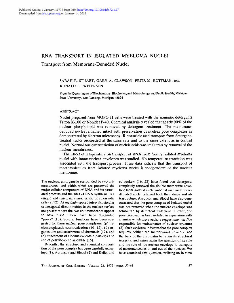
It has been established that structural features, e.g. nucleoli, dense lamina, and pore complexes, of nuclei in intact cells are retained in nuclei iso- lated in aqueous systems (8). Fig. 2 shows repre- sentative thin sections of MOPC-21 tissue culture cell nuclei prepared by Dounce homogenization, washed with transport buffer, and then treated with NP-40 or Triton X-100, or washed again with buffer. It can be seen that nuclei treated with low levels of either Triton X-100 (Fig. 2 C) or NP-40 (Fig. 2 B) do not show an electron-dense double unit membrane structure at the boundary of the nucleus. The buffer-washed nuclei, however, clearly have both the inner and outer membranes (Fig. 2 A). Although the morphology of deter- gent-treated nuclei is similar to that of control nuclei, the internal architecture has been altered, i.e., note condensed chromatin. However, this alteration is not manifested when comparing the
!o
8
6
4
2
lo Z u 8 ~ 6
8 6
~, ~ A 4S 18 S 28 S
, , i [
5 10 15 2O 25 FRACTION
FmURE 1 Sucrose-SDS gradient analysis of RNA re- leased from myeloma nuclei. (A) RNA released after a 30-min incubation of isolated nuclei without ATP (total cpm = 6.5 x 10a). (B) and (C) Sucrose-SDS gradient analysis of polysomal poly(A)+ RNA and total cytoplas- mic RNA from MOPC-21 cells, respectively. MOPC-21 cells were labeled for 30 rain with [3H]uridine, washed, resuspended in fresh medium without radioactive uri- dine, and incubated for an additional 30 min. The cells were washed and lysed as described in Materials and Methods. After removal of nuclei, 0.1 vol of 5% (vol/ vol) solution of Trition X-100 and deoxycholate was added to the postnuclear supernate. Polysomes were purified by pelleting and RNA was isolated by phenol- chloroform extraction. (B) Poly(A)+ polysomal RNA isolated by oligo (dT) cellulose chromatography (total cpm = 3.5 x 10a). (C) Purified total cytoplasmic RNA from MOPC-21 cells (total cpm = 1.1 x 10⁵).
restriction and release of macromolecules (see be- low), but may reflect changes in the nucleoplasmic ionic environment due to removal of the nuclear membranes. Fig. 3 shows a higher magnification of thin sections of the periphery of control and detergent-treated nuclei after a 20-min incubation in vitro. Comparison of these micrographs shows complete morphological absence of the nuclear envelope in nuclei treated with either Triton X- 100 or NP-40. The unit membrane structures of the nuclear envelope were intact in the control nuclei. Pore complexes were seen at the periphery of both control and detergent-treated nuclei. Iden- tical results were obtained when samples were fixed in glutaraldehyde and postfixed with osmium tetroxide.
60 THE JOURNAL OF CELL BXOLOGY" VOLUME 72, 1977
FI6URE 2 Thin sections through representative nuclei: (A) prepared by Dounce homogenization in hypotonic buffer and washed in transport buffer x 18,500; (B) treated with 0.5% NP-40 x 14,400; (C) treated with 0.5% Triton X-100 x 18,500 (see Materials and Methods). Bars, 1 /~m. Nucleolus, nu.
Chemical Composition o f Control and
Detergent- Treated Nuclei
Although evidence has been presented previ- ously that Triton X-100 causes a loss of phospho- lipid from isolated nuclei, and by extension, mem- brane structure (1, 2), we felt that this should be directly demonstrated by phospholipid phospho-
rus analysis of myeloma nuclei. Table II shows the effect of detergent on the composition of MOPC- 21 nuclei. Treatment of isolated myeloma nuclei with either NP-40 or Triton X-100 resulted in loss of almost 90% nuclear phospholipid to the wash. Control nuclei, washed with buffer, however, re- tained 70% nuclear phospholipid.
A relatively high proportion of nuclear lipid was
STUART ET AL. RNA Transport in Isolated Myeloma Nuclei 61

revealed very little cytoplasmic contamination of nuclei prepared by Dounce homogenization, it is possible that parts of the outer nuclear membrane are stripped by repeated buffer washes.
Analysis of nuclear membrane phospholipid was based on phosphorus content of a total lipid extract of whole nuclei. To insure that the extracts for phospholipid analysis were not contaminated with phosphorus from nucleic acids, cells were prelabeled with [3H]thymidine and each stage of lipid extraction was monitored by liquid scintilla- tion spectrometry. Less than 3.5% of the total input nuclear radioactivity appeared in the first chloroform: methanol extract, and this quantity was slightly reduced by subsequent KCI washes of this extract. If uniform labeling of DNA is as- sumed, a maximum of 0.01 t~g of phosphorus could be accounted for by contamination of the lipid extract with nucleic acids.
Transport from Membrane-Denuded
MOPC-21 Nuclei
The transport of RNA from isolated nuclei pre- pared by Dounce homogenization, NP-40, or Tri- ton X-100 treatment was examined. Conditions employed were optimum for the release of RNA in this system while maintaining the normal physi- ological in vivo nuclear restriction of macromole- cules. If the nuclear envelope regulates or modu- lates transport of RNA from the nucleus to the
FIGURE 3 Thin sections through representative nuclei after 20 rain at 37~ in transport buffer: (A) washed with 0.5% Triton X-100 x 96,000; (B) washed with 0.5% NP-40 x 76,200; (C) prepared by Dounce homogeniza- tion and washed with buffer x 60,000. Bars, 0.1 /,~m. Inner nuclear membrane, im; outer nuclear membrane, o m .
lost to the buffer wash in the control nuclei as compared to other studies (1, 20). This may be due to our purification procedures which did not include centrifugation of the nuclear pellet through heavy sucrose. Since electron microscopy
TABLE II
Effect of Detergent on Composition of MOPC-21 Nuclei
Phospholipid/ Phospholipid 10 6 nuclei
% gg
Control Supernate 29.3 1.60 Pellet 70.7 3.86 Total 100.0 5.46*
Triton X-100 (0.5% vol/vol) Supernate 86.3 5.65 Pellet 13.7 0.90 Total 100.0 6.55
Nonidet P-40 (0.5 % vol/vol) Supernate 88.7 5.46 Pellet 11.3 0.70 Total 100.0 6.16
* Sum of supemate and pellet values.
62 THE JOURNAL OF CELL BIOLOGY' VOLUME 72, 1977

cytoplasm, some alteration in the efficacy of release in membrane-denuded nuclei should be observed. As can be seen from Fig. 4, MOPC-21 nuclei treated with either detergent released R N A to the surrounding medium at the same rate and to the same extent as control nuclei. Further, when detergent-treated nuclei were incubated in reac- tion mixtures containing 8 mM Mg §247 conditions nonpermissive for release in control nuclei, no R N A was released. Fig. 5 shows the ratio of released R N A to D N A ( R N A / D N A ) with time. Nuclei treated with either NP-40 or Triton X-100 selectively released R N A (ratio increased) as did control nuclei which had both the inner and outer membranes intact. Control and detergent-treated nuclei incubated in 8 mM Mg §247 did not release R N A (Fig. 4) or D N A (Fig. 5, ratio remained unchanged). In these reactions, both control and experimental, the percentage of total radioactive D N A released ranged from 0.6 to 1 .7%. This D N A release and the parallel release of very low levels (1 .2-1 .4%) of R N A from nuclei incubated
12
11
10
9
8
" 7
~d ~e 3
2
!
o j - - - - - - o
t ~ _ ~ i = = = ~ _ z - I
0 .5 I0 20 30 TIME
FIGURE 4 Release of prelabeled [aH]RNA from iso- lated MOPC-21 nuclei treated with buffer, [] []; 0.5% Triton X-100, O O; 0.5% NP-40, A A in 3 mM Mg ++. Control reactions contained 8 mM Mg ++, buffer-washed nuclei �9 II; NP-40-treated nuclei, �9 � 9 and Triton X-lOO-treated nuclei, --" 11. TCA-precipitable RNA released to the postnuclear su- pemate was analyzed as described in Materials and Methods for each time sample. Each reaction contained approximately 4 x 10 s cpm TCA-precipitable RNA.
< 3 Z
Im
a
o
1
~ l l l I
5 I0 20 3O TIME
FIGURE 5 Ratio of released [aH]RNA to released [~C]DNA. MOPC-21 cells at 3 x I05 ceUs/ml were labeled for 18 h with [2-14C]thymidine and then pulsed for 30 rain with [5,6-3H]uridine immediately before iso- lation of nuclei as described in Materials and Methods. Ratio of release of TCA-precipitable [ZH]RNA to []4C]DNA in incubation mixtures containing 3 mM Mg ++ is shown for control nuclei, [] D; Triton X- 100-treated nuclei, O O; and NP-40-treated nuclei, A A. The RNA/DNA ratio in 8 mM Mg ++ is also shown for buffer-washed nuclei, ~- II; Triton X-100; O------O; and NP-40, �9 � 9 In these reactions, both control and experimental, the percentage of total radio- active DNA released to the supernate ranged from 0.6 to 1.7. At 30 min, 8-10% of the total precipitable RNA had been released to the supernate in each of the experi- mental reactions.
in 8 mM Mg §247 probably reflect background lysis of fragile nuclei.
The effect of temperature on the release of R N A from myeloma nuclei with intact nuclear envelopes was studied to determine whether the transport process was regulated or modulated by a membrane-associated activity. The effect of tem- perature on the rate of an enzyme-catalyzed reac- tion can frequently be predicted by the Arrhenius equation which relates the velocity constant of the process with the absolute temperature. Further, when membrane functions are analyzed, these predictions are not followed, in that plotting the logarithm of the velocity of the reaction vs. the reciprocal of the absolute temperature results in a line with a discontinuous slope. The "break" oc- curs at a specific temperature. Above this charac-
STUART ET AL. RNA Transport in Isolated Myeloma Nuclei 63

teristic temperature, the energy of activation (E) for the process is decreased, and this change in E can be correlated with lateral phase changes in the membrane (20, 21). When this method of analysis was applied to the transport of RNA from isolated nuclei between 10 and 37~ no such "break", or transition, was observed (data not shown). Rather, the energy of activation remained con- stant over the entire range of temperatures tested. The calculated Arrhenius energy of activation for the transport of RNA was Ea = 19.9 kcal/mol.
DISCUSSION
Regulation of nucleocytoplasmic communication is one of the most important functions of eukar- yotic cells. We have monitored a number of pa- rameters to ascertain the validity of using isolated myeloma nuclei to study selective macromolecular transport from the nuclear space. Under appropri- ate conditions, i.e. in the absence of added ATP and in the presence of cytosol, myeloma nuclei selectively release RNA (25). The released RNA is heterogeneous in size and a significant propor- tion is polyadenylated. Additional data suggest that in vitro release of RNA reflects both quantita- tively and qualitatively the transport of nuclear RNA in intact MOPC-21 cells.
Since the ultrastructure of the nuclear envelope in eukaryote cells as diverse as amphibian oocytes, amoebae, and rat liver cells is highly conserved, this membrane barrier with its associated pore complexes appears to be a likely site for regulation of nucleocytoplasmic exchange. The pore com- plexes are a consistent feature of nuclear enve- lopes (9, 12). These structures are annular when viewed in cross section and as shown by Aaronson and Blobel (2) remain intact, as does the nucleus itself, after removal of the nuclear envelope by detergent treatment. Hildebrand et al. (14) and Berezney and Coffey (5, 6) have shown that a residual nuclear structure remains after detergent removal of the membrane envelope, digestion of the RNA and DNA, and 2 M NaCl extraction of mammalian cell nuclei. This protein matrix retains the shape and size of the nucleus and can be visualized by electron microscopy.
Evidence from several different systems has suggested that the nuclear pores are the sites of nucleocytoplasmic transfer of macromolecules (10, 13, 15). Further, restricted diffusion through the pores as defined and described by Paine et al. (19) could account for different rates of solute
transport from cytoplasm to nucleus simply by a small alteration in the radii of the pores. In addi- tion, our data indirectly support the conclusion that the nuclear pore complexes are important in nueleocytoplasmic exchange since transport of RNA was not dependent on the presence of the nuclear envelope.
Our studies with intact freshly isolated MOPC- 21 nuclei indicate that treatment with either NP- 40 or Triton X-100 at concentrations employed for nuclear preparation from animal tissue (23) removes the nuclear envelope completely. No unit membrane structure was visible at the periphery of the nuclei and nearly 90% phospholipid was re- moved from the detergent-treated nuclei. The de- tergent-treated nuclei were intact and the normal morphology, including pore complexes, was ob- served.
Transport studies in membrane-denuded nuclei have shown that RNA was released with the same efficacy from detergent-treated nuclei as from control nuclei with intact nuclear envelopes. Fur- ther, the pattern of release was identical and the normal in vivo restriction of macromolecules was conserved in both control and detergent-treated nuclei. In retrospect, evidence for this was sup- plied by Chatterjee and Weissbach (7) in a study of transport of RNA from isolated HeLa cell nu- clei prepared by detergent treatment and frozen before use. These authors found that nuclei pre- pared by detergent wash released RNA and pro- tein concomitantly, but did not release DNA. No comparison with transport from nuclei with intact nuclear envelopes was made, however.
In nuclei with both membranes intact, no change in Arrhenius activation energy was associ- ated over the range of temperatures of incubation. The Arrhenius activation energy, E, calculated from this equation is a constant. In contrast, when membrane-associated functions are examined, the predictions of the Arrhenius equation may not be followed (i.e., a change in E results in a line with a discontinuous slope at a characteristic temperature [20, 21]). In many systems a phase change has been demonstrated in the lipid component of the membrane at, or close to, the same temperature as that of the change in activation energy of the associated enzyme system (20). In particular, a number of bacterial transport systems have been examined in this way, yielding good correlation between the characteristic temperature of the change in energy of activation for the transport of
64 THE JOURNAL OF CELL BIOLOGy-VOLUME 72, 1977

a given molecule and the lateral phase change(s) of the m e m b r a n e (17, 20).
One should point ou t tha t these studies do not rule out the possibility that release of R N A from m e m b r a n e - d e n u d e d nuclei is due to the break- down of nuclear RN P structural e lements . This appears unlikely, however , since normal nuclear restriction ( D N A not released) and the extent and kinetics of R N A release are comparable in intact and m e m b r a n e - d e n u d e d myeloma nuclei.
I t would appear tha t the nuclear m e m b r a n e s are not directly involved in the regulat ion and /or mod- ulation of the t ranspor t of R N A from the nucleus in myeloma cells. Since the pore complex itself seems to be an intrinsic par t of the residual pro te in matrix of the nucleus, it may well be the site of regulation of nucleocytoplasmic communica t ion .
This is journal article 7592 from the Michigan Agricul- tural Experiment Station.
We wish to thank Dr. Gary Hooper, Director of the Electron Optics Laboratory, Michigan State University, and Mr. H. Stuart Pankratz for assistance in interpreta- tion and preparation of micrographs.
This investigation was supported by the National Insti- tutes of Health grant CA13175 to Fritz M. Rottman and NIH grant AIl1493 to Ronald J. Patterson. Sarah E. Stuart is a postdoctoral fellow of NIH. Gary A. Clawson is supported by NIH grant GM01422 and by funding from the College of Osteopathic Medicine, MSU.
Received for publication 22 March 1976, and in revised form 9 September 1976.
R E F E R E N C E S
1. AARONSON, R. P., and G. BLOBEL. 1974. On the attachment of the nuclear pore complex. J. Cell Biol. 62:746-754.
2. AAXONSON, R. P., and G. BLOEEL. 1975. Isolation of the nuclear pore complexes in association with a lamina. Proc. Natl. Acad. Sci. U. S. A. 72:1007- 1011.
3. Avrv, H., and P. LEVER. 1972. Purification of biologically active globin messenger RNA by chro- matography on oligothymidylic acid cellulose. Proc. Natl. Acad. Sci. U. S. A. 69:1408-1412.
4. B~rLErr, G. R. 1959. Phosphorus assay in col- umn chromatography. J. Biol. Chem. 234:466-468.
5. BE~ZNEY, R., and D. S. CO~EY. 1974. Identifica- tion of a nuclear protein matrix. Biochem. Biophys. Res. Commun. 611:1410-1417.
6. BE~ZNEY, R., and D. S. Co~Y. 1975. Associa- tion of newly replicated DNA within the nuclear matrix. J. Cell Biol. 67:29 a. (Abstr.)
7. CuArrE~EE, N. K., and H. WEISSRACH. 1973.
Release of RNA from HeLa cell nuclei. Arch. BIO- chem. Biophys. 157:160-167.
8. DAWSON, P. F., and E. H. MEgIER. 1956. Electron microscopy of cell nuclei isolated in aqueous media. Exp. Cell Res. 11:237-239.
9. FAaEa6f~, A. C. 1974. The nuclear pore complex: Its free existence and an hypothesis as to its origin. Cell Tissue Res. 151:403-415.
10. FELDHERR, C. M. 1972. Structure and function of the nuclear envelope. II. Ultrastructure. Adv. Cell Mol. Biol. 2:273-307.
11. FOLCH, J., M. HuS, and G. H. SL6ANE STANLEY. 1957. A simple method for the isolation and purifi- cation of total lipids from animal tissues. 1. Biol. Chem. 226:497-509.
12. FRANKE, W. W. 1970. Universality of the nuclear pore complex structure. Z. Zellforsch. Mikrosk. Anat. 105:405-429.
13. FXANKE, W. W. 1974. Nuclear envelopes, structure and biochemistry of the nuclear envelope. Philos. Trans. R. Soc. Lond. B. Biol. Sci. 268:67-93.
14. HILDESRANO, C. E., R. T. OrdNXDA, and L. R. GURLEY. 1975. Existence of a residual nuclear pro- tein matrix in cultured Chinese hamster cells. J. Cell Biol. 67:169 a. (Abstr.)
15. JACOB, J., and G. A. DANmLLI. 1972. Electron microscope observations on nuclear pore-polysome association. Cell Differ. 1:119-125.
16. KELLER, J. M., and D. E. RILEY. 1976. Nuclear ghosts: A nonmembranous structural component of mammalian cell nuclei. Science (Wash. D. C.). 193:399-401.
17. LnqDEN, C. D., K. L. WgIC;HT, H. M. Mc- CONNELL, and C. F. Fox. 1973. Lateral phase separations in membrane lipids and the mechanisms of sugar transport in Escherichia coli. Proc. Natl. Acad. Sci. U. S. A. 70:2271-2275.
18. LowRY, O. H., N. J. ROSEBROU~H, A. C. FAgg, and R. J. RANOALL. 1951. Protein measurement with the Folin phenol reagent. J. Biol. Chem. 193:265-275.
19. PAINE, P. L., L. C. MOORE, and S. B. HoRowrrz. 1975. Nuclear envelope permeability. Nature (Lond.). 254:109-114.
20. RAISON, J. K., J. M. LYONS, and W. W. THOMSON. 1970. The influence of membranes on the tempera- ture-induced changes in the kinetics of some respira- tory enzymes of mitochondria. Arch. Biochem. BIO- phys. 142:83-90.
21. RAISON, J. K., J. M. LYONS, R. J. MELHORN, and A. D. KErra. 1971. Temperature-induced phase changes in mitochondrial membranes detected by spin labeling. J. Biol. Chem. 246:4036-4046.
22. RILEY, D. E., J. M. KELLER, and B. BYERS. 1975. The isolation and characterization of nuclear ghosts from cultured HeLa cells. Biochemistry. 14:3005- 3013.
STUART El' AL. RNA Transport in Isolated Myeloma Nuclei 65

23. SINGER, R. H., and S. PESMAN. 1973. Messenger RNA in HeLa cells: kinetics of formation and de- cay. J. Mol. Biol. 78:321-334.
24. SPu~, A. R. 1969. A low-viscosity epoxy resin embedding medium for electron microscopy. J. Ul-
trastruct. Res. 26:31-43. 25. STUART, S. E., F. M. ROTTMAN, and R. J. PArrEX-
SON. 1975. Nuclear restriction of nucleic acids in the presence of ATP. Biochem. Biophys. Res. Com- mun. 62:439-447.
66 THE JOURNAL OF CELL BIOLOGY" VOLUME 72, 1977