risk of occupational radiation-induced cataract in … · equivalent doses at working places and...
TRANSCRIPT
RISK OF OCCUPATIONAL RADIATION-INDUCED CATARACT
IN MEDICAL WORKERS
SNEZANA MILACIC1,*
Faculty of Medicine, University of Belgrade, Institute of Occupational Medicine and
Radiological Protection; Belgrade, Serbia1
*Author and address for correspondence:
Snezana Milacic, MD
Faculty of Medicine
University of Belgrade
Institute of Occupational Medicine and Radiological Protection
Deligradska 29
11000 Belgrade
Serbia
Tel: + 381 11 3674 515
Fax: + 381 11 643 675
E-mail: [email protected]

ABSTRACT
Objective. The objective of this study was determination of criteria for recognition of a
pre senile cataract as a professional disease in healthcare personnel exposed to small
doses of ionizing radiation.
Method. The study included 3240 health workers in medical centers of Serbia in the
period 1992-2002. A total of 1560 workers were employed in the zone (group A) and
1680 out of ionizing radiation zone (group B). Among group A, two groups had been
selected:
1. Group A-1: Health workers in the ionizing radiation zone who contracted lens
cataract during their years of service while dosimetry could not reveal higher
absorbed dose (A-1=115);
2. Group A-2: Health workers in the ionizing radiation zone with higher incidence of
chromosomal aberrations and without cataract (A-2=100);
Results. More significant incidence of cataract was found in group A, χ²=65.92; p<0.01.
Radiation risk was higher in health workers in radiation zone than in others, relative risk
is 4, 6.
Elevated blood sugar level was found in higher percentage with health workers working
in radiation zone who developed cataract.
Conclusion. Low doses of radiation are not the cause of occupational cataract as
individual occupational disease. X-ray radiation may be a significant cofactor of cataract
in radiological technicians.
Key words: Low doses ionizing radiation, occupational cataract.
INTRODUCTION
The cataract is the most common degenerative opacity of the crystalline lens, developing
with aging.
The interior capsule of the lens is lined with transparent layer of epithelial cells, which
maintains the function of lens, what is accomplished by cell division at the equator of
crystalline lens and moderate growth towards the center. Multiple factor may interfere
with division process and growth of lenticular epithelial cells, especially in case of
genetic predisposition (1-3).
Damaged cells are moved towards the posterior pole of the crystalline lens, forming
irregularly the young lenticular fibers which lose their transparency and prevent straight
light movement, what is manifested by opacification in the posterior subcapsular region
(4-7).
Risk evaluation of presenile cataract revealed that it was in relation to phenotype
produced by the expression of several different genes and exposure to environmental
noxae (8, 9).
Physical agents causing the cataract may be as follows: infrared, ultraviolet and
microwave non-ionizing radiation, electric power and ionizing radiation (6).
Exposure to sunlight may be a risk factor of cataract development. Exposure to the
ultraviolet-B (UVB) component of light may be associated with the severity of cortical
opacities in men, 43 to 84 years of age (2, 4, and 7).
The X-rays are the largest threat to crystalline lens. Mostly higher doses, over 500 mSv
caused the cataract, and only in 15% of cases the doses were low, below 100 mSv (10).
Other risk factors of cataract are the following: alcohol abuse, tobacco smoke, systemic
diseases (endocrine and metabolic), impaired eye circulation, ocular pressure, refractory
eye abnormalities (myopia), blood pressure, heart conditions (3, 5, 9), low food
antioxidant level. Antioxidative prophylaxis such as intake of vitamins C and E over 10
years and more, had the impact on lowering the risk of cataract (11).
The symptoms of cataract onset are slow, progressive and painless impairment of vision,
sensitivity to light, more defective color perception and diplopia. All these directly
diminish the working and living ability, and given it may be of occupational origin, the
analysis of risks of cataract and possibilities of its detection and treatment are significant
for rights protection workers and their return to work places.
The workers with manifested cataract within and beyond the area of ionizing radiation
were analyzed and compared with the intent to evaluate the effect of low doses to
development of cataract.
METHODOLOGY
The study involved 3240 health workers of Medical Centers in Serbia in the period 1992-
2002, who used to work within and beyond the area of ionizing radiation.
The annual periodical-preventive controls of health workers included the eye examination
as well. After visual acuity measurement, the lens was examined by retroillumination
method (red reflex) and using the biomicroscope. In case of impaired visual acuity or
presence of cloudy lens, the examination was performed in mydriasis.
The factors causing the cataract of the crystalline lens were analyzed: age, sex,
occupation, duration of occupational exposure, metabolic disorders (glycemia),

cardiovascular diseases (high blood pressure and heart conditions – arrhythmia), etc. Data
on smoking, alcohol consumption and corticosteroid therapy as well as exposure to
chemical and other physical noxae, genetic burden, were obtained from their medical
histories entered in periodical control charts. A total of 1560 (Group A) health workers of
different occupation, age and sex were employed in the ionizing radiation zone, and 1680
(Group B) out of the zone. The percentage of smokers was nearly equal in both groups
(31% and 33%). Alcoholism, toxins and medicaments were excluded as well as
occupation exposition to other radiation. Food regime, i.e., antioxidants in food, obesity,
genetic factor and sun-exposure varied equally in both groups.
Two subgroups were singled out from the Group A:
1. Group A-1 of exposed 115 workers with cataract chronically exposed to low
doses of radiation, mean age of 46 years, both genders, i.e. 56 females and 59
males –; and
2. Group A-2 of exposed 100 workers without cataract, selected on the basis of
higher incidence of chromosomal aberrations due to chronic exposure to low
doses, mean age 41 years, sexes, 54 females and 46 males –.
The Group B consisted of health workers with different occupations and employed at
different working places within public health institutions, which were not exposed to
ionizing radiation. The subjects with cataract were singled out from this group as a
controls (Group B-C), n=26, mean age 40 years, genders, 7 females and 19 males.
By vocation, the subjects from groups B-C and A-2 were: radiographers, radiologists,
pneumo-phthisiologists, nuclear medicine workers, dental radiographers, nurses,

anesthesiologists and other medical technicians who were, within their job description,
present in the ionizing radiation area. They are exposed to X-ray radiation (in X-ray
diagnostics, radiotherapy, and interventional radiology) and gamma and radiation (in
nuclear medicine).
The controls B-C consisted of physicians, medical technicians, nurses and lab technicians
at working places beyond the ionizing radiation area.
Absorbed dose on body surface of the exposed subjects in groups A-1 and A-2 was
measured by thermoluminescent personal dosimeters (TPD) worn during the entire shift
in the radiation zone, below the protective gown and they were read on monthly basis.
Doses were expressed as annual or 5-year equivalent to absorbed dose, in millisievert
(mSv). Equivalent doses at working places and exposition dose in the air were measured
as well.
Chromosomal aberrations in lymphocytes were analyzed. Chromosomal aberrations are
useful for evaluation of radiation risk in occupation exposure to ionizing radiation.
Dicentric form of chromosome is a specific, radiation-induced change of DNA. The
presentation of chromosomal aberrations (dicentrics) indicates higher exposition to lower
doses. Higher frequency of dicentric suggests higher radiobiological risk of disease due
to ionizing radiation exposure at working place (complete biomarker). Dicentric
frequency was measured in 200 mitoses of lymphocytes sampled and prepared from
blood of subjects in groups A-1, A-2 and B-C.
Owing to exposure of all people to some kind of natural radiation, dicentrics are possible
even in occupationally non-exposed individuals, but such dicentric frequency is below
1%.
Statistical analysis
The study used methods of descriptive statistics: central tendency measures: arithmetical
mean (X), variability measures: variation interval (max-min) and standard deviation
(±SD); analytical statistics: to assess the significance of difference: Student’s t-test and χ2
test and methods of correlation significance: Spearman’s rank correlation coefficient.
The incidences (I) (I=rate/100,000/1 year) were compared and represented as relative
risk, RR.
RESULTS
Equivalent doses at working places of subjects in the ionizing radiation zones were low,
ranging from 0.5 μSv/h to 8 μSv/h, and 0.41 Bq/100cm2 to 330 Bq/100cm2 at working
surfaces, what was tolerated limit for occupationally exposed individuals; exposure dose
in the air was at the level of natural phon at all working places of subjects in Groups A
and B.
Absorbed doses in exposed subjects with cataract, measured by thermoluminescent
personal dosimeter (TPD) ranged from 2.64 (min.) to 48.10 (max), mean value: 7.58 ±
4.78 mSv/5 years (1.59 ± 30 mSv/year), and were not different from mean absorbed
annual doses in the exposed subjects without cataract (1.63 ± 1.45mSv/1g), being below
maximally tolerated dose of 100 mSv/5 years 920-50 mSv annually).
In Group A-1, there was a total of 100 dicentrics in 23.000 mitoses (115 subjects x 200
lymphocytes); frequency of chromosomal aberrations f = 0,43% → f< 1%.
In group A-2, the frequency of chromosomal aberrations was 1.5% → f > 1% (300
dicentrics per 20.000 mitoses; fmin 0.5% - fmax 2,5%).
Dicentric type aberrations were not found in Group B-C.
Radiation risk was significant in Group EA-2 on account of highest frequency of
chromosomal aberrations.
There was a significant difference (χ2=65,92; p<0.01) of the crystalline lens cataract
between the studied groups employed in public health institutions, i.e. Group A in the
zone and Group B out of the ionizing radiation zone.
Cataract was 4-5 times more common in Group A; relative risk RR was 4.6
(IA/IB=700:150).
Mean age of subjects with cataract developed during the occupational ionizing radiation
exposure (Group A-1) was higher than the mean age of cataract patients who did not
work in the ionizing radiation zone (Group BC, Table 1) as well as exposed workers with
no developed cataract (Group A-2, Table 2), p ≤ 0.01.
Occupationally exposed health workers (Group A-1) who developed cataract had
approximately longer years of service in the ionizing radiation zone in comparison to
exposed workers of Group A-2 (Table 3). However, the correlation of cataract incidence
and duration of exposure was not linear: in 10 years of exposure, the incidence was
31,3%; 11-20 years – 31,3%; 20-30 years – 30,4% and the rest of 7% was found in older
workers with over 31 years of service.
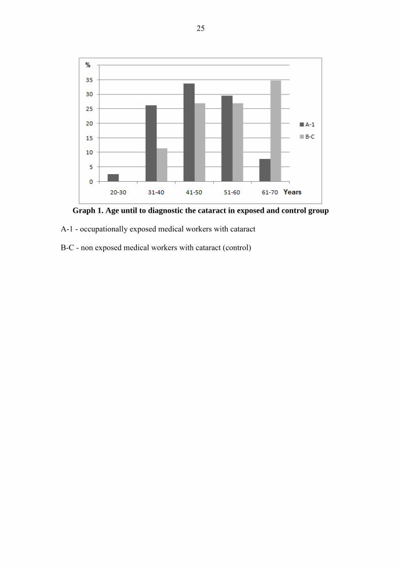
The cataract was most frequently diagnosed in the age 41 to 50 years in Group E, and
thereupon the incidence of cataract tended to decline. The lowest was in the age up to 31
years and over 60 years. In the control group B-C, cataract incidence correlated with the
age, covering the age period of 41 to 70 years (Graph 1). The incidence of cataract tended
to be higher with the age both in the exposed and control group, but such correlation was
not linear in the exposed group. The peak of the highest cataract incidence in workers in
the ionizing radiation was shifted by 10 years forward, to younger age in relation to the
controls (Graph 1).
Among cataract patients, the prevalence of males was higher. Nevertheless, the ratio of
genders and cataract incidence was significantly overturned in favor of female sex
(p<0,05), when they were exposed to ionizing radiation, i.e. in the Group A-1 (Table 4).
There was no difference of gender in the exposed workers with and without cataract
(Groups A-1 and A-2) (Table 5).
Significant was the difference in occupations (working places) of workers who developed
and did not develop cataract during their duration of occupation exposure (p < 0,05).
Higher percentage was found in workers with cataract exposed to x rays (radiographers,
radiologists and pneumo-phthisiologists), and the lowest percentage was verified in those
exposed to radionuclides in nuclear medicine (iodine and technetium), i.e. gamma and
beta rays (Table 6).
The frequency of chromosomal aberrations did not correlate with age and sex,
coefficients of correlation were approximately: 0,09 (p=0,47) and 0.01 (p=0,93),
respectively.
Equivalent doses measured by TLD did not correlate with age (p=0,09) and sex (p=0,29),
too.
Among cataract patients affected during the occupational exposition, radiological
technicians were the most prevalent (63,5%), while the physicians – radiologists and

pneumo-phthisiologists were the second and third, respectively. Time interval of
exposition when the cataract was primarily diagnosed in the majority of cases in
radiographers was 11-20 years (21% out of 63,5%, or 1/3 of the time), while all other
cases were diagnosed in the period 1 to 10 years and 21 to 40 years of service in the
ionizing radiation zone, and the least number was identified over 31 years (7%). In
addition, the smallest number of cataracts was recorded in younger and oldest age, which
is, only 1,7% up to 30 years and 2,6% in over 60 years of age, while the diseased
radiographers most commonly were 40-49 years of age.
Among doctors, distribution of contracting the disease by time exposure intervals was
approximately the same.
There was a significant difference in cataract morphology between the exposed group E
and control group B-C (p<0,01; χ2=12,55). In Group A-1, the incidence of cortical
cataract was 80% (n=92). In the controls B-C, with 53,8% of nuclear cataract (n=14), the
cortical one was prevalent in 46,2% (n=12).
The effects of other risk factors, such as glycemia, blood pressure and heart conditions,
were also analyzed.
Elevated blood sugar level was found in higher percentage with health workers, working
in radiation zone, who developed cataract (p≤0.01) (Table 8).
Neither cardiovascular diseases nor blood pressure differed in studied groups of workers
in radiation zone (A-1/A-2: χ²=1,14; p=0.28, and χ²=1,45; p=0,48, respectively) as well
as between the exposed and control group (A-1/B-C: χ²=1,39; p=0.24, and χ²=2,92;
p=0,23, respectively).
DISCUSSION
There have been diverse opinions on ionizing radiation dose sufficient to cause the
cataract (12, 13). In our subjects, absorbed doses measured by TPD were very low, far
from limit values for occupationally exposed individuals. However, many
ophthalmologists think that there is no harmless dose for the crystalline lens (4, 6, 14,
15). Cataract is a dose-related manifestation, but it may develop after perennial exposure
to low doses due to impaired metabolism and albumin denaturation as the result of
cumulative effects of radiotoxins, free radicals, exhausted antioxidative reserve and DNA
damage in the lens epithelial cells.
The initial dotted opacity in the posterior lens pole subsequently keeps on spreading, and
cumulative damage due to radiation exposure gradually involves the entire lens, resulting
in cataract and vision loss. Damaged epithelial cells are retained in the lens even after its
removal for lens replacement surgery; therefore, chromosomal aberrations can be
detected in these cells which otherwise cannot be found in peripheral blood lymphocytes
of these subjects, as indicator of earlier increased exposure. DNA changes in these
epithelial cells may be construed as stochastic, dose-non related effect of radiation (15,
16).
Regardless of low doses, they could directly or indirectly cause the lens opacification as
cumulative effect of long-term exposure to radiation (12-16).
The controls consisted of health workers out of radiation zone who developed cataract
after all, but in older age and different by morphology, probably due to effect of other
risk factors (9, 11, 14, 15).
It is not necessary for all workers at higher radiobiological risk (higher frequency of
chromosomal aberration – dicentrics) to get occupational disease. Development of
cataract as occupational disease depends upon several factors (10, 12, 13, 16).
The largest proportion of health workers without developing the cataract in spite of
higher frequency of chromosomal aberrations were those engaged in nuclear medicine,
and the lowest proportion was among the x-ray workers. On the contrary, among exposed
workers with developed cataract, the majority was employed in the x-ray room, i.e.,
radiological technicians, who generally have the largest work burden in the x-ray
radiation zone. Moreover, it is possible that low LET (Linear energy transfer) radiation
such as x-ray has higher penetrating power and longer range, and consequently, it causes
the crystalline lens damage much easier than high LET radiation, such as corpuscular (-
ray) radiation with shorter range but higher radiobiological effect on most sensitive DNA
material, especially in case of contamination which is likely when handling with
radionuclides in nuclear medicine.
Sex and age had no effect on chromosomal aberrations, but interfered with higher risk of
cataract. Longer life and years of service contribute to longer exposure to ionizing
radiation, as well as to other factors having the impact on oxidative damage of the
crystalline lens. In our subjects, risk of cataract depended upon the type of radiation (x),
occupation (radiographers), working place (x-ray room, radiology department), age,
duration of occupational exposure, gender, glycemia, etc. The majority of subjects got
sick between 40 and 50 years of age, and the least up to 30 and over 60 years. However,
in relation to duration of exposure, the highest incidence was noted between 11 and 20
years, but the cataract also developed with shorter duration of occupational exposure, in
younger age. It is known that hereditary cataract may develop even after the age of 20,
and senile cataract after the age of 60, while the age of 45 is quite adequate for
development of cataract due to environmental risk factors (13-15). Moreover,
approximately one-third of subjects with cataract in both groups (exposed and control)
were smokers.
Risk of cataract due to smoking is relative and related to smoking intensity and years of
smoking, although the effect of smoking on cortical cataract, which was more frequent in
the exposed workers, could not be verified (14).
Radiologists and technicians exposed to x-rays in interventional radiology were at highest
risk of occupational, radiation cataract if they spent more time in direct beam without
wearing protective glasses (12). The initial opacification in posterior subcapsular region
(PSC) was reported in even 37% of radiologists, while cataract after long latent period
was developed in 8% of the time, and upon retirement in the older age, even 20.6% of
cases (12, 13). Protection, which primarily implies reduction of radiation dose at working
places (17), is an imperative, because otherwise the cataract could be avoided even if it
were developed after termination of working life.
Most commonly, the initial localization of cataract was in posterior subcapsular region –
PCS, with gradual development as cortical (7, 17). In our subjects exposed to ionizing
radiation, the incidence of cortical cataract was significantly higher. Typical majority, i.e.
90%, of senile cataracts belong to other types of opacity. Different risks correspond to
different morphological types.
Diabetes enhances the risk of posterior subcapsular cataract, as well as ionizing radiation,
but it also has impact on other forms, cortical and mixed forms. Among other factors
contributing to cataract risk, higher glycemia level had impact on our subjects, too.
Higher risk of cortical cataract was described in women. In our study, sex ratio was in
favor of females, if they were employed in radiation zone. The level of blood pressure
had different influence to cataract development in males (5). Hypertension had no effect
on PSC cataract. Nevertheless, in our study, neither blood pressure nor heart conditions
had any effect to development of cataract.
The incidence of nuclear form of cataract was significantly lower in our subjects working
in the ionizing radiation zone. Nuclear cataract is commonly caused by non-occupational
exposure to various noxae, such as smoking, obesity, excessive exposure to sun light,
while long-term wearing of glasses and myopia increases the risk of mixed cataract.
CONCLUSION
It is evident that chronic exposure to low doses is not the cause of occupational radiation
cataract as an independent occupational disease. The effects of radiation to biological
DNA material failed to be proven on cataract development, it was rather related to type of
radioactive emission, mode and type of radiation, working burden and working
conditions as well as type of job.
Reference
1. Hennis A, Wu SY, Nemesure B, Leske MC. Risk Factors for Incident Cortical
and Posterior Subcapsular Lens Opacities in the Barbados Eye Studies. Arch
Ophthalmol. 2004; 122:525-530.
2. Cruickshanks KJ, Klein BE, Klein R. Ultraviolet light exposure and lens
opacities: the Beaver Dam Eye Study. Am J Public Health. 1992; 82(12): 1658–
1662.

3. West SK, Valmadrid CT. Epidemiology of risk factors for age-related cataract.
Surv Ophthalmol. 1995; 39:323-334.
4. Rosmini F, Stazi MA, Milton RC. A dose-response effect between a sunlight
index and age-related cataracts: Italian-American Cataract Study Group. Ann
Epidemiol. 1994;4: 266-270.
5. Schaumberg DA, Glynn RJ, Christen WG, et al. A prospective study of blood
pressure and risk of cataract in men. Ann Epidemiology. 2001; 11:104-110.
6. Dennis M. Marcus, Chris Sheils, Maribeth H. Johnson, Sandra B. McIntosh,
Diane B. Leibach, Albert Maguire, Judith Alexander,Chander N. Samy. External
Beam Irradiation of Subfoveal Choroidal Neovascularization Complicating Age-
Related Macular Degeneration. Arch Ophthalmol. 2001; 119:171-180.
7. Bochow TW, West SK, Azar A, Munoz B, Sommer A, Taylor HR. Ultraviolet
light exposure and risk of posterior subcapsular cataracts. Arch Ophthalmology
1989; 107: 369 - 372.
8. Shiels A, Hejtmancik FJ. Genetic Origins of Cataract. Arch Ophthalmology
2007;125:165-173.
9. Mukesh BN, Le A, Dimitrov PN, Ahmed S, Taylor HR, McCarty CA.
Development of Cataract and Associated Risk Factors. Arch Ophthalmology
2006; 124:79-85.
10. Meecham WJ, Char DH, Kroll S, Castro JR, Blakely EA. Anterior segment
complications after helium ion radiation therapy for uveal melanoma. Radiation
cataract. Arch Ophthalmol 1994; 112: 197 - 203.
11. Mares-Perlman JA, Lyle BJ, Klein R, Alicia I. Fisher AI; William E. Brady WE;
Gina M. VandenLangenberg GM, Trabulsi JN, Palta M. Vitamin Supplement Use
and Incident Cataracts in a Population-Based Study Arch Ophthalmology 2000;
118: 1556 - 1563.
12. Vano E, Gonzalez L, Beneytez F,Moreno F. Lens injuries induced by
occupational exposure in non-optimized interventional radiology laboratories. The
British Journal of Radiology 1998; 171(847):728-733.
13. Baruch SJ. Cataracts in retired actinide-exposed radiation workers. Radiation
Protection Dosimetry 2005; 113(1):123-125.
14. Lindblad BE, Håkansson N, Svensson H, Philipson B, Wolk A. Intensity of
Smoking and Smoking Cessation in Relation to Risk of Cataract Extraction: A
Prospective Study of Women. American Journal of Epidemiology 2005;
162(1):73-79.
15. Moore AT. Understanding the molecular genetics of congenital cataract may have
wider implications for age related cataract. Br J Ophthalmol. 2004; 88:2-3.
16. Worgul BV. Radiation cataract: a stochastic expression of genotoxic Damage. Eye
Radiation and Environmental Research Laboratory, Columbia University, New
York US; 1996:http://www.jinr.ru/~drrr /Timofeeff/conference/radbio/worgul.htm
17. WHO. (World Health Organization) Library Cataloguing in Publication Data,
Management of cataract in primary health care services- 2nd ed ISBN 92 4
154499 6, Geneva; 1996: 1-3.
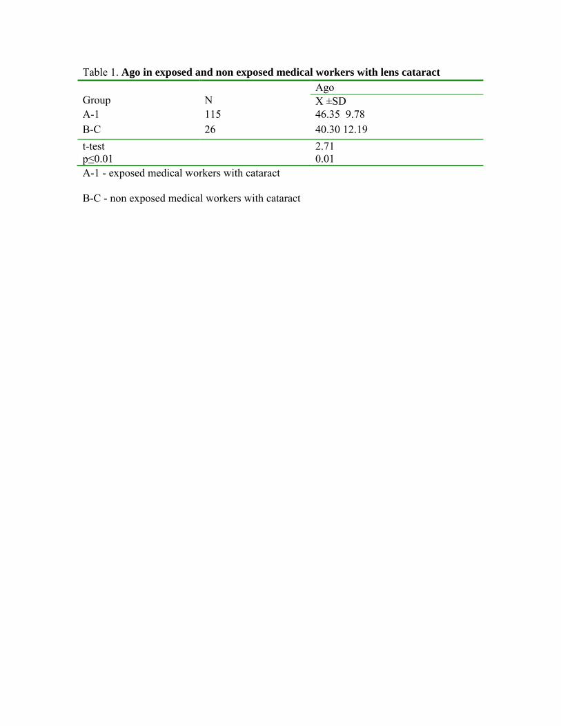
Table 1. Ago in exposed and non exposed medical workers with lens cataract
A-1 - exposed medical workers with cataract
B-C - non exposed medical workers with cataract
Group
N
Ago X ±SD
A-1 115 46.35 9.78 B-C 26 40.30 12.19 t-test p≤0.01
2.71 0.01
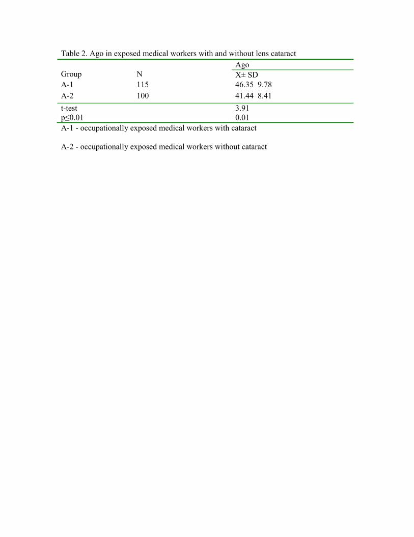
Table 2. Ago in exposed medical workers with and without lens cataract
A-1 - occupationally exposed medical workers with cataract
A-2 - occupationally exposed medical workers without cataract
Group
N
Ago X± SD
A-1 115 46.35 9.78 A-2 100 41.44 8.41 t-test p≤0.01
3.91 0.01

Table 3. Duration of occupationally exposure medical workers with
and without lens cataract
A-1 - occupationally exposed medical workers with cataract
A-2 - occupationally exposed medical workers without cataract
DOE - Duration of Occupationally Exposure
Group N DOE X±SD
A-1 115 16.86 9.59 A-2 100 14.22 8.37 t-test p<0.05
2.13 0.03
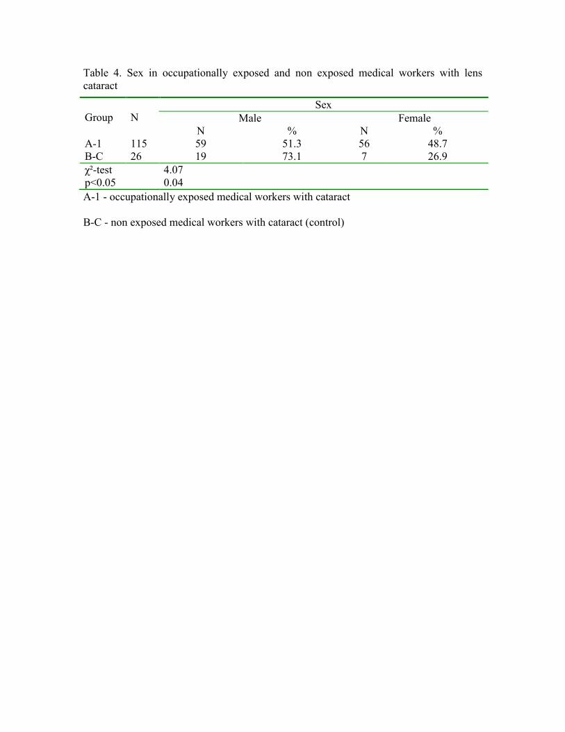
Table 4. Sex in occupationally exposed and non exposed medical workers with lens cataract
A-1 - occupationally exposed medical workers with cataract
B-C - non exposed medical workers with cataract (control)
Group
N
Sex Male Female
N % N % A-1 115 59 51.3 56 48.7 B-C 26 19 73.1 7 26.9 χ²-test p<0.05
4.07 0.04
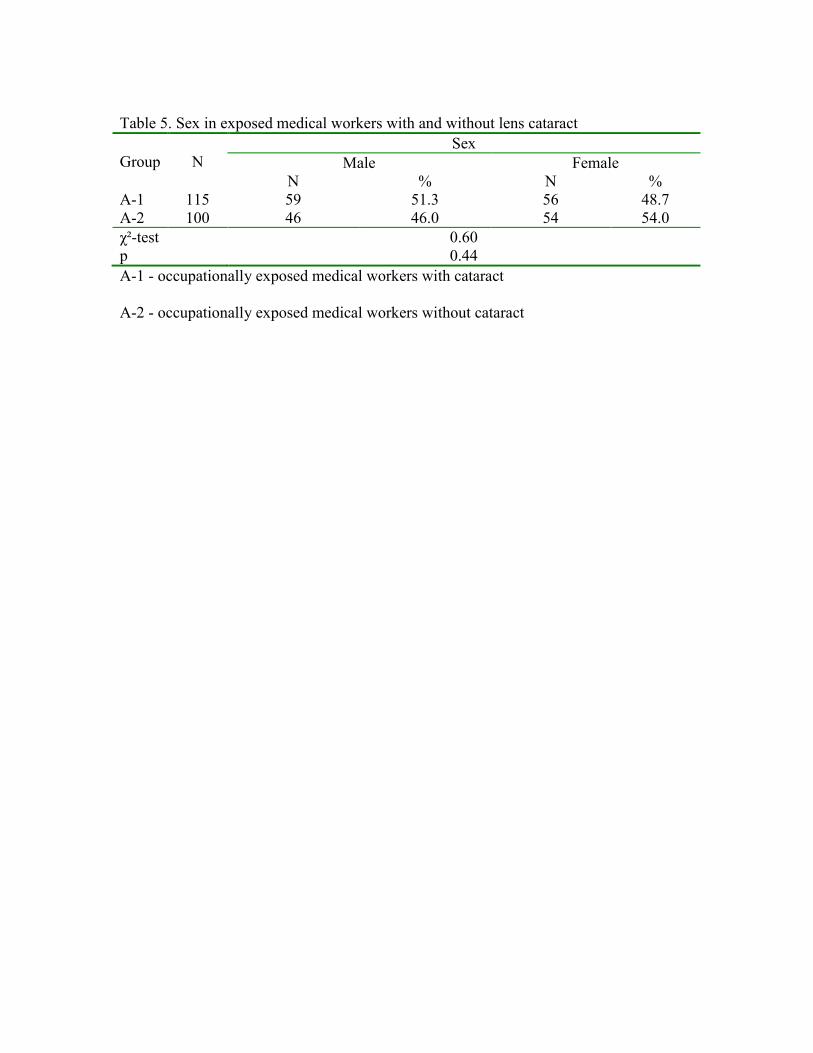
Table 5. Sex in exposed medical workers with and without lens cataract
Group
N
Sex Male Female
N % N % A-1 115 59 51.3 56 48.7 A-2 100 46 46.0 54 54.0 χ²-test p
0.60 0.44
A-1 - occupationally exposed medical workers with cataract
A-2 - occupationally exposed medical workers without cataract
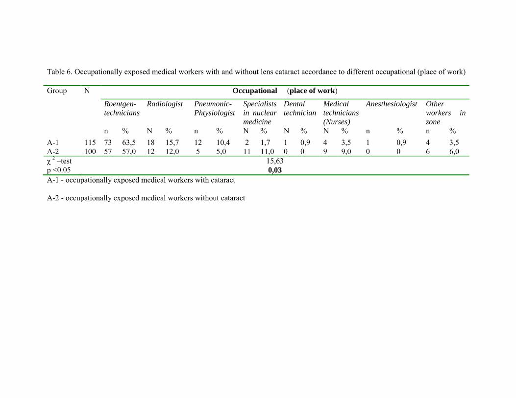
Table 6. Occupationally exposed medical workers with and without lens cataract accordance to different occupational (place of work)
Group N
Occupational (place of work)
Roentgen-
technicians Radiologist Pneumonic-
Phtysiologist
Specialists
in nuclear
medicine
Dental
technician Medical
technicians
(Nurses)
Anesthesiologist Other
workers in
zone n % N % n % N % N % N % n % n %
A-1 115 73 63,5 18 15,7 12 10,4 2 1,7 1 0,9 4 3,5 1 0,9 4 3,5 A-2 100 57 57,0 12 12,0 5 5,0 11 11,0 0 0 9 9,0 0 0 6 6,0 χ 2 –test p <0.05
15,63 0,03
A-1 - occupationally exposed medical workers with cataract
A-2 - occupationally exposed medical workers without cataract
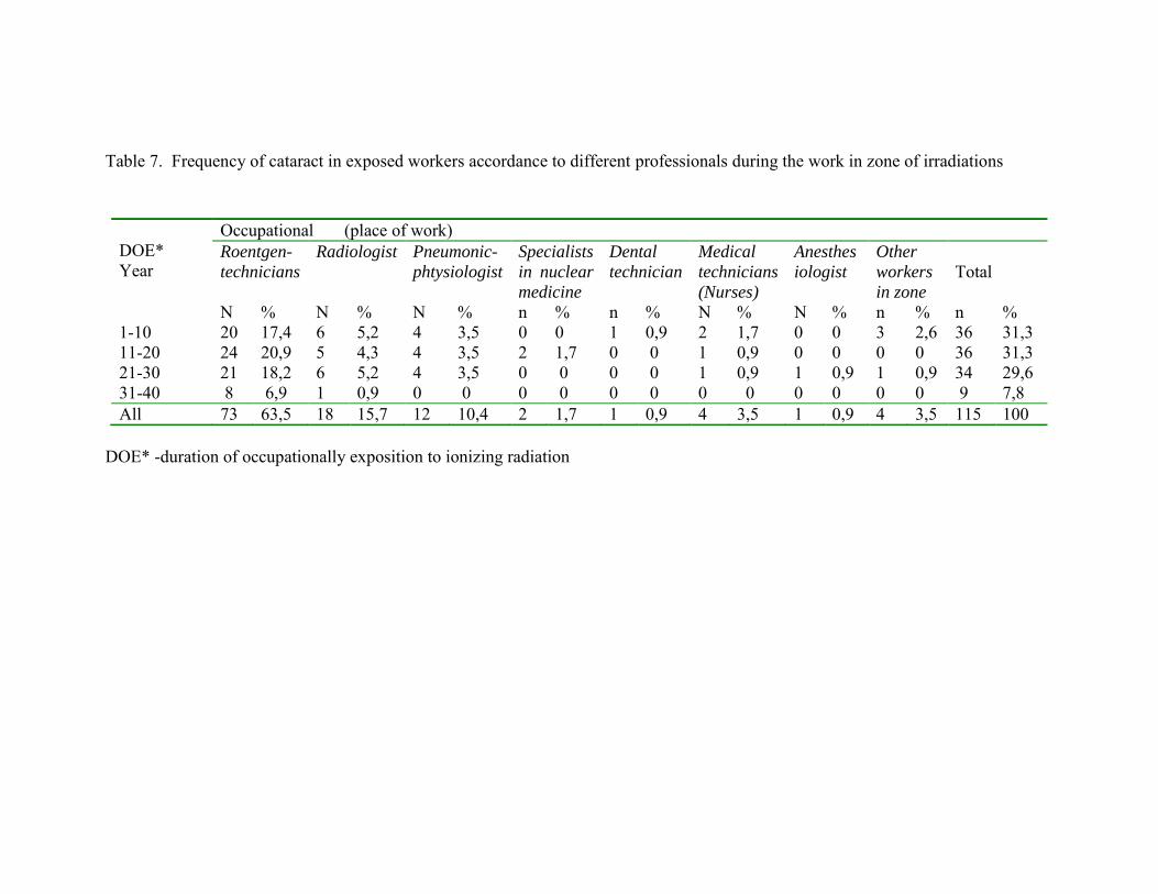
Table 7. Frequency of cataract in exposed workers accordance to different professionals during the work in zone of irradiations
DOE* -duration of occupationally exposition to ionizing radiation
DOE* Year
Occupational (place of work) Roentgen-
technicians Radiologist Pneumonic-
phtysiologist Specialists
in nuclear
medicine
Dental
technician Medical
technicians
(Nurses)
Anesthes
iologist
Other
workers
in zone
Total
N % N % N % n % n % N % N % n % n % 1-10 20 17,4 6 5,2 4 3,5 0 0 1 0,9 2 1,7 0 0 3 2,6 36 31,3 11-20 24 20,9 5 4,3 4 3,5 2 1,7 0 0 1 0,9 0 0 0 0 36 31,3 21-30 21 18,2 6 5,2 4 3,5 0 0 0 0 1 0,9 1 0,9 1 0,9 34 29,6 31-40 8 6,9 1 0,9 0 0 0 0 0 0 0 0 0 0 0 0 9 7,8 All 73 63,5 18 15,7 12 10,4 2 1,7 1 0,9 4 3,5 1 0,9 4 3,5 115 100
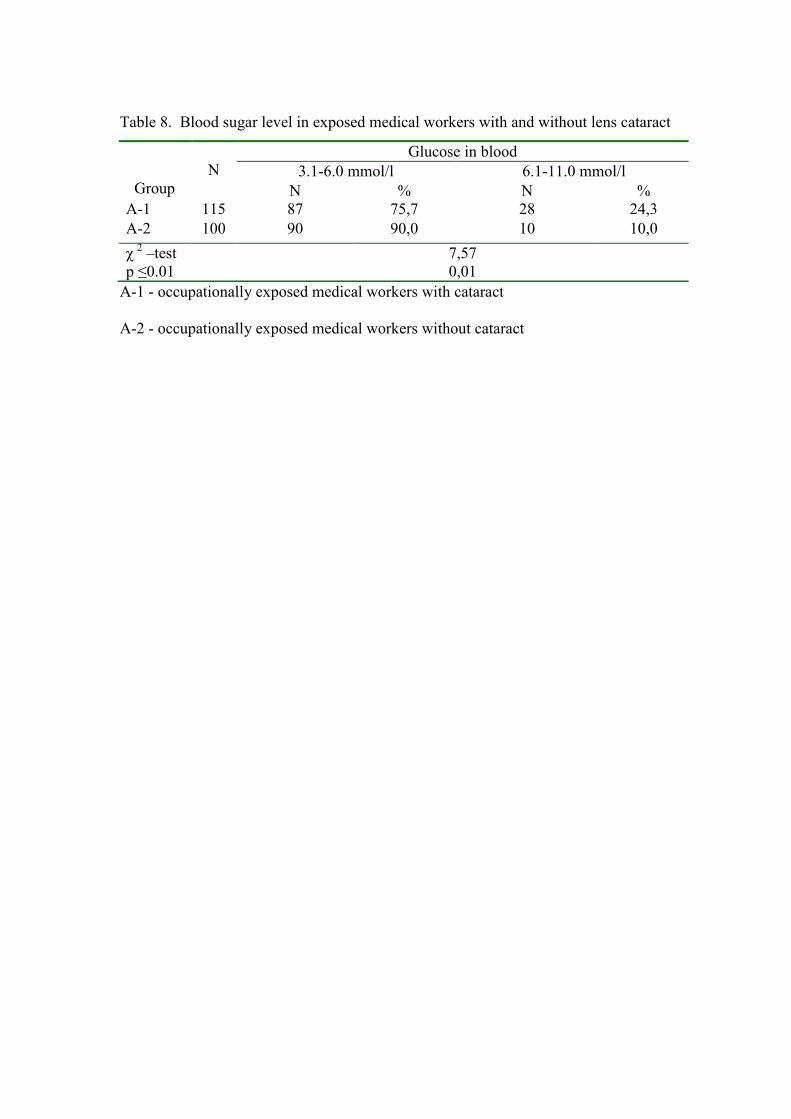
Table 8. Blood sugar level in exposed medical workers with and without lens cataract
A-1 - occupationally exposed medical workers with cataract
A-2 - occupationally exposed medical workers without cataract
Group
N
Glucose in blood 3.1-6.0 mmol/l 6.1-11.0 mmol/l
N % N % A-1 115 87 75,7 28 24,3 A-2 100 90 90,0 10 10,0 χ 2 –test p ≤0.01
7,57 0,01
25
Graph 1. Age until to diagnostic the cataract in exposed and control group
A-1 - occupationally exposed medical workers with cataract
B-C - non exposed medical workers with cataract (control)