ring testing of diagnostic protocols for identification ... · ring testing of diagnostic protocols...
TRANSCRIPT

EUPHRESCO Non-Competitive Project:
Ring testing of diagnostic protocols for identification and detection of
Gibberella circinata in pine seed.
Final Report
Project Coordinator:
Dr Renaud Ioos French Agency for Food, Environmental and Occupational Health and Safety (Anses) Plant Health Laboratory – Mycology Unit. Domaine de Pixérécourt, BP 90059, F54220 Malzéville, France Phone : +33 (0)3.83.29.00.80, Fax: +33 (0)3.83.33.29.00.02 Email: [email protected]
Topic Coordinator:
Dr Paul H.J.F. van den Boogert divisie Plant nieuwe Voedsel en Waren Autoriteit Address: 15, Geertjesweg, 6706 EA, Wageningen, The Netherlands PO Box: 9102, 6700 HC Wageningen, The Netherlands Phone: + 31 (0) 317 496694, Fax +31 (0) 317 421701 Email: [email protected]

TABLE
1. Background ......................................................................................................................................................... 5
1.1. Data on Gibberella circinata ......................................................................................................................... 5
1.2. Identification of the pathogen ..................................................................................................................... 5
1.3. Objectives of the project. ............................................................................................................................. 6
2. Project workplan ................................................................................................................................................. 9
3. List of the protocols to be ring-tested............................................................................................................... 11
4. Characteristics of the seed samples .................................................................................................................. 13
4. 1. Preparation of the samples ....................................................................................................................... 13
4.2. Size of the seed samples ............................................................................................................................ 13
4.2.1. Agreement on a sampling procedure. ................................................................................................. 13
4.2.2. Size of the samples for the project ring tests. ..................................................................................... 14
4.2.3. Number of samples and range of contamination levels ..................................................................... 15
4.2.4. Preparation of the samples ................................................................................................................. 16
4.2.5. Costs for seed samples production ..................................................................................................... 16
5. Results of the collaborative studies .................................................................................................................. 17
5.1. Protocol selection ...................................................................................................................................... 17
5.1.1. Result of the final vote ........................................................................................................................ 17
5.1.2. Description of the protocols retained. ................................................................................................ 18
5.1.3. Individual implication of the partners ................................................................................................. 19
5.2. Pre-trial results ........................................................................................................................................... 21
5.3. Main trials results ....................................................................................................................................... 22
5.3.1. Data recovery ...................................................................................................................................... 22
5.3.2. Treatment of the data ......................................................................................................................... 22

5.3.3. Performance criteria ........................................................................................................................... 23
5.3.4. Analytical sensitivity and specificity .................................................................................................... 25
5.4. Feedback from partners ............................................................................................................................. 29
5.5. Conclusion .................................................................................................................................................. 32
6. Results of the NPPO survey about the seed sampling strategy ........................................................................ 33
6.1. Setting of the statistical parameters to determine the sample size. ......................................................... 33
6.2. Results of EPPO country consultation ........................................................................................................ 34
6.3. Proposal for a harmonized sample size for pine seeds .............................................................................. 36
7. Conclusions of the Gibcir diagseed project ....................................................................................................... 38
References ............................................................................................................................................................ 41


1. BACKGROUND
1.1. DATA ON GIBBERELLA CIRCINATA
Gibberella circinata (anamorphic stage Fusarium circinatum) is the causal agent of pine pitch canker. The disease almost exclusively affects Pinus species, but was also described to occur on Douglas-fir (Pseudotsuga menziesii). This disease is a serious threat to the pine forests, due to extensive tree mortality, reduced growth and timber quality. Multiple branch infection may cause severe crown dieback and eventually lead to the death of the tree. This aggressive fungus may also cryptically infect the Pinus seeds and may cause damping-off in seedlings.
The fungus is officially reported in the USA, Mexico, Haiti, South Africa, Japan, Chile (Anonymous 2005) and it has recently been reported in the EPPO region. In Spain and France G. circinata is under eradication and in Italy the pest organism has been eradicated. The pathogen is subject to EC emergency measures and there are requirements for MS to conduct surveys. In nurseries there had been findings in Spain, Portugal and France; it has been found in forests, parks and gardens in Spain.
EFSA has recently presented their opinion, pest risk assessment and evaluation of risk management options. The conclusions were that protection was needed against imports which posed a risk and that requirements should be defined for the movement of seed, living plants, wood, soil, used machinery and vehicles from infested areas in the EU. Parts of Portugal, Spain, France, Italy and Greece were the areas mainly at risk, based on climatic data and host distribution, but other areas are may also be at risk.
Based on this report, seed is likely to the most significant pathway to spread the disease.
1.2. IDENTIFICATION OF THE PATHOGEN
There are several molecular methods available to confirm the identity of the anamorphic stage of G. circinata in pure culture, and to identify & detect the pest organism in planta. The methods that have been described in the EPPO diagnostic protocol PM 7/91(1) (Anonymous 2009) include, plating techniques followed by morphological identification in pure culture, a PCR-RFLP (Restriction Fragment Length Polymorphism) test for pure culture identification, real-time PCR’s and conventional PCR tests for direct in planta detection.

An ISTA protocol was published in 2002 to detect F. moniliforme f. sp. subglutinans (former taxonomic name for F. circinatum) in seeds of Pinus taeda and P. elliotii (International Seed Testing Association 2002), but the recent EPPO diagnostic protocol PM 7/91(1) discourages its use, owing to potential specificity issues.
Except the real-time PCR method (Ioos, Fourrier et al. 2009), the conventional PCR test developed by Ramsfield et al. (2008) and the ISTA protocol (International Seed Testing Association 2002), the different methods available are not accompanied with validation data.
1.3. OBJECTIVES OF THE PROJECT.
The first aim of this project is to ring test available and widely used detection methods and to provide validation and performance data for each of them. The validation data provided by this project will be useful to help the reference laboratories and mandated diagnostic laboratories to chose and implement efficient pine seed testing regarding this pathogen.
The second aim is to provide an agreement about the sample sizes of pine seeds for testing.
All the pine seed samples used for this project will be artificially contaminated with known quantities of the target pathogen. Preliminary tests should ensure the homogeneity of the different samples produced for the different levels of infestations and the different sample sizes. Preliminary investigation will be needed to explore the ISPM N°31 standard (International Plant Protection Convention 2008), and to determine which range of sample size should be assessed during this collaborative research project.
Policy, Science and Operational needs:
Research is needed to address the following objectives:
• Provision of harmonized sampling methods for pine seed
• Provision of validated detection tools for use by inspection services

The applications should address the following areas of work
• Sampling of seeds: Evaluation, optimisation and validation and comparative evaluation of sampling protocols in use at the various laboratories of inspection services and according to or based on ISTA guidelines, ISPM N°31, and EPPO protocol PM 7/91(1).
• Method validation: inventory of detection and identification methods in use at the various laboratories; method validation of selected methods for routine investigations in seeds. The method validation includes the analytical specificity, sensitivity, selectivity, reproducibility and repeatability performance characteristics.
• Ring testing: Performance of a ring test using the selected methods on seed samples spiked with known levels of infection, including positive and negative controls.
Specific outputs of the project:
• Production of a statistically-valid sampling protocol; validated detection protocols in seeds
• Demonstration of the usability of sampling and detection tools to inspection services and diagnostic laboratories
Beneficiaries of this research product
• Inspection services and mandated diagnostic laboratories of the National Plant Protection Organisations. Also EPPO and EFSA and the seed production and seed trade industries in and outside EU may benefit from the project results.
• Collaborations involving scientists where the pathogen occurs is encouraged where this adds value to the project in the European context.
Project participants
A total of 11 laboratories representing 10 countries signed up to the project through their local EUPHRESCO representatives (Table 1). Other laboratories have expressed their willingness to participate but finally withdrew from the project because of the lack of appropriate quarantine facilities for the containment and handling of a quarantine airborne fungus.

Table 1: List of the partner laboratories involved in the project.
Belgium (Flanders): Anne Chandelier [[email protected]] Walloon Agricultural Research Centre (CRAW) Department of Life Science - Marchal Building Rue de Liroux, 4 B-5030 Gembloux
Portugal : Eugénio Luís de Fraga Diogo [[email protected]] Instituto Nacional de Recursos Biológicos, IP / L-INIA, Unidade de Investigação de Protecção de Plantas (UIPP), Laboratório de Micologia Edificio 1 – Tapada da Ajuda 1349 - 018 Lisboa
France: Céline fourrier / Renaud Ioos [[email protected]] Anses Laboratoire de la Santé des Végétaux - Unité de Mycologie Domaine de Pixérécourt, BP 90059, F54220 Malzéville
Belgium (Wallonia): Sven Inghelbrecht [[email protected]] Institute for Agricultural and Fisheries Research Plant Sciences Unit - Crop protection Burg. van Gansberghelaan 96 bus 2, 9820 Merelbeke
Ireland : Choiseul, James [[email protected]] Department of Agriculture, Fisheries and Food DAFF Laboratory Complex, Backweston, Celbridge, Co. Kildare
Italy : Luca Riccioni and Tiziana Annesi [[email protected]] Consiglio per la Ricerca e la Sperimentazione in Agricoltura. Centro di Ricerca per la Patologia Vegetale (CRA-PAV). Via C.G. Bertero 22, I-00156 Rome
UK: Victoria Barton / Ann Barnes [[email protected]] The Food and Environment Research Agency 04GA08/09, Sand Hutton Y041 1LZ
Spain : Ana Mª Pérez Sierra [[email protected]] Grupo de Investigación en Hongos Fitopatógenos Instituto Agroforestal Mediterráneo Universidad Politécnica de Valencia Camino de Vera s/n 46022 Valencia
Denmark : Henrik Jørskov Hansen Seed and Plants, Diagnostic Laboratory in Plants, Seed and Fodder, Ministeriet for Fødevarer, Landbrug og Fiskeri, Plantedirektoratet Skovbrynet 20, 2800 Kgs. Lyngby
Latvia: Kristine Paruma [[email protected]] State Plant Protection Service National Phytosanitary Laboratory Lielvardes str. 36/38, Riga, LV-1006, Latvia
Romania: Adam Mariana [[email protected]] Central Laboratory for Phytosanitary Quarantine. 11 Afumati. 077190 Bucharest
The Netherlands : Patricia van Rijswick Plant Protection Service Wageningen, The Netherlands

2. PROJECT WORKPLAN
The project aimed at providing validation data for some of the available detection protocols targeting G. circinata in pine seed. According to EPPO PM7/98(1) (Anonymous 2010), a test is considered as fully validated when it provides data for the following performance criteria: analytical sensitivity, analytical specificity, reproducibility and repeatability. In this project, validation was performed with reference material made of artificially infected seed samples.
The ring tests were undertaken as collaborative studies, and within the bounds of possibility, taking into consideration to the requirements of EN ISO 16140 regarding the validation of alternative methods (International Standardization Organization 2003). Due to budget’s constraints with this non-competitive funding system, the number of required repetitions was decreased.
The timetable adopted for this project is described in Table 2.
Table 2: Project work plan timetable.
Task Partners involved Completion date Questionnaire via e-mail about the participation: participants indicate which protocols they wish to use for the ring tests (maximum 3 protocols).
Project coordinator Questionnaire sent by 2011 January the 1st
Response by the participants before 2011 january the 31st
Preparation of artificially infected seed samples. Number of samples to be prepared in accordance with the results of the questionnaire.
CRA-PAV February-April 2011
Preliminary studies to ensure stability and homogeneity of the artificially infected seed samples
CRA-PAV February-April 2011
NPPO questionnaire about the sampling procedure sent to each partner to be forwarded to their respective NPPO
Project coordinator + all partners
March 2011
Preparation of an official Letter of Authorization (EU Directive CE/2008/61) to be sent to CRA-PAV. (See §4.1)
All partners Before May 2001
Poster session during the annual European Mycological Network (EMN) held in Dublin (IE): presentation of the project, discussion on sample sizing and preparation of the questionnaire to the NPPO (APPENDIX 1)
Project coordinator + all partners attending the meeting
April 2011
Pre-trial test by all participating labs, to check their ability to run the main trial (one sample with a contamination level equals to ten times the limit of detection for all participants per protocol to be tested)
CRA-PAV + all partners May 2011
Production and distribution to all participants of a datasheet for results data (isolation, PCR, real-time PCR)
Project coordinator May 2011
Results of the pre-trial test to be sent to the project coordinators All partners June 2001 Distribution of seed samples for protocol validation to all participants (one series of CRA-PAV September 2011

samples per protocol per participant). Results of the trials to be sent by all participants to project coordinator. All partners November 2011 Results of the NPPO questionnaire to be sent to project coordinator All partners’ respective
NPPO November 2011
Statistical analysis of the ring tests’ data Project coordinator December 2011 Draft of a provisional report Project coordinator December 2011 Meeting Presentation and discussion of results Agree draft report (choice of a recommended protocol?) Agree publication of results in a peer-reviewed scientific journal
All partners + CRA-PAV + Project coordinator + Topic coordinator
January 2012
Final report to be delivered to the EUPHRESCO project office Project coordinator February 2012 Submission of joint publication Project coordinator +
CRA PAV March 2012
* CRA-PAV: Consiglio per la Ricerca e la Sperimentazione in Agricoltura, Centro di Ricerca per la Patologia Vegetale, Via C.G. Bertero 22, I-00156 Rome, Italy.

3. LIST OF THE PROTOCOLS TO BE RING-TESTED
Different diagnostic protocols are already published in the scientific literature or available as classical mycological methods. Some of them are recommended by the EPPO diagnostic protocol for G. circinata PM 7/91(1) (Anonymous 2009).
In order to provide a statistically-valid sampling protocol and validated detection protocols in seeds to the inspection services and diagnostic laboratories, the EUPHRESCO project ring-tested a range of selected protocols.
The partners involved had first to indicate which protocol(s) they would like to use in order to select the short list of protocols to be ring tested. After consultation, a short list of 3 “top ranked” protocols was proposed by the project coordinator, and then the partners had to specify which one(s) they want to ringtest (1 up to 3). Therefore, a maximum of three different protocols could be tested by each partner.
Table 3 lists a series of existing G. circinata detection protocols. As a recommendation, partners preferably had to choose protocols that were already used throughout Europe and listed in the EPPO diagnostic protocol for G. circinata PM 7/91(1) (see items marked with *).
Table 3: Currently available protocols for the diagnosis of G. circinata in pine seeds
Protocol Technique Reference Listed in the EPPO diagnostic protocol for G. circinata PM 7/91(1)
1* Isolation followed by morphological isolation
Agar plating (Komada’s medium) + morphological characterization
EPPO diagnostic protocol for G. circinata PM 7/91(1)
Yes, recommended
2* Isolation followed by morphological isolation
Agar plating (DCPA medium) + morphological characterization
EPPO diagnostic protocol for G. circinata PM 7/91(1)
Yes, recommended
3 Isolation followed by PCR-RFLP
Agar plating + PCR amplification of H3 gene + RFLP analysis
Steenkamp et al. (1999) Yes, recommended
4* Isolation followed by conventional PCR
Agar plating + PCR (mycelial DNA extraction followed by conventional PCR targeting G. circinata specific regions within IGS)
EPPO diagnostic protocol for G. circinata PM 7/91(1) and Schweigkofler et al. (2004)
Yes, recommended
5 Blotter paper incubation Incubation on blotter paper sprayed with PNCB** liquid medium
ISTA (2002) Yes, not recommended
6* IGS conventional PCR Total DNA extraction followed by conventional PCR targeting G. circinata specific regions within IGS
Schweigkofler et al. (2004) and Ioos et al. (2009)
Yes, recommended

7* IGS Sybrgreen real-time PCR Total DNA extraction followed by Sybrgreen real-time PCR targeting G. circinata specific regions within IGS
Schweigkofler et al. (2004) and Ioos et al. (2009)
Yes, recommended
8 Duplex SCAR-based conventional PCR
Total DNA extraction followed by a duplex conventional PCR test targeting G. circinata specific regions designed from SCARs
Ramsfield et al. (2008) Quoted but no experience about them
9* IGS hydrolysis probe real-time PCR
Total DNA extraction followed by real-time PCR using primers and a hydrolysis probe targeting G. circinata specific regions
Ioos et al. (2009) Yes, recommended
*Protocols that are already used throughout Europe and listed in the EPPO diagnostic protocol for G. circinata PM 7/91(1).
** PNCB is a toxic compound, and should be used very carefully.

4. CHARACTERISTICS OF THE SEED SAMPLES
4. 1. PREPARATION OF THE SAMPLES
In order to save time for the partners, and to mitigate the biosecurity hazard induced by the large-scale handling of G. circinata, the preparation of seed samples was be entrusted to CRA-PAV (Italy). The costs of sample preparation and shipment were charged to each participant in the ring test.
Pinus pinaster seeds were used throughout the ringtest.
For the shipment and importation of artificially infected seed lots, all partners had to prepare an official letter of Authorization (LOA, see APPENDIX 2), issued by their respective local phytosanitary authorities, according to EU directive CE/2008/61. The LOA will had to be sent to CRA-PAV and then endorsed by the Italian phytosanitary authorities. The endorsed LOA were send along with the samples by CRA-PAV.
4.2. SIZE OF THE SEED SAMPLES
4.2.1. AGREEMENT ON A SAMPLING PROCEDURE.
ISPM N°31 (International Plant Protection Convention 2008) extensively addresses the issue of sampling. The sampling concepts presented in this standard, initially devoted to sampling of consignments, may also apply to selection of units for testing. In other words, this standard may help to detail the sampling procedure(s) to apply when a seed lot has to be tested by a laboratory for a particular analysis, e.g. the diagnosis of G. circinata in a Pinus seed lot.
In the area of phytosanitary matters, and according to this standard, a statistically based sampling is designed to detect a certain percentage of infestation with a specific confidence level, and thus requires the national plant protection organisation (NPPO) to determine the following interrelated parameters: acceptance number, level of detection, confidence level, efficacy of detection and sample size. As some of the value for some of these parameters may be set by the NPPO, the sample size can be determined by calculation.

In addition, the most appropriate statistically based sampling method must be selected. Owing to the epidemiology of the pathogen and its cryptic nature in seed, the distribution of - and rate of infestation by- G. circinata in a Pinus seed lot is unpredictable. Simple random sampling method appears therefore as the fittest for sampling in this case.
One of the objectives of this project was to evaluate, optimize and validate sampling protocols in use at the various laboratories of inspection services. It is apparent from the literature that the sampling protocol must be drawn up based on statistic data, and in according to the parameters listed above. The project conducted an inventory of the different acceptable parameters values. This inventory was made by each partner by contacting its respective NPPO.
A questionnaire sheet (see APPENDIX 3) was prepared by the project coordinator and had to be filled by the NPPO. Data collected was discussed between the partners during the project meeting in order to end up with an agreed proposal.
This objective was therefore more or less independent from the ring tests that were organized to evaluate the different method, and could be fulfilled at any time before the end of the project. However, a feed back with the ring test results was useful, since one of the parameters to be used to determine the sample size was related to the efficacy of detection (see International Plant Protection convention (2008)), which was partially assessed during the ring testing.
4.2.2. SIZE OF THE SAMPLES FOR THE PROJECT RING TESTS.
No agreement was available at the beginning of the project regarding sample size and sampling procedure (see § 4.2.1.). For practical aspects, the ring tests was therefore organized with generally accepted and used sample sizes, regardless of the outcome of the agreement described above.
The G. circinata EPPO diagnosis protocol (Anonymous 2009) did not recommend a fixed sample size for the reasons already discussed in § 4.2.1. However, it reported that currently two sample sizes were classically used : 400 seeds (International Seed Testing Association 2002) and 1000 seeds (Ioos, Fourrier et al. 2009).
For practical reasons, it was decided to carry out the ring test with a constant sample size. Including the possibility to process two sample sizes would have introduced an additional parameter in the ring test, and as discussed above, the sample size was a question of agreement and statistics.
Using a sample size of 400 seeds was advisable for several reasons: easier and cheaper to prepare, easier and cheaper to analyze by agar plating (time and place). Depending on the outcome of the project, it may be advised to increase the size of sample to be tested, eventually. However, the results of the technical evaluation of the analysis method will be transposable to larger samples as well; except the parameters related to time- and room- consummation, price and user-friendliness.

To avoid counting individual seeds, the operator may resort to the mean-thousand seed weight for the major Pinus and Pseudotsuga menziesii table available in the EPPO G. circinata diagnosis protocol (Anonymous 2009)
4.2.3. NUMBER OF SAMPLES AND RANGE OF CONTAMINATION LEVELS
As recommended above, a constant sample size of 400 seeds was used throughout the ringtests, regardless of the nature of the diagnosis protocol used.
In order to assess the sensitivity of each protocol, a series of samples with decreasing quantity of target organism had to be tested by all participants, for each protocol.
According to EPPO’s guidelines (Anonymous 2010) and ISO 16140 (International Standardization Organization 2003) it was preferable for each participant to test at least eight replicates for three samples corresponding to :
i) a negative control (containing only G. circinata-free seeds),
ii) a contamination level slightly above the relative limit of detection (assumed to correspond to one contaminated seed out of 400),
iii) a contamination level equals to ten times the relative limit of detection (i.e. 10 seeds out of 400),
iv) in addition, a ‘specificity’ (negative) control should be tested in order to assess the specificity of the protocol. This seed sample would contain seeds artificially contaminated with one or several strains of Fusarium species phylogenetically or morphologically close to F. circinatum (e.g. F. subglutinans, F. verticillioides, F. oxysporum).
However, to lower the participating costs and ease the work for CRA-PAV, it was decided to decrease the number of replicates for each sample from 8 to 3. This meant that the ring test could not exactly meet the requirements of the standards.
Finally, the sample distribution was as follows:
- 12 samples (samples i to iv x 3 replicates) had to be analysed per participant per protocol.
- 3 x [nb participants] samples had to be prepared per contamination level (i to iv)
- A total of 12 x [nb participants] samples had to be prepared.

4.2.4. PREPARATION OF THE SAMPLES
Healthy seed samples were artificially infested with known quantities of individual artificially contaminated seeds, following the protocol described by Ioos et al. (2009). Healthy seeds of Pinus nigra were offered by CNBF-Italian for State Forestry Department of Pieve Santo Stefano, Arezzo.
The absence of G. circinata on the Pinus nigra seeds was verified on a set of 400 seeds by DPCA plating and in parallel, after a biological enrichment step, by conventional PCR (Schweigkofler, O'Donnell et al. 2004) and hydrolysis probe realtime PCR (Ioos, Fourrier et al. 2009).
In order to produce artificially infected seeds, 32 g of Pinus nigra seeds, were dipped in a solution of freshly harvested Gibberella circinata conidia (2x10⁶ conidia/mL) produced in pure agar plate culture, and then dried in sterile conditions. The seeds were previously sterilized with around 0,5 % of commercial hypoclorite and the sterilization were verified by plating on PDA. A unique reference strain of G. circinata was used, i.e. CBS 117843. Eleven g of Pinus nigra seeds were dipped in a solution of freshly harvested Fusarium oxysporum, F. subglutinans and F. fujikuroi microconidia (2x10⁶ conidia/mL each) belonging to the CRA-PAV collection.
Plating of of 100 randomly picked contaminated seeds ensured that living propagules of the target pathogen were present on the seed surface, and that 100% of the seeds prepared were contaminated and could be used for the preparation of calibrated artificially contaminated seed samples. Artificially prepared seed samples were not surface-sterilized before analysis, as in the real-world, G. circinata may be present on the seed husk as well as inside the seed.
The samples were prepared by adding one or 10 infected seeds in each 399- or 390-seed sample, and stored at 4-5 °C in three layers of plastic bags hermetically closed, before shipment (fast delivery service, within 24 hours). Before the two expeditions (pre-trial and main-trial), the stability of the contamination over time and during transportation, was verified by plating on PDA 100 contaminated seeds stored for three days at room temperature.
4.2.5. COSTS FOR SEED SAMPLES PRODUCTION
All the seed samples were prepared by CRA-PAV (Italy). The cost for a series of 12 samples (i.e. for one protocol) was set to 300 euros + tax (tax free within EU) and indicated to each partner by a specific quotation (APPENDIX 4).
Each partner had to pay directly CRA-PAV. The total amount charged depended of the number of protocols tested (one up to maximum three) and therefore ranged from 300 to 900 Euros.

5. RESULTS OF THE COLLABORATIVE STUDIES
5.1. PROTOCOL SELECTION
Nine protocols were subjected to a vote through an email consultation (APPENDIX 5). Each partner indicated its preference by choosing three out of the nine protocols available. All the partners answered and a ranking could be drawn up.
5.1.1. RESULT OF THE FINAL VOTE
The results were as followed.
1Isolation followedby morphologicalisolation
1 1 5
2Isolation followedby morphologicalisolation
1 1 1 1 1 5 33 Isolation followed
by PCR-RFLP 1 1 4
4Isolation followedby conventionalPCR
1 1 1 1 1 1 1 1 1 9 1
5 Blotter paperincubation 0 6
6 IGS conventionalPCR 1 1 2 4
7 IGS Sybrgreen real-time PCR 0 6
8Duplex SCAR-based conventional PCR
0 6
9IGS hydrolysisprobe real-timePCR
1 1 1 1 1 1 6 2
H I J K L
PARTNERS
TOTAL Final RankProtocol ID A B C D E F G

Based on the poll’s results, Protocol 4 was recognized as the most popular protocol, meaning that it was probably already used by a majority of the partners in their lab. Protocol 9 and 2 came second, and third, respectively. All three protocols are among the protocols that are already recommended by EPPO diagnostic PM 7/91(1) for G. circinata, which suggests that the protocols listed in the EPPO diagnostic are preferred by the partners, over the other ones.
The other protocols received fewer votes, suggesting they were less frequently used, or not used at all by the partners. This also meant that there was already a good consensus between partners about the preferable methods to be used in routine.
The three protocols finally retained were:
- Protocol 2: Isolation followed by morphological isolation. Agar plating (DCPA medium) + morphological characterization.
- Protocol 4: Isolation followed by conventional PCR. Agar plating + PCR (mycelial DNA extraction followed by conventional PCR targeting G. circinata specific regions within IGS).
- Protocol 9: IGS hydrolysis probe real-time PCR Biological enreichment followed by DNA extraction and real-time PCR using primers/hydrolysis probe targeting G. circinata specific regions.
5.1.2. DESCRIPTION OF THE PROTOCOLS RETAINED.
To ensure that each partner would work the same way for each protocol, it was decided that EPPO diagnostic protocol for G. circinata (PM 7/91) (Anonymous 2009) would serve as a common basis.
For each of these three protocols, the sections of the PM 7/91(1) that should be mandatory followed as instructions are indicated and sent to each partner by email (Table 4).
Table 4: Details about the protocols chosen by the partners after the poll.
Protocol Technique Reference in EPPO PM 7/91 (1)
2 Isolation followed by morphological isolation
- p 301, § “seeds” - appendix 1 - p 302-304, § “morphological characteristics in pure culture”
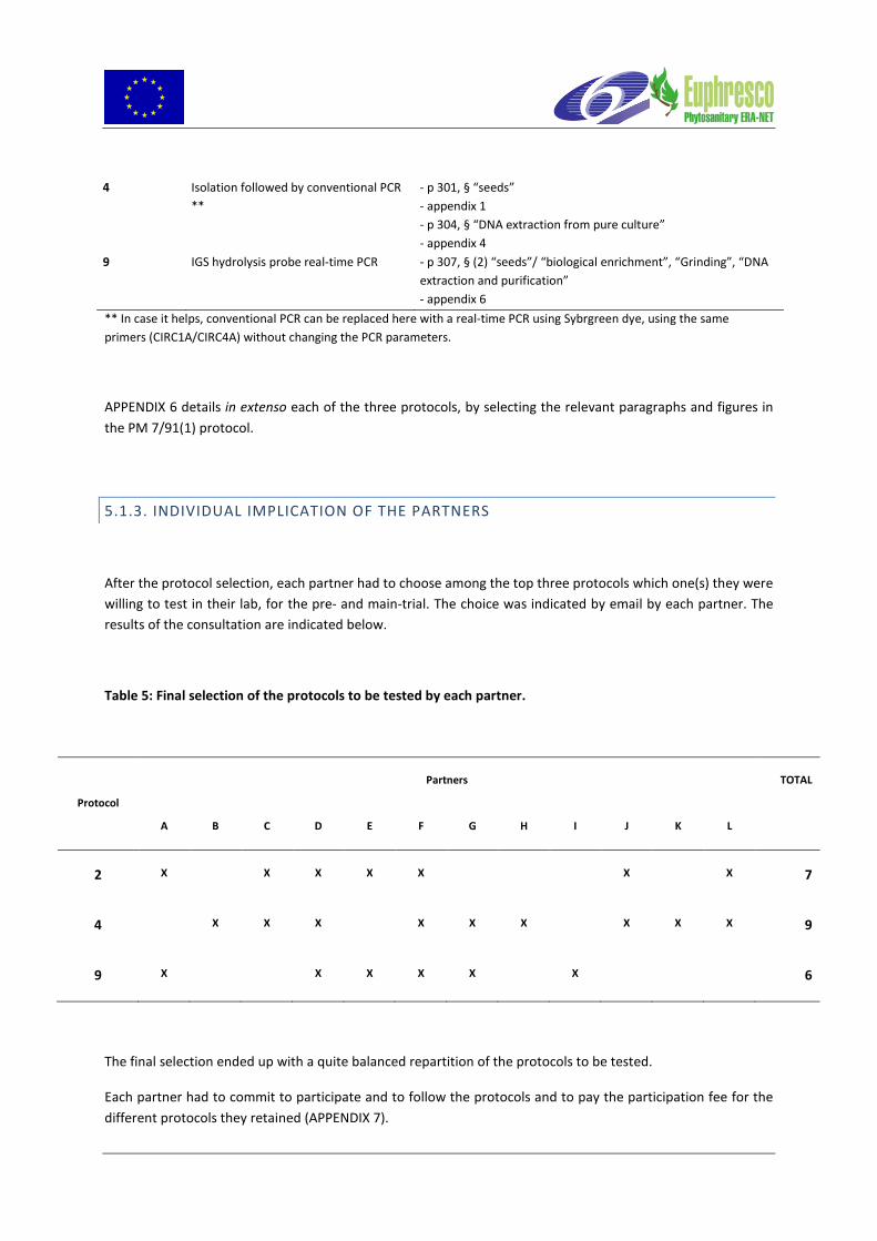
4 Isolation followed by conventional PCR **
- p 301, § “seeds” - appendix 1 - p 304, § “DNA extraction from pure culture” - appendix 4
9 IGS hydrolysis probe real-time PCR - p 307, § (2) “seeds”/ “biological enrichment”, “Grinding”, “DNA extraction and purification” - appendix 6
** In case it helps, conventional PCR can be replaced here with a real-time PCR using Sybrgreen dye, using the same primers (CIRC1A/CIRC4A) without changing the PCR parameters.
APPENDIX 6 details in extenso each of the three protocols, by selecting the relevant paragraphs and figures in the PM 7/91(1) protocol.
5.1.3. INDIVIDUAL IMPLICATION OF THE PARTNERS
After the protocol selection, each partner had to choose among the top three protocols which one(s) they were willing to test in their lab, for the pre- and main-trial. The choice was indicated by email by each partner. The results of the consultation are indicated below.
Table 5: Final selection of the protocols to be tested by each partner.
Protocol
Partners TOTAL
A B C D E F G H I J K L
2 X X X X X X X 7
4 X X X X X X X X X 9
9 X X X X X X 6
The final selection ended up with a quite balanced repartition of the protocols to be tested.
Each partner had to commit to participate and to follow the protocols and to pay the participation fee for the different protocols they retained (APPENDIX 7).


5.2. PRE-TRIAL RESULTS
In order to be allowed to participate to the main trial, and according to ISTA (ISTA 2007), only laboratories experienced with applying the evaluated techniques should be invited to participate. A pretrial test was therefore organised and on the basis of the results of this test, the test organiser decided which partner could be involved in the main comparative test.
Only partners that successfully obtained a positive result by analysing this blind sample were allowed to participate to the main trial.
For each protocol it decided to retain, each partner had first to analyse a blind sample, which was contaminated with 10 F. circinata infested seeds. This sample was prepared according to the protocols described above for sample preparation.
The pre-trial samples were sent by CRA-PAV on the beginning of June 2011 and according to the “acknowledgement of receipt for pre-trial samples” sheet sent back by the partners (APPENDIX 8), all the samples were received between the 8 and 9th of June. No sample quality problem was reported on receipt.
The results were reported by each partner using the result sheet presented in APPENDIX 9, and are summarized in Table 6.
Table 6: Pre-trial results obtained by each partner.
Protocol
Result expected / result obtained
Partners
TOTAL
A B C D E F G H I J K L
2 + / + + / + + / + + / + + / ? + / + + / ** 5/7
4 + / + + / + + / + + / ? + / + + / +* + / + + / + + / ** 7/9
9 + / + + / + + / + + / + + / + + / + 6/6
* This partner expressed its will to replace the conventional PCR followed by gel electrophoresis analysis by a real-time PCR using SybrGreen staining and
melting curve analysis. The real-time PCR was carried out using the same chemical and thermal conditions than initially expected for the conventional PCR test.

** No answer
Based on the pretrial results, it was decided not to retain Lab F for the evaluation of protocols 2 and 4, since it was not able to provide the expected results. After discussion between Lab F and the organiser, it was unfortunately not possible to point out the origin of the problem. Nevertheless, Lab F did participate to the main-trial for the protocol 4 but the data generated were not taken into consideration.
No answer was sent back from lab L, and therefore no data could be used for this partner.
All the other partners reported the expected results for each of the protocols they wanted to test, and were therefore retained for the corresponding main-trials.
5.3. MAIN TRIALS RESULTS
5.3.1. DATA RECOVERY
In good accordance with the timetable, all the main trial samples were sent by CRA-PAV by the middle of September 2011. According to the “acknowledgement of receipt for main-trial samples” (APPENDIX 10) sheet sent back by the partners, all the samples were received between the 22 and 26th of September. No sample quality problem was reported on receipt.
• Results for protocol 2 were received by the organiser between October the 27th and December the 2nd.
• Results for protocol 4 were received by the organiser between October the 14th and November the 26th.
• Results for protocol 9 were received by the organiser between October the 5th and December the 9th.
The results for the main-trial for each protocol were reported by each partner by the result sheet presented in APPENDICES 11, 12 and 13, for protocol 2, 4 and 9, respectively.
5.3.2. TREATMENT OF THE DATA
The raw results are presented in APPENDICES 14, 15 and 16, for protocol 2, 4 and 9, respectively.

The participants were asked to provide only ‘+’ or ‘–’ results. For each protocol, the results obtained for the blinded samples were processed according to EN ISO 16140, i) to compute the relative accuracy (AC), specificity (SP) and selectivity (SE) and their respective confidence intervals (CI).
- Relative Accuracy (AC)
AC represents the correlation between the expected results and the results obtained using the protocol.
AC = 100% × (PA + NA)/N, with N = NA + PA + PD + ND
- Relative specificity (SP)
SP provides an estimation of the ability of the protocol not to detect the target when it is not present (Healthy seed samples or samples contaminated by non target species).
SP = 100% × NA/N−, with N− = NA + PD
- Relative sensitivity (SE)
SE provides an estimation of the ability of the protocol to detect the target when it is present (artificially G. circinata-infected samples).
SE = 100% × PA/N+, with N+ = PA + ND
- Confidence intervals (CI)
CI was computed for each percentage p of AC, SE and SP
− If 10% < p < 90%, p is assumed to follow a normal distribution and the 95% confidence interval will by estimated as follows:
CI95% = p ± 2x sqr(p(1 − p)/n), with n = N, N+, or N− for AC,SE or SP, respectively.
− If p > 90%, tables of binomial distribution must be used.
5.3.3. PERFORMANCE CRITERIA
An overview of the performance criteria calculated for each protocol is presented in table 7.

Table 7: Performance criteria calculated for each protocol base on the main-trial test results.
Protocol 2 Isolation followed by
morphological identification
Protocol 4 Isolation followed by
conventional PCR
Protocol 9 Biological enrichement
followed by IGS hydrolysis probe real-time PCR
N partners involved 5 8 5 (6)*** Nb samples analysed 60 96 72
Nb samples analysed and retained 58* 81** 60***
Negative Accord (NA) 29 39 29 Positive Accord (PA) 28 37 26
Negative Deviation (ND) 1 1 4 Positive Deviation (PD) 0 4 1
Relative sensitivity 96.5% [91.4-100]**** 97.4% [95.5-99.3] 86.7%[78.0-95.4] Relative specificity 100% 90.7% [87.5-93.9] 96.7% [92.3-100]
Relative accuracy 98.3% [94.6-100] 93.8% [91.2-96.4] 91.7% [84.7-98.7]
Time spent for analysis (days) 12 to 26 7 to 15 4 to 8 * 2 results rated as undetermined were removed from the data set. ** 3 results rated as undetermined were removed from the data set, and data from lab F were removed. *** the samples analysed by Lab F were removed from the analysis since the problem the result deviations are probably better explained by contamination rather than inherent to the protocol in itself. **** CI95%
The performance criteria show that all three protocols provide very good values for relative sensitivity, specificity and accuracy.
5.3.3.1 Relative specificity
Protocol 2 (isolation – morphology) provided excellent specificity since by contrast with protocols 4 and 9, no false positive result was reported. In other words, isolation followed by morphological identification did not generate spurious positive results with other Fusarium strains, including strains morphologically or genetically close to the target species.
Protocol 9 (enrichment and real time PCR) provided an excellent level of relative specificity, although a single case of false positive was encountered with one sample contaminated with a non target species. Ioos et al. (2009) reported a relative specificity level of 100%, meaning that in their conditions, no false positive result was obtained. Though, the results of this project show that this protocol may generate false positive results in certain conditions, when contamination issues are not appropriately addressed.
On the other hand, protocol 4 (isolation – PCR) generated 9.3% false positive results. False positive results were obtained with non target Fusarium species, as well as with negative controls. This means that unexpected cross reactions as well as contamination issues have been met during the experiments.

Protocol 9 provided results within a maximum of 8 days, whereas protocols 4 and 2 required at least from 7 up to 26 days. The time spent for analysis was the highest with negative samples analyzed by following protocol 2, for which the suspect isolates are first transferred to SNA and PDA plate for incubation and final identification.
5.3.3.2. Relative sensitivity
Protocol 4 (isolation – PCR) and Protocol 2 (isolation – morphology) provided the best levels of relative sensitivity. By contrast, Protocol 9 (enrichment – real time PCR) was 10 % less sensitive, meaning that this protocol was less able to detect the target when it is present.
Analysis of the raw data shows that most of the false negative results obtained following Protocol 9, corresponded to samples containing a single contaminated seed. Since this protocol is mainly standardized by the use of DNA extraction kits and the use of real-time automatic equipment, there may be at least two explanations for these discrepancies between partners:
i) Heterogeneous grinding of the seed after the enrichment step, leading to the collection of both contaminated and/or non-contaminated macerates by the operator. This problem has been reported by two partners that could not use an automatic grinding system, and have to crush samples by hand.
ii) Different sensitivity fluorescence threshold set manually or automatically on the real-time equipment. Low fluorescence levels generated by DNA extracts with low target contamination levels may not be considered as background fluorescence depending of the settings.
Ioos et al. (2009) reported a relative sensitivity level of 79.1% (±4.3), which is inferior to the value obtained during this project. However, this value was calculated with DNA samples obtained with various DNA extraction procedures. The results obtained during this project shows that the relative sensitivity of the protocol 9 is improved by using a consistent DNA extraction procedure, as recommended by the EPPO diagnostic protocol.
5.3.4. ANALYTICAL SENSITIVITY AND SPECIFICITY
In order to compare the three protocols, the results may also be exploited by type of sample:
- Results obtained with samples contaminated with 10 G. circinata-infected seeds - Results obtained with samples contaminated with 1 G. circinata-infected seed (practical limit of
detection for plating) - Results obtained with samples containing 10 seeds contaminated with non target species - Results obtained with samples not contaminated

The three figures (1, 2, and 3) below illustrate and compare the results obtained vs the expected results for each protocol and each type of sample.
These graphs enable to compare the analytical sensitivity (detection of medium and low level target concentration) and specificity (non detection of non target species) for each protocol.
Figure 1: Results obtained following Protocol 2 (Isolation/morphology) for the different types of seed samples

Figure 2: Results obtained following Protocol 4 (Isolation/conventional PCR) for the different types of seed samples
Figure 3: Results obtained following Protocol 9 (Biological enrichment / qPCR) for the different types of seed samples

5.3.4.1 Results obtained with samples contaminated with 10 G. circinata-infected seeds
Protocols 4 and 9 both yielded 100% positive results with these medium level-infected samples, which support their ability to detect the target when present at a medium level in a sample.
For protocol 2, one single positive sample could not be detected by Lab J, who ended up with an “undetermined result” caused by over-competition by a fast growing fungus on the isolation medium. These results shows that protocol 2 is efficient to detect the target when present at a medium level in a sample, but may yield false negative results when fast growing fungus are present in the analysed samples.
5.3.4.2. Results obtained with samples contaminated with 1 G. circinata-infected seeds
Protocol 2 detected G. circinata in 17 out of 18 infected samples. There was no explanation for this single false negative result.
Procotol 4 detected G. circinata in 18 out of 21 infected samples. Two positive samples could not be detected by Lab J, who ended up with an “undetermined result” caused by over-competition by a fast growing fungus on the isolation medium. There was no explanation for the third false negative result.
Protocol 9 detected G. circinata in 11 out of 15 infected samples. The four false negative results were obtained by three different labs.
These results show that none of the three protocols enabled a 100% efficient detection of G. circinata in samples with a low level of infection. Protocols 2 and 4, based on a prior isolation of the target failed to detect G. circinata because of over competition by fast-growing fungi or no growth of the target. On the other hand, protocol 9 could not detect the target probably because of the limit of detection of the assay in certain conditions.
5.3.4.3. Results obtained with seed samples contaminated with non target species.
The non target species were chosen as to assess the ability of each protocol not to detect morphologicaly close species (i.e. F. oxysporum, F. fujikuroi or F. subglutinans) for the test based on morphology, and/or phylogenetically close species (F. fujikuroi and F. subglutinans) for the tests based on DNA sequences.
Protocol 2 successfully obtained negative results with 17 out of 18 seed samples contaminated with non target species. One single sample was rated as ‘undetermined’ caused by over-competition by a fast growing fungus on the isolation medium.
Protocol 4 successfully obtained negative results with 18 out of 21 seed samples contaminated with non target species. Two of these false positive results were obtained by Lab B, who reported that for these two samples, Fusarium isolates were recovered, and although morphologically doubtful, they were positive after species-specific conventional PCR test. The latter false positive result was obtained by lab H who reported a doubt about the specificity of the primers, since most of the samples of the set it tested yielded signal, although not always at an acceptable level.

Protocol 9 successfully obtained negative results with 14 out of 15 seed samples contaminated with non target species. There was no explanation for the single false negative result obtained.
These results show that even though undetermined results may be obtained by the protocol 2 because of over competition with fast-growing fungi, no false positive was obtained due to morphological confusion, supporting the analytical specificity of this protocol. On the other hand, protocols 4 and 9, resorting of molecular biology methods, both produced a few false positive results. Incomplete specificity of the primers may be an explanation for the conventional PCR, but false positive results should have been obtained with all the partners, since working with the same samples. On the other hand there is no obvious reason for the single realtime PCR discrepancy, since no cross reaction was observer for the other non target samples, in the other labs.
5.3.4.4. Results obtained with non contaminated seed samples.
Protocols 2 and 9 both yielded 100% negative results with these non infected samples, which supports the analytical specificity of these protocols.
Protocol 4 successfully obtained negative results with 19 out of 21 seed samples contaminated. Lab J ended up with an “undetermined result” caused by over-competition by a fast growing fungus on the isolation medium. The single false positive result was obtained by lab H who reported a doubt about the specificity of the primers, since most of the samples of the set it tested yielded signal, although not always at an acceptable level. Since Lab H also experienced false positive results with a sample contaminated with a non target species, it may by hypothesised that incomplete specificity of the primers may not be the cause of the problem, but rather a problem of background fluorescence (this lab replaced the electrophoresis gel by a sybrgreen staining).
5.4. FEEDBACK FROM PARTNERS
After the completion of the collaborative studies, the partners have been consulted by email, in order to give their views and opinions about the protocol(s) they have tested.
The result of this consultation is reported in table 8.

Table 8: Comments made by the partners about the protocols assessed in their respective laboratory.
Advantages Drawbacks Other comments
Protocol 2 (isolation/morphology)
Lab A: “- Simplicity of the analysis - Sensitivity ? (if a lot of seeds are analyzed ?)” Lab C: “Any laboratory with trained staff on Fusarium can do it even if they do not have PCR” Lab G: “does not require expensive instruments” Lab D: “Straight-forward set up and assessments. Lower cost for a few samples.” Lab E: “ Cheap, not requiring expensive equipment or reagents”
Lab A: “ - time consuming - Possible pollution development that may overcompete the target fungus on the growing medium” Lab C: “Time consuming (media preparation, plating of seeds, plates revision, subculturing, 10 days on SNA, morphological identification) Space consuming (space in incubators), plates (high number of plates, different media), Contamination with other fungi that can mask Fusarium, missing a seed during the plating (that could be the contaminated one!!!).” Lab G: “We did not participate to this protocol, but for our experience the operator should have a good expertise to morphological identification. Moreover the methods is time consuming” Lab D: “More time consuming in comparison with protocol 9 and large numbers of samples take up a lot of lab space.” Lab E: “Time consuming and takes a long time to provide results. Requires experience on morphological identification.”
Lab D: “This would be our preferred option for low number of samples. (1-3 samples)”
Protocol 4 (isolation/conventional PCR)
Lab C: “Time saving, no need of trained staff on identification of Fusarium.”
Lab C: “Time consuming (media preparation, plating of seeds, plates revision, subculturing, 10 days on SNA, morphological
Lab B: “Some isolates showed a positive results after conventional PCR, while based on morphology the isolates

Lab K: “The numerous observations made, helped us for the easier identification of fungus on media culture, because Fusarium circinatum it looks very typical.” Lab G: “The identification by PCR makes it more robust and reliable. Moreover it is not necessary to have a great experience on morphological identification.” Lab D: “ If there are lots of samples, then it is quicker to extract and PCR for a result. Also, cultures can be tested before they have grown for the required no. of days for morphological assessment PCR provides confirmation of morphology results, therefore ensuring accurate (double-checked) results.”
identification) Space consuming (space in incubators), plates (high number of plates, different media), Contamination with other fungi that can mask Fusarium, missing a seed during the plating (that could be the contaminated one!!!).” Lab G: “This method is time consuming for the preparation of the substrate and plating of the seeds.” Lab D: ” With only a few samples it would be quicker to assess on morphological features alone. More resource required (staff, equipment, consumables, time) to test by both isolation and PCR.”
were negative. Because no coiled hyphae and no polyphialidic conidiophores were visible. Because of the additional note in the flow diagram in the Eppo protocol we decided to give the samples a positive result but with the remark: morphological doubtful. In real life we would ask for an extra seed sample.” Lab K: “After the isolation on DCPA medium, for the seeds with low level of infection , it is naecessary to increase the quantity of micelium for the PCR test. Our observations have shown us that on OA medium F. circinatum is growing better
and faster than PDA medium.” Lab D: “In practice we would probably use molecular method only to confirm culture identity if in doubt and not cultures that are clearly identifiable by morphology only.”
Protocol 9 (biological enrichment/realtime PCR
Lab G: ”This method allows to easily analyze a greater number of seeds for each sample and requires much less time.” Lab D: “Quicker result than having to wait for cultures to grow” Lab E: “Fast and sensitive.” Lab I: “Rapid, specific”
Lab G: “Real time PCR is very sensitive and this could produce false positives” Lab D: “Blending the seeds in the broth is quite a messy process and difficult to ensure no cross-contamination” Lab I: “It is essential to control the sample preparation (see “other comments) to get a sufficient sensitivity It is necessary to define a cut off value”
Lab G: “ We also tested the method with Pinus pinea seeds, and it worked. It is necessary a pre-breaking of the seeds before the DNA enrichment.” Lab D: “This would be our preferred option for high numbers of sample.” Lab E: “The reagents are expensive but this can be compensated by savings in labor.” Lab I: “ The test provides good results in terms of specificity and sensitivity provided that the sample preparation is done properly (see comparative data in the attached file)”

Individual exchanges were made with partners that ended up with one or several unexpected results, in order to try to know whether the discrepancy was caused by the protocol in itself, or if other explanations may be hypothesized.
When a partner recognized that the discrepancy could be caused by a problem in the lab (contamination, insufficient training or information, etc.), another set of samples could be ordered to CRA-PAV, in order to check if improvement could be achieved and the problem(s) overcome.
5.5. CONCLUSION
Mandated diagnostic laboratories working under accreditation should only use validated tests. The validation process is carried out to provide objective evidence that the test is suitable for the routine diagnosis. Te minimum test performance criteria to be defined are: analytical sensitivity, analytical specificity, repeatability, reproducibility and if appropriate selectivity. In this project, we only assessed the relative sensitivity and specificity of three selected protocols for the detection of G. circinata in pine seed, by running an inter-laboratory comparison, using the known status of blind pine seed sample as standards.
To our knowledge, no validation data was available up to now for protocols 2 (isolation/morphology) and 4 (isolation/conventional PCR), and this project filled this gap. Despite validation data was already available for Protocol 9 (see Ioos et al., 2009), this project generated additional data, including results obtained with different equipment, chemical and staff. This will add value by taking into consideration some of the aspect of robustness of the protocol.
All three selected protocols appear as fit for diagnostic purpose based on their performance values. However, they showed different advantages or drawbacks, that cannot be quantified, but are based on observations or on practical experience. The final choice of the protocol remains up to the diagnostic lab, and should be discussed by combining several parameters: speed of process, availability of trained staff, or ad hoc equipment, number of samples to be processed simultaneously, etc.

6. RESULTS OF THE NPPO SURVEY ABOUT THE SEED SAMPLING STRATEGY
6.1. SETTING OF THE STATISTICAL PARAMETERS TO DETERMINE THE SAMPLE SIZE.
In the area of phytosanitary matters, and according to ISPM N°31 (3), a statistically based sampling is designed to detect a certain percentage of infestation with a specific confidence level, and thus requires the national plant protection organisation (NPPO) to determine the following interrelated parameters:
• Acceptance number (number of acceptable infested seeds in a sample taken from a lot),
• Efficacy of detection (refers to how effective the testing method is in finding infestation),
• Confidence level (probability that a pest infesting a specified proportion of seeds in a seed lot will be detected in the sample used for analysis),
• Detection level (minimum percentage of infestation that the sampling methodology will detect at the specified efficacy of detection and confidence level),
• Sample size.
As some of the value for some of these parameters may be set by the NPPO, the sample size can be determined by calculation.
Considering the nil tolerance applied to the organisms listed by the 2000/29/CE directive or organisms like Gibberella circinata for which specific emergency measures have been taken, some of the parameters are not adjustable. In case of nil tolerance for Gibberella circinata in a seed lot, the tolerance level (number of acceptable infested seeds in an entire lot), as well as the acceptance number (number of acceptable infested seeds in a sample taken from a lot), are automatically set to zero.
The efficacy of detection (i.e. diagnostic “sensitivity” of the test) is in our case expressed as the percentage of tested seeds that are correctly identified as infested by the analysis method. In the framework of this Euphresco project, three analysis methods will be tested, consisting in using either an isolation technique or a molecular method based on PCR. For the isolation technique, the sensitivity of the test is assumed to be one seed out all the seeds analyzed (e.g. 1/ 250, 1/ 400, 1 / 1000, etc., depending on the sample size) since each seed of the sample is observed and analyzed individually and assuming that the cryptically infesting fungus is not in a latent stage, meaning that it will grow out of the seed once plated (2). For the molecular technique, the sensitivity of the test has already been estimated experimentally to less than 1 seed /1000 (4). For practical reasons, it is reasonable to assume that only samples not exceeding 1000 seeds can routinely be analyzed by

[Gibcir diagseed]
the laboratories in charge of the official analysis. Therefore, the efficacy of detection may be set to 100% (or 1).
However, two other parameters may be discussed and set by the NPPO, according to their constraints and objectives:
1) The confidence level is the probability that a pest (G. circinata) infesting a specified proportion of seeds in a seed lot will be detected in the sample used for analysis (e.g. a 95% confidence level indicates that on average, if 100 samples are taken from a lot that has a specific proportion of seeds infested, 95 of the samples will detect the infestation, and 5 will not). The higher the confidence level, the larger the sample required to demonstrate it. However, a confidence level cannot be set to 100%, as sampling always involves error.
2) The detection level is the minimum percentage of infestation that the sampling methodology will detect at the specified efficacy of detection and level of confidence. If the infestation level of a seed lot is equal or larger than the detection level, then the sampling will detect at least on infested seed with the desired confidence level. In practical terms, if no G. circinata is found in the sample, the NPPO has the desired level of confidence that the infestation level in the entire lot does not exceed the detection level that it has set. A very low detection level requires a larger number of seeds to be sampled to have a high probability.
6.2. RESULTS OF EPPO COUNTRY CONSULTATION
Only 10 out of 50 countries consulted have answered to the country consultation using the questionnaire presented in APPENDIX 3.
The results of the questionnaire are reported in Table 9 for the consultation about the confidence level and in Table 10 for the tolerance level.
EUPHRESCO_Gcircinata2012_Final_report Page 34 of 75

[Gibcir diagseed]
Table 9: Values or range of values proposed by the NPPO for the confidence level
Confidence level Country 80% 90% 95% 99% Others Bulgaria X X Croatia X X Czech Rep. Estonia X France* X Italy X Poland X Romania X Slovenia X Spain X Total (%) 1 (9%) 7 (63.6%) 3 (27.3%)
* Comment from France: these values cannot be applied to lots with less than 5000 seeds, otherwise almost the entire lot is destroyed for the analysis.
Table 10: Values or range of values proposed by the NPPO for the tolerance level
Tolerance level Country 5% 2% 1% 0.1% 0.01% Other Bulgaria X X Croatia X Czech Rep. X X Estonia X France* 0.5% Italy X Poland X Romania X Slovenia X Spain X Total (%) 2 (16.7%) 8 (66.7%) 1 (8.3%) 1 (8.3%)
* Comment from France: these values cannot be applied to lots with less than 5000 seeds, otherwise almost the entire lot is destroyed for the analysis.
EUPHRESCO_Gcircinata2012_Final_report Page 35 of 75

[Gibcir diagseed]
6.3. PROPOSAL FOR A HARMONIZED SAMPLE SIZE FOR PINE SEEDS
According to the results of the NPPO questionnaire, the most popular values for confidence level and tolerance level may be used to determine a consensus sample size for pine seeds.
The agreed values for the interrelated parameters determining sample size are as follows:
• “0” for the acceptance number (number of acceptable infested seeds in a sample taken from a lot) ,
• “1” for the efficacy of detection (refers to how effective the testing method is in finding infestation),
• “0.95” for the confidence level (probability that a pest infesting a specified proportion of seeds in a seed lot will be detected in the sample used for analysis),
• “0.01” for the detection level (minimum percentage of infestation that the sampling methodology will detect at the specified efficacy of detection and confidence level),
According to these data and to table 11(extracted from ISPM N°31) the sample size would range from 95 seeds for a seed lot containing at least 100 units, to 298 seeds for a seed lot exceeding 200 000 units.
EUPHRESCO_Gcircinata2012_Final_report Page 36 of 75

[Gibcir diagseed]
Table 11: Table of minimum sample size for 95% and 99% confidence level at varying levels of detection according to lot size, assuming that the detection efficacy is 100%, and hypergeometric distribution.
However, it can be observed from the results of the performance criteria evaluated in this project, that none of the three protocols selected showed a 100% detection efficacy, which means that the minimal number of seeds to be analysed should be superior to the range of values quoted above. In this respect, it seems that the 400 seeds sampling strategy used for this project was appropriate and may be recommended for the future, providing that the values for the parameters used here are retained by the NPPO.
However, given the fact that 1000 seeds are easily processed following protocol 9, this mean that this protocol enables to consider a higher confidence level (99%) with a lower detection level (0.05%) (see Table 11).
EUPHRESCO_Gcircinata2012_Final_report Page 37 of 75

[Gibcir diagseed]
7. CONCLUSIONS OF THE GIBCIR DIAGSEED PROJECT
The project aimed at producing harmonized sampling method for pine seed and validated detection tools for use by inspection service.
This project gathered 12 partners from 11 European countries. The results may beneficiate to the inspection service and the mandated diagnostic laboratories of the NPPOs.
An inventory of the currently existing methods to detect G. circinata in pine seed was carried out and after consultation of the partners three protocols were finally selected for further assessment. The selection was made by the partners, based on the popularity of the protocols, the possibility to be easily implemented, the availability of trained staff, etc.
The interlaboratory test that was organised enabled to produce validation data for three protocols:
• Protocol 2 Isolation followed by morphological isolation
• Protocol 4 Isolation followed by conventional PCR
• Protocol 9 IGS hydrolysis probe real-time PCR.
The different partners followed the protocol guidelines by sticking to the requirements available in EPPO Gibberella circinata diagnostic PM 7/91(1). However, they used their own reagents, equipment and involved their own staff. The validation data generated by this project therefore include an assessment of the robustness of each protocol.
Protocol 2 Isolation followed by morphological isolation
Protocol 2 was judged as the most easy to implement. It requires few types of equipment, and is very sensitive. However, it is time and room consuming and thus does not seem to be appropriate for the analysis of numerous samples at the same time. In addition, overcontamination by non target species was reported by the participants and may lead to false negative results, especially at low infection levels. This protocol entirely relies on the expertise of the operator that must be trained for the correct identification of Fusarium circinatum in pure culture. Identification may be confusing when uncommon strains (eg with uncoiled sterile hyphae) are met.
Relative sensitivity: 96.5%
Relative specificity: 100%
EUPHRESCO_Gcircinata2012_Final_report Page 38 of 75

[Gibcir diagseed]
Protocol 4 Isolation followed by conventional PCR
Protocol 4 is very similar to protocol 2, except that the final identification of the candidate Fusarium strains are identified by conventional PCR instead of the observation of microscopic features. Therefore, taxonomic skills are less required but the project showed that cross reaction with close Fusarium species may occur and generate false positive results.
Relative sensitivity: 97.4%
Relative specificity: 90.7%
Protocol 9 IGS hydrolysis probe real-time PCR
Protocol 9 was deemed as the most convenient one since it required no expertise on taxonomy, may be easily standardized and is fit for the analysis of numerous samples (less time spent for the analysis, less room needed).
It was shown that protocol 9 may generate false negative results depending on the fluorescence threshold setting of the equipment. Likewise, the used of an efficient grinder appeared as of paramount importance in order to produce a sufficiently homogenized seed macerate before sampling.
Relative sensitivity: 86.7%
Relative specificity:96.7%
All three protocols may be used by official laboratories and showed acceptable performance data. The choice of the protocol may rely on the availability of trained staff, the number of seed samples to be analysed simultaneously, and the time that can be allocated for the analysis.
The second aspect of Gibberella circinata testing was about the need to define a harmonized sampling procedure.
The questionnaire prepared during this project in order to tackle the sampling issue was successful. A majority of the countries that answered advocated a confidence level of “0.95” (probability that a pest infesting a specified proportion of seeds in a seed lot will be detected in the sample used for analysis), combined with a detection level set at “0.01” (minimum percentage of infestation that the sampling methodology will detect at the specified efficacy of detection and confidence level).
According to ISPM N°31, the appropriate sample size required for analysis based on the general agreement after country consultation would range from 95 seeds for a seed lot containing at least 100 units, to 298 seeds for a seed lot exceeding 200 000 units.
EUPHRESCO_Gcircinata2012_Final_report Page 39 of 75

[Gibcir diagseed]
EUPHRESCO_Gcircinata2012_Final_report Page 40 of 75

[Gibcir diagseed]
REFERENCES
Anonymous (2005). "Gibberella circinata." EPPO Bulletin 35(3): 383-386.
Anonymous (2009). "PM 7/91(1): Gibberella circinata." EPPO Bulletin 39(3): 298-309.
Anonymous (2010). "PM 7/98 (1): Specific requirements for laboratories preparing accreditation for a plant pest diagnostic activity." EPPO Bulletin 40(1): 5-22.
International Plant Protection Convention (2008). ISPM N° 31. Methodologies for sampling of consignments. International Standards for phytosanitary measures. Rome, It., FAO: 19.
International Seed Testing Association (2002). International rules for testing. 7-009: Detection of Fusarium moniliforme var. subglutinans Wollenw. & Reinke on Pinus taeda and P. elliotii (Pine) Basseldorf, Switzerland.
.
International Standardization Organization (2003). ISO 16140:2003 Microbiology of food and animal feeding stuffs - Protocol for the validation of alternative methods. Geneva, Switzerland.
Ioos, R., C. Fourrier, et al. (2009). "Sensitive Detection of Fusarium circinatum in Pine Seed by Combining an Enrichment Procedure with a Real-Time Polymerase Chain Reaction Using Dual-Labeled Probe Chemistry." Phytopathology 99(5): 582-590.
ISTA (2007). ISTA Methode validation for seed testing. I. S. T. Association. Basseldorf, Switzerland: 70.
Ramsfield, T. D., K. Dobbie, et al. (2008). "Polymerase chain reaction-based detection of Fusarium circinatum, the causal agent of pitch canker disease." Molecular Ecology Resources 8(6): 1270-1273.
Schweigkofler, W., K. O'Donnell, et al. (2004). "Detection and quantification of airborne conidia of Fusarium circinatum, the causal agent of pine pitch canker, from two California sites by using a real-time PCR approach combined with a simple spore trapping method." Applied and Environmental Microbiology 70(6): 3512-3520.
Steenkamp, E. T., B. D. Wingfield, et al. (1999). "Differentiation of Fusarium subglutinans f. sp. pini by Histone Gene Sequence Data." Applied And Environmental Microbiology 65(8): 3401-3406.
EUPHRESCO_Gcircinata2012_Final_report Page 41 of 75

[Gibcir diagseed]
Appendix 1
EUPHRESCO_Gcircinata2012_Final_report Page 42 of 75

[Gibcir diagseed]
Appendix 2
EUPHRESCO_Gcircinata2012_Final_report Page 43 of 75

[Gibcir diagseed]
Appendix 3
EUPHRESCO_Gcircinata2012_Final_report Page 44 of 75

[Gibcir diagseed]
EUPHRESCO_Gcircinata2012_Final_report Page 45 of 75

[Gibcir diagseed]
EUPHRESCO_Gcircinata2012_Final_report Page 46 of 75

[Gibcir diagseed]
EUPHRESCO_Gcircinata2012_Final_report Page 47 of 75

[Gibcir diagseed]
EUPHRESCO_Gcircinata2012_Final_report Page 48 of 75

[Gibcir diagseed]
EUPHRESCO_Gcircinata2012_Final_report Page 49 of 75

[Gibcir diagseed]
Appendix 4
EUPHRESCO_Gcircinata2012_Final_report Page 50 of 75

[Gibcir diagseed]
Appendix 5
Please confirm that you are ready to pay for the seed samples prepared by CRA-PAV (Luca Riccioni and Tiziana Annesi) by indicating "YES" in next cell.
Protocol ID
Technique Reference
Please chose a maximum of three (3) protocols in the list by indicating "YES" in the appropriate cell.*
1 Isolation followed by morphological isolation
Agar plating (Komada’s medium) + morphological characterization
EPPO diagnostic protocol for G. circinata PM 7/91(1)
2 Isolation followed by morphological isolation
Agar plating (DCPA medium) + morphological characterization
EPPO diagnostic protocol for G. circinata PM 7/91(1)
3 Isolation followed by PCR-RFLP Agar plating + PCR amplification of H3 gene + RFLP analysis
Steenkamp et al. (1999)
4 Isolation followed by conventional PCR
Agar plating + PCR (mycelial DNA extraction followed by conventional PCR targeting G. circinata specific regions within IGS)
EPPO diagnostic protocol for G. circinata PM 7/91(1) and Schweigkofler et al. (2004)
5 Blotter paper incubation Incubation on blotter paper sprayed with PNCB** liquid medium
ISTA (2002)
6 IGS conventional PCR
Total DNA extraction followed by conventional PCR targeting G. circinata specific regions within IGS
Schweigkofler et al. (2004) and Ioos et al. (2009)
7 IGS Sybrgreen real-time PCR
Total DNA extraction followed by Sybrgreen real-time PCR targeting G. circinata specific regions within IGS
Schweigkofler et al. (2004) and Ioos et al. (2009)
EUPHRESCO_Gcircinata2012_Final_report Page 51 of 75

[Gibcir diagseed]
8 Duplex SCAR-based conventional PCR
Total DNA extraction followed by a duplex conventional PCR test targeting G. circinata specific regions designed from SCARs
Ramsfield et al. (2008)
9 IGS hydrolysis probe real-time PCR
Total DNA extraction followed by real-time PCR using primers and a hydrolysis probe targeting G. circinata specific regions
Ioos et al. (2009)
* A set of twelve 400-seed samples will have to be analyzed for each protocol chosen by each partner
(e.g if 2 or 3 protocols are chosen by the partner, 2x12= 24 or 3x12=36 seed samples, respectively, will have to be analyzed).
EUPHRESCO_Gcircinata2012_Final_report Page 52 of 75

[Gibcir diagseed]
Appendix 6
Detailed protocols as quoted in the EPPO diagnostic PM7/91(1)
Protocol 2: Isolation followed by morphological characterization
Seeds
Seeds are directly plated onto Fusarium semi-selective media (e.g. Komada’s medium. DCPA medium see Appendix 1) without previous surface disinfection. Plates are incubated at room temperature (22±6°C). During incubation, the plates are observed periodically and all the Fusarium spp. colonies are transferred to Potato dextrose agar (PDA) and to Spezieller-Nährstoffarmer Agar (SNA) (Appendix 1) for morphological identification. This method is efficient and reliable to isolate any Fusarium spp. from the seeds and does not require expensive equipment, though time- and space-consuming when serial analyses are conducted. However, the correct morphological identification of F. circinatum in pure culture requires experience and in case of uncertainty a molecular confirmation should be carried out. In addition, Storer et al. (1998) have demonstrated that agar plating of pine seeds may not be able to detect dormant (quiescent) propagules of F. circinatum, thus leading to an unknown risk of false negative results.
Morphological characteristics in pure culture
For morphological identification, the isolates are grown on PDA to study colony morphology and pigmentation, and on SNA (Appendix 1) to study formation and type of microconidia and conidiogenous cells. SNA and PDA plates are incubated at room temperature. All isolates are examined after 10 days and confirmed as F. circinatum based on the morphological features described by Nirenberg & O’Donnell (1998) and Britz et al. (2002). On PDA, F. circinatum grows relatively rapidly (average growth of 4.7 mm/day at 20°C; Nirenberg and O’Donnell, 1998). After 10 days, the colony should have an entire margin, white cottony or off-white aerial mycelium with a salmon tinge in the middle or with a purple or dark violet pigment in the agar (Fig. 6). On SNA, microconidia are aggregated in false heads (Figs 7a, b), with branched conidiophores, mono and polyphialidic- conidiophores (Figs 8a, b), obovoid microconidia in aerial mycelium, mostly nonseptate or with occasionally 1-septum. Chlamydospores are absent. The sterile hyphae (coiled/not distinctively coiled) are characteristic of F. circinatum and are observed clearly on this medium (Figs 9a, b). The epithet “circinatum” refers to these typical coiled hyphae, also called “circinate” hyphae.
Appendix 1: Composition of the different culture media
A- Komada medium (Komada, 1975):
This medium is suitable for isolation of Fusarium circinatum from plant tissue, including seeds, but not for identification. The basal medium contains:
• K2HPO4: 1.0 g
• KCl: 0.5 g
EUPHRESCO_Gcircinata2012_Final_report Page 53 of 75

[Gibcir diagseed]
• MgSO4 7H2O: 0.5 g
• Fe-Na-EDTA: 10 mg
• L-Asparagine: 2.0 g
• D-Galactose: 20.0 g
• Technical agar: 15.0 g
• Distilled water to 1.0 L
The pH is adjusted to 3.8 ± 0.2 with 10 % phosphoric acid. The basal medium is autoclaved and slightly cooled before adding the following filter-sterilized supplemental solutions:
• Pentachloronitrobenzene (PNCB, 75% w/w): 1.0 g
• Oxgall: 0.5 g
• Na2B4O7 10H2O: 1.0 g
• Streptomycin: 6 mL/L of stock solution (5 g of streptomycin in 100mL distilled water)
B -PDAS
Potato dextrose agar supplemented with 0.5 mg mL-1 of streptomycin sulphate salt (775 units/mg solid)
C- Dichloran Chloramphenicol Peptone Agar (DCPA) ( (slightly modified by Ioos et al., 2004; after Andrews & Pitt, 1986)
This medium is suitable for isolation of Fusarium circinatum from plant tissue, including seeds, but not for identification. The medium contains:
• Bacteriological peptone, 15.0 g
• KH2PO4, 1.0 g
• MgSO4(7H2O): 0.5 g
• Chloramphenicol: 0.2 g
• 2.6-dichloro-4-nitroanilin (dichloran) (0.2% W/V in ethanol, 1.0 mL): 2 mg
• Violet crystal (0.05 % W/V in water, 1.0 mL): 0.0005 g
• Technical agar: 20.0 g
• Distilled water: to 1.0L
EUPHRESCO_Gcircinata2012_Final_report Page 54 of 75

[Gibcir diagseed]
C- Spezieller-Nährstoffarmer Agar (SNA) (Gerlach & Nirenberg, 1982)
This medium should be mandatory used for identification of F. circinatum, based on morphological features. The medium contains:
• KH2PO4: 1.0 g
• KNO3: 1.0 g
• MgSO4 7H2O: 0.5 g
• KCl: 0.5 g
• Glucose: 0.2 g
• Sucrose: 0.2 g
• Technical agar: 20.0 g
• Distilled Water to 1.0 L
Optionnaly, two 1-cm2 square pieces of sterile filter paper may be laid on the surface of the agar since Fusarium sporodochia are sometimes more likely produced at the edge of the paper.
Figures :
EUPHRESCO_Gcircinata2012_Final_report Page 55 of 75

[Gibcir diagseed]
EUPHRESCO_Gcircinata2012_Final_report Page 56 of 75

[Gibcir diagseed]
Protocol 4: Isolation followed by conventional PCR
Seeds
Seeds are directly plated onto Fusarium semi-selective media (e.g. Komada’s medium. DCPA medium see Appendix 1) without previous surface disinfection. Plates are incubated at room temperature (22±6°C). During incubation, the plates are observed periodically and all the Fusarium spp. colonies are transferred to Potato dextrose agar (PDA) and to Spezieller-Nährstoffarmer Agar (SNA) (Appendix 1) for morphological identification. This method is efficient and reliable to isolate any Fusarium spp. from the seeds and does not require expensive equipment, though time- and space-consuming when serial analyses are conducted. However, the correct morphological identification of F. circinatum in pure culture requires experience and in case of uncertainty a molecular confirmation should be carried out. In addition, Storer et al. (1998) have demonstrated that agar plating of pine seeds may not be able to detect dormant (quiescent) propagules of F. circinatum, thus leading to an unknown risk of false negative results.
DNA extraction from pure culture
Fungal DNA should be extracted using an appropriate standard method for DNA extraction from fungi e.g. regular commercial plant DNA extraction kits (or other methods reviewed in Irlinger et al., 2008) and analyzed following any of the tests presented in Appendices 2, 4 or 6.
For PM7/91 (1) Appendix 1, see protocol 2 above.
Appendix 4: Identification at species level by conventional or SyBr green real-time PCR (Schweigkofler et al., 2004)
1-General informations
Schweigkofler et al. (2004) described a technique based on a conventional or a SyBrgreen real-time PCR designed from the rDNA IGS (Inter Genic Spacer) region to identify the anamorphic stage of G. circinata in pure culture or in trapped airborne spores, but may be adapted to the analysis of seeds following the biological enrichment step (See Identification section, Ioos R., pers. comm.).
The PCR test targets a region of the IGS and produces a 360 bp amplicon for G. circinata (sequences of the IGS region for G. circinata may be retrieved from Genbank, accessions AFAY249397 to AY249403). A specific region of the IGS is amplified with G. circinata DNA using the primer pair CIRC1A (forward) and CIRC4A (reverse) (Table 2).
2-Methods
Nucleic acid extraction and purification.
See Appendix 3.
Conventional PCR reaction.
A G. circinata-specific IGS portion is amplified by PCR as follows.
EUPHRESCO_Gcircinata2012_Final_report Page 57 of 75

[Gibcir diagseed]
The PCR reaction mixture includes:
• 1 X PCR buffer supplied with the DNA polymerase,
• 0.25 mM each dNTP,
• 2 mM MgCl2,
• 0.5 µM of each CIRC1A and CIRC4A primers,
• 0.05 U/µL DNA polymerase
• 6.25 µL of template DNA,
• Molecular grade water is (MGW) added to reach the final reaction volume of 25 µL).
Each DNA extract should be tested by at least two replicate reactions.
The PCR reaction conditions should be carried out in a thermocycler equipped with a heated lid and include an initial denaturation at 94°C for 3 min, followed by 45 cycles for denaturation at 94°C for 35 s, annealing at 66°C for 55 s and elongation at 72°C for 50 s. A final elongation step is done at 72°C for 12 min.
The PCR products are separated by electrophoresis in a 1 % agarose gel followed by ethidium bromide staining. A DNA template containing amplifiable G. circinata DNA will yield a 360-bp fragment after a CIRC1A/CIRC4A PCR.
SyBr green real-time PCR reaction
A G. circinata-specific IGS portion is amplified by PCR as follows.
The PCR reaction mixture includes:
• 1 X PCR buffer supplied with the DNA polymerase,
• 0.25 mM each dNTP,
• 5 mM MgCl2,
• 0.5 µM of each CIRC1A and CIRC4A primers,
• SyBrgreen dye (concentration to be adjusted following the manufacturer’s recommendation)
• 0.05 U/µL DNA polymerase
• 6.25 µL of template DNA,
• Molecular grade water is (MGW) added to reach the final reaction volume (25 µL).
Each DNA extract should be tested by at least two replicate reactions.
The real-time PCR reactions are carried out in a suitable PCR instrument equipped with a system capable of fluorescence monitoring.
EUPHRESCO_Gcircinata2012_Final_report Page 58 of 75

[Gibcir diagseed]
The PCR reaction conditions include an initial denaturation at 95°C for 3-10 min (according to the type of DNA polymerase), followed by 45 cycles for denaturation at 94°C for 35 s, annealing at 66°C for 55 s, and extension at 72°C for 50 s. The fluorescence of the reporter dye is monitored at the end of each extension step.
The accumulation of G. circinata PCR amplicons is monitored in real-time by the measurement of the specific fluorescence of the SyBr green dye incorporated into the PCR product. A DNA template containing amplifiable G. circinata DNA will yield a Cycle threshold (Ct) value. Ct value represents the estimated cycle number from which the level of fluorescence becomes significantly superior to the background fluorescence level.
The nature of the amplicons should be checked by yielding melting curves at the end of the amplification process and by comparison to the melting curves yielded with the PCR positive control.
3-Essential procedural information
A DNA extraction negative control (blank tube) should be included for each DNA extraction series in order to ensure the absence of contamination during this step.
A PCR negative control containing no target DNA should be included in every test in order to ensure the absence of contamination during PCR.
A PCR positive control should be used (genomic DNA from a reference strain of G. circinata, or subcloned G. circinata CIRC1A/CIRC4A PCR product). When testing plant and seed samples, the positive control should correspond to the limit of detection of the test (LOD, Limit Of Detection). This LOD positive control1 should be included in order to assess the performance of the PCR run and to ensure that the negative results are caused by an absence or a too low level of the PCR target in the DNA sample, rather than by an insufficient PCR efficiency.
The quality of the DNA extract should be assessed by a relevant mean e.g. by spectrophotometry, by using an ad hoc internal amplification control or by testing the extract in PCR with the fungal ribosomal genes primers ITS1 and ITS4 (White et al., 1990). In the latter case, the PCR conditions are those described above, simply replacing the FCIRC1A/CIRC4A primers with ITS1 and ITS4 primers (Table 2), and decreasing the annealing temperature to 50°C. A positive signal (approximately 600 bp) following this test would mean that the DNA extract was amplifiable: DNA was successfully extracted and the level of co-extracted inhibiting compounds was sufficiently low.
Interpretation of results:
• A sample will be considered positive if it produces amplicons of 360 bp and provided that the contamination controls are negative.
• A sample will be considered negative, if i) it produces no band of 360 bp, ii) provided that the sample DNA extract proved to be amplifiable and that no significant inhibition occurred, and iii) if used, that the LOD positive control tested in the PCR run yielded a 360 bp amplicon.
1 LOD positive control is made of diluted genomic DNA from a reference strain of G. circinata, or diluted subcloned G. circinata CIRC1A/CIRC4A PCR product. It can be defined as the lowest target amount giving positive result in at least 95% of the times, thus ensuring a ≤5% false negative rate.
EUPHRESCO_Gcircinata2012_Final_report Page 59 of 75

[Gibcir diagseed]
• Plant samples whose DNA extract yields a Ct inferior or equal to CtLOD should be considered as infected by G. circinata, provided that the negative controls (PCR and DNA extraction) do not yield Ct.
• Plant samples whose DNA extract does not yield a Ct inferior or equal to CtLOD should be considered as non-infected by G. circinata, or infected below the detection threshold of the technique, provided that the sample DNA extract proved to be amplifiable and that no significant inhibition occurred.
• Tests should be repeated if any contradictory or unclear results are obtained.
EUPHRESCO_Gcircinata2012_Final_report Page 60 of 75

[Gibcir diagseed]
Protocol 9: Biological enrichment followed by IGS hydrolysis real-time PCR
Biological enrichment
This procedure was initially described by Ioos et al. (2009) and should be followed when the presence of G. circinata will be checked by a conventional or real-time PCR test carried out directly on a seed DNA extract (Appendix 4 and 6). The purpose of this preliminar biological enrichment step is to increase the biomass of viable G. circinata propagules, prior to DNA extraction and molecular testing.
As recommended by ISTA for agar plating technique (ISTA, 2002), at least 400 seeds per seed lot are incubated at 22±3°C for 72 hrs in a cell culture flask with potato dextrose broth (PDB, Difco, Beckton, Dickinson and Co, Sparks, MD, USA). However, larger sample sizes (e.g. 1000 seeds in Ioos et al., 2009) can easily be processed by this test and may increase the chance to detect the fungus when present at low infection levels. The flask's size should be chosen so that the entire seed sample can be spread more or less as a “single seed”-thick layer. Depending on the species of Pinus, the average size of the seed may vary greatly and the quantity of PDB per flask should be manually adjusted in a way that the seed layer is almost completely overlaid by the liquid medium.
Grinding
After incubation, the whole content of the flask (seeds and PDB) is transferred aseptically into a decontaminated mixer bowl of appropriate volume, and is subsequently ground with a mixer mill till a homogenous solution is obtained. Sterile water or sterile PDB may be added at this step in case the ground sample remains too thick. Two sub-samples of ca 500 µL are then collected and transferred aseptically into individual 2-mL microcentrifuge tubes for DNA extraction.
DNA extraction and purification
Total DNA should be extracted preferably following the extraction protocol described by Ioos et al. (2009) using the commercial DNA extraction kit Nucleospin Plant II® miniprep (Macherey-Nagel, Hoerdt, France), which proved to be efficient, but other DNA extraction protocols may be used providing that they proved equivalent in yield and quality of DNA.
Total DNA is extracted individually from the two 500 µL sub-samples following the manufacturer's instructions with slight modifications.
DNA extraction and purification
Total DNA should be extracted preferably following the extraction protocol described by Ioos et al. (2009) using the commercial DNA extraction kit Nucleospin Plant II® miniprep (Macherey-Nagel, Hoerdt, France), which proved to be efficient. However, other DNA extraction protocols may be used providing that they proved to yield total DNA at least equivalent with at least similar quality and quantity.
Total DNA is extracted following the manufacturer's instructions with slight modifications. First, the chemical lysis incubation step is extended to 20 min, using the PL1 lysis buffer. After this incubation step, the sample is centrifuged for 5 min at approximately 11 000 g to compact the debris and only the supernatant is recovered to be further processed following the manufacturer's instructions. Total DNA is finally eluted with 100 µL of the elution buffer provided by the manufacturer and stored frozen until analysis. Total DNA is directly used as a template for conventional or real-time PCR (Appendices 4, 6).
Appendix 6: Identification at species level by dual-labelled probe real-time PCR (Ioos et al., 2009)
1-General information
Ioos et al. (2009) described a technique based on a real-time PCR designed from the rDNA IGS (Intergenic region) to identify the anamorphic stage of G. circinata (F. circinatum) in pure culture or directly in plant samples.
EUPHRESCO_Gcircinata2012_Final_report Page 61 of 75

[Gibcir diagseed]
The PCR test targets a region of the IGS and produces a 149 bp amplicon for G. circinata (sequences of the IGS region for G. circinata may be retrieved from Genbank, accessions AFAY249397 to AY249403). A specific region of the IGS is first amplified with G. circinata DNA using the primer pair FCIR-F (forward) and FCIR-R (reverse) and detected by a fluorescent probe FCIR-P (Table 2).
2-Methods
Nucleic acid extraction and purification.
See Appendix 3.
Real-time PCR reaction.
A G. circinata-specific IGS portion is amplified by real-time PCR as follows.
The real-time PCR reaction mixture includes:
• 1 X PCR buffer supplied with the DNA polymerase,
• 0.20 mM each dNTP,
• 5 mM MgCl2,
• 0.2 µM of each FCIR-F and FCIR-R primers,
• 0.1 µM of FCIR-P probe,
• 0.025 U/µL Hotstart DNA polymerase,
• 25-50 ng of template DNA,
• Molecular grade water (MGW) is added to reach the final reaction volume (20 µL).
Each DNA extract should be tested by at least two replicate reactions.
The real-time PCR reactions are carried out in a suitable PCR instrument equipped with a system capable of fluorescence monitoring.
The PCR reaction conditions include an initial denaturation at 95°C for 10 min, followed by 40 cycles for denaturation at 95°C for 15 s, annealing/extension at 70°C for 55 s. The fluorescence of the reporter dye is monitored at the end of each annealing/extension step.
The accumulation of G. circinata PCR amplicons is monitored in real-time by the measurement of the specific fluorescence of the reporter dye cleaved from the FCIR-P probe. A DNA template containing amplifiable G. circinata DNA will yield a Cycle threshold (Ct) value. Ct value represents the estimated cycle number from which the level of fluorescence becomes significantly superior to the background fluorescence level.
3-Essential procedural information
EUPHRESCO_Gcircinata2012_Final_report Page 62 of 75

[Gibcir diagseed]
A DNA extraction negative control should be included for each DNA extraction series in order to ensure the absence of contamination during this step (blank tube containing sterile MGW, or 500 µL of PD Broth for seed samples).
A PCR negative control (no template control, containing for instance MGW) should be included in every experiment to check the absence of contamination during PCR.
A PCR limit of detection (LOD) positive control2 should be used in order to assess the performance of the PCR run and to ensure that the negative results are caused by an absence or a too low level of the PCR target in the DNA sample, rather than by an insufficient PCR efficiency.
The quality of the DNA extract should be assessed by a relevant mean, e.g. by spectrophotometry, by testing the extract in conventional PCR, with the universal fungal ribosomal genes primers ITS1 and ITS4 (See Appendix 4) or in real-time PCR, with other universal plant and fungal primers and probe such as 18S uni-F/-R/-P (Ioos et al., 2009) or other universal tests described in the scientific literature. A positive signal (approximately 600 bp) following ITS1/ITS4 PCR or a Ct yielded with 18S uni-F/-R/-P real-time PCR test would mean that the DNA extract was amplifiable: DNA was successfully extracted and the level of co-extracted inhibiting compounds was sufficiently low.
Interpretation of results:
• Plant samples whose DNA extract yields a Ct inferior or equal to CtLOD should be considered as infected by G. circinata, provided that the negative controls (PCR and DNA extraction) do not yield Ct.
• Plant samples whose DNA extract doesn’t yield a Ct inferior or equal to CtLOD should be considered as non-infected by G. circinata, or infected below the detection threshold of the technique, provided that the sample DNA extract proved to be amplifiable and that no significant inhibition occurred.
• Tests should be repeated if any contradictory or unclear results are obtained. Doubtful or borderline results should be re-analyzed using the same or another technique (e.g. sequencing).
2 LOD positive control is made of diluted genomic DNA from a reference strain of G. circinata, or diluted subcloned G. circinata FCIR-F/FCIR-R PCR product. It can be defined as the lowest target amount giving positive result in at least 95% of the times, thus ensuring a ≤5% false negative rate.
EUPHRESCO_Gcircinata2012_Final_report Page 63 of 75

[Gibcir diagseed]
Appendix 7
Letter of Commitment
By this document we confirm that
[fill in full name of the participating institute]
will support the EUPHRESCO project ‘Ring testing of diagnostic protocols for identification and detection of Gibberella circinata in pine’ via a non-competitive funding mechanism.
We will contribute to [tick one or more research items] :
o the ring test on detection using isolation (DCPA) followed by morphological characterization with labour and laboratory facilities according to protocol 2 of the worplan
o the ring test on detection using isolation (DCPA) followed by conventional PCR with labour and laboratory facilities according to protocol 4 of the workplan
o the ring test on detection using IGS hydrolysis probe real-time PCR with labour and laboratory facilities according to protocol 9 of the workplan
We also provide budgets for (1 / 2 / 3) ____ lots of 12 seed samples to be ring tested in the indicated detections protocols and for additional costs attending project meetings, e.g. travel and hotel expenses [fill in a
budget in € or any wording to indicate that these costs will be compensate]
Signature [add signature of authorized person]
Date
Name [add name and position of signing person]
EUPHRESCO_Gcircinata2012_Final_report Page 64 of 75

[Gibcir diagseed]
Appendix 8
EUPHRESCO project ‘Ring testing of diagnostic protocols for identification and detection of Gibberella circinata in pine’
Acknowledgement of receipt for pretrial samples
Please fill this form immediately upon reception of the package and return in by email or fax to [email protected], fax : +33 3 83 29 00 22
The samples should be stored at 5± 3°C until analysis
Contact scientist : Signature Institute Date of receipt of the package :
We acknowledge the receipt of the following samples (based on your choices to participate:
Sample code Observed Quality*
PR2-T
PR4-T
PR9-T
* “OK” if bag not open or torn, otherwise description of the problem
we have to report the following additional problem(s):
EUPHRESCO_Gcircinata2012_Final_report Page 65 of 75

[Gibcir diagseed]
Appendix 9
EUPHRESCO project ‘Ring testing of diagnostic protocols for identification and detection of Gibberella circinata in pine’
Pre-trial sample(s) result sheet
[fill in full name of the participating institute]
We obtained the following results with the pre-trial sample(s):
Tick appropriate box(es) according to your initial commitment
Protocol Description Positive result Negative result Undetermined result (describe the cause if known)
2 isolation (DCPA) followed by morphological characterization
4 isolation (DCPA) followed by conventional PCR
9 IGS hydrolysis probe real-time PCR
Signature [add signature of authorized person]
Date
Name [add name and position of signing person]
Sheet to be filled and sent back to the project leader:
By attached scan to [email protected]
Or by fax : +33 3 83 29 00 22
EUPHRESCO_Gcircinata2012_Final_report Page 66 of 75

[Gibcir diagseed]
Appendix 10
EUPHRESCO project ‘Ring testing of diagnostic protocols for identification and detection of Gibberella circinata in pine’
Acknowledgement of receipt for maintrial samples
Please fill this form immediately upon reception of the package and return in by email or fax to [email protected], fax : +33 3 83 29 00 22
The samples should be stored at 5± 3°C until analysis
Contact scientist : Signature Institute Date of receipt of the package :
We acknowledge the receipt of the following samples (based on your choices to participate:
Protocol 2 sample set Protocol 4 sample set Protocol 9 sample set
Sample code Observed Quality* Sample code Observed Quality* Sample code Observed Quality*
T1-1 T2-1 T3-1
T1-2 T2-2 T3-2
T1-3 T2-3 T3-3
T1-4 T2-4 T3-4
T1-5 T2-5 T3-5
T1-6 T2-6 T3-6
T1-7 T2-7 T3-7
T1-8 T2-8 T3-8
T1-9 T2-9 T3-9
T1-10 T2-10 T3-10
T1-11 T2-11 T3-11
T1-12 T2-12 T3-12
* “OK” if bag not open or torn, otherwise description of the problem
we have to report the following additional problem(s):
Appendix 11
EUPHRESCO_Gcircinata2012_Final_report Page 67 of 75

[Gibcir diagseed]
EUPHRESCO project ‘Ring testing of diagnostic protocols for identification and detection of Gibberella circinata in pine’
Main-trial sample(s) result sheet PROTOCOL 2
isolation (DCPA) followed by morphological characterization
[fill in full name of the participating institute]
We obtained the following results with the main-trial sample(s):
Sample* Positive result Negative result Undetermined result (describe the cause if known)
Approx. time spent for the analysis (in hours or days)
T__ -1
T__-2
T__-3
T__-4
T__-5
T__-6
T__-7
T__-8
T__-9
T__-10
T__-11
T__-12
* please indicate below which set of samples was used (1, 2 or 3) for this protocol.
Signature [add signature of authorized person] Date:
Name [add name and position of signing person]
Sheet to be filled and sent back to the project leader:
By attached scan to [email protected]
Or by fax : +33 3 83 29 00 22
EUPHRESCO_Gcircinata2012_Final_report Page 68 of 75

[Gibcir diagseed]
Appendix 12
EUPHRESCO project ‘Ring testing of diagnostic protocols for identification and detection of Gibberella circinata in pine’
Main-trial sample(s) result sheet PROTOCOL 4
isolation (DCPA) followed by conventional PCR
[fill in full name of the participating institute]
We obtained the following results with the main-trial sample(s):
Sample* Positive result Negative result Undetermined result (describe the cause if known)
Approx. time spent for the analysis (in hours or days)
T__ -1
T__-2
T__-3
T__-4
T__-5
T__-6
T__-7
T__-8
T__-9
T__-10
T__-11
T__-12
* please indicate below which set of samples was used (1, 2 or 3) for this protocol.
Signature [add signature of authorized person] Date:
Name [add name and position of signing person]
Sheet to be filled and sent back to the project leader:
By attached scan to [email protected]
Or by fax : +33 3 83 29 00 22
EUPHRESCO_Gcircinata2012_Final_report Page 69 of 75

[Gibcir diagseed]
Appendix 13
EUPHRESCO project ‘Ring testing of diagnostic protocols for identification and detection of Gibberella circinata in pine’
Main-trial sample(s) result sheet PROTOCOL 9
IGS hydrolysis probe real-time PCR
[fill in full name of the participating institute]
We obtained the following results with the main-trial sample(s):
Sample* Positive result Negative result Undetermined result (describe the cause if known)
Approx. time spent for the analysis (in hours or days)
T__ -1
T__-2
T__-3
T__-4
T__-5
T__-6
T__-7
T__-8
T__-9
T__-10
T__-11
T__-12
* please indicate below which set of samples was used (1, 2 or 3) for this protocol.
Signature [add signature of authorized person] Date:
Name [add name and position of signing person]
Sheet to be filled and sent back to the project leader:
By attached scan to [email protected]
Or by fax : +33 3 83 29 00 22
EUPHRESCO_Gcircinata2012_Final_report Page 70 of 75

[Gibcir diagseed]
Appendix 14
Detailed results of the partners following the main trial tests
Protocol 2 Isolation followed by morphological characterization
Sample
Content Expec
ted result
Lab A Lab C Lab D Lab E Lab J Pers.co
de Result Pers.co
de Result Pers.co
de Result Pers.co
de Result Pers.co
de Result
1 Negative control R1 Neg. T2-9 Neg. T1-10 Neg. T1-10 Neg. T1-10 Neg. T1-10 Neg.
2 Negative control R2 Neg. T2-11 Neg. T1-2 Neg. T1-2 Neg. T1-2 Neg. T1-2 Neg.
3 Negative control R3 Neg. T2-3 Neg. T1-6 Neg. T1-6 Neg. T1-6 Neg. T1-6 Neg.
4 10 seeds inoculated with Fusarium spp. R1
Neg. T2-4 Neg. T1-12 Neg. T1-12 Neg. T1-12 Neg. T1-12 Undet.
5 10 seeds inoculated with Fusarium spp. R2
Neg. T2-6 Neg. T1-1 Neg. T1-1 Neg. T1-1 Neg. T1-1 Neg.
6 10 seeds inoculated with Fusarium spp. R3
Neg. T2-1 Neg. T1-9 Neg. T1-9 Neg. T1-9 Neg. T1-9 Neg.
7 1 seed inoculated with G. circinata R1
POS. T2-12 POS. T1-3 POS. T1-3 POS. T1-3 POS. T1-3 POS.
8 1 seed inoculated with G. circinata R2
POS. T2-8 POS. T1-4 POS. T1-4 POS. T1-4 POS. T1-4 Neg.
9 1 seed inoculated with G. circinata R3
POS. T2-10 POS. T1-8 POS. T1-8 POS. T1-8 POS. T1-8 POS.
10 10 seeds inoculated with G. circinata R1
POS. T2-2 POS. T1-5 POS. T1-5 POS. T1-5 POS. T1-5 POS.
11 10 seeds inoculated with G. circinata R2
POS. T2-5 POS. T1-7 POS. T1-7 POS. T1-7 POS. T1-7 POS.
12 10 seeds inoculated with G. circinata R3
POS. T2-7 POS. T1-11 POS. T1-11 POS. T1-11 POS. T1-11 Undet
Negative Accord 6 6 6 6 5 Positive Accord 6 6 6 6 4 Negative Deviation 0 0 0 0 1 Positive Deviation 0 0 0 0 0
*Lab J rated T1-11 and T1-12 as „of undetermined status“ because of overcontamination of the isolation medium.
TOTAL Protocol 2 N samples = 60 N samples retained
= 58* Negative Accord
(NA) 29
Positive Accord (PA)
28
Negative Deviation (ND)
1
Positive Deviation (PD)
0
SE 96.5% [91.4-100]** SP 100% AC 98.3% [94.6-100]
* 2 results rated as undetermined were removed from the data set.
** CI95%
EUPHRESCO_Gcircinata2012_Final_report Page 71 of 75

[Gibcir diagseed]
Appendix 15
Protocol 4 Isolation followed by conventional PCR
Sample Content Expected result
Lab B Lab C Lab D Lab G Lab J* Lab K Lab F** Lab H*** Pers.code Result Pers.code Result Pers.code Result Pers.code Result Pers.code Result Pers.code Result Pers.code Result Pers.code Result
1 Negative control R1 Neg. T1-10 Neg. T2-9 Neg. T2-9 Neg. T1-10 Neg. T2-9 Neg. T1-10 Neg. T2-9 Neg. T1-10 POS. 2 Negative control R2 Neg. T1-2 Neg. T2-11 Neg. T2-11 Neg. T1-2 Neg. T2-11 Undet. T1-2 Neg. T2-11 Neg. T1-2 Neg. 3 Negative control R3 Neg. T1-6 Neg. T2-3 Neg. T2-3 Neg. T1-6 Neg. T2-3 Neg. T1-6 Neg. T2-3 Neg. T1-6 Neg. 4 10 seeds inoculated with Fusarium spp. R1 Neg. T1-12 POS. T2-4 Neg. T2-4 Neg. T1-12 Neg. T2-4 Neg. T1-12 Neg. T2-4 Neg. T1-12 POS. 5 10 seeds inoculated with Fusarium spp. R2 Neg. T1-1 Neg. T2-6 Neg. T2-6 Neg. T1-1 Neg. T2-6 Neg. T1-1 Neg. T2-6 Neg. T1-1 Neg. 6 10 seeds inoculated with Fusarium spp. R3 Neg. T1-9 POS. T2-1 Neg. T2-1 Neg. T1-9 Neg. T2-1 Neg. T1-9 Neg. T2-1 Neg. T1-9 Neg. 7 1 seed inoculated with G. circinata R1 POS. T1-3 POS. T2-12 POS. T2-12 POS. T1-3 POS. T2-12 Undet. T1-3 POS. T2-12 Neg. T1-3 POS. 8 1 seed inoculated with G. circinata R2 POS. T1-4 POS. T2-8 Neg. T2-8 POS. T1-4 POS. T2-8 POS. T1-4 POS. T2-8 Neg. T1-4 POS. 9 1 seed inoculated with G. circinata R3 POS. T1-8 POS. T2-10 POS. T2-10 POS. T1-8 POS. T2-10 Undet. T1-8 POS. T2-10 Neg. T1-8 POS.
10 10 seeds inoculated with G. circinata R1 POS. T1-5 POS. T2-2 POS. T2-2 POS. T1-5 POS. T2-2 POS. T1-5 POS. T2-2 Neg. T1-5 POS. 11 10 seeds inoculated with G. circinata R2 POS. T1-7 POS. T2-5 POS. T2-5 POS. T1-7 POS. T2-5 POS. T1-7 POS. T2-5 Neg. T1-7 POS. 12 10 seeds inoculated with G. circinata R3 POS. T1-11 POS. T2-7 POS. T2-7 POS. T1-11 POS. T2-7 POS. T1-11 POS. T2-7 Neg. T1-11 POS.
Negative Accord 4 6 6 6 5 6 6** 6 Positive Accord 6 5 6 6 4 6 0** 4 Negative Deviation 0 1 0 0 0 0 6** 0 Positive Deviation 2 0 0 0 0 0 0** 2
*Lab J rated T2-10, T2-11 and T2-12 as „of undetermined status“ because of overcontamination of the isolation medium.
** Lab F could not use the conventional PCR test and was therefore removed from the data analysis
*** Lab H carried out EtBr staining followed by melting curve analysis instead of gel electrophoresis analysis of the PCR product
EUPHRESCO_Gcircinata2012_Final_report Page 72 of 75

[Gibcir diagseed]
TOTAL Protocol 4 N samples = 96 N samples retained = 81*
Negative Accord (NA) 39 Positive Accord (PA) 37
Negative Deviation (ND) 1 Positive Deviation (PD) 4
SE 97.4% [95.5-99.3]** SP 90.7% [87.5-93.9] AC 93.8% [91.2-96.4]
* 3 results rated as undetermined were removed from the data set.
** CI95%
EUPHRESCO_Gcircinata2012_Final_report Page 73 of 75

[Gibcir diagseed]
Appendix 16
Protocol 9 IGS hydrolysis probe real-time PCR
Sample Content Expected result
Lab A Lab D Lab E Lab F* Lab G Lab I Pers.code Result Pers.code Result Pers.code Result Pers.code Result Pers.code Result Pers.code Result
1 Negative control R1 Neg. T1-10 Neg. T3-9 Neg. T2-9 Neg. T1-10 POS. T2-9 Neg. T1-10 Neg. 2 Negative control R2 Neg. T1-2 Neg. T3-11 Neg. T2-11 Neg. T1-2 POS. T2-11 Neg. T1-2 Neg. 3 Negative control R3 Neg. T1-6 Neg. T3-4 Neg. T2-3 Neg. T1-6 POS. T2-3 Neg. T1-6 Neg. 4 10 seeds inoculated with Fusarium spp. R1 Neg. T1-12 Neg. T3-6 Neg. T2-4 POS. T1-12 POS. T2-4 Neg. T1-12 Neg. 5 10 seeds inoculated with Fusarium spp. R2 Neg. T1-1 Neg. T3-5 Neg. T2-6 Neg. T1-1 POS. T2-6 Neg. T1-1 Neg. 6 10 seeds inoculated with Fusarium spp. R3 Neg. T1-9 Neg. T3-10 Neg. T2-1 Neg. T1-9 POS. T2-1 Neg. T1-9 Neg. 7 1 seed inoculated with G. circinata R1 POS. T1-3 POS. T3-12 POS. T2-12 POS. T1-3 POS. T2-12 POS. T1-3 Neg. 8 1 seed inoculated with G. circinata R2 POS. T1-4 POS. T3-1 POS. T2-8 POS. T1-4 POS. T2-8 Neg. T1-4 Neg. 9 1 seed inoculated with G. circinata R3 POS. T1-8 POS. T3-3 POS. T2-10 Neg. T1-8 POS. T2-10 POS. T1-8 POS.
10 10 seeds inoculated with G. circinata R1 POS. T1-5 POS. T3-2 POS. T2-2 POS. T1-5 POS. T2-2 POS. T1-5 POS. 11 10 seeds inoculated with G. circinata R2 POS. T1-7 POS. T3-7 POS. T2-5 POS. T1-7 POS. T2-5 POS. T1-7 POS. 12 10 seeds inoculated with G. circinata R3 POS. T1-11 POS. T3-8 POS. T2-7 POS. T1-11 POS. T2-7 POS. T1-11 POS.
Negative Accord 6 6 5 0 6 6 Positive Accord 6 6 5 6 5 4 Negative Deviation 0 0 1 0 1 2 Positive Deviation 0 0 1 6 0 0
*Lab F obtained positive results for all the samples, which may be doubtful. A contamination problem or a data interpretation problem may explain this.
TOTAL Protocol 9 N samples = 72 N samples = 72 N samples retained = 72 N samples retained = 60*
Negative Accord (NA) 29 29 Positive Accord (PA) 32 26
EUPHRESCO_Gcircinata2012_Final_report Page 74 of 75

[Gibcir diagseed]
Negative Deviation (ND) 4 4 Positive Deviation (PD) 7 1
SE 88.9%[81.5-96.3]* 86.7%[78.0-95.4]* SP 80.6% [71.4-89.8] 96.7% [92.3-100] AC 84.7% [76.3-93.1] 91.7% [84.7-98.7]
* CI95%
** the samples analysed by Lab F were removed from the analysis since the problem the result deviations are probably better explained by contamination rather than inherent to the protocol in itself.
EUPHRESCO_Gcircinata2012_Final_report Page 75 of 75