right ventricular infarction - wordpress.com · right ventricular infarction is rarely isolated; it...
TRANSCRIPT

1
CHAPTER 6Right Ventricular Infarction
KEY POINTS
Th e recognition of right ventricular infarction is relatively recent and dates to the 1930s—a patient with the typical symptoms of inferior territory infarction (pale, diaphoretic, ashen, bradycardic, hypotensive) and cardiogenic shock with elevated jugular venous pressure and no dyspnea (shock from right-sided heart failure).
Th e setting of right ventricular infarction is indeed that of acute inferior infarction in the great majority of cases, although uncommonly (<3%), right ventricular infarction may be isolated (i.e., without recognized inferior involvement of the left ventri-cle).1 Th e probability of right ventricular infarction rises in pro-portion to the size of the inferior infarction and the degree of hemodynamic compromise. Th e described incidence of right ven-tricular infarction has ranged widely (14% to 84%) according to the diagnostic technique used and the patient’s profi le. Right ven-tricular infarction is recognizable clinically in one seventh of hemodynamically stable inferior infarctions, two thirds of hypo-tensive inferior infarctions, most inferior infarctions with cardio-genic shock, and one tenth of anterior infarction cases. At autopsy, two thirds of inferior infarctions have pathologic evidence of associated right ventricular infarction.
Right ventricular infarction is associated with all lesions and other complications of right coronary artery occlusion, including bradyarrhythmias and atrioventricular (AV) block; all forms of myocardial rupture, including right ventricular rupture and right ventricular papillary muscle rupture; true and false aneurysms; and right ventricular thrombi. Th e occurrence of
� Right ventricular infarction is rarely isolated; it is
usually part of an inferior infarction.
� The syndrome of right ventricular infarction is that of
low output and hypotension.
� Right ventricular infarction is responsible for some
cardiogenic shock cases and a larger proportion of
hypotensive inferior infarction cases.
� Bedside physical diagnosis and electrocardiographic
fi ndings are strongly suggestive in most cases.
� Right ventricular wall motion abnormalities correlate
with the site of occlusion and the coronary anatomy.
� Particular pathophysiologic issues:
� Prominent sensitivity of the ischemic right ventricle to
preload
� Ventricular interdependence may develop or be
provoked.
� Particular management strategies depend on the indi-
vidual patient’s status and response:
� Optimal range of volume status
� Inotropic support
� Reperfusion
right ventricular infarction has a major infl uence on the periop-erative course of patients with ventricular septal defects and is a prominent determinant of hemodynamics and of mortality.
Right ventricular infarction is prominently associated with a higher incidence of clinical events and risk, and it is an inde-pendent predictor of prognosis aft er acute inferior myocardial infarction. A series of right ventricular infarctions documented bradyarrhythmias in 40%, ventricular tachycardia (>15 beats) and ventricular fi brillation in 30%, and hypotension in 43% (12 of 23 responded to volume and bradycardia treatment, 11 of 23 required inotropes).2,3 From a meta-analysis of six studies of right ven-tricular infarction involving 1200 patients, the odds ratio of death was 3.2; of cardiogenic shock, 3.2; of atrioventricular block, 3.4; and of ventricular tachycardia and fi brillation, 2.7.4 Th e SHOCK registry established that the mortality of cardiogenic shock due to right ventricular infarction (55%) diff ered little from that of cardiogenic shock due to left ventricular infarction (59%), despite lesser incidence of anterior infarction, younger age, and greater incidence of single-vessel disease.5,6 Th e mere sign of ST elevation in V4R establishes a 30% in-hospital mortality.
Relevant coronary anatomy is illustrated in Figure 6-1 and Table 6-1. Th e coronary anatomy usually responsible for right ventricular infarction is an occlusion of a proximal dominant right coronary artery before acute marginal branches (Fig. 6-2). Th e relative risk of right ventricular infarction with occlusion of a proximal dominant right coronary artery before acute marginal
PROPERTY OF E
LSEVIE
R
SAMPLE C
ONTENT - NOT FIN
AL
FB: Cardiologia Siglo XXI

2 COMPLICATIONS OF MYOCARDIAL INFARCTION: CLINICAL DIAGNOSTIC IMAGING ATLAS WITH DVD
Figure 6-1. Coronary anatomy and right ventricular perfusion. UPPER IMAGES, The posterior descending (interventricular) coronary artery and its blood supply to the lower third of the interventricualr septum and the medial posterior right and left ventricles. MIDDLE IMAGES, Right coronary perfusion of the right ventricle through acute marginal branches and the posterior descending (interventricular) artery. The acute marginal branches supply the free wall of the right ventricle. Hence, proximal right coronary occlusion (before the acute marginal branches) results in extensive right ventricular dysfunction, segmentally involving the free wall (lateral) and posterior walls of the right ventricle as well as the inferior septum. LOWER IMAGES, The LAD delivers, through diagonal branches to either side of the anterior interventricualr groove, perfusion to the medial anterior right and left ventricles. LAD occlusion will therefore result in failure of the anterior right ventricle and the anterior two thirds of the interventricular septum. Septal function contributes signifi cantly to right ventricular function.
Table 6-1. Branches of the Right Coronary Artery
Right Coronary Branches Supplies
Artery to the sinoatrial (SA) node SA node in 60%
Conus branch RVOT, may collateralize to LAD
Atrial branches RA
Acute marginal branches RV lateral wall
Posterior descending
(interventricular) artery
Septal perforators Inferobasal septum
Diagonal branches to the LV Posteromedial LV, posteromedial
papillary muscle
Diagonal branches to the RV Posteromedial RV
Distal (ongoing) right coronary
artery
Artery to the atrioventricular (AV)
node
AV node in 90%
Posteroventricular branches Posterior LV
Posterolateral branches Posterolateral LV
LAD, left anterior descending coronary artery; LV, left ventricle; RA, right atrium; RV, right
ventricle; RVOT, right ventricular outfl ow tract.
branches is 3.0 [1.2-8.1] (P = .02), although not all proximal right coronary artery occlusions result in right ventricular infarction.7 Uncommonly, an occlusion of a dominant left circumfl ex artery, of an isolated acute marginal branch alone, of a nondominant right coronary artery, or of the left anterior descending (LAD) coronary artery results in a recognized right ventricular infarc-tion (Fig. 6-3). An angiographically signifi cant LAD coronary lesion (75% have more than 75% stenosis) is common in large, clinically severe right ventricular infarctions due to occlusion of a dominant right coronary artery.8,9 A proximal dominant right coronary occlusion interrupts blood fl ow through the acute mar-ginal branches to the lateral and inferior walls of the right ven-tricle—resulting in right ventricular dysfunction of a suffi cient extent to be recognized in most cases. Interestingly, only half of proximal right coronary occlusions produce right ventricular infarction, suggesting resistance of the (nonhypertrophied) right ventricle to ischemia and potentially of collateralization from the left coronary artery in an important proportion of cases. Th e rel-evance of right ventricular thebesian veins to right ventricular oxygen supply is controversial.1
Although the most prominent right ventricular infarctions are the consequence of right coronary artery occlusions, 10% of LAD-generated infarctions have pathologically and clinically apparent right ventricular infarction.10
PROPERTY OF E
LSEVIE
R
SAMPLE C
ONTENT - NOT FIN
AL
FB: Cardiologia Siglo XXI

RIGHT VENTRICULAR INFARCTION 3
6
DIFFERENCES OF RIGHT VENTRICULAR INFARCTION AND LEFT VENTRICULAR INFARCTION
• Right-sided heart failure versus left -sided heart failure• Prominent need for adequate right ventricular preload• Pericardium-mediated ventricular interdependence is an
active phenomenon in right ventricular infarction because the right ventricle is more prone to rapid dilation.
• Recoverability of the right ventricle is greater and faster than that of the left ventricle.
• Preinfarction angina protects against right ventricular infarction.
• Right atrial pressure mechanical function is important and may be lost in atrial fi brillation, heart block, and atrial infarction.
• Right ventricular hypertrophy increases right ventricular ischemia.
COMPLICATIONS OF RIGHT VENTRICULAR INFARCTION
• Low output• Shock (usually with little pulmonary congestion)• Right ventricular papillary muscle rupture (1/20 of papillary
muscle ruptures)• Right ventricular free wall rupture (1/7 of myocardial
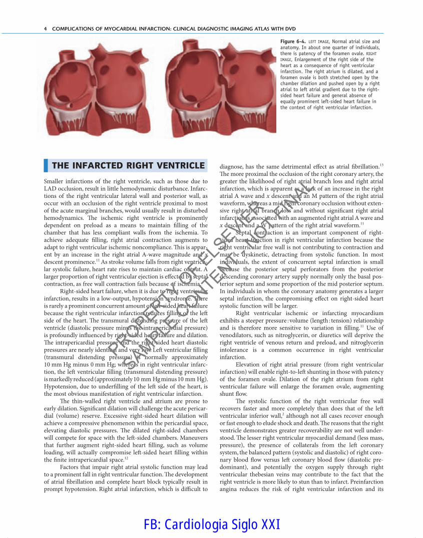
ruptures)• Patency of a foramen ovale with right-to-left shunting (hypox-
emia unresponsive to supplemental oxygen) (Fig. 6-4)• Pulmonary embolism of a right ventricular apical thrombus• Pacer perforations• Pericarditis
THE NORMAL RIGHT VENTRICLE
Th e stroke volume of the right ventricle is the same as that of the left ventricle, but the right ventricle pumps against a lesser arterial pressure (one fi ft h of systemic) and resistance (one tenth of sys-temic); hence, the right ventricle in its usual state is a less forceful pump, having one sixth of the muscle mass, having one third of the wall thickness, and performing one fourth of the stroke work of the systemic ventricle. Th e right ventricle receives systemic venous blood at lower diastolic fi lling pressures than does the left ventricle and also with greater respiratory fl uctuations in fi lling pressure. Th e normal right ventricle functions despite variations in preload. In contradistinction, the ischemic right ventricle is highly sensitive to •• despite variations in preload. Th e pattern of contraction of the right ventricle diff ers from that of the left ven-tricle, which contracts radially and longitudinally. Th e right ven-tricular free wall contracts toward the interventricular septum, with also some longitudinal shortening. Th e interventricular septum normally pushes into the right ventricle, contributing to right ventricular function.
Figure 6-2. Blood supply to the right ventricle. The wall of the right ventricle is depicted as a pale blue rim and the cavity as a darker space. The normal vascular anatomy that supplies the right ventricle is composed of the conus branch that perfuses the RVOT region and that may evolve to collateralize to the LAD; acute marginal branches, of which there are usually two or three angiographically apparent branches that perfuse the lateral-free wall of the right ventricle; small diagonal branches that arise from the LAD anteriorly to supply the medial anterior wall of the right ventricle and often its apex; and small diagonal branches that arise from the posterior descending coronary artery that supply the medial posterior aspect of the right ventricle. The inferior septum, which contributes to right ventricular systolic and diastolic function, is supplied by inferior septal branches of the posterior descending coronary artery (not shown) as the anterior superior septum is similarly supplied by septal perforator branches by the LAD, which also contribute to right ventricular function or dysfunction. Atrial branches also arise from the right coronary artery, and the artery to the sinoatrial node is usually the fi rst branch of the right coronary artery. The artery to the atrioventricular node arises in most patients about 1 cm distal to the “crux” of the right coronary–posterior descending artery. The posteromedial papillary muscle is perfused by branches of the right coronary from the posterior descending coronary artery or posteroventricluar branches off the ongoing distal right coronary artery. The right coronary artery supplies a large number of structures within the heart, and the permutations of cardiac dysfunction are greater in right coronary infarction than in infarction caused by either other coronary artery.
Figure 6-3. In ≤10% of patients, the left circumfl ex coronary artery supplies perfusion to a larger extent of the right ventricle than usual, through posterior diagonal branches off the posterior descending coronary artery and from acute marginal branches of an ongoing circumfl ex after the crux.
1
PROPERTY OF E
LSEVIE
R
SAMPLE C
ONTENT - NOT FIN
AL
FB: Cardiologia Siglo XXI
4 COMPLICATIONS OF MYOCARDIAL INFARCTION: CLINICAL DIAGNOSTIC IMAGING ATLAS WITH DVD
THE INFARCTED RIGHT VENTRICLE
Smaller infarctions of the right ventricle, such as those due to LAD occlusion, result in little hemodynamic disturbance. Infarc-tions of the right ventricular lateral wall and posterior wall, as occur with an occlusion of the right ventricle proximal to most of the acute marginal branches, would usually result in disturbed hemodynamics. Th e ischemic right ventricle is prominently dependent on preload as a means to maintain fi lling of the chamber that has less compliant walls from the ischemia. To achieve adequate fi lling, right atrial contraction augments to adapt to right ventricular ischemic noncompliance. Th is is appar-ent by an increase in the right atrial A-wave magnitude and x descent prominence.11 As stroke volume falls from right ventricu-lar systolic failure, heart rate rises to maintain cardiac output. A larger proportion of right ventricular ejection is eff ected by septal contraction, as free wall contraction fails because of ischemia.
Right-sided heart failure, when it is due to right ventricular infarction, results in a low-output, hypotension syndrome. Th ere is rarely a prominent concurrent amount of left -sided heart failure because the right ventricular infarction reduces fi lling of the left side of the heart. Th e transmural distending pressure of the left ventricle (diastolic pressure minus the intrapericardial pressure) is profoundly infl uenced by right-sided heart failure and dilation. Th e intrapericardial pressure and the right-sided heart diastolic pressures are nearly identical and very low. Left ventricular fi lling (transmural distending pressure) is normally approximately 10 mm Hg minus 0 mm Hg; whereas in right ventricular infarc-tion, the left ventricular fi lling (transmural distending pressure) is markedly reduced (approximately 10 mm Hg minus 10 mm Hg). Hypotension, due to underfi lling of the left side of the heart, is the most obvious manifestation of right ventricular infarction.
Th e thin-walled right ventricle and atrium are prone to early dilation. Signifi cant dilation will challenge the acute pericar-dial (volume) reserve. Excessive right-sided heart dilation will achieve a compressive phenomenon within the pericardial space, elevating diastolic pressures. Th e dilated right-sided chambers will compete for space with the left -sided chambers. Maneuvers that further augment right-sided heart fi lling, such as volume loading, will actually compromise left -sided heart fi lling within the fi nite intrapericardial space.12
Factors that impair right atrial systolic function may lead to a prominent fall in right ventricular function. Th e development of atrial fi brillation and complete heart block typically result in prompt hypotension. Right atrial infarction, which is diffi cult to
diagnose, has the same detrimental eff ect as atrial fi brillation.13 Th e more proximal the occlusion of the right coronary artery, the greater the likelihood of right atrial branch loss and right atrial infarction, which is apparent as a lack of an increase in the right atrial A wave and x descent and an M pattern of the right atrial waveform, whereas a mid right coronary occlusion without exten-sive right atrial branch loss and without signifi cant right atrial infarction is associated with an augmented right atrial A wave and x descent and a W pattern of the right atrial waveform.11
Septal contraction is an important component of right-sided heart function in right ventricular infarction because the right ventricular free wall is not contributing to contraction and may be dyskinetic, detracting from systolic function. In most individuals, the extent of concurrent septal infarction is small because the posterior septal perforators from the posterior descending coronary artery supply normally only the basal pos-terior septum and some proportion of the mid posterior septum. In individuals in whom the coronary anatomy generates a larger septal infarction, the compromising eff ect on right-sided heart systolic function will be larger.
Right ventricular ischemic or infarcting myocardium exhibits a steeper pressure : volume (length : tension) relationship and is therefore more sensitive to variation in fi lling.11 Use of venodilators, such as nitroglycerin, or diuretics will deprive the right ventricle of venous return and preload, and nitroglycerin intolerance is a common occurrence in right ventricular infarction.
Elevation of right atrial pressure (from right ventricular infarction) will enable right-to-left shunting in those with patency of the foramen ovale. Dilation of the right atrium from right ventricular failure will enlarge the foramen ovale, augmenting shunt fl ow.
Th e systolic function of the right ventricular free wall recovers faster and more completely than does that of the left ventricular inferior wall,2 although not all cases recover enough or fast enough to elude shock and death. Th e reasons that the right ventricle demonstrates greater recoverability are not well under-stood. Th e lesser right ventricular myocardial demand (less mass, pressure), the presence of collaterals from the left coronary system, the balanced pattern (systolic and diastolic) of right coro-nary blood fl ow versus left coronary blood fl ow (diastolic pre-dominant), and potentially the oxygen supply through right ventricular thebesian veins may contribute to the fact that the right ventricle is more likely to stun than to infarct. Preinfarction angina reduces the risk of right ventricular infarction and its
Figure 6-4. LEFT IMAGE, Normal atrial size and anatomy. In about one quarter of individuals, there is patency of the foramen ovale. RIGHT IMAGE, Enlargement of the right side of the heart as a consequence of right ventricular infarction. The right atrium is dilated, and a foramen ovale is both stretched open by the chamber dilation and pushed open by a right atrial to left atrial gradient due to the right-sided heart failure and general absence of equally prominent left-sided heart failure in the context of right ventricular infarction.
PROPERTY OF E
LSEVIE
R
SAMPLE C
ONTENT - NOT FIN
AL
FB: Cardiologia Siglo XXI
RIGHT VENTRICULAR INFARCTION 5
6
complications, presumably because of ischemic preconditioning and collateral recruitment (Table 6-2).7 Less than half of proximal dominant occlusions produce recognized right ventricular infarc-tion. Th e extent of recovery of the right ventricle has prompted some to propose that the right ventricle experiences ischemia rather than infarction.
CLINICAL PRESENTATIONS OF RIGHT VENTRICULAR INFARCTION
• Hypotensive inferior infarction• Inferior infarction, cardiogenic shock• Inferior infarction with venous distention, Kussmaul sign• Inferior infarction, blood pressure intolerant of nitroglycerin• Larger biomarker rise than anticipated from inferior
infarction• Greater amount of hypotension than expected from a fi rst
infarct with a small or moderate creatine kinase rise
Diff erential diagnosis of myocardial infarction with prominent hypotension but without proportionally as much pulmonary edema:
• Underfi lling of the left ventricle• Hypovolemia, may be from medications• Right ventricular infarction
• Vasodepression• Excessive eff ect of medications• Medication-induced anaphylaxis• Vagal vasodepressant state
• Tamponade• Ventricular septal rupture
DIAGNOSIS OF RIGHT VENTRICULAR INFARCTION
Physical Diagnosis
Right ventricular infarction can be diagnosed at the bedside. Th e combination of jugular venous pressure and >8 cm and a Kuss-maul sign is 88% sensitive and 76% specifi c for right ventricular
infarction. Th e triad of hypotension, venous distention, and clear lung fi elds in the setting of inferior infarction is 96% specifi c for right ventricular infarction but only 25% sensitive.14 Hypovole-mia and hypervolemia obscure jugular venous fi ndings of right ventricular infarction.
Electrocardiography
Electrocardiography (ECG) is useful for the diagnosis of right ventricular infarction, as long as the baseline ECG recording is normal and if ECG is performed very early in the infarction. Several ECG patterns have been advanced. Right-sided chest leads (V4R) demonstrating more than 0.5 mm or more than 1 mm of ST elevation suggest right ventricular infarction. Use of 0.5 mV or mm is more sensitive but less specifi c; 1 mm of ST elevation 0.08 second aft er the J point is seen in most (>80%) cases and has a high pathologic correlation with right ventricular infarction (100%).15 A signifi cant caveat is that the fi nding of ST elevation in V4R is evanescent, lasting only 24 to 48 hours and normalizing in half of cases within 10 hours. False-positives are also common and may be generated by pulmonary embolism, pericarditis, acute anteroseptal ST elevation myocardial infarction (STEMI), and anteroseptal aneurysm. ST elevation in V1 through V4 is associ-ated with pure right ventricular infarction.16 ST elevation in stan-dard limb lead III more than in standard limb lead II is 97% sensitive and 70% specifi c.17
Other common, clinically important but not diagnostic ECG fi ndings in right ventricular infarction include sinus brady-cardia, sinus tachycardia, atrial fi brillation (seen in up to one third of cases),18 atrioventricular block and third-degree block, and right bundle branch block. Right bundle branch block has been noted in up to 48% of cases of right ventricular infarction and is associated with a poor prognosis.19,20
Echocardiography
Echocardiography is useful to corroborate fi ndings of right ven-tricular systolic dysfunction and to identify or to exclude associa-tions and complications of inferior infarction or of the right ventricular infarction itself. Older echocardiographic and nuclear signs of right ventricular infarction were simply right ventricular dilation and overall systolic dysfunction. With improved imaging, assessment of regional right ventricular systolic function is now possible and useful because it correlates with the right coronary anatomy and the location of occlusion. Th e posterior wall of the right ventricle is most commonly aff ected by right ventricular infarction because it is the most distal right coronary territory.21 Combined right ventricular lateral and posterior wall motion abnormalities are produced by more proximal right coronary artery occlusions and are the typical fi ndings of hemodynamically signifi cant right ventricular infarction. In the context of inferior infarction, right ventricular akinesis or hypokinesis (segmental) is 83% sensitive and 93% specifi c for right ventricular infarction.22
Right ventricular wall motion abnormalities do occur in other disease states, such as pulmonary hypertension, pulmonary embolism, and trauma, and aft er poor pump protection. Views that assist with right ventricular regional wall motion assessment are listed in Table 6-3. Echocardiographic fi ndings in right ven-tricular infarction are summarized in Table 6-4.
Table 6-2. Preinfarction Angina Decreases the Risk of Right
Ventricular Infarction and Complications
With Angina
(N = 62)
Without Angina
(N = 51)
ST elevation V4R 27% 71% <.001
Complete AV block 11% 33% .04
Hypotension or shock 8% 53% <.001
In-hospital death 5% 10% .26
From Shiraki H, Yoshikawa T, Anzai T, et al: Association between preinfarction angina and a
lower risk of right ventricular infarction. N Engl J Med 1998;338:941-947.
2
3
4
PROPERTY OF E
LSEVIE
R
SAMPLE C
ONTENT - NOT FIN
AL
FB: Cardiologia Siglo XXI

6 COMPLICATIONS OF MYOCARDIAL INFARCTION: CLINICAL DIAGNOSTIC IMAGING ATLAS WITH DVD
becomes broad and sometimes bifi d, refl ecting lesser dP/dt (upslope from systolic failure), impaired relaxation (reduced −dP/dt), and septal bulge into the right ventricle.
Th e most prominent pressure waveform of right ventricu-lar infarction is the combination of diastolic failure (elevated right atrial and right ventricular diastolic pressure) and systolic failure (lower right ventricular systolic pressure). Th us, a right atrial pressure : pulmonary capillary wedge pressure (RAP : PCWP) above 0.86 (normal <0.6) is 82% sensitive and 97% specifi c for right ventricular infarction in the setting of inferior infarction.26
MANAGEMENT
Management principles are based on the following:
• Optimize right ventricular preload
• Optimize volume status (central venous pressure [CVP] of 15 mm Hg)
• Maintain atrioventricular synchrony by maintaining or restoring sinus rhythm (if in atrial fi brillation) or sequen-tial pacing for complete heart block
• Reduce right ventricular aft erload
• Normalize left atrial pressure• Consider use of nitric oxide
• Inotrope stimulation of stunned right ventricular tissue
• Maintain arterial pressure
• Vasopressor support• Intra-aortic balloon counterpulsation (IABP) use
• Reperfusion (early)• In case of intractable circulatory failure, consider:
• Assist device• Transplantation
Table 6-3. Echocardiographic Views That Assist with Right Ventricular Regional Wall Motion Assessment
Segment of the
Right Ventricle
Vascular Supply If Wall Motion Assessment Suggests Site of Occlusion
Echocardiographic view
Parasternal long-axis
view
RVOT Conus branch Very proximal RCA
Parasternal short-axis
view
Anterior RV Diagonal branches off the LAD LAD
Lateral wall Acute marginal branches Before one (mid RCA) or all acute marginal (proximal RCA)
branches
Posterior wall Diagonal branches off the PDA/PIV PDA/PIV or anywhere proximal to PDA/PIV, including dominant
left circumfl ex artery
RV infl ow view
Apical 4-chamber view Lateral wall Acute marginal branches Before one (mid RCA) or all acute marginal (proximal RCA)
branches
Subcostal long-axis view Lateral wall Acute marginal branches Before one (mid RCA) or all acute marginal (proximal RCA)
branches
Subcostal long-axis view Posterior wall Diagonal branches off the PDA/PIV PDA/PIV or anywhere proximal to PDA/PIV, including dominant
left circumfl ex artery
Lateral wall Acute marginal branches Before one (mid RCA) or all acute marginal (proximal RCA)
branches
Anterior RV Diagonal branches off the LAD LAD
LAD, left anterior descending coronary artery; PDA/PIV, posterior descending (interventricular) artery; RCA, right coronary artery; RV, right ventricle; RVOT, right ventricular outfl ow tract.
Radionuclide Angiography
Th e diagnosis of right ventricular infarction by radionuclide tech-nique is possible by use of the combination of overall depression of right ventricular systolic function (<40%; normal range is 45% to 75%) and regional right ventricular systolic dysfunction.1 Tech-netium pyrophosphate scanning is specifi c but not sensitive.1
Cardiac Magnetic Resonance
Cardiac magnetic resonance (MR) is able to image the increased myocardial water (edema) by T2 weighting and by gadolinium contrast enhancement.25 Cardiac MR SSFP sequences elegantly depict right and left ventricular regional wall motion.
Hemodynamics, Cardiac Catheterization, and Angiography
Right-sided heart catheterization is seldom necessary for diagno-sis of right ventricular infarction, but waveforms are revealing of the physiology. As described before, as long as right atrial infarc-tion has not occurred, right atrial systolic function (Starling forces) is recruited to enable fi lling of a noncompliant right ven-tricle, resulting in an augmented A wave and x descent of the right atrial pressure and a W pattern. When signifi cant right atrial infarction has occurred, the right atrial A wave is therefore dimin-ished, as is the x descent, and the right atrial waveform has an M pattern.12 Pericardial restraint will also contribute to the M pattern (“square root” or “dip and plateau” pattern) typical of severe and larger right ventricular infarction with impaired right ventricular compliance and pericardial restraint.21 Th e x descent predomi-nance can also be seen. Th e right ventricular systolic waveform
PROPERTY OF E
LSEVIE
R
SAMPLE C
ONTENT - NOT FIN
AL
FB: Cardiologia Siglo XXI

RIGHT VENTRICULAR INFARCTION 7
6
However, excess volume loading, in addition to congesting the venous vasculature and potentially compromising organ (especially hepatic) function, leads to overfi lling of the right side of the heart and, because of ventricular interdependence, increases left -sided heart diastolic pressures while decreasing left -sided heart volumes, stroke volume, and cardiac output. Furthermore, experimental canine models of right ventricular infarction estab-lish that volume loading in the context of intact pericardium increases right ventricular infarction twofold to threefold com-pared with pericardiectomy, probably by reducing right ventricu-lar perfusion gradient (Table 6-5; Figs. 6-5 to 6-7).27,28
Above a right atrial pressure of 15 mm Hg, volume loading does not further improve cardiac index or mean arterial blood pressure, but inotropic stimulation (5 to 10 μg/min/m2 dobuta-mine) does (Figs. 6-8 and 6-9).29
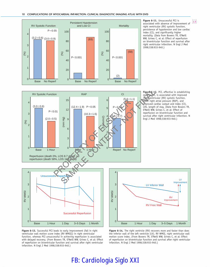
A small series demonstrated the need and the benefi ts of maintaining atrioventricular synchrony; atrial pacing at the same rate increased the stroke volume by a mean of 42%.30 Observa-tional data suggest that when reperfusion can be established, the mean right atrial pressure nearly normalizes (Fig. 6-10),31 and right ventricular systolic function (ejection fraction) may also nearly normalize.32 Unsuccessful percutaneous coronary inter-vention (PCI) is associated with absence of improvement of right ventricular systolic function, persistence of hypotension and low cardiac index, and signifi cantly higher mortality (Fig. 6-11).2 Among patients in whom PCI was successful, the percentage with persistent hypotension and low cardiac output is signifi -cantly less, as is mortality.2 Th e eff ect of successful PCI on right ventricular systolic function, right atrial pressure, and cardiac index is demonstrable at 1 hour aft er the procedure and is yet further •• at 1 and 3 days (Figs. 6-12 to 6-14).2 Th e percentage of cases in which right ventricular systolic function has normalized at 1 month is signifi cantly greater aft er successful reperfusion (Fig. 6-15).2
Although PCI has not been shown in large prospec -tive trials to reduce mortality in right ventricular infarction, the
Table 6-4. Echocardiographic Findings in Right Ventricular Infarction
Right ventricular dilation
Right ventricular segmental systolic dysfunction
Reduced motion of the right atrioventricular groove due to reduced
right ventricular longitudinal contraction
Right ventricular dilation (+ small left ventricle = interdependence)
Signs of elevated right atrial pressure
Right atrial dilation
Inferior vena cava dilation and reduced collapsibility
Interatrial septal deviation to the left side
Signs of elevated right ventricular diastolic pressure, low pulmonary
artery diastolic pressure
Complete (presystolic) pulmonic valve opening23
Flow across a patent foramen ovale from the right to the left side
(color Doppler fl ow mapping or agitated saline study)
Tricuspid regurgitation
Right ventricular papillary muscle rupture
Reduced cardiac output or index
Right ventricular systolic pressure not elevated, due to right
ventricular systolic failure
Premature (post A wave) pulmonary valve opening (right ventricular
noncompliance)
Right ventricular thrombus
Right atrial thrombus24
Right ventricular pseudoaneurysm
Complications of associated inferior wall or inferior septal infarction
Wall motion abnormalities
Aneurysms
False aneurysms
Intramyocardial hematoma
Septal rupture
Papillary muscle rupture
(Nonrupture) mitral insuffi ciency
Table 6-5. Effect of Volume Loading on Experimental Right
Ventricular Infarction
Experimental RVMI Volume Loading
RVSP −25% +28%
RVEF % −57% +67% (stroke work)
Aortic pressure −36% +35%
Cardiac output −32% +35%
RVEF, right ventricular ejection fraction; RVMI, right ventricular myocardial infarction; RVSP,
right ventricular systolic pressure.
From Goldstein JA, Vlahakes GJ, Verrier ED, et al: Volume loading improves low cardiac output
in experimental right ventricular infarction. J Am Coll Cardiol 1983;2:270-278.
0
1.00
(mm
Hg)
1.25
0.75
0.50
0.25
1.50
Peak A/Mean RAP
*
*
RVMI �RAMIBase0
0.1
(gm
m/m
2 )
0.2
0.3RA SW
*
*
RVMI �RAMIBase
Figure 6-5. Experimental right ventricular (RV) infarction increases right atrial (RA) function (stroke work [SW]) because of Starling recruitment. Right atrial infarction results in right atrial mechanical failure. MI, myocardial infarction; RAP, right atrial pressure. (Data from Goldstein JA, Vlahakes GJ, Verrier ED, et al: The role of right ventricular systolic dysfunction and elevated intrapericardial pressure in the genesis of low output in experimental right ventricular infarction. Circulation 1982;65:513-522.)
It is important to achieve adequate preload for the context of right ventricular infarction. Alleviation or avoidance of under-fi lling is benefi cial, and optimization of the right ventricular dia-stolic pressure may improve the cardiac output. In many patients, judicious volume infusion corrects hypotension.
5
6
7
PROPERTY OF E
LSEVIE
R
SAMPLE C
ONTENT - NOT FIN
AL
FB: Cardiologia Siglo XXI

8 COMPLICATIONS OF MYOCARDIAL INFARCTION: CLINICAL DIAGNOSTIC IMAGING ATLAS WITH DVD
0
25
(mm
Hg)
30
20
15
10
5
35
RV SP
*
*
RVMI �RAMIBase0
5
(gm
m/m
2 )
6
4
3
2
1
7RV SW
*
*
RVMI �RAMIBase0
(%)
80
60
40
20
100
LV EDA
*
RVMI �RAMIBase0
5
(mm
Hg)
10
15LV EDP
*
RVMI �RAMIBase0
100
(gm
m/m
2 )
120
80
60
40
20
140
Syst SBP
*
*
RVMI �RAMIBase0
25
(gm
m/m
2 )
30
20
15
10
5
SV
*
*
RVMI �RAMIBase
0
15
(mm
Hg)
20
10
25RV SBP
*
*
MI MI/PericPre0
3
2
1
(mm
Hg)
4
5
0
3
2
1
4
5RV MDP
*
*
MI MI/PericPre
0
40
60(mm
Hg) 80
20
140
120
100
Syst MAP
*
*
MI MI/PericPre 0
(mL)
5
20
15
10
SV
*
*
MI MI/PericPre 0
(L/m
in/m
2 )
1.0
0.5
2.5
2.0
1.5
CI
*
*
MI MI/PericPre 0
(%)
60
40
20
120
100
80
LV EDV
*
MI MI/PericPre
(gm
/m/m
2 )
RV SWI
*
*
MI MI/PericPre
Figure 6-6. Experimental right atrial infarction worsens right ventricular (RV) systolic function in right ventricular infarction and results in underloading of the left ventricle and worsened arterial pressure. LV EDA, ••; LV EDP, left ventricular end-diastolic pressure; SBP, systolic blood pressure; SP, stroke power; SV, stroke volume; SW, stroke work. (Data from Goldstein JA, Vlahakes GJ, Verrier ED, et al: The role of right ventricular systolic dysfunction and elevated intrapericardial pressure in the genesis of low output in experimental right ventricular infarction. Circulation 1982;65:513-522.)
Figure 6-7. In experimental right ventricular infarction, hemodynamic indices are improved by pericardiotomy, revealing that pericardium-mediated ventricular interdependence contributes to left-sided heart underloading and output. CI, cardiac index; LV EDV, left ventricular end-diastolic volume; MAP, mean arterial pressure; MDP, mean diastolic pressure; RV, right ventricle; SBP, systolic blood pressure; SWI, stroke work index; SV, stroke volume. (Data from Goldstein JA, Vlahakes GJ, Verrier ED, et al: The role of right ventricular systolic dysfunction and elevated intrapericardial pressure in the genesis of low output in experimental right ventricular infarction. Circulation 1982;65:513-522.)
8
9
10
PROPERTY OF E
LSEVIE
R
SAMPLE C
ONTENT - NOT FIN
AL
FB: Cardiologia Siglo XXI

RIGHT VENTRICULAR INFARCTION 9
6
0
5
20
15
10
RAPDobutamine
11 patientswith IWIand RVMI,RAP�10,BP�100,CI�2,and STeinferiorlyand V4R
* *
DBTM5
DBTM10
Base DBTM5
DBTM10
Base DBTM5
DBTM10
Base0
1.0
0.5
2.5
3.0
2.0
1.5
CI
*
*
Dobutamlne (best dose 10 mg/kg/min), increases CI and MBP.
0
60
40
20
120
100
80
Mean BP (mm Hg)
* *
0
5
20
15
10
RAPVolumeLoading
11 patientswith IWIand RVMI,RAP�10,BP�100,CI�2,and STeinferiorlyand V4R
*
PostPre PostPre PostPre0
1.0
0.5
1.5
2.0
CI
p�NS
p�NS
Above an RAP of 15, there is no increase in CI or systemic BP.
0
60
40
20
120
100
80
Mean BP (mm Hg)
0
4
16
10
14
12
8
6
2
(mm
Hg)
0
4
16
10
14
12
8
6
2
(mm
Hg)
RAP
27 patientswith RVMIof 141 ptswith InfMI,17 rec’dPTCAwithin24 hours,10 did not
ReperfusionMI NoReperfusion
MI
RAP
p�0.05
p�NS
Figure 6-8. In patients with right ventricular infarction and inferior left ventricular infarction optimized with right atrial pressure above 10 mm Hg but still hypotensive and with low output, inotropic doses of dobutamine lower right atrial pressure (RAP) and increase cardiac index (CI) and arterial blood pressure. IWI, inferior wall infarction; RVMI, right ventricular myocardial infarction. (Data from Ferrario M, Poli A, Previtali M, et al: Hemodynamics of volume loading compared with dobutamine in severe right ventricular infarction. Am J Cardiol 1994;74:329-333.)
Figure 6-9. In patients with right ventricular infarction and inferior left ventricular infarction already with optimal right ventricular fi lling pressures, further volume loading does not improve arterial hemodynamics. CI, cardiac index; IWI, inferior wall infarction; RAP, right atrial pressure; RVMI, right ventricular myocardial infarction. (Data from Ferrario M, Poli A, Previtali M, et al: Hemodynamics of volume loading compared with dobutamine in severe right ventricular infarction. Am J Cardiol 1994 1994;74:329-333.)
Figure 6-10. Successful reperfusion reduces mean right atrial pressure (RAP). PTCA, percutaneous transluminal coronary angioplasty; RVMI, right ventricular myocardial infarction. (Data from Kinn JW, Ajluni SC, Samyn JG, et al: Rapid hemodynamic improvement after reperfusion during right ventricular infarction. J Am Coll Cardiol 1995;26:1230-1234.)
11
12
PROPERTY OF E
LSEVIE
R
SAMPLE C
ONTENT - NOT FIN
AL
FB: Cardiologia Siglo XXI
10 COMPLICATIONS OF MYOCARDIAL INFARCTION: CLINICAL DIAGNOSTIC IMAGING ATLAS WITH DVD
0
1
4
3
2
RV Systolic Function(%
)
(%)
No ReperfBase No ReperfBase No ReperfBase
(3.0�0.9)(3.2�0.6)(83)
(58)
(2)0
60
40
20
80
100
(%) 60
40
20
0
80
100
Persistent Hypotensionand Low CI
P�0.001
P�0.55
P�0.001
Mortality
0
1
4
3
2
RV Systolic Function
(%)
(mm
Hg)
1 HourBase 1 HourBase No ReperfReperf
(2.0�0.5)
(3.0�0.6)
(10.3�1.6)
(12.4�1.9)
(5.6�1.1)
(3.6�0.7)
0
9
6
3
12
15(L
/min
/m2) 4
3
2
1
0
5
6
RAP
P�0.05
P�0.01
Reperfusion (death 0%, LOS 8.7 days), noreperfusion (death 58%, LOS 11.8 days)
P�0.04
CI
0
3
2
1
4
1 HourBase 1 Day
Successful Reperfusion
Unsuccessful Reperfusion
3–5 Days 1 Month
RV
WM
SI
0
3
2
1
4
1 HourBase 1 Day
RV Free Wall
LV Inferior Wall
LVImprovement
RVImprovement
3–5 Days 1 Month
RV
WM
SI
Figure 6-11. Unsuccessful PCI is associated with absence of improvement of right ventricular (RV) systolic function, persistence of hypotension and low cardiac index (CI), and signifi cantly higher mortality. (Data from Bowers TR, O’Neill WW, Grines C, et al: Effect of reperfusion on biventricular function and survival after right ventricular infarction. N Engl J Med 1998;338:933-940.)
Figure 6-12. PCI, effective in establishing reperfusion, is associated with improved right ventricular (RV) systolic function, lower right atrial pressure (RAP), and improved cardiac output and index (CI). LOS, length of stay. (Data from Bowers TR, O’Neill WW, Grines C, et al: Effect of reperfusion on biventricular function and survival after right ventricular infarction. N Engl J Med 1998;338:933-940.)
Figure 6-13. Successful PCI leads to early improvement (fall in right ventricular wall motion score index [RV WMSI]) in right ventricular function, whereas PCI unsuccessful in achieving reperfusion is associated with delayed recovery. (From Bowers TR, O’Neill WW, Grines C, et al: Effect of reperfusion on biventricular function and survival after right ventricular infarction. N Engl J Med 1998;338:933-940.)
Figure 6-14. The right ventricle (RV) recovers more and faster than does the inferior wall of the left ventricle (LV). RV WMSI, right ventricular wall motion score index. (From Bowers TR, O’Neill WW, Grines C, et al: Effect of reperfusion on biventricular function and survival after right ventricular infarction. N Engl J Med 1998;338:933-940.)
13
PROPERTY OF E
LSEVIE
R
SAMPLE C
ONTENT - NOT FIN
AL
FB: Cardiologia Siglo XXI

RIGHT VENTRICULAR INFARCTION 11
6
0
60
% N
orm
al a
t 1 M
onth
80
40
20
100
LVRV
(8)
(98)
p�0.001
Figure 6-15. At 1 month after infarction, almost all survivors have normal right ventricular (RV) systolic function, but few have normalized left ventricular (LV) systolic function. (Data from Bowers TR, O’Neill WW, Grines C, et al: Effect of reperfusion on biventricular function and survival after right ventricular infarction. N Engl J Med 1998 1998;338:933-940.)
available evidence favors its use, particularly early use, and sug-gests that it is underused.
Th erapy for right ventricular infarction cases should be individualized. General principles of therapy are summarized in Table 6-6.33
OUTCOMES
Th e in-hospital mortality of right ventricular infarction is, as expected, graded according to the severity of the hemodynamic disturbances (Killip I: 5% mortality; Killip IV, cardiogenic shock: 55% mortality) and the occurrence of complications or associated lesions. Th e in-hospital mortality of inferior infarction without right ventricular infarction is 6%; with right ventricular infarc-tion, 31%; and with right ventricular infarction and cardiogenic shock, 50% to 60%.34
Table 6-6. General Principles of Therapy for Right Ventricular
Infarction Cases
Type of Case Therapy
Asymptomatic Avoid diuretics and vasodilators that
may precipitate low output and
hypotension.
Symptomatic low-output
state and normotensive
with right atrial
pressure or pulmonary
capillary wedge pressure
<15 mm Hg
Add fl uid to increase the pulmonary
capillary wedge pressure to
15-18 mm Hg.
If cardiac output does not increase
adequately, add a vasodilator.
If the cardiac output still does not
increase adequately, add dobutamine
or amrinone.
Reperfusion therapy should be
considered.
Symptomatic low-output
state and normotensive
with right atrial
pressure or pulmonary
capillary wedge pressure
>15 mm Hg
Add intravenous dobutamine or
amrinone.
Additional vasodilator with fl uid therapy
support may be added.
Reperfusion therapy should be
considered.
Cardiogenic shock Sustain blood pressure with dopamine;
additional dobutamine may increase
cardiac output.
Consider right ventricular assist or
pulmonary artery counterpulsation for
select cases.
Reperfusion therapy should be strongly
considered.
Th e long-term survival of patients discharged from the hospital is good. Most do recover a large proportion of right ventricular systolic function and therefore have their prognosis determined by the degree of postinfarction left ventricular func-tion and the extent and severity of coronary artery disease. However, postinfarction persistence of right ventricular dysfunc-tion is an independent predictor of the development of heart failure and of death.35
CASE 1
History
� 83-year-old man with chronic renal insuffi ciency, presenting with 9
hours of ischemic chest pain
Physical Examination
� BP 110/60 mm Hg, HR 48 bpm, RR 12/min
� Venous distention to the angle of the jaw
� Normal S1, S2; S4 present
� No murmurs or rubs
� Extremities warm, mentation normal
Management
� Avoidance of preload-reducing maneuvers
� As the neck veins were indicative of adequate preload, no volume
was given.
� As the output was clinically adequate, no inotropes were given.
Evolution and Outcome
� Right-sided heart failure resolved after 3 hours.
� The remainder of the hospital course was uneventful: no recurrent
pains, no reversible defect on perfusion scanning.
� Well at follow-up
Comments
� Right ventricular myocardial infarction diagnosed by elevated neck
veins in the context of an acute inferior STEMI; echocardiography
corroborative
� No major hemodynamic disturbances other than mild hypotension
� Early spontaneous recovery of right ventricular systolic function
PROPERTY OF E
LSEVIE
R
SAMPLE C
ONTENT - NOT FIN
AL
FB: Cardiologia Siglo XXI

12 COMPLICATIONS OF MYOCARDIAL INFARCTION: CLINICAL DIAGNOSTIC IMAGING ATLAS WITH DVD
I aVR V1 V4
II aVL V2 V5
III
V1
aVF V3 V6
Figure 6-16. TOP, ECG shows sinus rhythm, inferior ST elevation, and inverted T waves. Lateral ST elevation and inverted T waves suggest acute inferolateral infarction. BOTTOM, Chest radiograph shows right lung fi eld, no pulmonary edema, and large left pleural effusion.
PROPERTY OF E
LSEVIE
R
SAMPLE C
ONTENT - NOT FIN
AL
FB: Cardiologia Siglo XXI

RIGHT VENTRICULAR INFARCTION 13
6
Figure 6-17. Transthoracic echocardiography. TOP ROW, Apical 4-chamber views oriented to the right. There is dilation and “sphericalization” of the right ventricle, consistent with infarction and systolic dysfunction. There is moderate tricuspid insuffi ciency due to the right ventricular systolic dysfunction. MIDDLE LEFT, Tricuspid regurgitation spectral profi le. Right ventricular systolic pressure: 4 × V2 + RAP = 13 + 15 = 28 mm Hg. The right ventricular systolic pressure is low for a typical myocardial infarction case with shock, and the diastolic pressure (the right atrial pressure) is disproportionately high (50% of the systolic pressure). The delayed velocity rise (slope) of the tricuspid regurgitation is consistent with impaired right ventricle dP/dt. MIDDLE RIGHT, Right atrial and right ventricular dilation. There is marked systolic bulging of the right atrium due to right atrial pressure exceeding that of the left atrium. BOTTOM ROW, Diastolic (LEFT) and systolic (RIGHT) views. There is diastolic fl attening of the interventricular septum due to elevation of the right ventricular diastolic pressure from the right ventricular infarction.
CASE 2
History
� 75-year-old man presenting with 3 syncopal episodes; no chest
pain
� Found by EMS to be in third-degree aortic valve block, BP
55/– mm Hg
� Mechanical ventilation and vasopressors started at community
hospital
� Transferred for a pacer
Physical Examination
� BP 90/60 mm Hg, HR 55 bpm
� Cool extremities
� Head trauma from syncope, 5-cm laceration across the forehead
� Venous distention to the angle of the jaw
� Quiet heart sounds
� No gallops, rubs, or murmurs
� CK 3000, creatinine 300
Management
� Temporary pacer inserted, with poor capture
� High-dose vasopressors and inotropes used
� IABP inserted
Outcome
� Arrived in shock, remained in shock
� Died within an hour of arrival, within 4 hours of presentation to
the community hospital
PROPERTY OF E
LSEVIE
R
SAMPLE C
ONTENT - NOT FIN
AL
FB: Cardiologia Siglo XXI

14 COMPLICATIONS OF MYOCARDIAL INFARCTION: CLINICAL DIAGNOSTIC IMAGING ATLAS WITH DVD
I aVR V1 V4
II aVL V2 V5
III aVF V3 V6
Figure 6-18. TOP, ECG shows complete heart block with an irregular wide complex ventricular rhythm. There is no pacemaker capture due to right ventricular infarction. MIDDLE, Chest radiograph shows normal-sized cardiopericardial silhouette, no pulmonary edema. BOTTOM, ECG on arrival shows asystole with ventricular escape beats.
Figure 6-19. Transthoracic echocardiography. LEFT IMAGE, Parasternal long-axis view. Contracting anterior septum, akinesis of the posterior wall. Prominent jet of mitral regurgitation. RIGHT IMAGE, Subcostal view (right ventricle free wall akinesis). Right ventricular and atrial dilation and tricuspid insuffi ciency.
� Died despite the preceding measures and before angiography could
be obtained
Comments
� Massive right ventricular infarction associated with inferior
infarction
� Cardiogenic shock in the context of an inferior STEMI is usually due
to right ventricular infarction, as it was here.
� Acute right-sided heart failure (venous distention and hypotension)
is usually due to tamponade, pulmonary embolism, or right
ventricular myocardial infarction.
� The diffi culty in achieving capture with the pacer was likely due to
the right ventricle’s infarction.
� Pulseless electrical activity likely resulted from progression of the
right ventricle’s failure, worsened by acidosis; right ventricle
perforation, after infarction or due to the pacer.
� An unfortunate example of the real risk that some right ventricular
infarctions confer.
PROPERTY OF E
LSEVIE
R
SAMPLE C
ONTENT - NOT FIN
AL
FB: Cardiologia Siglo XXI

RIGHT VENTRICULAR INFARCTION 15
6
CASE 3
History
� 79-year-old man collapsed at home
� Recent prolonged air travel
� Various chest pains the day before
Physical Examination
� Awake, intubated, very weak, cool extremities
� BP 80/50 mm Hg, HR 110 bpm, tachypneic
� Venous distention to the angle of the jaw, S3
� No murmurs or rubs
� CK 500
� Oliguric
Clinical Impression
� Although there was a high index of suspicion of pulmonary
embolism, CT scanning was normal, as were leg Doppler studies.
� The patient was ventilated for airway management.
� Transesophageal echocardiography was performed because
transthoracic images were poor.
Management
� Avoidance of preload reduction
� With pulmonary artery catheter guidance, the CVP was kept at 15
to 18 mm Hg.
� Dobutamine was given to increase cardiac output.
� Hemodynamics were acceptable with this regimen, and his
hemodynamics spontaneously normalized on day 5.
Outcome
� Discharged angina and heart failure free
Comments
� Large submassive right ventricular infarction
� Late presentation
� Volume optimizing and inotropes corrected the hypotension and
low output.
� Spontaneous recovery
� Postdischarge echo showed normalized right ventricular systolic
function.
I aVR V1 V4
II aVL V2 V5
III
V1
aVF V3 V6
Figure 6-20. TOP, ECG shows sinus tachycardia with one premature ventricular contraction and nonspecifi c repolarization abnormalities. BOTTOM, Chest radiograph shows cardiomegaly and mild pulmonary edema.
Figure 6-21. Transesophageal echocardiography images show marked right atrial and right ventricular dilation. The left-sided heart chambers are small because of underloading. Interatrial septal bulging to the left side suggests that right atrial pressure is greater than left atrial pressure. The right ventricular free wall is severely hypokinetic or akinetic, despite inotropic support. There is no pericardial effusion.
PROPERTY OF E
LSEVIE
R
SAMPLE C
ONTENT - NOT FIN
AL
FB: Cardiologia Siglo XXI
16 COMPLICATIONS OF MYOCARDIAL INFARCTION: CLINICAL DIAGNOSTIC IMAGING ATLAS WITH DVD
CASE 4
History
� 54-year-old man developed indigestion at 5 PM
� At 7 PM, experienced a cardiac arrest on the street
� 20 minutes of bystander CPR, defi brillation by EMS
Physical Examination
� Deeply comatose, basal skull laceration
� BP 80/50 mm Hg, HR 100 bpm, ventilated
� Marked venous distention
� S1, S2 normal
� S4
� No murmurs or rubs
Impression and Evolution
� Although there was a high index of suspicion of pulmonary
embolism, CT scanning and leg Doppler studies were normal.
� The patient was ventilated for airway management.
� Transesophageal echocardiography was performed because
transthoracic images were poor.
Management
� Avoidance of preload reduction
� With pulmonary artery catheter guidance, the CVP was kept at 15
to 18 mm Hg.
� Dobutamine was given to increase cardiac output.
� Hemodynamics were acceptable with this regimen, and his
hemodynamics spontaneously normalized on day 5.
Outcome
� Arrived in shock, remained in shock
� After 5 days, hemodynamic therapy was no longer needed, but
ventilatory support was continued because of persistent neurologic
impairment.
� Eventually (60 days later), he died of sepsis while still on a
ventilator.
Comments
� Large right ventricular infarction complicating inferior STEMI
� Initial hypotension due to right ventricular myocardial infarction
and recent cardiac arrest
� Hemodynamic resuscitation was straightforward.
� Clinical outcome was determined by neurologic sequelae of the
out-of-hospital cardiac arrest, not cardiac performance.
I aVR V1 V4
II aVL V2 V5
III aVF V3 V6
I aVR V1 V4
II aVL V2 V5
III aVF V3 V6
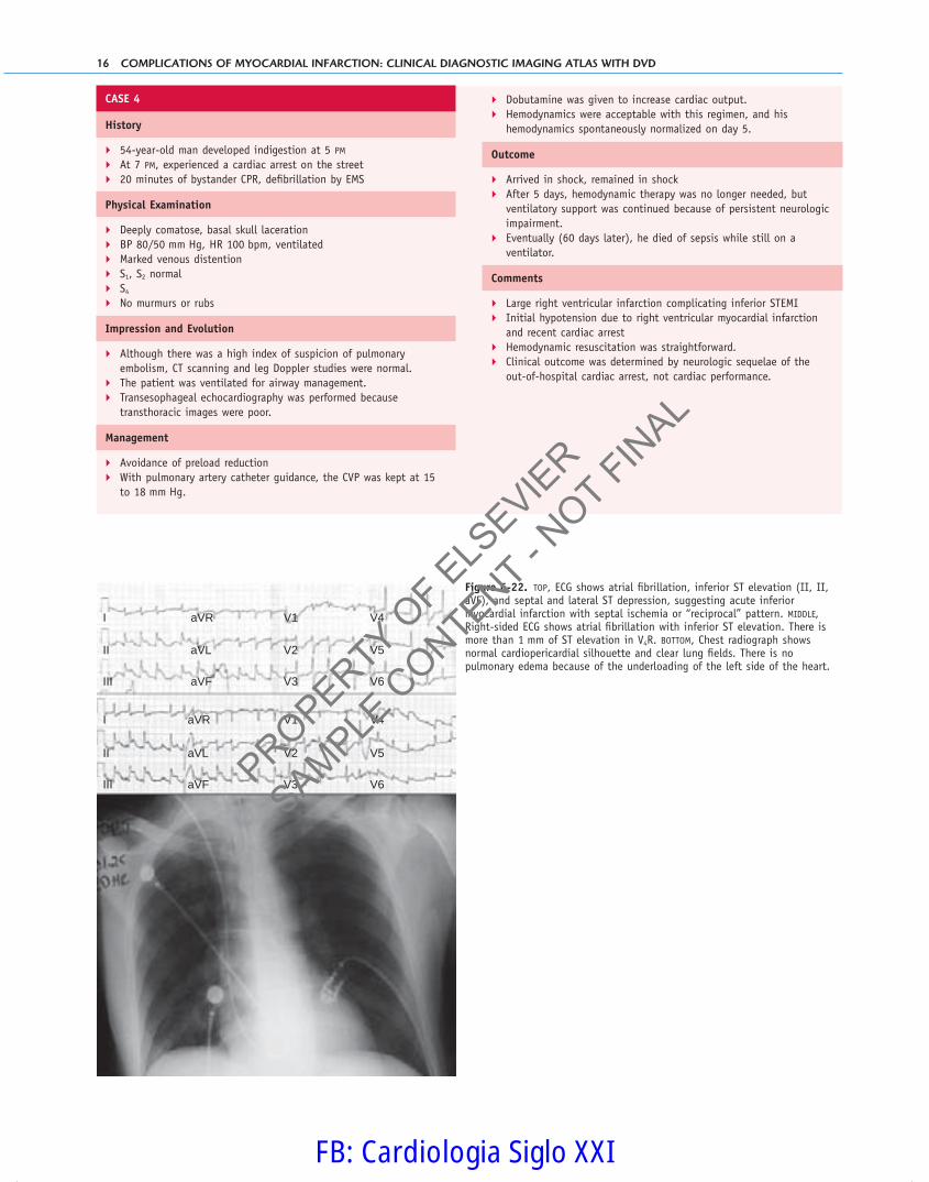
Figure 6-22. TOP, ECG shows atrial fi brillation, inferior ST elevation (II, II, aVF), and septal and lateral ST depression, suggesting acute inferior myocardial infarction with septal ischemia or “reciprocal” pattern. MIDDLE, Right-sided ECG shows atrial fi brillation with inferior ST elevation. There is more than 1 mm of ST elevation in V4R. BOTTOM, Chest radiograph shows normal cardiopericardial silhouette and clear lung fi elds. There is no pulmonary edema because of the underloading of the left side of the heart.
PROPERTY OF E
LSEVIE
R
SAMPLE C
ONTENT - NOT FIN
AL
FB: Cardiologia Siglo XXI

RIGHT VENTRICULAR INFARCTION 17
6
VmaxVmeanPmaxPmeanEnv. TiVTIHRDiaCo
�0.75 m/s 0.49 m/s 2.27 mmHg 1.18 mmHg 0.22 s 10.63 cm 83.73 BPM 2.20 cm 3.38 l/min
Figure 6-23. Transthoracic echocardiography. TOP ROW, Apical 3-chamber views in systole (LEFT) and diastole (RIGHT). There is dyskinesis of the basal inferior wall. The two bodies of the posterior papillary muscle are intact. BOTTOM LEFT, Subcostal M-mode study of the inferior vena cava. Caval (inferior vena cava) dilation without respiratory collapse = elevated central venous pressure. BOTTOM RIGHT, Left ventricular outfl ow tract pulsed wave Doppler sampling: reduced VTI (10.6 cm; normal, 18 to 22 cm) consistent with severely reduced stroke volume.
CASE 5
History
� 62-year-old man presented to a community hospital with 9 hours
of chest pain
� Received tPA at hour 10, but pain continues, as do ST elevations
Physical Examination
� Ventilated, BP 85/50 mm Hg, HR 110 bpm
� Prominent venous distention
� S1, S2 normal
� No gallops, rubs, or murmurs
� Chest clear
Impression and Evolution
� Inferior myocardial infarction with large right ventricular
myocardial infarction
� Hypoxemia due to large amount of shunting through a patent
foramen ovale (PFO)
� The ventilator time became protracted because of persistent
elevation of the CVP (18 mm Hg) and shunting through the PFO.
Maintenance of adequate oxygenation was diffi cult.
� There was no signifi cant left ventricular failure (PCWP 13 mm Hg).
� PFO closure with a device was considered, but fi rst it was thought
worth exploring whether occlusion of the PFO would really increase
the oxygenation.
Comments
� Right ventricular infarction complicating inferior STEMI
� Right-to-left shunting through a PFO contributing to arterial
hypoxemia
� Consideration of percutaneous closure of the PFO was given
because the time course on the ventilator was becoming prolonged,
but fevers precluded.
� Eventual recovery of right ventricular systolic function; concurrent
with this was recovery of diastolic function as seen by spontaneous
closure of the PFO and hypoxemia resolving.
� Postdischarge echo showed only mild left ventricular and right
ventricular systolic function.
PROPERTY OF E
LSEVIE
R
SAMPLE C
ONTENT - NOT FIN
AL
FB: Cardiologia Siglo XXI

18 COMPLICATIONS OF MYOCARDIAL INFARCTION: CLINICAL DIAGNOSTIC IMAGING ATLAS WITH DVD
I aVR V1 V4
II aVL V2 V5
III aVF V3 V6
Figure 6-24. ECG shows sinus rhythm, ST elevation in II and aVF consistent with acute inferior infarction, and ST depression in V2 and early R-wave transition, suggestive of acute infarction of the contiguous posterior wall.
Figure 6-25. TOP IMAGES, Transthoracic echocardiography. BOTTOM IMAGES, Transesophageal echocardiography. TOP LEFT, The left ventricle on this plane appears normal. TOP RIGHT, The right ventricle is substantially dilated and “sphericalized.” BOTTOM LEFT, The right atrium is prominently dilated, as is the coronary sinus, because of the elevation of the right atrial pressure. BOTTOM RIGHT, The anterolateral papillary muscle is intact.
Figure 6-26. Transesophageal echocardiography. TOP LEFT, The interatrial septum is mobile, and there is fl ow across it because of the elevation of the right atrial pressure, above that of the left atrial pressure, and the presence of a patent foramen ovale. TOP RIGHT, A catheter is being advanced across the interatrial septum. BOTTOM LEFT, A wire has been advanced across the interatrial septum. BOTTOM RIGHT, A balloon has been infl ated across the interatrial septum, resulting in prominent reverberations.
PROPERTY OF E
LSEVIE
R
SAMPLE C
ONTENT - NOT FIN
AL
FB: Cardiologia Siglo XXI

RIGHT VENTRICULAR INFARCTION 19
6
References
1. Kinch JW, Ryan TJ: Right ventricular infarction. N Engl J Med 1994;330:
1211-1217.
2. Bowers TR, O’Neill WW, Grines C, et al: Eff ect of reperfusion on biven-
tricular function and survival aft er right ventricular infarction. N Engl J
Med 1998;338:933-940.
3. López-Sendón J, López de Sá E, Roldán I, et al: Inversion of the normal
interatrial septum convexity in acute myocardial infarction: incidence,
clinical relevance and prognostic signifi cance. J Am Coll Cardiol 1990;15:
801-805.
4. Mehta SR, Eikelboom JW, Natarajan MK, et al: Impact of right ventricular
involvement on mortality and morbidity in patients with inferior myocar-
dial infarction. J Am Coll Cardiol 2001;37:37-43.
5. Hochman JS, Buller CE, Sleeper LA, et al: Cardiogenic shock complicating
acute myocardial infarction—etiologies, management and outcome: a
report from the SHOCK Trial Registry. SHould we emergently revascular-
ize Occluded Coronaries for cardiogenic shocK? J Am Coll Cardiol
2000;36(Suppl A):1063-1070.
6. Jacobs AK, Leopold JA, Bates E, et al: Cardiogenic shock caused by right
ventricular infarction: a report from the SHOCK registry. J Am Coll
Cardiol 2003;41:1273-1279.
7. Shiraki H, Yoshikawa T, Anzai T, et al: Association between preinfarction
angina and a lower risk of right ventricular infarction. N Engl J Med 1998;
338:941-947.
8. Isner JM, Roberts WC: Right ventricular infarction complicating left ven-
tricular infarction secondary to coronary heart disease. Frequency, loca-
tion, associated fi ndings and signifi cance from analysis of 236 necropsy
patients with acute or healed myocardial infarction. Am J Cardiol 1978;
42:885-894.
9. Ratliff NB, Hackel DB: Combined right and left ventricular infarction:
pathogenesis and clinicopathologic correlations. Am J Cardiol 1980;45:217-
221.
10. Tahirkheli NK, Edwards WD, Nishimura RA, Holmes DR Jr: Right ven-
tricular infarction associated with anteroseptal myocardial infarction: a
clinicopathologic study of nine cases. Cardiovasc Pathol 2000;9:175-179.
11. Goldstein JA: Pathophysiology and management of right heart ischemia.
J Am Coll Cardiol 2002;40:841-853.
12. Goldstein JA, Vlahakes GJ, Verrier ED, et al: Th e role of right ventricular
systolic dysfunction and elevated intrapericardial pressure in the genesis
of low output in experimental right ventricular infarction. Circulation
1982;65:513-522.
13. Goldstein JA, Tweddell JS, Barzilai B, et al: Right atrial ischemia exacer-
bates hemodynamic compromise associated with experimental right ven-
tricular dysfunction. J Am Coll Cardiol 1991;18:1564-1572.
14. Dell’Italia LJ, Starling MR, O’Rourke RA: Physical examination for exclu-
sion of hemodynamically important right ventricular infarction. Ann
Intern Med 1983;99:608-611.
15. Erhardt LR, Sjogren A, Wahlberg I: Single right-sided precordial lead in
the diagnosis of right ventricular involvement in inferior myocardial
infarction. Am Heart J 1976;91:571-576.
Figure 6-27. Transesophageal echocardiography. TOP LEFT, View of the right and left atria. The interatrial septum is revealing a patent foramen ovale. A saline injection has resulted in marginal contrast effect in the right atrium. Two bubbles have crossed the PFO into the left atrium. TOP RIGHT, Color fl ow mapping depicts the right-to-left fl ow at the atrial level. MIDDLE LEFT, Inferior vena cava in long axis as it enters the right atrium. A guide wire is seen extending to the inferior vena cava : right atrial junction. MIDDLE RIGHT, A catheter (note the near and far wall echoes) has been advanced over the guide wire and has just traversed the PFO. BOTTOM LEFT, The guide wire has been advanced through the PFO into the left atrium. BOTTOM RIGHT, A balloon has been infl ated within the left atrium and has fi lled the PFO. There are near and far wall artifacts from the balloon. With infl ation of the balloon within the left atrium and occlusion of the PFO by the balloon, the arterial oxygenation increased.
PROPERTY OF E
LSEVIE
R
SAMPLE C
ONTENT - NOT FIN
AL
FB: Cardiologia Siglo XXI

20 COMPLICATIONS OF MYOCARDIAL INFARCTION: CLINICAL DIAGNOSTIC IMAGING ATLAS WITH DVD
16. Logeart D, Himbert D, Cohen-Solal A: ST-segment elevation in precordial
leads: anterior or right ventricular myocardial infarction? Chest 2001;119:
290-292.
17. Saw J, Davies C, Fung A, et al: Value of ST elevation in lead III greater than
lead II in inferior wall acute myocardial infarction for predicting in-hos-
pital mortality and diagnosing right ventricular infarction. Am J Cardiol
2001;87:448-450, A6.
18. Sugiura T, Iwasaka T, Takahashi N, et al: Atrial fi brillation in inferior wall
Q-wave acute myocardial infarction. Am J Cardiol 1991;67:1135-1136.
19. Berger PB, Ryan TJ: Inferior myocardial infarction. High-risk subgroups.
Circulation 1990;81:401-411.
20. Braat SH, de Zwaan C, Brugada P, et al: Right ventricular involvement with
acute inferior wall myocardial infarction identifi es high risk of developing
atrioventricular nodal conduction disturbances. Am Heart J 1984;107:
1183-1187.
21. López-Sendón J, Garcia-Fernandez MA, Coma-Canella I, et al: Segmental
right ventricular function aft er acute myocardial infarction: two-dimen-
sional echocardiographic study in 63 patients. Am J Cardiol 1983;51:
390-396.
22. Bellamy GR, Rasmussen HH, Nasser FN, et al: Value of two-dimensional
echocardiography, electrocardiography, and clinical signs in detecting
right ventricular infarction. Am Heart J 1986;112:304-309.
23. López-Sendón J, Gonzalez Garcia A, Sotillo Marti J, Roldán I: Complete
pulmonic valve opening during atrial contraction aft er right ventricular
infarction. Am J Cardiol 1985;56:486-487.
24. Come PC: Transient right atrial thrombus during acute myocardial infarc-
tion: diagnosis by echocardiography. Am J Cardiol 1983;51:1228-1229.
25. Kumar A, Abdel-Aty H, Kriedemann I, et al: Contrast-enhanced cardio-
vascular magnetic resonance imaging of right ventricular infarction. J Am
Coll Cardiol 2006;48:1969-1976.
26. Shah PK, Maddahi J, Berman DS, et al: Scintigraphically detected pre-
dominant right ventricular dysfunction in acute myocardial infarction:
clinical and hemodynamic correlates and implications for therapy and
prognosis. J Am Coll Cardiol 1985;6:1264-1272.
27. Goldstein JA, Vlahakes GJ, Verrier ED, et al: Volume loading improves low
cardiac output in experimental right ventricular infarction. J Am Coll
Cardiol 1983;2:270-278.
28. Johnston WE, Lin CY, Feerick AE, et al: Volume expansion increases right
ventricular infarct size in dogs by reducing collateral perfusion. Chest
1996;109:494-503.
29. Ferrario M, Poli A, Previtali M, et al: Hemodynamics of volume loading
compared with dobutamine in severe right ventricular infarction. Am J
Cardiol 1994;74:329-333.
30. Topol EJ, Goldschlager N, Ports TA, et al: Hemodynamic benefi t of atrial
pacing in right ventricular myocardial infarction. Ann Intern Med 1982;96:
594-597.
31. Kinn JW, Ajluni SC, Samyn JG, et al: Rapid hemodynamic improvement
aft er reperfusion during right ventricular infarction. J Am Coll Cardiol
1995;26:1230-1234.
32. Schuler G, Hofmann M, Schwarz F, et al: Eff ect of successful thrombolytic
therapy on right ventricular function in acute inferior wall myocardial
infarction. Am J Cardiol 1984;54:951-957.
33. Chatterjee K: Pathogenesis of low output in right ventricular myocardial
infarction. Chest 1992;102(Suppl 2):590S-595S.
34. Zehender M, Kasper W, Kauder E, et al: Right ventricular infarction as an
independent predictor of prognosis aft er acute inferior myocardial infarc-
tion. N Engl J Med 1993;328:981-988.
35. Zornoff LA, Skali H, Pfeff er MA, et al: Right ventricular dysfunction and
risk of heart failure and mortality aft er myocardial infarction. J Am Coll
Cardiol 2002;39:1450-1455.
PROPERTY OF E
LSEVIE
R
SAMPLE C
ONTENT - NOT FIN
AL
FB: Cardiologia Siglo XXI

AUTHOR QUERY-FORM
Dear Author:
During the preparation of your manuscript for publication, the questions listed below have arisen. Please attend to these matters and return this form with your proof.
Many thanks for your assistance.
Query Query RemarksReferences
1. AU: Pls. supply missing text.
2. AU: higher than 8 cm meant?
3. AU: Is mV meant here?
4. AU: Should this be 0.5 mV or 5 mm?
5. AU: Pls. check dosage & initial it
6. AU: Pls. supply missing text.
7. AU: Please add an explanation for the asterisks in Figure 6-5 to the legend.
8. AU: Please add an explanation for the asterisks in Figure 6-6 to the legend.
9. AU: Pls. supply the spelled out term for LV EDA and check that the other acronyms are defi ned correctly.
10. AU: Please add an explanation for the asterisks in Figure 6-7 to the legend.
11. AU: Please add an explanation for the asterisks in Figure 6-8 to the legend.
12. AU: Please add an explanation for the asterisk in Figure 6-9 to the legend.
13. AU: Is there a parenthetical number for the Base data in the middle image of Figure 6-11? Pls. review.
HUM (006)
006-form-X5272.indd 1006-form-X5272.indd 1 5/22/2008 11:41:57 AM5/22/2008 11:41:57 AM
PROPERTY OF E
LSEVIE
R
SAMPLE C
ONTENT - NOT FIN
AL
FB: Cardiologia Siglo XXI