right-sided endocarditis in the setting of a ventricular...
TRANSCRIPT

Proceedings of UCLA Healthcare -VOLUME 19 (2015)-
CLINICAL VIGNETTE
Right-sided Endocarditis in the Setting of a Ventricular Septal Defect and Patent
Foramen Ovale
Shilpa Agrawal, B.S., and Kamran Shamsa, M.D.
Case Report A 25-year-old male with a history of intravenous drug use and a heart murmur since childhood presented to the emergency department with headache and slurred speech. His headache began 2 days prior and was described as a constant pounding sensation in the frontal region 3/10 in intensity without response to ibuprofen. On the morning of presentation, he developed slurred speech and word-finding difficulty without focal weakness. He was living in a drug rehabilitation program for the past 4 months and used intravenous drugs since age 17. His last intravenous drug use was prior to entering the rehabilitation program. The patient was told about a heart murmur in childhood and had taken antibiotics for endocarditis prophylaxis. He was also PPD positive and was started on isoniazid for latent TB seven weeks before. While on treatment, he developed high fevers, night sweats, and discontinued isoniazid. He was seen by his PCP who obtained blood cultures and an infectious disease specialist who started the patient on rifampin, approximately 3 weeks prior to presentation. After starting rifampin, his fevers resolved. The patient’s vital signs in the Emergency room included temperature of 37.6oC, heart rate 85 bpm, blood pressure of 125/91 mmHg, and respiratory rate of 18 with oxygen saturation of 100% on room air. His physical exam was significant for a widely heard 3/6 holosystolic murmur loudest at the left lower sternal border and bibasilar coarse crackles. Neurologic exam revealed no focal neurologic deficits. Laboratory studies were significant for a WBC of 12.01x10³/µL. EKG revealed normal sinus rhythm, right axis deviation, and an incomplete right bundle branch block. Brain MRI (Figure 1) showed a wedge-shaped focus of diffusion restriction in the left inferior parietal lobe with adjacent local inflammation, compatible with a small septic embolus. Focal T2 hypointensity near the margin of this lesion was also noted, raising concern for a mycotic aneurysm; however, follow-up brain CTA revealed that the suspicious vessel was a cortical vein. CTA chest (Figure 2) revealed a 4.3x2.1 cm pleural-based opacification in the peripheral right lower lobe suspicious for lung infarction and multifocal diffuse solid pulmonary nodules concerning for septic emboli. Bubble contrast transthoracic echocardiogram (TTE) showed a large
vegetation on the tricuspid valve leaflets (Figure 3) measuring 2.6x0.75 cm, moderate tricuspid regurgitation (peak velocity 4.86 m/s), severe pulmonary hypertension (pulmonary artery pressure 98-103 mmHg), and a patent foramen ovale with substantial right to left shunt. A small diastolic jet between the aorta and RV adjacent to the tricuspid valve vegetation was seen, suspicious for an aorto-RV fistula. Transesophageal echocardiogram (TEE) showed the suspected fistula was actually a small restrictive membranous VSD (Figure 4) with left to right shunting and confirmed the large TV vegetation (Figure 5). The patient was initially started on broad-spectrum antibiotic coverage with CNS penetration consisting of vancomycin, cefepime, and metronidazole. Coverage was subsequently narrowed to ceftriazone and gentamicin after review of outpatient cultures obtained by the patient’s PCP, which grew streptococcus salivarius. The patient was felt to require urgent tricuspid valve surgery and was managed medically during the pre-operative workup. During the pre-operative period, the patient developed acute aphasia and confusion. Brain MRI revealed a large acute left parieto-temporal lobe intraparenchymal hemorrhage in the region of the previously seen septic embolus and infarct. Brain MRA revealed a new, bleeding mycotic pseudoaneurysm arising from the left middle cerebral artery. The patient underwent left parietal craniotomy with evacuation of the intracerebral hematoma following by coiling of his aneurysm. Since the hemorrhagic stroke, the patient continues to be medically managed until he is stable for tricuspid valve repair. Discussion Right-sided endocarditis accounts for 10% of all cases of infective endocarditis (IE) and, in western populations, is most commonly seen in intravenous drug users (IVDU), especially young males in their 20s and 30s.1 The pathogenesis of IE involves damage to valvular endothelium, which then allows bacteria with specific adherent properties to colonize the valve. IVDU are at risk for IE because injected drugs contain particulate matter that damages valvular endothelium.2 Although the tricuspid valve is at greatest risk for damage, left-sided valves can also be affected because particulate matter as small as 8-10 µm can traverse the pulmonary capillaries.3

In the absence of intravenous drug use, congenital heart disease is the most likely cause for right-sided endocarditis.4 Specifically in individuals with ventricular septal defects (VSD), the most common form of congenital heart disease, IE is the most frequent and serious complication. One study showed that IE occurred in 11% of individuals with a VSD.5 IE tends to occur with relatively small VSD with large pressure gradients. The vegetation usually occurs on the side of the defect with lower pressure. In vitro experiments found the highest concentration of bacteria in this region. Furthermore, high velocity through the VSD tends to produce a drop in downstream lateral pressure and decreased perfusion of the intima, creating an environment for vegetation formation. In most individuals, blood flows from left to right through a VSD. Therefore, vegetations tend to occur on the right side of the defect and extend to the tricuspid valve.6 Our patient’s risk for right sided IE was especially high since he was both an IVDU and had a presumed history of small restrictive membranous VSD with left to right shunting, as was seen on TEE. The most common presentation of right-sided endocarditis is persistent fever, bacteremia, and multiple pulmonary emboli. Due to involvement of the pulmonary vasculature, chest pain, dyspnea, cough, and hemoptysis may also be present. Complications of right-sided endocarditis include pulmonary infarction, mycotic aneurysms of the pulmonary arteries followed by pulmonary hemorrhage, and right heart failure. Unlike left-sided endocarditis, neurologic and peripheral embolic manifestations are usually absent. If these manifestations are present, either left sided endocarditis or paradoxical embolism should be considered.1 A paradoxical embolus is an embolus that travels from the systemic venous circulation to the systemic arterial circulation due to right to left shunting through an abnormal direct pathway from the right to left side of the heart. A patent foramen ovale (PFO), present in 27% of adults, is a common direct pathway between the two sides of the heart. Most individuals do not have symptoms from a PFO and therefore are undiagnosed.7 Our patient presented with a septic embolus followed by a hemorrhagic mycotic pseudoaneurysm in the setting of right sided IE, and therefore, a paradoxical embolus was suspected. Our patient had both a PFO and a VSD, with concomitant high right-sided pressure, which heightens the risk for paradoxical embolus. The key aspects of the IE workup include blood cultures and imaging. The Duke criteria, which is based on clinical, microbiologic, and echocardiographic findings, has sensitivity and specificity of more than 80% and is commonly used in diagnosing IE.2 When three sets of blood cultures are performed, the pathogen is identified in about 90% of cases. Seventy percent of right-sided IE cases are a result of Staphylococcus aureus infection with the next most common infectious agent being streptococcal species.1 Imaging includes chest x-ray to evaluate for pulmonary emboli and
echocardiography to visualize the vegetation(s).2 For individuals with suspected IE who are IVDU, transthoracic echocardiography is usually first performed due to its simplicity and reduced cost as compared to transesophageal echocardiography. If transthoracic imaging does not provide diagnostic quality images or is negative in a case where the suspicion for IE is high, then transesophageal imaging should be performed.1 Our patient’s blood cultures grew streptococcus salivarius, chest x-ray revealed multiple pulmonary emboli, and transthoracic echocardiography revealed a tricuspid vegetation but transesophageal echocardiography was obtained to clearly define the vegetation and, more importantly, to better visualize a suspected left to right shunt. Overall, the prognosis for right-sided endocarditis is good with in-hospital mortality of less than 10%.1 Treatment usually consists of antibiotics for 2-6 weeks depending on the type of microorganism involved and whether the valve is native versus prosthetic. The choice of antibiotic depends on the microorganism but commonly involves a beta-lactam antibiotic combined with a aminoglycoside.2 Indications for surgery in IVDU with IE include heart failure, persistent sepsis, or multiple systemic embolizations. Based on one study of 80 patients that met these criteria for surgical treatment, 5-year survival was 70%.8 Both anticoagulant and antiplatelet therapies are not recommended due to increased risk of death from cerebral hemorrhage compared with reduction in risk of embolic events. Our patient’s course of IE was complicated by a cerebral hemorrhage, which has prevented him from undergoing surgery, and therefore prognosis remains guarded. Figure 1. Brain MRI, axial FLAIR – small septic embolus in the left inferior parietal lobule
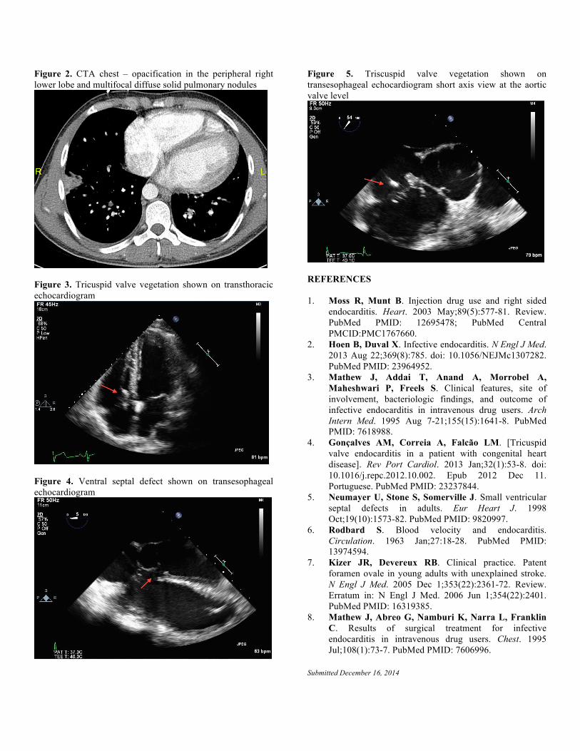
Figure 2. CTA chest – opacification in the peripheral right lower lobe and multifocal diffuse solid pulmonary nodules
Figure 3. Tricuspid valve vegetation shown on transthoracic echocardiogram
Figure 4. Ventral septal defect shown on transesophageal echocardiogram
Figure 5. Triscuspid valve vegetation shown on transesophageal echocardiogram short axis view at the aortic valve level
REFERENCES 1. Moss R, Munt B. Injection drug use and right sided
endocarditis. Heart. 2003 May;89(5):577-81. Review. PubMed PMID: 12695478; PubMed Central PMCID:PMC1767660.
2. Hoen B, Duval X. Infective endocarditis. N Engl J Med. 2013 Aug 22;369(8):785. doi: 10.1056/NEJMc1307282. PubMed PMID: 23964952.
3. Mathew J, Addai T, Anand A, Morrobel A, Maheshwari P, Freels S. Clinical features, site of involvement, bacteriologic findings, and outcome of infective endocarditis in intravenous drug users. Arch Intern Med. 1995 Aug 7-21;155(15):1641-8. PubMed PMID: 7618988.
4. Gonçalves AM, Correia A, Falcão LM. [Tricuspid valve endocarditis in a patient with congenital heart disease]. Rev Port Cardiol. 2013 Jan;32(1):53-8. doi: 10.1016/j.repc.2012.10.002. Epub 2012 Dec 11. Portuguese. PubMed PMID: 23237844.
5. Neumayer U, Stone S, Somerville J. Small ventricular septal defects in adults. Eur Heart J. 1998 Oct;19(10):1573-82. PubMed PMID: 9820997.
6. Rodbard S. Blood velocity and endocarditis. Circulation. 1963 Jan;27:18-28. PubMed PMID: 13974594.
7. Kizer JR, Devereux RB. Clinical practice. Patent foramen ovale in young adults with unexplained stroke. N Engl J Med. 2005 Dec 1;353(22):2361-72. Review. Erratum in: N Engl J Med. 2006 Jun 1;354(22):2401. PubMed PMID: 16319385.
8. Mathew J, Abreo G, Namburi K, Narra L, Franklin C. Results of surgical treatment for infective endocarditis in intravenous drug users. Chest. 1995 Jul;108(1):73-7. PubMed PMID: 7606996.
Submitted December 16, 2014