ribosomal dna sequence divergence within internal transcribed
TRANSCRIPT

Mycologia, 85(3), 1993, pp. 415-427. ? 1993, by The New York Botanical Garden, Bronx, NY 10458-5126
RIBOSOMAL DNA SEQUENCE DIVERGENCE WITHIN
INTERNAL TRANSCRIBED SPACER 1 OF THE
SCLEROTINIACEAE
IGNAZIO CARBONE AND LlNDA M. KOHN
Department of Botany, University of Toronto, Erindale College, Mississauga, Ontario L5L 1C6, Canada
ABSTRACT
Based on morphological and immunological studies, we hypothesize that there are two lineages within the Sclerotiniaceae, a family of plant-infecting ascomycetes in the order Helotiales: 1) genera producing sclerotia, which are tuberlike, melanized masses of hyphae, and 2) genera producing substratal stromata, which are mats of compact hyphae that incorporate plant tissues. We sequenced the Internal Transcribed Spacer (ITS 1), defined by primers ITS 1 and 2, in 43 isolates: 29 sclerotial isolates (19 species in 9 genera), 11 substratal isolates (8 species in 4 genera), and 3 outgroup isolates in the Leotiaceae (3 species in 3 genera). Direct, double-stranded sequencing yielded ca 170 bases for sclerotial isolates and ca 200 bases for substratal and outgroup isolates. MACVECTOR and MULTALIN were used for global alignment, and multiple alignment with hierarchical clustering, respectively. The Internal Transcribed Spacer showed close similarity among most of the sclerotial taxa (76 to 100% similarity to Sclerotinia sclerotiorum). This supports our hypothesis that a sclerotial lineage exists and suggests that this lineage has evolved relatively recently. Isolates of the asexual (mitotic) species Sclerotium cepivorum showed 98% similarity to those of the genus Sclerotinia. Sequence divergence was greater (45 to 65% similarity to S. sclerotiorum) amongst the substratal taxa and our outgroups. Parsimony analysis produced one statistically strongly supported tree for a group of species in the genus Rutstroemia, including Sclerotinia homoeocarpa. Although such subclusters of species can be distinguished using parsimony analysis, we conclude that a substratal lineage cannot be discerned based on sequence data from the ITS. Among these more distantly related taxa, including some substratal ingroup taxa and the outgroup taxa, ITS 1 is saturated with changes and shows relatively equal dissimilarity. The variation observed in the ITS does not resolve among more distantly related taxa. Key Words: ascomycetes, discomycetes, internal transcribed spacer, taxonomy
All members of the Sclerotiniaceae produce one of two basic types of stromata, the deter- minate tuberlike sclerotium or the indeterminate platelike substratal stroma (Whetzel, 1945; Kohn and Grenville, 1989a, b). The determinate scle- rotial genera include those genera which are nec- rotrophic plant pathogens, such as Sclerotinia and Botryotinia. The indeterminate substratal stromatal genera include those genera which are presumed to be plant saprobes, such as Rutstroe- mia.
In pure culture many of these fungi produce only mycelium and stroma. Production of the apothecial teleomorph may require further prep- aration and incubation of stromata, in addition to spermatization of stromata in heterothallic species. In nature, some ubiquitous plant infect- ing sclerotiniaceous species rarely or never pro? duce apothecia. One good example is Sclerotinia homoeocarpa, causal agent of dollar spot of turf, a common disease in North America, Europe, and Australasia, whose teleomorph has not been
conclusively reported and examined since the species was described. Several studies have sug- gested that S. homoeocarpa is not a member of the genus Sclerotinia and that it should be re- classified among the substratal stromatal Sclero- tiniaceae (Kohn, 1979; Kohn and Grenville, 1989a, b; Novak and Kohn, 1991). Examination of apothecia putatively thought to belong to this species suggested placement in either Lanzia or Moellerodiscus (Korf, pers. comm.), but unfor- tunately none of these apothecia were retained. Rutstroemia (= Poculum), Lanzia and Moeller? odiscus are delimited primarily on the basis of their apothecial microanatomy, with ectal excip- ula composed of filamentous cells in gel, fila- mentous cells lacking gel, and globose cells, re- spectively (Dumont, 1976). White's (1941) broader definition of Rutstroemia, still accepted by some workers, includes Lanzia and some el? ements of Moellerodiscus (Dumont, 1976). No description of the microanatomy, in particular, the ectal excipular structure of S. homoeocarpa
415

416 Mycologia
was furnished in the original description, no type or authentic herbarium material remains, and attempts to produce apothecia in vitro have been unsuccessful. Using the taxonomic methods nec- essary to compare the morphological characters that define Rutstroemia, Lanzia, and Moeller- odiscus, it is impossible to reclassify S. homoeo- carpa without having the teleomorph in hand. There have been no comparative molecular stud? ies of any of these taxa, which have not been extensively or copiously collected and, with the exception of S. homoeocarpa, have rarely been accessioned in culture collections. Another ex- ample is Sclerotium cepivorum, causal agent of soft rot of onions, which does not produce a tel? eomorph, only mycelia and determinate sclerotia and, consequently, under conventional interpre- tations of Article 59 of the International Code of Botanical Nomenclature (Greuter, 1988), can? not be classified among the holomorphic scle? rotial taxa in the Sclerotiniaceae with which it shares many features (Kohn and Grenville, 1989a, b; Novak and Kohn, 1991).
Depending on the relative character weighting for stromatal anamorphs (sclerotia versus sub? stratal stromata, morphological type), conidial anamorphs (presence or absence, morphological type), and apothecial microanatomy (gross mor? phology, ectal excipular type), holomorphic gen? era have been defined very broadly or very nar- rowly. Rutstroemia sensu White versus sensu Dumont is one example. In another example, based on a broad interpretation stressing apo? thecial morphology and the production of scle? rotia, Dennis (1978) and, to an even greater ex- treme, von Arx (1981) recognized virtually all sclerotial taxa as one genus Sclerotinia. These workers perhaps recognized a clade or generic lineage rather than a single genus. Other authors (Korf, 1973; Kohn, 1979; Schumacher and Kobn, 1985) have considered more equally character- istics of apothecial morphology along with those of anamorph morphology (conidia and stromata) and ecology, recognizing and segregating many genera. However, these workers have not re- solved relationships among genera. While teleo? morph and anamorph morphology have largely defined genera in the Sclerotiniaceae, the type of stroma produced and the relationship of the fun? gus to the host have afforded key characters for grouping genera within the family and for rec? ognizing relationships between the pleomorphic, sexual (meiotic) species in the family and mainly
or exclusively asexual (mitotic) species, such as Sclerotium cepivorum and Sclerotinia homoeo? carpa, both of which lack conidial anamorphs.
A recent study in our laboratory (Novak and Kohn, 1991) has shown that developmental stro? matal storage proteins occur in both the sclerotial and substratal stromatal taxa and that it is pos- sible to distinguish between these groups of taxa on the basis ofthe ability of these stromatal pro? teins to cross-react with the major 36-kDa pro? tein of Sclerotinia sclerotiorum. Using western hybridizations, only sclerotial proteins were shown to cross-react when treated with antibody raised to the 36-kDa protein of S. sclerotiorum. No cross-reactivity was observed with substratal stromatal proteins. Based on these immunolog- ical studies and on morphological studies (Kohn and Grenville, 1989a, b), we hypothesized that there are two lineages within the Sclerotiniaceae: the sclerotial lineage and the substratal stromatal lineage.
In the present study, sequence divergence in the nuclear ribosomal Internal Transcribed Spacer (ITS 1), bounded by the 18S and 5.8S genes of the nuclear ribosomal DNA, was ex- plored as a potential source of characters for studies at the generic level in the Sclerotiniaceae. Although the noncoding ITS regions are gener- ally more variable than the coding regions ofthe adjacent nuclear ribosomal RNA genes, given our previous observations of no intraspecific variation within Sclerotinia sclerotiorum (Kohn and Anderson, unpubl.), we postulated three things: 1) that the sclerotial taxa might show se? quence similarity to each other and sequence di? vergence from the substratal stromatal taxa, 2) that Sclerotium cepivorum would show a high level of sequence similarity to the sclerotial taxa, and 3) that Sclerotinia homoeocarpa would show sequence similarity to the substratal stromatal taxa and a lack of relatedness to Sclerotinia. The primary objective of this study was to sequence the ITS 1 in a larger sample within the Sclero? tiniaceae, including closely related species and genera within the two proposed lineages, as well as outgroup isolates representing the morpho- logically and ecologically most closely related family, the Leotiaceae. Outgroup isolates from the Leotiaceae were chosen based on the alter- native hypotheses that either substratal stro? matal taxa show closer relatedness to the Leo? tiaceae than to the sclerotial lineage or that substratal stromatal taxa show equal relatedness

Carbone and Kohn: Molecular Systematics of Sclerotiniaceae 417
to the Leotiaceae and to the sclerotial lineage. Neurospora crassa Shear & Dodge was included in the sample to facilitate alignment of the coding ends of the region amplified with these primers, although no close relationship to the Scleroti? niaceae was anticipated. In order to determine where, and what type of variation was present in the ITS 1, sequences were first aligned and grouped using programs for global alignment and multiple alignment with visual adjustment. Aligned sequences were then compared and grouped by hierarchical clustering and parsi? mony analysis. The results of this study were compared with immunological data from a pre- vious study (Novak and Kohn, 1991) for a subset of our isolates.
materials and methods
Cultures and DNA extraction.?Isolates used in this study are listed in Table I. Cultures were grown on potato-dextrose agar (PDA) in the dark and at room temperature (20-22 C). Mycelium was grown on liquid complete yeast medium (CYM), harvested after 2-4 days and freeze-dried for DNA extraction (Kohn et al., 1991; Kohli et al., 1992). DNA extraction was by the small scale (mini-prep) method of Zolan and Pukkila (1986). DNA was stored at -20 C until required for amplification.
Amplification of genomic DNA. ?The ITS 1 region de? fined by primers ITS 1 and ITS 2 (White et al., 1990) was amplified via the polymerase chain reaction (PCR). DNA was diluted 200-fold and 50 iA was placed in a 0.5 ml polypropylene tube to which 50 ^1 of a 2 x mix of all other reaction components was added as de- scribed by Kohn et al. (1991). Double-stranded DNA amplification products were generated by using equi- molar (50 pM) amounts of each primer. A DNA-free control tube was included with each run as a safeguard against contamination. The thermal program followed was as described in Kohn et al. (1991). PCR products were observed by electrophoresing 10 yX from each tube on 1.5% agarose gels in Tris-acetate-EDTA (TAE) buff? er. The presence of a single bright band in each lane was a check for a successful amplification, while the absence of a band in the control lane was a check against contamination. The remaining PCR reaction product (90 iA) was purified using GENECLEAN (BioCan Scientific). All purified DNA samples were stored at -20 C until required for sequencing.
Sequencingprocedure.?UNA was sequenced directly using the dideoxy chain termination method (Sanger et al., 1977). The ITS 1 region was sequenced for a sample of 43 isolates (Table I), consisting of 29 sclero? tial isolates (19 species in 9 genera), 11 substratal stro? matal isolates (8 species in 4 genera) and 3 outgroup isolates (3 species in 3 genera). For each isolate both complementary ITS 1 strands were sequenced follow? ing the protocol of Winship (1989) as modified by An? derson and Stasovski (1992).
Analysis of DNA sequences. ?Sequence data were checked between complementary strands and com- pared with other sequence data with the assistance of MACVECTOR (Lipman and Pearson, 1985; Pearson and Lipman, 1988) and MULTALIN (Corpet, 1988). The first step in sequence analysis was the comparison of complementary strands to check for reading errors and resolve ambiguities if possible. Complete sequenc? es were then globally aligned with a computerized alignment algorithm (MACVECTOR). This algorithm compares all the residues in one sequence to all posi- tions in a specified reference sequence only, in this case Sclerotinia sclerotiorum. To every pair of residues compared, a positive weight is assigned for a match and a negative weight for a mismatch, deletion or a gap. The generated value is the score ofthe alignment and represents the best (optimal) alignment ofthe non- reference sequence to the reference sequence for the weights used. The final alignment of all sequences to S. sclerotiorum was analyzed visually and the deletion/ gap weights were adjusted to obtain the best visual alignment of all sequences. The order ofthe sequences and weight parameters in the final MACVECTOR alignment were the order and weight parameters used for MULTALIN. MULTALIN performs all possible pairwise comparisons using an algorithm similar to MACVECTOR, with S. sclerotiorum as the query se? quence. Initially a hierarchical clustering of the se? quences is done using these scores (Corpet, 1988). This is followed by pairwise comparisons of clusters of aligned sequences to obtain the complete multiple alignment. MULTALIN then builds a hierarchical clustering ofthe sequences using the scores of all pair? wise comparisons in the multiple alignment as a mea- sure of the similarity between sequences. Similarity scores were represented on a fractional (or percentage) similarity scale where a value of 1.0 (or 100%) corre- sponds to no sequence divergence (i.e., identical se? quences). In general, as sequence divergence increases, the fractional similarity (S) between two sequences de- creases (S < 1.0) and the distance from the reference sequence, defined as 1 -S increases (Swofford and Olsen, 1990). The final multiple alignment was visually ad? justed and parsimony analysis of the multiple align? ment, both to compare branching topologies and branch lengths with the hierarchical clustering, was performed using PAUP 3.0n (Swofford, 1990).
RESULTS
Direct, double-stranded DNA sequencing yielded ca 170 bases for sclerotial isolates and ca 200 bases for the substratal stromatal and out? group isolates. In general, DNA sequences could be determined with accuracy by reading only the antisense strand but as a further check for reading errors and for resolution of ambiguous bases, it was necessary to align both strands in all isolates.
A preliminary MACVECTOR global align? ment of all the sequences with S. sclerotiorum (LMK 2) as the reference sequence revealed very little sequence divergence within the sclerotial

418 Mycologia
Table I Isolates representing the Sclerotiniaceae and selected outgroups

Carbone and Kohn: Molecular Systematics of Sclerotiniaceae 419
Table I Continued
a Collection no., in parentheses, designates accession number in the collection of L. M. Kohn. b Localities are abbreviated as follows: N.W.T., Ellesmere Island, North West Territories; N.S.W., New South
Wales, Australia. c Sources of cultures are abbreviated as follows: ATCC, American Type Culture Collection; CUP, Cornell
University Plant Pathology Herbarium; ATRI, Australian Turfgrass Research Institute. d Isolate represents an asexual (mitotic) species known only from anamorphs. e Isolate represents an anamorph of a pleomorphic species with a known teleomorph in the Sclerotiniaceae.
Unless designated otherwise, isolates in this table represent pleomorphic species with known teleomorphs in the Sclerotiniaceae.
isolates and a high level of divergence within the substratal stromatal and outgroup isolates. Be? cause of this, visual sequence alignment within the sclerotial isolates was easily accomplished, but alignment between sclerotial and substratal stromatal and outgroup isolates was difncult, with dubious results. The best visual alignment that we could achieve of all of the sequences with MACVECTOR was obtained with a match score of 4 and a mismatch score of 2, a deletion penalty of 20 (rather than the default of 12), and a gap penalty of 4. The order ofthe sequences did not have any effect on the final MACVECTOR align? ment but was found to have an effect on the final multiple alignment (data not shown). Therefore, the order of the sequences given by MACVEC? TOR was the order used for MULTALIN.
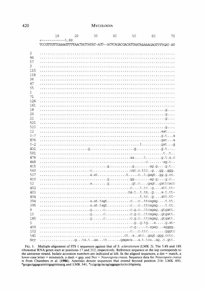
Multiple alignment of all the sequences (Fig. 1) revealed that there was very little divergence close to the ends of the sequences, and no di? vergence when the ends extended into the coding regions ofthe 18S and 5.8S rRNA genes. Little intraspecific sequence variation was observed. Among the sclerotial isolates most of the vari- ability was located in the middle of the ITS 1 region, and was either in the form of single base substitutions only [Sclerotinia spp., Sclerotium cepivorum, and Cristulariella moricola (teleo? morph = Grovesinia pyramidalis M. Cline, J. L.
Crane, & S. Cline)] or base substitutions accom- panied by a 2 bp insertion/deletion motif (Bo- trytis spp., Moniliniafructicola, Myriosclerotinia spp., and Ciborinia ciborium). Among the sub? stratal and outgroup isolates single or multiple base substitutions and insertion/deletion events were observed throughout the ITS 1 region.
Hierarchical clustering based on the multiple alignment by MULTALIN, with S. sclerotiorum (LMK 2) as the query sequence, is shown in Fig. 2; immunological characters from a previous study of stromatal proteins (Novak and Kohn, 1991) and morphological types of the macroco- nidial anamorphs are also shown in Fig. 2. The purpose of the cluster analysis was mainly to order the data set and to compartmentalize like and unlike isolates, not to estimate true evolu? tionary distance. The final sequence alignment within those clusters with >70?/o similarity was found to be relatively insensitive to the choice of weights and a reasonable alignment could also be attained by eye. Sequences that were outside these clusters had ambiguous alignments and gaps were inserted as necessary by the alignment al- gorithm to maximize the total similarity score when summed over all positions. The choice of query sequence did not affect the clustering; the same clustering was obtained when isolates of each of four other taxa in the sample [Sclerotinia
420 Mycologia
10 20 30 40 50 60 70 <-5.8S TCCGTIUITGAMGTITrAACTATTATAT- ACT- - AC^CAGACGACATTAATAAAAAGAGITrrGAT-AT
2 . 44 . 57 . 3 . 115 . 118 . 36 . 47 . 55 . 1 . 71 . 126 . 161 . 18 .g_ 20 .g_ 21 .g_ 521 . 523 .g_ 12 .aat_ 0-7 .g.t. . .a E76 .gat. . .a 0-2 .t.gat. . .g 431 .g.g.g.t.... 501 .t. .t. . 476 .aa.t.g.t .a.c 448 .c.-ag.t. . 415 .g.g.ag.g. . . .g.t. . 540 .c.cat.c.ttc. .g. . .gg. .ggg. 537 .a. at.t.c. . t.gagt. .gg.g.cc. 410 .g.gg.ag.g. . . .g.c. . 52 .a.g.gt. c. . . . gagt.. gattcacc 402 .c_t.tc. .g_att.tt- 403 .na.t. .t.tn. .g. . . .a.t.tt- 404 .t.tc. .g_att.tt- 394 .a.at.tagt.c. . .c. .ttcagag. . . .t.tt. 395 .a.at.tagt.c. . .c. .ttcagag. . . .t.tt. 8 .g.c.c.g.c.ttcagag. .gtgatt. 10 .g.c.c.g.c. .ttcagag. .gtgatt. 185 .g.c.c.g.c. .ttcagag. .gtgatt. 5 .g. .g.tg. . .a.g.at- 400 .e.g.c. agag. . . agggg. 102 .c... c. ttc.gggtc 541 .ct. . a. . atc. . gagt. ggg. cctc. Ncr .g. . . ta. t. . aa. . . tt.gagaca. . . a. t. tca. . ag. . t. gtt.
Fig. 1. Multiple alignment of ITS 1 sequences against that of S. sclerotiorum (LMK 2). The 5.8S and 18S ribosomal RNA genes start at positions 17 and 212, respectively. Reference sequence at the top corresponds to the antisense strand. Isolate accession numbers are indicated at left. In the aligned sequences, a dot = match, a lower-case letter = mismatch, a dash = gap, and Ncr = Neurospora crassa. Sequence data for Neurospora crassa is from Chambers et al. (1986). Asterisks denote sequences that extend beyond position 216: LMK 400, *gccgactggagcattttttgagtttttaatgand LMK 541, *cctgctgctacagtaggagactcctcctttgtaatg.

Carbone and Kohn: Molecular Systematics of Sclerotiniaceae 421
80 90 100 110 120 130 140
TCTCTGGCGAGCATACA- -AGGCCCCGA- -AGAQCAGCTCGCC
2 44 57 3 115 118 36 47 55 1 71 126 161 18 20 21 521 523 12 0-7 E76 0-2 431 501 476 448 415 540 537 410 52 402 403 404 394 395 8 10 185 5 400 102 541 Ncr
c ct c c ctgt
a cgag-
.g.
.g.
gag .cg .c-
ctgaca.gcgcgggc .gg.g.. ...ggc.
.c..gaggc ?c.tggc
-.ct.c.gcgaacg. -.et.c.gcgaacg. -.ctcc.gcg.gcg. -.ctcc.gc..gcg. -.ctcc.gc..gcg. ..ctcc.gc..gcg. ..ctcc.gc..gcg. ..ctcc.gc..gcg. -.ctct.gcg.gcg. c...c...ac..gggg-. ct.tg.-. g.gggca.tca.cag.gg
a a a
?g ?g ?g
?g
g-
gt.cg tga.gcattttaga ?g?.
cc.g..ga.gcggtggccc gccgaagccagcggt ccgg ccaggg..g. g. . gng. an.
g g g g
ag ag ag ag ag ag ag ag ag ag ag a.a at ag ?g
.cgg.
tc
tctcaggctcgaaagcttgaggctctggg-caattaagccc..g tctcaggctcgaaagcctggggctctggg-caattaagccc..g tctcaggctcgaaagcttgaggccttggg.cagttaaggcc.ag tcccaggcccg..aaggcgctgg...tgtcccccgagggtgc-. tcccaggcccg..aaggcgctgg...tgtcccccgagggtgc-. tctccggccccggagggcgctg....tgtccccggaagggtc.. tctccggccccggagggcgctg....tgtccccggaagggtc.. tctccggccccggagggcgctg....tgtccccggaagggtc.. tcttcggcttgtgaaagctaa.t.aacggtttagggct..<
-.cccggtggccgat.
ct ct ct ct ct c. g-
g.gcgt g..c..
cgg. act. cg. . ggg.. gccgcgagcgggagacccgaggat.. gg.. ggcccgaaggcctttccggac
Fig. 1. See p. 420 for explanation.
homoeocarpa (LMK 8), Botrytis cinerea (LMK 18), Rutstroemia petiolorum (LMK 402), and Monilinia megalospora (LMK 415)] were used as query sequences (data not shown). Most ofthe
sclerotial isolates, including the three isolates of Sclerotium cepivorum, were tightly clustered to- gether showing close similarity to Sclerotinia sclerotiorum (S = 0.76-1.0). The remaining iso-

422 Mycologia
150 160 170 180 190 200 210 18S->
AAAGC AACAAAGTAATAATACACAAGGGTGGGAGGTCTACCCTTT-CGGGCATG-AACICTGTAATG
2 44 57 3 115 118 36 47 55 1 71 126 161 18 20 21 521 523 12 0-7 E76 0-2 431 501 476 448 415 540 537 410 52 402 403 404 394 395 8 10 185 -J 400 102 541 Ncr
.t. . . .ta.g.a.
.cg.a. . gt. . . . agg. t. tgcc. ttttt. a. . . g. a. .
.gtt. gtta. . ataca.gcg.a.
. . g.gt.t. . . . a. . gt. . ga. atcta. . ccga. . . . a.
.tggg...g...a..gt....agg.t.tgcccctgt......g.-..
. n.g.. .. g.cg.... c.gc. ..
.ta.g.a.
.ta.-.g.a. . tag.g.g.a. .ta.g.g.a. .ta.g.g.a. ? gag.t.g.g.a-g. ? gag.t.g.g.a-g. ? gag.t.g.g.a-g.
.ta.g.g.act. .g.tcg.ga.agcc.t- . g.g.g. . . .c. . . .tc.ttc.aa.g.ggt.ggcacttcaatc.cgctgacgg. . . . g.t....t..c.a.tg. actataa. . cg. g-. .gt...g.c.g..c....t..g... acc. t. gggagggggcc. tggtcgtcgagcag. a. gcga. *
cc...gc.g.g.c..ccg...gggtaa.attcgc.atggtttg.gggagtttt..? .
g. .a.
at. .a. .g...g..
.tt
. .t. .
.cg., ? tg., .tg., .tg., .tg.
? g. ?g. ? g. . .a. .gc.g.gc*
Fig. 1. See p. 420 for explanation.
lates, representing all of the substratal stromatal taxa, the outgroups, and three sclerotial taxa, Monilinia megalospora, M. oxycocci, and Ci- borinia erythronii, showed less similarity to S.
sclerotiorum (S = 0.45-0.65) and formed several clusters. Within the sclerotial cluster were two subclusters of very similar taxa, the Sclerotinia cluster (S > 0.98) and the Botrytis cluster (S >

Carbone and Kohn: Molecular Systematics of Sclerotiniaceae 423
0.96). Among the predominantly substratal stro? matal and outgroup isolates there was one no- table large cluster including a subcluster with Rutstroemia henningsiana and Sclerotinia ho- moeocarpa (S = 0.81), and a subcluster with R. petiolorum, R. sydowianaandR.firma(S > 0.87). Although this cluster of Rutstroemia species in? cluding S. homoeocarpa could be distinguished, a substratal stromatal lineage could not be dis- cerned. As shown in Fig. 2, similarity to the query sequence among two of the three outgroup isolates from the Leotiaceae (LMK 540, Bispo- rella citrina and LMK 537, Ionomidotis sp.), a closely related family in the same order (Helo- tiales), was within the same range (S = 0.45-0.65) as among the ingroup substratal stromatal iso? lates and three sclerotial isolates from the Sclero? tiniaceae. Neurospora crassa rooted most deeply in the clustering (S = 0.30, with an expected random match of S = 0.25); although the align? ment of the internal portion of ITS 1 of N. crassa to those of the other isolates in our sample was very poor, the ends extending into the coding regions aligned well.
Even with powerful computing, parsimony analysis of the data set was problematic because of the site saturation and the larger sequence size of ITS 1 in the substratal stromatal and outgroup isolates, especially when compared to the rela? tively conserved sequences (yielding few char? acters) of most of the sclerotial isolates. The branch-and-bound search function with PAUP failed to produce a tree, even when taxa were reordered and one isolate was used to represent each taxon. A heuristic search with PAUP with only one isolate representing each taxon pro? duced a tree with similar topology to that pro? duced by hierarchical clustering, but with branches supported no more than 46-78%. Sub- clusters defined by hierarchical clustering with MULTALIN and branch-and-bound searches of certain subsets of taxa with PAUP were similar. Trees produced by these branch-and-bound searches are shown in Figs. 3-8. In trees shown in Figs. 3 and 5, no branch is supported in >95 of 100 bootstrap replications. Sequences of iso? lates in the Botrytis cluster produced 57 most parsimonious trees, with little support for branching by bootstrap resampling (Fig. 5) or 50% majority rules (Fig. 6), and with little res- olution by strict consensus (Fig. 7). Parsimony analysis of the Rutstroemia cluster resulted in one most parsimonious tree (Fig. 8) with the
branch to all Rutstroemia species, including Sclerotinia homoeocarpa, supported in 100 out of 100 bootstrap replications.
DISCUSSION
Results of multiple alignment and hierarchical clustering in this study show that most of the sclerotial taxa, including Sclerotium cepivorum, cluster together, showing close similarity to S. sclerotiorum. This supports our hypothesis that a sclerotial lineage exists and suggests that this lineage has evol ved relatively recently. Although a cluster of species within Rutstroemia can be distinguished, we conclude that a substratal stro? matal lineage cannot be discerned based on this genomic region. Among these taxa, the ITS 1 sequences are saturated with changes. The vari? ation in ITS 1 does not resolve among the more distantly related taxa. The positions among deeply rooted branches in Fig. 2 are therefore not sig- nificant. As a result, it is not surprising that the three outgroup taxa representing the Leotiaceae are found among the substratal stromatal in- group in Fig. 2. With the exception ofthe rela? tionships within the Rutstroemia cluster, essen- tially all of the substratal stromatal taxa and the three outgroup taxa are equally unrelated with respect to ITS 1. Our alternative hypotheses con- cerning the relatedness of the substratal stro? matal taxa to taxa in the sclerotial lineage and the Leotiaceae cannot be tested with sequence data from the ITS. Within the Rutstroemia clus? ter, however, the position of Sclerotinia homoeo? carpa is significant among the other Rutstroemia species.
The results of this study are in agreement with immunological data from a previous study (No- vak and Kohn, 1991), particularly the western blot data as indicated in Fig. 2 of this paper, and the discriminant analysis of competitive en- zyme-linked immunosorbent assays (ELISAs; Fig. 6; Novak and Kohn, 1991). In both the discriminant analysis ofthe competitive ELISAs and our hierarchical clustering and parsimony analyses, the sclerotial isolates including Scle? rotium cepivorum were clustered together, show? ing close similarity to S. sclerotiorum, while the substratal stromatal isolates showed less simi? larity and did not cluster. Immunological and sequence similarity data also show that Sclero? tinia homoeocarpa is more closely related to the substratal stromatal genera than to other Sclero-

424 Mycologia
WBMS
?
Sclerotinia sclerotiorum (2) S. sclerotiorum (44) S. sclerotiorum (57)
' S. minor (3) S. minor (115) S. minor (118)
r S. trifoliorum (36) i S. trifoliorum (47) ' S. trifoliorum (55)
HSclerotium
cepivorum (1) S. cepivorum (71) S. cepivorum (126)
? Cristulariella moricola (161)
HBotrytis
clnerea (18) B. clnerea (20) B. cinerea (21)
KBotrytis sp. (521)
B. calthae (523) Monilinia fructicola (12)
i? Myriosclerotinia dennisii (0-7) Ti- Ciborinia ciborium (E76)
*- Myriosclerotinia scirpicola (0-2) ? Monilinia fructigena (431) ? Ciboria caucus (501) ? C. acerina (476) ? Verpatinia calthicola (448) ? Monilinia megalospora (415) ? Bisporella citrina (540) ? lonomidotis sp. (537) ? Monilinia oxycocci (410) ^? Ciborinia erythronii (52) ? Rutstroemia petiolorum (402) ? R. sydowiana (403) ? R. firma (404)
r R. henningsiana (394) i- R. henningsiana (395)
Sclerotinia homoeocarpa (8) S. homoeocarpa (10) S. homoeocarpa (185) Lambertella subrenispora (5) L. langei (400) Piceomphale bulgarioides (102) Ascocoryne cylichnium (541)
0.4 0.6 0.8 1.0
Similarity scale
Fig. 2. Hierarchical clustering based on multiple alignment of ITS 1 sequence data and comparison with other characters. Pairwise similarity scores are represented on a fractional similarity scale where a value of 1.0 (or 100%) corresponds to identical sequences. Isolate accession numbers in the collection of L. M. Kohn are shown in brackets. Column WB shows results of western blot analysis from Novak and Kohn (1991); +, cross- reaction; ?, no cross-reaction; n, no data available. Column MS designates macroconidial state; B, Botrytis; C, Cristulariella; MJ, Monilia lacking disjunctors; MD, Monilia with disjunctors; o, no macroconidia; *, have distinctive macroconidial states that are morphologically different from those of the Sclerotiniaceae.

Carbone and Kohn: Molecular Systematics of Sclerotiniaceae 425
B. cinerea (18) C. moricola (161)
S. cepivorum (1) ? S. trifoliorum (36)
l?j. S. minor (3) S. sclerotiorum (2)
Fig. 3. Unrooted phylogenetic tree based on the multiple alignment of the ITS 1 of five species in the Sclerotinia cluster with Botrytis cinerea (LMK 18) as the outgroup. One of two most parsimonious trees is shown. Horizontal distance is proportional to branch length; vertical distance does not contribute to branch length. The scale bar equals two character state changes. The consistency index of both trees was 1.000. No branch was supported in >95 of 100 bootstrap repli? cations.
tinia spp., suggesting that it should be reclassi- fied. Parsimony analysis ofthe Rutstroemia sub- cluster (Fig. 8) with bootstrapping suggests that S. homoeocarpa may be accommodated in Rut? stroemia, but a comparison with other species in closely related genera, such as Lanzia and Moel? lerodiscus, is needed.
With the exception of a single base substitution in one isolate of S. minor (LMK 118), and vari? ation in a single base in the two isolates of Rut? stroemia henningsiana (LMK 394, 395), the ab? sence of intraspecific variation in ITS 1 within our sample of sclerotiniaceous isolates was no- table. In contrast, O'Donnell (1992) recently re? ported multiple ITS types within Fusarium sam- bucinum Fckl. (teleomorph = Gibberella pulicaris (Fr.) Sacc). Different rates of ITS evolution, with examples in the literature of resolution at intra? specific, species, genus, and suprageneric levels among different groups of fungi, could be due
- B. cinerea (18) . C. moricola (161) ? S. cepivorum (1) ? S. trifoliorum (36) S. minor (3)
L? S. sclerotiorum (2)
Fig. 4. Strict consensus of the two most parsimo? nious trees in which only the branching topology is significant. Branch lengths are not drawn to scale.
- S. sclerotiorum (2)
J- B. cinerea (18)
B. calthae (523)
,? M. dennisii (0-7) C. ciborium (E76)
? M. scirpicola (0-2) ?? Al fructigena (431) M. fructicola (12)
Botrytis sp. (521)
Fig. 5. Unrooted phylogenetic tree based on the multiple alingment ofthe ITS 1 of seven species in the Botrytis cluster with S. sclerotiorum (LMK 2) and M. fructigena (LMK 431) as outgroups. One of 57 most parsimonious trees is shown. Horizontal distance is proportional to branch length; vertical distance does not contribute to branch length. The scale bar of branch lengths equals two character state changes. The con- sistency index ofthe tree shown was 0.880. No branch was supported in >95 of 100 bootstrap replications.
either to a molecular clock with variable rates or to the as yet unexplained effects of life cycle strat- egies on rates of speciation (Bruns et al., 1991).
There are several interesting taxonomic im- plications in our study. First, clustering based on similarity does not suggest that all sclerotial taxa should be accommodated under one genus Scle? rotinia. Sequences of Sclerotium cepivorum and Cristulariella moricola show 98% similarity to that of Sclerotinia sclerotiorum, suggesting a close relationship that is also supported by other bio- chemical and morphological characters. Taxo? nomic placement ofthe apparently "mitotic spe-
S. sclerotiorum (2) B. cinerea (18) B. calthae (523) M. fructicola (12) M. dennisii (0-7) C. ciborium (E76) M. scirpicola (0-2) M. fructigena (431) Botrytis sp. (521)
Fig. 6. Unrooted phylogenetic tree based on 50% majority rule of the 57 trees. Numbers indicate the percentage of trees that support the branch.

426 Mycologia
? S. sclerotiorum (2) ? B. clnerea (18) ? Botrytis sp. (521) ? B. calthae (523) ? M. fructicola (12) ? M. dennisii (0-7) ? C. ciborium (E76) ? M. scirpicola (0-2) ? M. fructigena (431)
Fig. 7. Strict consensus of the 57 most parsimo? nious trees in which only the branching topology is significant. Branch lengths are not drawn to scale.
cies" (see Reynolds and Taylor, 1992) within a genus in the Sclerotiniaceae would require both more molecular characters, yielding a statisti- cally well-supported phylogenetic placement within a genus, and revision of Article 59 of the International Code of Botanical Nomenclature. Another intriguing question is whether the un- usual Cristulariella conidial anamorph is derived from sclerotia or appressoria rather than a mod- ification of the Botrytis conidial form.
Isolates of all Botrytis species examined, in? cluding LMK 521, a new species on Rubus from Norway (Holst-Jensen and Schumacher, in manuscript), show >96% similarity. Although a well-supported lineage cannot be discerned from this data, the Botrytis cluster (Figs. 2, 5-7) shares a unique deletion/substitution motif between po? sitions 110 and 140 (Fig. 1).
The >97% similarity of Ciborinia ciborium to Myriosclerotinia isolates is cause to question the recent transfer of this Carex- and Eriophorum- infecting species from Myriosclerotinia, a genus of fungi infecting members of the Cyperaceae and Juncaceae, to Ciborinia (Schumacher and Kohn, 1985). The divergence in ITS 1 between C. ci? borium and C. erythronii is notable; we think that Ciborinia as presently circumscribed is a hetero- geneous grouping in need of monographic revi? sion.
While Monilinia fructicola and M. fructigena in the Junctoriae group of the genus Monilinia (Batra, 1991) fall within the exclusively sclerotial cluster (both with >76% similarity to S. scleroti? orum), M. megalospora and M. oxycocci, in the Disjunctoriae group, are outside of the sclerotial cluster (with 65% and 56% similarity to S. scle-
. S. sclerotiorum (2) - P. bulgarioides (102)
r R. petiolorum (402) I? R. sydowiana (403) R. firma (404)
R. henningsiana (394) S. homoeocarpa (8)
? L subrenispora (5)
8
Fig. 8. Unrooted phylogenetic tree based on the multiple alignment of the ITS 1 of six species in the Rutstroemia cluster (including Lambertella subrenis? pora) with S. sclerotiorum (LMK 2) and P. bulgarioides (LMK 102) as outgroups. Horizontal distance is pro- portional to branch length; vertical distance does not contribute to branch length. The scale bar equals ten character state changes. The consistency index of the tree shown was 0.902. Asterisks indicate those branch? es that are supported in >95 of 100 bootstrap repli? cations.
rotiorum, respectively). In an independent study, utilizing some ofthe same isolates as in our sam? ple, Holst-Jensen (1992) analysed morphologi? cal, ecological, and restriction fragment length polymorphism (RFLP) characters using UPGMA and PAUP, deriving a strongly supported branch (>95 out of 100 bootstrap replications) separat- ing the Junctoriae (lacking disjunctors between macroconidia and infecting rosaceous hosts with fleshy fruits) from the Disjunctoriae (possessing disjunctors and infecting ericaceous or rosaceous hosts with relatively dry fruits), in agreement with our ITS 1 data. Interestingly, the branching to- pologies of both his dendrograms and clado- grams support the separation of M. megalospora and M. oxycocci that we observed; he hypothe- sizes that this reflects the earlier divergence time of the former species.
Lastly, the rejection of Honey's lectotypifica- tion of Rutstroemia (Kohn and Schumacher, 1984) with Peziza bulgarioides Rabenh. in Kalchbr. (= Piceomphale bulgarioides) is sup? ported by the lack of similarity in ITS 1 of our isolate of this species to the Rutstroemia cluster (Fig. 8) especially in comparison to our isolate of the species that was ultimately conserved as the type species R. firma.
In addition to the taxonomic implications, ITS variability is being exploited for disease diag- nosis and identification of other phytopathogenic fungi (Xue et al., in press; Nazar et al., 1991) and could be used to identify mycelia and sclerotia

Carbone and Kohn: Molecular Systematics of Sclerotiniaceae 427
of the sclerotial Sclerotinaceae infecting or in- festing plants and seeds. Data from this study could be used in the development of diagnostic oligonucleotide probes for the identification of taxa or groups of taxa.
acknowledgments
This research was supported by a Summer Bursary for IC and an Operating Grant to LMK from the Nat- ural Sciences and Engineering Research Council of Canada. We thank all those who furnished cultures. Thanks also to Jim Anderson and Jerry Brunner.
LITERATURE CITED
Anderson, J. B., and E. Stasovski. 1992. Molecular phylogeny of northern hemisphere species of Ar? millaria. Mycologia 84: 505-516.
Batra, L. R. 1991. World species o/Monilinia (Fungi): their ecology, biosystematics and control. Mycolo? gia Memoir No. 16. Cramer, Berlin, Germany.
Bruns, T. D., T. J. White, and J. W. Taylor. 1991. Fungal molecular systematics. Annual Rev. Ecol. Syst. 22: 525-564.
Chambers, C, S. K. Dutta, and R. J. Crouch. 1986. Neurospora crassa ribosomal DNA: sequence of internal transcribed spacer and comparison with N. intermedia and N. sitophila. Gene 44: 159-164.
Corpet, F. 1988. Multiple sequence alignment with hierarchical clustering. Nucl. Acids Res. 16: 10881- 10890.
Dennis, R. W. G. 1978. British Ascomycetes. Cramer, Vaduz, Liechtenstein.
Dumont, K. P. 1976. Sclerotiniaceae XI. On Moel- lerodiscus(=Ciboriopsis). Mycologia6S: 233-267.
Greuter, W. 1988. International code of botanical nomenclature. Koeltz Scientific Books, Konig- stein, Germany.
Holst-Jensen, A. 1992. Morphology and rDNA re? striction site based phylogeny in the genus Monili? nia (Sclerotiniaceae). Master's Thesis, cand. scient., University of Oslo, Oslo, Norway.
Kohli, Y., R. A. A. Morrall, J. B. Anderson, and L. M. Kohn. 1992. Local and trans-Canadian clonal distribution of Sclerotinia sclerotiorum on canola. Phytopathology 82: 875-880.
Kohn, L. M. 1979. A monographic revision of the genus Sclerotinia. Mycotaxon 9: 365-444.
-, and D. J. Grenville. 1989a. Anatomy and histochemistry of stromatal anamorphs in the Sclerotiniaceae. Canad. J. Bot. 67: 371-393.
-, and-. 1989b. Ultrastructure of stro? matal anamorphs in the Sclerotiniaceae. Canad. J. Bot. 67: 394-406.
-, and T. Schumacher. 1984. Two counter-pro- posals for the conservation of Rutstroemia (Fun? gi). Taxon 34: 507-509.
-, E. Stasovski, I. Carbone, J. Royer, and J. B. Anderson. 1991. Mycelial incompatibility and molecular markers identify genetic variability in field populations of Sclerotinia sclerotiorum. Phy? topathology 81: 480-485.
Korf, R. P. 1973. Discomycetes and Tuberales. Pp.
249-319. In: The fungi. An advanced treatise. Eds., G. C Ainsworth, F. K. Sparrow, and A. D. Suss- man. Academic Press, San Diego, California.
Lipman, D. J., and W. R. Pearson. 1985. Rapid and sensitive protein similarity searches. Science 227: 1435-1441.
Nazar, R. N., X. Hu, J. Schmidt, D. Culham, and J. Robb. 1991. Potential use of PCR amplified ri? bosomal intergenic sequences in the detection and differentiation of verticillium wilt pathogens. Physiol. Molec. Pl. Pathol. 39: 1-11.
Novak, L. A., and L. M. Kohn. 1991. Electrophoretic and immunological comparisons of developmen- tally regulated proteins in members ofthe Scleroti? niaceae and other sclerotial fungi. Appl. Environ. Microbiol. 57: 525-534.
O'Donnell, K. 1992. Ribosomal DNA internal tran- scribed spacers are highly divergent in the phy- topathogenic ascomycete Fusarium sambucinum (Gibberella pulicaris). Curr Genet. 22: 213-220.
Pearson, W. R., and D. J. Lipman. 1988. Improved tools for biological sequence comparison. Proc. Natl. Acad. Sci. USA 85: 2444-2448.
Reynolds, D. R., and J. W. Taylor. 1992. Article 59: reinterpretation or revision? Taxon 41: 91-98.
Sanger,R.,S.Nicklen,andA.R.Coulson. 1977. DNA sequencing with chain-terminating inhibitors. Proc. Natl. Acad. Sci. USA 73: 5463-5467.
Schumacher, T., and L. M. Kohn. 1985. A mono- graphic revision of the genus Myriosclerotinia. Canad. J. Bot. 63: 1610-1640.
Swofford, D. L. 1990. Phylogenetic analysis using parsimony (PAUP Version 3.0n). Illinois Natural History Survey, Champaign, Illinois.
-, and G. J. Olsen. 1990. Phylogeny reconstruc? tion. Pp. 411-501. In: Molecular systematics. Eds., D. M. Hillis and C. Moritz. Sinauer Associates, Sunderland, Massachusetts.
von Arx, J. A. 1981. The genera of fungi sporulating in pure culture. Cramer, Vaduz, Liechtenstein.
Whetzel, H. H. 1945. A synopsis of the genera and species ofthe Sclerotiniaceae, a family of stromatic inoperculate discomycetes. Mycologia 37: 648-714.
White, T. J., T. Bruns, S. Lee, and J. Taylor. 1990. Amplification and direct sequencing of fungal ri? bosomal DNA for phylogenetics. Pp. 315-322. In: PCR protocols: a guide to the methods and appli- cations. Eds., M. A. Innis, D. H. Gelfand, J. J. Sninsky, and T. J. White. Academic Press, San Diego, California.
White, W. L. 1941. A monograph of the genus Rut? stroemia (Discomycetes). Lloydia 4: 153-240.
Winship, P. R. 1989. An improved method for di- rectly sequencing PCR amplified material using dimethyl sulphoxide. Nucl. Acids Res. 17: 1266.
Xue, B., P. H. Goodwin, and S. L. Annis. Pathotype identification of Leptosphaeria maculans with PCR and oligonucleotide primers from ribosomal in? ternal transcribed spacer sequences. Physiol. Mo? lec. Pl. Pathol. (In press)
Zolan, M. E., and P. J. Pukkila. 1986. Inheritance of DNA methylation in Coprinus cinereus. Molec. CellBiol. 6: 195-200.
Accepted for publication February 11, 1993