rheological and molecular characterization of equine synovial fluid nikki buck advisor: dr. skip...
TRANSCRIPT
Nikki BuckAdvisor: Dr. Skip RochefortOregon State UniversitySchool of Chemical, Biological
and Environmental Engineering
Summer, 2008

Connect rheological properties to the molecular characterization of equine synovial fluid.
Characterize the properties of hyaluronic acid within synovial fluid.

Compound word derived from GreekPoly: manyMeros: partA polymer is a long chain of repeating units
covalently bonded together. Spaghetti Analogy
One polymer is one noodle entangled within a plate of spaghetti.

the main polymeric component of synovial fluid
Repeating units of hyaluronan

Viscoelastic fluid that acts in both lubrication and shock absorption of articular joints.
Equine synovial fluid is being studied from hock and stifle joints of racehorses.

Stifle (knee)
Hock (ankle)

How do we study polymers?◦Rheology: The study of
the deformation and flow of matter
◦Elasticity: The ability to return to its natural shape after deformation, restoring force
◦Viscosity: Resistance to shear or extensional stress

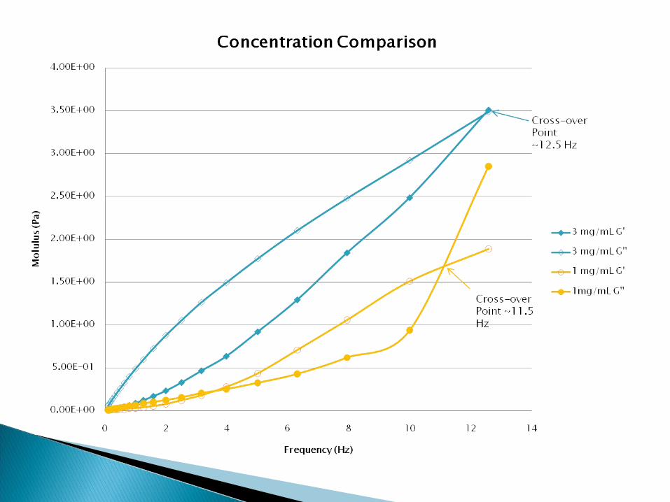
The molecular weight of synovial fluid makes a difference in the viscosity and elasticity of samples.
Prediction: samples with higher molecular weights will demonstrate more elasticity and viscosity at given shear rates and frequencies.

The cone oscillates at a specific range of frequencies and the machine measures the viscosity and elasticity of the fluid.
G’ = elastic modulus “stored energy”G’’ = viscous modulus“lost energy”

40mm 2°cone Peltier plate geometry25°Celcius

A cone or plate rotates at a constant shear rate (deformation rate), while the machine measures the shear stress exerted on the instrument by the fluid.
Fluid
Viscosity = shear stress
shear rate
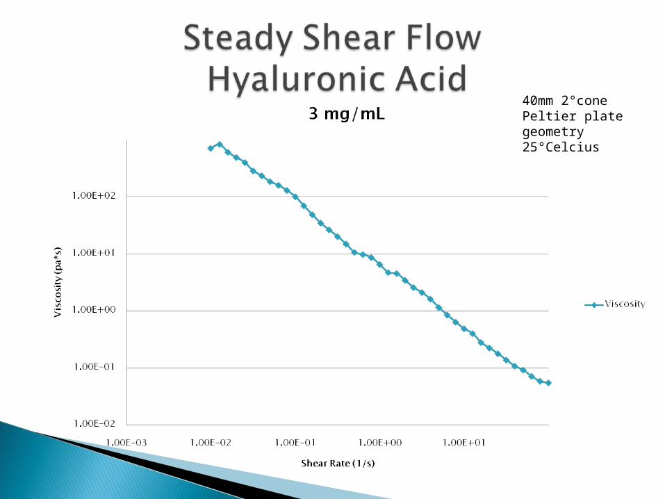
40mm 2°cone Peltier plate geometry25°Celcius


0.001
0.01
0.1
1
0.01 0.1 1 10 100
Shear Rate (1/s)
34-089 RS Steady Shear Comparison
New 34-089 RS
Old 34-089 RS

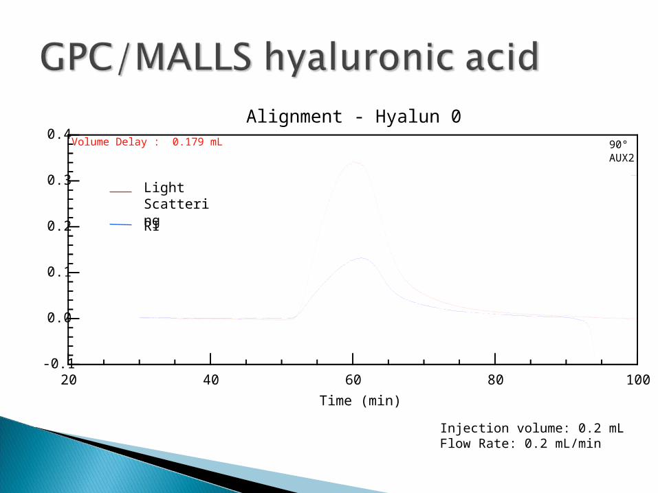
Two detector system:◦ Sample first separated by size exclusion
chromatography (porous columns)◦ Refractive Index detector determines the
concentration◦ Light scattering determines the molecular weight

Detector measures the intensity of light as a function of deflection angle and concentration.
Detector, I()Detector, Io
Polymer SolutionLight Source
-0.1
0.0
0.1
0.2
0.3
0.4
20 40 60 80 100
AUX, 90° Detector
Time (min)
Alignment - Hyalun 0
90°AUX2
Volume Delay : 0.179 mL
Light ScatteringRI
Injection volume: 0.2 mLFlow Rate: 0.2 mL/min
-0.2
0.0
0.2
0.4
0.6
0.8
20 40 60 80 100
AUX, 90° Detector
Time (min)
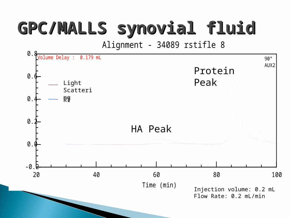
Alignment - 34089 rstifle 8
90°AUX2
Volume Delay : 0.179 mL
Light ScatteringRI
HA Peak
Protein Peak
GPC/MALLS synovial fluidGPC/MALLS synovial fluid
Injection volume: 0.2 mLFlow Rate: 0.2 mL/min

Sample ID: 34089 rstifle in 1:10 PBS August 1, 2008 Operator: Nikki Buck Collection InformationCollection time : Fri Aug 01, 2008 10:06 AM PSTSolvent name : PBS pH 7Solvent RI: 1.334Calibration constants DAWN : 8.2930e-06 » AUX2 : 5.1727e-05Flow rate : 0.200 mL/minCalculation method : dn/dc + AUX Constantdn/dc (mL/g) : 0.167 0.167 RESULTS:Molar Mass Moments (g/mol)Mw : 3.384e+05 (0.5%) 6.171e+04 (0.17%)

An enzyme that hydrolyzes the peptide bond between amino acids of a protein Enzyme used: Dipase from Bacillus polymyxa Protocol:
◦ Dilute synovial fluid 1:3 in PBS◦ Add 0.78 units Protease per mL synovial fluid◦ Incubate 15 min in 37°C water bath◦ Filter◦ Extract HA using phenol-chloroform◦ Filter
Protease

Hypothesis: Part 2
Proteins cause the second light scattering peak but do not interfere with the molecular weight reading of GPC/MALLS light scattering.
Prediction: Synovial fluid samples allowed to incubate in protease will not demonstrate a protein peak during light scattering analysis, and will have molecular weights in the same range as that of the undigested samples.

-0.2
0.0
0.2
0.4
0.6
0.8
20 40 60 80 100
AUX, 90° Detector
Time (min)
Alignment - 34089 rstifle 8
90°AUX2
Volume Delay : 0.179 mL
-0.04
0.00
0.04
0.08
0.12
20 40 60 80 100
AUX, 90° Detector
Time (min)
Alignment - 34089 rstifle 8
90°AUX2
Volume Delay : 0.179 mL
34089 Right Stifle
MW: 3.384*105 g/mol
34089 Right Stifle digested in Protease
MW: 3.819*105 g/mol
Light Scattering
RI
Light Scattering
RI
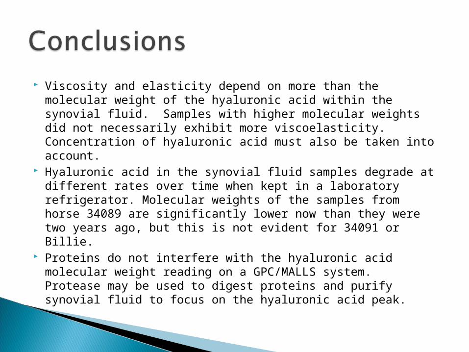
Viscosity and elasticity depend on more than the molecular weight of the hyaluronic acid within the synovial fluid. Samples with higher molecular weights did not necessarily exhibit more viscoelasticity. Concentration of hyaluronic acid must also be taken into account.
Hyaluronic acid in the synovial fluid samples degrade at different rates over time when kept in a laboratory refrigerator. Molecular weights of the samples from horse 34089 are significantly lower now than they were two years ago, but this is not evident for 34091 or Billie.
Proteins do not interfere with the hyaluronic acid molecular weight reading on a GPC/MALLS system. Protease may be used to digest proteins and purify synovial fluid to focus on the hyaluronic acid peak.

Howard Hughes Medical Institute Dr. Kevin Ahern Dr. Skip Rochefort, OSU School of Chemical
Biological and Environmental Engineering Sara Tracy, M.S. Chemical Engineering Dr. Jill Parker, OSU College of Veterinary
Medicine Haley Thompson, Coralie Backlund, and
Jesse McKiernan