rgs14 restricts plasticity in hippocampal ca2 by … · jupiter, fl, 33458. email:...
TRANSCRIPT
1
RGS14 restricts plasticity in hippocampal CA2 by limiting
postsynaptic calcium signaling
Abbreviated Title: RGS14 limits calcium to block CA2 plasticity
Authors and Affiliations: Paul R. Evans1,2, Paula Parra-Bueno1, Michael S. Smirnov1, Daniel
J. Lustberg3, Serena M. Dudek3, John R. Hepler2*, and Ryohei Yasuda1*
1Max Planck Florida Institute for Neuroscience, Jupiter, FL, 33458, USA
2Department of Pharmacology, Emory University School of Medicine, Atlanta, GA 30322, USA
3Neurobiology Laboratory, National Institute of Environmental Health Sciences, National
Institutes of Health, Research Triangle Park, NC, 27709, USA
*Co-correspondence
Author Contributions: PE, PP, and DL performed research; PE, PP, and MS analyzed data;
PE, SD, JH, and RY designed research; PE wrote and PE, SD, JH and RY edited the paper.
Correspondence should be addressed to: 1) John R. Hepler, Ph.D., Rollins Research
Center, 1510 Clifton Road, Suite G205, Atlanta, GA, 30322. Email: [email protected]
2) Ryohei Yasuda, Ph.D., Max Planck Florida Institute for Neuroscience, One Max Planck Way,
Jupiter, FL, 33458. Email: [email protected]
Number of figures: 5
Number of tables: 0
Number of multimedia: 0
Number of words for Abstract: 249
Number of words for Significance Statement: 111
Number of words for Introduction: 722
Number of words for Discussion: 1439
(which was not peer-reviewed) is the author/funder. All rights reserved. No reuse allowed without permission. The copyright holder for this preprint. http://dx.doi.org/10.1101/297499doi: bioRxiv preprint first posted online Apr. 8, 2018;

2
Acknowledgements: We thank Jaime Richards for preparing organotypic slice cultures and
reagents, David Kloetzer for laboratory management, and Drs. Lesley Colgan and Tal Laviv for
assistance with two-photon microscopy.
Conflict of Interest: The authors declare no competing financial interests.
Funding Sources: P.R.E. was supported by a predoctoral fellowship from NINDS
(1F31NS086174). J.R.H. was supported by NINDS grants 5R01NS37112 and 1R21NS074975.
S.M.D. was supported by the Intramural Research Program of NIEHS (Z01ES100221). R.Y.
was supported by NIMH grants R01MH080047 and DP1NS096787.
(which was not peer-reviewed) is the author/funder. All rights reserved. No reuse allowed without permission. The copyright holder for this preprint. http://dx.doi.org/10.1101/297499doi: bioRxiv preprint first posted online Apr. 8, 2018;
3
Abstract
Pyramidal neurons in hippocampal area CA2 are distinct from neighboring CA1 in that they
resist synaptic long-term potentiation (LTP) at CA3 Schaffer Collateral synapses. Regulator of G
Protein Signaling 14 (RGS14) is a complex scaffolding protein enriched in CA2 dendritic spines
that naturally blocks CA2 synaptic plasticity and hippocampus-dependent learning, but the
cellular mechanisms by which RGS14 gates LTP are largely unexplored. A previous study has
attributed the lack of plasticity to higher rates of calcium (Ca2+) buffering and extrusion in CA2
spines. Additionally, a recent proteomics study revealed that RGS14 interacts with two key
Ca2+-activated proteins in CA2 neurons: calcium/calmodulin, and CaMKII. Here, we investigate
whether RGS14 regulates Ca2+ signaling in its host CA2 neurons. We find the nascent LTP of
CA2 synapses due to genetic knockout (KO) of RGS14 in mice requires Ca2+-dependent
postsynaptic signaling through NMDA receptors, CaMK, and PKA, revealing similar
mechanisms to those in CA1. We report RGS14 negatively regulates the long-term structural
plasticity of dendritic spines of CA2 neurons. We further show that wild-type (WT) CA2 neurons
display significantly attenuated spine Ca2+ transients during structural plasticity induction
compared with the Ca2+ transients from CA2 spines of RGS14 KO mice and CA1 controls.
Finally, we demonstrate that acute overexpression of RGS14 is sufficient to block spine
plasticity, and elevating extracellular Ca2+ levels restores plasticity to RGS14-expressing
neurons. Together, these results demonstrate for the first time that RGS14 regulates plasticity in
hippocampal area CA2 by restricting Ca2+ elevations in CA2 spines and downstream signaling
pathways.
Significance Statement
Recent studies of hippocampal area CA2 have provided strong evidence in support of a clear
role for this apparently plasticity-resistant subregion of the hippocampus in social, spatial, and
temporal aspects of memory. Regulator of G Protein Signaling 14 (RGS14) is a critical factor
(which was not peer-reviewed) is the author/funder. All rights reserved. No reuse allowed without permission. The copyright holder for this preprint. http://dx.doi.org/10.1101/297499doi: bioRxiv preprint first posted online Apr. 8, 2018;
4
that inhibits synaptic plasticity in CA2, but the molecular mechanisms by which RGS14 limits
LTP remained unknown. Here we provide new evidence that RGS14 restricts spine calcium
(Ca2+) in CA2 neurons and that key downstream Ca2+-activated signaling pathways are required
for CA2 plasticity in mice lacking RGS14. These results define a previously unrecognized role
for RGS14 as a natural inhibitor of postsynaptic Ca2+ signaling in hippocampal area CA2.
Introduction
Pyramidal neurons in hippocampal area CA2 differ markedly from neighboring subfields in that
synaptic long-term potentiation (LTP) is not readily induced (Zhao et al., 2007). A number of
genes are selectively expressed in CA2 pyramidal neurons (Lein et al., 2005, 2007;
Cembrowski et al., 2016), and a few of these proteins have been shown to contribute to the
atypical plasticity features of CA2 (Simons et al., 2009, 2012; Lee et al., 2010; Caruana et al.,
2012; Pagani et al., 2015; Carstens et al., 2016; Dudek et al., 2016). Regulator of G Protein
Signaling 14 (RGS14) is one such gene that is highly expressed in CA2 pyramidal neurons
(Evans et al., 2014), where it is enriched postsynaptically in dendrites and spines (Lee et al.,
2010; Squires et al., 2017). RGS14 knockout (KO) mice possess an unusually robust capacity
for LTP in CA2, which is absent in wild-type (WT) mice, and exhibit markedly enhanced spatial
learning (Lee et al., 2010). Thus, RGS14 acts as a critical factor restricting synaptic plasticity in
CA2 pyramidal neurons and hippocampal-based learning and memory.
RGS14 is a complex scaffolding protein with an unconventional protein architecture that allows
it to integrate G protein signaling and ERK/MAPK signaling (Shu et al., 2010; Vellano et al.,
2013; Evans et al., 2015). RGS14 interacts with active Gai/o-GTP subunits through an RGS
domain and inactive Gai1/3-GDP subunits through its GPR motif (Cho et al., 2000; Traver et al.,
2000; Hollinger et al., 2001). RGS14 also binds active HRas-GTP and Raf kinases through the
(which was not peer-reviewed) is the author/funder. All rights reserved. No reuse allowed without permission. The copyright holder for this preprint. http://dx.doi.org/10.1101/297499doi: bioRxiv preprint first posted online Apr. 8, 2018;

5
tandem Ras-binding domains (Kiel et al., 2005; Shu et al., 2010; Vellano et al., 2013). The
nascent plasticity of CA2 synapses in RGS14 KO mice requires ERK signaling (Lee et al.,
2010), but additional cellular mechanisms have yet to be explored.
An independent study has attributed the lack of plasticity in CA2 pyramidal neurons to smaller
spine Ca2+ transients due to higher Ca2+ extrusion rate and buffering compared to CA1 and CA3
pyramidal neurons (Simons et al., 2009), and Ca2+-dependent mechanisms underlie some forms
of CA2 synaptic potentiation (Simons et al., 2009; Pagani et al., 2015). Elevations in
postsynaptic Ca2+ are critical to induce activity-dependent LTP in CA1 hippocampal neurons
(Lynch et al., 1983; Malenka et al., 1988) as well as in CA2 (Simons et al., 2009). Ca2+ binds
calmodulin (Ca2+/CaM), which then potently activate many plasticity-inducing pathways,
including CaMK, Ras/ERK, and PKA (Malenka et al., 1989; Abel et al., 1997; Otmakhova et al.,
2000; Yasuda et al., 2006; Lee et al., 2010; Tang and Yasuda, 2017). A very recent proteomic
analysis found that RGS14 natively interacts with proteins that regulate actin binding, CaMK
activity, and CaM binding, in mouse brain (Evans et al., 2018). From these candidate
interactors, two new bona fide RGS14 binding partners were identified that are Ca2+-activated
and required for plasticity: Ca2+/CaM and CaMKII. Moreover, proximity ligation assays revealed
that RGS14 interacts with CaM and CaMKII in murine hippocampal CA2 neurons (Evans et al.,
2018). However, to date, no evidence has functionally linked RGS14 to Ca2+ signaling pathways
required for LTP induction. Therefore, we investigated whether RGS14 modulates key Ca2+-
stimulated pathways to restrict plasticity in CA2 hippocampal neurons.
Here, we performed electrophysiology and two-photon fluorescence imaging experiments in
brain slices from RGS14 WT and KO littermates. Using a reporter mouse line to localize CA2
dendrites to perform field recordings, we found the nascent LTP of CA2 synapses in adult
RGS14 KO mice requires Ca2+-dependent signaling through NMDARs, CaMK, and PKA. Given
(which was not peer-reviewed) is the author/funder. All rights reserved. No reuse allowed without permission. The copyright holder for this preprint. http://dx.doi.org/10.1101/297499doi: bioRxiv preprint first posted online Apr. 8, 2018;
6
the similarity to mechanisms supporting structural plasticity of dendritic spines in CA1, we next
discovered that RGS14 limits CA2 spine structural plasticity. We found that RGS14 likely
accomplished this by attenuating spine Ca2+ levels during spine plasticity induction as spines of
CA2 neurons from WT mice showed smaller Ca2+ transients during synaptic stimulation
compared to CA2 neurons from RGS14 KO mice. Lastly, we found that acute overexpression of
RGS14 in brain slices from RGS14 KO mice can “rescue” the impairment in long-term spine
plasticity; sustained spine plasticity can be restored to RGS14-expressing neurons by elevating
extracellular calcium levels suggesting that Ca2+ signaling is central to this process. Our results
presented here define a new role for RGS14 as a novel regulator of postsynaptic Ca2+ signaling
and identify a new mechanism upstream of the Ras-ERK pathway whereby RGS14 exerts
control over plasticity signaling in CA2 pyramidal neurons.
Materials and Methods
Animals
Animals in all experiments were house under a 12h:12h light/dark cycle with access to food and
water ad libitum. All experimental procedures conform to US NIH guidelines and were approved
by the animal care and use committees of the Max Planck Florida Institute for Neuroscience and
the National Institute of Environmental Health Sciences. RGS14 KO mice were generated and
maintained as previously described (Lee et al., 2010). Both male and female RGS14 WT/KO
animals were used in all experiments. Reporter mice expressing enhanced green fluorescent
protein (EGFP) in CA2 pyramidal neurons under the Amigo2 promoter (Amigo2-EGFP;
Tg(Amigo2-EGFP)LW244Gsat; RRID:MMRRC_033018-UCD) were crossed with RGS14
WT/KO mice to label CA2 dendrites for field recordings.
Acute slice preparation
(which was not peer-reviewed) is the author/funder. All rights reserved. No reuse allowed without permission. The copyright holder for this preprint. http://dx.doi.org/10.1101/297499doi: bioRxiv preprint first posted online Apr. 8, 2018;
7
Adult RGS14 WT or KO;Amigo2-EGFP+ mice (P20-P50) were sedated by isoflurane inhalation,
and perfused intracardially with a chilled choline chloride solution. Brains were removed and
placed in the same choline chloride solution composed of 124 mM choline chloride, 2.5 mM KCl,
26 mM NaHCO3, 3.3 mM MgCl2, 1.2 mM NaH2PO4, 10 mM glucose and 0.5 mM CaCl2, pH 7.4
equilibrated with 95% O2/5% CO2. Coronal slices (400 µm) were prepared on a vibratome (Leica
VT1200), and slices were maintained in a submerged holding chamber at 32°C for 1h and then
at room temperature in oxygenated ACSF.
Extracellular Recordings and LTP protocol
Experiments were performed at room temperature (~21°C), and submerged slices
were perfused with oxygenated ACSF containing 2 mM CaCl2, 2 mM MgCl2 and 100 µM
picrotoxin. One or two glass recording electrodes (resistance ~4 MΩ) containing the
same ACSF solution was placed in the stratum radiatum of CA2 or CA1 respectively (~100–200
µm away from the soma) while stimulating Schaffer Collateral fibers with square current pulses
(0.1 ms duration) using a concentric bipolar stimulation electrode (FHC) every 30 s. For clarity
of display, every other data point was removed from averaged time course field recording
experiments; all data points were included in the statistical analyses. Area CA2 was detected by
Amigo2-EGFP fluorescence. The initial slope of the EPSP was monitored with custom software.
The stimulation strength was set to ~50% saturation. LTP was assessed by applying three sets
of high frequency stimuli (100 Hz, 1 s) with 20 s intervals. All data was analyzed with an in-
house program written in MATLAB (MathWorks). Data are presented as mean ± SEM. For all
LTP experiments, predetermined statistical comparisons were made between the normalized
average fEPSP slope 40-60 mins following LTP induction to assess long-lasting changes in
synaptic efficacy. Statistical comparisons were performed using two-way ANOVA, and Sidak’s
multiple comparisons test was used to compare the same CA region between RGS14 WT and
(which was not peer-reviewed) is the author/funder. All rights reserved. No reuse allowed without permission. The copyright holder for this preprint. http://dx.doi.org/10.1101/297499doi: bioRxiv preprint first posted online Apr. 8, 2018;

8
KO animals. Differences between datasets were judged to be significant at p ≤ 0.05. Statistical
analyses were performed in GraphPad Prism 7.
To assess whether LTP could be inhibited pharmacologically, slices from RGS14 KO/Amigo2-
EGFP+ animals were perfused with either ACSF for controls or ACSF supplemented with either
APV (50 µM, Sigma), KN-62 (10 µM, Tocris), or PKI (14-22) amide myristoylated (1 µM, Enzo
Life Sciences), to inhibit NMDA receptors, CaMK, or PKA, respectively. For KN-62 experiments,
RGS14 KO control slices were perfused with ACSF containing 0.01% DMSO as a vehicle
control. Electrophysiological recordings and LTP induction protocol were performed as
described above. Data are presented as mean ± SEM. Statistical comparisons were performed
using Student’s t test to compare each inhibitor with respective KO control, and differences
between datasets were judged to be significant at p ≤ 0.05. Statistical analyses were performed
in GraphPad Prism 7.
Tissue preparation for histology
Adult Amigo2-EGFP mice were anesthetized by isoflurane inhalation and transcardially
perfused with 4% paraformaldehyde in PBS. Brains were postfixed for 24h, submerged in 30%
sucrose in PBS, and sectioned coronally at 40 µm on a cryostat. Sections were washed in PBS,
blocked for at least 1h in 5% normal goat serum (NGS, Vector Labs) diluted in 0.1% PBS-X
(0.1% Triton X-100 in PBS) at room temperature, and incubated in primary antibodies diluted in
the same buffer overnight. A chicken polyclonal anti-GFP antibody (Abcam) was used at a
1:2,000 dilution to enhance Amigo2-EGFP fluorescence with either a rabbit polyclonal anti-
PCP4 antibody (Santa Cruz) or a rabbit polyclonal anti-Wfs1 antibody (ProteinTech). Sections
were thoroughly washed in 0.1% PBS-X and incubated in secondary antibodies (Alexa goat
anti-chicken 488 and Alexa goat anti-rabbit 568, Invitrogen) diluted at 1:500 for 2h at room
temperature. Finally, sections were washed in 0.1% PBS-X and mounted under ProLong Gold
(which was not peer-reviewed) is the author/funder. All rights reserved. No reuse allowed without permission. The copyright holder for this preprint. http://dx.doi.org/10.1101/297499doi: bioRxiv preprint first posted online Apr. 8, 2018;

9
Antifade fluorescence media with DAPI (Invitrogen). Sections were then imaged on a Zeiss 710
meta confocal microscope using a 40X oil-immersion lens.
Organotypic slice preparation
Hippocampal slice cultures were prepared from postnatal day 6-8 RGS14 WT/KO mice as
described previously (Stoppini et al., 1991). In brief, hippocampi were dissected and sliced at
320 μm thickness using a tissue chopper. The slices were plated on a membrane filter (Millicell-
CM PICMORG50, Millipore). These cultures were maintained at 37 °C in an environment of
humidified 95% O2 and 5% CO2. The culture medium was exchanged with fresh medium every
three days. After 7-10 days in culture, neurons were sparsely transfected with ballistic gene
transfer (McAllister, 2000) using gold beads (9-11 mg) coated with plasmid containing mEGFP
cDNA (10 µg) for sLTP experiments. For RGS14 overexpression experiments, cultured slices
from RGS14 KO mice were co-transfected with plasmids containing GFP-tagged RGS14 (20
µg) and CyRFP1 (10 µg, volume marker) cDNAs or mEGFP cDNA (10 µg) as a control. Slices
were imaged after 3-12 days following transfection. CA2/CA1 neurons were identified by
somatic location within the slice and branching morphology of the apical dendrites.
Two-photon fluorescence microscopy and two-photon glutamate uncaging
Glutamate uncaging and imaging of live neurons were performed under a custom-built two-
photon microscope with two Ti:Sapphire lasers (Chameleon, Coherent). In brief, the lasers were
tuned at the wavelength of 920 nm and 720 nm for imaging and uncaging, respectively. The
intensity of each laser was independently controlled with electro-optical modulators (Conoptics).
The fluorescence was collected with an objective (60x, 1.0 numerical aperture, Olympus),
divided with a dichroic mirror (565dcxr) and detected with photoelectron multiplier tubes (PMTs)
placed after wavelength filters (ET520/60M-2P for green, ET620/60M-2p for red, Chroma). MNI-
caged L-glutamate (4-methoxy-7-nitroindolinyl-caged L-glutamate, Tocris) was uncaged with a
(which was not peer-reviewed) is the author/funder. All rights reserved. No reuse allowed without permission. The copyright holder for this preprint. http://dx.doi.org/10.1101/297499doi: bioRxiv preprint first posted online Apr. 8, 2018;
10
train of 4-8 ms laser pulses (2.7-3.0 mW under the objective, 30 times at 0.5 Hz) near a spine of
interest. Experiments were performed at room temperature in ACSF solution containing (in mM):
127 NaCl, 2.5 KCl, 25 NaHCO3, 1.25 NaH2PO4, 4 CaCl2, 25 glucose, 0.001 tetrodotoxin (Tocris)
and 4 MNI-caged L-glutamate, bubbled with 95% O2 and 5% CO2. We examined
secondary/tertiary branches of apical dendrites of CA1 and CA2 pyramidal neurons (located in
stratum radiatum) in organotypic cultured hippocampus slices at 10-22 days in vitro.
Calcium Imaging
For Ca2+ imaging, we performed whole-cell patch recordings of CA2/CA1 pyramidal neurons in
cultured hippocampus slices with the patch pipette containing the Ca2+ indicator Fluo-4FF (500
µM; Thermofisher) and Alexa-594 (500 µM) diluted in potassium gluconate internal solution
(containing in mM:130 K gluconate, 10 Na phosphocreatine, 4 MgCl2, 4 Na2ATP, 0.3 MgGTP, L-
Ascorbic acid 3, HEPES 10, pH 7.4, 300 mosm). Both dyes were excited simultaneously by a
Ti:Sapphire laser (Chameleon, Coherent) at 920 nm. Fluo-4FF (green) and Alexa-594 (red)
fluorescence were used to quantify the change in [Ca2+] (∆[Ca2+]) using the following formula:
Δ[Ca%&] =Δ𝐺𝑅 𝐾,𝐺𝑅 -./
where (G/R)sat is the ratio of green and red fluorescence at a saturating Ca2+ concentration
measured in the patch pipette (after each day of imaging), and the dissociation constant (KD) for
Fluo-4FF is 10.4 µM (Yasuda et al., 2004; Lee et al., 2009). Images were acquired using fast-
framing two-photon fluorescence microscopy (15.63 Hz frame rate) over a ~4 s baseline before
inducing structural plasticity by glutamate uncaging with a train of 4-8 ms laser pulses (2.7-3.0
mW under the objective, 30 times at 0.5 Hz) near a spine of interest. Images were analyzed with
MATLAB (MathWorks). Data are presented as the averaged time courses from baseline and the
uncaging-triggered average change in [Ca2+] (µM) for all 30 glutamate uncaging pulses from
(which was not peer-reviewed) is the author/funder. All rights reserved. No reuse allowed without permission. The copyright holder for this preprint. http://dx.doi.org/10.1101/297499doi: bioRxiv preprint first posted online Apr. 8, 2018;

11
baseline ± SEM. To analyze the Ca2+ decay kinetics from CA2 spines, the maximum and
minimum values from the uncaging-triggered averages were normalized and decay time
constants were extrapolated by curve fitting (nonlinear regression) in GraphPad Prism 7.
Imaging automation
For sLTP experiments images were acquired as a Z stack of five slices with 1 µm separation,
averaging five frames/slice. Using multi-position imaging of spines with high-throughput
automation, dendritic spines at four positions on separate secondary/tertiary dendrites were
imaged simultaneously employing algorithms for autofocusing and drift correction to maintain
position and optimal focus during long imaging experiments as recently described (Smirnov et
al., 2017). Baseline images were acquired over 5 mins prior to two-photon glutamate uncaging
(1 min, 30 pulses at 0.5 Hz) followed by 30 mins of imaging post-uncaging. A 5 min baseline
stagger was incorporated to avoid data loss during uncaging events.
Post hoc immunostaining
Immediately following two-photon imaging experiments, organotypic hippocampal slices were
fixed in 4% paraformaldehyde for 30 mins at room temperature. Slices were then washed
thoroughly in 0.01M PBS, permeabilized for 15 mins in 0.3% PBS-X (0.3% Triton X-100 in
PBS), and thoroughly washed in PBS. Slices were blocked for at least one hour at room
temperature in a blocking solution containing 10% NGS (Vector Labs) diluted in 0.1% PBS-X
prior to incubation in primary antibodies diluted in the same blocking solution for 42 hours at
room temperature. A rabbit polyclonal anti-PCP4 antibody (Santa Cruz) was used at a 1:500
dilution to delineate area CA2 in all slice culture experiments. For sLTP experiments with
mEGFP-expressing neurons, a chicken polyclonal anti-GFP antibody (Abcam) was used at a
dilution of 1:1,000 to visualize imaged neurons. For Ca2+ imaging experiments, Alexa-594
fluorescence was used to identify imaged neurons. Sections were thoroughly washed in 0.1%
(which was not peer-reviewed) is the author/funder. All rights reserved. No reuse allowed without permission. The copyright holder for this preprint. http://dx.doi.org/10.1101/297499doi: bioRxiv preprint first posted online Apr. 8, 2018;
12
PBS-X and incubated in secondary antibodies (Alexa goat anti-chicken 488 and Alexa goat anti-
rabbit 568, Invitrogen) diluted at 1:500 for 2h at room temperature. After rinsing samples
thoroughly in 0.1% PBS-X, slices were optically cleared by incubating in a 60% 2,2′-
Thiodiethanol solution (v/v, Sigma) for 30 mins at room temperature (Aoyagi et al., 2015). Intact
organotypic slices were imaged in the same clearing solution in glass bottom dishes (Willco) on
a Zeiss 880 laser scanning confocal microscope.
Image and Data Processing
Confocal laser scanning microscope images were processed using FIJI software (NIH v2.0.0).
Images were only adjusted for brightness/contrast and cropped for presentation.
Experimental Design and Statistical Analysis
All statistical analyses were performed in GraphPad Prism 7. Statistical analyses of CA2/CA1
neurons from RGS14 WT/KO mice were made using two-way ANOVA (non-repeated
measures) with post hoc Sidak’s multiple comparison test between genotypes (i.e. WT CA2-KO
CA2 and WT CA1-KO CA1). For pharmacological experiments, LTP in the presence of each
inhibitor was compared with paired KO CA2 controls using two-tailed, unpaired t tests. For
RGS14 overexpression experiments, CA2 neurons transfected with RGS14-GFP were
compared to mEGFP transfected CA2 neurons by unpaired t-test. For RGS14 overexpression
experiments in CA1 neurons, all groups were compared by one-way ANOVA followed by
Fisher’s LSD test. Complete results of the statistical analyses for each experiment are reported
in the results section and sample sizes are noted in the figure legends. In figures * denotes
p<0.05, ** denotes p≤0.01, *** denotes p<0.001, **** denotes p<0.0001.
Results
Synaptic potentiation can be induced in CA2 in slices from adult RGS14 KO mice
(which was not peer-reviewed) is the author/funder. All rights reserved. No reuse allowed without permission. The copyright holder for this preprint. http://dx.doi.org/10.1101/297499doi: bioRxiv preprint first posted online Apr. 8, 2018;
13
RGS14 has been previously shown to inhibit LTP induction at Schaffer collateral synapses onto
CA2 pyramidal neurons by performing whole cell recordings in brain slices prepared from young
mice while stimulating in stratum radiatum (Lee et al., 2010). However, the postsynaptic
mechanisms underlying RGS14’s actions are poorly understood. Here, we sought to investigate
mechanisms by which RGS14 constrains CA2 synaptic potentiation in adulthood when RGS14
levels are highest and stable (Evans et al., 2014). To reliably localize CA2 dendrites in slices,
we crossed RGS14 KO mice with an Amigo2-EGFP reporter mouse line that fluorescently labels
CA2 pyramidal neurons (Carstens et al., 2016). We validated that this mouse line selectively
labels CA2 pyramidal neurons by immunolabeling for the dentate gyrus (DG)- and CA2-enriched
protein Purkinje cell protein 4 (PCP4) as well as the CA1 molecular marker Wolframin (WFS1).
We found that Amigo2-EGFP fluorescence colocalizes with PCP4 immunoreactivity (Figure 1A)
but does not overlap with immunostaining for the CA1 marker WFS1 (Figure 1B).
We next performed field potential recordings in acute brain slices prepared from adult RGS14
WT and KO (both Amigo2-EGFP+) mice and replicated previous findings that high-frequency
stimulation (3 x 100 Hz) of stratum radiatum induces robust LTP in CA2 neurons of RGS14 KO
mice, which is absent in WT mice, and similar to CA1 controls (Lee et al., 2010; Figure 1C).
Comparing the mean field excitatory postsynaptic potential (fEPSP) slope averaged from 40-60
minutes after LTP induction, we found that the RGS14 KO CA2 fEPSP slope was significantly
larger than WT CA2; LTP induced in CA1 was not significantly different between genotypes
(Figure 1D, two-way ANOVA results for RGS14 genotype were F(1,55)=6.64, p=0.0127; results
for CA region were F(1,55)=2.478, p=0.1212; and results for interaction were F(1,55)=3.992,
p=0.0507. Sidak’s post hoc WT CA2–KO CA2 p=0.0036, WT CA1–KO CA1 p=0.9028).
Baseline synaptic responses were not altered as input-output curves of fEPSP slope and
amplitude were not different in CA2 or CA1 between adult RGS14 WT and KO mice (data not
shown).
(which was not peer-reviewed) is the author/funder. All rights reserved. No reuse allowed without permission. The copyright holder for this preprint. http://dx.doi.org/10.1101/297499doi: bioRxiv preprint first posted online Apr. 8, 2018;

14
CA2 LTP in RGS14 KO mice requires NMDARs, CaMK, and PKA activity
Previous work has shown that LTP in CA2 neurons is naturally suppressed by both robust Ca2+
handling mechanisms in spines (Simons et al., 2009) and RGS14 (Lee et al., 2010). To
determine if the nascent LTP present in CA2 neurons of RGS14 KO mice requires Ca2+
signaling, we next performed the same LTP induction protocol in area CA2 using brain slices
prepared from RGS14 KO (Amigo2-EGFP+) mice in the presence of pharmacological inhibitors
of Ca2+-dependent signaling pathways (Figure 2). The LTP in area CA2 of slices from RGS14
KO mice was effectively blocked by bath application of the NMDAR antagonist APV (50 µM,
blue) as well as inhibitors of CaMK (KN-62, 10 µM) or PKA (PKI, 1 µM; Figure 2A-C).
Comparing the mean fEPSP slope 40-60 minutes following LTP induction, CA2 plasticity was
significantly reduced by each inhibitor relative to paired RGS14 KO controls (Figure 2D;
unpaired two-tailed t-tests: for APV t(30) = 2.868, p=0.0075; for KN-62 t(37)=2.59, p=0.0136; for
PKI t(37) = 2.502, p=0.0169). These findings indicate that the nascent LTP in CA2 neurons of
RGS14 KO mice requires NMDARs and subsequent CaMK and PKA activity, which can be
activated – directly or indirectly in the case of PKA – by Ca2+ (Figure 2E). Similar mechanisms
are thought to support plasticity in CA1 (Malenka et al., 1989; Abel et al., 1997; Otmakhova et
al., 2000).
RGS14 suppresses structural plasticity of CA2 dendritic spines
After determining that CA2 synapses from RGS14 KO mice utilize similar mechanisms as CA1
neurons to support synaptic potentiation, we next asked if RGS14 might also play a role in
activity-dependent spine structural plasticity (sLTP, Figure 3). This enlargement of dendritic
spines induced upon synaptic stimulation, is strongly associated with LTP in hippocampus and
relies on similar cellular mechanisms (Matsuzaki et al., 2004; Nishiyama and Yasuda, 2015).
RGS14 is well positioned to regulate sLTP as it is enriched in CA2 spines and dendrites (Lee et
(which was not peer-reviewed) is the author/funder. All rights reserved. No reuse allowed without permission. The copyright holder for this preprint. http://dx.doi.org/10.1101/297499doi: bioRxiv preprint first posted online Apr. 8, 2018;
15
al., 2010); however, no previous studies have examined spine plasticity in CA2 pyramidal
neurons. To determine whether RGS14 modulates sLTP in CA2, we cultured hippocampal
slices from RGS14 KO mice and WT littermate controls and sparsely transfected neurons with
mEGFP using ballistic gene transfer. We then performed two-photon fluorescence microscopy
during two-photon glutamate uncaging to induce spine structural plasticity on secondary/tertiary
apical dendrites of CA2 and CA1 pyramidal neurons (Figure 3). In order to optimize data
collection from each neuron during long experiments, we utilized an automated method to
image multiple dendritic spine positions simultaneously (Smirnov et al., 2017). This interface
tracks coordinates of multiple dendritic segments on the same neuron and employs autofocus
and drift correction algorithms during the experiment to maintain optimal focus. To validate the
hippocampal subregion of all neurons imaged in these studies, slices were fixed immediately
following two-photon imaging and immunostained for PCP4 to delineate the boundaries of
hippocampal area CA2 (Figure 3A, red).
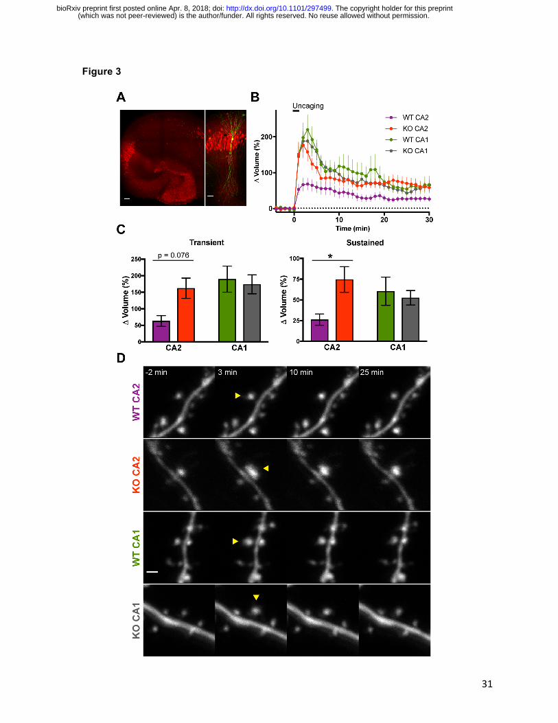
Dendritic spines of RGS14 WT CA2 neurons exhibited significantly smaller volume change
following repetitive glutamate uncaging to induce sLTP compared to RGS14 KO CA2 neurons
or CA1 controls (Figure 3B-D). Comparing the mean spine volume change between samples,
the spines of CA2 neurons from RGS14 WT mice trended toward reduced growth in the
transient phase of sLTP immediately following uncaging, but the results were not significantly
different from the spine volume change of KO CA2 neurons (Figure 3C, left; two-way ANOVA
results for genotype were F(1,69) = 1.676, p = 0.1998; results for CA region were F(1,69) =
4.624, p = 0.0350; and results for interaction were F(1,69) = 3.174, p = 0.0792. Sidak’s post hoc
WT CA2–KO CA2 p=0.0749, WT CA1–KO CA1 p=0.9236.). CA2 spines from slices prepared
from RGS14 WT mice exhibited significantly less enlargement compared to RGS14 KO CA2
spines during the later sustained phase of sLTP (Figure 3C, right; two-way ANOVA results for
genotype were F(1,69) = 2.172, p = 0.1451; results for CA region were F(1,69) = 0.1954, p =
(which was not peer-reviewed) is the author/funder. All rights reserved. No reuse allowed without permission. The copyright holder for this preprint. http://dx.doi.org/10.1101/297499doi: bioRxiv preprint first posted online Apr. 8, 2018;

16
0.6598; and results for interaction were F(1,69) = 4.198, p = 0.0443. Sidak’s post hoc WT CA2–
KO CA2 p=0.0360, WT CA1–KO CA1 p=0.8953.). These results indicate that RGS14 naturally
restricts the structural plasticity of dendritic spines in CA2 pyramidal cells, similar to the case
with functional plasticity.
RGS14 attenuates spine Ca2+ elevations in CA2
Spine Ca2+ is critical for the induction of synaptic plasticity (Lynch et al., 1983; Malenka et al.,
1988; Harvey et al., 2008), and we found the LTP of CA2 synapses in RGS14 KO mice requires
Ca2+-driven signaling (Figure 2). Therefore, we investigated if the diminished spine plasticity
observed in WT CA2 neurons containing RGS14 could be due to reduced Ca2+ levels during
glutamate uncaging. Performing two-photon fluorescence microscopy, we monitored Ca2+-
dependent fluorescence changes in neurons elicited during glutamate uncaging to induce sLTP.
Neurons were intracellularly perfused with the synthetic Ca2+ indicator Fluo-4FF and the
structural dye Alexa-594 to simultaneously monitor Ca2+ elevations and spine volume changes
elicited by glutamate uncaging. Spine Ca2+ responses are presented as averaged time courses
(Figure 4 A,B) and the uncaging-triggered average change in Ca2+ concentration (∆[Ca2+])
across all 30 glutamate uncaging pulses (Figure 4C-E).
Uncaging-evoked peak Ca2+ elevations were significantly smaller in spines of WT CA2 neurons
compared to spines of CA2 neurons from KO littermates, which were comparable to Ca2+
responses in CA1 controls (Figure 4A-D; two-way ANOVA results for genotype were F(1,79) =
6.698, p = 0.0115; results for CA region were F(1,79) = 2.367, p = 0.1279; and results for
interaction were F(1,79) = 6.675, p = 0.0116. Sidak’s post hoc WT CA2–KO CA2 p<0.0001, WT
CA1–KO CA1 p>0.9999). Nonlinear regression analysis of normalized uncaging-triggered
average Ca2+ responses reveals similar decay kinetics in CA2 spines of RGS14 WT and KO
mice (Figure 4E; WT CA2 tau = 0.212 s, KO CA2 tau = 0.240 s). Glutamate uncaging also
(which was not peer-reviewed) is the author/funder. All rights reserved. No reuse allowed without permission. The copyright holder for this preprint. http://dx.doi.org/10.1101/297499doi: bioRxiv preprint first posted online Apr. 8, 2018;

17
elicited significantly reduced Ca2+ transients in WT CA2 dendrites relative to KO CA2 neurons,
which were similar to those in CA1 (data not shown). These results demonstrate that spines of
CA2 neurons from WT mice display reduced spine Ca2+ transients, which correlates with the
attenuated spine structural plasticity in the sustained phase, and genetic deletion of RGS14
unmasks synaptic plasticity and restore Ca2+ responses to similar levels as those observed in
CA2. Together, these data suggest that RGS14 may have a previously unrecognized role in
regulating spine Ca2+ to constrain CA2 synaptic plasticity.
Acute RGS14 expression blocks sLTP, and spine plasticity can be recovered by elevating
extracellular calcium
To more directly test if there is a causal relationship between RGS14 and the diminished
capacity for spine plasticity, we acutely overexpressed GFP-tagged RGS14 (RGS14-GFP) or
GFP (control) in organotypic slice cultures prepared from RGS14 KO mice and induced spine
plasticity by glutamate uncaging. RGS14-GFP overexpression significantly reduces spine
volume enlargement in the sustained phase of sLTP in CA2 pyramidal neurons as expected
(Figure 5 A,B; unpaired t-test, ** p = 0.01). RGS14-GFP overexpression similarly blocked
transient and sustained phases of spine plasticity in CA1 pyramidal neurons (Figure 5 C,D;
Fisher’s LSD test, ** p < 0.01, *** p < 0.001). Given the low probability of transfecting CA2
pyramidal neurons using ballistic gene transfer, we attempted to reverse the plasticity blockade
in RGS14-expressing CA1 pyramidal neurons by increasing the extracellular Ca2+ concentration
from [4 mM] to [8 mM] as a similar manipulation increased spine Ca2+ and restored LTP to WT
CA2 neurons (Simons et al., 2009). We find that performing sLTP induction in [8mM] external
Ca2+ significantly rescues the sustained phase of spine plasticity that is otherwise abrogated in
RGS14-positive CA1 pyramidal neurons (Figure 5 C,D; Fisher’s LSD test, * p < 0.05, *** p <
0.001). These results demonstrate that RGS14 is able to block long-term structural plasticity in
hippocampal neurons, and increasing extracellular [Ca2+] restores sustained phase sLTP to
(which was not peer-reviewed) is the author/funder. All rights reserved. No reuse allowed without permission. The copyright holder for this preprint. http://dx.doi.org/10.1101/297499doi: bioRxiv preprint first posted online Apr. 8, 2018;

18
RGS14-expressing CA1 neurons. Together, these data suggest that limited Ca2+ signaling is
central to RGS14’s ability to gate the structural plasticity of dendritic spines as boosting Ca2+
can unmask plasticity.
Discussion
In this study, we have identified a previously unknown role for RGS14 in the regulation of
postsynaptic Ca2+ signaling in hippocampal CA2 neurons. Specifically, we find that the synaptic
potentiation present in CA2 neurons lacking RGS14 requires NMDAR activation along with
downstream CaMK and PKA activity, revealing a striking similarity to Ca2+-driven mechanisms
underlying LTP in CA1 (Malenka et al., 1989; Abel et al., 1997; Otmakhova et al., 2000). We
further show that RGS14 naturally inhibits the structural plasticity of CA2 dendritic spines
induced by local glutamate uncaging, and loss of RGS14 unleashes robust CA2 spine plasticity
observed as long-lasting spine enlargement that is similar to spines in CA1. Although somatic
integration of distal and proximal dendritic spines has been examined (Srinivas et al., 2017), to
our knowledge this is the first report of the diminished capacity for spine structural plasticity in
WT CA2 neurons, similar to the lack of functional synaptic potentiation in stratum radiatum
(Zhao et al., 2007). We also demonstrate that the lack of functional and structural plasticity in
WT CA2 neurons correlates with attenuated spine Ca2+ transients, and plastic spines from CA2
neurons of RGS14 KO mice display larger spine Ca2+ transients that are comparable to CA1
controls. Lastly, we find that acute re-expression of RGS14 in brain slices from RGS14 KO mice
blocks sustained phase plasticity in CA2 and CA1 pyramidal neurons, and raising the
extracellular Ca2+ concentration can reverse the blockade of long-lasting sLTP induced by
RGS14. Taken together, RGS14 is a critical regulator of CA2 plasticity and Ca2+ signaling that
seemingly differentiates CA2 pyramidal neurons from the neighboring CA1 subfield, best known
for its robust plasticity.
(which was not peer-reviewed) is the author/funder. All rights reserved. No reuse allowed without permission. The copyright holder for this preprint. http://dx.doi.org/10.1101/297499doi: bioRxiv preprint first posted online Apr. 8, 2018;

19
Here, we used a pharmacological approach to reveal the nascent plasticity of CA2 synapses in
brain slices of adult RGS14 KO mice requires intact NMDAR, CaMK, and PKA activity (Figure
2), and we previously demonstrated a requirement for ERK activity as well (Lee et al., 2010).
These mechanisms are not unique to CA2 pyramidal neurons as they are also thought to
support LTP in CA1 as well (Malenka et al., 1989; Abel et al., 1997; Otmakhova et al., 2000).
Interestingly, RGS14 engages upstream signaling proteins in each of these kinase cascades:
Ca2+/CaM upstream of CaMKs (Evans et al., 2018), Gαi/o subunits that regulate PKA (Cho et
al., 2000; Traver et al., 2000; Hollinger et al., 2001), and H-Ras that activates ERK (Kiel et al.,
2005; Shu et al., 2010; Vellano et al., 2013). Thus, RGS14 is well positioned to regulate the
activities of these pathways, likely in CA2 neurons to naturally gate plasticity therein. Moreover,
feedback from these pathways likely regulates the function of RGS14 as it can be
phosphorylated by CaMKII (Evans et al., 2018) and PKA (Hollinger et al., 2003). Future studies
are necessary to determine how phosphorylation affects RGS14 functions in hippocampal
neurons.
Similar to the nascent plasticity of CA2 neurons in RGS14 KO mice shown here and before (Lee
et al., 2010), boosting spine Ca2+ levels in WT CA2 neurons to levels comparable to those found
in CA1 spines, by brief application of high extracellular [Ca2+] or blocking Ca2+ extrusion through
plasma membrane pumps, permits LTP in CA2 pyramidal neurons (Simons et al., 2009). These
results demonstrate that CA2 neurons contain the endogenous machinery to support LTP, and
led us to hypothesize a common mechanism may mediate the lack of plasticity in CA2. The
reduced spine Ca2+ transients we observe in WT CA2 neurons is consistent with previous
findings that action potential-evoked spine Ca2+ transients in CA2 are smaller than in CA3 and
CA1, owing to higher rates of Ca2+ extrusion and buffering (Simons et al., 2009). These two
factors counteract each with regard to decay time, but both reduce peak Ca2+ elevation. We
found highly similar decay kinetics of spine Ca2+ responses between WT and KO CA2 neurons
(which was not peer-reviewed) is the author/funder. All rights reserved. No reuse allowed without permission. The copyright holder for this preprint. http://dx.doi.org/10.1101/297499doi: bioRxiv preprint first posted online Apr. 8, 2018;

20
(Figure 4E), which could be attributed to the Ca2+ handling that has been previously observed in
CA2 spines (Simons et al., 2009). From these data, RGS14 does not likely affect Ca2+ extrusion
rate or buffering alone, but it may regulate both processes in CA2 spines to reduce peak Ca2+
without affecting decay time. One possible mechanism by which RGS14 could regulate spine
Ca2+ levels is through direct interactions with Ca2+/CaM, which has been shown to influence
Ca2+ dynamics in CA2 and CA1 pyramidal neurons (Simons et al., 2009). Another likely factor
contributing to the Ca2+ handling properties of CA2 pyramidal neurons, in addition to RGS14, is
PCP4/Pep-19, a CA2-enriched protein that modulates calmodulin, which could underlie the
robust spine Ca2+ extrusion (Simons et al., 2009). Our finding here showing that RGS14 limits
peak Ca2+ levels in CA2 spines was unexpected, and indicates a shared mechanism of robust
Ca2+ regulation to inhibit synaptic plasticity in hippocampal CA2.
RGS14 may additionally limit peak Ca2+ influx through NMDA receptors, although it has been
shown that CA2 NMDAR currents from whole-cell recordings did not differ between RGS14 WT
and KO mice (Lee et al., 2010). However, NMDAR Ca2+ influx in dendritic spines can be
modulated by cAMP-PKA signaling (Skeberdis et al., 2006), which may not be observed by
recording current at the soma. NMDAR function and linked signaling in CA2 neurons could be
further regulated by the striatal-enriched tyrosine phosphatase (STEP), which is also highly
expressed in hippocampal CA2. Future studies are aimed at defining the exact molecular
mechanisms by which RGS14 attenuates Ca2+ levels in hippocampal CA2 neurons.
Previous studies in hippocampal CA1 neurons have shown that Ca2+/CaM inhibitors block both
transient and sustained phase of spine plasticity (Matsuzaki et al., 2004) while inhibitors of
CaMKII selectively inhibit the sustained phase of sLTP (Matsuzaki et al., 2004; Lee et al., 2009).
Therefore, in a WT CA2 spine RGS14 may naturally restrict CaMKII from integrating Ca2+
signals – either by regulating the Ca2+ levels in spines (Figure 4) or through interactions with
(which was not peer-reviewed) is the author/funder. All rights reserved. No reuse allowed without permission. The copyright holder for this preprint. http://dx.doi.org/10.1101/297499doi: bioRxiv preprint first posted online Apr. 8, 2018;

21
Ca2+/CaM or CaMKII (Evans et al., 2018) - to achieve full activation necessary to induce long-
lasting structural and functional plasticity. Our finding that increasing the concentration of
extracellular Ca2+ during sLTP induction is sufficient to restore sustained, but not transient,
phase plasticity to RGS14-expressing CA1 neurons (Figure 5B) could possibly suggest that this
manipulation is sufficient to rescue CaMKII activity levels, but perhaps not those of Ca2+/CaM as
transient phase plasticity was not restored. Consistently, the LTP of CA2 synapses in brain
slices of RGS14 KO mice requires intact CaMKII signaling (Figure 2). Future experiments will
investigate if RGS14 regulates the activity of CaMKII in dendritic spines of CA2 neurons.
RGS14 also likely modulates other forms of plasticity in area CA2 in additional to tetanus-
induced LTP. RGS14 is well placed to regulate the CA2 synaptic potentiation mediated by
oxytocin/vasopressin, which requires postsynaptic Ca2+ as well as NMDAR, CaMKII, and ERK
activity (Pagani et al., 2015), similar to RGS14 KO mice (Figure 2). RGS14 may also play a role
in the caffeine-induced LTP of CA2 synapses, which is G protein- and cAMP-PKA-dependent,
but does not require postsynaptic Ca2+ (Simons et al., 2012). Lastly, RGS14 could potentially
participate in the suppression of CA2 plasticity mediated by perineuronal nets (PNNs, Carstens
et al., 2016); acute degradation of PNNs restores plasticity to CA2 in slices, but the relation of
RGS14 to PNNs is unknown. While the complex mechanisms that naturally gate LTP in CA2
neurons are not fully understood, it is clear that RGS14 plays a critical role in repressing
plasticity therein.
Why would multiple repressive mechanisms exist to block synaptic plasticity in stratum radiatum
of area CA2? A study of the expression and localization of RGS14 across development in the
postnatal mouse brain showed that RGS14 levels gradually increase with age until reaching
highest, stable levels in adulthood (Evans et al., 2014). Given that RGS14 KO mice exhibit
markedly enhanced spatial learning and object recognition memory, this developmental
(which was not peer-reviewed) is the author/funder. All rights reserved. No reuse allowed without permission. The copyright holder for this preprint. http://dx.doi.org/10.1101/297499doi: bioRxiv preprint first posted online Apr. 8, 2018;

22
upregulation may allow for a period of CA2 plasticity and heightened learning during early
postnatal development when spatial navigation, and possibly social bonding with conspecifics,
are crucial for survival. We hypothesize that the increase in RGS14 expression after this time
allows it to act as a salience filter, such that only specific experiences or forms of memory are
encoded as long-term memories by potentiation of CA2 synaptic transmission (Evans et al.,
2014).
Although much remains to be explored in the mechanistic underpinnings of restricted plasticity
in hippocampal CA2, here we define an unexpected role for RGS14 as a novel regulator of
spine Ca2+ levels revealing a mechanism upstream of Ras-ERK whereby RGS14 controls
synaptic plasticity in CA2. These results provide new insight into the cellular regulation of
plasticity in area CA2 and the central role that RGS14 plays in this process.
References
Abel T, Nguyen P V, Barad M, Deuel TA, Kandel ER, Bourtchouladze R (1997) Genetic
demonstration of a role for PKA in the late phase of LTP and in hippocampus-based long-
term memory. Cell 88:615–626.
Aoyagi Y et al. (2015) A Rapid Optical Clearing Protocol Using 2,2′-Thiodiethanol for
Microscopic Observation of Fixed Mouse Brain. PLoS One 10:e0116280.
Carstens KE, Phillips ML, Pozzo-Miller L, Weinberg RJ, Dudek SM (2016) Perineuronal Nets
Suppress Plasticity of Excitatory Synapses on CA2 Pyramidal Neurons. J Neurosci
36:6312–6320.
Caruana D a., Alexander GM, Dudek SM (2012) New insights into the regulation of synaptic
plasticity from an unexpected place: Hippocampal area CA2. Learn Mem 19:391–400.
Cembrowski MS, Wang L, Sugino K, Shields BC, Spruston N (2016) Hipposeq: a
comprehensive RNA-seq database of gene expression in hippocampal principal neurons.
(which was not peer-reviewed) is the author/funder. All rights reserved. No reuse allowed without permission. The copyright holder for this preprint. http://dx.doi.org/10.1101/297499doi: bioRxiv preprint first posted online Apr. 8, 2018;

23
Elife 5:e14997.
Cho H, Kozasa T, Takekoshi K, De Gunzburg J, Kehrl JH (2000) RGS14, a GTPase-activating
protein for Gialpha, attenuates Gialpha- and G13alpha-mediated signaling pathways. Mol
Pharmacol 58:569–576.
Dudek SM, Alexander GM, Farris S (2016) Rediscovering area CA2: unique properties and
functions. Nat Rev Neurosci 17:89–102.
Evans PR, Gerber KJ, Dammer EB, Duong DM, Goswami D, Lustberg DJ, Zou J, Yang JJ,
Dudek SM, Griffin PR, Seyfried NT, and Hepler JR (2018) Interactome Analysis Reveals
Regulator of G Protein Signaling 14 (RGS14) is a Novel Calcium/Calmodulin (Ca2+/CaM)
and CaM Kinase II (CaMKII) Binding Partner. J Proteome Res DOI:
10.1021/acs.jproteome.8b00027
Evans PR, Dudek SM, Hepler JR (2015) Regulator of G Protein Signaling 14: A Molecular
Brake on Synaptic Plasticity Linked to Learning and Memory. Prog Mol Biol Transl Sci
133:169–206.
Evans PR, Lee SE, Smith Y, Hepler JR (2014) Postnatal developmental expression of regulator
of G protein signaling 14 (RGS14) in the mouse brain. J Comp Neurol 522:186–203.
Harvey CD, Yasuda R, Zhong H, Svoboda K (2008) The spread of Ras activity triggered by
activation of a single dendritic spine. Science 321:136–140.
Hollinger S, Ramineni S, Hepler JR (2003) Phosphorylation of RGS14 by protein kinase a
potentiates its activity toward Gαi. Biochemistry 42:811–819.
Hollinger S, Taylor JB, Goldman EH, Hepler JR (2001) RGS14 is a bifunctional regulator of
Galphai/o activity that exists in multiple populations in brain. J Neurochem 79:941–949.
Kiel C, Wohlgemuth S, Rousseau F, Schymkowitz J, Ferkinghoff-Borg J, Wittinghofer F,
Serrano L (2005) Recognizing and Defining True Ras Binding Domains II: In Silico
Prediction Based on Homology Modelling and Energy Calculations. J Mol Biol 348:759–
775.
(which was not peer-reviewed) is the author/funder. All rights reserved. No reuse allowed without permission. The copyright holder for this preprint. http://dx.doi.org/10.1101/297499doi: bioRxiv preprint first posted online Apr. 8, 2018;
24
Lee S-JR, Escobedo-Lozoya Y, Szatmari EM, Yasuda R (2009) Activation of CaMKII in single
dendritic spines during long-term potentiation. Nature 458:299–304.
Lee SE, Simons SB, Heldt SA, Zhao M, Schroeder JP, Vellano CP, Cowan DP, Ramineni S,
Yates CK, Feng Y, Smith Y, Sweatt JD, Weinshenker D, Ressler KJ, Dudek SM, Hepler JR
(2010) RGS14 is a natural suppressor of both synaptic plasticity in CA2 neurons and
hippocampal-based learning and memory. Proc Natl Acad Sci U S A 107:16994–16998.
Lein ES et al. (2007) Genome-wide atlas of gene expression in the adult mouse brain. Nature
445:168–176.
Lein ES, Callaway EM, Albright TD, Gage FH (2005) Redefining the boundaries of the
hippocampal CA2 subfield in the mouse using gene expression and 3-dimensional
reconstruction. J Comp Neurol 485:1–10.
Lynch G, Larson J, Kelso S, Barrionuevo G, Schottler F (1983) Intracellular injections of EGTA
block induction of hippocampal long-term potentiation. Nature 305:719–721.
Malenka RC, Kauer JA, Perkel DJ, Mauk MD, Kelly PT, Nicoll RA, Waxham MN (1989) An
essential role for postsynaptic calmodulin and protein kinase activity in long-term
potentiation. Nature 340:554–557.
Malenka RC, Kauer JA, Zucker RS, Nicoll RA (1988) Postsynaptic calcium is sufficient for
potentiation of hippocampal synaptic transmission. Science 242:81–84.
Matsuzaki M, Honkura N, Ellis-Davies GCR, Kasai H (2004) Structural basis of long-term
potentiation in single dendritic spines. Nature 429:761–766.
McAllister AK (2000) Biolistic transfection of neurons. Sci STKE 2000:pl1.
Nishiyama J, Yasuda R (2015) Biochemical Computation for Spine Structural Plasticity. Neuron
87:63–75.
Otmakhova NA, Otmakhov N, Mortenson LH, Lisman JE (2000) Inhibition of the cAMP pathway
decreases early long-term potentiation at CA1 hippocampal synapses. J Neurosci
20:4446–4451.
(which was not peer-reviewed) is the author/funder. All rights reserved. No reuse allowed without permission. The copyright holder for this preprint. http://dx.doi.org/10.1101/297499doi: bioRxiv preprint first posted online Apr. 8, 2018;

25
Pagani JH, Zhao M, Cui Z, Avram SKW, Caruana DA, Dudek SM, Young WS (2015) Role of the
vasopressin 1b receptor in rodent aggressive behavior and synaptic plasticity in
hippocampal area CA2. Mol Psychiatry 20:490–499.
Shu F, Ramineni S, Hepler JR (2010) RGS14 is a multifunctional scaffold that integrates G
protein and Ras/Raf MAPkinase signalling pathways. Cell Signal 22:366–376.
Simons SB, Caruana DA, Zhao M, Dudek SM (2012) Caffeine-induced synaptic potentiation in
hippocampal CA2 neurons. Nat Neurosci 15:23–25.
Simons SB, Escobedo Y, Yasuda R, Dudek SM (2009) Regional differences in hippocampal
calcium handling provide a cellular mechanism for limiting plasticity. Proc Natl Acad Sci U
S A 106:14080–14084.
Skeberdis VA, Chevaleyre V, Lau CG, Goldberg JH, Pettit DL, Suadicani SO, Lin Y, Bennett
MVL, Yuste R, Castillo PE, Zukin RS (2006) Protein kinase A regulates calcium
permeability of NMDA receptors. Nat Neurosci 9:501–510.
Smirnov MS, Evans PR, Garrett TR, Yan L, Yasuda R, Juskaitis R (2017) Automated Remote
Focusing, Drift Correction, and Photostimulation to Evaluate Structural Plasticity in
Dendritic Spines Dunaevsky A, ed. PLoS One 12:e0170586.
Squires KE, Gerber KJ, Pare J-F, Branch MR, Smith Y, Hepler JR (2017) Regulator of G protein
signaling 14 (RGS14) is expressed pre- and postsynaptically in neurons of hippocampus,
basal ganglia, and amygdala of monkey and human brain. Brain Struct Funct. doi:
10.1007/s00429-017-1487-y. [Epub ahead of print] PMID: 28776200.
Srinivas K V., Buss EW, Sun Q, Santoro B, Takahashi H, Nicholson DA, Siegelbaum SA (2017)
The Dendrites of CA2 and CA1 Pyramidal Neurons Differentially Regulate Information Flow
in the Cortico-Hippocampal Circuit. J Neurosci 37:3276–3293.
Stoppini L, Buchs PA, Muller D (1991) A simple method for organotypic cultures of nervous
tissue. J Neurosci Methods 37:173–182.
Tang S, Yasuda R (2017) Imaging ERK and PKA Activation in Single Dendritic Spines during
(which was not peer-reviewed) is the author/funder. All rights reserved. No reuse allowed without permission. The copyright holder for this preprint. http://dx.doi.org/10.1101/297499doi: bioRxiv preprint first posted online Apr. 8, 2018;
26
Structural Plasticity. Neuron 93:1315–1324.e3.
Traver S, Bidot C, Spassky N, Baltauss T, De Tand MF, Thomas JL, Zalc B, Janoueix-Lerosey
I, Gunzburg JD (2000) RGS14 is a novel Rap effector that preferentially regulates the
GTPase activity of galphao. Biochem J 350 Pt 1:19–29.
Vellano CP, Brown NE, Blumer JB, Hepler JR (2013) Assembly and Function of the Regulator
of G protein Signaling 14 RGS14·H-Ras Signaling Complex in Live Cells Are Regulated by
Gαi1 and Gαi-linked G Protein-coupled Receptors. J Biol Chem 288:3620–3631.
Yasuda R, Harvey CD, Zhong H, Sobczyk A, van Aelst L, Svoboda K (2006) Supersensitive Ras
activation in dendrites and spines revealed by two-photon fluorescence lifetime imaging.
Nat Neurosci 9:283–291.
Yasuda R, Nimchinsky EA, Scheuss V, Pologruto TA, Oertner TG, Sabatini BL, Svoboda K
(2004) Imaging Calcium Concentration Dynamics in Small Neuronal Compartments. Sci
Signal 2004.
Zhao M, Choi Y-S, Obrietan K, Dudek SM (2007) Synaptic plasticity (and the lack thereof) in
hippocampal CA2 neurons. J Neurosci 27:12025–12032.
FIGURES and LEGENDS
(which was not peer-reviewed) is the author/funder. All rights reserved. No reuse allowed without permission. The copyright holder for this preprint. http://dx.doi.org/10.1101/297499doi: bioRxiv preprint first posted online Apr. 8, 2018;

27
Figure 1
Figure 1. RGS14 KO mice exhibit plasticity of CA2 synapses into adulthood.
A. Amigo2-EGFP fluorescence (green) labels CA2 pyramidal neurons as shown by overlap
with another CA2 molecular marker PCP4 (red). Scale bar = 100 µm.
B. Amigo2-eGFP fluorescence (green) does not colocalize with immunoreactivity for the CA1
pyramidal neuron marker WFS1 (magenta). Scale bar = 100 µm.
(which was not peer-reviewed) is the author/funder. All rights reserved. No reuse allowed without permission. The copyright holder for this preprint. http://dx.doi.org/10.1101/297499doi: bioRxiv preprint first posted online Apr. 8, 2018;
28
C. Summary graph of field potential recordings from acute hippocampal slices prepared from
adult RGS14 WT and KO mice (both Amigo2-EGFP+) validate that RGS14 KO mice
possess a capacity for LTP in CA2 in adulthood (red), which is absent in WT mice (purple).
RGS14 WT and KO mice do not differ in CA1 plasticity (green, gray). LTP was induced by
high-frequency stimulation (HFS; 3 x 100 Hz) at time 0 (arrow). Data are represented as
mean normalized fEPSP slope ± SEM (dotted reference line at 1.0). Sample sizes (in
slices/animals) are WT CA2 n = 14/5; KO CA2 n = 16/4; WT CA1 n = 17/4; KO CA1 n =
12/5. Insets (top) are representative traces of field potentials recorded from areas CA2 and
CA1 from slices of RGS14 WT and KO mice before (light line) and after (heavy line) LTP
induction.
D. Quantification of the mean normalized fEPSP slope 40-60 mins following LTP induction (C)
with error bars representing SEM. The difference in the degree of LTP induced in RGS14
WT and KO CA2 synapses was significant, whereas no difference was detected between
RGS14 WT and KO synapses recorded in CA1. Sidak’s, **p≤0.01, ns = not significant.
(which was not peer-reviewed) is the author/funder. All rights reserved. No reuse allowed without permission. The copyright holder for this preprint. http://dx.doi.org/10.1101/297499doi: bioRxiv preprint first posted online Apr. 8, 2018;

29
Figure 2
Figure 2. The nascent CA2 LTP in RGS14 KO mice requires Ca2+-activated pathways.
A. Summary graph of LTP induction experiments performed in area CA2 of RGS14 KO
(Amigo2-EGFP+) mice either in the presence (blue) or absence (gray) of bath-applied
(which was not peer-reviewed) is the author/funder. All rights reserved. No reuse allowed without permission. The copyright holder for this preprint. http://dx.doi.org/10.1101/297499doi: bioRxiv preprint first posted online Apr. 8, 2018;

30
NMDA receptor antagonist APV (50 µM). LTP was induced by HFS (3 x 100 Hz) in stratum
radiatum at time 0 (arrow). Data are represented as mean normalized fEPSP slope ± SEM
(dotted reference line at 1.0). Insets (top) are representative traces of field potentials
recorded in CA2 stratum radiatum of RGS14 KO mice before (light line) and after (heavy
line) LTP induction.
B. Summary graph of LTP induction experiments performed in area CA2 of RGS14 KO
(Amigo2-EGFP+) mice either in the presence (yellow) or absence (gray) of bath-applied
CaMK inhibitor KN-62 (10 µM).
C. Summary graph of LTP induction experiments performed in area CA2 of RGS14 KO
(Amigo2-EGFP+) mice either in the presence (blue) or absence (gray) of bath applied PKA
inhibitor PKI (1 µM).
D. Bar graph displaying the mean normalized field potential slope (mV sec-1) from data shown
in Figure 2A-C, at 40-60 mins following LTP induction with error bars representing SEM.
Sample sizes (in slices/animals) are for APV: drug n = 14/10, KO control n = 13/8. For KN-
62: drug n = 19/9, KO control n = 16/9. For PKI: drug n = 18/10, KO control n = 16/10.
Sample sizes are the same for averaged time courses of each group in Figure 2 A-C. Each
inhibitor was compared with paired KO CA2 controls by unpaired two-tailed, t-test; **p <
0.01, *p < 0.05.
E. Signaling diagram of a CA2 spine from a RGS14 KO mouse depicting mechanistic targets of
the pharmacological inhibitors used in Figure 2A-C.
(which was not peer-reviewed) is the author/funder. All rights reserved. No reuse allowed without permission. The copyright holder for this preprint. http://dx.doi.org/10.1101/297499doi: bioRxiv preprint first posted online Apr. 8, 2018;
31
Figure 3
(which was not peer-reviewed) is the author/funder. All rights reserved. No reuse allowed without permission. The copyright holder for this preprint. http://dx.doi.org/10.1101/297499doi: bioRxiv preprint first posted online Apr. 8, 2018;
32
Figure 3. RGS14 suppresses CA2 spine structural plasticity.
A. Representative post hoc immunostaining to delineate hippocampal region CA2 region after
imaging biolistically transfected neurons. Left: Organotypic hippocampus slice culture
stained for the DG- and CA2-enriched gene PCP4 (red). Scale bar = 100 µm. Right:
Magnified view of area CA2 in PCP4 immunostained (red) hippocampus on left with a
biolistically labeled CA2 pyramidal neuron expressing mEGFP (green). Scale bar = 50 µm.
B. Averaged time course of spine volume change during the induction of spine structural
plasticity (sLTP) by repetitive two-photon glutamate uncaging (top bar; 30 pulses at 0.5 Hz)
in the absence of extracellular Mg2+. The number of samples (spines/neurons/animals) for
stimulated spines are 19/6/5 for WT CA2, 15/8/6 for KO CA2, 23/7/5 for WT CA1, and 16/6/5
for KO CA1. Sample size applies to Figure 3 B and C. Error bars denote SEM.
C. Quantification of the average volume change for stimulated spines during the transient (1-3
mins) and sustained (21-25 mins) phases of sLTP induction. Sidak’s, *p < 0.05.
D. Representative two-photon fluorescence images of dendritic spines during sLTP induction in
mEGFP-expressing hippocampal pyramidal neurons. Arrowheads indicate location of
glutamate uncaging. Scale bar = 1 µm.
(which was not peer-reviewed) is the author/funder. All rights reserved. No reuse allowed without permission. The copyright holder for this preprint. http://dx.doi.org/10.1101/297499doi: bioRxiv preprint first posted online Apr. 8, 2018;

33
Figure 4
Figure 4. RGS14 restricts Ca2+ levels in CA2 spines.
A. Averaged time courses of spine Ca2+ transients measured with Fluo-4FF during two-photon
glutamate uncaging to induce sLTP (30 pulses, 0.5 Hz). Data are shown as the average
change in spine Ca2+ concentration (∆[Ca2+]) from CA2 pyramidal neurons.
(which was not peer-reviewed) is the author/funder. All rights reserved. No reuse allowed without permission. The copyright holder for this preprint. http://dx.doi.org/10.1101/297499doi: bioRxiv preprint first posted online Apr. 8, 2018;

34
B. Averaged time courses of spine Ca2+ transients measured with Fluo-4FF and Alexa-594
during two-photon glutamate uncaging to induce sLTP (30 pulses, 0.5 Hz). Data are shown
as the average change in spine Ca2+ (∆[Ca2+]) from CA1 pyramidal neurons.
C. Uncaging-triggered averages for the change in spine Ca2+ concentration during sLTP
induction. Error bars denote SEM.
D. RGS14 limits CA2 spine Ca2+ transients. Bar graphs displaying the average peak ∆[Ca2+] ±
SEM (****p < 0.0001). The number of samples (spines/neurons/animals) are 40/5/4 for WT
CA2, 17/3/3 for KO CA2, 16/3/3 for WT CA1, and 10/2/2 for KO CA1.
E. Normalized uncaging-triggered averages for the change in spine Ca2+ during sLTP induction
reveal similar Ca2+ decay kinetics for RGS14 WT and KO CA2 neurons. Error bars denote
SEM. WT CA2 average tau is 0.212 s; KO CA2 average tau is 0.240 s.
(which was not peer-reviewed) is the author/funder. All rights reserved. No reuse allowed without permission. The copyright holder for this preprint. http://dx.doi.org/10.1101/297499doi: bioRxiv preprint first posted online Apr. 8, 2018;

35
Figure 5
Figure 5. RGS14 expression blocks long-term spine plasticity in CA2 and CA1 neurons lacking
RGS14, and high extracellular Ca2+ restores structural plasticity to RGS14-expressing neurons.
A. Averaged time course of spine volume change in the presence or absence of RGS14 during
the induction of spine structural plasticity by repetitive two-photon glutamate uncaging in the
absence of extracellular Mg2+ in CA2 pyramidal neurons from RGS14 KO mice. The
number of samples (spines/neurons/animals) for stimulated spines are 11/4/3 for GFP
(black) and 11/4/3 for RGS14-GFP (red).
B. Quantification of the average volume change for stimulated spines either in the presence or
absence of RGS14 during the transient (1-3 mins) and sustained (21-25 mins) phases of
sLTP induction for CA2 neurons. Unpaired t-test, **p = 0.01.
(which was not peer-reviewed) is the author/funder. All rights reserved. No reuse allowed without permission. The copyright holder for this preprint. http://dx.doi.org/10.1101/297499doi: bioRxiv preprint first posted online Apr. 8, 2018;
36
C. Averaged time course of spine volume change during the induction of spine structural
plasticity in the presence or absence of RGS14 by repetitive two-photon glutamate uncaging
in the absence of extracellular Mg2+ in CA1 pyramidal neurons from RGS14 KO mice. The
number of samples (spines/neurons/animals) for stimulated spines are 11/4/4 for GFP
(black) and 23/9/6 for RGS14-GFP 4mM external [Ca2+] (red), and 9/4/4 for RGS14-GFP
external 8mM [Ca2+] (blue).
D. Quantification of the average volume change for stimulated spines in the presence or
absence of RGS14 during the transient and sustained phases of sLTP induction for CA1
neurons. Fisher’s LSD test, * p < 0.05, **, p < 0.01, *** p < 0.001. Error bars, SEM. Dotted
reference line drawn at 0% volume change.
(which was not peer-reviewed) is the author/funder. All rights reserved. No reuse allowed without permission. The copyright holder for this preprint. http://dx.doi.org/10.1101/297499doi: bioRxiv preprint first posted online Apr. 8, 2018;