revostmm vol 7-2-2015 ingles maquetaciÛn 1 · león vázquez f, bonis j, bryant cerezo v, herrero...
TRANSCRIPT


On-line version: http://www.revistadeosteoporosisymetabolismomineral.com
Submit originals: [email protected]
EDITORIALHip fracture: an opportunity to treat osteopo-rosis?Hernández Hernández JL
ORIGINAL ARTICLESUse of bisphosphonates in postmenopausalwomen with rheumatoid arthritis; results of amulticentre studyNaranjo A, Ojeda S, Hernández‐Beriaín JA,Talaverano S, Nóvoa‐Medina J, Álvarez F y grupoToARCan
Prevention of osteoporotic fracture in Spain:use of drugs before and after a hip fractureLeón Vázquez F, Bonis J, Bryant Cerezo V, HerreroHernández S, Jamart Sánchez L, Díaz Holgado A
CLINICAL NOTESGitelman syndrome and Chondrocalcinosis. Aclinical case reviewRosselló Aubach Ll, Vélez Cedeño VK, MontalàPalou N, Conde Seijas M, Palliso Folch F
Stress fracture in metatarsals: concerning twocasesDel Río Martínez PS, Moreno García MS, CasorránBerges MP, Baltanás Rubio P
REVIEWVitamin D and multiple sclerosis. Prevalence ofhypovitaminosis DLópez Méndez P, Sosa Henríquez M
SUMMARY Vol. 7 - Nº 2 - April-June 2015Our coverSubmetafisariarat femur regionstudied by microTAC
Authors:José A. Riancho yDiego Ferreño(Dep. Medicina Interna yMateriales, Universidadde Cantabria-IDIVAL,Santander)
Sociedad Española de Investigación Ósea y del Metabolismo Mineral (SEIOMM)
PresidentFrancesc Xavier Nogués Solán
VicepresidentJosé Manuel Olmos Martínez
SecretariatCarmen Gómez Vaquero
TreasureArancha Rodríguez de Cortazar
Vocal 1Cristina Carbonell Abella
Vocal 2Antonio Cano Sánchez
Velázquez, 94 (1ª planta)28006 Madrid (Spain)
Telf: +34-625 680 737Fax: +34-917 817 020
e-mail: [email protected]
http://www.seiomm.org
Editing
Avda. Reina Victoria, 47 (6º D)28003 Madrid (Spain)
Telf. +34-915 538 297 e-mail: [email protected]://www.ibanezyplaza.com
Graphic designConcha García García
English translationAndrew Stephens
ImpresionGráficas 82, S.L.
Valid Support32/09-R-CM
Legal DepositM-3643-2013
ISSN 1889-836X
DirectorManuel Sosa Henríquez
Editor HeadMª Jesús Gómez de Tejada Romero
Indexed in: Scielo, IBECS, SIIC Data Bases, embase, Redalyc,Open J-Gate, DOAJ, Free Medical Journal, Google Academic,Medes, Electronic Journals Library AZB, e-revistas, WorldCat,Latindex, EBSCOhost, MedicLatina, Dialnet, SafetyLit,Mosby’s, Encare, Academic Keys.
47
49
54
63
67
71

Pilar Aguado AcínMaría José Amérigo GarcíaMiguel Arias PacienciaEmilia Aznar VillacampaChesús Beltrán AuderaPere Benito RuizSantiago Benito UrbinaMiguel Bernard PinedaJosep Blanch i RubióJosé Antonio Blázquez CabreraJosé Ramón Caeiro ReyJavier Calvo CataláMª Jesús Cancelo HidalgoJorge Cannata AndíaAntonio Cano SánchezCristina Carbonell AbellaJordi Carbonell AbellóPedro Carpintero BenítezEnrique Casado BurgosSantos Castañeda SanzSonia Dapia RobledaJesús Delgado CalleBernardino Díaz LópezCasimira Domínguez CabreraFernando Escobar JiménezJosé Filgueira RubioJordi Fiter AresteJuan José García BorrásJuan Alberto García VadilloEduardo Girona Quesada
Carlos Gómez AlonsoMilagros González BéjarJesús González MacíasEmilio González ReimersJenaro Graña GilSilvana di GregorioDaniel Grinberg VaismanNuria Guañabens GayRoberto Güerri FernándezFederico Hawkins CarranzaDiego Hernández HernándezJosé Luis Hernández HernándezGabriel Herrero-Beaumont CuencaEsteban Jódar GimenoPau Lluch MezquidaMª Luisa Mariñoso BarbaGuillermo Martínez Díaz-GuerraMaría Elena Martínez RodríguezLeonardo Mellivobsky SaldierManuel Mesa RamosAna Monegal BrancosJosefa Montoya GarcíaMaría Jesús Moro ÁlvarezManuel Muñoz TorresLaura Navarro CasadoManuel Naves GarcíaJosé Luis Neyro BilbaoXavier Nogués SolánJoan Miquel Nolla SoléJosé Antonio Olmos Martínez
Norberto Ortego CentenoSantiago Palacios Gil-AntuñanoEsteban Pérez AlonsoRamón Pérez CanoJosé Luis Pérez CastrillónPilar Peris BernalConcepción de la Piedra GordoJosé Manuel Quesada GómezEnrique Raya ÁlvarezRebeca Reyes GarcíaJosé Antonio Riancho MoralLuis de Río BarqueroLuis Rodríguez ArboleyaArancha Rodríguez de Gortázar
Alonso-Villalobos Minerva Rodríguez GarcíaAntonia Rodríguez HernándezManuel Rodríguez PérezInmaculada Ros VillamajóRafael Sánchez BorregoOscar Torregrosa SuauAntonio Torrijos EslavaCarmen Valdés y LlorcaCarmen Valero Díaz de LamadridAna Weruaga Rey
METHODOLOGY AND DESIGN OF DATA
Pedro Saavedra SantanaJosé María Limiñana Cañal
Committee of experts
Editorial Committee
Teresita Bellido. PhDDepartment of Medicine, Division of Endocrinology.Indiana University School of Medicine. Indianapolis,Indiana. Estados Unidos
Ernesto Canalis. MD, PhDDirector, Center for Skeletal Research. Professor ofOrthopedic Surgery and Medicine New EnglandMusculoskeletal Institute University of Connecticut HealthCenter. Farmington, CT. Estados Unidos
Dr. Oswaldo Daniel MessinaFacultad de Medicina. Universidad de Buenos Aires.Hospital Cosme Argerich. Buenos Aires. Argentina
Patricia Clark Peralta. MD, PhDFacultad de Medicina, UNAM. Unidad ClínicaEpidemiológica. Hospital Infantil Federico Gómez. MéxicoDF. México
Dr. Carlos MautalenProfesor Consultor Titular de la Facultad de Medicina.Universidad de Buenos Aires. Director de "Mautalen, Salude Investigación". Buenos Aires. Argentina.
Lilian I Plotkin. PhDAnatomy and Cell Biology. Indiana University School ofMedicine. Indianapolis, Indiana. Estados Unidos
Dr. Manuel Díaz CurielUniversidad Autónoma de Madrid. Unidad de MetabolismoÓseo. Hospital Fundación Jiménez Díaz. Instituto deInvestigación FJD. Fundación Hispana de Osteoporosis yMetabolismo Mineral (FHOEMO). Madrid. España
Dr. Adolfo Díez PérezUniversidad de Barcelona. Servicio de Medicina Interna.Instituto Municipal de Investigación Médica. (IMIM).Hospital del Mar. Barcelona. España
Dr. Francesc Xavier Nogués SolánUniversidad Autónoma de Barcelona. Unidad deInvestigación en Fisiopatología Ósea y Articular (URFOA).Departamento de Medicina Interna, Parc de Salut Mar –RETICEF. Barcelona. España
Dr. Manuel Sosa Henríquez (Director)Universidad de Las Palmas de Gran Canaria. Grupo deInvestigación en Osteoporosis y Metabolismo Mineral.Hospital Universitario Insular. Servicio de Medicina Interna.Unidad Metabólica Ósea. Las Palmas de Gran Canaria.España
Dra. María Jesús Gómez de Tejada Romero (Redactora Jefe)Universidad de Sevilla. Departamento de Medicina. Sevilla.España
46COMMITTEESS / Rev Osteoporos Metab Miner 2015 7;2:46

EDITORIAL / Rev Osteoporos Metab Miner 2015 7;2:47-4847
Hernández Hernández JLUnidad de Metabolismo Óseo - Servicio de Medicina Interna - Hospital Marqués de Valdecilla-IDIVAL. Santander - Universidad de Cantabria - RETICEF
Hip fracture: an opportunity to treatosteoporosis?
racture of the hip is the most seriouscomplication of osteoporosis, not onlydue to the morbimortality it entails butdue to the social-health costs which itgenerates1. However, in spite of thisenormous impact, in practice the iden-tification and treatment of osteoporosis
and the adequate monitoring of those who havesuffered a hip fracture is highly irregular2.In Spain, the use of antiosteoporotic medication is,in general and in the primary care setting in particu-lar, higher in the group of women with an averageage of 65 years. However, it is much lower in thoseat ages with a greater propensity to hip fracture3,4.Furthermore, in spite of the fact that the therapeuticarsenal for osteoporosis has increased notably in thelast decade, the use of antiresorptive or osteofor-ming drugs after a hip fracture occurs is low, andhas even reduced in countries such as the US5.The reasons for this low use of antiosteoporotic tre-atment in patients with fragility fractures are com-plex and probably different in different healthsystems. Nevertheless, it should be said, firstly, thatthe understanding of osteoporosis, and the risk offracture and of its complications on the part of thepopulation and the people who look after thesepatients, is not always adequate6. Secondly, thesecondary effects associated with the use of antire-sorptive drugs (osteonecrosis of the jaw, atypicalfemoral fractures, auricular fibrillation…) have pla-yed a role in recent years in the decision to initiatean antiosteoporotic treatment7. Lastly, one of themost significant reasons is the fragmentation in thecare of these patients in different clinical settings(emergency services, traumatology and orthopae-dic surgery, rheumatology, internal medicine, geria-trics, rehabilitation, primary care). In fact, in the lastfew years, the development of multidisciplinaryfracture units have been promoted by the differentmedical societies. In line with this, a recent work,carried out in the United States, has demonstratedthat this type of unit would be cost-effective andwould result in a reduction in new fractures inthose subjects presenting a hip fracture8.
In this number of the Review of Osteoporosis andMineral Metabolism, León Vásquez et al.,9 analysethe variation in antiosteoporotic treatment beforeand after the occurrence of a hip fracture throughthe review of the database for pharmaco-epidemio-logical research in primary care (BIFAP), in theyears from 2005 to 2010. However, with the limita-tions in the clinical records and those mentioned bythe authors, they observed that around a quarter ofthe subjects who had suffered a fragility fracture ofthe hip received some anti-osteoporotic drug in theyear before the fracture (in fact only 15% had had adiagnosis of osteoporosis recorded). Approximatelyhalf of the medicines prescribed were bisphospho-nates, followed by calcitonin (12%), with the use ofteriparatide being around 2% (no patient was recor-ded as having been treated with denosumab, giventhat is was not yet commercialised). As a whole, itrepresents a striking figure, which could be evenlower, given that a patient is only considered tohave been treated if they had completed at least twoprescriptions of one of these agents or a single pres-cription if it had been completed in the last 6months. It was also not possible to obtain informa-tion regarding the dose or the period of exposure tothe drug. Furthermore, neither the persistence oradherence to treatment were analysed. In the case of prescription of antiosteoporotic tre-atment after hip fracture, there was only evidenceof a small increase (39% of patients). A third of thepatients with fractures were receiving calciumand/or vitamin D supplements, while, overall, theprescription of an antiresorptive drug with efficacyin the hip (bisphosphonates and strontium ranela-te) was around 25% (mainly alendronate and rise-dronate, in 20% of cases). The prescription of teri-paratide after fracture was very low, (2%). Thestrongest predictor associated with the receipt ofantiosteoporotic treatment after fracture was thatthe patient was female (OR: 2.4), followed byhaving had an earlier diagnosis of osteoporosis(OR: 1.61). It is worth noting that in this study alldrugs prescribed within a year after the fracturewere considered, without specifying the moment
FCorrespondence: José L. Hernández - Unidad de Metabolismo Óseo - Servicio de Medicina Interna - HospitalUniversitario Marqués de Valdecilla - Avda. Valdecilla, s/n - 39008 Santander (Spain)e-mail: [email protected]

48EDITORIAL / Rev Osteoporos Metab Miner 2015 7;2:47-48
at which their consumption was initiated, or thepersistence or adherence to the treatments sche-duled. Given that the study dealt with records ina primary care setting there were also no dataregarding the prescription of zoledronic acid.So, even with these limitations, these data from theBIFAP record, among others, do nothing but con-firm the low use of antiosteoporotic drugs after afragility fracture, and specifically, a fracture of thehip. Hence, the clinical records of patients withosteoporosis, such as the OSTEOMED register ofthe working group on osteoporosis of the SpanishSociety of Internal Medicine, may be useful toolsfor identifying areas of improvement in the mana-gement of this disease and its complications. In accordance with the above, the scientific and cli-nical societies involved must join forces to identify,adequately assess and closely monitor thosepatients with osteoporosis and fragility fractureswith the aim of reducing the patients’ risk of newfractures and improving their quality of life, whilecontributing to more efficient health systems. Theformation of multidisciplinary clinical fracture unitsmay contribute to the improvement in the appro-ach to these patients, especially in ensuring ade-quate treatment of osteoporosis after a hip fracture.
Bibliography
1. Hernández JL, Olmos JM, Alonso MA, González-Fernández CR, Martínez J, Pajarón M, et al. Trend inhip fracture epidemiology over a 14-year period in aSpanish population. Osteoporos Int 2006;17:464-70.
2. Eisman JA, Bogoch ER, Dell R, Harrington JT,McKinney RE Jr, McLellan A, et al. ASBMR Task Forceon Secondary Fracture Prevention. Making the firstfracture the last fracture: ASBMR task force report onsecondary fracture prevention. J Bone Miner Res2012;27:2039-46.
3. De Felipe R, Cáceres C, Cimas M, Dávila G, FernándezS, Ruiz T. Características clínicas de los pacientes contratamiento para la osteoporosis en un centro deAtención Primaria: ¿a quién tratamos en nuestras con-sultas? Aten Primaria 2010;42:559-63.
4. Martínez Laguna D, Sancho Almela F, Cano Collado E,Gardeñes Morón JM, Morró i Pla J, Cos Claramunt FX.Uso adecuado en Atención Primaria de los fármacosantirresortivos frente a la osteoporosis. RevOsteoporos Metab Miner 2011;3:77-83.
5. Solomon DH, Johnston SS, Boytsov NN, McMorrow D,Lane JM, Krohn KD. Osteoporosis medication use afterhip fracture in U.S. patients between 2002 and 2011. JBone Miner Res 2014;29:1929-37.
6. Beaton DE, Dyer S, Jiang D, Sujic R, Slater M, Sale JE,et al. Osteoporosis Fracture Clinic Screening ProgramEvaluation Team. Factors influencing the pharmacolo-gical management of osteoporosis after fragility fractu-re: results from the Ontario Osteoporosis Strategy'sfracture clinic screening program. Osteoporos Int2014;25:289-96.
7. Reyes C, Hitz M, Prieto-Alhambra D, Abrahamsen B.Risks and Benefits of Bisphosphonate Therapies. J CellBiochem 2015. doi: 10.1002/jcb.25266.
8. Solomon DH, Patrick AR, Schousboe J, Losina E.potential economic benefits of improved postfracturecare: a cost-effectiveness analysis of a fracture liaisonservice in the US health-care system. J Bone Miner Res2014;29:1667-74.
9. León Vázquez F, Bonis J, Bryant Cerezo V, HerreroHernández S, Jamart Sánchez L, Díaz Holgado A.Prevención de la fractura osteoporótica en España:uso de fármacos antes y después de una fractura decadera. Rev Osteoporos Metab Miner 2015;7(2):54-62.
ORIGINALS / Rev Osteoporos Metab Miner 2015 7;2:49-5349
Naranjo A1, Ojeda S1, Hernández-Beriaín JA2, Talaverano S3, Nóvoa-Medina J2, Álvarez F4 y grupo ToARCan1 Hospital Universitario de Gran Canaria Dr. Negrín2 Complejo Hospitalario Universitario Materno Insular de Gran Canaria3 Hospital Dr. Molina Orosa - Lanzarote4 Hospital Universitario Ntra. Sra. de la Candelaria - Tenerife
Use of bisphosphonates in postmenopausalwomen with rheumatoid arthritis; resultsof a multicentre study
Correspondence: Antonio Naranjo - c/Barranco de la Ballena, s/n - 35010 Las Palmas de Gran Canaria - Las Palmas (Spain)e-mail: [email protected]
Date of receipt: 13/04/2015Date of acceptance: 11/06/2015
SummaryObjective: The objective of this study was to analyse the use of bisphosphonates in women with rheuma-toid arthritis (RA) in the Canary Islands.Material and methods: This multicentre observational study included women aged 50 years or over. At asingle visit, demographic variables and those relating to the RA, history of fragility fractures, use of cor-ticoids, performance of bone densitometry (DXA) and current treatment with bisphosphonates wererecorded. The simplified FRAX ® tool was used and the recommendations of the American College ofRheumatology (ACR) for the prophylaxis of osteoporosis with corticoids were applied. Results: 192 women were included, with an average age of 62 years. A total of 91 (48%) patients were recei-ving corticoids; 17 of these (9%) had suffered a fracture; 123 (66%) had had a DXA; and 52 (28%) were takingbisphosphonates (70% of the patients with osteoporosis or fracture and 45% of those with criteria for prophy-lactic use of corticoids for osteoporosis). Those factors having a significant association with the use of bisphos-phonates were age, duration of the disease, the HAQ functional capacity questionnaire, the risk of fracturedetermined by FRAX®, treatment with corticoids, history of fracture and the previous performance of DXA. Inthe multivariate study only the DXA (p=0.03) and history of fracture (p=0.02) were significantly associated. Conclusions: In postmenopausal women from the Canary Islands with RA the prescription of bisphos-phonates could conform better to the guidelines, especially in patients receiving treatment with corticoids.
Key words: rheumatoid arthritis, osteoporosis, fracture, bisphosphonates, bone densitometry.

ORIGINALS / Rev Osteoporos Metab Miner 2015 7;2:49-5350
IntroductionPatients with rheumatoid arthritis (RA) have anincreased risk of osteoporosis (OP) and of fractu-re. The prevalence of osteoporosis in RA is betwe-en 17% and 32% in the spine and between 15%and 36% in the hip1,2. In addition to the classic riskfactors, the disease itself and the use of corticoidsare considered as independent factors for the riskof fracture, as is outlined in the FRAX® tool3. In theclinical monitoring of the patient with RA, therheumatologist takes into account the guides to themanagement of OP4,5, as well as the guides to theprevention of corticoid-induced osteoporosis6,7.
This study analyses the use of bisphosphonatesin postmenopausal women with RA in clinicalpractice.
Material and methodsA multicentre observational study was carried outin five hospitals in the Canary Islands (four univer-sity hospitals and one district hospital), whichincluded consecutive patients attending the rheu-matology clinic. The study was approved by theethics committee for clinical research of theUniversity of Gran Canaria Dr Negrin Hospital,and the patients gave their written consent. Theinclusion criteria were: women of 50 or moreyears of age attending a clinic with a diagnosis ofRA (1997 and/or 2010 criteria). The exclusion cri-terion was that the arthritis had been developingfor less than 6 months.
The collection of the data was carried out in asingle visit by the doctor who regularly treated thepatient. Thus, the data collected were the follo-wing: age of the patient, sex, period of develop-ment of the disease, presence/absence of rheuma-toid factor, extra-articular manifestations, erosivedisease, performance of a DXA, history of fragilityfracture after the age of 50, the taking of corti-coids, duration and dose, and treatment with bis-phosphonates. Also collected in that visit weredisease-modifying (DMDs)) and biological treat-ments. The patient completed the questionnaireon functional capacity – the Health AssessmentQuestionnaire (HAQ)8. The risk of fracture wasquantified using a simplified FRAX® index usingage, sex, smoking habit, history of fragility fractu-re after the age of 50, RA and the use of corticoids.The reason for using the simplified FRAX® indexwas the non-availability of all the necessary data,such as family history of hip fracture in forebears,alcohol consumption or early menopause. Aweight and height of 60 kg and 160 cm, respecti-vely, were established to obtain a BMI of 23.4 forall the patients.
The percentage of patients treated with bis-phosphonates was analysed and the ACR criteriafor the prophylaxis of corticoid-induced osteopo-rosis were applied7. In short, all postmenopausalwomen or those over 50 years of age with RA andcorticoids are candidates for bisphosphonates,except those who have a risk of major fractureaccording to FRAX® of less than 10%, as well as adose of less than 7.5 mg/d of prednisone, and
who neither present osteoporosis by DXA, norhistory of fragility fracture.
A descriptive statistical analysis was performedwith parametric and non-parametric tests for com-parison of groups. The differences between hospi-tals were analysed using Fisher’s exact test. Toanalyse the factors associated with the use of bis-phosphonates a multiple regression multivariatemodel was used with thoseparameters with statis-tical significance in the bivariate analysis. SPSS(Statistical Package for Social Sciences version15.0) was used and the statistical significance wasplaced at p<0.5.
ResultsThe fieldwork was carried out between March2013 and March 2014. 192 women were included,whose characteristics are set out in Table 1.
At their visit, 48% of the patients were receivingcorticoids, with an average dose of 6 mg of predni-sone (standard deviation – SD - 2.8 mg): 27% of thetotal were taking ≥5 mg for at least 3 months.
The average risk of fracture, measured byFRAX® in 185 patients was 8.2±7.3% for a majorfracture and 3.55% for a hip fracture. In 149patients (77%), the risk of major fracture was lessthan 10%, in 23 patients (12%) it was between 10%and 20%, while in 20 patients (10%) it was above20%. The risk of hip fracture was higher than 3%in 46 patients (24%).
A DXA had been performed on 66% of thepatients with a range according to hospital from36% to 87% (p<0.001), the results being osteopo-rosis in 26%, low bone mass in 49% and normal in24%. In comparison with those patients who hadnot had a DXA, the patients who had had the testmore commonly had a risk of fracture >20% (3%vs 12%: p=0.04).
At the current visit 28% were receiving bis-phosphonates, with a range of 14% to 39% depen-ding on the hospital (p=0.09). Table 2 shows thepatients in treatment with or without bisphospho-nates, and the associated factors.
33 of the of the 88 patients (37%) in treatmentwith corticoids were taking bisphosphonates. 44patients met the ACR criteria for OP prophylaxis,of whom 20 (45%) were taking bisphosphonates.21 of the 30 cases (70%) with osteoporosis accor-ding to the DXA, and 12 of the 17 cases with aprevious fracture (70%) were receiving bisphos-phonates. In nine patients more than one of theseconditions applied.
A significant association was observed betwe-en treatment with bisphosphonates and age, theduration of the disease, the average incapacityaccording to the HAQ, the average risk of fractureaccording to FRAX®, history of fragility fracture(OR 9.86; 95% CI: 9.26-10.47), treatment with cor-ticoids (OR 2.49; 95% CI: 2.15-2.83) and the per-formance of a DXA (OR 9.59; 95% CI: 9.04-10.14)(Table 2). In the multivariate study, in which thevariable dependent was the use of bisphosphona-tes, only the DXA (p=0.03) and history of fracture(p=0.02) were significant.

ORIGINALS / Rev Osteoporos Metab Miner 2015 7;2:49-5351
DiscussionThe multicentre study which we present is a snaps-hot from real clinical practice of the approach toosteoporosis in patients with RA being monitoredby a rheumatologist. A significant difference isobserved in the request for DXA between the dif-ferent hospitals, there being a less marked differen-ce in the use of bisphosphonates. The ordering ofa DXA is more frequent in patients at higher risk offracture, a fact reported in a study of Japanesewomen with RA9. The use of bisphosphonates inour study was associated with the carrying out ofa DXA and with history of fracture, but not withthe risk determined by FRAX®, or with the use ofcorticoids after the multivariate study. Thus,slightly less than half of the patients with criteriafor the prophylaxis of osteoporosis by corticoidswere taking bisphosphonates, a similar figure tothat reported in a North American study10. Theguide of the Spanish Society of Internal Medicinerecommends prophylactic treatment for corticoid-induced osteoporosis in postmenopausal women ifthey are going to receive, or are receiving, >5mg/day of prednisone or equivalent for more than3 months6. On their part, the consensus of theSpanish Rheumatology Society advises preventati-ve measures in those patients who are going totake doses equivalent to ≥5 mg/day of prednisonefor more than 3 months, reserving pharmacologi-cal treatment for those patients with a risk factor4.Neither consensus is specific to patients with RA.
In the CANAL study, which included femalepostmenopausal primary care patients with anaverage age of 63 years referred for DXA, the ave-rage FRAX® for major fracture was 6.1% in the sub-group from the Canary Islands11, while in thisstudy with RA the average FRAX® was 8.2%. Thepercentage of women treated in the Canariangroup of the CANAL study was 28%, exactly thesame as the patients with RA in this study, in spiteof the fact that the risk of fracture in RA is higher.The results of our work suggest that in the absen-ce of DXA, the prescription of bisphosphonates inRA is not appropriate since neither the risk of frac-ture nor the taking of corticoids are evaluated asthey should be. Two studies have analysed theprescription of treatment for osteoporosis inwomen of all ages with RA, varying between 22%and 32%12,13. A Japanese study of 3,970 patientswith RA found that only 44% of those with a highrisk had been prescribed bisphosphonates9, a simi-lar figure to that in the north American CORRONAstudy13, as well as in our study, in which 50% ofwomen with a FRAX® higher than 10% were recei-ving bisphosphonates.
This study had various limitations: not all therisk factors for fracture were recorded, such as hipfracture of their forebears, alcohol, low weight,early menopause or other causes of secondaryosteoporosis. Furthermore, the risk of fracture cal-culated by FRAX® is a simplification of the origi-nal. It has also been reported that other, simpler,tools may predict the risk of fracture in a similarway to FRAX®14. In any case, this simplified tool
may always err due to its underestimation of therisk of fracture. On the other hand, we considerimportant the fact that our study includes a signi-ficant sample of patients being seen in five hospi-tals in real clinical practice.
In conclusion, in those patients with RA over50 years of age in the Canary Islands the prescrip-tion of bisphosphonates by rheumatologists showsareas of improvement, especially in the evaluationof risk of fracture and in the prophylaxis of corti-coid-induced osteoporosis.
Acknowledgements: Carmen Alonso, for admi-nistrative tasks, and Teresa Guzman for logisticalsupport.
Financing: This study has a scholarship Abbvie(ACA-SPAI-12-17). Abbvie has not participated inthe drafting of the summary or the decision toreturn.
ToARCan group (treatment for rheumatoidarthritis target in the Canaries): AntonioNaranjo, Félix Francisco, Soledad Ojeda, JuanCarlos Quevedo, Carlos Rodríguez-Lozano, LauraCáceres (Hospital Universitario de Gran CanariaDr. Negrín); José Ángel Hernández-Beriaín, JavierNóvoa-Medina, Sergio Machín, (ComplejoHospitalario Universitario Materno Insular de GranCanaria); Sigrid Talaverano, José Adán Martín(Hospital Dr. Molina Orosa, Lanzarote); EsmeraldaDelgado, Elisa Trujillo, Beatriz-Rodríguez Lozano,Lorena Expósito, María Vanesa Hernández(Hospital Universitario de Canarias, Tenerife);Fátima Álvarez, Laura Magdalena Armas (HospitalUniversitario NS Candelaria, Tenerife).
Conflict of interest: The first author, in the nameof the other co-authors, declares that there are noconflicts of interest.
Bibliography
1. Haugeberg G, Uhlig T, Falch JA, Halse JI, Kvien TK.Bone mineral density and frequency of osteoporosis infemale patients with rheumatoid arthritis-results from394 patients in the Oslo County Rheumatoid ArthritisRegister. Arthritis Rheum 2000;43:522-30.
2. Sinigaglia L, Nervetti A, Mela Q, Bianchi G, Del PuenteA, Di Munno O, et al. A multicenter cross sectionalstudy on bone mineral density in rheumatoid arthritis.J Rheumatol 2000;27:2582-9.
3. Disponible en: http://www.shef.ac.uk/FRAX/tool.jsp[consultado 2 de septiembre de 2014].
4. Pérez Edo L, Alonso Ruiz A, Roig Vilaseca D, GarcíaVadillo A, Guañabens Gay N, Peris P, et al.Actualización 2011 del consenso Sociedad Española deReumatología de osteoporosis. Reumatol Clin2011;76:357-79.
5. Clinician's Guide to Prevention and Treatment ofOsteoporosis. Washington, DC: National OsteoporosisFoundation; 2010. National Osteoporosis Foundation.
6. Sosa Henríquez M, Díaz Curiel M, Díez Pérez A,Gómez Alonso C, Gonzalez Macias J, FarreronsMinguella J, et al. Guía de prevención y tratamiento dela osteoporosis inducida por glucocorticoides de laSociedad Española de Medicina Interna. Rev Clin Esp
ORIGINALS / Rev Osteoporos Metab Miner 2015 7;2:49-5352
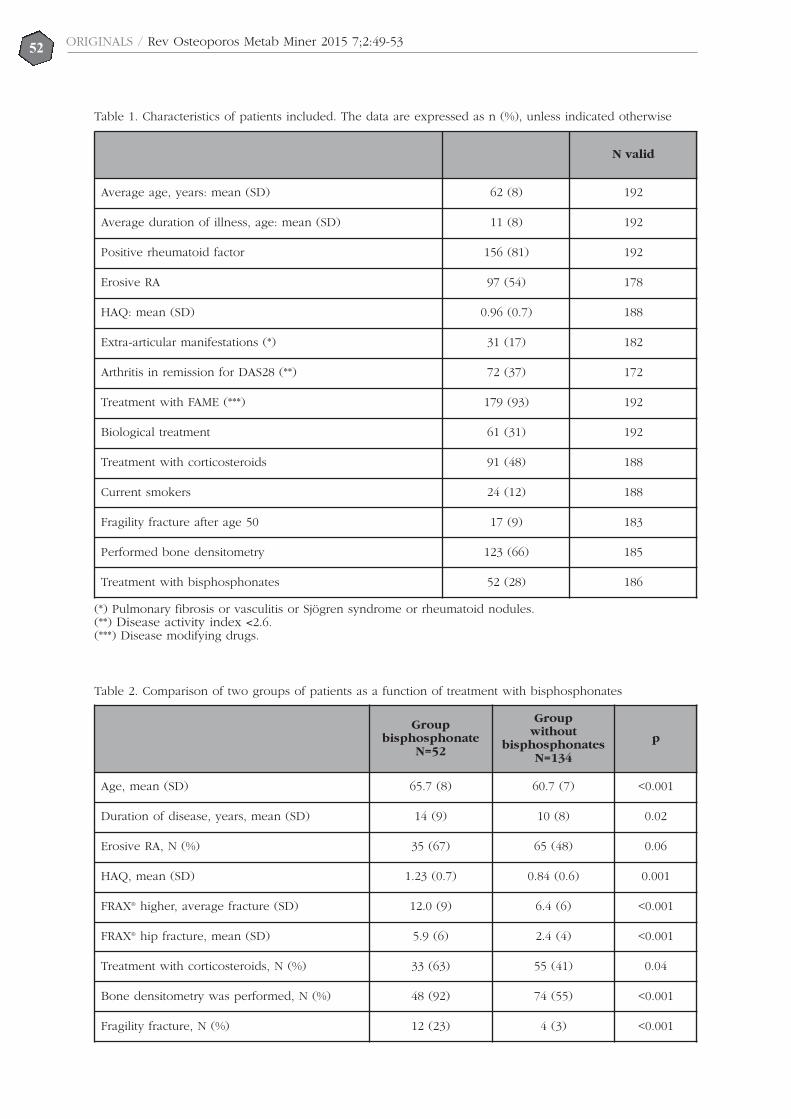
Table 1. Characteristics of patients included. The data are expressed as n (%), unless indicated otherwise
N valid
Average age, years: mean (SD) 62 (8) 192
Average duration of illness, age: mean (SD) 11 (8) 192
Positive rheumatoid factor 156 (81) 192
Erosive RA 97 (54) 178
HAQ: mean (SD) 0.96 (0.7) 188
Extra-articular manifestations (*) 31 (17) 182
Arthritis in remission for DAS28 (**) 72 (37) 172
Treatment with FAME (***) 179 (93) 192
Biological treatment 61 (31) 192
Treatment with corticosteroids 91 (48) 188
Current smokers 24 (12) 188
Fragility fracture after age 50 17 (9) 183
Performed bone densitometry 123 (66) 185
Treatment with bisphosphonates 52 (28) 186
(*) Pulmonary fibrosis or vasculitis or Sjögren syndrome or rheumatoid nodules.(**) Disease activity index <2.6.(***) Disease modifying drugs.
Table 2. Comparison of two groups of patients as a function of treatment with bisphosphonates
Groupbisphosphonate
N=52
Groupwithout
bisphosphonatesN=134
p
Age, mean (SD) 65.7 (8) 60.7 (7) <0.001
Duration of disease, years, mean (SD) 14 (9) 10 (8) 0.02
Erosive RA, N (%) 35 (67) 65 (48) 0.06
HAQ, mean (SD) 1.23 (0.7) 0.84 (0.6) 0.001
FRAX® higher, average fracture (SD) 12.0 (9) 6.4 (6) <0.001
FRAX® hip fracture, mean (SD) 5.9 (6) 2.4 (4) <0.001
Treatment with corticosteroids, N (%) 33 (63) 55 (41) 0.04
Bone densitometry was performed, N (%) 48 (92) 74 (55) <0.001
Fragility fracture, N (%) 12 (23) 4 (3) <0.001

ORIGINALS / Rev Osteoporos Metab Miner 2015 7;2:49-5353
2008;208:33-45.7. Grossman JM, Gordon R, Ranganath VK, Deal C,
Caplan L, Chen W, et al. American College ofRheumatology 2010 Recommendations for thePrevention and Treatment of Glucocorticoid-InducedOsteoporosis. Arthritis Care Res 2010;62:1515-26.
8. Esteve-Vives J, Batlle-Gualda E, Reig A. Spanish ver-sion of the Health Assessment Questionnaire: reliabi-lity, validity and transcultural equivalency. Grupo parala Adaptación del HAQ a la Población Española. JRheumatol 1993;20:2116-22.
9. Furuya T, Hosoi T, Saito S, Inoue E, Taniguchi A,Momohara S, et al. Fracture risk assessment and oste-oporosis treatment disparities in 3,970 Japanesepatients with rheumatoid arthritis. Clin Rheumatol2011;30:1105-11.
10. Watt J, Thompson A, Le Riche N, Pope J. There is still acare gap in osteoporosis management for patients withrheumatoid arthritis. Joint Bone Spine 2014;81:347-51.
11. Naranjo A, Rosas J, Ojeda S, Salas E. Manejo de la oste-
oporosis en atención primaria antes y después delresultado de la densitometría; tratamiento instauradoversus tratamiento recomendado en los consensos(estudio CANAL). Reumatol Clin 2013;9:269-73.
12. Heberlein I, Demary W, Bloching H, Braun J,Buttgereit F, Dreher R, et al. Prophylaxis and treatmentof osteoporosis in patients with rheumatoid arthritis(ORA study). Z Rheumatol 2011;70:793-8.
13. Coulson KA, Reed G, Gilliam BE, Kremer JM,Pepmueller PH. Factors influencing fracture risk, Tscore, and management of osteoporosis in patientswith rheumatoid arthritis in the consortium of rheuma-tology researchers of north america (CORRONA)registry. J Clin Rheumatol 2009;15:155-60.
14. Rubin KH, Abrahamsen B, Friis-Holmberg T, HjelmborgJV, Bech M, Hermann AP, et al. Comparison of differentscreening tools (FRAX®, OST, ORAI, OSIRIS, SCORE andage alone) to identify women with increased risk offracture. A population-based prospective study. Bone2013;56:16-22.

ORIGINALS / Rev Osteoporos Metab Miner 2015 7;2:54-6254
León Vázquez F1, Bonis J2, Bryant Cerezo V2, Herrero Hernández S3, Jamart Sánchez L3, Díaz Holgado A4
1 Centro de Salud Universitario San Juan de la Cruz - Dirección Asistencial Noroeste - Gerencia de Atención Primaria - Servicio Madrileño de Salud - Pozuelode Alarcón (Madrid)2 BIFAP - Base de Datos para la Investigación Farmacoepidemiológica en Atención Primaria - División de Farmacoepidemiología y Farmacovigilancia - AgenciaEspañola de Medicamentos y Productos Sanitarios - Madrid3 Servicio de Farmacia Atención Primaria - Dirección Asistencial Noroeste - Gerencia de Atención Primaria - Servicio Madrileño de Salud - Majadahonda (Madrid)4 Dirección Técnica de Sistemas de Información Sanitaria - Gerencia Adjunta de Planificación y Calidad - Servicio Madrileño de Salud - Madrid
Prevention of osteoporotic fracturein Spain: use of drugs before andafter a hip fracture
Correspondence: Fernando León Vázquez - Centro de Salud Universitario San Juan de la Cruz - Camino deAlcorcón, 8 - 28224 Pozuelo de Alarcón - Madrid (Spain)e-mail: [email protected]
Date of receipt: 04/04/2015Date of acceptance: 07/07/2015
SummaryIntroduction: Treatment of osteoporosis is focussed on the prevention fragility fractures, fractures of thehip being those which produce the highest rates of morbidity and mortality. The existence of a previousfracture is an important predictor of a new fracture.Objective: we intend to analyse how treatment for osteoporosis varies before and after a hip fracture.Material and methods: Using the 4,126,030 clinical records in the database for pharmaco-epidemiologicalresearch in primary care (Base de Datos para la Investigación Farmacoepidemiológica en Atención Primaria[BIFAP] ) 2011 for the whole of Spain, information was obtained regarding patients who had a first hip frac-ture recorded between 2005-2011, having been monitored for at least a year before and after. We analyse theprevious and subsequent treatment for osteoporosis (including calcium and vitamin D supplements).Results: 2,763 patients over 60 years of age (average 81 years) had suffered a hip fracture, of whom 81.6%were women. Before the fracture 26.5% (95% confidence interval [CI]: 24.8-28.1%) had received someantiosteoporotic treatment, of which 12% (95% CI: 11.0-13.5%), were bisphosphonates. 38.6% (95%CI:36.8-40.4%) received treatment after the fracture, 20.4% (95%: 18.9-22%) treated with bisphosphonates.The factors associated with the initiation of treatment after the fracture were being a woman, being youn-ger and having a previous diagnosis of osteoporosis.Conclusions: Most of the patients studied were not receiving preventative treatment before their hip frac-ture. After the fracture the prescription of treatment increased a little. The drugs most commonly addedwere calcium, vitamin D and bisphosphonates.
Key words: osteoporosis, hip fracture, secondary prevention.

ORIGINALS / Rev Osteoporos Metab Miner 2015 7;2:54-6255
IntroductionOsteoporosis is a bone disorder characterised by adeficit in both bone mineral density (quantity) andbone architecture (quality), which results in lowerbone strength, greater fragility and a higher risk offracture after minor trauma (fragility or osteoporo-tic fracture)1. According to the densitometric crite-ria proposed in 1994 by the World HealthOrganisation (WHO)2, in Spain, the prevalence ofosteoporosis is around 26% of women aged 50years or over, increasing with age3.
Among the osteoporotic fractures, vertebralfractures are those with the highest incidence,along with those of the radius, generating signifi-cant morbidity, although little mortality. But it isfractures of the hip, which appear later on, whichpresent the greatest mortality4, in addition to gene-rating greater dependency and higher health costs.In a third of cases the patient had already had anearlier fragility fracture, with 21% of these even inthe other hip5. A previous fragility fracture is,along with age, the most significant risk factor forsuffering a new osteoporotic fracture. The appea-rance of a hip fracture due to a low impact trau-ma in older age permits the establishment with ahigh degree of suspicion of the diagnosis of esta-blished osteoporosis, making its confirmationthrough the use of other diagnostic measures,such as densitometry, unnecessary6.
Currently, various drugs are used for the pre-vention of osteoporotic fractures such as the bis-phosphonates (alendronate, risedronate, etidrona-te, ibandronate and zoledronate) strontium ranela-te (which has recently seen its authorisation foruse limited) estrogen receptor modulators (raloxi-fene and bazedoxifene), denosumab, teriparatideand parathyroid hormone. In the past, hormonereplacement therapy or calcitonin were also used,but are now in disuse due to the existence of saferand more efficacious alternatives. The use of cal-cium7 and vitamin D supplements8 was alsorecommended, associated or not with the afore-mentioned drugs, to which have been attributedimprovements in bone mineral density, whoseefficacy in the prevention of fractures is currentlycompromised when used without being associa-ted with other drugs9.
The main aim of this study was to analyse, ina primary care setting, the prevalence of the useof pharmacological drugs for the treatment or pre-vention of osteoporosis before and after a first hipfracture of osteoporotic aetiology. The secondaryaim was to analyse the possible factors associatedwith the decision to initiate treatment with bis-phosphonates after a fracture in patients whowere not taking them previously.
Material and methodsThe study was carried out using the BIFAP databa-se (Database for pharmaco-epidemiological rese-arch in primary care [Base de Datos para laInvestigación Farmacoepidemiológica en AtenciónPrimaria]) 2011, which includes anonymised infor-mation from the clinical records of 4,126,030
patients (with an average monitoring period of 4.8years per patient), recorded by 2,239 family doc-tors and primary care paediatricians across thewhole of Spain10.
The computerised clinical history for eachpatient is composed of episodes, each of whichhas an associated diagnosis, coded according tothe International Classification for Primary Care(ICPC)11. Each prescription issued for the patient isassociated with a specific ICPC episode.
A study of transverse design was carried out ofthe use of medications for osteoporosis before andafter a first episode of fracture. Those patientsover 60 years of age with a first record of hip frac-ture coded as ICPC L75 in the period between 1stJanuary 2005 and 1st January 20011, and with arecord covering at least a year before and after thedate of the fracture, were included. Those patientswith a history of cancer and of Paget’s diseasewere excluded.
For each patient selected, the sex, the age at thetime of fracture, the date of the hip fracture and thepresence of earlier diagnoses coded using ICPCcorresponding to possible absolute or relative con-traindications for the use of bisphosphonates, werenoted from the medical record (Annex 1), as wellas the presence of previous episodes of diabetesmellitus type 1, rheumatoid arthritis, hyperthyroi-dism, masculine hypogonadism, malabsorption,malnutrition, early menopause and osteoporosis(Annex 2).
The previous use of corticoids was also analy-sed, with, for the purposes of this study, a pre-vious user being a patient who had had at least 3prescriptions, and with an estimated 90 days ormore of usage (based on the dosage) of predniso-lone ≥ 5 mg/day (or equivalent) at any time befo-re the date of the hip fracture.
Lastly, the use before or after the hip fractureof bisphosphonates (etidronate, alendronate, iban-dronate, risedronate), vitamin D, calcium, calcito-nin, estrogens, parathyroid hormones, teriparatide,raloxifene, bazedoxifene strontium ranelate anddenosumab, were considered (Annex 3).
For each of the aforementioned drugs thepatient was considered to be under primary pre-vention if they had received, at any time beforethe fracture, at least two prescriptions for one ofthe drugs listed, or in the case of having receiveda single prescription, if this was issued within 180days before the fracture. The patient was conside-red to be under subsequent prevention for hipfracture if they had had at least one prescription ofone of the drugs for osteoporosis described withina year after the date of the fracture.
In order to analyse which factors were associa-ted with the initiation of treatment with bisphos-phonates after a hip fracture in those who had notreceived earlier treatment, a logistical regressionmodel was constructed, using as independentvariables the year of the fracture, the age of thepatient, the sex, the presence of diabetes, rheuma-toid arthritis, record of osteoporosis or any con-traindication for the use of bisphosphonates, as
ORIGINALS / Rev Osteoporos Metab Miner 2015 7;2:54-6256
well as previous exposure to corticoids. A back-ward selection strategy was used based on thelikelihood ratio model for the selection of varia-bles finally included in the model. For the descrip-tive analysis the proportion of patients who werereceiving each of the treatments studied before,and in the year following, the fracture was calcu-lated, as well as the average age and duration ofthe monitoring before and after the fracture, withcorresponding confidence intervals of 95% (95%CI). For hypothesis testing regarding the differen-ces in the proportion of use of each of the drugsbefore and after the fracture, the McNemar test forpaired data was used.
Results2,763 patients over 60 years of age (average of 81years) were identified who had presented a firsthip fracture in the period of the study, 2,225 ofwhom were women (81.6%). The average dura-tion of the period of registration prior to the frac-ture was 5.8 years. The rest of the demographicand comorbidity data are described in Table 1.
A total of 731 patients (26.5%; 95% CI: 24.8-28.1%) had received one of the drugs analysedbefore the fracture (Table 2). Of these, 338patients (12.2%; 95% CI: 11.0-13.5%) had receivedsome treatment with bisphosphonates.
In the year following the hip fracture, 1,066patients (38.6%; 95% CI: 36.8-40.4%) had receivedsome antiosteoporotic treatment (Table 2), ofwhom 564 (20.4%; 95% CI: 18.9-22.0%) had recei-ved a bisphosphonate (Figure 1). The increase inthe use of drugs against osteoporosis (p<0.0001),as well as the increase in the use of a bisphospho-nate (p<0.0001) were statistically significant accor-ding to the McNemar test.
The most commonly prescribed drugs, bothbefore and after the fracture, were calcium (23.2%and 32.4% respectively) and vitamin D (19.6% and31.0% respectively ). Among the bisphosphonatesthe most common were alendronate (6.6% and10.4%) and risedronate (5.4% and 8.1%). On theother hand, it was notable that of the 508 men inthe study, 11 (2.2%) were receiving alendronatebefore the fracture, and 29 (5.2%) took themwithin the year following the fracture.
Of the 338 patients who took bisphosphonatesat any time before the fracture, 104 (30.8%) did nottake them in the year after it. On the other hand, ofthe 2,425 patients who had not taken it before, 330(13.6%) started treatment with bisphosphonatesafresh in the year following the fracture. A total of369 patients (13.4%; 95% CI: 12.1-14.6%) presentedsome absolute and relative contraindications for theuse of bisphosphonates, including any diagnosis ofgastritis or dyspepsia (complete criteria in Annex 1).Of the 642 patients who were taking calcium sup-plements at some point before the fracture, 31%(200 patients) did not receive them in the year afterthe fracture; while of 2,121 patients who were nottaking them, 462 (21.8%) started to receive themafter it. We obtained almost identical percentageswith vitamin D supplements.
The logistic regression model (Table 3) regar-ding patients who were not taking treatment beforethe fracture (n=2,425) showed that the factors asso-ciated with a higher probability of initiating a treat-ment with bisphosphonates after fracture (n=330)were: being a woman (OR=2.44; p<0.0001), havinga previous diagnosis of osteoporosis recorded(OR=1.61; p=0.009), being younger (OR per yearof age=0.96; p<0.0001) and having some absoluteor relative contraindication for the use of bisphos-phonates (OR=1.41; p=0.033). No association wasobserved between the start of treatment with bis-phosphonates after fracture and the fact of havingdiabetes, previous exposure to corticoids, historyof rheumatoid arthritis or the year in which thefracture occurred. No significant interactions wereobserved between the independent variablesanalysed.
DiscussionThe natural course of osteoporosis has a prolon-ged asymptomatic phase. In this period of primaryprevention it is necessary to influence modifiablerisk factors12, although the use of drugs is contro-versial and the benefits, if any, are of low magni-tude13. On the other hand, there is a consensus innot recommending population screening of bonemineral density with densitometry, and that thistest is reserved for high risk cases and in order totake key therapeutic decisions14.
After the first fragility fracture the risk of suffe-ring future fractures increases considerably15,16. So,after a first vertebral fracture, the risk of a new ver-tebral fracture increases 4.4 times, and of a hipfracture by 2.3 times17. The usefulness of drugs forprevention subsequent to the fracture (which isusually called secondary prevention, but whichwould strictly be tertiary prevention),18 has bettertests available for its use in primary prevention6,13.
Various studies have analysed the prescriptionof drugs for osteoporosis after a hip fracture,Some evaluate the treatment prescribed on dis-charge from hospital after a hip fracture, withlevels of treatment which vary between 6%19 and19%20. Other works address treatment after anyosteoporotic fracture over the course of a year,obtaining levels from 15% for treatment after theevent21, in other cases up to 24% after any fractu-re, with levels of 44% after vertebral fracture and21% after a hip fracture22. In our case we obtainedrates somewhat higher than the 38% for osteopo-rotic treatment, even though our data include tre-atment initiated up to a year after the fracture,and excluded patients with early mortality (withless than a year of records available after the frac-ture), which probably limits its comparability withother studies. The majority of the patients (73.5%)in our sample had not received drug treatment forosteoporosis before their hip fracture. After thefirst fracture, the doctors initiated some treatmentafresh in a minority of patients, both with bis-phosphonates (13.6%) and calcium-vitamin D(21.8%). By comparing the prevalence of its usebefore and after the fracture an increase was con-

ORIGINALS / Rev Osteoporos Metab Miner 2015 7;2:54-6257
firmed in the proportion of patients who receivedsome drug treatment (from 26.5% to 38.6%),which was, furthermore, statistically significant(p<0.0001 for the McNemar test). In a northAmerican study23, the probability of receiving tre-atment after a hip fracture diminished from 40.2%in 2002 to 20.5% in 2011. Whether this incrementis slight or not, is a matter of controversy,although the guides6,15,17,24 include people withfractures as the target population, who obtain thegreatest benefit from pharmacological treatmentin normal clinical practice.
The highest consumption of antiresorptivedrugs in our setting is found in women at relati-vely early ages (66 years on average)25 in whomosteoporotic fracture is less frequent in compari-son with the age group of older women, in whichfractures are more common and (in the hip) moreserious. However, a review concluded that alen-dronate does reduce clinically and statistically sig-nificantly vertebral, non-vertebral, hip and wristfractures in secondary prevention, without therebeing statistically significant results for primary
prevention, except for vertebral fractures13,although this is a controversial point26.
The logistical regression model allows us toanalyse the factors related to the decision to initia-te a treatment with bisphosphonates after a firsthip fracture in patients who were not receivingthem previously. The data suggest that doctors inprimary care use criteria similar to those used forthe initiation of treatment before fracture and inprimary prevention. So, being female, youngerand having an earlier diagnosis of osteoporosisincreases the probability of initiating treatmentafter a first hip fracture.
Notable among the drugs which have mostbeen used in our analysis, both before and after afracture, are the bisphosphonates, alendronateand risedronate, similar to other series27. On theother hand there are the recommendations in theguides for efficacy, safety and price10. The datafrom the study showed the existence of men intreatment with alendronate; even though alendro-nate has shown definite efficacy in improvingbone mass in males28, its indication in the data
Table 1. Description of the population. Clinical characteristics, exposure to corticoids and contraindications forthe use of bisphosphonates, before a hip fracture
n
Total 2,763
Average SD (min-max)
Age (years) 81.6 7.76 (60-105)
Preregistration period (days) 2,130 999 (366-10,909)
n Percentages
Women 2,255 81.6%
Diabetes mellitus type 2 454 16.4%
Hyperthyroidism 30 1.1%
Rheumatoid arthritis 32 1.2%
Hypogonadism 0 0.0%
Malabsorption 0 0.0%
Malnutrition 4 0.1%
Early menopause 5 0.2%
Osteoporosis 428 15.5%
Prior exposure to corticosteroids 144 5.2%
Contraindications for bisphosphonates 369 13.36%

ORIGINALS / Rev Osteoporos Metab Miner 2015 7;2:54-6258
sheet is restricted to postmenopausal osteoporo-sis29,30. Only 15.5% of those patients with hip frac-ture had included in their diagnosis “osteoporo-sis”, although they had received treatment withantiresorptive drugs, which suggests an additionalproblem of under-registration.
Our study has some limitations. It does not dis-tinguish as to whether the treatment before thefracture was for primary prevention, given that thepatient could have had a previous fragility fractu-re, as long as it was different from the hip. Neitherdoes it analyse the dose or duration of the drugsused, since after the fracture there could havebeen patients treated for a short period, as againstothers who could have been treated for the wholeperiod of the study after the hip fracture. Theprescription of drugs subsequent to the fracturereflects the preoccupation by the professional withthe risk of new fractures, which results in theinitiation of treatment aimed at secondary preven-
tion. However, it does not tell us about its persis-tence over time.
Another limitation is that, given the nature ofthe record from which the data was obtained, it isnot possible to differentiate with certainty betwe-en absolute contraindications and precautions forthe use of bisphosphonates. The association bet-ween the existence of an earlier contraindicationbefore the fracture and the start of treatment afterthe fracture (OR=1.41) should be interpretedwithin this context. A possible hypothesis wouldsuggest that the professionals, faced with precau-tion on use, don’t initiate preventative treatmentwith bisphosphonates, but that once the fractureoccurs, reconsider the risk-benefit balance infavour of pharmacological treatment. It is impor-tant to note that in our study only those patientswith a survival of at least one year after fracturewere included. This selection criterion adds con-sistency to our data and facilitates their interpreta-
Table 2. Prevalence of pharmacological treatment for osteoporosis before and after a first hip fracture
Beforefracture
(a)
Afterfracture
(b)
n % n % Suspended(c)
Begin(d)
p(e)
Total 2,763 2,763
Bisphosphonates (f) 338 12.2% 564 20.4% 104 330 <0.0001
Alendronate 183 6.6% 288 10.4% 84 189 <0.0001
Etidronate 21 0.8% 3 0.1% 18 0 <0.0001
Ibandronate 26 0.9% 74 2.7% 9 57 <0.0001
Risedronate 149 5.4% 224 8.1% 66 141 <0.0001
Calcium 642 23.2% 904 32.7% 200 462 <0.0001
Vitamin D 542 19.6% 857 31.0% 167 482 <0.0001
Ca + vitamin D 535 19.3% 828 30.0% 173 466 <0.0001
Calcitonin 91 3.3% 42 1.5% 72 23 <0.0001
Teriparatide/PTH 13 0.5% 58 2.1% 6 51 <0.0001
Estrogens 15 0.5% 6 0.2% 13 4 0.0490
Raloxifene/bazedoxifene 41 1.5% 17 0.6% 29 5 <0.0001
Strontium ranelate 21 0.8% 71 2.6% 14 64 <0.0001
Denosumab 0 0.0% 0 0.0% 0 0 <0.0001
In treatment (g) 731 26.5% 1,066 38.6% 194 529 <0.0001
(a): at any time before the first hip fracture; (b): within the 365 days subsequent to the first hip fracture; (c):treatment stopped after the hip fracture; (d): treatment initiated after the hip fracture; (e): McNemar test for pai-red data; (f): in treatment with at least one bisphosphonate; (g): in treatment with one of the earlier drugs.

59ORIGINALS / Rev Osteoporos Metab Miner 2015 7;2:54-62
tion, but makes it difficult to compare them withthe results of other studies in which patients withearly mortality after a fracture are included.
Notable among the strengths of the study is thehigh number of hip fractures analysed (n=2,763)and the variety of drugs studied. The fact that theclinical record was used as a source of data retros-pectively, and the inclusion of treatment initiatedup to a year after the date of the fracture, and notonly immediately after it, means that the resultsare probably a good refection of real clinical prac-tice in the primary care context. Using episodes ofhip fractures in people over 60 years of age as amarker for established osteoporosis offers advan-tages since, given its gravity, it is not usually omit-ted from their record, and it rarely has a differentorigin from bone fragility6. Contrarily, the analysisof other types of fracture such as of the wrist orvertebrae are less specific, since they may haveother origins, may pass unnoticed, or be variablein the register. A piece of data in favour of theexternal validity of the study is that the averageage at fracture in our sample, 81 years, coincideswith other Spanish studies with different methodo-logies, and coincides also in the ratio betweenwomen and men of 4:14,5.
The majority of patients in our study were notin treatment before suffering their hip fracture.After it there was a moderate increase in the pres-cription of drugs for osteoporosis. There arecurrently no data on the efficacy of these drugs inthe prevention of hip fracture in patients whohave already suffered a previous hip fracture, andit would therefore be very interesting to carry outnew studies to determine whether the preventati-ve treatment after a first hip fracture is effective ornot in preventing new fractures.
Acknowledgements: The authors would like tothank the inestimable collaboration of the familydoctors and paediatricians in primary care partici-pating in BIFAP, whose contribution of a high qua-lity record of their daily activity has made the rea-lisation of this study possible.
Source of funding: This work was carried outwithout external funding.
Bibliography
1. National Institutes of Health (USA). ConsensusDevelopment Panel on Osteoporosis Prevention,Diagnosis, and Therapy. JAMA 2001;285:785-95.
2. World Health Organization: Assessment of FractureRisk and its application to screening for postmenopau-sal Osteoporosis. Report of WHO Study group(Technical report series 843: 1-129). GenevaSwitzerland; 1994.
3. Díaz-Curiel M, García JJ, Carrasco JL, Honorato J,Pérez-Cano R, Rapado A, et al. Prevalencia de osteo-porosis determinada por densitometría en la poblaciónfemenina española. Medicina Clínica (Barcelona)2001;116:86-8.
4. Serra JA, Garrido G, Vidán M, Marañón E, Brañas F,Ortiz J. Epidemiología de la fractura de cadera enancianos en España. Ann Med Intern (Madrid)2002;19:389-95.
5. Herrera A, Martínez AA, Ferrández L, Gil E, Moreno A.Epidemiology of osteoporotic hip fractures in Spain.Int Orthop 2006;30:11-4.
6. National Institute for Health and Care Excellence.Alendronate, etidronate, risedronate, raloxifene, stron-tium ranelate and teriparatide for the secondary pre-vention of osteoporotic fragility fractures in postmeno-pausal women (amended) (TA161). NICE, 2010http://www.nice.org.uk/guidance/ta161.
7. Shea B, Wells G, Cranney A, Zytaruk N, Robinson V,Griffith L, et al. Osteoporosis Methodology Group andThe Osteoporosis Research Advisory Group. Meta-
Figura 1. Evolución del tratamiento antes y después de la fractura de cadera
100%
90%
80%
70%
60%
50%
40%
30%
20%
10%
0%
Before fracture After fracture
Untreated
Treated without BF
Treated with BF
60ORIGINALS / Rev Osteoporos Metab Miner 2015 7;2:54-62
analyses of therapies for postmenopausal osteoporo-sis. VII. Meta-analysis of calcium supplementation forthe prevention of postmenopausal osteoporosis.Endocr Rev 2002;23:552-9.
8. Papadimitropoulos E, Wells G, Shea B, Gillespie W,Weaver B, Zytaruk N, et al. Osteoporosis MethodologyGroup and The Osteoporosis Research Advisory
Group. Meta-analyses of therapies for postmenopausalosteoporosis. VIII: Meta-analysis of the efficacy of vita-min D treatment in preventing osteoporosis in postme-nopausal women. Endocr Rev 2002;23:560-9.
9. Moyer VA. U.S. Preventive Services Task Force.Vitamin D and calcium supplementation to preventfractures in adults: U.S. Preventive Services Task Forcerecommendation statement. Ann Intern Med2013;158:691-6.
10. Salvador-Rosa A, Moreno-Pérez JC, Sonego D, García-Rodríguez LA, de Abajo-Iglesias FJ. El Proyecto BIFAP: Basede datos para la Investigación Farmacoepidemiológica enAtención Primaria. Aten Primaria 2002;30:655-61.
11. Lamberts H, Wood M (Eds.). Clasificación Internacionalde la Atención Primaria (CIAP). Barcelona: Masson/SG;1990.
12. National Clinical Guideline Centre (UK). Osteoporosis:Fragility Fracture Risk: Osteoporosis: Assessing the riskof fragility fracture. London: Royal College ofPhysicians (UK); 2012.
13. Wells GA, Cranney A, Peterson J, Boucher M, Shea B,Robinson V, et al. Alendronate for the primary andsecondary prevention of osteoporotic fractures in pos-tmenopausal women. Cochrane Database Syst Rev2008;(1):CD001155.
14. Malabanan AO, Rosen HN, Vokes TJ, Deal CL, AleleJD, Olenginski TP, et al. Indications of DXA inwomen younger than 65 yr and men younger than 70yr: The 2013 Official Positions. J Clin Densitom2013;16:467-71.
15. Grupo de trabajo de la Guía de Práctica Clínica sobreOsteoporosis y Prevención de Fracturas por Fragilidad.Guía de Práctica Clínica sobre Osteoporosis yPrevención de Fracturas por Fragilidad. Plan deCalidad para el Sistema Nacional de Salud delMinisterio de Sanidad y Política Social. Agènciad Avaluació de Tecnologia i Recerca Mèdiques deCataluña (AATRM). Madrid, 2010.
16. Akesson K, Marsh D, Mitchell PJ, McLellan AR,Stenmark J, Pierroz DD, et al. IOF Fracture WorkingGroup. Capture the Fracture: a Best PracticeFramework and global campaign to break the fragilityfracture cycle. Osteoporos Int 2013;24:2135-52.
17. Dirección General de Farmacia y Productos Sanitarios.Recomendaciones para la valoración y tratamiento dela osteoporosis primaria en mujeres de la Comunidadde Madrid. Madrid: Consejería de Sanidad; 2007.
18. Martínez-González MA, Guillén-García F, Delgado-Rodríguez M. Conceptos en Salud Pública. En:
Table 3. Factors related to the initiation of bisphosphonate therapy after first hip fracture (a)
OR adjusted (b) IC 95%
Woman 2.44 1.69 - 3.52
Prior osteoporosis 1.61 1.13 - 2.30
Contraindication prior bisphosphonate (c) 1.41 1.03 - 1.94
Age 0.96 0.94 - 0.97
(a): logistical regression model with 2,425 patients who did not receive primary prevention with bisphospho-nates prior to the fracture; (b): dependent variable: receiving secondary prevention with bisphosphonates inthe 365 days subsequent to a first hip fracture; (c) absolute or relative contraindication for the use of bisphos-phonates.
Gastric pathology:
• Oesophagitis: oesophagitis, caustic oesopha-
gitis, reflux oesophagitis.• Duodenal ulcer: duodenal ulcer, duodenal
ulceration.• Gastric ulcer: stomach ulcer, stomach ulce-
ration, perforated stomach ulceration, gastroin-testinal ulceration, peptic ulceration.
• Gastritis: disturbance in stomach function,dyspepsia, duodenitis
Annex 1. Absolute or relative contraindications to thebisphosphonates
• Hyperthyroidism• Diabetes mellitus type 2 • Malabsorption syndrome• Malnutrition• Masculine hypogonadism• Early menopause • Rheumatoid arthritis• Osteoporosis
Annex 2. Other clinical characteristics analysed

61ORIGINALS / Rev Osteoporos Metab Miner 2015 7;2:54-62
Annex 3. Drug study
• Corticosteroids:
H02AB01 Betamethasone
H02AB13 Deflazacort
H02AB02 Dexamethasone
H02AB09 Hydrocortisone
H02AB04 Methylprednisolone
H02AB06 Prednisolone
H02AB07 Prednisolone
H02AB08 Triamcinolone
• Vitamin D
A11CC05 Cholecalciferol
• Calcium supplements
A12AA01 Calcium phosphate
A12AA04 Calcium phosphate
A12AA10 Calcium glucoheptonate
A12AA12 Calcium acetate, anhydrous
A12AA20 Calcium (different salts in combination)
A12AA91 Calcium pidolate
A12AA92 Oseina-hydroxyapatite complex
• Calcium + vitamin D partnerships
A12AX91 Calcium phosphate + cholecalciferol
A12AX92 Calcium lactate + cholecalciferol
A12AX93 Calciocarbonato + cholecalciferol
A12AX94 Calcium glucoheptonate + cholecalciferol
A12AX96 Calcium pidolate + cholecalciferol
• Estrogens
G03CA03 Estradiol
G03CA04 Estriol
G03CA57 Conjugated estrogens
• Selective estrogen receptor modulators
G03XC01 Raloxifene
G03XC02 Bazedoxifene
• Calcitonins
H05BA01 Calcitonin (salmon, synthetic)
H05BA03 Calcitonin (human synthetic)
• Bisphosphonates
M05BA01 Etidronic acid
M05BA04 Alendronate acid
M05BA06 Ibandronic acid
M05BA07 Risedronic acid
M05BA91 Alendronate acid + cholecalciferol
• Other endocrine drugs
H05AA02 Teriparatide
H05AA03 Parathyroid hormones
• Other drugs bone diseases
M05BX03 Strontium ranelate
M05BX04 Denosumab

ORIGINALS / Rev Osteoporos Metab Miner 2015 7;2:54-6262
Martínez-González MA (Ed). Conceptos de SaludPública y Estrategias Preventivas. Un manual paraCiencias de la Salud. Barcelona: Elsevier; 2013:9-13.
19. Rabenda V, Vanoverloop J, Fabri V, Mertens R, SumkayF, Vannecke C, et al. Low incidence of anti-osteoporo-sis treatment after hip fracture. J Bone Joint Surg Am2008;90:2142-8.
20. Andrade SE, Majumdar SR, Chan KA, Buist DS, Go AS,Goodman M, et al. Low frequency of treatment of oste-oporosis among postmenopausal women following afracture. Arch Intern Med 2003;163:2052-7.
21. Ensrud KE, Schousboe JT. Clinical practice. Vertebralfractures. N Engl J Med 2011;364:1634-42.
22 Panneman MJ, Lips P, Sen SS, Herings RM.Undertreatment with anti-osteoporotic drugs after hos-pitalization for fracture. Osteoporos Int 2004;15:120-4.
23. Solomon DH, Johnston SS, Boytsov NN, McMorrow D,Lane JM, Krohn KD. Osteoporosis medication use afterhip fracture in U.S. patients between 2002 and 2011. JBone Miner Res 2014;29:1929-37.
24. Guías de actuación. Osteoporosis Manejo: prevención,diagnóstico y tratamiento. 1ª Edición. Barcelona:Semfyc Ediciones, 2014.
25. De Felipe R, Cáceres C, Cimas M, Dávila G, FernándezS, Ruiz T. Características clínicas de los pacientes con
tratamiento para la osteoporosis en un centro deAtención Primaria: ¿a quién tratamos en nuestras con-sultas? Aten Primaria 2010;42:559-63.
26. Erviti J, Alonso A, Oliva B, Gorricho J, López A,Timoner J, et al. Oral bisphosphonates are associatedwith increased risk of subtrochanteric and diaphysealfractures in elderly women: a nested case-controlstudy. BMJ Open 2013 30;3(1).
27. Carbonell-Abella C, Guañabens-Gay N, Regadera-Anechina L, Marín-Rives JA, Taverna-Llauradó E, Ayechu-Redín MP. Análisis del cumplimiento terapéutico enmujeres con osteoporosis. Reumatol Clin 2011;7:299-304.
28. Orwoll E, Ettinger M, Weiss S, Miller P, Kendler D,Graham J, et al. Alendronate for the treatment of oste-oporosis in men. N Engl J Med 2000;343:604-10.
29. Centro de Información online de Medicamentos CIMA.Agencia Española de Medicamentos y ProductosSanitarios. Ficha técnica del Alendronato. Disponible enhttp://www.aemps.gob.es/cima/pdfs/es/ft/69193/FT_69193.pdf [Consultada el 4/04/2015].
30. León-Vázquez F, Herrero-Hernández S, Cuerpo-Triguero C, Andrés-Prado MJ,Cabello-Ballesteros L.Prescripción de ácidos alendrónico y risedrónico envarones: uso fuera de la ficha técnica en un área desalud. Reumatol Clin 2015;11:64-7.

CLINICAL NOTES / Rev Osteoporos Metab Miner 2015 7;2:63-6663
Rosselló Aubach Ll1, Vélez Cedeño VK2, Montalà Palou N1, Conde Seijas M1, Palliso Folch F1
1 Dirección Clínica del Aparato Locomotor - Hospital de Santa María de Lleida2 CAP Les Borges Blanques - Lleida
Gitelman syndrome and chondrocalcinosis.A clinical case review
Correspondence: Lluís Rosselló Aubach - Hospital de Santa María de Lleida - Dirección Clínica del Aparato Locomotor -Avda. Rovira Roure, 44 - 25198 Lleida (Spain)e-mail: [email protected]
Date of receipt: 06/05/2015Date of acceptance: 19/06/2015
SummaryGitelman syndrome is a tubulopathy of autosomal recessive inheritance which presents with, amongother manifestations, hypomagnesemia and hypocalciuria. We present the case of a woman of 68 yearsof age who came for a consultation due to arthritis in the large joints, in the absence of other sympto-mology. The X-ray study showed deposits of calcium pyrophosphate in the knees, pubic symphysis andother joints. Blood tests revealed hypomagnesemia and hypocalciuria compatible with Gitelman syndro-me, which was confirmed following a genetic study.
Key words: Gitelman syndrome, chondrocalcinosis, hypomagnesemia.

CLINICAL NOTES / Rev Osteoporos Metab Miner 2015 7;2:63-6664
IntroductionGitelman syndrome is a disease transmitted byrecessive autosomal inheritance, and is caused bymutations in the gene SLC12A3, located in the16q13 chromosome, which codes for the synthesisof the Na+-Cl– cotransporter of the distal convolu-ted tubule1, which produces a defect in the reab-sorption of sodium. This increase in the loss ofsalt, in turn causes a moderate volume depletionwhich activates the renin-angiotensin-aldosteronesystem2. It is a tubulopathy characterised by hypo-magnesemia, hypopotassemia with metabolicalkalosis and hypocalciuria. In most cases it mani-fests itself in adolescence or in adulthood andfollows a more benign course than what is knownas Bartter syndrome3. Most patients have low ornormal arterial tension and may present with signsof volume depletion4. Their levels of urinary pros-taglandin E2 are normal. It is important to empha-sise that the severity of the symptoms is not rela-ted to the genotype pattern, nor is there a correla-tion with the laboratory test results in thesepatients. The differential diagnosis should becarried out with diuretic or laxative abuse andwith patients with chronic emetic syndrome5-6. Inspite of the fact that the association betweenGitelman syndrome and chondrocalcinosis hasalready been known for some years, only in rarecases are chondrocalcinosis and hypomagnesemiapresented together, such as occurred in ourpatient, due to the accumulation of calcium pyro-phosphate crystals in the joints stimulated by thehypomagnesemia.
Clinical caseA female patient, 68 years of age, with no patho-logical history of interest, who came for a consul-tation due to repeated episodes of pain andinflammation in both knees, attributed until thento a degenerative process, and which improvedwith non-steroidal anti-inflammatories. During thelast two years she also had pain in both wrists andcervical spine of a mechanical nature. She said shehad not suffered episodes of diarrhoea or vomi-ting, did not consume diuretics or any other typeof pharmaceutical drugs.
The examination showed a patient in a gene-rally good state of health, normohydrated, withblood pressure of 120/80 mmHg. The rest of theexamination showed pain and flexion/extensionlimitation in the right knee with positive meniscalmanoeuvres, without signs of leaking joints. Thehands showed degenerative signs in the distalinterphalangeal joints suggestive of Heberden’snodes.
In the analyses, the haemogram and formulawere normal. The biochemical analysis showedthe following results: urea, 37 mg/dl; creatinine,0.71 mg/dl; glomerular filtrate, >60 mL/min/1.73m;total calcium, 9.45 mg/dl; inorganic phosphate,3.51 mg/dl; alkaline phosphatase, 56 U/L;sodium(Na), 140 mEq/l; potassium (K), 3.4 mEq/l;TSH, 3.45 mUL; blood PTH, 2.9 pmol/L (1.6-6.9);25-hydroxicolecalciferol, 30.9 ng/ml (30-100);
bone alkaline phosphatase, 9.7 ug/L; magnesium(Mg), 0,54 mmol/L (0,66-0,99). In urine at 24hours: negative proteinuria; calciuria, 69.56 mg(100-250); phosphaturia, 588.30 mg; Mg, 1.31mg/dL (1.7-5.7); phosphate in the first urine of theday, 15.9 mg/dL (40-136). The acute phase reac-tants, rheumatoid factor, anti-citrullinated antibodyand antinuclear antibodies (ANA, anti-ENA) werenormal or negative.
The X-ray study showed calcification in themenisci of both knees with additional degenerati-ve signs (Figure 1), of the pubic symphysis, ofboth carpi, in the hyaline coxofemoral cartilage, aswell as in the metatarsophalangeal joint in the bigtoe of both feet (Figure 2).
The nuclear magnetic resonance (NMR) of theright knee showed severe degenerative signs ofpatellofemoral, and internal and external tibiofe-moral osteoarthritis with degenerative rupture ofboth menisci.
A molecular genetic study was requested usingPCR amplification and sequencing of theSCL112A3 gene, detecting homozygosis of thec2576T>C(p.L859P) mutation in the exon of thisgene and which confirmed the diagnosis ofGitelman syndrome. The treatment consisted oforal supplements of magnesium at variable dosesdepending on the results of the monitoring analy-sis, and 0.5 mg colchicine a day to avoid episodesof pseudogout which the patent was suffering.
DiscussionGitelman syndrome was described by this authorin 1966. It is an autosomal recessive hereditarydisease resulting from the mutation in the longarm of chromosome 16 in which the SLC12A3gene which codes for the thiazide-sensitive Na-Clcotransporter in the distal tubule is affected. Itsincidence is one case in every 40,000 people7.
In most cases the symptoms do not appearbefore the age of seven, and the disease is gene-rally diagnosed during adolescence or adulthoodwith very light symptoms, in some cases evenbeing asymptomatic, and whose definitive diagno-sis has to be made through a genetic study, aswith our patient8.
The physiopathology of Gitelman syndrome isthe disturbance of the function of the thiazide-sen-sitive ClNa cotransporter (TSC) which results inthe tubular reabsorption of chloride and sodium inthe distal nephron, causing a loss of salt andwater, with the consequent hypovolemia. Thereduction in vascular volume activates the renin-angiotensin system, promoting an increase in theconcentrations of renin and aldosterone. This, inturn, facilitates in the cortical collector duct anincrease in the reabsorption of sodium in the api-cal membrane and an activation of the Na+-K+-ATPase in the basolateral membrane. The increasein the concentration of aldosterone stimulates theH+-ATPase in the cortical and medullar collectorducts, causing an increase in the secretion of H+ inthe apical membrane. At the same time, the uri-nary secretion of potassium is increased due to the

CLINICAL NOTES / Rev Osteoporos Metab Miner 2015 7;2:63-6665
increase in the activity in inthe basolateral membrane ofthe Na+-K+-ATPase. All thisfosters the appearance ofhypopotassemia alkalosis.The low intracellular contentof sodium raises the tubularreabsorption of calciumthrough the activation of theNa+/Ca+ basolateral exchan-ge, resulting in hypocalciu-ria. The magnesuria is incre-ased by activating theMg2+/Na+ exchange, given theexistence of a negative tran-sepithelial potential, whichleads to the appearance ofhypomagnesemia9.
With regard to the renalfunction, a reduction in therenal tubular threshold forthe reabsorption of magne-sium without affectation ofTmMg2 is confirmed. Thesedata are compatible with thefact that most of the filteredmagnesium is reabsorbed inthe thick ascending limb ofthe loop of Henle, and thatthe distal tubule only reab-sorbs around 5% of the filte-red magnesium. The mecha-nisms for concentration andacidification are intact. In thehydrosaline overload the dis-tal absorption of chlorideand sodium are reduced10.
The molecular mecha-nisms which link the hypo-magnesemia to the chondro-calcinosis are not fully unders-tood. While it is known thatmagnesium is a cofactor formany pyrophosphatases, such as alkaline phospha-tase, which allows, in turn, the conversion of inorga-nic pyrophosphate into orthophosphate. The mag-nesium also increases the solubility of the crystals ofcalcium pyrophosphate. In states of hypomagnese-mia this solubility of the calcium pyrophosphataseis changed, and the precipitation takes place of thecrystals in the joints producing a crisis of pseudo-gout and also reduces the natural dissolution ofthese pyrophosphate crystals11,12.
In Gitelman syndrome the patients are fre-quently asymptomatic, except for the appearanceof recurrent episodes of muscular weakness andtetany, which may be accompanied by abdominalpain, vomiting and fever. The intervals of apparenthealth may be very prolonged and the diagnosis isnot usually established until adulthood. However,almost half of the patients present lessersymptoms such as an appetite for salt, fatigue,muscle weakness, general aching, dizziness, noc-turia and polydipsia6,10.
Our patient had presented different episodesof pain and inflammation of the joints, especiallyin both knees, although due to the fact that secon-dary evolved osteoarthritis was probably added tothe chondrocalcinosis and degenerative rupture ofboth menisci in the right knee, its association withGitelman syndrome was not suspected until lowlevels of magnesium in the blood and of calciumin 24 hour urine were observed, which was sub-sequently confirmed by the genetic study.
In the treatment of Gitelman syndrome, theefficacy of the administration of salts of Mg exclu-sively (preferably MgCl, which compensates forthe loss of both Mg and Cl in the urine) has beendemonstrated, with normalisation of the biochemi-cal parameters and clinical remission. A correctionof the hypopotassemia is performed occasionallywith the administration of potassium salts.
Indomethacin or the potassium-sparing diure-tics (spironolactone or amiloride) are reserved forthe most refractory cases.
Figure 1. X-ray of the front of both knees. Intraarticular calcification
Figure 2. Plantar plate of the front of both feet. Metacarpophalangeal periar-ticular calcification

CLINICAL NOTES / Rev Osteoporos Metab Miner 2015 7;2:63-6666
With the presentation of this case we considerthe determination of levels of magnesium ions inthe blood, as well as calcium and magnesium in24 hour urine, to be important in all those patientswith chondrocalcinosis and in those in whomhypomagnesemia is suspected, in order to exclu-de Gitelman syndrome.
Bibliography
1. Konrad M, Weber S. Recent advances in moleculargenetics of hereditary magnesium-losing disorders. JAm Soc Nephrol 2003;14:249-60.
2. De Jong JC, Van Der Vliet WA, Van Den Heuvel L,Willems PH, Knoers NV, Bindels RJ. Functional expres-sion of mutations in the human Na-Cl cotransporter:Evidence for impaired routing mechanism in Gitelman’ssyndrome. J Am SocNephrol 2002;13:1442-8.
3. Simon DB, Nelson-Williams C, Bia MJ, Ellison D, KaretFE, Molina AM, et al. Gitelman’s variant of Bartter’ssyndrome, in hereditary hypokalemic alkalosis, is cau-sed by mutations in the thiazide-sensitive sodium chlo-ride cotransporter. Nat Genet 1996;12:24-30.
4. Jones AC, Chuck AJ, Arie EA, Green DJ, Doherty M.Diseases associated with calcium pyrophosphate depo-sition disease. Semin Arthritis Rheum 1992;22:188-202.
5. González Domínguez J, Escudero Contreras A, PérezGuijo V, Martínez Sánchez FG, Caracuel Ruiz MA,Collantes Estévez E. Condrocalcinosis e hipomagnese-mia: evolución clínicorradiológica. Reumatol Clin2008;4:37-9.
6. Puchades MJ, González Rico MA, Pons S, Miguel A,Bonilla B. Alcalosis metabólica hipopotasémica: a pro-pósito de un síndrome de Gitelman. Nefrología2004;24,Suppl III:72-5.
7. Schlingmann KP, Konrad M, Seyberth HW. Genetics ofhereditary disorders of magnesium homeostasis.Pediatr Nephrol 2004;19:13-25.
8. Martín V, Lafarga M, Garcia L, Rodrigo MD.Diagnóstico casual de un síndrome de Gitelman.Semergen 2014;40:95-8.
9. García Nieto V, Catambrana A, Müller D, Claverie-Martin F. Condrocalcinosis e Hipomagnesemia en unpaciente portador del Gen cotransportador de unanueva mutación del CLNa sensible a tiazidas.Nefrología 2003;6:504-9.
10. Molina A, Mon C. Variabilidad Clínica del Síndrome deGitelman. Nefrología 2006;26:504-6.
11. Richette P, Ayoub G, Lahalle S, Vicaut E, Badran AM,Joly F, et al. Hypomagnesemia assocaited with chon-drocalcinosis: a cross-sectional study. Arhritis Rheum2007;57:1496-501.
12. Caló L, Punzi L, Semplicini A. Hypomagnesemia andchondrocalcinosis in Bartter’s and Gitelman‘s syndro-me; review of the pathogenic mechanisms. Am JNephrol 2000;20:347-50.

CLINICAL NOTES / Rev Osteoporos Metab Miner 2015 7;2:67-7067
Del Río Martínez PS1, Moreno García MS1, Casorrán Berges MP1, Baltanás Rubio P2
1 Servicio de Reumatología - Hospital Clínico Universitario “Lozano Blesa” - Zaragoza2 Servicio de Anestesia y Reanimación - Hospital Clínico Universitario “Lozano Blesa” - Zaragoza
Stress fracture in metatarsals: concerningtwo cases
Correspondence: Pilar del Río Martínez - Servicio de Reumatología - Hospital Clínico Universitario “LozanoBlesa”- Avda. San Juan Bosco, 15 - 50009 Zaragoza (Spain)e-mail: [email protected]
Date of receipt: 25/04/2015Date of acceptance: 26/06/2015
SummaryStress fractures occur when a bone with normal elastic strength is subjected to higher loads than itsmechanical strength. Although they may occur in any location they are more frequent in the metatarsals,these being the areas subject to greatest load. The clinical presentation for stress fractures is highly non-specific, which means that a detailed history is key to a suspected diagnosis. X-rays may be normal inthe first stages, with gammagraphy and magnetic resonance being the gold standards for diagnosis in theinitial stages. It is recommended that a study of possible underlying causes which may have contributedto the fracture is carried out. Generally the treatment is conservative, although in some cases, such asthose occurring in the 5th metatarsal, surgical treatment may be necessary.
Key words: fracture, stress, metatarsals.

CLINICAL NOTES / Rev Osteoporos Metab Miner 2015 7;2:67-7068
IntroductionStress fractures occur when a bone with a normalelastic strength is subject to repeated force by ten-sion or compression. We need to differentiate bet-ween stress fractures and fractures due to insuffi-ciency, which are those which are produced byphysiological tensions on a bone with reducedbone strength.
Stress fractures may appear in any location,being more common in the metatarsals (MTT),mainly in the neck of the 2nd and 3rd MTT. Theclinical presentation and physical examinationpermit a suspected diagnosis, confirmed with anX-ray. However, in the initial stages the X-ray maybe normal or inconclusive, which means that thecarrying out of a CT, NMR or bone gammagraphyis necessary. Below, we describe two cases ofstress fractures.
Case 1We describe the case of a female patient of 47years of age being monitored due to psoriaticspondyloarthritis and fibromyalgia, in treatmentwith leflunomide, celecoxib and gabapentin,without history of interest and who maintained aregular menstrual cycle. She reported no toxichabits and her body mass index (BMI) was nor-mal. In a routine examination she reported pain inher feet and ankles, with no history of trauma,with a mechanical rhythm, which had increasedprogressively until claudication occurred, with
modification of the foot statics due to the pain.The examination highlighted the inflammation ofthe ankles and feet with pain from the pressureand bilateral fovea. An echography was carriedout at the surgery which showed up a very mar-ked inflammation of the subcutaneous cell tissue(SCT) with no signs of synovitis or Doppler signal.An X-ray was requested of the feet, which showedno pathological signs. Due to the significant SCToedema, the patient was referred to angiology forassessment. From this service a lymphographywas requested which confirmed severe bilaterallymphatic insufficiency. With the persistence ofthe symptoms of intense pain with claudication,the carrying out of an NMR of the feet was initia-ted, which showed in the right foot a fracturecallus in the 3rd and 4th MTT and oedema in the2nd MTT (Figure 1); and in the left foot a fractu-re line in the 1st MTT, and oedema in the 3rd and4th MTT and in the surrounding tissue (Figure 2).Given the findings of the NMR, the patient wasassessed by the traumatology service which indi-cated conservative treatment with non-weight-bearing and rehabilitation (magnet therapy). Dueto the finding of multiple stress fractures, the studyproceeded in our clinics, with analysis of renalfunction, calcium in blood and urine, ionic cal-cium, magnesium and PTH being carried out,which were normal. Only vitamin D was confir-med to be 19.5 ng/ml, for which treatment forsupplements was indicated.
Assessing the case of this patient as a whole,we suggested, as a predisposing factor to theappearance of stress fractures, the significantantalgic alterations in foot statics which had deve-loped due to the pain produced by the severelymphatic insufficiency the patient had suffered.
Case 2We describe the case of a 58 year old femalepatient. Her history includes a hysterectomy at 42years of age due to metrorrhagia secondary tomyoma. Two years before, she had been assessedby the gynaecology department due to densitome-tric lumbar osteoporosis (T-score in L1-L4 of -3,with normal figures in the femoral neck), treatedwith denosumab and vitamin D supplements. Notoxic habits or personal or family history of fractu-re were reported, and her BMI was normal. Sheattended the clinic due to mechanical pain in theleft foot, acute onset, without inflammation or trig-gering cause, which had increased in intensityuntil it became refractory to NSAIDs. In the exami-nation there were no notable findings, except painon the movement of the left forefoot. No altera-tions in the foot statics were observed. The patientwas given an X-ray of the feet with showed nopathological signs. An NMR of the left foot wasrequested, which revealed a stress fracture in the2nd MTT with periosteal callus and soft tissueoedema (Figure 3). An analytic study was carriedout, which highlighted an increase in levels ofPTH and vitamin D. (103.7 pg/ml and 272 ng/ml,respectively), attributed to an excess in the sup-
Figure 1. NMR image of right foot which shows frac-ture callus in the 3rd and 4th MTT (Case 1)

CLINICAL NOTES / Rev Osteoporos Metab Miner 2015 7;2:67-7069
plementation of vitamin D. Renal function and cal-cium in blood and urine were normal. Treatmentwith vitamin D supplements and denosumab werestopped. Assessed by the traumatology service,conservative treatment was indicated, with non-weight-bearing, relative rest, NSAIDs and magnettherapy, with progressive improvement. Due tothe age of the patient, 58 years, and the predomi-nance of osteoporosis in the lumbar region, thepatient was considered an appropriate candidatefor treatment with SERMs (bazedoxifene), associa-ted with supplements of calcium with vitamin D.One year later, in the same month in which thepain started in the earlier episode, the patientagain reported the same symptoms in the left foot,without a triggering cause. An X-ray was reques-ted which showed a callus from an old fracture inthe 2nd MTT due to an earlier stress fracture, withno other findings. An NMR was carried out of theleft foot to complete the study, which showedoedema in the 1st and 3rd MTT, cuneiform, sca-phoid and astragalus bones, and posterior tibialtenosynovitis. A new bone densitometry wasrequested which showed a T-score in the lumbarspine of -3.5. The patient had not been takingbazedoxifene and vitamin D continuously, so theimportance of resuming them was emphasisedgiven that the levels of bone mineral density hadworsened. The fractures were treated with rehabi-litation and non-weight-bearing with progressiveimprovement.
Assessing the case overall, we suggest osteopo-rosis as the predisposing factor, since the patientwas not obese, nor had she presented trauma orother risk factors. The fact that the two episodesof pain started in the same month (coinciding witha change of season) with a year’s difference, appe-ars to us striking. The patient reported no changein her habits or in her state of physical activity(sedentary) at these times, which is why we con-sider that the change in type of footwear mayhave put an overload on the left foot causing theappearance of new stress fractures.
DiscussionThe first description of stress fractures are attribu-table to Dr Briethaupt, who studied pain in thefeet of recruits which worsened with standing andtraining. Stress fractures are located in the MTTs in25% of cases, this being the area of greatest load1.Those located in the 2nd, 3rd or 4th MTT are con-sidered to be low risk because they usually res-pond to conservative treatment, while those in the5th MTT are of high risk2,3 since they may requiremore aggressive treatment4,5. Although differentcauses have been suggested risk factors are consi-dered to be6,7,8: anatomical anomalies (flat feet,dorsiflexion or plantar flexion of MTT, contractedgastrocnemius, excessively long 2nd MTT); physi-cal anomalies, obesity, osteoporosis and relateddiseases, lack of exercise, muscular insufficiencyand external factors (footwear, changes in theintensity or amount of training, change in the trai-ning surface).
Figure 2. NMR image of the left foot with fracture linein the 1st MTT, bone oedema in 2nd, 3rd and 4thMTT, and in surrounding tissues (Case 1)
Figure 3. NMR image of left foot with fracture of 2ndMTT with periosteal callus and soft tissue oedema(Case 2)

CLINICAL NOTES / Rev Osteoporos Metab Miner 2015 7;2:67-7070
The diagnosis is based on a detailed clinicalhistory which includes data on working and spor-ting habits. A stress fracture should be suspectedin a case of foot pain which is poorly located andworsens gradually after the start of a new activityor very hard training weeks before the start of thepain. The radiological evidence does not usuallyappear before 2-6 weeks, cortical stretching andperiosteal thickening and hypertrophy being theinitial radiological signs. The degree of bonelesion may be of different intensity: bone contu-sion, cortical microfracture, extended periostealmicrofracture and macroscopic transcortical fractu-re9. Gammagraphy and NMR are the “gold stan-dards” for the initial diagnosis of cases, those inwhich X-ray tests may show normal findings. Theclassification of Arendt10 correlates the histopatho-logical studies with the imaging tests and treat-ment (Table 1). Grades I and II correspond to thestage of medullar oedema, grade III correspondsto periosteal changes and bone stress, and gradeIV to clear cortical fracture.
The treatment is initially conservative, althoughin some cases, especially in fractures in the 5thMTT, surgical treatment may be necessary11.
Bibliography
1. Raghavan P, Christofides E. Role of teriparatide inaccelerating metatarsal stress fracture healing: a caseseries and review of literature. Clin Med InsightsEndocrinol Diabetes 2012;5:39-45.
2. Schmoz S, Voelcker AL, Burchhardt H, Tezval M,Schleikis A, Stürmer KM, et al. Conservative therapy formetatarsal 5 basis fractures-retrospective and prospecti-ve analysis. Sportverletz Sportschaden 2014;28:211-7.
3. DeVries JG, Taefi E, Bussewitz BW, Hyer CF, Lee TH.The fifth metatarsal base: anatomic evaluation regar-ding fracture mechanism and treatment algorithms. JFoot Ankle Surg 2015;54:94-8.
4. Lee KT, Park YU, Jegal H, Kim KC, Young KW, Kim JS.Factors associated with recurrent fifth metatarsal stressfracture. Foot Ankle Int 2013;34:1645-53.
5. Perron AD, Brady WJ, Keats T.A. Management of com-mon stress fractures; when to apply conservative the-rapy, when to take an aggressive approach. PostgradMed 2002;111:95-106.
6. Childers RL Jr, Meyers DH, Turner PR. Lesser metatar-sal stress fractures: a study of 37 cases. Clin PodiatrMed Surg 1990;7:633-44.
7. Pegrum J, Dixit V, Padhiar N, Nugent I. The pathophy-siology, diagnosis, and management of foot stress frac-tures. Phys Sportsmed 2014;42:87-99.
8. Shindle MK, Endo Y, Warren RF, Lane JM, Helfet DL,Schwartz EN, et al. Stress fractures about the tibia, foot,and ankle. J Am Acad Orthop Surg 2012;20:167-76.
9. Hatch RL, Alsobrook JA, Clugston JR. Diagnosis andmanagement of metatarsal fractures. Am FamPhysician 2007;76:817-26.
10. Arendt EA, Griffiths HJ. The use of MR Imaging in theassessment and clinical management of stress reactionof bone in high-perfomance athletes. Clin Sports Med1997;16:291-306.
11. Zwitser EW, Breederveld RS. Fractures of the fifth meta-tarsal; diagnosis and treatment. Injury 2010;41:555-62.
Table 1. Radiological gradation of stress fractures
X-ray Gammagraphy NMR Treatment
Grade I Normal Poorly definedhypercaptant areas
STIR positive T1 andT2 negative 3 weeks rest
Grade II Normal More intense capturebut not defined
STIR and T2 positiveT2 negative Rest of 3-6 weeks
Grade IIIBarely perceptiblelines. Incipientperiosteal reaction
Well defined areasof capture with wellcontrasted margins
T1 and T2 positiveswithout corticalrupture
Rest for 12-16 weeks
Grade IV Fracture orperiosteal reaction
Intense transcorticalcapture
T1 and T2 positivewith fracture line
More than 16 weeksrest

REVIEW / Rev Osteoporos Metab Miner 2015 7;2:71-7871
López Méndez P1, Sosa Henríquez M2,3
1 Hospital Universitario Insular de Gran Canaria - Servicio de Neurología - Complejo Hospitalario Insular Materno-Infantil - Las Palmas de Gran Canaria2 Universidad de Las Palmas de Gran Canaria - Instituto Universitario de Investigaciones Biomédicas y Sanitarias (IUIBS) - Las Palmas de Gran Canaria3 Hospital Universitario Insular de Gran Canaria - Unidad Metabólica Ósea - Complejo Hospitalario Insular Materno-Infantil - Las Palmas de Gran Canaria
Vitamin D and multiple sclerosis.Prevalence of hypovitaminosis D
Correspondence: Pino López Méndez - Hospital Universitario Insular de Gran Canaria - Servicio de Neurología - ComplejoHospitalario Insular Materno-Infantil - Avenida Marítima del Sur, s/n - 35001 Las Palmas de Gran Canaria (Spain)e-mail: [email protected]
SummaryMultiple sclerosis (MS) is a chronic inflammatory autoimmune disease of the central nervous systemwhose etiology is unknown. Certain environmental factors, such as vitamin D, may have an influence onits pathogenesis, although the optimum threshold for vitamin D necessary to maximise its extraosseousbenefits is not known. This article reviews, non-systematically, studies world-wide which relate vitaminD with MS. Overall, there are no significant differences between cases of MS and controls. In the caseseries, hypovitaminosis D with respect to values considered to be normal is seen in patients with MS, anobservation which may also apply to healthy individuals. To be able to clarify the extent of the relations-hip between vitamin D and MS, further prospective studies are needed.
Key words: vitamin D, multiple sclerosis, epidemiology, prevalence, deficit.

REVIEW / Rev Osteoporos Metab Miner 2015 7;2:71-7872
IntroductionMultiple sclerosis (MS) is a chronic autoimmunedisease of the central nervous system (CNS) trig-gered by an inflammatory disorder which causesfocussed infiltrations of lymphocytes into the brainand spinal cord, causing demyelination and axo-nal damage over time1. Although we are able toestablish a diagnosis of MS, its aetiology remainsunknown. It appears that certain environmentalfactors may contribute to a susceptibility to thisdisease, without any one of them alone being suf-ficient to trigger it. Among those factors proposedare the geographical latitude of residence beforepuberty, which is associated with exposure to sun,and blood levels of vitamin D2. 1,25-dihydroxy-vitamin D (1,25(OH)2D3) is the form responsiblefor most, but not all, of the biological actions ofvitamin D, while 25-hydroxy-vitamin D(25(OH)D3) is the form most common in theblood3, which is why it is this metabolite which isdetermined in most studies of vitamin D.
The importance of vitamin D for muscular-ske-letal health and bone metabolism is widelyknown4-7. With reference to this, the optimum levelof 25(OH)D3 in the blood has been established asbeing between 32-50 nM/L (12.8-20 ng/ml), thisbeing the level associated with the maximum sup-pression of PTH8. According to the Institute ofMedicine (IOM) of the United States, the recom-mendations regarding blood levels of vitamin Dare that: values of 25(OH)D3 <30 nM/L are defi-cient; those between 30-50 nM/L may be insuffi-cient for some people; and levels >50 nM/L aresufficient for nearly the whole population9.However, it is not completely clear what are theoptimum levels necessary in relation to theextraosseous effect of vitamin D.
The prevalence of MS and its north-south gra-dient in the northern hemisphere is inverselycorrelated with exposure to UVB ultraviolet light10.However, this latitudinal gradient has been atte-nuated in the last 25 years, which suggests thatenvironmental factors may play a determining role,exposure to sun and vitamin D being potentialcandidates which may explain this phenomenon.
This is because an inverse relationship betwe-en levels of vitamin D and the risk of developingMS has been observed, as well as which changesin lifestyle associated with lower exposure to sunand, therefore, less synthesis of vitamin D, maycontribute to the attenuation of the latitudinal gra-dient11.
The significance of vitamin D in relation tosolar exposure dependent on the latitude of resi-dence is due to the immunomodulatory propertiesattributed to vitamin D. The activated T & Blymphocytes have nuclear receptors specific tovitamin D, so that this vitamin increases the diffe-rentiation of the monocytes to macrophages andreduces the proliferation of activated lymphocytes,the synthesis of IgG by the B cells, the generationand activation of natural killer cells and theexpression of various inflammatory cytokines,such as TNF-α, IL-1, IL-6 and IL-812.
In this article a non-systematic review is carriedout of the literature to evaluate the prevalence ofhypovitaminosis D in patients with MS in differentregions across the world (Table 1), in search of acommon pattern which could help us form a hypo-thesis for new lines of clinical research in this field.
Prevalence of hypovitaminosis in multi-ple sclerosisHypovitaminosis D is a phenomenon prevalent insouthern Europe, the Middle East, India, Chinaand Japan, on which the skin-type, sex, type ofclothing usually worn, nutrition, the use of vitamincomplexes, the body mass index and degree ofurbanisation all have an influence13. These zonescorrespond according to latitudinal gradient withareas of average prevalence for MS, except thenorth of Europe, which would be a zone of highprevalence14. So, the question is, to what extent ishypovitaminosis D associated with MS as a causalfactor, as a consequence, or simply an incidentalfinding which leads us to erroneous associationswith this phenomenon.
EuropeIn Europe, various studies have been carried
out which have tried to determine the influence ofblood levels of vitamin D on MS. Data taken froma transverse case-control study in Finland publis-hed in 200515 show that there are no differences inblood levels of 25(OH)D3 between the groups inthe study, with average values of 50 nM/L forpatients with MS and 57 nM/L for the controls.When the data is segmented according to whetherthe samples were taken during winter or summermonths, there continues to be no statistically sig-nificant differences in the winter months (41 nM/Lfor MS and 44 nM/L for the controls), but thevalues were significantly lower in those patientswith MS in the summer months (58 nM/L in MS vs85 nM/L in the controls). A detail to be taken intoaccount in this study is that the patients in thecontrol group were not completely healthy, butthat 65% were neurological patients without MS,but with diagnoses of Bell’s palsy, hemiplegicmigraine, migraine with aura, post-lumbar-punctu-re cephalgia, paraesthesia, paroxysmal positionalvertigo, dizziness, central scotoma, extrapyramidalsyndrome, depression, epileptic crisis and fibrom-yalgia. In addition, in Finland in 2008 anothercase-control study was published16 with healthycontrols drawn from laboratory staff, matchedaccording to age, sex and place of residence. Inthis study it was observed that the seasonal varia-tions in blood levels of vitamin D were the samefor the patients with MS as for the healthy subjects.The average values obtained were 57.6±20.5 nM/Lfor those with MS and 55.3±22.4 nM/L for thehealthy controls. Establishing a cut-off point of≤37 nM/L, 43% of the patients with MS and 53% ofthe controls had a deficit of 25(OH)D3, while, ifthe cut-off point was set at 50 nM/L only 17% ofthe patients with MS and 22% of the controls hadinsufficient levels of vitamin D. Also in Finland,due to its high prevalence of MS, a study has

REVIEW / Rev Osteoporos Metab Miner 2015 7;2:71-7873
recently been published which studied bloodlevels of 25(OH)D3 during pregnancy, and after, inpatients with MS in comparison with healthy con-trols17. This study revealed that the patients withMS had lower levels of 25(OH)D3 during thewhole of the pregnancy compared with thehealthy controls, with a striking decrease occu-rring in the first month post-partum. The authorsdescribed how this decrease was statistically signi-ficant in the group of patients with MS whoselevels moved from 46.9 nM/L in the third trimesterof pregnancy to 36.5 nM/L in the first month post-partum, and that 73% of the patients with MS hada vitamin D deficit, defined as <50 nM/L, duringpregnancy. This was postulated to be related to apossible interaction between vitamin D metabo-lism and the hormonal state of these patients.
In Sweden, another Nordic country with a highprevalence of MS, a case-control study was publis-hed in 2012, subdivided into two groups, oneconsisting of patients with MS, matched 2:1 withtheir controls, and the other group consisting ofpregnant patients matched 5:1 with their con-trols18. The results produced by this study showthat the average blood levels of 25(OH)D3 weresimilar between cases and controls, both in thesubgroup of pregnant subjects (39 nM/L in thecases and 40 nM/L in the controls) and in the non-pregnant subgroup 40 nM/L in the cases and 39nM/L in the controls). The study also confirmedthe presence of seasonal variations, such thatlevels >75 nM/L were four times more frequent inthe summer than in the winter.
There are authors who suggest that for hypovi-taminosis D to have a real influence on the deve-lopment of MS it needs to be present before theonset of the disease, meaning that if normal levelsof vitamin D are maintained in the early stages oflife, the risk of MS is reduced. For this reason, in2014 a study was published, also carried out inSweden, which tried to establish the risk of suffe-ring MS according to the vitamin D status in thenew born19. It was carried out in a cohort of allnew born babies born in Sweden since 1975 andcompared data from 459 cases and 663 controls atbirth and of 298 cases and 307 controls at the startof the disease. The results obtained were that thevitamin D status at birth was not associated withthe risk of MS in a wide section of the populationof a city with moderate levels of sun. The valuesof 25(OH)D3 at birth were 29.4 nM/L in the casesand 29.9 nM/L in the controls, at the point of diag-nosis with MS, the blood levels were 65.0 nM/L forthe cases and 67.8 nM/L for the controls, datawhich do not support the idea proposed to dateof the role of vitamin D in the aetiology of MS.
In the Netherlands, the data available in rela-tion to levels of 25(OH)D3 in patients with MS arethose from a prospective longitudinal study of 73patients with relapsing-remitting MS published in201220. The average value of 25(OH)D3 in this caseseries is 69 nM//L with a coefficient of variation of41%. As in other similar articles, the seasonalvariation in values of vitamin D follows a sinusoi-
dal curve, and concludes that levels of 25(OH)D3<50 nM/L are associated with a 1.9 times higherrisk of an exacerbation of the disease in an inter-val of 4 weeks compared with patients with levels>50 nM/L.
In Ireland, three cities are described with diffe-rent prevalence for MS, which, from highest tolowest are: Donegal, Wexford and South Dublin.In 2011 a study was published regarding the pre-valence of MS in Ireland, looking for an associa-tion between MS and vitamin D or genotypeHLA21. The average value of 25(OH)D3 was 38.6nM/L in the cases and 36.4 nM/L in the controls,with no statistically significant difference. Whatwas striking was that the levels of vitamin D weresignificantly higher in South Dublin (50.7 nM/L),which has the lowest prevalence of MS in Ireland,than in the two other cities (36.9 nM/L in Donegaland 39.7 in Wexford).
To demonstrate the possible involvement ofvitamin D in patients with MS resident in Paris, amulticentre regional case-control study wascarried out during the first quarter of 201022 whichrevealed lower levels of vitamin D in those affec-ted by MS than in the control group, these being14.5 nM/L and 16.7 nM/L, respectively. In anotherstudy carried out between June 2008 and February200923 it was reported that 83% of the patients hadinsufficient vitamin D, defined as levels of25(OH)D3 <75 nM/L, and that 17% had a deficit of25(OH)D3, with an average value of 52 nM/L.
In terms of Spain, the data available to date arefrom a case-control study carried out in Cataluñawhich was published in 201224. In this study, sea-sonal variations in blood levels of vitamin D werealso reported, such that in the summer no statisti-cally significant differences were found betweenthe groups studied, but in the winter the resultsconfirmed that the patients with relapsing-remit-ting multiple sclerosis (RRMS) had levels of25(OH)D3 lower than that of the controls (16.6nM/L and 24.1 nM/L, respectively). However, thiswas not the case in patients with primary progres-sive multiple sclerosis (PPMS). These findings aresimplified by the authors who conclude that inwinter 65% of the patients with RRMS had insuffi-cient levels of 25(OH)D3 (<20 nM/L) in compari-son with 45% of the healthy controls.
AmericaIn America, the contribution made in 200625 by
Munger et al. in relation to levels of 25(OH)D3 andthe risk of MS is notable. This was a case-controlstudy carried out in seven million US military per-sonnel, of whom 257 became cases of MS, and whowere subsequently compared with two controls foreach case of the same age, sex, race and date oftaking the blood sample. It was observed that theaverage levels of 25(OH)D3 were 75.2 nM/L in thewhite population, 29.7 nM/L higher than in theblack population in which the average value was45.5 nM/L, and 8.6nM/L higher than in the Hispanicpopulation and other ethnic groups which had anaverage level of 66.6 nM/L. The study concludedthat among white people the risk of developing MS

REVIEW / Rev Osteoporos Metab Miner 2015 7;2:71-7874
is reduced by 41% for each 50 nM/L increase in thelevel of 25(OH)D3, with no statistically significantdifferences between the sexes. In addition, it wasobserved that there was 51% less risk of MS amongpatients who had levels of 25(OH)D3 equal to orgreater than 100 nM/L in comparison with thosewho had levels lower than 75 nM/L.
In New York a study was carried out andpublished in 199426 in which it was concluded thatthere was a high prevalence of vitamin D deficitand a reduction in bone mass in patients with MS.This study was carried out in 80 women with MSwho were admitted to a tertiary hospital, withouttheir being matched with healthy controls. Theaverage blood levels of 25(OH)D3 in 52 samplesobtained from this population was 42.9 nM/Lwithout there being seasonal variations.
In California, another study on vitamin D andMS compared the blood levels in white patientswith that of Hispanic patients, all of whom hadalready been diagnosed with MS, without healthycontrols. This transverse study published in 201227
showed that the average levels of 25(OH)D3 were32.1 nM/L among the Hispanic patients and 24.6nM/L among the white patients, and that theselevels did not experience seasonal fluctuations inthe Hispanic population.
At the time of conducting this review regardingvitamin D in patients with MS there were no datafrom South America published in PubMed. Even ina publication by Brum in 210428 it is stated thatthere are no comparative studies on blood levelsof vitamin D in Brazil’s regions, although therehad been studies in other selected risk groupssuch as postmenopausal women without MS29.
AsiaWe take as an example of the prevalence of
hypovitaminosis D in Asia a study carried out inIndia published in 201330 which obtained averagevalues for 25(OH)D3 of 39.0 nM/L in thosepatients with MS, significantly lower than thehealthy controls who had levels of 45.5 nM/L. Ifonly those patients who were in clinical remission(without exacerbations) were studied these levelswould increase to 46.0 nM/L, while in thosepatients with exacerbations it was 37.0 nM/L.
OceaniaA study was carried out in Australia, published
in 2011, whose aim was to evaluate whether expo-sure to sun and the state of vitamin D in the bloodmeasured as 25(OH)D3 were associated withdeveloping a first demyelinating event31. This wasa multicentre case-control study with patients infour Australian cities: Brisbane, Newcastle,Geelong and the western district of Victoria, andin the island of Tasmania. For those patients whohad had their first demyelinating event the avera-ge levels of vitamin D were 75.1.nM/L, and for thecontrols, 80.4 nM/L.
Another case-control study carried out inTasmania and published in 200732 concludes thatthe average values of vitamin D were similar bet-ween the two groups studied, being 51.4 nM/L forthe cases and 53.1 nM/L for the controls.
Prevalence of hypovitaminosis D beyondmultiple sclerosisUp to this point we have discussed how bloodlevels of vitamin D may have an influence on theoverall increase in the epidemiology of MS due toits association with exposure to sun according tothe latitudinal gradient. However, it is suggestedthat environmental factors such as lifestyle or die-tary habits may be modifying factors whichinfluence the prevalence of hypovitaminosis Dacross the globe.
The main source of vitamin D for most peopleis exposure to sun and the phototype of the skin,since the pigmentation related to melanin allowseach person’s skin to have sufficient vitamin D tosatisfy their requirements. However, internationalrecommendations which have been made in rela-tion to exposure to sun to prevent skin cancer(avoiding exposure to the sun, clothing which lea-ves little skin exposed to the sun, sun protec-tion…) has resulted in a world population at riskof hypovitaminosis D33.
Reduced blood levels of 25(OH)D3 have beenassociated with other diseases apart from MS, suchas cancer, cardiovascular disease, other autoimmu-ne rheumatic diseases3 and even with autism andcephalgia34, as well as in the healthy population.
It is notable how populations which we pre-viously assumed to be without vitamin D defi-ciency and healthy, may come to have low bloodlevels of 25-OH-vitamin D. This situation has beenreported amongst medical students, researchers inhealth sciences and resident doctors due to theirlong working days without exposure to sun35-38, aswell as amongst inhabitants of urban areas whichallow little access to sunlight39. But it is even moreunlikely to find that individuals who appear tohave adequate exposure to sun may also have rea-ched reduced levels of 25(OH)D3 , with averagevalues of 32 ng/ml (80 nM/L), as is the case withsurfers40. In any case, there are populations which,due to sociocultural or religious factors, have ahigh prevalence of hypovitaminosis D, such as hasbeen reported in regions of Turkey and Moroccorelating the clothing worn, this deficiency beinghigher among women41, above all if the veil isworn, possibly dropping as low as 3.6 ng/ml (9nM/L) in Turkey42.
The importance of these findings is reinforcedwhen we see that the blood levels of vitamin D arerelated to cardiovascular health43-46, such that higherlevels of 25(OH)D3 are associated with a reductionin the risk of cardiovascular disease, diabetes, meta-bolic syndrome and arterial hypertension, as wellas in the risk of death, be it of cardiovascular ori-gin, cancer47,48 or due to other causes49.
In the same way that vitamin D deficiency isconsidered to be an immunomodulatory factor inmultiple sclerosis, as we reported at the start ofthis article, this effect has also been reported inother rheumatic inflammatory diseases such asrheumatoid arthritis, psoriatic arthritis, lupus orBehçet’s disease50-54, in intestinal inflammatorydisease and in coeliac disease55-57.

REVIEW / Rev Osteoporos Metab Miner 2015 7;2:71-7875
Tab
le 1
. Le
vels
of
25(O
H)D
3in
pat
ients
with
multi
ple
scl
erosi
s in
diffe
rent co
untrie
s of
the
world
RRM
S: r
elap
sing-
rem
ittin
g m
ulti
ple
scl
erosi
s; P
PM
S: p
rim
ary
pro
gres
sive
multi
ple
scl
erosi
s.
Au
tho
rY
ear
Co
un
try/
Cit
ySa
mp
le s
ize
25-O
H-D
3(n
M/L
)25-O
H-D
3(n
g/m
l)p
val
ue
Ref
eren
ce
Soilu
-Han
nin
en20
05Fi
nla
nd
40 c
ases
/ 4
0 co
ntrols
50 c
ases
/ 5
7 co
ntrols
20 /
22.
80.
202
(15)
Soilu
-Han
nin
en20
08Fi
nla
nd
23 c
ases
/ 2
3 co
ntrols
57.6
cas
es /
55.
3 co
ntrols
23.0
/ 2
2.1
0.81
(16)
Jalk
anen
2015
Finla
nd
15 c
ases
6 co
ntrols
46.9
ante
par
tum
- 3
6.5
post
par
tum
62.7
ante
par
tum
- 5
2.8
post
par
tum
18.8
- 1
4.6
25.1
- 2
1.1
0.02
0.54
(17)
Salz
er20
12Sw
eden
37 c
ases
pre
gnan
cy (
control 5:
1)
102
case
s no p
regn
ancy
(co
ntrol 2:
1)
40 c
ases
- 3
9 co
ntrols
39 c
ases
- 4
0 co
ntrols
16 -
14.
414
.4 -
16
0.99
0.97
(18)
Ued
a20
14Sw
eden
459
case
s66
3 co
ntrols
Neo
nat
es: 29
.4 c
ases
- c
ontrols
29.
9D
ebut:
65.0
cas
es -
controls
67.
811
.8 -
12.
026
- 2
7.1
>0.
05(1
9)
Runia
2012
Net
her
lands
73 c
ases
6927
.6<0.
05(2
0)
Loner
gan
2011
Irel
and
632
case
s /
632
controls
38.5
7 /
36.4
115
.4 -
14.
6>0.
05(2
1)
Nea
u20
11Fr
ance
170
case
s /
170
controls
14.5
cas
es /
16.
7 co
ntrols
5.8
- 6.
7(2
2)
Pie
rrot-D
esei
llign
y20
09Fr
ance
167
case
s52
20.8
(23)
Gra
u L
ópez
2012
Spai
n40
cas
es R
RM
S15
cas
es P
PM
S40
controls
16.6
24.1
22.1
6.6
9.6
8.8
0.00
010.
7(2
4)
Munge
r20
06U
nite
dSt
ates
148
cauca
sian
cas
es29
6 w
hite
chec
ks10
9 bla
ck /
his
pan
ic c
ases
218
bla
ck /
his
pan
ic c
ontrols
75.2
white
45.5
bla
ck66
.6 h
ispan
ic
30.2
18.2
26.6
0.00
1(2
5)
Nie
ves
1994
New
York
80 c
ases
42.9
17.2
(26)
Am
ezcu
a20
12Cal
iforn
ia80
his
pan
ic c
ases
80 c
auca
sian
cas
es80
.261
.532
.124
.60.
001
(27)
Pan
dit
2013
India
110
case
s (6
3 outb
reak
s)10
8 co
ntrols
39 46.5
15.6
18.6
0.00
3(3
0)
Luca
s20
11Aust
ralia
216
case
s /
395
controls
75.1
cas
es /
80.
4 co
ntrols
30.0
/ 3
2.2
>0.
05(3
1)
van d
er M
ei20
07Tas
man
ia13
6 ca
ses
/ 27
2 co
ntrols
51.4
cas
es /
53.
1 co
ntrols
20.6
/ 2
1.2
>0.
05(3
2)

REVIEW / Rev Osteoporos Metab Miner 2015 7;2:71-7876
ConclusionsVitamin D as an environmental factor influencingthe pathogeny of MS is an increasingly acceptedhypothesis in view of the existing evidence in thisrespect but, although its role in bone mineralmetabolism is indisputable, it is still not comple-tely clear what the threshold for vitamin D shouldbe considered optimum to achieve its extraosse-ous benefits, among which is its immunomodula-tory effect. On conducting this review to unders-tand the state of hypovitaminosis D in the popula-tion with MS across the world we see that, overall,there are no statically significant differences bet-ween cases and controls15,16,18,21,24,31,32. One point totake into account is the correct choice of controls.For example, in a study by Soilu et al.15, healthycontrols were not selected, but controls unaffectedby MS, and in the study by Nieves et al.26 the caseswere drawn from hospitalised patients, whichmeans we already start with a series of cases witha higher degree of clinical affectation or othertypes of bias resulting from the hospitalisationitself.
In those studies in which only cases affectedby MS were studied20,23,26,27 the focus was on hypo-vitaminosis D with respect to the values conside-red normal for the general population, which areusually 20 ng/ml or 50 nM/L, but by not havinghealthy controls with which to compare them, it isnot possible to attribute this observation to thedisease itself, since we do not know if there areother factors involved such as lifestyle habits orclothing worn.
A study which concluded that there was hypo-vitaminosis D attributable to MS is that of Jalkenenet al.17, which compared the situation pre- andpost-partum in patients affected by MS and whichobserved how in the first month post-partum thedrop in blood levels of vitamin D is clearly grea-ter in the cases. The study by Pandit el al.30 alsoshowed that those patients with MS had lowerlevels of vitamin D than the controls, but it shouldbe noted that approximately half of the cases werein clinical relapse at the time the sample was takenfor the determination of vitamin D, which couldmean a confusion factor since when only the sub-group without relapse was studied the averagevalues of 25(OH)D3 were similar to that of thecontrol group.
It appears that the clinical form of the diseaseinfluences the levels of vitamin D, as is describedin the study by Grau-López et al.24, which revealedthat the primarily progressive forms had higherhypovitaminosis D than the relapsing-remittingforms, but only in the summer months. Anotherfactor which seems to influence the prevalence ofhypovitaminosis D in patients with MS, as descri-bed in the article by Munger et al.25, is the patien-t’s race, such that levels of 25(OH)D3 are higher inwhite people, followed by Hispanic people, withblack people having the lowest levels.
What is still not clear after all this discussion iswhen is the right moment to avoid this hypovita-minosis. The study by Ueda et al.19 is revealing
with respect to this question. While the other stu-dies discussed refer to a determination of a crosssection, the Ueda study refers to a prospectivecohort, in which those patients with blood levelsof vitamin D lower at birth are those who subse-quently most commonly develop MS. This obser-vation reaffirms the importance of supplementingvitamin D in pregnant women as a measure of pri-mary prevention, not only in MS but in as manyother pathological situations where vitamin D hasbeen seen to be involved. Thus we come to thedilemma – is hypovitaminosis a predisposing fac-tor for MS, or is it a consequence of the disease? –since by being incapacitated they are less exposedto sun, and their fatigability increases with expo-sure to sun.
Taking into account the fact that most of thestudies do not demonstrate differences betweenthe cases and the controls, and that hypovitamino-sis D exists in healthy individuals35-42, could it bethe case that vitamin D has an immunomodulatoryeffect only in individuals with a predisposition ofsuffering a particular disease? So, it seems thatnew, well-designed prospective studies will beneeded in order to be able to glimpse in the futu-re the extent and scope of the extraosseous effectsof vitamin D.
Conflict of interest: The first author, in the nameof the other co-authors, declares that there are noconflicts of interest.
Bibliography
1. Miller D, Barkhof F, Montalban X, Thompson A, FilippiM. Clinically isolated syndromes suggestive of multiplesclerosis, part I: natural history, pathogenesis, diagno-sis, and prognosis. Lancet Neurol 2005;4:281-8.
2. Handel AE, Giovannoni G, Ebers GC, RamagopalanSV. Environmental factors and their timing in adult-onset multiple sclerosis. Nat Rev Neurol 2010;6:156-66.
3. Holick MF. Sunlight and vitamin D for bone health andprevention of autoimmune diseases, cancers, and car-diovascular disease. Am J Clin Nutr 2004;80 (6 Suppl):1678-88.
4. Cashman KD, Hill TR, Cotter AA, Boreham CA,Dubitzky W, Murray L, et al. Low vitamin D statusadversely affects bone health parameters in adoles-cents. Am J Clin Nutr 2008;87:1039-44.
5. Moreno LA, Valtuena J, Perez-Lopez F, Gonzalez-GrossM. Health effects related to low vitamin D concentra-tions: beyond bone metabolism. Ann Nutr Metab2011;59:22-7.
6. LeBoff MS, Kohlmeier L, Hurwitz S, Franklin J, WrightJ, Glowacki J. Occult vitamin D deficiency in postme-nopausal US women with acute hip fracture. JAMA1999;28:1505-11.
7. Holick MF, Siris ES, Binkley N, Beard MK, Khan A,Katzer JT, et al. Prevalence of Vitamin D inadequacyamong postmenopausal North American women recei-ving osteoporosis therapy. J Clin Endocrinol Metab2005;90:3215-24.
8. Dawson-Hughes B, Mithal A, Bonjour JP, Boonen S,Burckhardt P, Fuleihan GE, et al. IOF position state-ment: vitamin D recommendations for older adults.Osteoporos Int 2010;21:1151-4.
9. Spiro A, Buttriss JL. Vitamin D: An overview of vitaminD status and intake in Europe. Nutr Bull 2014;39:322-50.

REVIEW / Rev Osteoporos Metab Miner 2015 7;2:71-7877
10. Orton SM, Wald L, Confavreux C, Vukusic S, Krohn JP,Ramagopalan SV, et al. Association of UV radiationwith multiple sclerosis prevalence and sex ratio inFrance. Neurology 2011;76:425-31.
11. Alonso A, Hernan MA. Temporal trends in the inciden-ce of multiple sclerosis: a systematic review.Neurology 2008;71:129-35.
12. Pozuelo-Moyano B, Benito-Leon J. Vitamin D and mul-tiple sclerosis. Rev Neurol 2013;56:243-51.
13. Lips P. Vitamin D status and nutrition in Europe andAsia. J Steroid Biochem Mol Biol 2007;103:620-5.
14. Compston A, Coles A. Multiple sclerosis. Lancet2008;372:1502-17.
15. Soilu-Hanninen M, Airas L, Mononen I, Heikkila A,Viljanen M, Hanninen A. 25-Hydroxyvitamin D levelsin serum at the onset of multiple sclerosis. Mult Scler2005;11:266-71.
16. Soilu-Hanninen M, Laaksonen M, Laitinen I, EralinnaJP, Lilius EM, Mononen I. A longitudinal study ofserum 25-hydroxyvitamin D and intact parathyroidhormone levels indicate the importance of vitamin Dand calcium homeostasis regulation in multiple sclero-sis. J Neurol Neurosurg Psychiatry 2008;79:152-7.
17. Jalkanen A, Kauko T, Turpeinen U, Hamalainen E,Airas L. Multiple sclerosis and vitamin D during preg-nancy and lactation. Acta Neurol Scand 2015;131:64-7.
18. Salzer J, Hallmans G, Nystrom M, Stenlund H, WadellG, Sundstrom P. Vitamin D as a protective factor inmultiple sclerosis. Neurology 2012;79:2140-5.
19. Ueda P, Rafatnia F, Baarnhielm M, Frobom R,Korzunowicz G, Lonnerbro R, et al. Neonatal vitaminD status and risk of multiple sclerosis. Ann Neurol2014;76:338-46.
20. Runia TF, Hop WC, de Rijke YB, Buljevac D, HintzenRQ. Lower serum vitamin D levels are associated witha higher relapse risk in multiple sclerosis. Neurology2012;79:261-6.
21. Lonergan R, Kinsella K, Fitzpatrick P, Brady J, MurrayB, Dunne C, et al. Multiple sclerosis prevalence inIreland: relationship to vitamin D status and HLAgenotype. J Neurol Neurosurg Psychiatry 2011;82:317-22.
22. Neau JP, Artaud-Uriot MS, Lhomme V, Bounaud JY,Lebras F, Boissonnot L, et al. Vitamin D and multiplesclerosis. A prospective survey of patients of Poitou-Charentes area. Rev Neurol (Paris) 2011;167:317-23.
23. Pierrot-Deseilligny C. Clinical implications of a possi-ble role of vitamin D in multiple sclerosis. J Neurol2009;256:1468-79.
24. Grau-Lopez L, Granada ML, Raich-Regue D, Naranjo-Gomez M, Borras-Serres FE, Martinez-Caceres E, et al.Regulatory role of vitamin D in T-cell reactivity againstmyelin peptides in relapsing-remitting multiple sclero-sis patients. BMC Neurol 2012;12:103.
25. Munger KL, Levin LI, Hollis BW, Howard NS, AscherioA. Serum 25-hydroxyvitamin D levels and risk of mul-tiple sclerosis. JAMA 2006;296:2832-8.
26. Nieves J, Cosman F, Herbert J, Shen V, Lindsay R. Highprevalence of vitamin D deficiency and reduced bonemass in multiple sclerosis. Neurology 1994;44:1687-92.
27. Amezcua L, Chung RH, Conti DV, Langer-Gould AM.Vitamin D levels in Hispanics with multiple sclerosis. JNeurol 2012;259:2565-70.
28. Brum DG, Comini-Frota ER, Vasconcelos CC, Dias-Tosta E. Supplementation and therapeutic use of vita-min D in patients with multiple sclerosis: consensus ofthe Scientific Department of Neuroimmunology of theBrazilian Academy of Neurology. Arq Neuropsiquiatr2014;72:152-6.
29. Arantes HP, Kulak CA, Fernandes CE, Zerbini C,Bandeira F, Barbosa IC, et al. Correlation between 25-hydroxyvitamin D levels and latitude in Brazilian pos-tmenopausal women: from the Arzoxifene GenerationsTrial. Osteoporos Int 2013;24:2707-12.
30. Pandit L, Ramagopalan SV, Malli C, D'Cunha A, KunderR, Shetty R. Association of vitamin D and multiple scle-rosis in India. Mult Scler 2013;19:1592-6.
31. Lucas RM, Ponsonby AL, Dear K, Valery PC, Pender
MP, Taylor BV, et al. Sun exposure and vitamin D areindependent risk factors for CNS demyelination.Neurology 2011;76:540-8.
32. van der Mei IA, Ponsonby AL, Dwyer T, Blizzard L,Taylor BV, Kilpatrick T, et al. Vitamin D levels in peo-ple with multiple sclerosis and community controls inTasmania, Australia. J Neurol 2007;254:581-90.
33. Holick MF, Chen TC. Vitamin D deficiency: a worldwi-de problem with health consequences. Am J Clin Nutr2008;87:1080-6.
34. Rosecrans R, Dohnal JC. Seasonal vitamin D changesand the impact on health risk assessment. ClinBiochem 2014;47:670-2.
35. Haney EM, Stadler D, Bliziotes MM. Vitamin D insuffi-ciency in internal medicine residents. Calcif Tissue Int2005;76:11-6.
36. Gonzalez-Padilla E, Soria LA, Gonzalez-Rodriguez E,Garcia-Santana S, Mirallave-Pescador A, Groba MM, V,et al. High prevalence of hypovitaminosis D in medi-cal students in Gran Canaria, Canary Islands (Spain).Endocrinol Nutr 2011;58:267-73.
37. Tangpricha V, Pearce EN, Chen TC, Holick MF. VitaminD insufficiency among free-living healthy youngadults. Am J Med 2002;112:659-62.
38. Calatayud M, Jodar E, Sanchez R, Guadalix S, HawkinsF. Prevalence of deficient and insufficient vitamin Dlevels in a young healthy population. Endocrinol Nutr2009;56:164-9.
39. Gonzalez SM, Romagosa Perez-Portabella A, Zabaletadel OE, Gudina EN, Pozo DC, Moreno FR, et al.Vitamin D deficiency in women of reproductive age.Aten Primaria 2008;40:393-9.
40. Binkley N, Novotny R, Krueger D, Kawahara T, DaidaYG, Lensmeyer G, et al. Low vitamin D status despiteabundant sun exposure. J Clin Endocrinol Metab 2007;92:2130-5.
41. Quesada Gomez JM, Diaz-Curiel M. Vitamin DDeficiency and Consequences in MediterraneanCountries. In: Holick MF, editor. Nutrition and Health:Vitamin D.LLC: Humana Press; 2010. p. 453-67.
42. Alagol F, Shihadeh Y, Boztepe H, Tanakol R, YarmanS, Azizlerli H, et al. Sunlight exposure and vitamin Ddeficiency in Turkish women. J Endocrinol Invest2000;23:173-7.
43. Vaidya A, Forman JP. Vitamin D and hypertension:current evidence and future directions. Hypertension2010;56:774-9.
44. Vaidya A. Vitamin D and cardio-metabolic disease.Metabolism 2013;62:1697-9.
45. Skaaby T. The relationship of vitamin D status to riskof cardiovascular disease and mortality. Dan Med J2015;61(2).
46. Parker J, Hashmi O, Dutton D, Mavrodaris A, StrangesS, Kandala NB, et al. Levels of vitamin D and cardio-metabolic disorders: systematic review and meta-analysis. Maturitas 2010;65:225-36.
47. Bertrand KA, Rosner B, Eliassen AH, Hankinson SE,Rexrode KM, Willett W, et al. Premenopausal plasma25-hydroxyvitamin D, mammographic density, andrisk of breast cancer. Breast Cancer Res Treat2015;149:479-87.
48. Klampfer L. Vitamin D and colon cancer. World JGastrointest Oncol 2014;6:430-7.
49. Chowdhury R, Kunutsor S, Vitezova A, Oliver-WilliamsC, Chowdhury S, Kiefte-de-Jong JC, et al. Vitamin Dand risk of cause specific death: systematic review andmeta-analysis of observational cohort and randomisedintervention studies. BMJ 2014;348:1903.
50. Grazio S, Naglic DB, Anic B, Grubisic F, Bobek D,Bakula M, et al. Vitamin d serum level, disease activityand functional ability in different rheumatic patients.Am J Med Sci 2015;349:46-9.
51. Yap KS, Morand EF. Vitamin D and systemic lupus ery-thematosus: continued evolution. Int J Rheum Dis2015;18:242-9.
52. Sanguesa GC, Flores Robles BJ, Andreu JL. Bonehealth, vitamin D and lupus. Reumatol Clin 2014. doi:10.1016/j.reuma.2014.10.001.

REVIEW / Rev Osteoporos Metab Miner 2015 7;2:71-7878
53. Schoindre Y, Jallouli M, Tanguy ML, Ghillani P, GalicierL, Aumaitre O, et al. Lower vitamin D levels are asso-ciated with higher systemic lupus erythematosus acti-vity, but not predictive of disease flare-up. Lupus SciMed 2014;1:e000027. doi: 10.1136/lupus-2014-000027.
54. Khabbazi A, Rashtchizadeh N, Ghorbanihaghjo A,Hajialiloo M, Ghojazadeh M, Taei R, et al. The status ofserum vitamin D in patients with active Behcet's dise-ase compared with controls. Int J Rheum Dis2014;17:430-4.
55. Raftery T, O'Sullivan M. Optimal vitamin D levels inCrohn's disease: a review. Proc Nutr Soc 2015;74:56-66.
56. O'Sullivan M. Vitamin D as a novel therapy in inflam-matory bowel disease: new hope or false dawn? ProcNutr Soc 2015;74:5-12.
57. Rabelink NM, Westgeest HM, Bravenboer N, JacobsMA, Lips P. Bone pain and extremely low bone mine-ral density due to severe vitamin D deficiency in celiacdisease. Arch Osteoporos 2011;6:209-13.