revision of new world loxocera (diptera: psilidae), with ... · loxocera californica capelle is...
TRANSCRIPT

INTRODUCTION
The Psilinae genus Loxocera Meigen, 1803 currentlyincludes 61 described species, most of which (56 spp.)occur in the Old World. Previous authors have dividedthe genus into four subgenera: the Nearctic and OldWorld Loxocera s. str. (38 spp.), the Palaearctic and Ori-ental Platystyla Macquart, 1835 (5 spp.), the eastPalaearctic Tropeopsila Shatalkin, 1983 (2 spp.), and theOriental Asiopsila Shatalkin, 1998 (14 spp.) (2 Orientalspecies are unplaced to subgenus and might not belong inLoxocera, see Shatalkin, 1998). The subgenus Asiopsila
was erected for a distinct clade of Oriental species(Shatalkin, 1998), but Shatalkin (l. c.) expressed doubtsabout its placement in Loxocera s. l. An additional subge-nus, Imantimyia, was proposed by Frey (1925) for speciesrelated to the European L. albiseta (Schrank, 1803) butthe subgenus was considered a synonym of Loxocera s.str. by subsequent authors. Three species currently placedin the Afrotropical genus Loxocerosoma Verbeke, 1968,probably belong in Loxocera.
The Nearctic species of Loxocera were first revised byJohnson (1920), who recognised three species (L. cylin-
drica Say, 1823 (Fig. 1), L. collaris Loew, 1869 and L.
fumipennis Coquillett, 1901) and four colour varieties ofL. cylindrica. Shortly thereafter Melander (1920) added anew species from the western US (L. microps). The mostrecent taxonomic paper on New World Loxocera is
Capelle’s (1953) revision, in which one more species wasdescribed (L. californica). We here redefine Loxocera,review its subgenera and species groups, and revise theNew World species of the genus with the addition of afirst Neotropical Loxocera and a new northeasternNearctic species. Descriptions of the eggs of New Worldspecies and of exemplars of most Old World speciesgroups/subgenera are also provided, including a key to allknown eggs of Loxocera s. l.
MATERIAL AND METHODS
Preparation methods
Male and female abdomens were cleared in hot 10% KOH,neutralized in glacial acetic acid and stored in glycerine. Alleggs described in this paper were obtained by dissection fromgravid females (museum specimens). Females usually containmature eggs, providing an easily accessible additional characterset for taxonomic and/or phylogenetic analyses. The eggs ofPsilidae offer useful specific characters as well as characters ofgreat phylogenetic value at the generic level. We therefore sug-gest that it should be standard procedure to include descriptionsof eggs when describing new taxa in this family.
Photography
Photographs of museum specimens (Figs 3–6, 17–20) weretaken with a Microptics Digital Lab XLT imaging system usinga Canon EOS 1 Ds camera and Microptics ML-1000 flash fibreoptic illumination system. Each image was assembled from a
Eur. J. Entomol. 103: 193–219, 2006ISSN 1210-5759
Revision of New World Loxocera (Diptera: Psilidae), with phylogenetic
redefinition of Holarctic subgenera and species groups
MATTHIAS BUCK and STEPHEN A. MARSHALL
Department of Environmental Biology, University of Guelph, Guelph, Ontario N1G 2W1, Canada; e-mail: [email protected]
Key words. Loxocera, Asiopsila, Psilidae, phylogeny, redefinition, subgenera, species groups, key, new species, new synonymy,New World, egg
Abstract. The New World species of Loxocera Meigen are revised including two new species, L. (Imantimyia) ignyodactyla Bucksp. n. from Costa Rica (first record of the genus from the Neotropical region) and L. (Imantimyia) ojibwayensis Buck sp. n. fromOntario, Canada. Loxocera californica Capelle is synonymized with L. collaris Loew and lectotypes are designated for L. pleuritica
Loew and L. cylindrica var. obsoleta Johnson (both synonyms of L. cylindrica Say). The New World species are diagnosed and akey to species is provided. The male and female terminalia of Loxocera are described in detail for the first time, and their functionalmorphology is discussed. Eggs of most species are described and a key to the known eggs of Loxocera is provided. A phylogeneticframework for the Holarctic subgenera and species groups of Loxocera is developed based on morphological characters of the adultflies. The Old World subgenus Platystyla Macquart is synonymized with Loxocera s. str., and Imantimyia Frey is reinstated as avalid subgenus including all Holarctic species previously placed in Loxocera s. str. except the L. aristata species group. This leads tothe following new subgeneric combinations: L. (L.) malaisei Frey comb. n., L. (L.) matsumurai Iwasa comb. n., L. (L.) monstrata
Iwasa, comb. n., and L. (L.) omei Shatalkin comb. n. The species groups of Imantimyia are redefined, i.e. the L. achaeta-group (7spp.), the L. fulviventris-group (4 spp.), and the L. albiseta-group (1 sp.). The Oriental subgenus Asiopsila Shatalkin is referred toPsila Meigen s. l. as a subgenus based on characters of the egg, resulting in fourteen new generic combinations: Psila (Asiopsila)brevibuccata (Shatalkin) comb. n., P. (A.) burmanica (Frey) comb. n., P. (A.) decorata (de Meijere) comb. n., P. (A.) derivata
(Shatalkin) comb. n., P. (A.) formosana (Hennig) comb. n., P. (A.) freidbergi (Shatalkin) comb. n., P. (A.) humeralis (de Meijere)comb. n., P. (A.) kambaitensis (Frey) comb. n., P. (A.) limpida (Shatalkin) comb. n., P. (A.) maculipennis (Hendel) comb. n., P. (A.)michelseni (Shatalkin) comb. n., P. (A.) pleuralis (Frey) comb. n., P. (A.) primigena (Shatalkin) comb. n., and P. (A.) vittipleura
(Shatalkin) comb. n.
193

series of photographs (with different focal planes) using thecomputer freeware CombineZ, version 4.6 (Hadley, 2004).
Terminology
The morphological terminology follows McAlpine (1981)except for certain terms pertaining to the phallic complex, whichfollow Andersson (1977):
As in most Schizophora the psilid hypandrium consists of amore or less developed, anterior, horizontal, transverse, oftenplate-like portion (“hypandrial plate”) and a pair of posteriorarms (“hypandrial arms”) (e.g., Fig. 57). Rarely, the hypandrialarms are fused posteriorly forming a hypandrial bridge (Fig. 7:hb). The hypandrial arms have been erroneously termed“surstylar apodemes”, “gonopods” (Shatalkin, 1986, 2002;Steyskal, 1987) or “processi longi” [= subepandrial sclerite](Wang, 1988). We agree with Hennig (1941) in consideringthese structures of hypandrial origin. The phallapodeme [=aedeagal apodeme (p.p.) of McAlpine, 1981] is a laterally(rarely dorsoventrally) flattened, cuticular ingrowth that servesfor the attachment of muscles (e.g., Fig. 21: pa). It is supportedventrally by the phallapodemic sclerite [= aedeagal guide +aedeagal apodeme (p.p.) of McAlpine, l. c.], whose anterior por-tion forms a broad, flat or longitudinally folded plate (e.g., Figs21, 22: ps). The narrower posterior portion of the phallapodemicsclerite bifurcates into two arms (“phallapodemic arms”) (e.g.,Fig. 34: psa), which embrace the base of the phallus (= aedeagusof McAlpine, 1981). Along its margins the phallapodemicsclerite is membranously connected or partially fused to thehypandrium. A special feature of Psilidae is the “phallic pouch”(e.g., Fig. 57: pp), which is delimited ventrally by the hypan-drial plate and dorsally by the anterior part of the phallapodemicsclerite.
Acronyms of depositories
CASC – California Academy of Sciences, San Francisco,California, USA; CNCI – Canadian National Collection ofInsects, Ottawa, Ontario, Canada; DEBU – Department of Envi-ronmental Biology, University of Guelph, Guelph, Ontario,Canada; INBC – Instituto Nacional de Biodiversidad, SantoDomingo de Heredia, Costa Rica; MCZC – Museum of Com-parative Zoology, Harvard University, Cambridge, Massachu-setts, USA; ROME – Royal Ontario Museum, Toronto, Ontario,Canada; SEMC – Snow Entomological Museum, University ofKansas, Lawrence, Kansas, USA; USNM – United States
National Museum, Washington, D.C., USA; ZSMC – Zoolo-gische Staatssammlung, München, Germany.
Genus Loxocera Meigen, 1803
Loxocera Meigen, 1803: 275. Type species: Musca aristata
Panzer, 1801: 24 (by monotypy).
Type species of the genus. Under the original descrip-tion of Loxocera Meigen (1803: 276) somewhat ambigu-ously mentions a single species: “Mulio ichneumoneus
Fabr. s. [= synonym] Musca aristata Panzer”. “Mulio ich-
neumoneus Fabr.” in fact refers to Musca ichneumonea
Linnaeus, 1761, which is usually cited as the type speciesof Loxocera (e.g., Frey, 1925; Hennig, 1941; Shewell,1965; Cogan, 1977). Unfortunately, the identity of thisspecies is doubtful, and the name possibly refers to a spe-cies of Syrphidae (Soós, 1984: 35). Traditionally, Musca
ichneumonea has been considered a senior synonym ofeither Loxocera aristata (Panzer, 1801) (e.g., Frey, 1925;Hennig, 1941) or L. albiseta (Schrank, 1803) (e.g.,Becker, 1905). Apparently, Meigen (1803) himself feltthe need to clarify the concept of Musca ichneumonea bygiving the synonymy with Musca aristata (in his whole1803 work this is the only instance, where a synonymy ismentioned). Due to the fact that the identity of Musca ich-
neumonea cannot be ascertained, and Meigen (1803)intended to refer but to a single species, we are followingSoós (1984) in considering Musca aristata the type spe-cies of Loxocera.
Sister group. The most recent discussion of the phylo-genetic relationships of Loxocera was presented byShatalkin (1998). In Fig. 2 his written description of Psil-inae phylogeny is translated into a cladogram and com-pared to our own phylogenetic hypothesis (see below).Both hypotheses agree in regarding Psila Meigen, 1803s. l. as the sister group of Loxocera s. l. (for the mono-phyly of Psila s. l. see Buck & Marshall, 2006).However, Shatalkin (1998) excluded Pseudopsila John-son, 1920 from Psila s. l., leading to a very differentgroundplan for Psila s. l. and the Psilinae, and resulting in
194
Fig. 1. Loxocera (Imantimyia) cylindrica male perched onleaf (Canada, Ontario).
Fig. 2. Phylogeny of the subfamily Psilinae. Explanations:Black circles represent apomorphic character states (for expla-nation of characters 1–16 see text: Groundplan of subgenusLoxocera). In the present study we include Pseudopsila andAsiopsila in Psila s. l., synonymize Platystyla with Loxocera s.str., and reinstate Imantimyia as a valid subgenus of Loxocera.

different polarities for certain characters of phylogeneticimportance within Loxocera. In a separate paper (Buck &Marshall, 2006) we clarify the identity of Pseudopsila,confirm its placement in Psila s. l. and redefine Psila s. l.based on egg morphology. Characters used in the phylo-genetic analysis of subgenera and species groups of Loxo-
cera are polarized through outgroup comparison with
Psila s. l. and Chyliza Fallén, 1820 (the putative sistergroup of Psilinae, see Shatalkin, 2002).
Monophyly. Originally, the genus Loxocera includedonly species possessing a preapical patch of dense, felt-like microtomentum below the hind femur (Fig. 56: fp).The felt patch has obviously evolved in the stem speciesof this group because no similar structure is found in anyrelated group of flies. Unlike Shatalkin (1998), who inter-
195
Figs 3–6. External characters of Loxocera s. str. and Loxocera (Imantimyia). L. (L.) hoffmannseggi: 3 – female, head, dorsal view(Germany, Bayern); 4 – mid coxa, anteroventral view (Germany, Baden-Württemberg). L. (I.) ignyodactyla sp. n.: 5 – female, head,dorsal view (paratype; Costa Rica, Puntarenas). L. (I.) albiseta: 6 – mid coxa, anteroventral view (Germany, Nordrhein-Westfalen).cxp – mid coxal prong, lu – lunule, va – velvety area of frons.

preted this character as “probably plesiomorphic” withoutproviding any explanation, we consider it a defining char-acter of Loxocera. No further apomorphies for this cladewere found. Loxocera in this original sense has beendivided into two subgenera (treated as genera by Frey,1925): Platystyla and Loxocera s. str. For reasonsexplained below, the subgeneric limits are redefined hereand Imantimyia (previously treated as a synonym ofLoxocera s. str.) is recognised as the sister group of Loxo-
cera s. str.Shatalkin (1989) added Tropeopsila as a third subgenus
to Loxocera, after previously describing it as a separategenus (Shatalkin, 1983). Tropeopsila shares with Loxo-
cera s. str. and Imantimyia the modified, coriaceous, lat-erally compressed ovipositor and moderately elongatefirst flagellomere but lacks the femoral patch of felt-likemicrotomentum. The modified ovipositor is a putativesynapomorphy for Tropeopsila + (Imantimyia + Loxocera
s. str.). However, Psilosoma Zetterstedt, 1860, which is
currently considered a subgenus of Psila (e.g., Iwasa,1998), shows a similar ovipositor morphology, and therelationships of this group require further study. Tradi-tionally, elongate antennae have been considered an auta-pomorphy of Loxocera, as well, but this character alsooccurs in several subgenera of Psila, i.e. Freyopsila
Shatalkin, 1986 (see Shatalkin, 1998), Asiopsila (seebelow), Psila s. str. and Xenopsila Buck, 2006 (Buck &Marshall, 2006), and it is possible that elongate antennaeare part of the ground plan of the Psilinae. Without accessto material of Tropeopsila we are unable to further clarifyits relationships.
Several Oriental species, which were added to Loxo-
cera s. str. by various earlier authors, share with Tropeop-
sila, Imantimyia and Loxocera s. str. the elongate firstflagellomere but lack all other apomorphic characters thatdefine Loxocera. Shatalkin (1998) demonstrated thatthese species from a monophyletic group which is charac-terized by three unique autapomorphies: a pale microto-mentose patch on the katepisternum, small alula, and thelack of postgonites. Shatalkin (1998: 92) erected a newsubgenus Asiopsila for this group (type species: Loxocera
maculipennis Hendel, 1913), and expressed strong reser-vations about its placement in Loxocera s. l. Based onnew evidence from egg morphology we are now con-vinced that Asiopsila is part of Psila s. l. and does notbelong in Loxocera. Asiopsila shares with other sub-genera of Psila the highly characteristic, disk-like, micro-pylar cap and wide, reticulated, chorionic air canals (seebelow in section on eggs), which we consider putativeautapomorphies of Psila s. l. (Buck & Marshall, 2006).We therefore transfer Asiopsila to Psila s. l. as asubgenus, proposing the following fourteen new genericcombinations: P. (A.) brevibuccata (Shatalkin, 1998)comb. n., P. (A.) burmanica (Frey, 1955) comb. n., P.(A.) decorata (de Meijere, 1914) comb. n., P. (A.) deri-
vata (Shatalkin, 1998) comb. n., P. (A.) formosana (Hen-nig, 1940) comb. n., P. (A.) freidbergi (Shatalkin, 1998)comb. n., P. (A.) humeralis (de Meijere, 1916) comb. n.,P. (A.) kambaitensis (Frey, 1955) comb. n., P. (A.) lim-
pida (Shatalkin, 1998) comb. n., P. (A.) maculipennis
(Hendel, 1913) comb. n., P. (A.) michelseni (Shatalkin,1998) comb. n., P. (A.) pleuralis (Frey, 1928) comb. n.,P. (A.) primigena (Shatalkin, 1998) comb. n., P. (A.) vitti-
pleura (Shatalkin, 1998) comb. n.Subgeneric classification. The subgenus Loxocera s.
str. as defined by previous authors is paraphyletic withregard to Platystyla. A small group of species in Loxo-
cera s. str., which includes the type species of the genus,forms a well-defined monophyletic group with the speciesof Platystyla. It is therefore necessary to synonymize thelatter with the former. The remainder of the species previ-ously placed in Loxocera s. str. also form a monophyleticgroup, and include the type species of Imantimyia, a sub-generic name that has been considered a synonym ofLoxocera s. str. by all workers except its original authorFrey (1925). Imantimyia is reinstated here as a subgenus,including all the species formerly placed in Loxocera s.
196
Figs 7–10. Male genitalia of Loxocera s. str. L. aristata (Swe-den, Värmland): 7 – pregenital sclerite and hypopygium, caudalview; 8 – hypandrium and associated structures, left lateralview; 9 – ditto, ventral view, rectangular window showingmicrotrichose inner surface of phallic pouch. L. hoffmannseggi
(Germany, Baden-Württemberg): 10 – phallus, ventral view.Scale 0.1 mm. ce – cercus, ea – ejaculatory apodeme, ep – epan-drium, ha – hypandrial arm, hb – hypandrial bridge, hl – hypan-drial lobe, pa at – phallapodeme, anterior transverse portion, papl – phallapodeme, posterior longitudinal portion, pg – postgo-nite, pgs – pregenital sclerite, ph – phallus, pp – phallic pouch,ps – phallapodemic sclerite, pt – phallotrema, pt pr – cuticularprocesses around phallotrema.

str. except the L. aristata-group, which remains in Loxo-
cera s. str.Diagnosis. Loxocera s. l. in the present redefined sense
is diagnosed as follows: face more or less receding (astypical for the subfamily) (Figs 17–20); first flagellomerelong, at least 5.5× as long as broad (high) (Figs 17–20), ifshorter (most Loxocera s. str.) then arista removed frombase of first flagellomere (Fig. 3), inserted in distal half orslightly before middle of flagellomere; posterior half ofanepisternum with a patch of dense, downcurved hairventrally (Fig. 19: hp) (poorly developed in somespecies); laterotergite slightly convex (as typical for thesubfamily), not protruding; hind femur usually withpreapical patch of very dense, felt-like microtomentum onlower surface; female terminalia laterally compressed andcoriaceous.
Key to the subgenera of Loxocera
1 Ventral surface of hind femur without patch of dense micro-tomentum (east Palaearctic). . . . . . . . . . . . . . . . Tropeopsila
– Ventral surface of hind femur with a patch of dense, felt-likemicrotomentum in distal half. . . . . . . . . . . . . . . . . . . . . . . . 2
2 Frontal vitta largely velvety and dull, desclerotized (Fig. 3).Lunule (Fig. 3: lu) sclerotized and broadly exposed betweenantennal base and anterior margin of frons (rarely with-drawn in dry specimens). Alula bare except margin. Malepregenital sclerite large and exposed, setulose (Fig. 7: pgs).Female cerci contiguous with tergite 10 but separate (Fig.11) (Palaearctic, Oriental). . . . . . . . . . . . . . . Loxocera s. str.
– Frontal vitta sclerotized (Fig. 5), at least subshining, nevervelvety. Lunule usually hidden (Fig. 5), at most very nar-rowly exposed between antennal base and anterior margin offrons. Alula microtrichose. Male pregenital sclerite smalland withdrawn, bare. Female cerci completely fused to ter-gite 10, forming one continuous sclerite (e.g., Fig. 12)(widespread). . . . . . . . . . . . . . . . . . . . . . . . . . . . . Imantimyia
Morphology and function of male and female
terminalia
The male and female terminalia of Loxocera have neverbeen described in detail, although Capelle (1953) illus-trated postgonites of the Nearctic species, Griffiths (1972)provided a brief description of the male genitalia ofPsilidae (including brief references to L. cylindrica), andShatalkin (1989, 1998) discussed certain features of themale genitalia providing simple illustrations for most sub-genera. Even recent species descriptions include little ifany information on the male and female genitalia (Iwasa,1992, 1993, 1996; Shatalkin, 1998). As the followingphylogenetic reassessment of the subgenera of Loxocera
relies heavily on genitalic characters it is necessary toprovide a detailed generic description.
The following description of the male and female ter-minalia and their function apply only to the subgeneraLoxocera s. str. and Imantimyia (Tropeopsila was notavailable for examination). Notes on Loxocera s. str. arebased on the European species L. hoffmannseggi Meigen,1826, L. aristata and L. maculata Rondani, 1876; noteson Imantimyia are mainly based on the New World spe-cies and on the European L. albiseta.
Male terminalia
Morphology. Abdomen with symmetrical pregenitalsclerite between tergite 6 and epandrium. This sclerite isprobably homologous to tergite 7 but possibly incorpo-rates fused remnants of sternite 8 (homology with tergite7 suggested by location of spiracles 7 at ventrolateralmargins of pregenital sclerite). Pregenital sclerite ofImantimyia bare and short (illustrated by Griffiths, 1972:Fig. 48), often weakened or interrupted medially, lateralportions folded inward and thus not visible in lateralview; pregenital sclerite of Loxocera s. str. large (dis-tinctly larger than epandrium) (Fig. 7: pgs), exposed andextensively setulose, fused to epandrium along posteriormargin. Sternite 7 absent but some species (L. (I.) albi-
seta and some African species, see Verbeke, 1952) pos-sess a very short, transverse, bare sclerite behind sternite6, which might be homologous to sternite 7. Spiracles 7symmetrically placed behind spiracles 6. Epandriumforming a simple arch. Surstyli absent. Subepandrialsclerite absent in Loxocera s. str., present but oftenweakly sclerotized in Imantimyia (not absent as stated byGriffiths, 1972), medially divided into two broadly sepa-rated halves; each half with a vertical ventral portion (Fig.
197
Figs 11–14. Female genitalia of Loxocera s. str., L. (Iman-
timyia) and Chyliza. Loxocera (L.) aristata (Sweden,Värmland): 11 – ovipositor from segment 8 onward, lateralview. Loxocera (Imantimyia) albiseta (England, Devon): 12 –ovipositor from segment 8 onward, lateral view; 13 – apex ofovipositor, lateral view. Chyliza apicalis (Canada, Ontario): 14– left cercus, lateral view. Scale 0.1 mm (ovipositors) and 0.05mm (cercus). ce – cercus, d – dorsal, se8 – segment 8, ss – peg-like sensilla, S10 – sternite 10, T10 – tergite 10, v – ventral.

45: se vp), and a horizontal dorsal portion (Fig. 46: sedp). Hypopygium in Imantimyia with a cavity extendinganteriorly below cerci (Fig. 45), dorsally and laterallydelimited by subepandrial sclerite, ventrally delimited byhypandrial/phallic complex; cavity absent in Loxocera s.str. (Fig. 7) and hypopygium closed by a vertical mem-brane connecting base of cerci with hypandrial bridge.Hypandrial plate present in Imantimyia but short andoften weakly sclerotized, divided into two halves in L.
albiseta, completely desclerotized in Loxocera s. str.(Figs 8, 9). Hypandrial arms in Imantimyia forming moreor less extensive vertical plates (e.g., Fig. 33: hy), con-nected to phallapodemic sclerite through membranousarea; in Loxocera s. str. hypandrial arms fused to phalla-podemic sclerite anteriorly (Fig. 9: ha, ps); posteriorlyproduced into large, convex, setulose lobes (Figs 7, 8: hl),projecting below epandrium like surstyli. Hypandrialbridge present and robust in Loxocera s. str. (Fig. 7: hb);absent in Imantimyia. Phallic pouch variable in size anddegree of sclerotization, laterally compressed and some-times with denticulate surface structure in Nearctic Iman-
timyia (Figs 44, 47); phallic pouch very large and mostlyto completely membranous ventrally in Loxocera s. str.(Figs 8, 9: pp). Phallapodeme of Imantimyia laterallycompressed and of variable size, completely fused tophallapodemic sclerite (Fig. 21: pa), rarely free anteriorly(Fig. 43: pa); phallapodeme of Loxocera s. str. anteriorlydivided into two perpendicular arms (i.e., T-shaped, Figs8, 9: pa at), which form a transverse crest on phallapo-demic sclerite; posterior longitudinal portion well devel-oped (Fig. 8: pa pl) or more or less reduced.Phallapodemic sclerite bearing at least a few setulae, lon-gitudinally folded or arched, forming a V- or U-shapedroof over genital cavity, posteriorly bifurcate, bifurcateportion sometimes much longer than anterior undividedportion. Postgonites directed posteriorly (Fig. 15), ven-trally (Fig. 8), laterally (e.g., Fig. 34) or (in African spe-cies, teste Shatalkin, 1998) anteriorly, articulated more orless apically on phallapodemic arms (e.g., Fig. 34) or nearmiddle (e.g., Fig. 58), articulated laterally to hypandrialarms. Phallus simple and symmetrical, not divided intobasi- and distiphallus, oblong in Loxocera s. str. (Figs 9,
10), roughly T-shaped (base abruptly widened) in Iman-
timyia (e.g., Fig. 30), apical, membranous portion short tolong. Phallotrema of Loxocera s. str. very large, flankedby peculiar, short or long, straight or curved, simple orapically bifurcate, cuticular processes (Figs 9, 10: pt pr).Ejaculatory apodeme moderately developed in Loxocera
s. str. (Fig. 8: ea), absent or very small in Imantimyia
(e.g., Fig. 21)Discussion of morphology. All species of Loxocera
lack surstyli as is typical for the subfamily (Steyskal,1987; Shatalkin, 2002). The structures Iwasa (1992) inter-preted as surstyli in some east Palaearctic Loxocera s. str.are in fact hypandrial lobes. The hypandrial origin ofthese lobes is clearly demonstrated by the fact that theyare continuous with (fused to) the hypandrium, but sepa-rated from the epandrium.
Function. The different morphologies described herereflect different functional mechanisms of the male geni-talia. The moveability of certain parts of the genitalia caneither be observed directly or through comparison ofspecimens of the same species that died with their geni-talia in different functional positions.
Four principal types of movements can be observed inLoxocera genitalia: (1) Hypandrium (plus associatedstructures) against epandrium. (2) Phallapodemic sclerite(plus phallapodeme) against hypandrium. (3) Postgoniteagainst hypandrium. (4) Phallus against phallapodemicsclerite. The full spectrum of movements occurs only inthe L. (I.) achaeta- and the L. (I.) fulviventris-speciesgroups. In Loxocera s. str. and apparently L. (I.) albiseta
movements (2) and (3) are not enabled. This is due to thefusion or close connection between the phallapodemicsclerite and the hypandrium, which as a consequence alsoprecludes movement (3). Movements (1) and (2) arecaused directly by muscle action. In the L. achaeta- andthe L. fulviventris-group movements (3) and (4) are indi-rectly effected through movement of the phallapodemicsclerite against the hypandrium.
Female terminalia
Morphology. Intersegmental membrane 6–7 veryreduced in Loxocera s. str., partially to almost completelysclerotized in Imantimyia. Segment 7 slightly compressedlaterally, with a pair of spiracles; pleural area reduced,tergite almost reaching sternite (e.g., Fig. 31). Tergite 7 insome Imantimyia with a pair of paramedian posterior pro-jections, in some species membrane below each projec-tion with a minute pit (Figs 49, 50). Sternite 7 keeledalong mid-line (Loxocera s. str.) or flat to slightly convex(Imantimyia). Anterior portion of intersegmental mem-brane 7–8 permanently exposed, not retractable into seg-ment 7 (Fig. 62: im nr). Ovipositor beyond segment 7strongly compressed. Segment 8 more or less coriaceousand longitudinally striate, without differentiated tergiteand sternite, sometimes with slightly sclerotized areas,especially laterally; without microtrichia except some-times anteroventrally. Posterodorsal margin of segment 8shallowly emarginate (Figs 11, 12) or triangularly incised(e.g., Fig. 65). Segment 8 laterally clearly delimited fromfollowing segment (e.g., Fig. 64) or not (Fig. 52); pos-
198
Fig. 15. Hypandrium and associated structures of Loxocera
(Imantimyia) albiseta (Austria, Oberösterreich), left lateralview. Scale 0.1 mm. ha – hypandrial arm, hp – hypandrial plate,pa – phallapodeme, pg – postgonite, ph – phallus, ph la – phal-lus, lateral arm, pp – phallic pouch, ps – phallapodemic sclerite.

teroventral margin simple or with a deep linear incision,sometimes bearing a few long microtrichia. Tergite 10and cerci separate in Loxocera s. str. (Fig. 11), indistin-guishably fused in Imantimyia (e.g., Fig. 12), bearingshort setulae and some longer hairs. Sternite 10 with shortsetulae and few longer hairs, posterior margin with adense fringe of long microtrichia in Imantimyia (e.g., Fig.52). Cerci separate and firmly appressed to each other(e.g., Fig. 51) or fused along midline, flattened laterallyor dorsoventrally (Figs 51–53). In some species of Iman-
timyia (L. albiseta and L. fulviventris-group) each cercusbears a pair of peg-like sensilla laterally (probably che-moreceptive basiconic sensilla) (e.g., Fig. 13: ss). Thischaracter is noteworthy because it appears to be part ofthe groundplan of Psilidae and has been preserved inChyliza (Fig. 14), Psila s. l. and the more primitive spe-cies of Loxocera. Spermathecae not observed, desclero-tized as usual in the family.
Discussion of morphology. The small pits that occurbehind tergite 7 in most Imantimyia have not beendescribed before in Psilidae. Their function is unknown.A similar structure is present in at least two undescribedspecies of Psila s. str. from northern Quebec and NewMexico (Buck, unpublished), but in these species the pitsare much larger and more conspicuous than in Loxocera.
Discussion of function. The oviposition behaviour ofLoxocera has never been observed. Based on its verycompressed shape the Loxocera ovipositor was thought tobe adapted for piercing tissue of the host plant (Steyskal,1987; Iwasa, 1998). Examination of the fine structuredoes not support this hypothesis. The cerci or the fusedtergite 10 + cerci bear several longer hairs dorsally andventrally, some of which are directed more or less poste-riorly (e.g., Figs 53, 65). These hairs would hinder pene-tration of the plant tissue and would probably break off atthe first oviposition attempt. In contrast to the piercingovipositors of Agromyzidae and Tephritidae, the ovi-positor of Loxocera does not possess any raspers or sharpedges enabling it to cut into plant tissue. It seems morelikely that the ovipositor is adapted for inserting eggs intonarrow crevices. Based on existing knowledge of the hostplants of Loxocera (see below) this could be the spacebetween leaf sheaths and stems of Carex or Juncus.
Biology
All members of the family Psilidae appear to be phy-tophagous (Steyskal, 1987), though host plants of mostspecies are unknown. The most common eastern Nearcticspecies of Loxocera, L. cylindrica, breeds in stems of thesedge Carex interior Bailey (Valley et al., 1969), andapparently attacks healthy plants. The European L. albi-
seta was reared from stems of Juncus effusus L. (deMeijere, 1941). Immature stages are largely unknownexcept for the two European species L. albiseta (larva: deMeijere, 1945; puparium: de Meijere, 1941) and L. aris-
tata (egg: see Taxonomic treatment of eggs below).
Subgenus Loxocera Meigen, 1803
Platystyla Macquart, 1835: 374, syn. n. Type species: Loxocera
hoffmannseggi Meigen, 1826: 366 (by monotypy).
Included species. Loxocera s. str. in the revised senseis comprised of nine species including four new subgen-eric combinations: L. aristata (Panzer, 1801) (Europe,Caucasus); L. atriceps Bigot, 1886 (Europe, Caucasus;species status unclear); L. glandicula Iwasa, 1993(Nepal); L. hoffmannseggi Meigen, 1826 (Europe); L.maculata Rondani, 1876 (Europe); L. malaisei Frey,1955, comb. n. (Burma, Nepal); L. matsumurai Iwasa,1992, comb. n. (Far East); L. monstrata Iwasa, 1992,comb. n. (Japan); and L. omei Shatalkin, 1998, comb. n.(China). The only species examined were L. aristata, L.
hoffmannseggi and L. maculata (material in ZSMC,CNCI). Based on their very brief original descriptions L.
annulata Wang & Yang, 1996 and L. tianmuensis Wang& Yang, 1998 (both from China), which were originallyassigned to the subgenus Loxocera, cannot be confidentlyplaced to subgenus.
Monophyly. Loxocera s. str. in the traditional sense(e.g., Hennig, 1941) excluded species with an elongatedscape and pedicel (L. hoffmannseggi, L. malaisei), whichwere placed in a separate subgenus Platystyla. Iwasa(1992, 1998) slightly modified the concept of Platystyla
after discovering species with a short scape and pedicel,emphasizing the more distal insertion and thickening ofthe arista in this subgenus. Frey (1925) was the firstauthor to realize that L. (L.) aristata (the type species ofLoxocera) and its relatives are not closely related to theremaining species of Loxocera s. str.: He elevatedPlatystyla and Loxocera s. str. to genus rank and created anew subgenus Imantimyia for the species of Loxocera s.str. outside the L. aristata-group. His classification wasnot adopted by subsequent authors, and Imantimyia hassince been treated as a synonym of Loxocera s. str. (e.g.,Hennig, 1941; Soós, 1984). Shatalkin (1998) confirmedL. aristata plus related species as a distinct species groupwithin Loxocera s. str. but continued to use the traditionalsubgeneric classification. None of the previous workersrealized that Platystyla is closely related to the L. (L.)aristata-group, and that both together form a well-characterized monophyletic group. In combining the twogroups the name Loxocera retains priority over Platystyla
and is hereby instated as the valid name for the clade. Thesister group, Frey’s Imantimyia, includes the remainder ofthe species of the previous Loxocera s. str. The charactersused by Frey (1925) to define Imantimyia are ambiguousand unsuitable but the monophyly of this subgenus isdemonstrated by other characters (see below).
Groundplan. The hypothesized groundplan charactersof Loxocera s. str. are listed below (based on examinationof the European species L. hoffmannseggi, L. aristata andL. maculata). The corresponding character states found inthe sister group Imantimyia are given in parentheses.
Autapomorphic characters (plesiomorphic charactersstates = groundplan characters of Imantimyia in parenthe-ses): (1) Frontal vitta desclerotized, matt and velvety as inFig. 3 (sclerotized, at least subshining, not velvety as inFig. 5). (2) Lunule broadly exposed, extending laterallybetween base of antenna and anterior margin of frons asin Fig. 3: lu (lunule hidden or only slightly exposed
199

between antennal bases as in Fig. 5). (3) Mid coxal prongshortened, more or less appressed to coxo-trochanteralmembrane as in Fig. 4: cxp (well developed, distinctlyprojecting above surface as in Fig. 6: cxp). (4) Alula bare,devoid of microtrichia except along margin (surfacemicrotrichose). (5) Tergite 6 fused to pregenital sclerite(separate). (6) Pregenital sclerite fused to epandrium as inFig. 7, sclerites delimited from each other by distinctsuture (separate). (7) Subepandrial sclerite absent (presentas in Figs 45, 46; weakly developed in some species). (8)Hypopygium without cavity between cerci andhypandrial/phallic complex (with large cavity extendinganteriorly below cerci, delimited ventrally byhypandrial/phallic complex). (9) Hypandrial plate veryreduced or absent as in Fig. 9, not connected to hypan-drial arms, which are secondarily fused to phallapodemicsclerite (hypandrial plate moderately developed as in Figs33, 34, continuous with hypandrial arms, which are sepa-rate from phallapodemic sclerite). (10) Hypandrial armsposteroventrally produced into a pair of large shield-likelobes around phallus as in Figs 7–9 (lobes absent). (11)Phallotrema surrounded by cuticular processes as in Figs9, 10: pt pr (phallotrema simple). (12) Female sternite 7keeled along midline (not keeled). (13) Female cercuswithout peg-like sensilla laterally (with two close-set peg-like sensilla laterally as in Fig. 13: ss; lost in some Iman-
timyia).Plesiomorphic characters (apomorphic characters states
= defining characters of Imantimyia in parentheses): (14)Pregenital sclerite large, setulose and exposed: Fig. 7: pgs(small, bare and hidden). (15) Ejaculatory apodeme mod-erately developed: Fig. 8: ea (very small or absent: e.g.,Fig. 33). (16) Female tergite 10 separate from cerci: Fig.11 (fused: e.g., Fig. 12).
Characters of uncertain polarity (character states ofImantimyia in parentheses): (17) Hypandrial bridge pre-sent: Fig. 7: hb. According to drawings provided byIwasa (1993) the hypandrial bridge might be mediallyinterrupted in L. monstrata and L. matsumurai (hypan-drial bridge absent). (18) Phallapodeme with anteriortransverse portion: Fig. 8: pa at (transverse portionabsent). (19) Phallus oblong: Fig. 10 (T-shaped: e.g., Fig.30). (20) Postgonite with irregularly serrate posteriormargin: Fig. 9 (not serrate). (21) Postgonite in fixed posi-tion, pointing ventrally: Fig. 8 (postgonite either move-able or pointing in different direction).
Discussion. The monophyly of Loxocera s. str. is wellsupported by a large number of unique autapomorphies.Only characters (6) and (9) occur elsewhere in the Psil-inae (i.e., in Psila, subgenus Xenopsila), where they havedeveloped independently (Buck & Marshall, 2006). Themonophyly of Imantimyia on the other hand is lessstrongly supported. The most convincing autapomorphyis the fusion of the cerci with tergite 10 in the female(character 16). The remaining defining characters appearto be prone to homoplasy to at least some degree: Thereduction of the pregenital sclerite (character 14) is awidespread condition in Psilidae, and Loxocera s. str.appears to be the only group in the whole family that has
preserved a well-developed, setulose pregenital sclerite(tergite 7). Its reduction must therefore have taken placeat least three times independently within the family. Twocharacter states of unknown polarity also invite comment:A hypandrial bridge (character 17) is present in Loxocera
s. str., Chyliza and apparently in Tropeopsila (Shatalkin,1998: Fig. 4), and must have been independently lost orgained more than once in the evolution of Psilidae. Thetransverse phallapodeme (character 18) of Loxocera s. str.is shared with Psilosoma and Psila dimidiata Loew,1869, where it must have developed independently. Theoblong phallus (character 16) of Loxocera s. str. appearsto be similar to Tropeopsila (Shatalkin, 1998: Fig. 4),whereas the T-shaped phallus of Imantimyia occurs invery similar form in Psila s. str., indicating further casesof homoplasy.
Subgenus Imantimyia Frey, 1925, stat. restit.
Imantimyia Frey, 1925: 50. Type species: Nemotelus albiseta
Schrank, 1803: 104 (original designation).
Species groups. Loxocera was first divided into speciesgroups by Shatalkin (1998), who recognised four groups,two of which (L. achaeta- and L. fulviventris-group) arenow placed in Imantimyia. The taxonomic limits of bothgroups are corrected and their definitions are based on amore stringent character analysis. The European L. albi-
seta, which was placed in the L. fulviventris-group byShatalkin, is removed from this group based on the phylo-genetic analysis presented below. It is treated separatelyhere as the only member of the L. albiseta-group. The L.fulviventris-group in the revised sense is quite homoge-neous and consists of morphologically similar species.This is also true for the L. achaeta-group if the somewhataberrant Central American species L. ignyodactyla sp. n.is excluded. In the present phylogenetic analysis weinclude the following four operational taxonomic units: L.
achaeta-group s. str. (excl. L. ignyodactyla sp. n.), L. ign-
yodactyla sp. n., L. fulviventris-group, and L. albiseta.Loxocera algerica Villeneuve, 1913, L. chinensis Iwasa,1996 and L. triplagata Wang & Yang, 1996 were notavailable for examination, and their group affiliationremains unknown.
L. achaeta-group
Included species. L. achaeta Shatalkin, 1989 (Far East,not examined); L. cylindrica Say, 1823 (eastern Nearctic);L. fumipennis Coquillett, 1901 (central Nearctic; wronglyattributed to L. fulviventris group by Shatalkin, 1998); L.
ignyodactyla Buck sp. n. (Costa Rica), L. lutulenta Iwasa,1992 (Far East, 1% examined in CNCI); L. nigrifrons
Macquart, 1835 (Europe, material examined in ZSMC);L. ojibwayensis Buck sp. n. (northeastern Nearctic).According to Wang & Yang (1996) L. lunata Wang &Yang, 1996 and L. univitatta Wang & Yang, 1996 fromChina are closely related to L. nigrifrons, and mighttherefore also belong in the L. achaeta-group.
Monophyly. The monophyly of the L. acheata-groupin the strict sense (excl. L. ignyodactyla sp. n.) is estab-lished by four apomorphic characters (plesiomorphiccharacter states in parentheses): (A1) Inner space of
200

phallic pouch strongly constricted by membranous lateralwalls leaving only a narrow median gap: Figs 44, 47(phallic pouch wide). (A2) Female segment 7 with pair ofpits behind tergite 7: Figs 49, 50 (pits absent). (A3) Poste-rior margin of female tergite 8 indistinctly delimited fromfollowing segment laterally: Fig. 52 (distinctly delimited,forming broad rounded lobe: Fig. 64). (A4) Femalecercus distinctly dorsoventrally flattened: Figs 51–53 (lat-erally flattened).
L. fulviventris-group
Included species. L. collaris Loew, 1869 (= L.
californica Capelle, 1953, syn. n., see below) (Nearctic);L. fulviventris Meigen, 1826 (Transpalaearctic, materialexamined in ZSMC, CNCI); L. microps Melander, 1920(western Nearctic); L. sylvatica Meigen, 1826 (Europe,material examined in ZSMC, CNCI). According to Wang& Yang (1996) L. planivena Wang & Yang, 1996 and L.
sinica Wang & Yang, 1996 from China are similar to L.
sylvatica, and might therefore also belong in the L.
fulviventris-group.Monophyly. The monophyly of the group is
established by six apomorphic characters (plesiomorphiccharacter states in parentheses): (F1) Pedicel higher thanlong as in Figs 17, 18 (at least as long as high as in Figs19, 20). (F2) First flagellomere somewhat swollen at baseas in Figs 17, 18 (not swollen as in Figs 19, 20). (F3)Sternites 2–5 conspicuously desclerotized and foldedalong mid-line (not desclerotized and folded). (F4)Postgonite with well-defined apical tooth present: e.g.,Figs 26–29 (tooth absent, but apex of postgonite more orless tapered: e.g., Figs 39–42). (F5) Postgonite withabundant microtrichia ventrally: e.g., Figs 26–29 (withvery few or no microtrichia: e.g., Figs 39–42). (F6)Female cerci completely fused along midline (free: e.g.,Fig. 51). According to the phylogenetic analysis (seebelow) the enlarged, plate-like hypandrial arms (seecharacter 5 below) comprise another synapomorphy forthe species group. This character appears to have evolvedindependently in L. albiseta.
L. albiseta-group
Included species. L. albiseta (Schrank, 1803) (Europe,material examined in ZSMC, CNCI). According to Wang& Yang (1996) L. pauciseta Wang & Yang, 1996 fromChina is closely related to L. albiseta, and might thereforebelong in the same group.
Autapomorphic characters (plesiomorphic characterstates in parentheses). – (L1) Phallapodemic sclerite withvery long posterior arms (arms shorter). (L2)Phallapodeme asymmetrically continued as ridge oninternal surface of left arm of phallapodemic sclerite(continued symmetrically on both arms of phallapodemicsclerite). (L3) Postgonite completely rounded distally:Fig. 15 (either toothed or tapered: e.g., Figs 36–42).
Species-group relationships within Imantimyia:
Phylogenetic analysis. The analysis is based on a 10-character matrix (Table 1), including the following char-
acters (plesiomorphic character state coded 0; apomorphiccharacter state coded 1):
(1) Gena, short silvery pubescence: present – 0; absent– 1. (2) Arista pubescence: short – 0 (e.g., Figs 17, 18);long – 1 (e.g., Figs 19, 20). (3) Wing pattern/infuscation:absent – 0; present – 1. (4) Phallic pouch, inner membra-nous surface: smooth – 0; sculptured with spinules orrounded scales (Fig. 47) – 1. (5) Hypandrial arms: rela-tively slender – 0; forming large, vertical plates (e.g., Fig.33: hy) – 1. (6) Ejaculatory apodeme: present – 0; absent– 1. (7) Postgonite insertion on phallapodemic arms:apical or preapical (e.g., Fig. 34) – 0; near middle (e.g.,Fig. 58) – 1. (8) Postgonite moveability: fixed – 0; move-able – 1. (9) Female tergite 10, shape: not or very littleprojecting anteriorly (Fig. 12) – 0; strongly projectinganteriorly into segment 8 (e.g., Fig. 65) – 1. (10) Femalecercus: with two peg-like sensilla (Fig. 12) – 0; withoutsensilla – 1.
Discussion. The data support two equally most parsi-monious cladograms (Fig. 16) that differ with regard tothe position of L. albiseta. Cladogram 1 with L. albiseta
as the sister group of the remaining Imantimyia speciesseems slightly more likely than cladogram 2 with thisspecies as the sister species of the L. achaeta-group.Shatalkin (1998) included L. albiseta in the L.
fulviventris-group because of the apical position of thepostgonites (character 7), which he considered apomor-phic. However, this is not supported by comparison withstaggered outgroups (Loxocera s. str. – Tropeopsila –
201
Fig. 16. Phylogeny of species groups of Loxocera (Iman-
timyia). Explanations: Circles represent apomorphic characterstates (black circles = unique synapomorphies, open circles =convergences; character number given above circles, charactersexplained in text). Tree statistics: tree length = 13, consistencyindex (CI) = 0.769, retention index (RI) = 0.700. Charactermatrix see Table 1.
1111101111L. ignyodactyla sp. n.
1111101111L. achaeta-group (s. str.)
0010010001L. fulviventris-group
0000110010L. albiseta
10987654321Character #:
TABLE 1. Character matrix for phylogenetic analysis of spe-cies groups in Imantimyia.
Psila s. l.; ordered by decreasing degree of relatedness),all of which have apically or preapically inserted postgo-nites. Basally inserted postgonites do occur in derivedsubgroups of Psila s. l., but these are clearly cases of con-vergence (Buck & Marshall, 2006).
The sister group relationship between the NeotropicalL. ignyodactyla sp. n. and the Holarctic remainder of theL. achaeta species group is strongly supported by charac-ters 3, 4, 7, 9 and 10, especially by the basal insertion ofthe postgonites (character 7). It is noteworthy that the lossof peg-like sensilla (character 10) has occurred independ-ently in Loxocera s. str. (see above).
Taxonomic treatment: adults
Key to adults of the New World species of Loxocera
Note: All New World species of Loxocera belong to the sub-genus Imantimyia.
1 Wing with a transverse brown band at level of posteriorcross vein, apex hyaline (Fig. 55). Face (betweenparafacials) and frontal orbits without microtomentum. Malehind femur with slender posteroventral process (Fig. 56).Postgonite moderately sclerotized, pale brown (Fig. 60).Large species, ca. 12 mm (Costa Rica). . . . . . . . . . . . . . . . . .. . . . . . . . . . . . . . . . . . . . . . . . . . . . . . . L. ignyodactyla sp. n.
– Wing unpatterned or infuscated at apex and around posteriorcross vein, lacking transverse band (Fig. 54). Face laterally(including parafacials) and lower frontal orbits with silveryto brown microtomentum. Male hind femur simple. Postgo-nite strongly sclerotized, dark brown with black apex.Smaller species, 6–8 mm (Nearctic). . . . . . . . . . . . . . . . . . . 2
2 Pedicel short, higher than long (Figs 17, 18). Wing hyaline.Tergite 1 smooth, with minute transverse wrinkles or minuteroughening. Sternites 2–5 longer than broad, desclerotizedand conspicuously folded along midline. Postgonite shortand rounded, with slender apical process (Figs 26–29, 36,37). Female cerci laterally compressed and completely fusedalong midline. . . . . . . . . . . . . . . . . . . L. fulviventris-group 3
– Pedicel long, longer than high (Figs 19, 20). Wing patternedor evenly infuscated. Tergite 1 distinctly rugose. Sternites2–5 at least as broad as long (except sometimes S2), neitherdesclerotized (or just slightly so) nor folded medially. Post-gonite elongate, tapered at one or both ends (Figs 39–42,48). Female cerci dorsoventrally flattened, separate butfirmly appressed to each other (Figs 51–53). . . . . . . . . . . . . .. . . . . . . . . . . . . . . . . . . . . . . . . . . . . . L. achaeta-group, in part 4
202
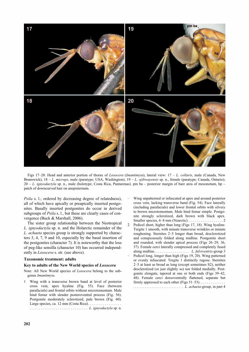
Figs 17–20. Head and anterior portion of thorax of Loxocera (Imantimyia), lateral view: 17 – L. collaris, male (Canada, NewBrunswick); 18 – L. microps, male (paratype; USA, Washington); 19 – L. ojibwayensis sp. n., female (paratype; Canada, Ontario);20 – L. ignyodactyla sp. n., male (holotype; Costa Rica, Puntarenas). pm ba – posterior margin of bare area of mesonotum, hp –patch of downcurved hair on anepisternum.

3 Vertical bristles short, inner vertical shorter than distancebetween its socket and midline. Parafacial broad (Fig. 18),at least 2.0× as wide as swollen basal part of arista. Firstflagellomere relatively short (Fig. 18), 1.8–1.9× (%) or1.3–1.5× (&) as long as face. Hind tibia with anteroventralapical bristle hardly differentiated from surrounding hairs.Postgonite as in Figs 36, 37 (western Nearctic). . L. microps
– Vertical bristles long, inner vertical at least as long as dis-tance between its socket and midline. Parafacial narrower(Fig. 17), at most 1.5× as wide as swollen basal part ofarista. First flagellomere long (Fig. 17), 2.3–3.0× (%) or1.9–2.6× (&) as long as face. Hind tibia with distinctanteroventral apical bristle. Postgonite as in Figs 26–29(Nearctic). . . . . . . . . . . . . . . . . . . . . . . . . . . . . . . . L. collaris
4 Scutum: length of bare area behind anterior declivityapproximately as long as postpronotal lobe (Fig. 19). Malepostgonite sickle-shaped, tapered at both ends (Fig. 48).Wing patterned (Fig. 54). Arista long-pubescent (Fig. 19)(Ontario). . . . . . . . . . . . . . . . . . . . . . . . L. ojibwayensis sp. n.
– Scutum: length of bare area behind anterior declivity at mosthalf as long as postpronotal lobe. Male postgonite withrounded base, tapering towards apex (Figs 39–42). Wingand arista variable. . . . . . . . . . . . . . . . . . . . . . . . . . . . . . . . . 5
5 Wing evenly infuscated. Arista short-pubescent (as in Fig.17), longest rays as long as diameter of swollen base ofarista. Postgonite more elongate (Fig. 41, 42), with moreslender and more curved apex than in L. cylindrica (centralNearctic). . . . . . . . . . . . . . . . . . . . . . . . . . . . . . L. fumipennis
– Wing infuscated at apex, around posterior crossvein andpenultimate sector of M (as in Fig. 54). Arista long-pubescent (as in Fig. 19), longest rays twice as long asdiameter of swollen base of arista. Postgonite shorter (Figs39, 40), with stouter and less curved apex than in L.
fumipennis (eastern Nearctic). . . . . . . . . . . . . . L. cylindrica
Loxocera fulviventris -group
Diagnosis. Bare, non-tomentose, median strip of facealmost reaching base of antennae (except in L. microps).Eye generally smaller (eye height 1.6–3.2× genal height)than in L. achaeta-group. Pedicel higher than long, firstflagellomere with somewhat swollen base, especially inmale (Figs 17, 18). Arista (Figs 17, 18) distinctlyremoved from base of first flagellomere (inserted clearlydistal of apicoventral margin of pedicel). Arista short-pubescent, longest rays as long as diameter of swollen
203
Figs 23–30. Male genitalia of Loxocera (Imantimyia) collaris
(showing individual variation). Hypandrium, lateral view: 23 –USA, Oregon; 24 – USA, California; 25 – Canada, New Bruns-wick. Right postgonite, inner lateral view: 26 – holotype of L.
californica syn. n.; 27, 28 – USA, Tennessee; 29 – Mexico,Durango. Phallus, posterior view: 30 – Mexico, Durango. Scale0.05 mm (postgonites) and 0.1 mm (others). hy – hypandrium,hc – hypandrium, dorsal condyle, hs – hypandrium, anterodorsalshoulder, pa – phallapodeme, pg – postgonite, ps – phallapo-demic sclerite.
Figs 21–22. Male genitalia of Loxocera (Imantimyia) collaris
(Canada, Nova Scotia): 21 – lateral view; 22 – ventral view.Scale 0.1 mm. ce – cercus, ea – ejaculatory apodeme, ep – epan-drium, hy – hypandrium, pa – phallapodeme, pg – postgonite,ph – phallus, pp – phallic pouch, ps – phallapodemic sclerite.
base of arista. Wing not infuscated. Tergite 1 smooth,with very fine wrinkles or roughening. Sternites 2–5longer than broad, desclerotized and conspicuouslyfolded along midline. Male terminalia: Dorsal portion ofsubepandrial sclerite well developed, ventral portionpoorly developed to absent. Hypandrium large, forminglarge, conspicuous, almost vertical plates (Figs 21, 33:hy), largely ventral to epandrium even in resting position.Phallic pouch poorly developed, not spinulose. Phallapo-deme connected to phallapodemic sclerite throughout itsentire length (e.g., Fig. 21: pa). Phallapodemic scleritewith scattered setulae, lacking setulose protuberances atbase of posterior arms. Postgonites inserted at apex ofphallapodemic arms (Fig. 34); shape rounded with apicalprocess; ventral portion with abundant, long microtrichiaand a few hairs (Figs 26–29, 36, 37). Ejaculatory apo-deme small to minute but well sclerotized (e.g., Fig. 21).Female terminalia: Intersegmental area between segments6 and 7 long, ca. 1/3 as long as segment 6; intersegmentalmembrane sclerotized laterally and ventrally. Female ter-gite 7 anteromedially produced (projection hidden belowprevious tergite, only visible in cleared specimens). Seg-ment 7 without tiny pits behind tergite 7. Segment 8 withmore or less developed, sclerotized, lateral strips; poste-rior margin slightly incised dorsally, laterally clearlydelimited from following segment and forming distinctrounded lobe. Cerci laterally compressed and completelyfused along midline. Two peg-like sensilla present oneach cercus (Fig. 32: ss).
Loxocera collaris Loew, 1869
(Figs 17, 21–32)
Loxocera collaris Loew, 1869: 184.Loxocera californica Capelle, 1953: 106, syn. n.
Diagnosis. Bare, non-tomentose, median strip of faceextending to level of antennae, narrow near dorsal endand restricted to median carina. Parafacial narrow (Fig.17), width less than ocellar diameter; eye large, in lateralview almost attaining anterior margin of head. Eye height1.7–3.2× genal height. First flagellomere 2.3–3.0× (%) or1.9–2.6× (&) as long as face (Fig. 17). Pleuron with orwithout black markings. Hind tibia with well developedanteroventral apical bristle. Median desclerotized area ofmale sternite 6 triangular, widening posteriorly. Malegenitalia: Hypandrium height ca. 0.5× its length, shape ofanterodorsal shoulder and dorsal condyle (for articulationwith epandrium) somewhat variable (Figs 21, 23–25: hc,hs). Phallapodeme large, size very variable (Figs 21,23–25: pa). Phallapodemic sclerite parallel-sided, notexpanded anteriorly (Fig. 22: ps). Postgonite variable,especially with regard to development of apical spine(Figs 26–29), more rounded than in L. microps. Phalluswithout pair of short processes at base (Fig. 30). Femaleovipositor as in Figs 31, 32.
Discussion of synonymy. Like other species in thegenus, L. collaris is quite variable, especially in colora-tion, genal width, length of antenna, shape of postgoniteand development of the phallapodeme. Capelle (1953),who examined only one male of L. collaris whendescribing his new species L. californica, was obviouslyunaware of this variability. After studying long series ofL. collaris from various localities and additional materialof “californica” from Mexico it became clear that thelatter is just a variety of the former. The length of theocellar bristles especially the vertical bristles, which wereCapelle’s main diagnostic characters, and also the eyeheight is less on average in western specimens but easternspecimens sometimes also have shorter bristles. Thedarker coloration of the thorax and abdomen of “califor-
nica” might be climatically induced because all availablespecimens are from higher elevations. Specimens fromcomparable elevations in the east (Tennessee: GreatSmoky Mts) show the same dark body pattern asdescribed for “californica”. No difference betweeneastern and western specimens was found in other diag-nostic characters used by Capelle (1953) like the length offirst flagellomere, body size, length of body hair, headshape and shape of postgonite. The latter varies consid-erably in size and curvature of the posterior spine evenamong specimens from the same locality (e.g., Figs 27,28). The unusual shape of the basal portion of the postgo-nite in “californica” as depicted in Capelle’s Fig. 7 wasnot confirmed by examination of the holotype (Fig. 26).No differences were found between the eggs of easternand Mexican specimens (eggs from specimens from thewestern US were not available).
Type material. L. collaris: not examined (MCZC).L. californica: Holotype %, USA, California, Sequoia Natl.
Pk., 6.viii.1940, R.H. Beamer (SEMC). Paratypes: 3%, 1&,
204
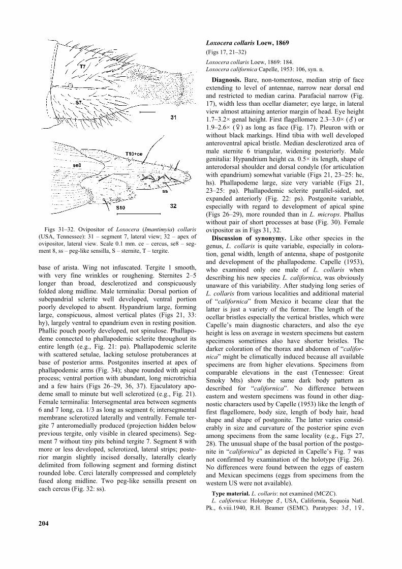
Figs 31–32. Ovipositor of Loxocera (Imantimyia) collaris
(USA, Tennessee): 31 – segment 7, lateral view; 32 – apex ofovipositor, lateral view. Scale 0.1 mm. ce – cercus, se8 – seg-ment 8, ss – peg-like sensilla, S – sternite, T – tergite.

USA, California: 2%, 1&, Mono Lake, 31.vii.1940, R.H.Beamer (SEMC); 1%, Yosemite Natl. Pk., 1.viii.1940, D.E.Hardy (SEMC). (One paratype & from Huntington Lake, Cali-fornia at CASC not examined).
Other material examined. Canada. Newfoundland: 1%, Por-tugal Cove, 10.viii.1967, J.F. McAlpine (CNCI); 4%, St. John’s,Agric. Exp. Stn., 5., 6. and 9.viii.1967, malaise trap, J.F.McAlpine (CNCI). Nova Scotia: 6%, Bridgetown, 29.viii. and2.ix.1912, “G[?] E S” (CNCI, 1 DEBU); 1% Kentville,6.viii.1958, J.R. Vockeroth (CNCI); 1% Aldershot, 15.viii.1950,A. McPhee (CNCI); 1&, Truro, 9.viii.1919, “W” (CNCI). NewBrunswick: St. Andrews, 1% 1.viii.1978, S.A. Marshall, 1%,19.viii.1978, Marshall & Konecny (DEBU); 1%, Kouchibou-guac Natl. Pk., 7.ix.1977, S.J. Miller (CNCI). Ontario: 1%, Pres-cott, 5.ix.1977, K.N. Barber (DEBU); 1%, Icewater Creekwatershed, ca. 12.7 km NNE Searchmont, mi. 10.5 WhitmanDam Rd., 21.–28.vii.1986, riparian meadow – alder thicket,malaise trap, K.N. Barber (DEBU). USA, Virginia: 1%, Blacks-burg, 2100 ft, 29.v.1962, J.R. Vockeroth (CNCI); 1&, Giles Co.,Bald Knob 10 km NW Blacksburg, 28.v.2005, S.M. Paiero(DEBU). Tennessee: 8%, 7&, Great Smoky Mts Natl. Pk., Col-lins Gap, 5700 ft, 22.viii.1957, J.G. Chillcott (CNCI); 6%, 3&,same data as previous except: Clingmans Dome, 6600 ft(CNCI); 1%, 2&, same as previous except: Indian Gap to Cling-mans Dome, 5200–6600 ft, 6.viii.1957 (CNCI). North Carolina:1%, Mt. Mitchell, 12.viii.1957, L.A. Kelton (CNCI); 1%,Mitchell Co., Roan Mt., 6200 ft, 13.viii.1957, J.G. Chillcott(CNCI). Oregon: 1%, Lincoln Co., Saddleback Mt., 12.ix.1959,J.C. Dirks-Edmunds (USNM); 1%, Benton Co., 5 mi WNWCorvallis, 15.ix.–1.x.1984, D.C. Darling (ROME). California:1%, Sequoia Natl. Park, Stony Creek, 5.vii.1947, W.W. Wirth(USNM). Mexico, Durango: 11%, 5&, 10 mi W El Salto, 9000ft, 10.vi.–3.vii.1964 (various dates), J.F. McAlpine (CNCI); 1%,24 mi W La Ciudad, 7000 ft, 28.vi.1964, W.R.M. Mason(CNCI).
Distribution. Widespread in the northeastern Nearctic, in theAppalachians south to North Carolina and Tennessee; in thewest from Montana, Idaho and Washington (Capelle, 1953)south to California; also in Durango, Mexico at high elevations.The species has not been recorded from either the midwesternUS or the western half of Canada. The apparent gap betweeneastern and western populations probably does not reflect theactual distribution of this rarely collected species.
Loxocera microps Melander, 1920
(Figs 18, 33–38)
Loxocera microps Melander, 1920: 92.
Diagnosis. Upper 1/5–2/5 of face with sparse microto-mentum; bare, non-tomentose, median strip of facerestricted to lower 3/5–4/5. Parafacial broad (Fig. 18),width at narrowest point ca. 2× ocellar diameter; eyesmall, in lateral view broadly separated from anteriormargin of head. Eye height 1.3–2.1× genal height. Firstflagellomere 1.8–1.9× (%) 1.3–1.5× (&) as long as face(Fig. 18). Katepisternum more or less darkened ventrally.Hind tibia with anteroventral apical bristle hardly differ-entiated from surrounding hairs. Median desclerotizedarea of male sternite 6 parallel-sided, not widening poste-riorly. Male genitalia: Cercus (Fig. 33: ce) better devel-oped than in L. collaris, with more numerous hairs, apexdistinctly pointing ventrally. Hypandrium (Fig. 33: hy)very large, height ca. 0.8× its length. Phallapodeme rela-tively small (Fig. 33: pa). Phallapodemic sclerite
expanded anteriorly (Fig. 34). Postgonite (Figs 36, 37)more triangular than in other Nearctic species of L.
fulviventris-group. Phallus with a pair of short processesat base (Fig. 35: php). Female ovipositor very similar toL. collaris.
Variation. The coloration of this species is extremelyvariable. Southern specimens (incl. the paratype, Fig. 18)and a male from Willow, Alaska are predominantlyochreous with largely brown abdomen, restricted darkbrown markings on the mesoscutum, dark brown ocellartubercle, and usually some infuscation of the frons, lowerportion of katepisternum and postnotum. On the contrary,specimens from the Yukon Territory, and Unalakleet,Alaska, are almost completely blackish brown excludinganterior margin of frons narrowly, lateral portions of face,lower eye margin, anterior vertical stripe of gena, anteriorface of all coxae, knees, tibiae distally, and tarsi. Anunusual character for Psilidae is the presence of a fewsetulae on the meron of some specimens. The unusualshape of the postgonite illustrated by Capelle (1953: Plate
205
Figs 33–35. Male genitalia of Loxocera (Imantimyia) microps
(paratype; USA, Washington): 33 – lateral view; 34 – ventralview; 35 – phallus, caudal view. Scale 0.1 mm. ce – cercus, ea –ejaculatory apodeme, ed – ejaculatory duct, ep – epandrium, hy– hypandrium, pa – phallapodeme, pg – postgonite, ph –phallus, php – phallus, basal process, pp – phallic pouch, ps –phallapodemic sclerite, psa – ditto, apex of posterior arm(s).

2, Fig. 8) (reproduced in Fig. 38) is probably due to dis-tortion of the specimen and/or inaccurate illustration. Thepostgonite of the paratype (with identical collection dataas holotype) is depicted in Figs 36, 37.
Type material. Holotype %, USA, Washington, Mt. Rainier,Paradise Park, viii.1917, A.L. Melander (USNM, not exami-ned). Paratype: 1%, same data as holotype (USNM).
Other material examined. Canada. British Columbia: 2%,1&, Lisadele Lk., 58°41´N 133°4´W, 4000 ft, 7.viii.1960, R.Pilfrey & W.W. Moss (CNCI); 1%, Moosehorn Lk., 58°10´N132°7´W, 4500 ft, 27.vii.1960, W.W. Moss (CNCI); 1&, Mt.Revelstoke, 6200 ft, 18.viii.1952, G.J. Spencer (CNCI); 1&, Mt.Revelstoke Natl. Pk., Eva Lk. Trail, 6000 ft, 14.viii.1952, G.J.Spencer (CNCI). Yukon Territory: 1%, Firth River, 17.vii.1956,R.E. Leech (CNCI); 1%, Dempster Hwy km 420, 66°35´N,136°18´W, 3.–7.vii.1979, malaise trap, ROM field party(ROME). USA. Alaska: 1&, Unalakleet, 5.vii.1961, R. Madge(CNCI); 1%, Willow, 18.vii.1948, F.S. Blanton (USNM). Wash-ington: 1%, Mt. Rainier, Yakima Park, 22.vii.1924, A.L.Melander (USNM); 1&, (labelled “Parallotype” by Capelle),Mt. Rainier Natl. Pk., 16.viii.1941, L.J. Lipovski (SEMC).Idaho, 1&, Priest Lake, Lookout Mt., 20.viii.1919, A.L.Melander (USNM).
Distribution. Northwestern Nearctic from Alaska and theYukon Territory south to Washington and Idaho.
Loxocera achaeta-group
Diagnosis. Upper 2/5 of face completely tomentose(bare in L. ignyodactyla sp. n.). Eye large (eye height2.7–7.6× genal height). Pedicel longer than high (Figs 19,20), base of first flagellomere not swollen. Arista (Figs19, 20) inserted close to base of first flagellomere (at orslightly beyond level of apicoventral margin of pedicel).Arista long-pubescent, longest rays twice as long asswollen base of arista (short in L. fumipennis). Wingweakly patterned or evenly infuscated. Tergite 1 stronglyrugose (except in L. ignyodactyla sp. n.). Sternites 2–5 or3–5 at least as broad as long, not conspicuously desclero-tized and folded medially. Male genitalia: Dorsal portionof subepandrial sclerite partially to completely desclero-tized (Fig. 46: se dp), ventral portion well developed (Fig.45: se vp). Hypandrium moderately developed, hypan-drial arms not large and plate-like (e.g., Fig. 43: ha).Phallic pouch membranous, with spinulose inner surface(Fig. 47). Phallapodeme usually free anteriorly (Fig. 43:pa), and separate from phallic pouch (fused in L. ignyo-
dactyla sp. n.). Phallapodemic sclerite usually with a pairof setulose protuberances at base of posterior arms (Fig.44: pst) (absent in L. ignyodactyla sp. n.), otherwise withfew to many setulae. Postgonites articulated near middleof phallapodemic arms (e.g., Fig. 44: pg); shape slender,tapered on one or both ends (e.g., Figs 39–42, 48),bearing a few setulae, rarely also with a few microtrichia(Fig. 60). Ejaculatory apodeme apparently absent. Femaleterminalia: Intersegmental area between segments 6 and 7apparently absent, i.e. intersegmental membrane com-pletely sclerotized except for pleural membrane, distin-guished from tergites or sternites by absence of bristles.Tergite 7 with straight anterior margin. Segment 7 with atiny pit behind tergite 7 (Figs 49, 50) (absent in L. ignyo-
dactyla sp. n.). Segment 8 without sclerotized lateralstrips, posterior margin deeply incised dorsally (e.g., Fig.51), usually poorly delimited from following segment lat-erally (Fig. 52: se8) (well delimited in L. ignyodactyla sp.n.). Cerci usually dorsoventrally depressed (laterally com-pressed in L. ignyodactyla sp. n.), not fused along midline(Figs 51–53).
Loxocera cylindrica Say, 1823
(Figs 1, 39–40)
Loxocera cylindrica Say, 1823: 98.Loxocera pleuritica Loew, 1869: 38.Loxocera pectoralis Loew, 1869: 38.Loxocera cylindrica var. obsoleta Johnson, 1920: 15.
Diagnosis. Habitus as in Fig. 1. Extent of black mark-ings of thorax very variable (described by Johnson, 1920;geographical variation studied by Capelle, 1953); infusca-tion of wing also variable. Gena on average slightlyhigher than in L. ojibwayensis sp. n., eye height 3.8–5.8×genal height. Scutum: length of bare area behind anteriordeclivity at most half as long as postpronotal lobe. Malegenitalia very similar to L. ojibwayensis sp. n., exceptpostgonite. Postgonite (Figs 39–40) with pointed apexand rounded base, apex stouter than in L. fumipennis.Shape of postgonite subject to allometric variation similar
206
Figs 36–42. Postgonites of Loxocera (Imantimyia). L.
microps: 36, 37 – paratype, USA, Washington; 38 – redrawnfrom Capelle (1953: Plate II, Fig. 8). L. cylindrica: 39, 40 –Canada, Ontario (showing allometric variation). L. fumipennis:41 – redrawn from Capelle (1953: Plate II, Fig. 5); 42 – USA,Colorado. Lateral inner view except following: 37 – anteriorview; 38, 41 – angle of view unknown. Scale 0.05 mm(unknown for 38, 41).

to L. ojibwayensis sp. n.: postgonite stouter, and marginnext to setulose patch more convex in large (Fig. 39) thanin small males (Fig. 40). Female ovipositor similar to L.
ojibwayensis sp. n. (see below).Taxonomy. The type of L. cylindrica is destroyed and
Say’s (1823) description is inadequate to distinguish thisspecies from the very similar L. ojibwayensis sp. n.(described below). Considering that the new species israre and only known from the type locality but L. cylin-
drica (in the interpretation of previous authors) iscommon and widespread, we continue to apply this nameto the common species, i.e. we are using it as a seniorsynonym of L. pleuritica, L. pectoralis and L. obsoleta.Lectotypes of the latter two are designated to clarify theidentity of these nominal species with regard to L. ojib-
wayensis sp. n.
Type material. L. cylindrica. “Inhabits Pennsylvania”;destroyed (Capelle, 1953).
L. pectoralis. Holotype &, USA, Washington [D.C.] (seeLoew, 1869, not mentioned on labels), “Loew Coll.”, “pec-toralis m”, “Type 13334” (MCZC).
L. pleuritica. Lectotype % (M. Buck, by present designation):USA, New York, Schoharie Co., “Sharon Springs”, “LoewColl.”, “pleuritica m.”, “Type 13335” (MCZC). Paralectotypesnot examined (Capelle, 1953, erroneously considered aspecimen from Connecticut to be the only type specimen of thistaxon).
L. cylindrica var. obsoleta. Lectotype % (M. Buck, by presentdesignation): USA, “Plymouth Mass VII 28 83 [?]”, “CotypeNo”, “CW Johnson Collector”, “M.C.Z. Paratype 27036” (firstletters struck through) (MCZC). Paralectotypes not examined.
Other material examined. 179%, 237&, (CNCI, DEBU,ROME, SEMC, USNM; only marginal localities given).Canada. Every province from Newfoundland to Manitoba(Ninette, CNCI). USA. New Hampshire, Vermont, Massachu-setts, Connecticut, New York, Pennsylvania, Ohio, Michigan,Wisconsin, Iowa, Maryland, Virginia, Kentucky, Tennessee,North Carolina, South Carolina (Aiken Co.: Aiken, CNCI;Charleston Co.: Francis Marion Natl. Forest, Santee River,DEBU; Charleston Co.: Hobcaw Barony, Belle Baruch MarineField Stn., DEBU), Georgia (Floyd Co.: Cave Spring, CNCI),Missouri, Kansas (Kiowa Co.: 7.6 mi SE Haviland, SEMC),Mississippi, Louisiana, Texas (Austin, DEBU; Navasota, CNCI;Liberty Co.: 16 mi E Cleveland, DEBU).
Distribution. Widespread in the eastern Nearctic excludingthe extreme southeast, west to Manitoba, Minnesota, Nebraska,central Kansas, Arkansas and eastern Texas (in part afterCapelle, 1953).
Loxocera fumipennis Coquillett, 1901
(Figs 41–42)
L. fumipennis Coquillett, 1901: 617.
Diagnosis. Gena slightly higher than in L. cylindrica;eye height 2.7–3.7× genal height. Differs from all speciesin the L. achaeta-group by the very short-pubescent arista(as in Fig. 17). Scutum: length of bare area behind ante-rior declivity at most half-length of postpronotal lobe.Male genitalia very similar to L. ojibwayensis sp. n.,except postgonite. Postgonite (Fig. 42) with pointed apexand rounded base, very similar to L. cylindrica but apexmore slender. Allometric variation of the shape of thepostgonite similar to L. cylindrica (see above) is to be
expected. This phenomenon might also account for thesomewhat different shape of the postgonite illustrated byCapelle (1953: Plate II, Fig. 5) (reproduced in Fig. 41).Female ovipositor similar to L. ojibwayensis sp. n. (seebelow).
Distribution. Prairie provinces of Canada and American mid-west south to Texas and Arizona (Shewell, 1965). This speciesreplaces L. cylindrica in the mid-west; the ranges of both spe-cies narrowly overlap along the eastern limit of L. fumipennis.
Material examined. Canada. Manitoba: 1%, Aweme,22.vi.1953, A.R. Brooks & L.A. Kelton (CNCI). Saskatchewan:16%, 2&, Wood Mt., 49°19´N 106°21´W, 17.vi.1955, A.R.Brooks & J.R. Vockeroth (CNCI); 12%, Elbow, 23.vi.1954 and3.–17.vi.1960, Brooks & Wallis (CNCI); 1&, Swift Current,21.vi.1937, A.R. Brooks (CNCI); 1%, Scout Lk., 17.vi.1955,A.R. Brooks (CNCI); 3%, Lisieux, 49°16´N 105°59´W,21.vi.1955, J.R. Vockeroth (CNCI). Alberta: 1%, Brooks,13.vi.1957, Brooks & MacNay (CNCI); 1%, Elkwater Pk.,11.vii.1952, L.A. Konotopetz (CNCI). USA. North Dakota: 1%,Bismarck, 14.vi.1918, J.M. Aldrich (USNM). Colorado: 20%,14&, 4.5 mi N Boulder, 10., 13. and 20.vi.1961, C.H. Mann &
207
Figs 43–44. Male genitalia of Loxocera (Imantimyia) ojib-
wayensis sp. n. (holotype; Canada, Ontario): 43 – lateral view;44 – ventral view. Scale 0.1 mm. ep – epandrium, ha – hypan-drial arm, hp – hypandrial plate, pa – phallapodeme, pg – post-gonite, ph – phallus, pp – phallic pouch, ps – phallapodemicsclerite, psa – ditto, apex of posterior arm(s), pst – ditto, setu-lose tubercle at base of posterior arm.

W.R.M. Mason (CNCI); 2&, Boulder, Valmont Butte, 5300 ft,1.vi.1961, J.R. Stainer (CNCI); 1%, Longmont, 5000 ft,3.vi.1961, B.H. Poole (CNCI); 1%, “1563”, collection Coquil-lett (USNM); Ft. Collins, 1%, 30.v.1902, L.A. Titus, 1%,1.ix.1899, C.W. Johnson collection (USNM). Missouri: 1%,Williamsville, x.–xi, J.T. Becker (CNCI). Arkansas: 1&,Granite Dells, 12.vii.1947, R.H. Beamer (SEMC). Kansas: 1&,Lawrence, Nat. Hist. Res., 25.vi.1956, J.G. Chillcott (CNCI);1&, Lawrence, 4.iv.1938, D.E. Hardy (SEMC); 1&, DouglasCo., 13.v.1923, W.J. Brown (CNCI); 2%, 3&, Tonganoxie,11.v.1940, D.E. Hardy (SEMC); 2%, Chanute, 29.viii.1939,R.H. Beamer (SEMC); 2%, Cherokee Co., 4.v.1939 and15.v.1940, R.H. Beamer (SEMC); 1&, Hutchinson, 7.ix.1938,D.E. Hardy (SEMC); 2%, 1&, Menlo, 23.viii.1940, R.H.Beamer (SEMC). Texas: 3%, 1&, Brazos Co., College Station,26.iii. and 6.iv.1966, J.C. Schaffner & D.M. Wood (CNCI); 6%,4&, Carthage, 31.iii.1968, D.M. Wood (CNCI).
Loxocera ojibwayensis Buck sp. n.
(Figs 19, 43–54)
Description. Body length 5.5–6.0 mm, wing length4.0–4.5 mm, length of antenna 1.5 mm. Colorationsimilar to L. cylindrica, extent of black markings variable.Body red except: scape, first flagellomere (except some-times extreme base), frons excluding frontal orbits (rarelyocellar triangle only), face excluding parafacials and ven-tromedial triangular area, occiput (rarely not), upperextreme of gena and postgena (rarely not), postpronotal
lobes excluding anterior surface (sometimes only nar-rowly along margins), anterior portion of scutum to levelof posterior margin of postpronotal lobes (usually),median scutal stripe in anterior half or posterior 3/4 oralong whole length (rarely completely absent), sometimesa pair of free, paramedian spots at level of posteriormargin of postpronotal lobes (sometimes connected toanterior black area), lower edge of notopleuron (usually),lower half or lower margin of mediotergite (rarely not),upper half of propleuron sometimes, most of tergite 1 oronly median third (rarely not), female tergite and sternite7, and rarely female tergite 6, black. Black body markingsin some specimens medium to dark brown. Lower surfaceof pedicel, apical tarsal segments of all legs and some-times apical portion of palpus darkened. Major bristles ofhead black. Swollen basal portion of arista reddishyellow, becoming white distally (darkened in two speci-mens). Gena, especially anterior half, yellowish to ivory.Haltere off-white, stem yellowish.
Head (Fig. 19). Frons not or hardly projecting beyondlevel of anterior eye margin, slightly transverselydepressed at level of anterior third of eye. Frontal orbitssilvery microtomentose in anterior half, with a row ofabout 10 hairs, fronto-orbital bristle not differentiated.Frontal vitta sparsely setulose in anterior half, especiallynear anterior margin. Ocellar triangle slightly depressedalong midline, with slightly convex lateral margins,extending anteriorly about 0.85× length of frons. Ocellarbristles relatively short, length 2.0–2.5× ocellar diameter.
208
Figs 49–50. Abdominal segment 7 of female Loxocera (Iman-
timyia) ojibwayensis sp. n. (paratype; Canada, Ontario): 49 –lateral view; 50 – ditto, pit region enlarged. Scale 0.1 mm and0.05 mm (pit region). pe – pit entrance.
Figs 45–48. Male genitalia of Loxocera (Imantimyia) ojib-
wayensis sp. n. (Canada, Ontario): 45 – epandrium and associ-ated structures, caudal view (holotype); 46 – phallic complex,caudal view (holotype); 47 – lateral walls of phallic pouch, ven-tral view (holotype); 48 – right postgonite, lateral inner view(paratype). Scale 0.05 mm (postgonite) and 0.1 mm (others). ce– cercus, ha – hypandrial arm, pg – postgonite, ph – phallus, phla – phallus, lateral arm, ps – phallapodemic sclerite, se dp –subepandrial sclerite, dorsal portion, se vp – ditto, ventral por-tion.

Postvertical bristles not differentiated, 2–6 postocellarhairs present. Vertical bristles short; outer vertical bristleslightly longer than inner vertical, ca. 3× as long asocellar diameter. Face moderately slanting, belowantennae in upper half with slightly elevated midline.Upper half of face with inconspicuous brownish microto-mentum, lower half with more distinct silvery microto-mentum, the two halves reflecting light at differentangles. Microtomentum of lower face interrupted bymedian, triangular (dorsally tapered), bare, shining area.Parafacials narrow, at narrowest point barely as wide asswollen base of arista, with a row of setulae and with dis-tinct silvery microtomentum. Gena with several setulaeventrally, disc very sparsely setulose, lacking silverymicrotomentum. Eye large, protuberant; dorsal eyemargin about level with most elevated parts of frons. Eyeheight 4.3–7.6× genal height. Scape slightly longer thanbroad (dorsal view), its length equal to maximum widthof first flagellomere, with several setulae near dorsoapicalmargin, otherwise bare. Pedicel 1.3× as long as scape,with distinct dorsal seam, setulose. First flagellomerevery long, twice as long as face, more or less parallel-sided in lateral view, base hardly widened. Arista insertedclose to base of first flagellomere, basal margin of socketat same level as ventroapical margin of pedicel; length ofarista ca. 0.7× length of first flagellomere, with swollenbasal fourth. Pubescence of swollen basal portion short,shorter than maximum diameter of arista; pubescence ofslender apical portion long, longest rays twice as long asmaximum diameter of arista. Palpus with scatteredbrownish to blackish setulae on outer and ventral surface,without outstanding bristles.
Thorax with the usual complement of bristles.Notopleural bristle poorly differentiated, brownish (notblack), not much longer than surrounding hairs. Supra-alar and postalar bristle well developed, always black.One prescutellar pair of dorsocentral bristles, brown toblack, as long as supra-alar and postalar bristle or slightly
shorter. Scutum bare anteriorly to level of posteriormargin of postpronotal lobe. Hairs of scutum short, palebrown, denser along dorsocentral lines, slightly sparserbetween, much sparser lateral of dorsocentral lines,denser on notopleuron. Sculpture of scutum associatedwith density of hairing, stronger in areas that are moredensely haired, smooth in bare areas. Postpronotal lobessmooth, hairs sparse, sometimes reduced to a few setulae.Scutellum with finely wrinkled disc, apical bristles ca.0.8–1.0× as long as scutellum, preapical pair smaller,0.4–0.6× as long as apical pair, absent in half of the speci-mens examined. Pleuron mostly smooth and shining;propleuron with dense, felt-like microtomentum alonglower and posterior margin; posterior portion of lateroter-gite and lateral portions of mediotergite with sparsermicrotomentum. Anepisternum in posterior 3/5 withshort, moderately dense, upcurved brownish hair, ven-trally with the usual patch of dense, pale, downcurvedhair. Katepisternum with brownish hair, ventrally rela-tively dense and pale, dorsally sparser, central portionalmost bare. Posterior spiracle with numerous longsetulae along margin.
Legs haired except bare posteroventral surface of forefemur and largely bare lower surface of mid femur. Hindfemur of male without ventral process. Mid tibia with onelong and (1–)2 shorter ventroapical bristles, blackish orreddish except long bristle, which is black. Hind tibiawith moderately developed, blackish or reddish, anterov-entral, apical bristle. Hairing of tarsi becoming darkertowards apex, distinctly darkened in apical 2–3 tarso-meres. Wing as in Fig. 54, membrane slightly tinged withbrown, pattern somewhat variable: apical fifth of winginfuscated, sometimes just barely extending into cells r1
anteriorly and m posteriorly, infuscation sometimes inter-rupted medially in cells r2+3 and r4+5. Area around poste-rior crossvein, last sector of CuA1 and penultimate sectorof M (between crossveins) also infuscated, rarely areaaround last sector of M infuscated, as well, connectingapical spot with darkening around posterior crossvein.Wing veins brown in dorsal view excluding most ofcosta, subcosta, stem vein, R1 and Rs, which are yel-lowish brown. Last sector of vein M with moderatelystrong curvature.
Preabdomen moderately slender. Syntergite 1+21.2–1.3× as long as broad, laterally without conspicu-ously enlarged bristles in apical half of segments 1 and 2.Tergite 1 coarsely rugose, following tergites smooth withfine transverse wrinkles. Preabdominal sternites notfolded lengthwise along middle, not desclerotized medi-ally.
Male terminalia very similar to L. cylindrica exceptpostgonites. Male pregenital sclerite well defined butbroadly interrupted dorsally. Epandrium of holotypedarkened in ventral half. Cerci setulose (Fig. 45: ce), ven-trally fused, largely membranous excluding ventral sur-face. Subepandrial sclerite with well-developed ventralportion (Fig. 45: se vp); dorsal portion very faintly sclero-tized (Fig. 46: se dp). Phallic pouch laterally compressed(Fig. 44: pp), widening anteriorly and posteriorly; inner
209
Figs 51–53. Apex of ovipositor of Loxocera (Imantimyia)ojibwayensis sp. n. (paratype; Canada, Ontario): 51 – dorsalview; 52 – lateral view; 53 – ventral view. Scale 0.1 mm. ce –cercus, se8 – segment 8; S8 – ditto, posteroventral lobe (sternite8); S10 – sternite 10; T10 – tergite 10.

surface of pouch with minute aciculate sculpture in com-pressed middle portion (Fig. 47). Phallapodeme free ante-riorly from phallic pouch (Fig. 43: pa). Setulae ofphallapodemic sclerite few in number, posterior ones on amore or less developed protuberance near base of poste-rior arms (Fig. 44: pst). Apex of phallapodemic armstruncate (Figs 43, 44: psa). Lateral surface of hypandriumwith a few widely scattered setulae and a small patch ofmicrotrichia just above postgonite (Fig. 43: ha). Postgo-nite sickle-shaped (Fig. 48), strongly sclerotized andmostly dark brown with black apices, medial (ventral)surface with 2–4 setulae in anterior portion, lackingmicrotrichia. Shape of postgonite subject to slight allo-metric variation: in larger specimens margin next to patchof setulae more convex than in smaller specimens.Phallus T-shaped in posterior view (Fig. 46: ph), withrelatively long, membranous distal portion.
Female terminalia extremely similar to L. cylindrica; nodifferences of taxonomic value were detected. Interseg-mental membrane between segments 6–7 almost com-pletely sclerotized (excl. pleural portions and dorsomedialemargination), sclerotization continuous with tergite andsternite. Segment 7 laterally compressed (Fig. 49), tergitesubcarinate along midline, especially in anterior half. Ter-gite 7 on each side with short posterior extension and asmall pit below it (Fig. 50); exposed (non-retractable)portion of intersegmental membrane bare and minutelylongitudinally striate in anterior 2/3 of dorsal surface, oth-erwise finely microtrichose, retractable portion of inter-segmental membrane entirely microtrichose. Segment 8(Figs 51–53) distinctly striate on whole surface, with scat-tered short setulae, completely devoid of microtrichia;posterior margin with deep, acute, medial emarginationdorsally (Fig. 51), not clearly delimited laterally, and witha simple, narrowly rounded sternal lobe. Tergite 10 andcerci fused (Fig. 51: T10, ce). Tergite 10 dorsally withscattered, short setulae and one pair of bristles just basalof free apical portion of cerci. Sternite 10 with a few scat-tered setulae and 1–2 longer bristles, apex with a fringe oflong felt-like microtrichia (Fig. 52, 53: S10). Cerci (Figs51–53: ce) moderately flattened dorsoventrally, relativelylong, clearly discernable even in undissected specimens.Each cercus bare dorsally, with a lateral row of short bris-tles that appear to arise from a small groove, and with aventral row of longer bristles, two of which are enlarged.
Type material. Holotype % (debu01117471). Canada.Ontario: Essex Co., Windsor, Ojibway Prairie nr. SprucewoodAve. observation point, 42°15´51˝N, 83°4´30˝W, 26.–27.viii.2002, yellow pans, M. Buck & S.M. Paiero (DEBU). Paratypes2%, 14& (all DEBU). Canada, Ontario: 1&, (debu01117472),same data as holotype; 2& (debu01118320, debu01118321),same data as holotype except 12.–13.ix.2002; 1%, 5&(debu00251754 to ...759), same locality as holotype, 28.–29.vii.2005, yellow and white pans, S.M. Paiero & M.D. Bergeron;Windsor, Ojibway Prairie, 42°15´51˝N, 83°4´30˝W, 12.–13.ix.2002, 1& (debu00216770), yellow pans in loamy depression,1& (debu00216735), netted, M. Buck; 1& (debu00253172),same locality as previous, 25.vii.2003, yellow pans, S.M.Paiero; 1&, Windsor, Ojibway Prairie Reserve, 11.vii.1980,K.N. Barber; Windsor, ~1.5 km S Ojibway Prairie, 42°13´34˝N,83°4´27˝W, forest-prairie edge, 1% (debu01118304), 18–31.vii.
2001, 1& (debu01113897), 1.–22.ix.2001, S.M. Paiero, 1&(debu01109235), 1.–19.viii.2001, P. Pratt, all in malaise trap.
Etymology. The name refers to the type locality, OjibwayPrairie in Windsor, Ontario.
Distribution. At present only known from extreme south-western Ontario in Canada.
Relationships. This species shows similarities withboth the Palaearctic L. achaeta / L. lutulenta / L. nigri-
frons and the Nearctic L. cylindrica / L. fumipennis. Mostremarkably, the postgonite of the new species has thesame two-pronged shape as the three Palaearctic species(Shatalkin, 1989: Pl. 3, Fig. 2; Iwasa, 1992: Fig. 21).Loxocera ojibwayensis sp. n., L. lutulenta and L. nigri-
frons also share the large bare anterior area of the scutum(L. achaeta not examined). The three Nearctic species, onthe other hand, possess a notopleural and one pair of dor-socentral bristles, which are reportedly absent in L.
achaeta and L. lutulenta (Iwasa, 1992). However, a maleof L. lutulenta (Japan: Mt. Atago nr. Kyoto, CNCI) exam-ined by us has well developed dorsocentral bristles and apale, inconspicuous notopleural bristle but otherwiseagrees well with the original description (Iwasa, 1992).Variation in the development of dorsocentral bristles alsooccurs in L. aristata (see Greve & Skartveit, 2001) so thetaxonomic importance of this character should not beoverestimated.
210
Figs 54–56. External characters of Loxocera (Imantimyia)achaeta-group. L. ojibwayensis sp. n.: 54 – left wing (paratype;Canada, Ontario). L. ignyodactyla sp. n.: 55 – left wing (para-type; Costa Rica, Puntarenas); 56 – male hind femur and tibia,posterior view (holotype). fp – patch of felt-like micro-tomentum.

Loxocera ignyodactyla Buck sp. n.
(Figs 5, 20, 55–65)
Description. Largest species of the subgenus and oneof the largest species in the genus, body length 12 mm,wing length 8 mm, length of antenna 3 mm. Body redexcept: Scape, first flagellomere except extreme base,ocellar prominence and small area right behind posteriorocelli, postpronotal lobes excluding margins and anteriorsurface, black. Major bristles of head and thorax alsoblack. Palpus blackish in male, hardly infuscated infemale (difference probably due to individual variability,not sexual dimorphism). Pedicel on outer surface andventrally, first flagellomere basal of arista, dark reddish.Swollen basal portion of arista reddish yellow, becomingwhite distally. Face and gena slightly yellowish red. Hal-tere yellowish.
Head (Figs 5, 20). Frons strongly projecting beyondlevel of anterior eye margin (much more than in Nearctic
species), slightly transversely depressed at level of ante-rior third of eye. Frontal orbits with a row of about 15hairs, last hair enlarged, ca. twice as long as previousones, forming a small fronto-orbital bristle. Frontal vittasparsely setulose in anterior half, especially near anteriormargin. Ocellar triangle slender and very pointed, withslightly concave lateral margins, extending anteriorlyabout halfway between level of anterior margin of eyeand anterior margin of frons. Ocellar bristles moderatelydeveloped, length ca. 3× ocellar diameter. Postverticalbristles not differentiated, 3–7 postocellar hairs present.Vertical bristles relatively short; inner vertical bristleslightly longer than outer vertical, ca. 4× as long asocellar diameter. Face strongly slanting, with short, deli-cate median carina at level of antennal base and slightlybelow. Parafacials narrow, at narrowest point barely aswide as swollen base of arista, with a row of setulae and
211
Figs 57–58. Male genitalia of Loxocera (Imantimyia) ignyo-
dactyla sp. n. (holotype; Costa Rica, Puntarenas): 57 – lateralview; 58 – ventral view. Scale 0.1 mm. ha – hypandrial arm, hp– hypandrial plate, pa – phallapodeme, pg – postgonite, ph –phallus, pp – phallic pouch, ps – phallapodemic sclerite, psa –ditto, apex of posterior arm(s).
Figs 59–61. Male genitalia of Loxocera (Imantimyia) ignyo-
dactyla sp. n. (holotype; Costa Rica, Puntarenas): 59 – epan-drium and associated structures, caudal view; 60 – postgonite,lateral inner view; 61 – phallic complex, caudal view. Scale0.05 mm (postgonite) and 0.1 mm (others). ce – cercus, ha –hypandrial arm, hp – hypandrial plate, pg – postgonite, ph –phallus, ps – phallapodemic sclerite, psa – ditto, apex of poste-rior arm(s).

with weak silvery microtomentum. Gena swollen, withseveral stronger setulae ventrally, disc very sparsely setu-lose, lacking silvery microtomentum. Eye large, protuber-ant; dorsal eye margin projecting slightly above level offrons. Eye height 3.3–3.4× genal height. Scape slightlylonger than broad (dorsal view), its length equal tomaximum width of first flagellomere, with several setulaenear dorsoapical margin, otherwise bare. Pedicel slightlylonger than scape, with distinct dorsal seam, setulose.First flagellomere very long, twice as long as face, itsdiameter slightly increasing towards apex (lateral view),base hardly widened. Arista inserted close to base of firstflagellomere, basal margin of socket removed from (levelof) ventroapical margin of pedicel by 1.0–1.5× ocellardiameter; length of arista ca. 0.8× length of first flagello-mere, with swollen basal fourth. Pubescence of swollenbasal portion short, shorter than maximum diameter ofarista; pubescence of slender apical portion long, longestrays twice as long as maximum diameter of arista. Palpuswith scattered blackish setulae on outer and ventral sur-face, without outstanding bristles.
Thorax with the usual complement of bristles.Notopleural bristle slightly over half as long as the supra-alar and postalar bristle. One prescutellar pair of dorso-central bristles, 1.3–1.4× as long as supra-alar andpostalar bristles. Scutum with rough surface sculpture inhaired region, smooth in bare anterior region. Scutumbare up to level of anterior third of postpronotal lobe, oth-erwise uniformly setulose. Hairing of scutum short anddense, mostly black, reddish in notopleural region, notarranged in rows. Postpronotal lobes smooth, hairingmuch sparser than on scutum. Scutellum with finely wrin-kled disc, apical bristles ca. 1.4× as long as scutellum,preapical pair small, only ca. 0.3× as long as apical pair.Pleuron mostly smooth and shining; propleuron withdense, felt-like microtomentum along lower and posteriormargin; posterior portion of laterotergite and lateral por-tions of mediotergite with sparser microtomentum. Ane-pisternum posteriorly and dorsally with short, moderatelydense, upcurved reddish hair, in posteroventral cornerwith the usual patch of dense, pale, downcurved hair.Katepisternum with reddish hair, ventrally relativelydense, dorsally sparser, central portion almost bare. Pos-terior spiracle with numerous long setulae along margin.
Legs more or less uniformly haired except bare pos-teroventral surface of fore femur and largely bare lowersurface of mid femur. Hind femur of male with distinctposteroventral process before middle (Fig. 56). Mid tibiawith one long and 2–3 shorter ventroapical bristles,mostly reddish except long bristle, which is black in oneof the two specimens. Hind tibia with moderately devel-oped, reddish, anteroventral apical bristle. Hairing of tarsibecoming darker towards apex, distinctly darkened inapical three tarsomeres. Wing (Fig. 55) distinctly yel-lowish, with moderately developed dark band at level ofposterior crossvein; band more or less interrupted alongcentre of cells r2+3, r4+5 and dm. Wing veins mostly yel-lowish, brown in banded part of wing. Last sector of veinM with unusually strong curvature.
Preabdomen slender. Syntergite 1+2 ca. twice as longas broad (ratio influenced by shrinkage from drying), lat-erally with conspicuously enlarged bristles in apicalhalves of segments 1 and 2, on latter almost erect andnearly as long as width of segment at level of insertion (inair-dried specimens). Abdominal tergites smooth, withvery fine, evenly spaced, transverse wrinkles; tergite 1hardly with coarser sculpture.
Male pregenital sclerite weakly sclerotized and hardlydiscernible but apparently forming a complete dorsal archbetween spiracles 7. Epandrium evenly setose (Fig. 57).Cerci setulose (Fig. 59), ventrally more or less fused,largely membranous excluding ventral surface. Subepan-drial sclerite with well developed broad-elliptical ventralportion; extending from lower margin of epandrium tolower margin of cerci along posterior margin of epan-drium; dorsal portion completely desclerotized, undiffer-entiated. Phallic pouch large, curved towards left side ofbody (Fig. 58: pp), not laterally compressed as in Nearcticspecies of L. achaeta-group; inner surface of pouch withminute aciculate sculpture posteriorly, finely denticulatenear middle (denticles with broadly rounded apices),simple anteriorly. Phallapodeme moderately developed,connected to phallapodemic sclerite through its entirelength, anterior end not separated from phallic pouch(Fig. 57: pa). Phallapodemic sclerite with relativelynumerous, scattered setulae on ventral surface (Fig. 58:ps), lacking setulose protuberances at base of phallapo-demic arms; the latter with more slender apex (Figs57–59: psa) than in L. ojibwayensis sp. n. Lateral surfaceof hypandrium extensively microtrichose and with a fewsetulae above postgonites (Fig. 58, 61: ha). Postgoniteelongate (Fig. 60), with rounded base and pointed apex,medial (ventral) surface with seven setulae and in apicalthird with a few microtrichia, moderately sclerotized andrather pale brown throughout, not dark brown to black asin Nearctic species. Phallus T-shaped in posterior view
212
Figs 62–63. Ovipositor of Loxocera (Imantimyia) ignyodac-
tyla sp. n. (paratype; Costa Rica, Puntarenas), lateral view: 62 –segment 7; 63 – segment 8 and onward. Scale 0.2 mm. ce – cer-cus, im nr – intersegmental membrane, non-retractable portion,im r – ditto, retractable portion, se8 – segment 8, S – sternite, T– tergite.

(Fig. 61: ph), with broadly expanded lateral arms, andrelatively long, membranous distal portion.
Female terminalia. Intersegmental membrane betweensegments 6 and 7 long, sclerotized behind sternite 6 andlaterally behind tergite 6. Segment 7 laterally compressed(Fig. 62) but tergite not carinate medially. Tergite 7lacking paired, posterior extensions and pair of small pits.Exposed, non-retractable portion of intersegmentalmembrane (Fig. 62: im nr) with bare, minutelylongitudinally striate dorsal surface and finelymicrotrichose ventral surface; retractable portion (Fig. 62:im r) entirely microtrichose. Segment 8 distinctly striatedorsally and ventrally (Fig. 63: se8), smooth andsclerotized laterally, surface with scattered short setulae;posterior margin with deep, triangular incision dorsally(Fig. 65), produced into a pair of broad rounded lobeslaterally, and with a simple, narrowly rounded sternallobe (Fig. 64: S8). Tergite 10 and cerci fused (Fig. 65:T10, ce), with two pairs of bristles near base and twolongitudinal rows of bristles near apex, otherwise withscattered, very short and fine setulae. Sternite 10 with afew long bristles and few scattered setulae, posteriorlyterminating in a fringe of long felt-like microtrichia (Fig.64: S10). Cerci not dorsoventrally flattened, fused to eachother over most of their length except near apex(appearing completely fused in undissected specimens);ventral surface of tergite 10 + cerci with a longitudinalrow of bristles on each side (Fig. 64).
Type material. Holotype %, Costa Rica: Prov. Puntarenas,P[arque] Int[ernacional] La Amistad, de [from] Cerro Becón a[to] Tres Collinas, 2124 m, 17.iv.2002, M. Alfaro, libre, L_S_343750_565700, #69143 (INBC). Paratype &, Costa Rica: Prov.Puntarenas, Sabalito, Cerro Quijada del Diablo, 1950 m,11.vi.2000, M. Alfaro, manual, L_S_317400_600800, #59062(INBC).
Etymology. The name is a latinized compound Greek adjec-tive and refers to the finger-like ventral process of the male hindfemur (Gr. ignya: part of leg behind thigh and knee, ham; Gr.daktylos: finger).
Distribution. Only known from Puntarenas Province in CostaRica at elevations around 2000 m.
Relationships. Despite significant differences this spe-cies is best placed within the mainly Holarctic L. achaeta-group (see phylogeny of Imantimyia above). It preservesseveral plesiomorphic features that have been lost in theHolarctic species (see phylogenetic analysis of Iman-
timyia species groups above) and represents the mostbasal member of its group.
Taxonomic treatment: eggs
In order to allow comparisons between all availablesubgenera and species groups of Loxocera, three speciesrepresenting Old World-restricted groups were includedin the following treatment: L. (L.) hoffmannseggi, L. (L.)aristata, L. (Imantimyia) albiseta. Besides describing theeggs of Loxocera, we include a description and discus-sion of the egg of Psila (Asiopsila) decorata, justifyingthe removal of Asiopsila from Loxocera (see above).
Generic diagnosis (based on the species included inthe key below). Eggs roughly cylindrical, sometimes veryslightly curved or with straight lower (?) and archedupper (?) surface, length 0.95–2.3 mm, width 0.20–0.33mm, slightly tapered towards both ends, which arerounded or narrowly truncate; micropylar end alwaysmore obtuse or more broadly truncate than posterior end.Chorion with very finely to very coarsely granulartexture. Surface either with linear polygonal reticulationor with fine, close-set, longitudinal ridges connected byvariably developed transverse ridges or with a combina-tion of both. Surface in some species partially plain(without reticulation or ridging). Texture of chorion usu-ally becoming coarser at both poles, appearing micropo-rose or microreticulate, increasing in size to small or largepores. Micropyle located in centre of poorly to sharplydefined, flat to deeply depressed, central area, which is offiner texture than surrounding area.
Key to known eggs of Loxocera s. l.
Note: For a key to Psilidae genera see Buck (in press).
1 Reticulation of egg surface coarse (Figs 73, 76, 77, 85), atmost in small areas fine and linear; reticulation raised(sometimes developed as longitudinal ridges, e.g. Fig. 76),ridges prominent and well-defined or weakly developed asvery low, rounded ripples. . . . . . . . . . . . . . . . . . . . . . . . . . . 2
– Reticulation fine and linear (Figs 68, 78, 83, 84, 86), in cer-tain areas hardly noticeable to completely absent; reticula-tion never raised, not forming a ripple structure. . . . . . . . . 6
2 Micropylar pole broadly truncate (Fig. 69), micropylardepression deep and funnel-like, surrounding rim with smallteeth (Fig. 70). Posterior pole rounded (Palaearctic). . . . . . . .. . . . . . . . . . . . . . . . . . . . . . . . . . . . . . . L. (L.) hoffmannseggi
– Micropylar pole broadly truncate to rounded, micropylardepression shallow and often indistinct, surrounding rimnever toothed. Posterior pole truncate or rounded. . . . . . . . 3
3 Egg truncate at both poles (Fig. 72). Micropylar area (Fig.71) with scattered pores, central poreless area around micro-pyle large (Palaearctic). . . . . . . . . . . . . . . . . . L. (L.) albiseta
– At least posterior pole of egg rounded. Micropylar elevationwith close-set pores, central poreless area around micropylesmall to medium-sized. . . . . . . . . . . . . . . . . . . . . . . . . . . . . 4
213
Figs 64–65. Apex of ovipositor of Loxocera (Imantimyia)ignyodactyla sp. n. (paratype; Costa Rica, Puntarenas): 64 – lat-eral view; 65 – dorsal view. Scale 0.1 mm. ce – cercus, se8 –segment 8, S8 – ditto, posteroventral lobe (sternite 8), S10 –sternite 10, T10 – tergite 10.

4 Ventral(?) surface of egg with linear polygonal reticulationthat is not raised above surface (Fig. 86), elsewhere withraised longitudinal ridges connected by short transverseridges (Fig. 85). Micropylar pole (Fig. 87) and posteriorpole with small pores that increase in size toward centre(Nearctic). . . . . . . . . . . . . . . . . . . . L. (I.) ojibwayensis sp. n.
– Egg surface with raised ridges or raised reticulation onwhole surface (Figs 76, 77). Micropylar pole and posteriorpole with large pores of more or less uniform size (Fig. 74). .. . . . . . . . . . . . . . . . . . . . . . . . . . . . . . . . . . . . . . . . . . . . . . . 5
5 Egg surface with broader longitudinal ridges connected byfew, very narrow, inconspicuous, transverse ridges (Fig.76); interspaces without circular spots (Nearctic). . . . . . . . . .. . . . . . . . . . . . . . . . . . . . . . . . . . . . . . . . . . . . . L. (I.) collaris
– Egg surface polygonally reticulated (Fig. 77); width ofridges uniform regardless of orientation; each polygon with(0–)1–2 circular or elliptic spots (Nearctic). . L. (I.) microps
6 Posterior pole of egg with moderately coarse granular tex-ture, granulation not coarser than elsewhere on surface(Neotropical). . . . . . . . . . . . . . . . . L. (I.) ignyodactyla sp. n.
– Posterior pole of egg with very coarse granular texture(appearing microporose) or with small pores, texture muchcoarser than elsewhere on surface. . . . . . . . . . . . . . . . . . . . 7
7 Micropylar pole with ill-defined, very coarsely granularcentral area (Fig. 66). Granulation otherwise uniform overwhole surface (Fig. 68). Reticulation weakly developed ontwo opposite sides (Fig. 68), absent on the other two sides(Palaearctic). . . . . . . . . . . . . . . . . . . . . . . . . . L. (L.) aristata
– Micropylar pole with sharply defined, smooth(non-granular) central area (Fig. 79). Granulation of surfacecoarser in reticulated than in unreticulated areas. . . . . . . . . 8
8 Reticulation distinct in two broad bands (each ca. 8 poly-gons wide) on opposite sides, elsewhere weaker to absent(Nearctic). . . . . . . . . . . . . . . . . . . . . . . . . . . L. (I.) cylindrica
– Reticulation distinct in two narrow bands (each ca. 3–4polygons wide) on opposite sides (Fig. 78), elsewhere veryfaint to absent (Nearctic). . . . . . . . . . . . . . L. (I.) fumipennis
Loxocera (L.) aristata (Panzer, 1801)
(Figs 66–68)
Description. Egg (Fig. 67) about as long as abdominalsegments 3–4 combined (length ca. 1.35 mm, width ca.0.25 mm); micropylar pole minutely truncate, micropylararea slightly depressed below rim; posterior pole rounded.Two opposite sides very faintly reticulated with some-what irregular polygons (Fig. 68); remaining two sidesplain. Reticulation linear and not raised above surfacelevel. Chorion with uniform, slightly coarse, granular(microporose?) texture. Granular texture becomingincreasingly coarser near both poles; growing into small,close-set pores that increase in size towards centre.Micropylar pole (Fig. 66) with ill-defined, very coarselygranular central area lacking pores, with micropyle incentre.
Material examined. Sweden: 11 eggs from one female,Värmland, Ekshärad, 23.vii.1960, W.R.M. Mason (CNCI).
Loxocera (L.) hoffmannseggi (Meigen, 1826)
(Figs 69, 70)
Description. Egg (Fig. 70) about 0.75× as long asabdominal segments 3–4 combined (length 1.70–1.85mm, width 0.33–0.35 mm); micropylar pole broadly trun-cate (Fig. 69) micropylar area deeply depressed and sur-
rounded by a circle of small, irregular teeth; posteriorpole rounded. Chorion relatively thick, with very fine,uniform, granular texture. Surface reticulated with rowsof somewhat irregular polygons; reticulation very faint topractically absent in two narrow longitudinal bands(width of these bands ca. five polygons); bands separatedby an angle of roughly 130–140° in cross section. Rowsof polygons numbering ca. 90 across circumference atlevel of greatest diameter; polygons becoming shorternear posterior pole. Reticulation slightly raised above sur-face level (surface not distinctly ridged). Micropylardepression with coarse irregular pores (“spongy”) exceptbottom around micropyle, which is completely smooth.
Material examined. Germany: five eggs from one female,Oberbayern, Distr. Fürstenfeldbruck, Schöngeising, Pointl, 550m, 17.viii.1991, W. Schacht (ZSMC).
Loxocera (Imantimyia) albiseta (Schrank, 1803)
(L. albiseta-group)(Figs 71–73)
Description. Egg (Fig. 72) about as long as abdominalsegments 3–4 combined (length 1.4–1.5 mm, width ca.0.3 mm); micropylar pole distinctly truncate, surface oftruncation slightly depressed below rim; posterior pole
214
Figs 66–73. Eggs of Loxocera s. str. and L. (Imantimyia). L.
(L.) aristata (Sweden, Värmland): 66 – micropylar area, frontalview; 67 – egg, lateral view; 68 – chorion, lateral surface, finestructure. L. (L.) hoffmannseggi (Germany, Bayern): 69 –micropylar pole, lateral view; 70 – egg, lateral view. L. (Iman-
timyia) albiseta (England, Devon): 71 – micropylar area, frontalview; 72 – egg, lateral view; 73 – chorion, lateral surface, finestructure. Scale 0.1 mm (egg, total) and 0.05 mm (others). mp –micropylar pole, pp – posterior pole.

with small, even truncation. Chorion relatively thick, withvery fine, uniform, granular texture. Entire surface reticu-lated with rows of somewhat irregular polygons (Fig. 73);polygons becoming shorter near poles, especially nearposterior pole; polygons with 0–1(–2) very faint, circularspots of slightly coarser granular texture; rows of poly-gons numbering ca. 70 across circumference at level ofgreatest diameter. Reticulation slightly raised above sur-face level (surface not distinctly ridged). Micropylar trun-cation (Fig. 71) with scattered, roughly circular pores(aeropyles?) separated by smooth interspaces; central areaaround micropyle lacking pores. Posterior truncation alsowith pores, but pores separated by mostly very small,ridge-like interspaces and present in central area, as well.
Material examined. Great Britain: 16 eggs from one female,Devon, Paignton, Clennon Valley, 8.viii.1960, J.R. Vockeroth(CNCI).
Loxocera (Imantimyia) collaris Loew, 1869
(L. fulviventris-group)(Figs 74–76)
Description. Egg (Fig. 75) about as long as abdominalsegments 3–4 combined (length 0.95–1.00 mm, width ca.0.20 mm); micropylar pole more obtusely rounded thanposterior pole. Surface covered with fine, close-set, longi-tudinal ridges that are connected by short, very faintlyindicated, transverse ridges (Fig. 76); transverse ridgesmore prominent near poles, especially near micropylarpole. Number of ridges totalling ca. 55 across circumfer-ence at level of greatest diameter. Chorion with extremelyfine, granular texture between ridges; ridges itself fairlycoarsely granular. Both poles completely occupied bylarge pores that are separated by thin lamellar ridges, sizeof pores not increasing toward centre; micropyle in smallcircular depression in centre of micropylar pole (Fig. 74).
Material examined. USA: eight eggs from one female, Ten-nessee, Great Smoky Mts Natl. Pk., Collins Gap, 5,700 ft,22.viii.1957, J.G. Chillcott (CNCI).
Loxocera (Imantimyia) microps Melander, 1920
(L. fulviventris-group)(Fig. 77)
Description. Egg about as long as abdominal segments3–4 combined (length ca. 1.1 mm, width ca. 0.20 mm);micropylar pole more obtusely rounded than posteriorpole. Entire surface reticulated with somewhat irregularpolygons that are not arranged in distinct rows (Fig. 77);polygons becoming shorter near poles; number of poly-gons totalling ca. 55 across circumference at level ofgreatest diameter; reticulation slightly raised above sur-face. Chorion with extremely fine, granular texturebetween ridges; slightly coarser on ridges, with a largerspot at each intersection; centre of each polygon with(0–)1–2 circular to elliptic, slightly coarser granular areas.Micropylar pole and posterior pole similar to L. collaris
but pores slightly smaller.
Material examined. Canada: three eggs from one female,British Columbia, Lisadele Lk., 4,000 ft, 58°41´N 133°4´W,7.viii.1960, R. Pilfrey (CNCI).
Loxocera (Imantimyia) cylindrica Say, 1823
(L. achaeta-group)
Description. Egg slightly longer than abdominal seg-ments 3–4 combined (length ca. 1.25 mm, width ca. 0.20mm), both poles rounded, micropylar pole more obtuse.Surface on two opposite sides with distinct, linear, some-what irregular polygonal reticulation (area of distinctreticulation ca. 8 polygons wide), third side with similarbut fainter reticulation, fourth side almost plain, withextremely faint reticulation; polygons shorter near polesand reticulation distinct around whole circumference.Number of rows of polygons totalling ca. 35 across cir-cumference at level of greatest diameter. Chorion withmoderately coarse, granular texture in areas with distinctreticulation; granulation very fine in weakly reticulatedareas. Micropylar pole very coarsely granular, appearingirregularly microporose, with sharply defined, circular,smooth, central area bearing micropyle in centre. Poste-rior pole very coarsely granular, appearing microporose.
215
Figs 74–80. Eggs of Loxocera (Imantimyia). L. (I.) collaris
(USA, Tennessee): 74 – micropylar area, frontal view; 75 – egg,lateral view; 76 – chorion, lateral surface, fine structure. L. (I.)microps (Canada, British Columbia): 77 – chorion, lateral sur-face, fine structure. L. (I.) fumipennis (USA, Texas): 78 – cho-rion, lateral surface, fine structure; 79 – micropylar area, frontalview; 80 – egg, lateral view. Scale 0.1 mm (egg, total) and 0.05mm (others). mp – micropylar pole, pp – posterior pole.

Material examined. Canada: four eggs from one female,Newfoundland, St. John’s, Agricultural Experiment Station,26.vii.1967, J.F. McAlpine (CNCI).
Loxocera (Imantimyia) fumipennis Coquillett, 1901
(L. achaeta-group)(Figs 78–80)
Description. Egg (Fig. 80) long and slender, slightlylonger than abdominal segments 3–5 combined (length1.45–1.55 mm, width ca. 0.20 mm), very slightly curvedor with straight lower (?) and arched upper (?) surface;micropylar pole narrowly subtruncate, posterior polerounded. Concave (lower?) surface coarsely granular,lacking reticulation; arched (upper?) surface with veryfaint polygonal reticulation, granulation very coarse,appearing microporose, diameter of “micropores” greaterthan lines delimiting polygons. Surface on two oppositesides with narrow band of distinct, linear, somewhatirregular polygonal reticulation (each band only 3–4 poly-gons wide) (Fig. 78), third side with similar but fainterreticulation, fourth side almost plain, with extremely faintreticulation; polygons shorter near poles. Micropylar pole(Fig. 79) with very coarse irregular, dense granulation;central area surrounded by a slightly irregular ring of10–12 small, ill-defined pore-like structures (aeropyles?);central area well defined, circular, smooth, bearing micro-pyle in centre. Posterior pole coarsely granular, appearingmicroporose.
Material examined. USA: five eggs from one female, Texas,Panola Co., Carthage, 31.iii.1968, D.M. Wood (CNCI).
Loxocera (Imantimyia) ojibwayensis sp. n.
(L. achaeta-group)(Figs 85–88)
Description. Egg (Fig. 88) long and slender, as long asabdominal segments 3–5 combined (length ca. 1.6 mm,width ca. 0.25 mm); micropylar pole with small trunca-tion, posterior pole narrowly rounded. Chorion with veryfine, uniform, granular texture. Surface (excluding ventralside) covered with fine, close-set, longitudinal ridges con-nected by very short, narrow, transverse ridges (Fig. 85).Lower surface finely reticulated with somewhat irregular,elongate (mostly hexagonal) polygons (Fig. 86), whichbecome gradually less elongate at both poles; reticulationnot raised above surface level. Number of rows of poly-gons on lower surface plus number of ridges of remainingsurface totalling ca. 85 across circumference at level ofgreatest diameter. Micropylar pole (Fig. 87) withclose-set pores of variable size (mostly large), except incentral area, which is depressed and bears micropyle incentre. Posterior pole coarsely granular with smallclose-set pores in centre.
Material examined. Canada: two eggs from female paratypedebu01117472 (full data above).
Loxocera (Imantimyia) ignyodactyla sp. n.
(L. achaeta-group)(Figs 81–84)
Description. Egg (Fig. 82) long and slender, as long asabdominal segments 3–5 combined (length 2.2–2.3 mm,
width 0.30–0.33 mm), micropylar pole with small trunca-tion, gradually tapered towards rounded posterior pole.Chorion with slightly coarse granular texture, reticulatedwith somewhat irregular, elongate, linear, polygons,reticulation not elevated. Two opposite sides with distinctreticulation (Fig. 83), elsewhere reticulation practicallyobliterated and only visible under careful examination(Fig. 84). Micropylar pole (Fig. 81) with “spongy” tex-ture: the mostly medium-sized pores irregular-shaped andnot clearly delimited from each other (unlike in the pre-vious species). Posterior pole simple, without pores;granular texture not or hardly coarser than elsewhere.
Material examined. Costa Rica: two eggs from female para-type (full data above).
Discussion
Morphology. The only previously described Loxocera
egg is that of L. aristata (Gaponov, 1999). Gaponov’sdescription diverges radically from the L. aristata eggsstudied by us, and is obviously based on a different spe-cies. His eggs are smaller than any of our species, and are
216
Figs 81–88. Eggs of Loxocera (Imantimyia) achaeta-group.L. (I.) ignyodactyla sp. n. (Costa Rica, Puntarenas): 81 – micro-pylar area, frontal view; 82 – egg, lateral view; 83, 84 –chorion, lateral surface, fine structure of two different sides. L.(I.) ojibwayensis sp. n. (Canada, Ontario): 85 – chorion, dorsaland lateral (?) surfaces, fine structure; 86 – ditto ventral (?) sur-face; 87 – micropylar area, frontal view; 88 – egg, lateral view.Scale 0.1 mm (egg, total) and 0.05 mm (others). mp – micro-pylar pole, pp – posterior pole.

in fact only slightly more than half as long as the exam-ined L. aristata eggs from Sweden. Gaponov’s SEMmicrographs show strong longitudinal ridging connectedby small transverse ridges, while our specimens have acompletely smooth surface partially covered by linear(not raised), polygonal reticulation. The small size of theeggs and the surface sculpture is consistent with speciesof the L. fulviventris-group, perhaps pertaining to one ofthe Palaearctic species L. fulviventris and L. sylvatica. Weare also convinced that the two, highly unusual, promi-nent, circular ridges of the upper surface of the eggdescribed by Gaponov (1999) are due to a preparationartefact. None of the species studied here shows any indi-cation of circular ridging of the egg surface.
Phylogeny. Few characters of phylogenetic signifi-cance at the subgenus and species group level werefound. Some of the differences that appear to be consis-tent for certain species groups might turn out to be unreli-able when further species from other biogeographicregions are examined. The observed character states aredifficult to polarize because eggs of the sister group ofLoxocera (Psila s. l.) and the next outgroup (Chyliza) arevery different (see below, and Buck & Marshall, 2006).The following tentative conclusions can be drawn:
Eggs of the two examined species of Loxocera s. str.are quite different and do not show any obvious synapo-morphies. The most striking egg within the subgenusImantimyia is that of L. albiseta, which possesses aunique micropylar area. This might be considered anotherautapomorphic character for the L. albiseta-group. Spe-cies of the L. fulviventris-group have eggs that are morestrongly ridged than in other species and possess verylarge and uniformly sized pores at the micropylar pole.The latter character is probably apomorphic for the spe-cies group. Within the L. achaeta-group the NeotropicalL. ignyodactyla sp. n. stands out in egg characters as itdoes in adult characters. The egg is most significantlycharacterized by the very weakly sculptured posteriorpole. As a whole, the group is difficult to define based onegg morphology. Remarkably, characters like the strength(elevation) of the ridging of the egg surface and thetoughness of the chorion vary widely within certaingroups, indicating a high degree of homoplasy.
Psila (Asiopsila) decorata (de Meijere, 1914)
(Figs 89–92)
Description. Egg (Fig. 89) elliptical, ca. 0.65× as longas abdominal segments 3 and 4 combined (length ca. 0.55mm, width ca. 0.15 mm); micropylar pole with small, cir-cular, disk-like elevation (Fig. 90), posterior polerounded. Egg surface in mid section with ca. 24 broad,longitudinal, rounded (cross section approximately semi-circular) ridges (Fig. 92: clr) around its circumferenceconnected in regular intervals by narrower transverseridges (Fig. 92: ctr); number of longitudinal ridgesdecreasing toward poles through anastomoses; at micro-pylar pole number of ridges equalling number of aero-pyles on micropylar elevation; each ridge terminating atan interspace between base of two aeropyles. Both longi-
tudinal and transverse ridges with irregular reticulation(reticulation located on inner surface of chorionic aircanals; see Hinton, 1981: Plate 123 E, F). Areas betweenthe ridges roughly circular, unreticulated, with smallpatches of more granular texture. Micropylar elevation(Fig. 91) flat, bearing ten large, circular aeropyles (Fig.90: ae) around circumference. Micropyle tiny, locatedexactly in centre of disk-like process. Posterior polewithout specialized structures, bearing same reticulationas on longitudinal and transverse ridges.
Material examined. India: 13 eggs from one female, TamilNadu, Pulney Hills, Kodaikanal, 6,500 ft, iv.1953, P.S. Nathan(CNCI).
Discussion. The egg of Psila (Asiopsila) decorata isvery different from all known Loxocera eggs but isalmost identical to eggs of several Psila s. str. speciesdescribed by Hinton (1981), Gaponov (1999) and Meier& Hilger (2000) (see also discussion in Buck & Marshall,2006).
The most important difference between eggs of Loxo-
cera and Psila is the presence of a peculiar disk-like,micropylar cap with a corona of ca. 9–10 aeropyles in thelatter. In Loxocera the micropylar pole is tapered and notdeveloped as a clearly delimited cap; the micropylar aero-pyles are distributed in an irregular pattern and are usu-ally much smaller. Apart from the micropylar differences,the longitudinal ridges of the general surface (if present)
217
Figs 89–92. Egg of Psila (Asiopsila) decorata (India, TamilNadu): 89 – egg, lateral view; 90 – micropylar cap, lateral view;91 – micropylar cap, frontal view; 92 – chorion, lateral surface,fine structure. Scale 0.1 mm (egg, total) and 0.025 mm (others).ae – aeropyle, clr – longitudinal ridge of chorion, ctr – trans-verse ridge of chorion, mp – micropylar pole, pp – posteriorpole.

are much more numerous, very narrow (not broad androunded) and do not possess wide, internally reticulatedair canals as in Psila.
The disk-like micropylar cap and wide, reticulated, cho-rionic air canals of Psila appear to be unique withinPsilidae and other Diopsoidea, and clearly define a mono-phyletic group within the subfamily Psilinae (Buck &Marshall, 2006). Besides Psila (Asiopsila) and Psila s.str. this group probably includes all the subgenera attrib-uted to Psila s. l. by recent authors (Shatalkin, 1986,1989, 2002; Iwasa, 1998).
OUTLOOK
The present study provides a phylogenetic frameworkfor most of the Holarctic subgenera and species groups ofLoxocera. The following topics require further study: (1)Phylogenetic position of Tropeopsila. Critical to solvingthis question is egg morphology, which will clarifywhether this subgenus belongs in Psila s. l. or Loxocera.(2) Species groups of Loxocera s. str. ThisPalaearctic/Oriental subgenus was not organized into spe-cies groups because only three species were available forexamination. Antennal morphology varies widely withinthe subgenus, and it will be interesting to determinewhether external characters are consistent with phylogenyderived from genitalic characters. (3) Relationships ofAfrican Loxocera. According to Shatalkin (1998) theAfrican Loxocera species form a monophyletic group.Their subgeneric placement (in Imantimyia or Loxocera s.str.) and relationships with other species groups has yet tobe established. (4) Status of Loxocerosoma. The Africangenus Loxocerosoma was separated from Loxocera onvery tenuous grounds (Verbeke, 1968). Its three speciesprobably possess a felt patch on the hind femur and there-fore most likely belong in the clade that includes Iman-
timyia and Loxocera s. str.
ACKNOWLEDGEMENTS. The authors wish to express theirthanks to the following colleagues for arranging loans of speci-mens under their care: J.M. Cumming and J. Skevington(CNCI), Z.H. Falin (SEMC), M. Kotrba (ZSMC), A.L. Norrbom(USNM), M. Zumbado (INBC) and D.C. Currie (ROME). P.Perkins and staff at the MCZC is thanked for providing onlinedigital images of type material of Loxocera housed at their insti-tution. The Instituto Nacional de Biodiversidad (INBio) ofCosta Rica is thanked for financially supporting the publicationof the colour plates. The illustrations were skilfully executed byM. Musial and A. Cormier (only Fig. 54). This study wasfunded by NSERC (Natural Sciences and Engineering ResearchCouncil of Canada) through different grants to S.A. Marshall.
REFERENCES
ANDERSSON H. 1977: Taxonomic and phylogenetic studies onChloropidae (Diptera) with special reference to Old Worldgenera. Entomol. Scand. (Suppl.) 8: 200 pp.
BECKER T. 1905: Cyclorrhapha Schizophora: Holometopa. InBecker T., Bezzi M., Kertész K. & Stein P. (eds): Katalog der
paläarktischen Dipteren, Vol. 4. G. Wesselényi in Hód-mezövásárhely, Budapest, 272 pp.
BUCK M. (in press): Chapter 61. Psilidae. In Brown B.V.,Borkent A., Wood D.M. & Zumbado M. (eds): Manual of
Central American Diptera.
BUCK M. & MARSHALL S.A. 2006: The identity of Pseudopsila,description of a new subgenus of Psila, and redefinition ofPsila sensu lato (Diptera: Psilidae). Eur. J. Entomol. 103:183–192.
CAPELLE K.J. 1953: A revision of the genus Loxocera in NorthAmerica with a study of geographical variation in L. cylin-drica. Ann. Entomol. Soc. Am. 46: 99–114.
COGAN B.H. 1977: Family Psilidae. In Delfinado M.D. & HardyD.E. (eds): A Catalog of the Diptera of the Oriental Region.
Vol. 3: Suborder Cyclorrhapha (Excluding Division Aschiza).The University Press of Hawaii, Honolulu, pp. 24–27.
COQUILLETT D.W. 1901: New Diptera in the U.S. NationalMuseum. Proc. U. S. Natn. Mus. 23: 593–618.
FREY R. 1925: Zur Systematik der paläarktischen Psiliden(Dipt.). Notul. Entomol. 5: 47–50.
GAPONOV S.A. 1999: Ultrastructure of egg exochorion in thefamily Psilidae (Diptera). Zool. Zh. 78: 755–758 [in Russian,English abstr.].
GREVE L. & SKARTVEIT J. 2001: The genus Loxocera (Diptera,Psilidae) in Norway. Norw. J. Entomol. 48: 329–334.
GRIFFITHS G.C.D. 1972: The Phylogenetic Classification of Dip-
tera Cyclorrhapha with Special Reference to the Structure of
the Male Postabdomen. Series Entomologica 8, Dr. W. JunkN.V., The Hague, 340 pp.
HADLEY A. 2004: CombineZ version 4.6.http://www.microscopy-uk.org.uk/index.html.
HENNIG W. 1941: 41. Psilidae. In Lindner E. (ed.): Die Fliegen
der Palaearktischen Region. Vol. 5, Lieferung 140, Schweiz-erbart, Stuttgart, 38 pp.
HINTON H.E. 1981: Biology of Insect Eggs. 3 Vols. PergamonPress, Oxford, xxiv + 1125 pp.
IWASA M. 1992: Notes on the genus Loxocera Meigen (Diptera,Psilidae) from Japan, Sakhalin and the Kuril Islands. Jap. J.
Entomol. 60: 229–237.IWASA M. 1993: The genus Loxocera Meigen (Diptera, Psilidae)
from Nepal, with descriptions of two new species. Jap. J.
Entomol. 61: 243–249.IWASA M. 1996: Two new species of the genus Loxocera Meigen
(Diptera, Psilidae) from China and Sri Lanka. Jap. J.
Entomol. 64: 61–66.IWASA M. 1998: 3.11. Family Psilidae. In Papp L. & Darvas B.
(eds): Contributions to a Manual of Palaearctic Diptera (with
Special Reference to Flies of Economic Importance). Vol. 3:
Higher Brachycera. Science Herald, Budapest, pp. 177–183.JOHNSON C.W. 1920: A revision of the species of the genus
Loxocera, with a description of a new allied genus and a newspecies. Psyche (Camb.) 27: 15–19.
LOEW H. 1869: Diptera Americae septentrionalis indigena. Berl.
Entomol. Z. 13: 1–52, 129–186.MACQUART J. 1835: Histoire Naturelle des Insectes. Diptères.
Vol. 2. Paris, iv + 703 pp.MCALPINE J.F. 1981: Morphology and terminology – adults. In
McAlpine J.F., Peterson B.V., Shewell G.E., Teskey H.J.,Vockeroth J.R. & Wood D.M. (eds): Manual of Nearctic Dip-
tera. Vol. 1. Research Branch, Agriculture Canada, Mono-graph 27, Ottawa, pp. 9–63.
MEIER R. & HILGER S. 2000: On the egg morphology and phylo-genetic relationships of Diopsidae (Diptera: Schizophora). J.
Zool. Syst. Evol. Res. 38: 1–36.MEIGEN J.W. 1803: Versuch einer neuen GattungsEintheilung
der europäischen zweiflügligen Insekten. Mag. Insektenk. 2:259–281.
MEIGEN J.W. 1826: Systematische Beschreibung der bekannten
europäischen zweiflügligen Insekten, Vol. 5. SchulzischeBuchhandlung, Hamm, xii + 412 pp.
218

DE MEIJERE J.C.H. 1941: Pupariën van Loxocera in stengels vanJuncus. Entomol. Ber. (Amst.) 10: 286–287.
DE MEIJERE J.C.H. 1945: Over eenige Dipterenlarven, waarondereen galmug, die mijngangen maakt, en twee Dipteren, diegallen op paddenstoelen veroorzaken. Tijdschr. Entomol. 88:49–62.
MELANDER A.L. 1920: Synopsis of the dipterous family Psilidae.Psyche (Camb.) 27: 91–101.
PANZER G.W.F. 1801: Faunae insectorum germanicae initiae
oder Deutschlands Insekten Fasc. 73. Nürnberg, 24 pp.SAY T. 1823: Descriptions of dipterous insects of the United
States. J. Acad. Nat. Sci. Philad. 3: 9–54, 73–104.SCHRANK F. VON P. 1803: Fauna Boica. Durchgedachte
Geschichte der in Baiern einheimischen und zahmen Tiere,
Vol. 3(1). Landshut, 272 pp.SHATALKIN A.I. 1983: New species of flies of the family Psilidae
(Diptera) from the Far East. Entomol. Obozr. 62: 360–366 [inRussian; English translation in Entomol. Rev., Wash. 62:127–134].
SHATALKIN A.I. 1986: Review of the eastpalaearctic flies of PsilaMg. (Diptera, Psilidae), with the key of the Palaearctic spe-cies. Proc. Zool. Inst., Leningrad 146: 23–43 [in Russian,English abstr.].
SHATALKIN A.I. 1989: Notes on the Palaearctic Psilidae(Diptera). Archs Zool. Mus. Moscow State Univ. 27: 88–113[in Russian, English abstr.].
SHATALKIN A.I. 1998: Asian species of Loxocera Meigen (Dip-tera: Psilidae). Russ. Entomol. J. 6(3–4) [1997]: 87–97.
SHATALKIN A.I. 2002: Afrotropical Psilidae (Diptera). 1. GeneraBelobackenbardia gen. n. and Psila Meigen, 1803. Russ.
Entomol. J. 10 [2001]: 417–424.
SHEWELL G.E. 1965: Family Psilidae. In Stone A., SabroskyC.W., Wirth W.W., Foote R.H. & Coulson J.R. (eds): A
Catalog of the Diptera of America North of Mexico. UnitedStates Department of Agriculture, Washington, D.C., pp.638–641.
SOÓS A. 1984: Family Psilidae. In Soós A. & Papp L. (eds):Catalogue of Palaearctic Diptera. Vol. 9. Akadémiai Kiadó,Budapest, pp. 28–35.
STEYSKAL G.C. 1987: 60. Psilidae. In McAlpine J.F., PetersonB.V., Shewell G.E., Teskey H.J., Vockeroth J.R. & WoodD.M. (eds): Manual of Nearctic Diptera. Vol. 2. ResearchBranch, Agriculture Canada, Monograph 28, Ottawa, pp.781–784.
VALLEY K., WEARSCH T. & FOOTE B.A. 1969: Larval feedinghabits of certain Chloropidae (Diptera). Proc. Entomol. Soc.
Wash. 71: 29–34.VERBEKE J. 1952: Psilidae (Diptera Cyclorrhapha). Explor. Parc
Natn Albert, Miss. G.F. de Witte, 1933–1935, 78: 64 pp.VERBEKE J. 1968: Psilidae (Diptera Acalyptera). Explor. Parc
Natn Garamba, Miss. H. de Saeger 53: 65–78.WANG X. 1988: Bestimmungstabelle der westpaläarktischen
Chamaepsila-Arten (Diptera: Psilidae). Stuttg. Beitr. Naturk.
(A) 417: 13 pp.WANG X. & YANG CH. 1996: Psilidae. In Xue W. & Chao Ch.
(eds): Flies of China. Vol. 1. Liaoning Science and Tech-nology Press, Shenyang, pp. 424–456 [in Chinese, Englishdiagnoses for newly described taxa].
Received March 23, 2005; revised and accepted September 21, 2005
219