revision of acanthopleura guilding, 1829 (mollusca
TRANSCRIPT

Revision of Acanthopleura Guilding, 1829 (Mollusca:
Polyplacophora) based on light and electron
microscopy.
Volume I
This thesis is presented for the degree of Doctor of Philosophy of Murdoch University, Western Australia
2003
Submitted by
Lesley Rita Brooker, B.Sc. (Biology) Murd.
Murdoch University
Revision of Acanthopleura Guilding, 1829 (Mollusca:
Polyplacophora) based on light and electron
microscopy.
Volume II
This thesis is presented for the degree of Doctor of Philosophy of Murdoch University, Western Australia
2003
Submitted by
Lesley Rita Brooker, B.Sc. (Biology) Murd.
Murdoch University
i
I declare that this thesis is my own account of my research and
contains, as its main content, work which has not previously been
submitted for a degree at any tertiary institution.
Lesley Rita Brooker
i
Abstract
Light and scanning electron microscopy have been utilized to further resolve the
taxonomic status of the genus Acanthopleura Guilding, 1829 (Mollusca:
Polyplacophora) following Ferreira’s 1986 controversial revision, which
synonymised four well-established genera and numerous species. Specimens of the
19 nominal species of the genus Acanthopleura, together with those from five
widely disparate, geographic populations of one of these species (A. gemmata),
along with specimens of the outgroup, Onithochiton quercinus Gould, 1846), have
been utilised.
A consideration of gross morphological characters, including features of the
valves, girdle armature and gills, clearly separate A. rehderi from Acanthopleura,
aligning it with Onithochiton. They also suggest the synonymy of two pairs of
species (A. haddoni/A. vaillantii and A. testudo/A. brevispinosa), and indicate that
A. loochooana is closely aligned with species previously assigned to
Squamopleura (A. araucariana, A. curtisiana and A. miles).
Examination of microstructural characters of the intermediate valve, including
features of the tegmental micro architecture, the ocelli, the aesthetes and the
central anterior eaves, confirm the conclusions of the gross morphological study,
and, in addition, indicate a close relationship between A. echinata and A. nigra.
Investigation of the girdle armature indicates that Acanthopleura can be divided
into four groups based on the possession of predominantly scales, spines, spinelets
or spicules. However, there is wide intraspecific variation with regard to micro
architecture of the girdle elements. This section confirms the close relationships of

ii
A. haddoni/A. vaillantii and A. testudo/A. brevispinosa, and suggests affiliations
between other species.
A consideration of the morphological and morphometric characters of the radula,
including size, shape and orientation of the central, centrolateral, major lateral and
uncinal teeth, reveals that there are basically four distinct radula types. The
majority of Acanthopleura has a similar radula design with dominant, discoid
major lateral teeth, and much smaller centrolateral teeth. However, the radulae of
A. echinata and A. nigra have a distinctly robust and angular appearance, those of
A. brevispinosa and A. testudo are long with a greater number of rows of smaller
teeth, and that of A. rehderi is unique in the shape and orientation of its
centrolaterals and the possession of quadricuspid major laterals.
Biomineralization of the major lateral radular teeth, as determined using energy
dispersive spectroscopy, is remarkably consistent for most species of
Acanthopleura. However, the elemental percentages, and distribution throughout
the teeth, indicate a close relationship between A. loochooana and the species
formerly of the genus Squamopleura, while A. rehderi, aligns with the outgroup,
O. quercinus, in its elemental percentages and distribution, and the absence of a
lepidocrocite region.
Finally, the total 222 characters examined have been assessed and subjected to
cladistic analysis using PAUP (Phylogenetic Analysis Using Parsimony Swofford,
1991), generating a strict consensus tree of 1429 steps in length having a
homoplasy index (HI) of 0.6438. However, the high degree of homoplasy in more
than 70% of characters has obscured many relationships, preventing a good
resolution of the tree. A reassessment of all characters results in a subset of the
data containing only characters with unambiguously assigned states, and that have
a HI below 0.5. Subsequent cladistic analysis of this data set has resulted in a strict
consensus tree of 228 steps in length, having a consistency index of 0.6842 and HI
iii
of 0.3158 When the tree is rooted with O. quercinus as the outgroup, O. quercinus
and A. rehderi form sister clades to all other species examined. Hence, on the basis
of its separation from all Acanthopleura and its conformation to all diagnostic
characters for Onithochiton, A. rehderi is assigned to Onithochiton. The consensus
tree also depicts a close relationship between A. echinata and A. nigra, which form
a sister clade with all other Acanthopleura, while A. spinosa forms a sister clade
with the remaining species. The tree confirms the close relationship of four of the
subpopulations of A. gemmata. However, it places the specimens from Guam as a
sister clade, supporting separate species status. The Caribbean species,
A. granulata, is well resolved from A. gemmata and confirmed as a valid species.
The two Middle Eastern species, A. vaillantii from Egypt and A. haddoni from
Oman, form a tight clade with a limited range of characters separating them,
suggesting their synonymy. Similarly, specimens of A. brevispinosa from East
Africa and A. testudo from the Gulf of Oman, also form a tight clade, and the
presence of A. brevispinosa so far north extends its traditionally recognised range.
In support of Ferreira’s (1986) synonymy of Clavarizona, Liolophura and
Squamopleura with Acanthopleura, all species previously assigned to these genera
comprise a large clade within Acanthopleura.
Fifteen species of Acanthopleura Guilding, 1829 are here recognised, of which one
is new to science: A. granulata (Gmelin, 1791), A. spinosa (Bruguière, 1792),
A. gemmata (Blainville, 1825), A. hirtosa (Blainville, 1825), A. gaimardi
(Blainville, 1825), A. loochooana (Broderip & Sowerby, 1829), A. brevispinosa
(Sowerby, 1840), A. japonica (Lischke, 1873), A. vaillantii (Rochebrune, 1882),
A. curtisiana (Smith 1884), A. miles (Carpenter in Pilsbry, 1893), A. araucariana
(Hedley, 1898), A. tenuispinosa (Leloup, 1939), A. arenosa Ferreira, 1986,
Acanthopleura spec. nov. The genus Enoplochiton Gray, 1847 is comprised of
E. niger (Barnes, 1824) and E. echinatus (Barnes, 1824) (type Chiton niger
Barnes, 1824 by monotypy). Finally, A. rehderi Ferreira, 1986 is assigned to
Onithochiton as O. rehderi (Ferreira, 1986).
iv
Table of contents
Volume I Abstract ..............................................................................................................................i Table of contents ..............................................................................................................iv Acknowledgements .........................................................................................................xii Chapter 1: Introduction................................................................................................1
1.1. Introduction...................................................................................................1 1.2. Systematics in Acanthopleura.......................................................................2 1.3. Revision of the genus Acanthopleura ...........................................................4 1.4. Modern systematics.......................................................................................5 1.5. Aims of the current study..............................................................................6 1.6. Related studies ..............................................................................................7
1.6.1. Molecular analysis using RAPD PCR .....................................................7 1.6.2. Other biomineralization studies ...............................................................8
Chapter 2: History of the Systematics of Polyplacophora and the genus
Acanthopleura Guilding, 1829 ...................................................................9 2.1. The higher classification of Polyplacophora.................................................9 2.2. Subfamilial taxa ..........................................................................................16
2.2.1. Acanthopleurinae and its close relations................................................16 2.3. Generic taxa ................................................................................................19
2.3.1. Acanthopleura Guilding, 1829...............................................................19 2.3.2. Corephium Gray, 1847; Enoplochiton Gray, 1847 and Maugeria
Gray, 1857..............................................................................................20 2.3.3. Francisia Dall, 1882..............................................................................22 2.3.4. Sclerochiton Dall, 1881..........................................................................22 2.3.5. Mesotomura Pilsbry, 1893 and Amphitomura Pilsbry, 1893.................24 2.3.6. Liolophura Pilsbry, 1893 .......................................................................25 2.3.7. Rhopalopleura Thiele, 1893 ..................................................................26 2.3.8. Squamopleura Nierstrasz, 1905.............................................................27 2.3.9. Clavarizona Hull, 1923..........................................................................28 2.3.10. Acanthozostera Iredale & Hull, 1926 ....................................................29 2.3.11. Planispina Taki, 1962............................................................................29 2.3.12. Summary of genera synonymised with Acanthopleura by Ferreira
(1986a) ...................................................................................................30 2.4. Species of the genus Acanthopleura ...........................................................31 2.5. Specific taxa ................................................................................................40
2.5.1. Acanthopleura spinosa (Bruguière, 1792).............................................40 2.5.2. Acanthopleura granulata (Gmelin, 1791) .............................................41 2.5.3. Acanthopleura echinata (Barnes, 1824) ................................................42 2.5.4. Acanthopleura nigra (Barnes, 1824) .....................................................42 2.5.5. Acanthopleura gemmata (Blainville, 1825)...........................................43 2.5.6. Acanthopleura in the Middle East: A. haddoni (Winckworth 1927);
A. vaillantii (Rochebrune, 1882); A. testudo (Spengler, 1797); and A. spiniger (Sowerby, 1840) ..................................................................44
2.5.7. Acanthopleura hirtosa (Blainville, 1825)..............................................47 2.5.8. Acanthopleura gaimardi (Blainville, 1825)...........................................47
v
2.5.9. Acanthopleura loochooana (Broderip and Sowerby, 1829) ..................48 2.5.10. Acanthopleura brevispinosa (Sowerby, 1840).......................................49 2.5.11. Acanthopleura japonica (Lischke, 1873)...............................................50 2.5.12. Acanthopleura curtisiana (Smith, 1884), A. miles (Carpenter in
Pilsbry, 1893) and A. araucariana (Hedley, 1898)...............................52 2.5.13. Acanthopleura tenuispinosa (Leloup, 1939)..........................................55 2.5.14. Acanthopleura arenosa Ferreira, 1986a.................................................56 2.5.15. Acanthopleura rehderi Ferreira, 1986a..................................................56
2.6. Systematic Treatment of Acanthopleura.....................................................58 Chapter 3: Characters used in chiton systematics ...................................................60
3.1. Introduction.................................................................................................60 3.2. Characters used in contemporary chiton systematics..................................61
3.2.1. Size.........................................................................................................62 3.2.2. The shell.................................................................................................62
3.2.2.1. Insertion plates................................................................................64 3.2.2.2. Shell morphometrics.......................................................................64 3.2.2.3. Shell composition and microstructure ............................................66 3.2.2.4. Valve architecture...........................................................................70 3.2.2.5. Aesthetes and ocelli........................................................................72
3.2.3. Girdle armature ......................................................................................79 3.2.4. Gills........................................................................................................80
3.2.4.1. Number of ctenidia .........................................................................81 3.2.4.2. Gill arrangement .............................................................................81
3.2.5. Nephridiopore and gonopore .................................................................84 3.2.6. The radula ..............................................................................................85
3.2.6.1. Radula morphology ........................................................................85 3.2.6.2. Radula chemical composition.........................................................86
3.2.7. Reproductive features ............................................................................88 3.2.7.1. Egg hull shapes...............................................................................89 3.2.7.2. Sperm..............................................................................................90
3.2.8. Intestinal coiling.....................................................................................92 3.2.9. Molecular data........................................................................................92 3.2.10. Chromosomes ........................................................................................95
Chapter 4: Materials and Methods ............................................................................96
4.1. Specimen collection ....................................................................................96 4.2. Gross morphological and anatomical observations...................................107
4.2.1. Whole animal observations ..................................................................108 4.2.2. Excised tissue sections and internal organs .........................................110
4.3. The valves .................................................................................................111 4.3.1. Light microscopy: examination and photography ...............................111 4.3.2. The intermediate valves .......................................................................112
4.3.2.1. Electron micrographs....................................................................113 4.3.2.2. Morphometrics..............................................................................114
4.3.2.2.1. The ocellus ............................................................................114 4.4. Girdle tissue sections and girdle elements ................................................116
4.4.1. Scanning electron microscopy of girdle tissue sections.......................116 4.4.2. Light and scanning electron microscopic examination of individual
girdle elements .....................................................................................116 4.4.3. X-ray analysis of the girdle elements by energy dispersive
spectroscopy.........................................................................................119
vi
4.5. The radula..................................................................................................120 4.5.1. Light microscopy.................................................................................121 4.5.2. Scanning electron microscopy .............................................................124 4.5.3. Energy dispersive spectroscopy...........................................................127
4.5.3.1. Sample preparation.......................................................................127 4.5.3.2. Preliminary examination under the scanning electron
microscope....................................................................................128 4.5.3.3. Acquisition of X-ray spectra using energy dispersive
spectroscopy .................................................................................128 4.5.3.4. Standards and controls..................................................................130 4.5.3.5. Processing of spectra ....................................................................130
Chapter 5: Gross morphology ..................................................................................131
5.1. Introduction...............................................................................................131 5.2. Results .......................................................................................................139
5.2.1. Clarification of insertion plate nomenclature.......................................139 5.2.2. Species descriptions .............................................................................141
5.2.2.1 Acanthopleura spinosa .................................................................141 5.2.2.2. Acanthopleura gemmata...............................................................143
5.2.2.2.1. Specimens from Okinawa, Japan ..........................................143 5.2.2.2.2. Specimens from Queensland, Australia ................................145 5.2.2.2.3. Specimens from Western Australia.......................................147 5.2.2.2.4. Specimens from Tanzania, Africa.........................................149 5.2.2.2.5. Specimens from Guam..........................................................151
5.2.2.3. Acanthopleura vaillantii...............................................................153 5.2.2.4. Acanthopleura haddoni ................................................................155 5.2.2.5. Acanthopleura testudo..................................................................157 5.2.2.6. Acanthopleura granulata..............................................................159 5.2.2.7. Acanthopleura echinata................................................................161 5.2.2.8. Acanthopleura brevispinosa.........................................................163 5.2.2.9. Acanthopleura tenuispinosa .........................................................165 5.2.2.10. Acanthopleura japonica ...............................................................167 5.2.2.11. Acanthopleura gaimardi...............................................................169 5.2.2.12. Acanthopleura arenosa.................................................................171 5.2.2.13. Acanthopleura hirtosa ..................................................................173 5.2.2.14. Acanthopleura loochooana...........................................................175 5.2.2.15. Acanthopleura nigra.....................................................................177 5.2.2.16. Acanthopleura araucariana .........................................................179 5.2.2.17. Acanthopleura curtisiana .............................................................181 5.2.2.18. Acanthopleura miles.....................................................................183 5.2.2.19. Acanthopleura rehderi..................................................................185 5.2.2.20. Onithochiton quercinus ................................................................187
5.3. Discussion .................................................................................................189 5.4. Conclusions ...............................................................................................194
Chapter 6: Valve Microarchitecture ........................................................................195
6.1. Introduction...............................................................................................195 6.1.1. Tegmental sculpture.............................................................................196 6.1.2. Innervated structures ............................................................................196 6.1.3. Shell eaves............................................................................................199
6.2. Results .......................................................................................................202 6.2.1. Intermediate valve descriptions and morphometrics ...........................202
vii
6.2.1.1. Acanthopleura spinosa .................................................................202 6.2.1.2. Acanthopleura gemmata...............................................................205
6.2.1.2.1. Specimens from Japan...........................................................205 6.2.1.2.2. Specimens from Tanzania .....................................................209 6.2.1.2.3. Specimens from Australia .....................................................209 6.2.1.2.4. Specimens from Guam..........................................................211
6.2.1.3. Acanthopleura vaillantii...............................................................214 6.2.1.4. Acanthopleura haddoni ................................................................217 6.2.1.5. Acanthopleura testudo..................................................................220 6.2.1.6. Acanthopleura granulata..............................................................223 6.2.1.7. Acanthopleura echinata................................................................226 6.2.1.8. Acanthopleura brevispinosa.........................................................229 6.2.1.9. Acanthopleura tenuispinosa .........................................................234 6.2.1.10. Acanthopleura japonica ...............................................................237 6.2.1.11. Acanthopleura gaimardi...............................................................240 6.2.1.12. Acanthopleura arenosa.................................................................243 6.2.1.13. Acanthopleura hirtosa ..................................................................246 6.2.1.14. Acanthopleura loochooana...........................................................249 6.2.1.15. Acanthopleura nigra.....................................................................252 6.2.1.16. Acanthopleura araucariana .........................................................255 6.2.1.17. Acanthopleura curtisiana .............................................................258 6.2.1.18. Acanthopleura miles.....................................................................261 6.2.1.19. Acanthopleura rehderi..................................................................264 6.2.1.20. Onithochiton quercinus ................................................................267
6.3. Discussion .................................................................................................278 6.3.1. Tegmental microsculpture....................................................................278 6.3.2. Ocelli....................................................................................................280
6.3.2.1. Distribution...................................................................................280 6.3.2.2. Arrangement .................................................................................281 6.3.2.3. Density..........................................................................................281 6.3.2.4. Shape ............................................................................................282 6.3.2.5. Size ...............................................................................................283
6.3.3. The ocellus complex............................................................................285 6.3.3.1. Shape ............................................................................................285 6.3.3.2. Ocelli subsidiary pores .................................................................286 6.3.3.3. Medial pore...................................................................................287
6.3.4. Aesthetes ..............................................................................................289 6.3.4.1. Size ...............................................................................................289 6.3.4.2. Shape ............................................................................................290 6.3.4.3. Density..........................................................................................291 6.3.4.4. Ratio of subsidiary to apical pores ...............................................293 6.3.4.5. Comparisons between central and lateral valve regions...............293
6.3.4.5.1. Size ........................................................................................294 6.3.4.5.2. Densities ................................................................................294 6.3.4.5.3. Ratios.....................................................................................295
6.3.5. Eaves ....................................................................................................296 6.3.5.1. Tegmental layers of the eaves ......................................................297 6.3.5.2. Channel openings of the suprategmentum....................................298 6.3.5.3. Channel openings of the subtegmentum.......................................300 6.3.5.4. Aesthetes in the eaves...................................................................300
6.4. Conclusions ...............................................................................................302
viii
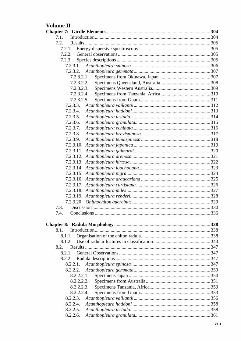
Volume II Chapter 7: Girdle Elements......................................................................................304
7.1. Introduction...............................................................................................304 7.2. Results .......................................................................................................305
7.2.1. Energy dispersive spectroscopy...........................................................305 7.2.2. General observations............................................................................305 7.2.3. Species descriptions .............................................................................305
7.2.3.1. Acanthopleura spinosa .................................................................306 7.2.3.2. Acanthopleura gemmata...............................................................307
7.2.3.2.1. Specimens from Okinawa, Japan ..........................................307 7.2.3.2.2. Specimens Queensland, Australia.........................................308 7.2.3.2.3. Specimens Western Australia................................................309 7.2.3.2.4. Specimens from Tanzania, Africa.........................................310 7.2.3.2.5. Specimens from Guam..........................................................311
7.2.3.3. Acanthopleura vaillantii...............................................................312 7.2.3.4. Acanthopleura haddoni ................................................................313 7.2.3.5. Acanthopleura testudo..................................................................314 7.2.3.6. Acanthopleura granulata..............................................................315 7.2.3.7. Acanthopleura echinata................................................................316 7.2.3.8. Acanthopleura brevispinosa.........................................................317 7.2.3.9. Acanthopleura tenuispinosa .........................................................318 7.2.3.10. Acanthopleura japonica ...............................................................319 7.2.3.11. Acanthopleura gaimardi...............................................................320 7.2.3.12. Acanthopleura arenosa.................................................................321 7.2.3.13. Acanthopleura hirtosa ..................................................................322 7.2.3.14. Acanthopleura loochooana...........................................................323 7.2.3.15. Acanthopleura nigra.....................................................................324 7.2.3.16. Acanthopleura araucariana .........................................................325 7.2.3.17. Acanthopleura curtisiana .............................................................326 7.2.3.18. Acanthopleura miles.....................................................................327 7.2.3.19. Acanthopleura rehderi..................................................................328 7.2.3.20. Onithochiton quercinus ................................................................329
7.3. Discussion .................................................................................................330 7.4. Conclusions ...............................................................................................336
Chapter 8: Radula Morphology ...............................................................................338
8.1. Introduction...............................................................................................338 8.1.1. Organisation of the chiton radula.........................................................338 8.1.2. Use of radular features in classification...............................................343
8.2. Results .......................................................................................................347 8.2.1. General Observations ...........................................................................347 8.2.2. Radula descriptions ..............................................................................347
8.2.2.1. Acanthopleura spinosa .................................................................347 8.2.2.2. Acanthopleura gemmata...............................................................350
8.2.2.2.1. Specimens Japan ...................................................................350 8.2.2.2.2. Specimens from Australia .....................................................351 8.2.2.2.3. Specimens Tanzania, Africa..................................................353 8.2.2.2.4. Specimens from Guam..........................................................353
8.2.2.3. Acanthopleura vaillantii...............................................................356 8.2.2.4. Acanthopleura haddoni ................................................................358 8.2.2.5. Acanthopleura testudo..................................................................358 8.2.2.6. Acanthopleura granulata..............................................................361
ix
8.2.2.7. Acanthopleura echinata................................................................363 8.2.2.7.1. Specimens from Peru ............................................................363 8.2.2.7.2. Specimens from Chile ...........................................................364
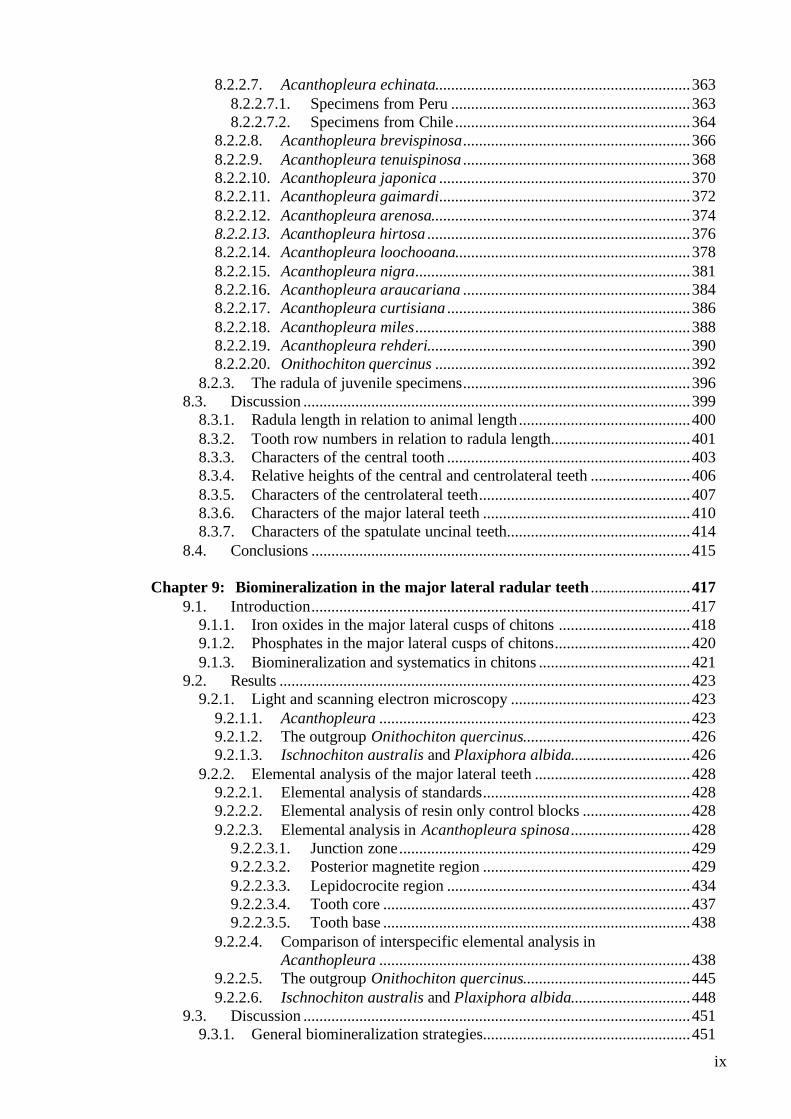
8.2.2.8. Acanthopleura brevispinosa.........................................................366 8.2.2.9. Acanthopleura tenuispinosa .........................................................368 8.2.2.10. Acanthopleura japonica ...............................................................370 8.2.2.11. Acanthopleura gaimardi...............................................................372 8.2.2.12. Acanthopleura arenosa.................................................................374 8.2.2.13. Acanthopleura hirtosa ..................................................................376 8.2.2.14. Acanthopleura loochooana...........................................................378 8.2.2.15. Acanthopleura nigra.....................................................................381 8.2.2.16. Acanthopleura araucariana .........................................................384 8.2.2.17. Acanthopleura curtisiana .............................................................386 8.2.2.18. Acanthopleura miles.....................................................................388 8.2.2.19. Acanthopleura rehderi..................................................................390 8.2.2.20. Onithochiton quercinus ................................................................392
8.2.3. The radula of juvenile specimens.........................................................396 8.3. Discussion .................................................................................................399
8.3.1. Radula length in relation to animal length...........................................400 8.3.2. Tooth row numbers in relation to radula length...................................401 8.3.3. Characters of the central tooth .............................................................403 8.3.4. Relative heights of the central and centrolateral teeth .........................406 8.3.5. Characters of the centrolateral teeth.....................................................407 8.3.6. Characters of the major lateral teeth ....................................................410 8.3.7. Characters of the spatulate uncinal teeth..............................................414
8.4. Conclusions ...............................................................................................415 Chapter 9: Biomineralization in the major lateral radular teeth .........................417
9.1. Introduction...............................................................................................417 9.1.1. Iron oxides in the major lateral cusps of chitons .................................418 9.1.2. Phosphates in the major lateral cusps of chitons..................................420 9.1.3. Biomineralization and systematics in chitons ......................................421
9.2. Results .......................................................................................................423 9.2.1. Light and scanning electron microscopy .............................................423
9.2.1.1. Acanthopleura ..............................................................................423 9.2.1.2. The outgroup Onithochiton quercinus..........................................426 9.2.1.3. Ischnochiton australis and Plaxiphora albida..............................426
9.2.2. Elemental analysis of the major lateral teeth .......................................428 9.2.2.1. Elemental analysis of standards....................................................428 9.2.2.2. Elemental analysis of resin only control blocks ...........................428 9.2.2.3. Elemental analysis in Acanthopleura spinosa..............................428
9.2.2.3.1. Junction zone.........................................................................429 9.2.2.3.2. Posterior magnetite region ....................................................429 9.2.2.3.3. Lepidocrocite region .............................................................434 9.2.2.3.4. Tooth core .............................................................................437 9.2.2.3.5. Tooth base .............................................................................438
9.2.2.4. Comparison of interspecific elemental analysis in Acanthopleura ..............................................................................438
9.2.2.5. The outgroup Onithochiton quercinus..........................................445 9.2.2.6. Ischnochiton australis and Plaxiphora albida..............................448
9.3. Discussion .................................................................................................451 9.3.1. General biomineralization strategies....................................................451
x
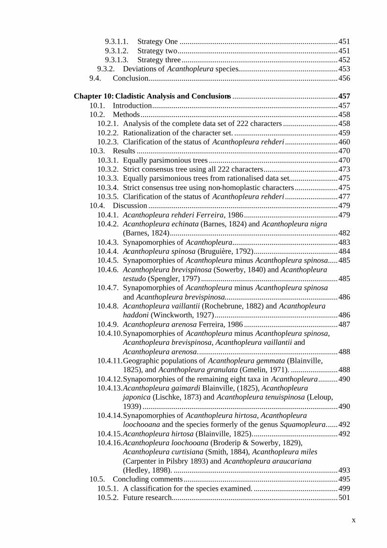
9.3.1.1. Strategy One .................................................................................451 9.3.1.2. Strategy two..................................................................................451 9.3.1.3. Strategy three................................................................................452
9.3.2. Deviations of Acanthopleura species...................................................453 9.4. Conclusion.................................................................................................456
Chapter 10: Cladistic Analysis and Conclusions ......................................................457
10.1. Introduction...............................................................................................457 10.2. Methods.....................................................................................................458
10.2.1. Analysis of the complete data set of 222 characters ............................458 10.2.2. Rationalization of the character set. .....................................................459 10.2.3. Clarification of the status of Acanthopleura rehderi ...........................460
10.3. Results .......................................................................................................470 10.3.1. Equally parsimonious trees ..................................................................470 10.3.2. Strict consensus tree using all 222 characters......................................473 10.3.3. Equally parsimonious trees from rationalised data set.........................475 10.3.4. Strict consensus tree using non-homoplastic characters ......................475 10.3.5. Clarification of the status of Acanthopleura rehderi ...........................477
10.4. Discussion .................................................................................................479 10.4.1. Acanthopleura rehderi Ferreira, 1986................................................479 10.4.2. Acanthopleura echinata (Barnes, 1824) and Acanthopleura nigra
(Barnes, 1824)......................................................................................482 10.4.3. Synapomorphies of Acanthopleura......................................................483 10.4.4. Acanthopleura spinosa (Bruguière, 1792)...........................................484 10.4.5. Synapomorphies of Acanthopleura minus Acanthopleura spinosa.....485 10.4.6. Acanthopleura brevispinosa (Sowerby, 1840) and Acanthopleura
testudo (Spengler, 1797) ......................................................................485 10.4.7. Synapomorphies of Acanthopleura minus Acanthopleura spinosa
and Acanthopleura brevispinosa..........................................................486 10.4.8. Acanthopleura vaillantii (Rochebrune, 1882) and Acanthopleura
haddoni (Winckworth, 1927)...............................................................486 10.4.9. Acanthopleura arenosa Ferreira, 1986 ................................................487 10.4.10. Synapomorphies of Acanthopleura minus Acanthopleura spinosa,
Acanthopleura brevispinosa, Acanthopleura vaillantii and Acanthopleura arenosa........................................................................488
10.4.11. Geographic populations of Acanthopleura gemmata (Blainville, 1825), and Acanthopleura granulata (Gmelin, 1971). ........................488
10.4.12. Synapomorphies of the remaining eight taxa in Acanthopleura..........490 10.4.13. Acanthopleura gaimardi Blainville, (1825), Acanthopleura
japonica (Lischke, 1873) and Acanthopleura tenuispinosa (Leloup, 1939) ....................................................................................................490
10.4.14. Synapomorphies of Acanthopleura hirtosa, Acanthopleura loochooana and the species formerly of the genus Squamopleura......492
10.4.15. Acanthopleura hirtosa (Blainville, 1825)............................................492 10.4.16. Acanthopleura loochooana (Broderip & Sowerby, 1829),
Acanthopleura curtisiana (Smith, 1884), Acanthopleura miles (Carpenter in Pilsbry 1893) and Acanthopleura araucariana (Hedley, 1898). ....................................................................................493
10.5. Concluding comments ...............................................................................495 10.5.1. A classification for the species examined. ...........................................499 10.5.2. Future research.....................................................................................501
xi
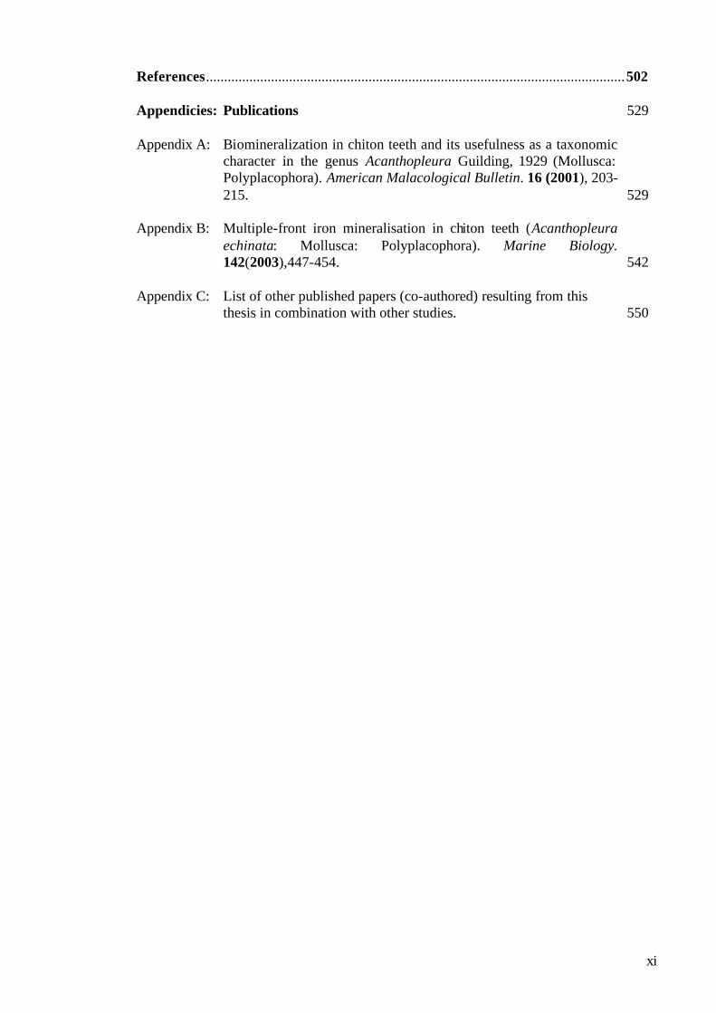
References....................................................................................................................502 Appendicies: Publications 529 Appendix A: Biomineralization in chiton teeth and its usefulness as a taxonomic
character in the genus Acanthopleura Guilding, 1929 (Mollusca: Polyplacophora). American Malacological Bulletin. 16 (2001), 203-215. 529
Appendix B: Multiple-front iron mineralisation in chiton teeth (Acanthopleura
echinata: Mollusca: Polyplacophora). Marine Biology. 142(2003),447-454. 542
Appendix C: List of other published papers (co-authored) resulting from this
thesis in combination with other studies. 550
xii
Acknowledgements
This research could not have been undertaken without the assistance of a large
number of people and I hope that in these acknowledgements I do not
inadvertently omit any of them.
I would like to thank my supervisors, Assoc. Prof. David Macey and Dr Fred
Wells for their advice, comments, never ending encouragement and, above all,
their friendship.
Foremost, my sincerest thanks go to my family, Keith, Graham, Rita, David and
Jane, for without their love and support I would not have had the fortitude to
complete this thesis. Especial thanks go to my husband, Keith, whose
encouragement and patience have sustained me, and to my son Graham, for whom
this PhD has been ongoing for most of his life. I am also grateful to them both for
their assistance in the collection of specimens from around the coast of Australia,
for without Keith risking “life and limb” in the surf, and without Graham’s keen
young eyes, my specimen collection would not have been complete.
The Division of Science and Engineering, at Murdoch University, abounds with
people who are liberal with their knowledge and expertise, and I am indebted to
many of them. My special thanks to Dr. Howard Gill for his guidance, generous
advice and corrections to the cladistics section of my thesis. My thanks also to
Alasdair Lee, for his preparation of the standard materials used in the
biomineralization section of the thesis, for his critical reading of many sections of
the thesis, and finally, for his contribution to the many papers we have published
together. Also my thanks go to Gordon Thomson and Peter Fallon, for their
assistance with histological and electron microscopical aspects of the study.
Special thanks go to the many friends and colleagues who have provided me with
their support, and more than a few laughs, over the years: to my students, Garth,
Jeremy and Imogen; and to all the people who asked how the thesis was going,
even if most of them were primed by my husband!
xiii
The collection of specimens, from a genus that is distributed from tropical and
temperate regions throughout the world, would not have been possible without the
assistance of many people. My sincere thanks go to Clay Bryce, of the Western
Australian Museum, who took receipt, and responsibility, for all of my
internationally acquired specimens. I was fortunate to be able to post a request for
specimens to the web site of the Mollusca list, located at Berkeley University,
USA, and was rewarded by an extremely generous international community.
Although the list of people who supplied me with ethanol preserved specimens of
Acanthopleura is long, I would not like to miss any of them out:
Julia Bell, Robert Bolland, Jason Court, Richer de Forges; Hiroshi Fukuda, Nan
Hauser, Heinz Hilbrecht, John Huisman, Lisa Kirkendale, Bruce Livett, Katherine
Liddiard, James Hockridge, Yasuhiro Nakamura, Cecelia Osorio, Hoyt Peckham,
Carlos Ramirez La Torre, Matthew Richmond, Hiroshi Saito, Jeremy Shaw,
Frances Stanley, Hermann Strack, John Taylor, Mary Villaume, John Webb, John
Wise and Hiroshi Yoshizaki.
I would like to mention various institutions, and thank their curators, for the loan
of museum specimens: Fred Wells and the Western Australian Museum; Richard
Kilburn and the Natal Museum; Robert van Syoc and the California Academy of
Sciences; Kathie Way and the British Museum of Natural History; Cheryl Bright
and the U.S. national Museum of Natural History; Georgia Cunningham and the
Victorian Museum; and the Leiden Museum.
I would like to thank the American Malacological Society (AMS) for monies from
the “Bernice Barbour AMS Travel Award”, which assisted my attendance and
presentation of a paper at the AMS 65th Annual Meeting in Pittsburgh (1999). My
thanks also to the Australian Marine Sciences Association for the International
Student Prize (1999), which enabled me to undertake fieldwork in the Cook
Islands, and facilitated the collection of rare specimens from this region. I am also
grateful to Gerald McCormack, for his hospitality and assistance on the Cook
Islands, and for supplying me with valuable contacts.
This research was partially funded by an Australian Research Council Small Grant
and a Murdoch University Research Infrastructure Grant. Many thanks also to
Murdoch University for a Murdoch University Research Scholarship from 1996 to
1999.

Figure 5.1 Light micrographs showing the variation in the development of the
insertion plate of the posterior valve of Acanthopleura species.
A, Acanthopleura spinosa showing the toothed insertion plate (ip) with
distinct slits (dashed arrows) and transverse callus (c).
B, A. japonica showing the broad flat insertion plate with round callus just
below it.
C and D, A. brevispinosa and A. arenosa displaying an intermediate
condition with the insertion plate poorly developed, but not flat, and few
slits (dashed arrows) that are degenerate in the centre of the valve.
E, F and G, specimens of A. gaimardi, A. japonica and a juvenile
A. hirtosa exhibiting two small symmetrical notches either side of the
valve (arrows).
All scale bars = 2mm.
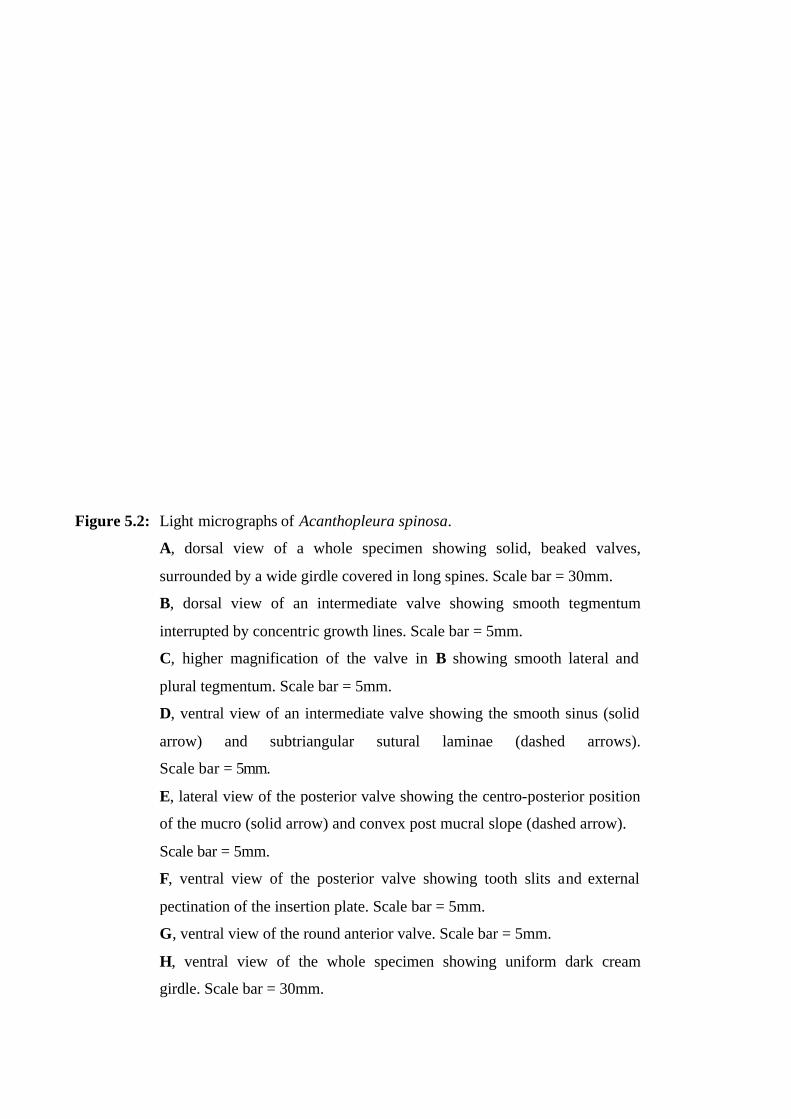
Figure 5.2: Light micrographs of Acanthopleura spinosa.
A, dorsal view of a whole specimen showing solid, beaked valves,
surrounded by a wide girdle covered in long spines. Scale bar = 30mm.
B, dorsal view of an intermediate valve showing smooth tegmentum
interrupted by concentric growth lines. Scale bar = 5mm.
C, higher magnification of the valve in B showing smooth lateral and
plural tegmentum. Scale bar = 5mm.
D, ventral view of an intermediate valve showing the smooth sinus (solid
arrow) and subtriangular sutural laminae (dashed arrows).
Scale bar = 5mm.
E, lateral view of the posterior valve showing the centro-posterior position
of the mucro (solid arrow) and convex post mucral slope (dashed arrow).
Scale bar = 5mm.
F, ventral view of the posterior valve showing tooth slits and external
pectination of the insertion plate. Scale bar = 5mm.
G, ventral view of the round anterior valve. Scale bar = 5mm.
H, ventral view of the whole specimen showing uniform dark cream
girdle. Scale bar = 30mm.
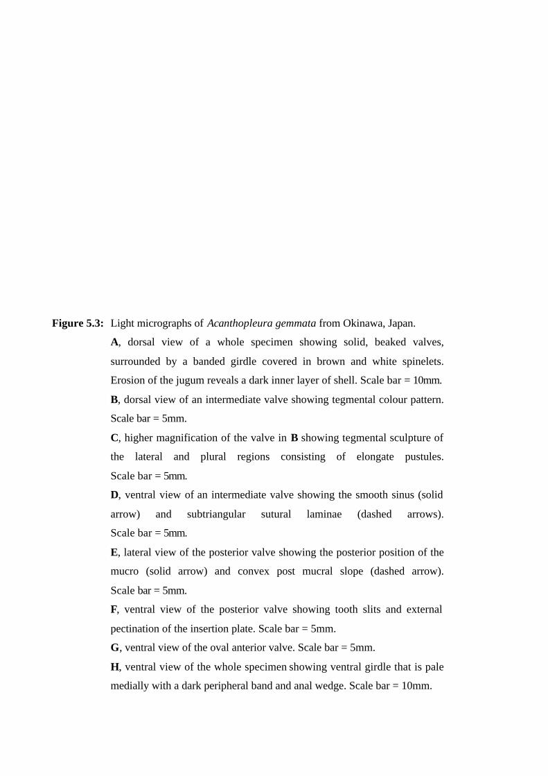
Figure 5.3: Light micrographs of Acanthopleura gemmata from Okinawa, Japan.
A, dorsal view of a whole specimen showing solid, beaked valves,
surrounded by a banded girdle covered in brown and white spinelets.
Erosion of the jugum reveals a dark inner layer of shell. Scale bar = 10mm.
B, dorsal view of an intermediate valve showing tegmental colour pattern.
Scale bar = 5mm.
C, higher magnification of the valve in B showing tegmental sculpture of
the lateral and plural regions consisting of elongate pustules.
Scale bar = 5mm.
D, ventral view of an intermediate valve showing the smooth sinus (solid
arrow) and subtriangular sutural laminae (dashed arrows).
Scale bar = 5mm.
E, lateral view of the posterior valve showing the posterior position of the
mucro (solid arrow) and convex post mucral slope (dashed arrow).
Scale bar = 5mm.
F, ventral view of the posterior valve showing tooth slits and external
pectination of the insertion plate. Scale bar = 5mm.
G, ventral view of the oval anterior valve. Scale bar = 5mm.
H, ventral view of the whole specimen showing ventral girdle that is pale
medially with a dark peripheral band and anal wedge. Scale bar = 10mm.
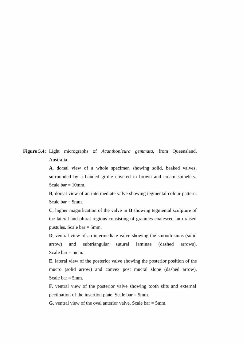
Figure 5.4: Light micrographs of Acanthopleura gemmata, from Queensland,
Australia.
A, dorsal view of a whole specimen showing solid, beaked valves,
surrounded by a banded girdle covered in brown and cream spinelets.
Scale bar = 10mm.
B, dorsal view of an intermediate valve showing tegmental colour pattern.
Scale bar = 5mm.
C, higher magnification of the valve in B showing tegmental sculpture of
the lateral and plural regions consisting of granules coalesced into raised
pustules. Scale bar = 5mm.
D, ventral view of an intermediate valve showing the smooth sinus (solid
arrow) and subtriangular sutural laminae (dashed arrows).
Scale bar = 5mm.
E, lateral view of the posterior valve showing the posterior position of the
mucro (solid arrow) and convex post mucral slope (dashed arrow).
Scale bar = 5mm.
F, ventral view of the posterior valve showing tooth slits and external
pectination of the insertion plate. Scale bar = 5mm.
G, ventral view of the oval anterior valve. Scale bar = 5mm.
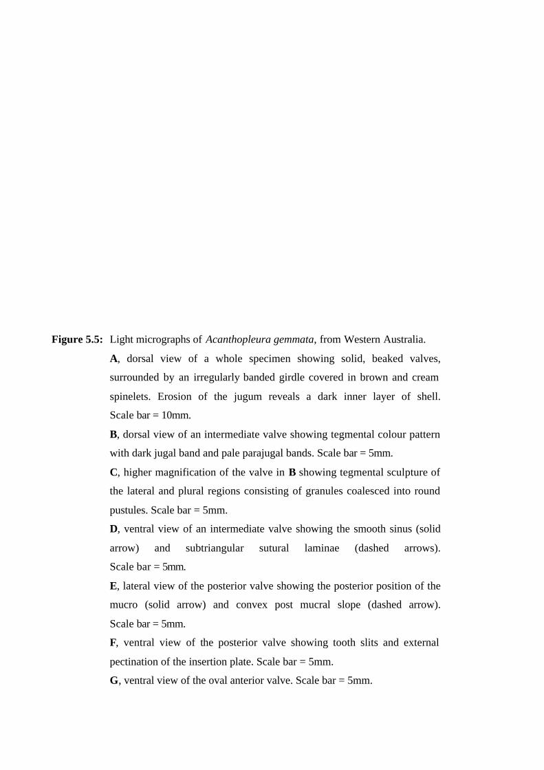
Figure 5.5: Light micrographs of Acanthopleura gemmata, from Western Australia.
A, dorsal view of a whole specimen showing solid, beaked valves,
surrounded by an irregularly banded girdle covered in brown and cream
spinelets. Erosion of the jugum reveals a dark inner layer of shell.
Scale bar = 10mm.
B, dorsal view of an intermediate valve showing tegmental colour pattern
with dark jugal band and pale parajugal bands. Scale bar = 5mm.
C, higher magnification of the valve in B showing tegmental sculpture of
the lateral and plural regions consisting of granules coalesced into round
pustules. Scale bar = 5mm.
D, ventral view of an intermediate valve showing the smooth sinus (solid
arrow) and subtriangular sutural laminae (dashed arrows).
Scale bar = 5mm.
E, lateral view of the posterior valve showing the posterior position of the
mucro (solid arrow) and convex post mucral slope (dashed arrow).
Scale bar = 5mm.
F, ventral view of the posterior valve showing tooth slits and external
pectination of the insertion plate. Scale bar = 5mm.
G, ventral view of the oval anterior valve. Scale bar = 5mm.

Figure 5.6: Light micrographs of Acanthopleura gemmata from Tanzania.
A, dorsal view of a whole specimen showing solid, beaked valves,
surrounded by an irregularly banded girdle covered in brown and cream
spinelets. Erosion of the jugum reveals a dark inner layer of shell.
Scale bar = 10mm.
B, dorsal view of an intermediate valve showing tegmental colour pattern
with dark jugal band and pale parajugal bands. Scale bar = 5mm.
C, higher magnification of the valve in B showing tegmental sculpture of
the lateral and plural regions consisting of flat-topped round to elongate
pustules. Scale bar = 5mm.
D, ventral view of an intermediate valve showing the smooth sinus (solid
arrow) and subtriangular sutural laminae (dashed arrows).
Scale bar = 5mm.
E, lateral view of the posterior valve showing the centro-posterior position
of the mucro (solid arrow) and convex post mucral slope (dashed arrow).
Scale bar = 5mm.
F, ventral view of the posterior valve showing tooth slits and external
pectination of the insertion plate. Scale bar = 5mm.
G, ventral view of the oval anterior valve. Scale bar = 5mm.
H, ventral view of the whole specimen showing ventral girdle that is pale
medially with a dark peripheral band and anal wedge. Scale bar = 10mm.
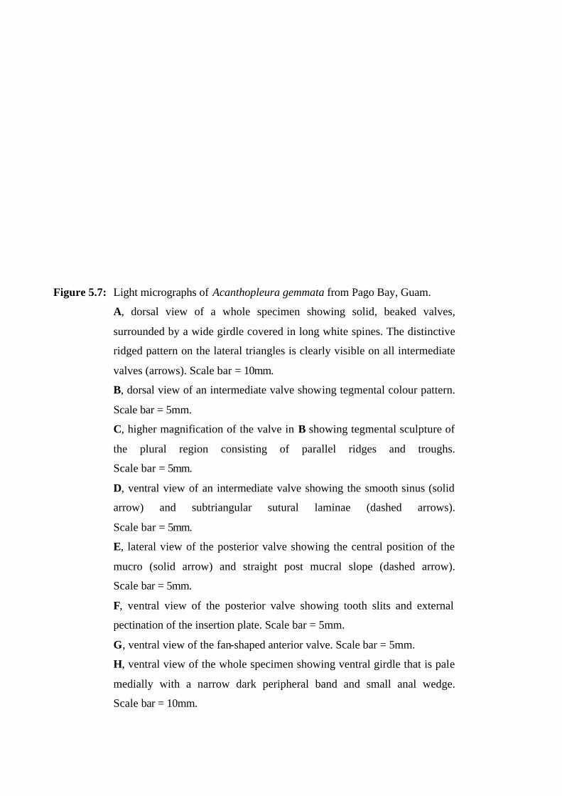
Figure 5.7: Light micrographs of Acanthopleura gemmata from Pago Bay, Guam.
A, dorsal view of a whole specimen showing solid, beaked valves,
surrounded by a wide girdle covered in long white spines. The distinctive
ridged pattern on the lateral triangles is clearly visible on all intermediate
valves (arrows). Scale bar = 10mm.
B, dorsal view of an intermediate valve showing tegmental colour pattern.
Scale bar = 5mm.
C, higher magnification of the valve in B showing tegmental sculpture of
the plural region consisting of parallel ridges and troughs.
Scale bar = 5mm.
D, ventral view of an intermediate valve showing the smooth sinus (solid
arrow) and subtriangular sutural laminae (dashed arrows).
Scale bar = 5mm.
E, lateral view of the posterior valve showing the central position of the
mucro (solid arrow) and straight post mucral slope (dashed arrow).
Scale bar = 5mm.
F, ventral view of the posterior valve showing tooth slits and external
pectination of the insertion plate. Scale bar = 5mm.
G, ventral view of the fan-shaped anterior valve. Scale bar = 5mm.
H, ventral view of the whole specimen showing ventral girdle that is pale
medially with a narrow dark peripheral band and small anal wedge.
Scale bar = 10mm.
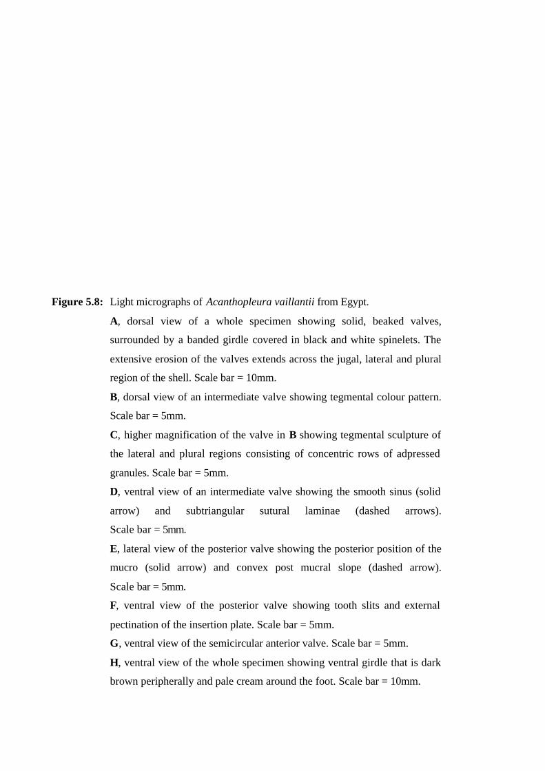
Figure 5.8: Light micrographs of Acanthopleura vaillantii from Egypt.
A, dorsal view of a whole specimen showing solid, beaked valves,
surrounded by a banded girdle covered in black and white spinelets. The
extensive erosion of the valves extends across the jugal, lateral and plural
region of the shell. Scale bar = 10mm.
B, dorsal view of an intermediate valve showing tegmental colour pattern.
Scale bar = 5mm.
C, higher magnification of the valve in B showing tegmental sculpture of
the lateral and plural regions consisting of concentric rows of adpressed
granules. Scale bar = 5mm.
D, ventral view of an intermediate valve showing the smooth sinus (solid
arrow) and subtriangular sutural laminae (dashed arrows).
Scale bar = 5mm.
E, lateral view of the posterior valve showing the posterior position of the
mucro (solid arrow) and convex post mucral slope (dashed arrow).
Scale bar = 5mm.
F, ventral view of the posterior valve showing tooth slits and external
pectination of the insertion plate. Scale bar = 5mm.
G, ventral view of the semicircular anterior valve. Scale bar = 5mm.
H, ventral view of the whole specimen showing ventral girdle that is dark
brown peripherally and pale cream around the foot. Scale bar = 10mm.
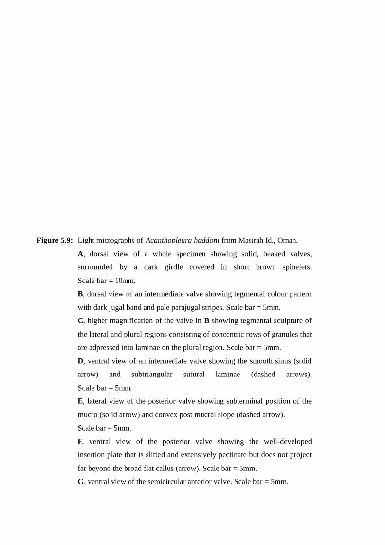
Figure 5.9: Light micrographs of Acanthopleura haddoni from Masirah Id., Oman.
A, dorsal view of a whole specimen showing solid, beaked valves,
surrounded by a dark girdle covered in short brown spinelets.
Scale bar = 10mm.
B, dorsal view of an intermediate valve showing tegmental colour pattern
with dark jugal band and pale parajugal stripes. Scale bar = 5mm.
C, higher magnification of the valve in B showing tegmental sculpture of
the lateral and plural regions consisting of concentric rows of granules that
are adpressed into laminae on the plural region. Scale bar = 5mm.
D, ventral view of an intermediate valve showing the smooth sinus (solid
arrow) and subtriangular sutural laminae (dashed arrows).
Scale bar = 5mm.
E, lateral view of the posterior valve showing subterminal position of the
mucro (solid arrow) and convex post mucral slope (dashed arrow).
Scale bar = 5mm.
F, ventral view of the posterior valve showing the well-developed
insertion plate that is slitted and extensively pectinate but does not project
far beyond the broad flat callus (arrow). Scale bar = 5mm.
G, ventral view of the semicircular anterior valve. Scale bar = 5mm.
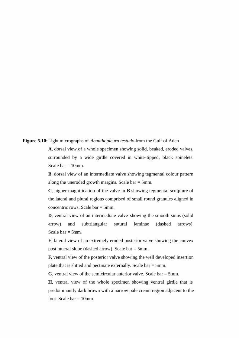
Figure 5.10: Light micrographs of Acanthopleura testudo from the Gulf of Aden.
A, dorsal view of a whole specimen showing solid, beaked, eroded valves,
surrounded by a wide girdle covered in white-tipped, black spinelets.
Scale bar = 10mm.
B, dorsal view of an intermediate valve showing tegmental colour pattern
along the uneroded growth margins. Scale bar = 5mm.
C, higher magnification of the valve in B showing tegmental sculpture of
the lateral and plural regions comprised of small round granules aligned in
concentric rows. Scale bar = 5mm.
D, ventral view of an intermediate valve showing the smooth sinus (solid
arrow) and subtriangular sutural laminae (dashed arrows).
Scale bar = 5mm.
E, lateral view of an extremely eroded posterior valve showing the convex
post mucral slope (dashed arrow). Scale bar = 5mm.
F, ventral view of the posterior valve showing the well developed insertion
plate that is slitted and pectinate externally. Scale bar = 5mm.
G, ventral view of the semicircular anterior valve. Scale bar = 5mm.
H, ventral view of the whole specimen showing ventral girdle that is
predominantly dark brown with a narrow pale cream region adjacent to the
foot. Scale bar = 10mm.

Figure 5.11: Light micrographs of Acanthopleura granulata from Florida Keys, USA.
A, dorsal view of a whole specimen showing solid, beaked valves,
surrounded by a banded girdle covered in short brown and cream spinelets.
Scale bar = 10mm.
B, dorsal view of an intermediate valve showing tegmental colour pattern
that is predominantly pale brown with a brown jugal band and broad,
salmon pink parajugal bands. Scale bar = 5mm.
C, higher magnification of the valve in B showing tegmental sculpture of
the lateral and plural regions comprised of concentric rows of round
granules that coalesce into elongate pustules on the lateral triangle.
Scale bar = 5mm.
D, ventral view of an intermediate valve showing the smooth sinus (solid
arrow) and subtriangular sutural laminae (dashed arrows).
Scale bar = 5mm.
E, lateral view of the posterior valve showing the centro-posterior position
of the mucro (solid arrow) and convex post mucral slope (dashed arrow).
Scale bar = 5mm.
F, ventral view of the posterior valve showing the well developed insertion
plate that is slitted and pectinate externally. Scale bar = 5mm.
G, ventral view of the fan-shaped anterior valve. Scale bar = 5mm.
H, ventral view of a whole specimen showing ventral girdle that is cream
medially and brown peripherally with a dark anal wedge.
Scale bar = 10mm.
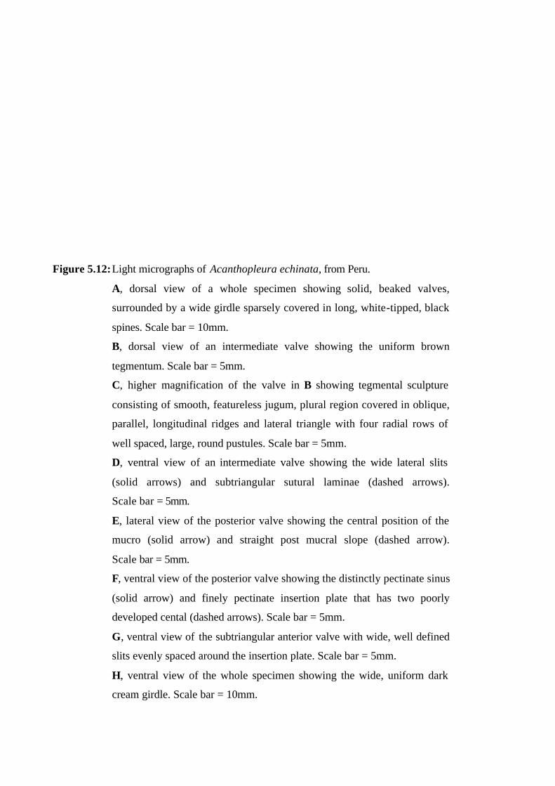
Figure 5.12: Light micrographs of Acanthopleura echinata, from Peru.
A, dorsal view of a whole specimen showing solid, beaked valves,
surrounded by a wide girdle sparsely covered in long, white-tipped, black
spines. Scale bar = 10mm.
B, dorsal view of an intermediate valve showing the uniform brown
tegmentum. Scale bar = 5mm.
C, higher magnification of the valve in B showing tegmental sculpture
consisting of smooth, featureless jugum, plural region covered in oblique,
parallel, longitudinal ridges and lateral triangle with four radial rows of
well spaced, large, round pustules. Scale bar = 5mm.
D, ventral view of an intermediate valve showing the wide lateral slits
(solid arrows) and subtriangular sutural laminae (dashed arrows).
Scale bar = 5mm.
E, lateral view of the posterior valve showing the central position of the
mucro (solid arrow) and straight post mucral slope (dashed arrow).
Scale bar = 5mm.
F, ventral view of the posterior valve showing the distinctly pectinate sinus
(solid arrow) and finely pectinate insertion plate that has two poorly
developed cental (dashed arrows). Scale bar = 5mm.
G, ventral view of the subtriangular anterior valve with wide, well defined
slits evenly spaced around the insertion plate. Scale bar = 5mm.
H, ventral view of the whole specimen showing the wide, uniform dark
cream girdle. Scale bar = 10mm.
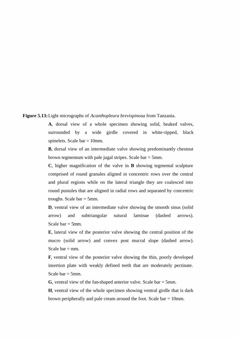
Figure 5.13: Light micrographs of Acanthopleura brevispinosa from Tanzania.
A, dorsal view of a whole specimen showing solid, beaked valves,
surrounded by a wide girdle covered in white-tipped, black
spinelets. Scale bar = 10mm.
B, dorsal view of an intermediate valve showing predominantly chestnut
brown tegmentum with pale jugal stripes. Scale bar = 5mm.
C, higher magnification of the valve in B showing tegmental sculpture
comprised of round granules aligned in concentric rows over the central
and plural regions while on the lateral triangle they are coalesced into
round pustules that are aligned in radial rows and separated by concentric
troughs. Scale bar = 5mm.
D, ventral view of an intermediate valve showing the smooth sinus (solid
arrow) and subtriangular sutural laminae (dashed arrows).
Scale bar = 5mm.
E, lateral view of the posterior valve showing the central position of the
mucro (solid arrow) and convex post mucral slope (dashed arrow).
Scale bar = mm.
F, ventral view of the posterior valve showing the thin, poorly developed
insertion plate with weakly defined teeth that are moderately pectinate.
Scale bar = 5mm.
G, ventral view of the fan-shaped anterior valve. Scale bar = 5mm.
H, ventral view of the whole specimen showing ventral girdle that is dark
brown peripherally and pale cream around the foot. Scale bar = 10mm.
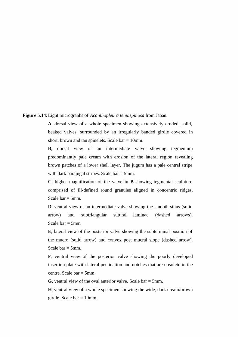
Figure 5.14: Light micrographs of Acanthopleura tenuispinosa from Japan.
A, dorsal view of a whole specimen showing extensively eroded, solid,
beaked valves, surrounded by an irregularly banded girdle covered in
short, brown and tan spinelets. Scale bar = 10mm.
B, dorsal view of an intermediate valve showing tegmentum
predominantly pale cream with erosion of the lateral region revealing
brown patches of a lower shell layer. The jugum has a pale central stripe
with dark parajugal stripes. Scale bar = 5mm.
C, higher magnification of the valve in B showing tegmental sculpture
comprised of ill-defined round granules aligned in concentric ridges.
Scale bar = 5mm.
D, ventral view of an intermediate valve showing the smooth sinus (solid
arrow) and subtriangular sutural laminae (dashed arrows).
Scale bar = 5mm.
E, lateral view of the posterior valve showing the subterminal position of
the mucro (solid arrow) and convex post mucral slope (dashed arrow).
Scale bar = 5mm.
F, ventral view of the posterior valve showing the poorly developed
insertion plate with lateral pectination and notches that are obsolete in the
centre. Scale bar = 5mm.
G, ventral view of the oval anterior valve. Scale bar = 5mm.
H, ventral view of a whole specimen showing the wide, dark cream/brown
girdle. Scale bar = 10mm.
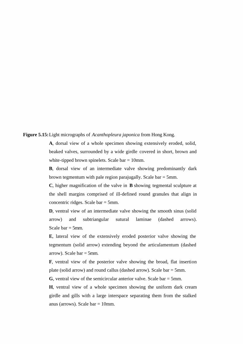
Figure 5.15: Light micrographs of Acanthopleura japonica from Hong Kong.
A, dorsal view of a whole specimen showing extensively eroded, solid,
beaked valves, surrounded by a wide girdle covered in short, brown and
white-tipped brown spinelets. Scale bar = 10mm.
B, dorsal view of an intermediate valve showing predominantly dark
brown tegmentum with pale region parajugally. Scale bar = 5mm.
C, higher magnification of the valve in B showing tegmental sculpture at
the shell margins comprised of ill-defined round granules that align in
concentric ridges. Scale bar = 5mm.
D, ventral view of an intermediate valve showing the smooth sinus (solid
arrow) and subtriangular sutural laminae (dashed arrows).
Scale bar = 5mm.
E, lateral view of the extensively eroded posterior valve showing the
tegmentum (solid arrow) extending beyond the articulamentum (dashed
arrow). Scale bar = 5mm.
F, ventral view of the posterior valve showing the broad, flat insertion
plate (solid arrow) and round callus (dashed arrow). Scale bar = 5mm.
G, ventral view of the semicircular anterior valve. Scale bar = 5mm.
H, ventral view of a whole specimen showing the uniform dark cream
girdle and gills with a large interspace separating them from the stalked
anus (arrows). Scale bar = 10mm.
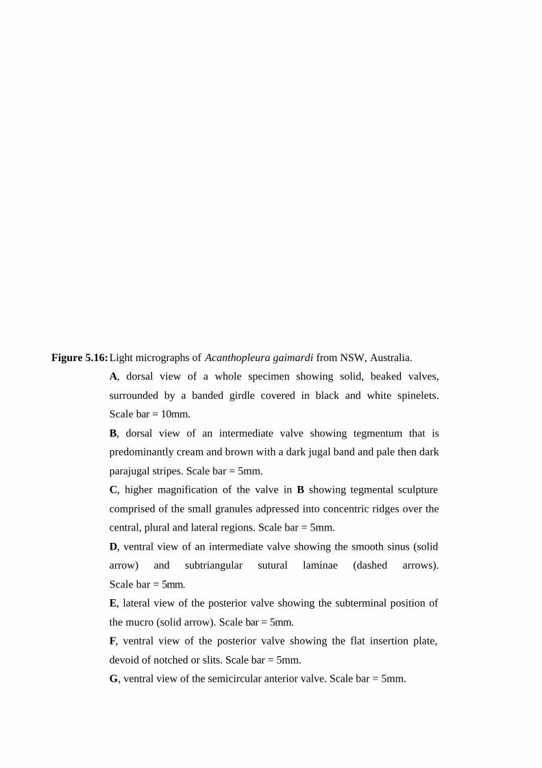
Figure 5.16: Light micrographs of Acanthopleura gaimardi from NSW, Australia.
A, dorsal view of a whole specimen showing solid, beaked valves,
surrounded by a banded girdle covered in black and white spinelets.
Scale bar = 10mm.
B, dorsal view of an intermediate valve showing tegmentum that is
predominantly cream and brown with a dark jugal band and pale then dark
parajugal stripes. Scale bar = 5mm.
C, higher magnification of the valve in B showing tegmental sculpture
comprised of the small granules adpressed into concentric ridges over the
central, plural and lateral regions. Scale bar = 5mm.
D, ventral view of an intermediate valve showing the smooth sinus (solid
arrow) and subtriangular sutural laminae (dashed arrows).
Scale bar = 5mm.
E, lateral view of the posterior valve showing the subterminal position of
the mucro (solid arrow). Scale bar = 5mm.
F, ventral view of the posterior valve showing the flat insertion plate,
devoid of notched or slits. Scale bar = 5mm.
G, ventral view of the semicircular anterior valve. Scale bar = 5mm.

Figure 5.17: Light micrographs of Acanthopleura arenosa from Qld., Australia.
A, dorsal view of a whole specimen showing solid, beaked valves,
surrounded by an irregularly banded girdle covered in brown and cream
spinelets. Scale bar = 10mm.
B, dorsal view of an intermediate valve showing brown tegmentum with a
dark brown jugal band and broad, pale parajugal bands. Scale bar = 5mm.
C, higher magnification of the valve in B showing tegmental sculpture
comprised of ill defined, round granules aligned in concentric ridges over
the central and plural regions and coalesced into randomly scattered, round
to elongate pustules over the lateral regions. Scale bar = 5mm.
D, ventral view of an intermediate valve showing the smooth sinus (solid
arrow) and subtriangular sutural laminae (dashed arrows).
Scale bar = 5mm.
E, lateral view of the posterior valve showing the centro-posterior position
of the mucro (solid arrow) and convex post mucral slope (dashed arrow).
Scale bar = 5mm.
F, ventral view of the posterior valve showing the thin, poorly developed
insertion plate with incomplete slits laterally. Scale bar = 5mm.
G, ventral view of the oval anterior valve. Scale bar = 5mm.
H, ventral view of a whole specimen showing the uniform cream girdle.
Scale bar = 10mm.

Figure 5.18: Light micrographs of Acanthopleura hirtosa from Western Australia.
A, dorsal view of a whole specimen showing solid, beaked valves,
surrounded by a banded girdle covered in black and white scales.
Scale bar = 10mm.
B, dorsal view of an intermediate valve showing tegmentum that is
predominantly black and brown with a dark jugal band and creamy yellow
parajugal stripe. Scale bar = 5mm.
C, higher magnification of the valve in B showing tegmental sculpture
comprised of low profile elongate pustules that form into concentric ridges
on the central region, and aligned in concentric rows on the lateral and
plural regions. Scale bar = 5mm.
D, ventral view of an intermediate valve showing the smooth sinus (solid
arrow) and subtriangular sutural laminae (dashed arrows).
Scale bar = 5mm.
E, lateral view of the posterior valve showing the subterminal position of
the mucro (solid arrow) and convex post mucral slope (dashed arrow).
Scale bar = 5mm.
F, ventral view of the posterior valve showing the flat insertion plate
(dashed arrow) with a thin ridge along the posterior margin (arrow). Scale
bar = 5mm.
G, ventral view of the fan-shaped anterior valve. Scale bar = 5mm.
H, ventral view of a whole specimen showing the uniform cream girdle.
Scale bar = 10mm.
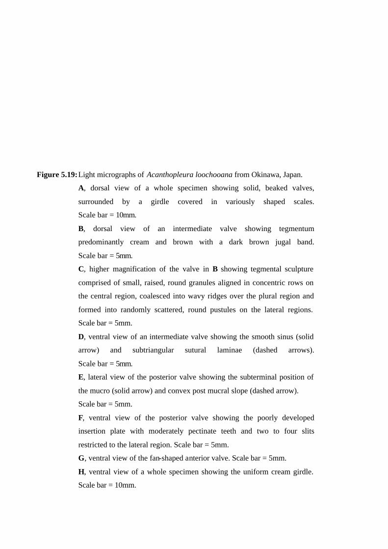
Figure 5.19: Light micrographs of Acanthopleura loochooana from Okinawa, Japan.
A, dorsal view of a whole specimen showing solid, beaked valves,
surrounded by a girdle covered in variously shaped scales.
Scale bar = 10mm.
B, dorsal view of an intermediate valve showing tegmentum
predominantly cream and brown with a dark brown jugal band.
Scale bar = 5mm.
C, higher magnification of the valve in B showing tegmental sculpture
comprised of small, raised, round granules aligned in concentric rows on
the central region, coalesced into wavy ridges over the plural region and
formed into randomly scattered, round pustules on the lateral regions.
Scale bar = 5mm.
D, ventral view of an intermediate valve showing the smooth sinus (solid
arrow) and subtriangular sutural laminae (dashed arrows).
Scale bar = 5mm.
E, lateral view of the posterior valve showing the subterminal position of
the mucro (solid arrow) and convex post mucral slope (dashed arrow).
Scale bar = 5mm.
F, ventral view of the posterior valve showing the poorly developed
insertion plate with moderately pectinate teeth and two to four slits
restricted to the lateral region. Scale bar = 5mm.
G, ventral view of the fan-shaped anterior valve. Scale bar = 5mm.
H, ventral view of a whole specimen showing the uniform cream girdle.
Scale bar = 10mm.
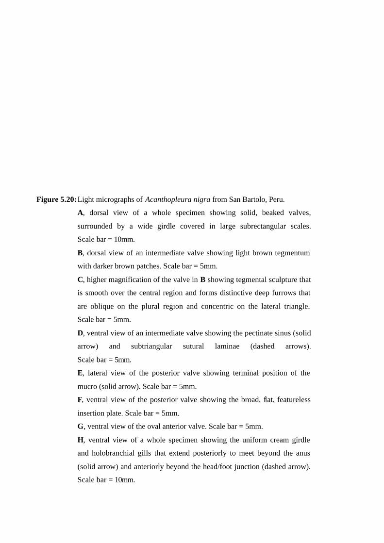
Figure 5.20: Light micrographs of Acanthopleura nigra from San Bartolo, Peru.
A, dorsal view of a whole specimen showing solid, beaked valves,
surrounded by a wide girdle covered in large subrectangular scales.
Scale bar = 10mm.
B, dorsal view of an intermediate valve showing light brown tegmentum
with darker brown patches. Scale bar = 5mm.
C, higher magnification of the valve in B showing tegmental sculpture that
is smooth over the central region and forms distinctive deep furrows that
are oblique on the plural region and concentric on the lateral triangle.
Scale bar = 5mm.
D, ventral view of an intermediate valve showing the pectinate sinus (solid
arrow) and subtriangular sutural laminae (dashed arrows).
Scale bar = 5mm.
E, lateral view of the posterior valve showing terminal position of the
mucro (solid arrow). Scale bar = 5mm.
F, ventral view of the posterior valve showing the broad, flat, featureless
insertion plate. Scale bar = 5mm.
G, ventral view of the oval anterior valve. Scale bar = 5mm.
H, ventral view of a whole specimen showing the uniform cream girdle
and holobranchial gills that extend posteriorly to meet beyond the anus
(solid arrow) and anteriorly beyond the head/foot junction (dashed arrow).
Scale bar = 10mm.
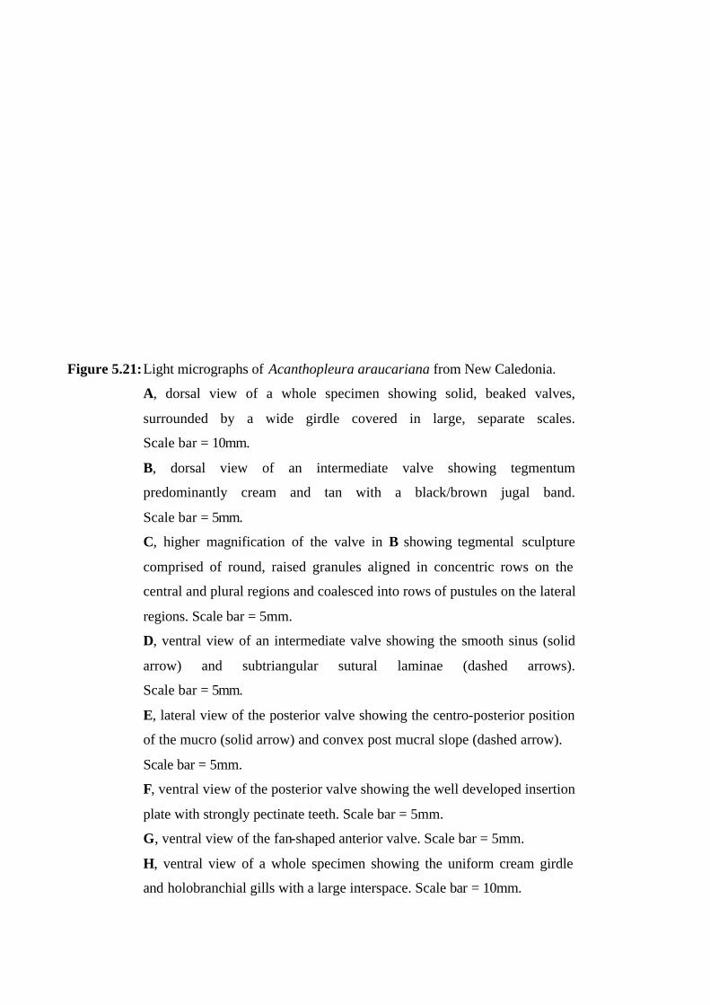
Figure 5.21: Light micrographs of Acanthopleura araucariana from New Caledonia.
A, dorsal view of a whole specimen showing solid, beaked valves,
surrounded by a wide girdle covered in large, separate scales.
Scale bar = 10mm.
B, dorsal view of an intermediate valve showing tegmentum
predominantly cream and tan with a black/brown jugal band.
Scale bar = 5mm.
C, higher magnification of the valve in B showing tegmental sculpture
comprised of round, raised granules aligned in concentric rows on the
central and plural regions and coalesced into rows of pustules on the lateral
regions. Scale bar = 5mm.
D, ventral view of an intermediate valve showing the smooth sinus (solid
arrow) and subtriangular sutural laminae (dashed arrows).
Scale bar = 5mm.
E, lateral view of the posterior valve showing the centro-posterior position
of the mucro (solid arrow) and convex post mucral slope (dashed arrow).
Scale bar = 5mm.
F, ventral view of the posterior valve showing the well developed insertion
plate with strongly pectinate teeth. Scale bar = 5mm.
G, ventral view of the fan-shaped anterior valve. Scale bar = 5mm.
H, ventral view of a whole specimen showing the uniform cream girdle
and holobranchial gills with a large interspace. Scale bar = 10mm.
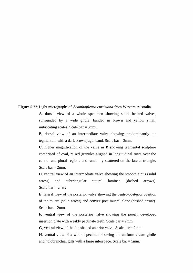
Figure 5.22: Light micrographs of Acanthopleura curtisiana from Western Australia.
A, dorsal view of a whole specimen showing solid, beaked valves,
surrounded by a wide girdle, banded in brown and yellow small,
imbricating scales. Scale bar = 5mm.
B, dorsal view of an intermediate valve showing predominantly tan
tegmentum with a dark brown jugal band. Scale bar = 2mm.
C, higher magnification of the valve in B showing tegmental sculpture
comprised of oval, raised granules aligned in longitudinal rows over the
central and plural regions and randomly scattered on the lateral triangle.
Scale bar = 2mm.
D, ventral view of an intermediate valve showing the smooth sinus (solid
arrow) and subtriangular sutural laminae (dashed arrows).
Scale bar = 2mm.
E, lateral view of the posterior valve showing the centro-posterior position
of the mucro (solid arrow) and convex post mucral slope (dashed arrow).
Scale bar = 2mm.
F, ventral view of the posterior valve showing the poorly developed
insertion plate with weakly pectinate teeth. Scale bar = 2mm.
G, ventral view of the fan-shaped anterior valve. Scale bar = 2mm.
H, ventral view of a whole specimen showing the uniform cream girdle
and holobranchial gills with a large interspace. Scale bar = 5mm.
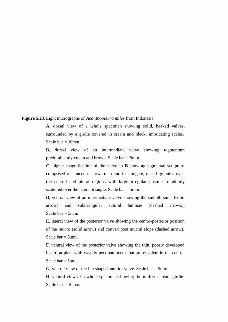
Figure 5.23: Light micrographs of Acanthopleura miles from Indonesia.
A, dorsal view of a whole specimen showing solid, beaked valves,
surrounded by a girdle covered in cream and black, imbricating scales.
Scale bar = 10mm.
B, dorsal view of an intermediate valve showing tegmentum
predominantly cream and brown. Scale bar = 5mm.
C, higher magnification of the valve in B showing tegmental sculpture
comprised of concentric rows of round to elongate, raised granules over
the central and plural regions with large irregular pustules randomly
scattered over the lateral triangle. Scale bar = 5mm.
D, ventral view of an intermediate valve showing the smooth sinus (solid
arrow) and subtriangular sutural laminae (dashed arrows).
Scale bar = 5mm.
E, lateral view of the posterior valve showing the centro-posterior position
of the mucro (solid arrow) and convex post mucral slope (dashed arrow).
Scale bar = 5mm.
F, ventral view of the posterior valve showing the thin, poorly developed
insertion plate with weakly pectinate teeth that are obsolete in the centre.
Scale bar = 5mm.
G, ventral view of the fan-shaped anterior valve. Scale bar = 5mm.
H, ventral view of a whole specimen showing the uniform cream girdle.
Scale bar = 10mm.

Figure 5.24: Light micrographs of Acanthopleura rehderi from the Cook Is.
A, dorsal view of a whole specimen showing solid, beaked valves,
surrounded by a banded girdle covered in minute, white and brown
spinelets. Scale bar = 10mm.
B, dorsal view of an intermediate valve showing tegmental colour pattern.
Scale bar = 5mm.
C, higher magnification of the valve in B showing the smooth central
tegmentum, raised parallel ridges on the plural region and concentrically
orientated elongate pustules on the lateral triangle. There is a single, radial
row of round ocelli. Scale bar = 5mm.
D, ventral view of an intermediate valve showing the pectinate sinus (solid
arrow) and triangular sutural laminae (dashed arrows). Scale bar = 5mm.
E, lateral view of the posterior valve showing the terminal position of the
mucro (solid arrow). Scale bar = 5mm.
F, ventral view of the posterior valve showing the porcelaneous texture of
the articulamentum, and the insertion plate (arrow) merged with the round
transverse callus (dashed arrow). Scale bar = 5mm.
G, ventral view of the fan-shaped anterior valve. Scale bar = 5mm.
H, ventral view of a whole specimen showing the uniform cream girdle
and the distinctive mantle flap (arrows) either side of the anus.
Scale bar = 10mm.
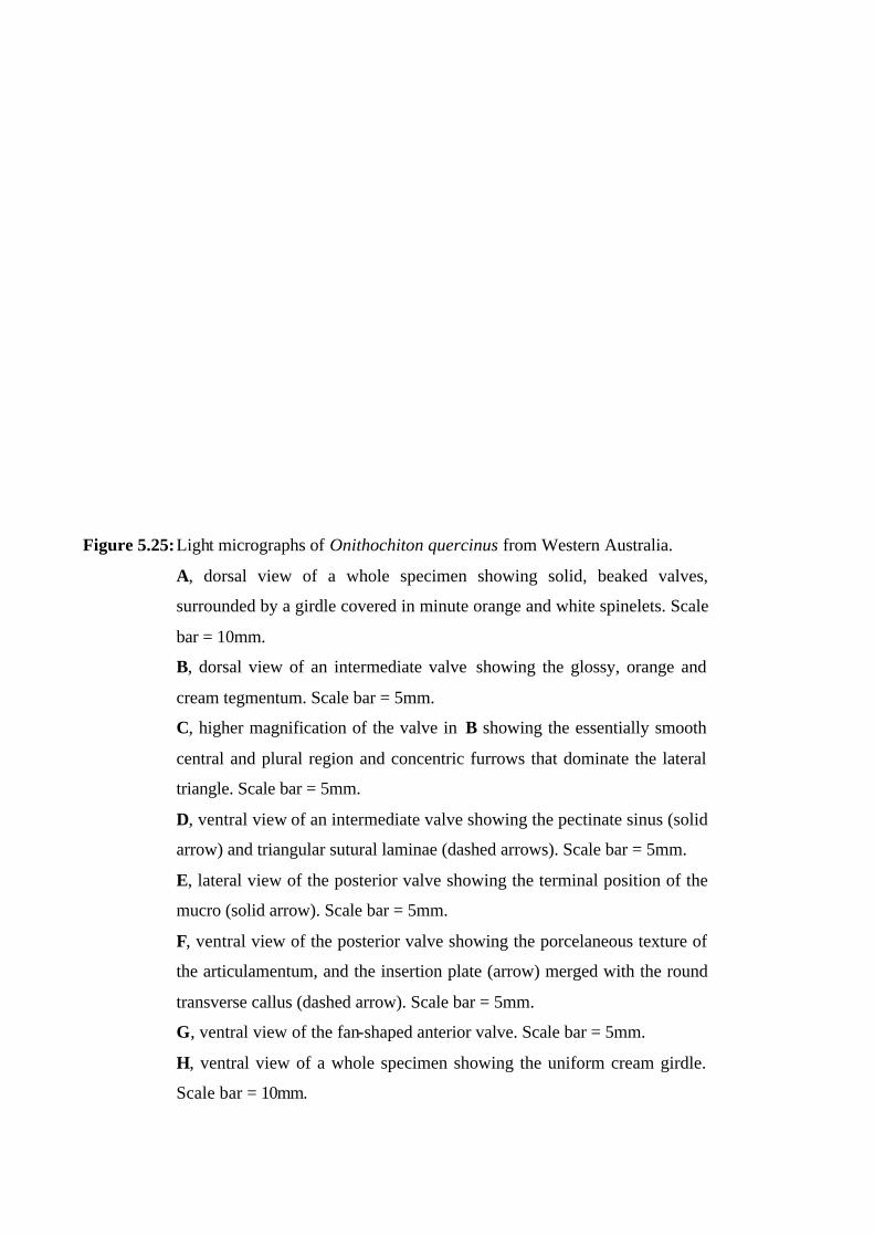
Figure 5.25: Light micrographs of Onithochiton quercinus from Western Australia.
A, dorsal view of a whole specimen showing solid, beaked valves,
surrounded by a girdle covered in minute orange and white spinelets. Scale
bar = 10mm.
B, dorsal view of an intermediate valve showing the glossy, orange and
cream tegmentum. Scale bar = 5mm.
C, higher magnification of the valve in B showing the essentially smooth
central and plural region and concentric furrows that dominate the lateral
triangle. Scale bar = 5mm.
D, ventral view of an intermediate valve showing the pectinate sinus (solid
arrow) and triangular sutural laminae (dashed arrows). Scale bar = 5mm.
E, lateral view of the posterior valve showing the terminal position of the
mucro (solid arrow). Scale bar = 5mm.
F, ventral view of the posterior valve showing the porcelaneous texture of
the articulamentum, and the insertion plate (arrow) merged with the round
transverse callus (dashed arrow). Scale bar = 5mm.
G, ventral view of the fan-shaped anterior valve. Scale bar = 5mm.
H, ventral view of a whole specimen showing the uniform cream girdle.
Scale bar = 10mm.
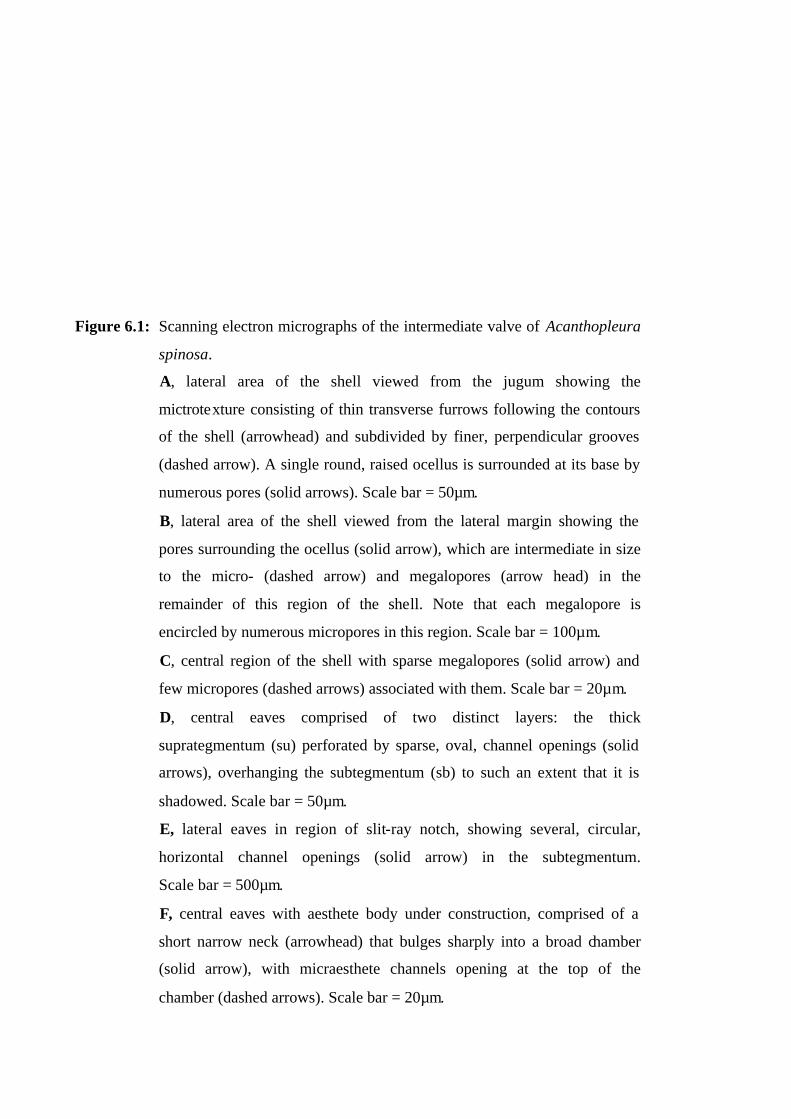
Figure 6.1: Scanning electron micrographs of the intermediate valve of Acanthopleura
spinosa.
A, lateral area of the shell viewed from the jugum showing the
mictrotexture consisting of thin transverse furrows following the contours
of the shell (arrowhead) and subdivided by finer, perpendicular grooves
(dashed arrow). A single round, raised ocellus is surrounded at its base by
numerous pores (solid arrows). Scale bar = 50µm.
B, lateral area of the shell viewed from the lateral margin showing the
pores surrounding the ocellus (solid arrow), which are intermediate in size
to the micro- (dashed arrow) and megalopores (arrow head) in the
remainder of this region of the shell. Note that each megalopore is
encircled by numerous micropores in this region. Scale bar = 100µm.
C, central region of the shell with sparse megalopores (solid arrow) and
few micropores (dashed arrows) associated with them. Scale bar = 20µm.
D, central eaves comprised of two distinct layers: the thick
suprategmentum (su) perforated by sparse, oval, channel openings (solid
arrows), overhanging the subtegmentum (sb) to such an extent that it is
shadowed. Scale bar = 50µm.
E, lateral eaves in region of slit-ray notch, showing several, circular,
horizontal channel openings (solid arrow) in the subtegmentum.
Scale bar = 500µm.
F, central eaves with aesthete body under construction, comprised of a
short narrow neck (arrowhead) that bulges sharply into a broad chamber
(solid arrow), with micraesthete channels opening at the top of the
chamber (dashed arrows). Scale bar = 20µm.
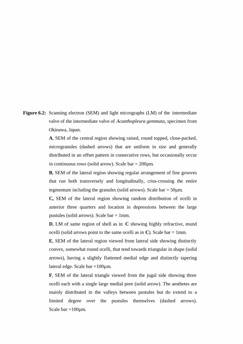
Figure 6.2: Scanning electron (SEM) and light micrographs (LM) of the intermediate
valve of the intermediate valve of Acanthopleura gemmata, specimen from
Okinawa, Japan.
A, SEM of the central region showing raised, round topped, close-packed,
microgranules (dashed arrows) that are uniform in size and generally
distributed in an offset pattern in consecutive rows, but occasionally occur
in continuous rows (solid arrow). Scale bar = 200µm.
B, SEM of the lateral region showing regular arrangement of fine grooves
that run both transversely and longitudinally, criss-crossing the entire
tegmentum including the granules (solid arrows). Scale bar = 50µm.
C, SEM of the lateral region showing random distribution of ocelli in
anterior three quarters and location in depressions between the large
pustules (solid arrows). Scale bar = 1mm.
D, LM of same region of shell as in C showing highly refractive, round
ocelli (solid arrows point to the same ocelli as in C). Scale bar = 1mm.
E, SEM of the lateral region viewed from lateral side showing distinctly
convex, somewhat round ocelli, that tend towards triangular in shape (solid
arrows), having a slightly flattened medial edge and distinctly tapering
lateral edge. Scale bar =100µm.
F, SEM of the lateral triangle viewed from the jugal side showing three
ocelli each with a single large medial pore (solid arrow). The aesthetes are
mainly distributed in the valleys between pustules but do extend to a
limited degree over the pustules themselves (dashed arrows).
Scale bar =100µm.
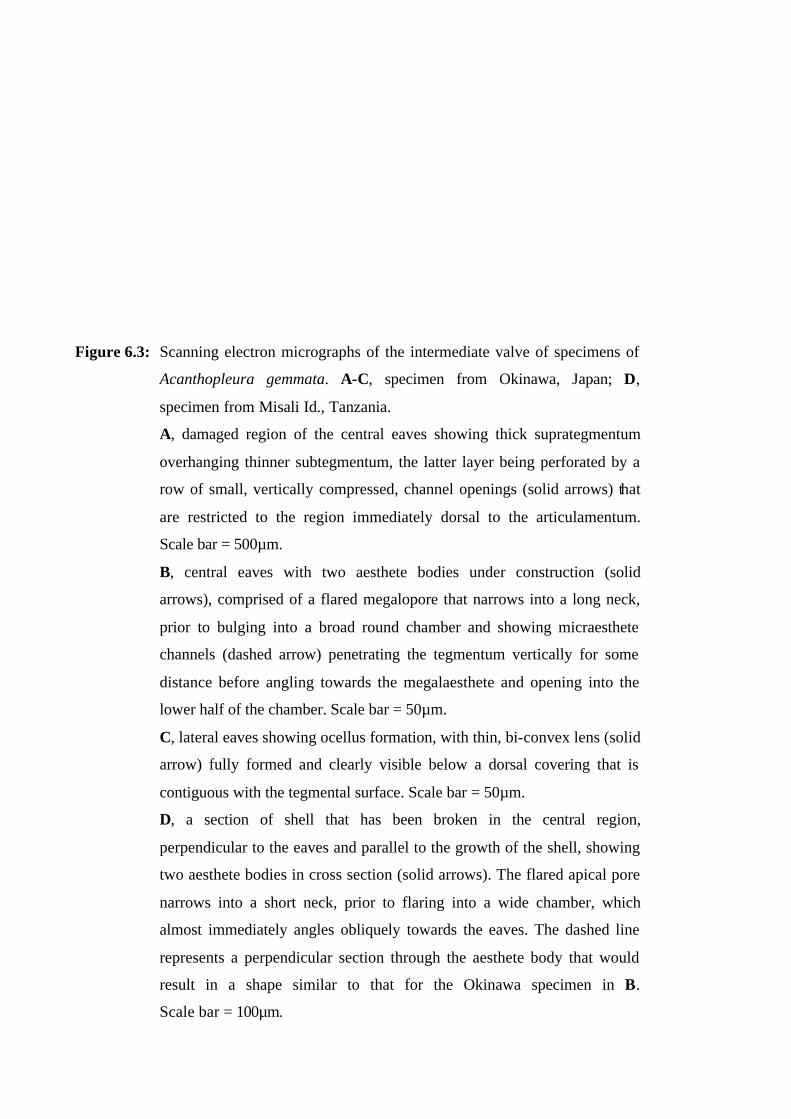
Figure 6.3: Scanning electron micrographs of the intermediate valve of specimens of
Acanthopleura gemmata. A-C, specimen from Okinawa, Japan; D,
specimen from Misali Id., Tanzania.
A, damaged region of the central eaves showing thick suprategmentum
overhanging thinner subtegmentum, the latter layer being perforated by a
row of small, vertically compressed, channel openings (solid arrows) that
are restricted to the region immediately dorsal to the articulamentum.
Scale bar = 500µm.
B, central eaves with two aesthete bodies under construction (solid
arrows), comprised of a flared megalopore that narrows into a long neck,
prior to bulging into a broad round chamber and showing micraesthete
channels (dashed arrow) penetrating the tegmentum vertically for some
distance before angling towards the megalaesthete and opening into the
lower half of the chamber. Scale bar = 50µm.
C, lateral eaves showing ocellus formation, with thin, bi-convex lens (solid
arrow) fully formed and clearly visible below a dorsal covering that is
contiguous with the tegmental surface. Scale bar = 50µm.
D, a section of shell that has been broken in the central region,
perpendicular to the eaves and parallel to the growth of the shell, showing
two aesthete bodies in cross section (solid arrows). The flared apical pore
narrows into a short neck, prior to flaring into a wide chamber, which
almost immediately angles obliquely towards the eaves. The dashed line
represents a perpendicular section through the aesthete body that would
result in a shape similar to that for the Okinawa specimen in B.
Scale bar = 100µm.
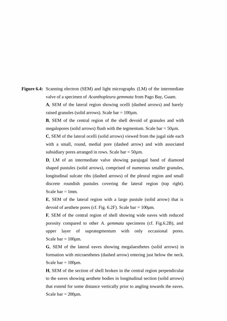
Figure 6.4: Scanning electron (SEM) and light micrographs (LM) of the intermediate
valve of a specimen of Acanthopleura gemmata from Pago Bay, Guam.
A, SEM of the lateral region showing ocelli (dashed arrows) and barely
raised granules (solid arrows). Scale bar = 100µm.
B, SEM of the central region of the shell devoid of granules and with
megalopores (solid arrows) flush with the tegmentum. Scale bar = 50µm.
C, SEM of the lateral ocelli (solid arrows) viewed from the jugal side each
with a small, round, medial pore (dashed arrow) and with associated
subsidiary pores arranged in rows. Scale bar = 50µm.
D, LM of an intermediate valve showing parajugal band of diamond
shaped pustules (solid arrows), comprised of numerous smaller granules,
longitudinal sulcate ribs (dashed arrows) of the pleural region and small
discrete roundish pustules covering the lateral region (top right).
Scale bar = 1mm.
E, SEM of the lateral region with a large pustule (solid arrow) that is
devoid of aesthete pores (cf. Fig. 6.2F). Scale bar = 100µm.
F, SEM of the central region of shell showing wide eaves with reduced
porosity compared to other A. gemmata specimens (cf. Fig.6.2B), and
upper layer of suprategmentum with only occasional pores.
Scale bar = 100µm.
G, SEM of the lateral eaves showing megalaesthetes (solid arrows) in
formation with micraesthetes (dashed arrow) entering just below the neck.
Scale bar = 100µm.
H, SEM of the section of shell broken in the central region perpendicular
to the eaves showing aesthete bodies in longitudinal section (solid arrows)
that extend for some distance vertically prior to angling towards the eaves.
Scale bar = 200µm.
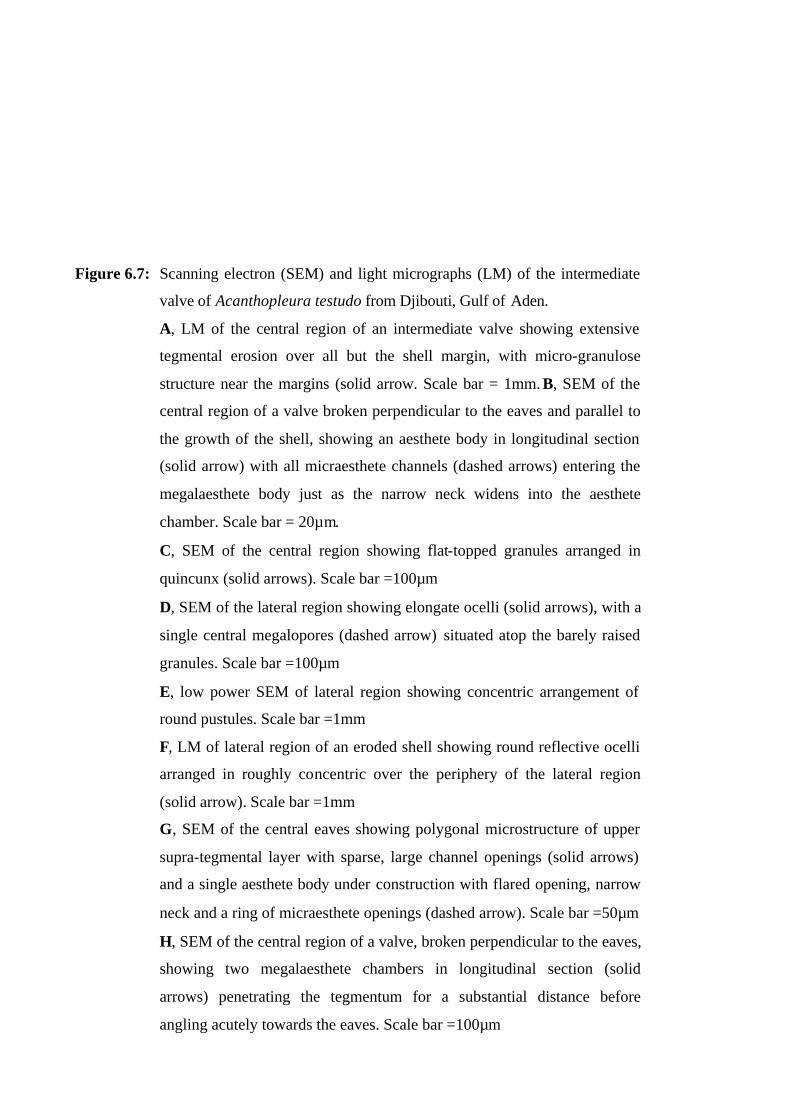
Figure 6.7: Scanning electron (SEM) and light micrographs (LM) of the intermediate
valve of Acanthopleura testudo from Djibouti, Gulf of Aden.
A, LM of the central region of an intermediate valve showing extensive
tegmental erosion over all but the shell margin, with micro-granulose
structure near the margins (solid arrow. Scale bar = 1mm. B, SEM of the
central region of a valve broken perpendicular to the eaves and parallel to
the growth of the shell, showing an aesthete body in longitudinal section
(solid arrow) with all micraesthete channels (dashed arrows) entering the
megalaesthete body just as the narrow neck widens into the aesthete
chamber. Scale bar = 20µm.
C, SEM of the central region showing flat-topped granules arranged in
quincunx (solid arrows). Scale bar =100µm
D, SEM of the lateral region showing elongate ocelli (solid arrows), with a
single central megalopores (dashed arrow) situated atop the barely raised
granules. Scale bar =100µm
E, low power SEM of lateral region showing concentric arrangement of
round pustules. Scale bar =1mm
F, LM of lateral region of an eroded shell showing round reflective ocelli
arranged in roughly concentric over the periphery of the lateral region
(solid arrow). Scale bar =1mm
G, SEM of the central eaves showing polygonal microstructure of upper
supra-tegmental layer with sparse, large channel openings (solid arrows)
and a single aesthete body under construction with flared opening, narrow
neck and a ring of micraesthete openings (dashed arrow). Scale bar =50µm
H, SEM of the central region of a valve, broken perpendicular to the eaves,
showing two megalaesthete chambers in longitudinal section (solid
arrows) penetrating the tegmentum for a substantial distance before
angling acutely towards the eaves. Scale bar =100µm
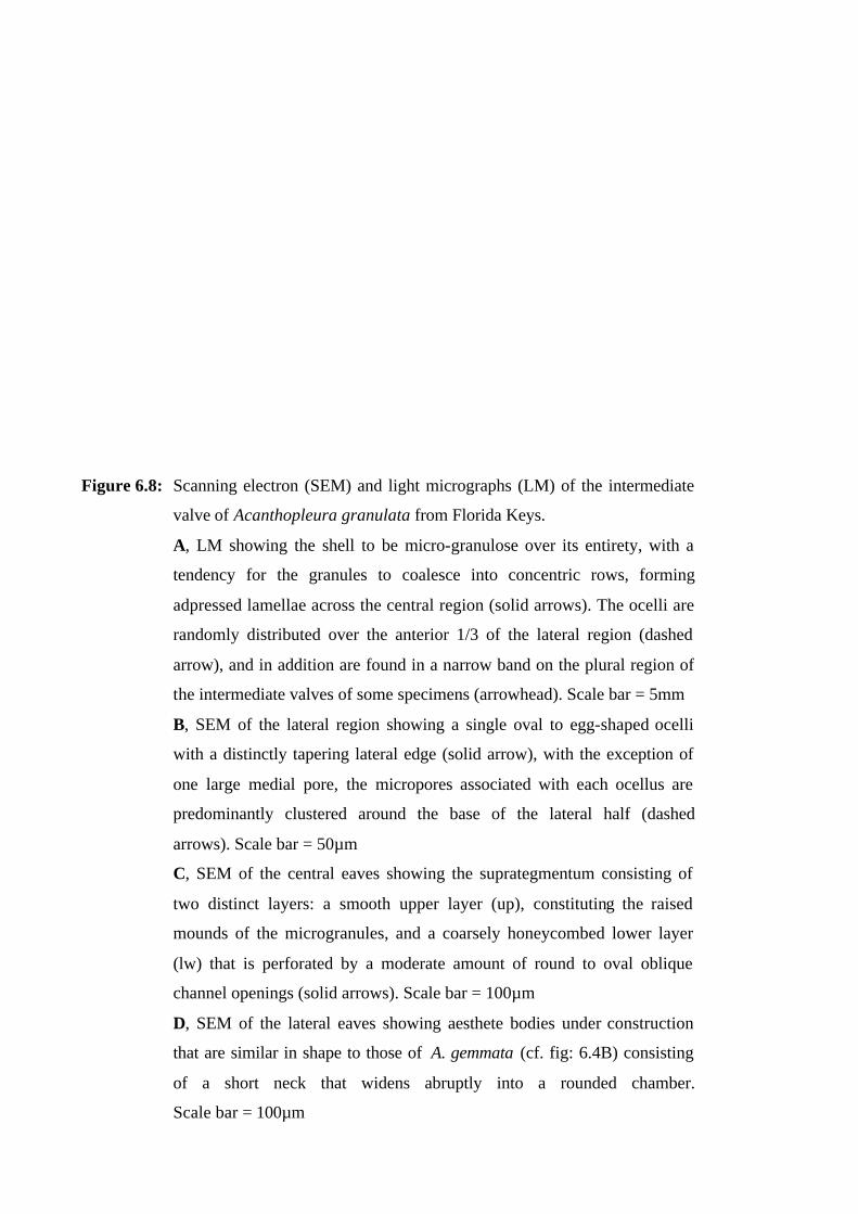
Figure 6.8: Scanning electron (SEM) and light micrographs (LM) of the intermediate
valve of Acanthopleura granulata from Florida Keys.
A, LM showing the shell to be micro-granulose over its entirety, with a
tendency for the granules to coalesce into concentric rows, forming
adpressed lamellae across the central region (solid arrows). The ocelli are
randomly distributed over the anterior 1/3 of the lateral region (dashed
arrow), and in addition are found in a narrow band on the plural region of
the intermediate valves of some specimens (arrowhead). Scale bar = 5mm
B, SEM of the lateral region showing a single oval to egg-shaped ocelli
with a distinctly tapering lateral edge (solid arrow), with the exception of
one large medial pore, the micropores associated with each ocellus are
predominantly clustered around the base of the lateral half (dashed
arrows). Scale bar = 50µm
C, SEM of the central eaves showing the suprategmentum consisting of
two distinct layers: a smooth upper layer (up), constituting the raised
mounds of the microgranules, and a coarsely honeycombed lower layer
(lw) that is perforated by a moderate amount of round to oval oblique
channel openings (solid arrows). Scale bar = 100µm
D, SEM of the lateral eaves showing aesthete bodies under construction
that are similar in shape to those of A. gemmata (cf. fig: 6.4B) consisting
of a short neck that widens abruptly into a rounded chamber.
Scale bar = 100µm
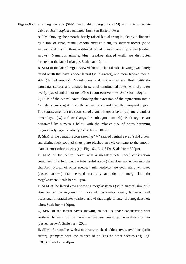
Figure 6.9: Scanning electron (SEM) and light micrographs (LM) of the intermediate
valve of Acanthopleura echinata from San Bartolo, Peru.
A, LM showing the smooth, barely raised lateral triangle, clearly delineated
by a row of large, round, smooth pustules along its anterior border (solid
arrows), and two or three additional radial rows of round pustules (dashed
arrows). Numerous minute, blue, teardrop shaped ocelli are distributed
throughout the lateral triangle. Scale bar = 2mm.
B, SEM of the lateral region viewed from the lateral side showing oval, barely
raised ocelli that have a wider lateral (solid arrows), and more tapered medial
side (dashed arrows). Megalopores and micropores are flush with the
tegmental surface and aligned in parallel longitudinal rows, with the latter
evenly spaced and the former offset in consecutive rows. Scale bar = 50µm
C, SEM of the central eaves showing the extension of the tegmentum into a
“V” shape, making it much thicker in the central than the parajugal region.
The suprategmentum (su) consists of a smooth upper layer (up) and granulose
lower layer (lw) and overhangs the subtegmentum (sb). Both regions are
perforated by numerous holes, with the relative size of pores becoming
progressively larger ventrally. Scale bar = 100µm.
D, SEM of the central region showing “V” shaped central eaves (solid arrow)
and distinctively toothed sinus plate (dashed arrow), compare to the smooth
plate of most other species (e.g. Figs. 6.4.A, 6.6.D). Scale bar = 500µm
E, SEM of the central eaves with a megalaesthete under construction,
comprised of a long narrow tube (solid arrow) that does not widen into the
chamber (typical of other species), micraesthetes are even narrower tubes
(dashed arrows) that descend vertically and do not merge into the
megalaesthete. Scale bar = 20µm.
F, SEM of the lateral eaves showing megalaesthetes (solid arrows) similar in
structure and arrangement to those of the central eaves, however, with
occasional micraesthetes (dashed arrow) that angle to enter the megalaesthete
tubes. Scale bar = 100µm.
G, SEM of the lateral eaves showing an ocellus under construction with
aesthete channels from numerous earlier rows entering the ocellus chamber
(dashed arrows). Scale bar = 20µm.
H, SEM of an ocellus with a relatively thick, double convex, oval lens (solid
arrow), (compare with the thinner round lens of other species (e.g. Fig.
6.3C)). Scale bar = 20µm.
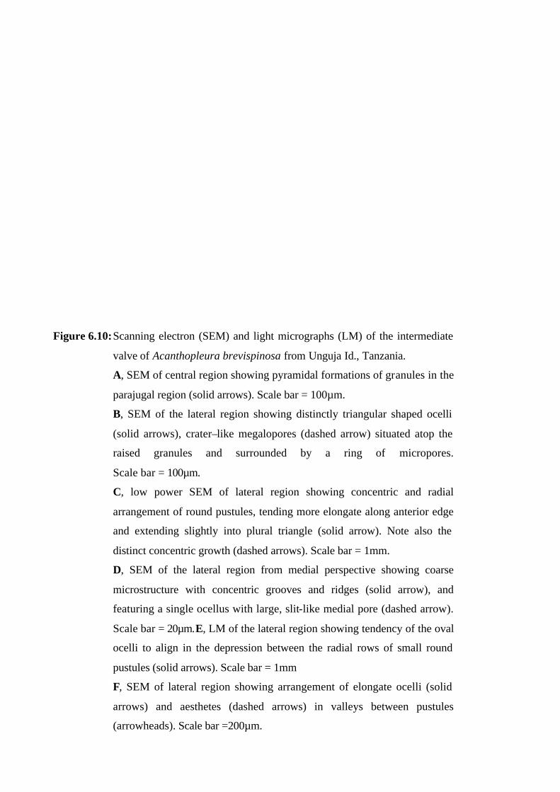
Figure 6.10: Scanning electron (SEM) and light micrographs (LM) of the intermediate
valve of Acanthopleura brevispinosa from Unguja Id., Tanzania.
A, SEM of central region showing pyramidal formations of granules in the
parajugal region (solid arrows). Scale bar = 100µm.
B, SEM of the lateral region showing distinctly triangular shaped ocelli
(solid arrows), crater–like megalopores (dashed arrow) situated atop the
raised granules and surrounded by a ring of micropores.
Scale bar = 100µm.
C, low power SEM of lateral region showing concentric and radial
arrangement of round pustules, tending more elongate along anterior edge
and extending slightly into plural triangle (solid arrow). Note also the
distinct concentric growth (dashed arrows). Scale bar = 1mm.
D, SEM of the lateral region from medial perspective showing coarse
microstructure with concentric grooves and ridges (solid arrow), and
featuring a single ocellus with large, slit-like medial pore (dashed arrow).
Scale bar = 20µm.E, LM of the lateral region showing tendency of the oval
ocelli to align in the depression between the radial rows of small round
pustules (solid arrows). Scale bar = 1mm
F, SEM of lateral region showing arrangement of elongate ocelli (solid
arrows) and aesthetes (dashed arrows) in valleys between pustules
(arrowheads). Scale bar =200µm.
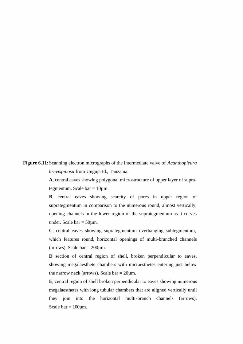
Figure 6.11: Scanning electron micrographs of the intermediate valve of Acanthopleura
brevispinosa from Unguja Id., Tanzania.
A, central eaves showing polygonal microstructure of upper layer of supra-
tegmentum. Scale bar = 10µm.
B, central eaves showing scarcity of pores in upper region of
suprategmentum in comparison to the numerous round, almost vertically,
opening channels in the lower region of the suprategmentum as it curves
under. Scale bar = 50µm.
C, central eaves showing suprategmentum overhanging subtegmentum,
which features round, horizontal openings of multi-branched channels
(arrows). Scale bar = 200µm.
D section of central region of shell, broken perpendicular to eaves,
showing megalaesthete chambers with micraesthetes entering just below
the narrow neck (arrows). Scale bar = 20µm.
E, central region of shell broken perpendicular to eaves showing numerous
megalaesthetes with long tubular chambers that are aligned vertically until
they join into the horizontal multi-branch channels (arrows).
Scale bar = 100µm.
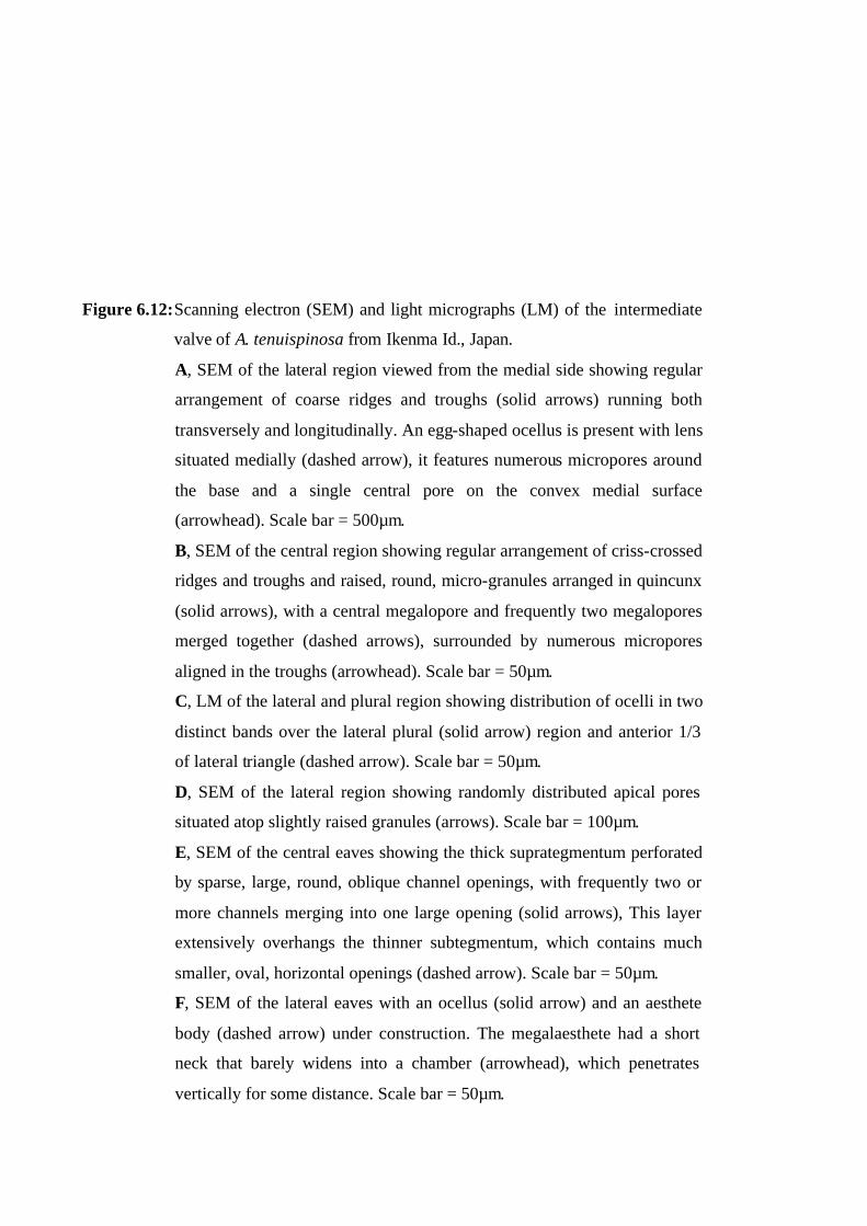
Figure 6.12: Scanning electron (SEM) and light micrographs (LM) of the intermediate
valve of A. tenuispinosa from Ikenma Id., Japan.
A, SEM of the lateral region viewed from the medial side showing regular
arrangement of coarse ridges and troughs (solid arrows) running both
transversely and longitudinally. An egg-shaped ocellus is present with lens
situated medially (dashed arrow), it features numerous micropores around
the base and a single central pore on the convex medial surface
(arrowhead). Scale bar = 500µm.
B, SEM of the central region showing regular arrangement of criss-crossed
ridges and troughs and raised, round, micro-granules arranged in quincunx
(solid arrows), with a central megalopore and frequently two megalopores
merged together (dashed arrows), surrounded by numerous micropores
aligned in the troughs (arrowhead). Scale bar = 50µm.
C, LM of the lateral and plural region showing distribution of ocelli in two
distinct bands over the lateral plural (solid arrow) region and anterior 1/3
of lateral triangle (dashed arrow). Scale bar = 50µm.
D, SEM of the lateral region showing randomly distributed apical pores
situated atop slightly raised granules (arrows). Scale bar = 100µm.
E, SEM of the central eaves showing the thick suprategmentum perforated
by sparse, large, round, oblique channel openings, with frequently two or
more channels merging into one large opening (solid arrows), This layer
extensively overhangs the thinner subtegmentum, which contains much
smaller, oval, horizontal openings (dashed arrow). Scale bar = 50µm.
F, SEM of the lateral eaves with an ocellus (solid arrow) and an aesthete
body (dashed arrow) under construction. The megalaesthete had a short
neck that barely widens into a chamber (arrowhead), which penetrates
vertically for some distance. Scale bar = 50µm.
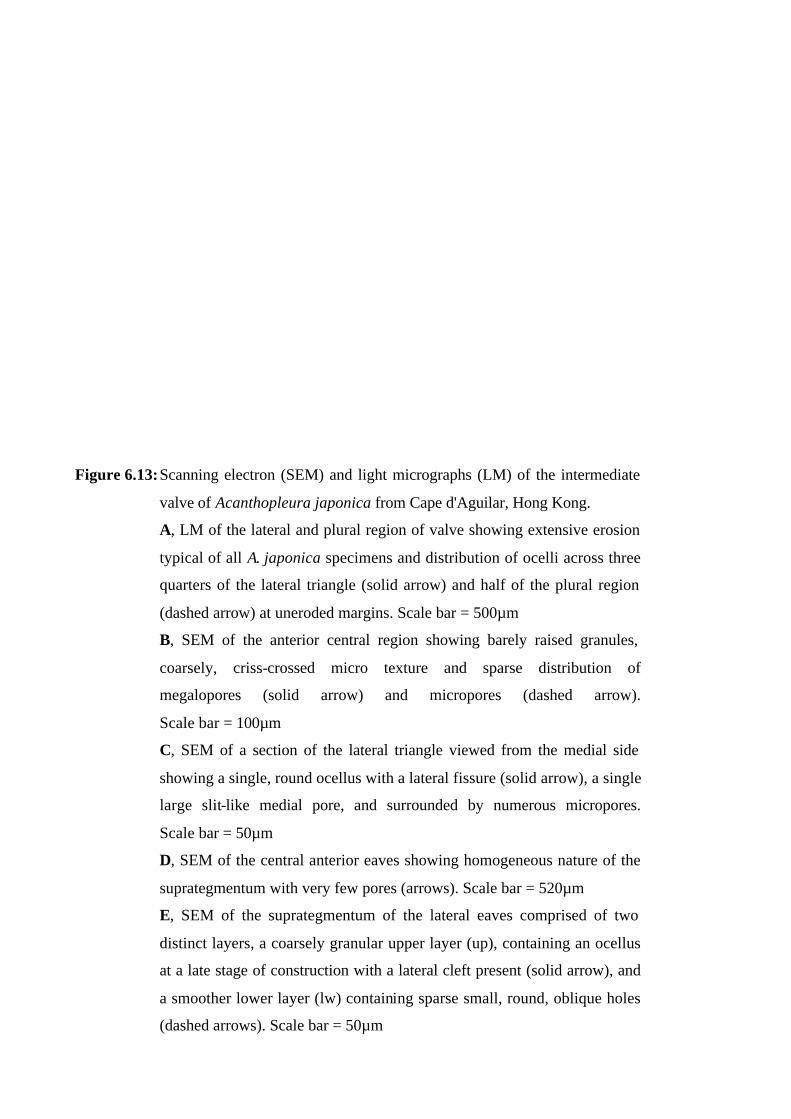
Figure 6.13: Scanning electron (SEM) and light micrographs (LM) of the intermediate
valve of Acanthopleura japonica from Cape d'Aguilar, Hong Kong.
A, LM of the lateral and plural region of valve showing extensive erosion
typical of all A. japonica specimens and distribution of ocelli across three
quarters of the lateral triangle (solid arrow) and half of the plural region
(dashed arrow) at uneroded margins. Scale bar = 500µm
B, SEM of the anterior central region showing barely raised granules,
coarsely, criss-crossed micro texture and sparse distribution of
megalopores (solid arrow) and micropores (dashed arrow).
Scale bar = 100µm
C, SEM of a section of the lateral triangle viewed from the medial side
showing a single, round ocellus with a lateral fissure (solid arrow), a single
large slit-like medial pore, and surrounded by numerous micropores.
Scale bar = 50µm
D, SEM of the central anterior eaves showing homogeneous nature of the
suprategmentum with very few pores (arrows). Scale bar = 520µm
E, SEM of the suprategmentum of the lateral eaves comprised of two
distinct layers, a coarsely granular upper layer (up), containing an ocellus
at a late stage of construction with a lateral cleft present (solid arrow), and
a smoother lower layer (lw) containing sparse small, round, oblique holes
(dashed arrows). Scale bar = 50µm
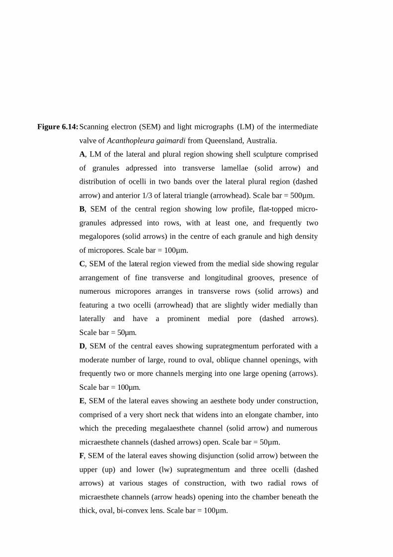
Figure 6.14: Scanning electron (SEM) and light micrographs (LM) of the intermediate
valve of Acanthopleura gaimardi from Queensland, Australia.
A, LM of the lateral and plural region showing shell sculpture comprised
of granules adpressed into transverse lamellae (solid arrow) and
distribution of ocelli in two bands over the lateral plural region (dashed
arrow) and anterior 1/3 of lateral triangle (arrowhead). Scale bar = 500µm.
B, SEM of the central region showing low profile, flat-topped micro-
granules adpressed into rows, with at least one, and frequently two
megalopores (solid arrows) in the centre of each granule and high density
of micropores. Scale bar = 100µm.
C, SEM of the lateral region viewed from the medial side showing regular
arrangement of fine transverse and longitudinal grooves, presence of
numerous micropores arranges in transverse rows (solid arrows) and
featuring a two ocelli (arrowhead) that are slightly wider medially than
laterally and have a prominent medial pore (dashed arrows).
Scale bar = 50µm.
D, SEM of the central eaves showing suprategmentum perforated with a
moderate number of large, round to oval, oblique channel openings, with
frequently two or more channels merging into one large opening (arrows).
Scale bar = 100µm.
E, SEM of the lateral eaves showing an aesthete body under construction,
comprised of a very short neck that widens into an elongate chamber, into
which the preceding megalaesthete channel (solid arrow) and numerous
micraesthete channels (dashed arrows) open. Scale bar = 50µm.
F, SEM of the lateral eaves showing disjunction (solid arrow) between the
upper (up) and lower (lw) suprategmentum and three ocelli (dashed
arrows) at various stages of construction, with two radial rows of
micraesthete channels (arrow heads) opening into the chamber beneath the
thick, oval, bi-convex lens. Scale bar = 100µm.
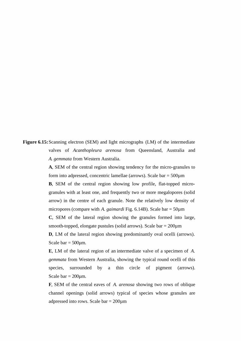
Figure 6.15: Scanning electron (SEM) and light micrographs (LM) of the intermediate
valves of Acanthopleura arenosa from Queensland, Australia and
A. gemmata from Western Australia.
A, SEM of the central region showing tendency for the micro-granules to
form into adpressed, concentric lamellae (arrows). Scale bar = 500µm
B, SEM of the central region showing low profile, flat-topped micro-
granules with at least one, and frequently two or more megalopores (solid
arrow) in the centre of each granule. Note the relatively low density of
micropores (compare with A. gaimardi Fig. 6.14B). Scale bar = 50µm
C, SEM of the lateral region showing the granules formed into large,
smooth-topped, elongate pustules (solid arrows). Scale bar = 200µm
D, LM of the lateral region showing predominantly oval ocelli (arrows).
Scale bar = 500µm.
E, LM of the lateral region of an intermediate valve of a specimen of A.
gemmata from Western Australia, showing the typical round ocelli of this
species, surrounded by a thin circle of pigment (arrows).
Scale bar = 200µm.
F, SEM of the central eaves of A. arenosa showing two rows of oblique
channel openings (solid arrows) typical of species whose granules are
adpressed into rows. Scale bar = 200µm
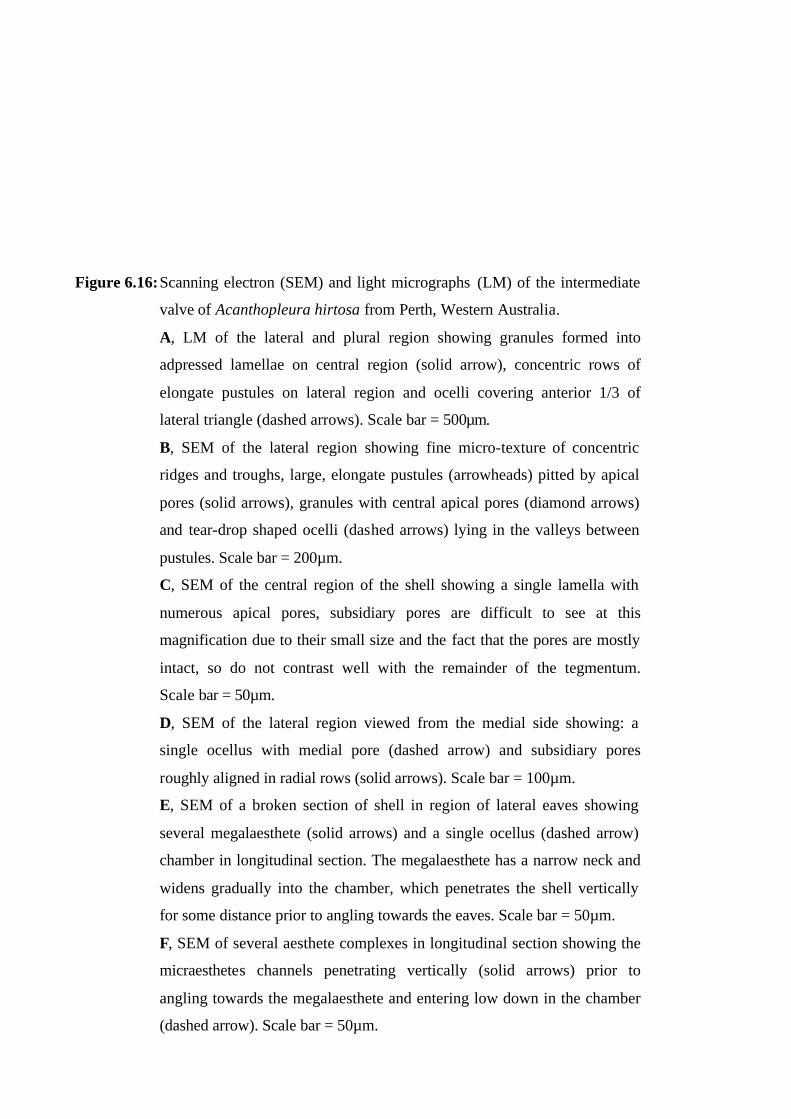
Figure 6.16: Scanning electron (SEM) and light micrographs (LM) of the intermediate
valve of Acanthopleura hirtosa from Perth, Western Australia.
A, LM of the lateral and plural region showing granules formed into
adpressed lamellae on central region (solid arrow), concentric rows of
elongate pustules on lateral region and ocelli covering anterior 1/3 of
lateral triangle (dashed arrows). Scale bar = 500µm.
B, SEM of the lateral region showing fine micro-texture of concentric
ridges and troughs, large, elongate pustules (arrowheads) pitted by apical
pores (solid arrows), granules with central apical pores (diamond arrows)
and tear-drop shaped ocelli (dashed arrows) lying in the valleys between
pustules. Scale bar = 200µm.
C, SEM of the central region of the shell showing a single lamella with
numerous apical pores, subsidiary pores are difficult to see at this
magnification due to their small size and the fact that the pores are mostly
intact, so do not contrast well with the remainder of the tegmentum.
Scale bar = 50µm.
D, SEM of the lateral region viewed from the medial side showing: a
single ocellus with medial pore (dashed arrow) and subsidiary pores
roughly aligned in radial rows (solid arrows). Scale bar = 100µm.
E, SEM of a broken section of shell in region of lateral eaves showing
several megalaesthete (solid arrows) and a single ocellus (dashed arrow)
chamber in longitudinal section. The megalaesthete has a narrow neck and
widens gradually into the chamber, which penetrates the shell vertically
for some distance prior to angling towards the eaves. Scale bar = 50µm.
F, SEM of several aesthete complexes in longitudinal section showing the
micraesthetes channels penetrating vertically (solid arrows) prior to
angling towards the megalaesthete and entering low down in the chamber
(dashed arrow). Scale bar = 50µm.
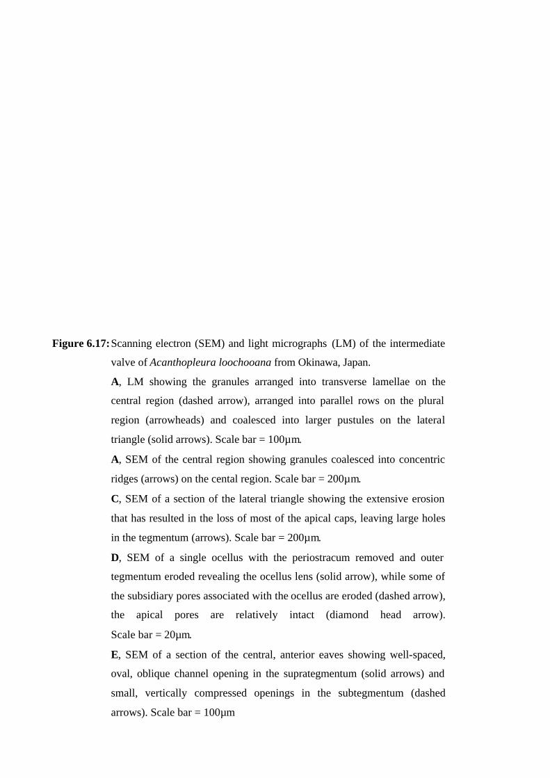
Figure 6.17: Scanning electron (SEM) and light micrographs (LM) of the intermediate
valve of Acanthopleura loochooana from Okinawa, Japan.
A, LM showing the granules arranged into transverse lamellae on the
central region (dashed arrow), arranged into parallel rows on the plural
region (arrowheads) and coalesced into larger pustules on the lateral
triangle (solid arrows). Scale bar = 100µm.
A, SEM of the central region showing granules coalesced into concentric
ridges (arrows) on the cental region. Scale bar = 200µm.
C, SEM of a section of the lateral triangle showing the extensive erosion
that has resulted in the loss of most of the apical caps, leaving large holes
in the tegmentum (arrows). Scale bar = 200µm.
D, SEM of a single ocellus with the periostracum removed and outer
tegmentum eroded revealing the ocellus lens (solid arrow), while some of
the subsidiary pores associated with the ocellus are eroded (dashed arrow),
the apical pores are relatively intact (diamond head arrow).
Scale bar = 20µm.
E, SEM of a section of the central, anterior eaves showing well-spaced,
oval, oblique channel opening in the suprategmentum (solid arrows) and
small, vertically compressed openings in the subtegmentum (dashed
arrows). Scale bar = 100µm

Figure 6.18: Scanning electron (SEM) and light micrographs (LM) of the intermediate
valve of Acanthopleura nigra from San Bartolo, Peru.
A, LM of the lateral region showing the smooth tegmentum, with deep
concentric furrows separating rounded plateaus (solid arrows) that are
covered in numerous minute, blue, oval ocelli. Scale bar = 1mm.
B, SEM of the lateral region featuring a single, oval, barely raised, ocellus
(solid arrow) displacing and distorting the subsidiary pores that are
otherwise evenly spaced along longitudinal rows (dashed arrows), with
apical pores offset from each other in adjacent rows (arrowheads).
Scale bar = 20µm.
C, SEM of the central region showing arrangement and density of
subsidiary and apical pores and the distinct extension of the
suprategmentum into a “V” shape (arrow). Scale bar = 500µm.
D, SEM of the central eaves showing two distinct layers of the tegmentum
with the suprategmentum (su) overhanging the recessed subtegmentum
(sb). The suprategmentum is further divided into three layers: a smooth
upper layer (up) containing numerous vertical micra- and megalaesthete
channels in longitudinal section; a more coarse middle layer (m) that is
perforated with numerous oblique channel openings, and finally a coarse
lower layer (lw) containing large, horizontally projecting plates (solid
arrows). Scale bar = 100µm.
E, SEM of a section of the suprategmentum showing long narrow tube-like
megalaesthetes (solid arrows) with numerous micraesthete channels
extending horizontally for some distance and often converging prior to
angling towards, and opening into, the aesthete complex (dashed arrows).
Scale bar = 20µm.

Figure 6.21: Scanning electron (SEM) and light micrographs (LM) of the intermediate
valve of Acanthopleura miles from Western Australia.
A, LM showing granules arranged in concentric rows across the central
and plural region (solid arrows) and coalesced into large round pustules on
the lateral triangle (dashed arrows). Scale bar = 500µm
B, SEM clearly showing the large, round, discrete pustules (solid arrows)
aligned in a row delineating the lateral and plural triangles and in radial
rows on the lateral region (dashed arrows). Scale bar = 500µm
C, SEM of the central region showing apical pores atop the granules that
are coalesced into concentric ridges (arrows). Scale bar = 200µm
D, SEM of the lateral region showing tegmentum crowded with prominent,
raised granules (solid arrows); apical pores (dashed arrows) atop the
granules and over the large pustule (top right); subsidiary pores covering
the entire tegmentum and a round, raised ocellus (arrow head).
Scale bar = 50µm.
E, SEM of the lateral region viewed, from the jugal side, showing an
ocellus with a distinctive pore on the medial side (arrow). Scale bar =
50µm.
F, SEM of the central, anterior eaves showing channel openings. Note that
the apical pores atop the raised granules penetrate the tegmentum for some
distance prior to opening into a wide, round chamber (arrows).
Scale bar = 50µm
G, SEM of the central eaves of a worn valve showing sparse, round to oval
channel openings (solid arrows) in the suprategmentum and smaller, oval,
horizontal openings (dashed arrows) in the recessed subtegmentum.
Scale bar = 100µm.

Figure 6.22: Scanning electron (SEM) and light micrographs (LM) of the intermediate
valve of Acanthopleura rehderi from Palmerston Id., Cook Is.
A, SEM of the lateral and plural region showing longitudinal riblets
(solid arrows) on the plural region and slightly raised lateral area with
radial rows of round to oval pustules (dashed arrows). Scale bar = 1mm.
B, LM of the lateral area of an intermediate valve showing a single row
of small, round ocelli (arrows). Scale bar =1mm. C, SEM of the lateral
region showing subsidiary pores evenly spaced along parallel,
longitudinal rows (solid arrows), and apical pores offset from each other
in consecutive rows (dashed arrows). A single round convex ocellus
(arrowhead) lies in a tegmental depression. The surrounding subsidiary
pores are distorted in shape by the depression around the ocellus, which
also displaces them out of their linear alignment. Scale bar = 50µm.
D, SEM of the central region showing arrangement of subsidiary and
apical pores in parallel rows. The eaves are thick with several aesthete
chambers in longitudinal section while the subtegmentum contains a row
of horizontally projecting, articulamental teeth (solid arrows).
Scale bar = 200µm.E, SEM of the anterior central eaves in the parajugal
region showing: the smooth, upper (up) suprategmentum (su) with an
aesthete complex (arrowhead) under construction, which has a long
vertical micraesthete channel (dashed arrow) angling to enter the
megalaesthete chamber and channels from previous rows of aesthetes
entering the chamber below this (solid arrows). The coarse, lower
suprategmentum (lw) has several oblique aesthete channel openings and
vertical openings under the overhang. Horizontal openings can also be
seen in the subtegmentum (sb). Scale bar = 50µm.
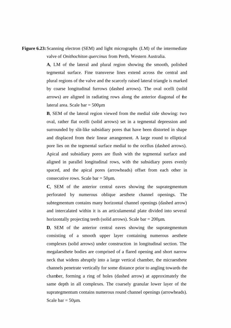
Figure 6.23: Scanning electron (SEM) and light micrographs (LM) of the intermediate
valve of Onithochiton quercinus from Perth, Western Australia.
A, LM of the lateral and plural region showing the smooth, polished
tegmental surface. Fine transverse lines extend across the central and
plural regions of the valve and the scarcely raised lateral triangle is marked
by coarse longitudinal furrows (dashed arrows). The oval ocelli (solid
arrows) are aligned in radiating rows along the anterior diagonal of the
lateral area. Scale bar = 500µm
B, SEM of the lateral region viewed from the medial side showing: two
oval, rather flat ocelli (solid arrows) set in a tegmental depression and
surrounded by slit-like subsidiary pores that have been distorted in shape
and displaced from their linear arrangement. A large round to elliptical
pore lies on the tegmental surface medial to the ocellus (dashed arrows).
Apical and subsidiary pores are flush with the tegmental surface and
aligned in parallel longitudinal rows, with the subsidiary pores evenly
spaced, and the apical pores (arrowheads) offset from each other in
consecutive rows. Scale bar = 50µm.
C, SEM of the anterior central eaves showing the suprategmentum
perforated by numerous oblique aesthete channel openings. The
subtegmentum contains many horizontal channel openings (dashed arrow)
and intercalated within it is an articulamental plate divided into several
horizontally projecting teeth (solid arrows). Scale bar = 200µm.
D, SEM of the anterior central eaves showing the suprategmentum
consisting of a smooth upper layer containing numerous aesthete
complexes (solid arrows) under construction in longitudinal section. The
megalaesthete bodies are comprised of a flared opening and short narrow
neck that widens abruptly into a large vertical chamber, the micraesthete
channels penetrate vertically for some distance prior to angling towards the
chamber, forming a ring of holes (dashed arrow) at approximately the
same depth in all complexes. The coarsely granular lower layer of the
suprategmentum contains numerous round channel openings (arrowheads).
Scale bar = 50µm.
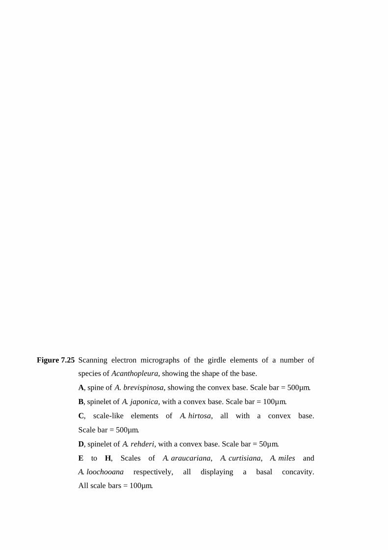
Figure 7.25 Scanning electron micrographs of the girdle elements of a number of
species of Acanthopleura, showing the shape of the base.
A, spine of A. brevispinosa, showing the convex base. Scale bar = 500µm.
B, spinelet of A. japonica, with a convex base. Scale bar = 100µm.
C, scale-like elements of A. hirtosa, all with a convex base.
Scale bar = 500µm.
D, spinelet of A. rehderi, with a convex base. Scale bar = 50µm.
E to H, Scales of A. araucariana, A. curtisiana, A. miles and
A. loochooana respectively, all displaying a basal concavity.
All scale bars = 100µm.
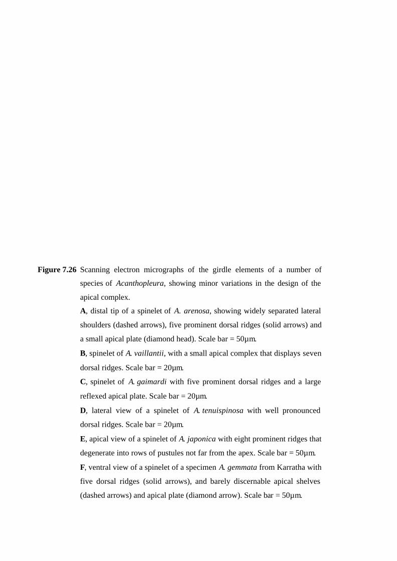
Figure 7.26 Scanning electron micrographs of the girdle elements of a number of
species of Acanthopleura, showing minor variations in the design of the
apical complex.
A, distal tip of a spinelet of A. arenosa, showing widely separated lateral
shoulders (dashed arrows), five prominent dorsal ridges (solid arrows) and
a small apical plate (diamond head). Scale bar = 50µm.
B, spinelet of A. vaillantii, with a small apical complex that displays seven
dorsal ridges. Scale bar = 20µm.
C, spinelet of A. gaimardi with five prominent dorsal ridges and a large
reflexed apical plate. Scale bar = 20µm.
D, lateral view of a spinelet of A. tenuispinosa with well pronounced
dorsal ridges. Scale bar = 20µm.
E, apical view of a spinelet of A. japonica with eight prominent ridges that
degenerate into rows of pustules not far from the apex. Scale bar = 50µm.
F, ventral view of a spinelet of a specimen A. gemmata from Karratha with
five dorsal ridges (solid arrows), and barely discernable apical shelves
(dashed arrows) and apical plate (diamond arrow). Scale bar = 50µm.
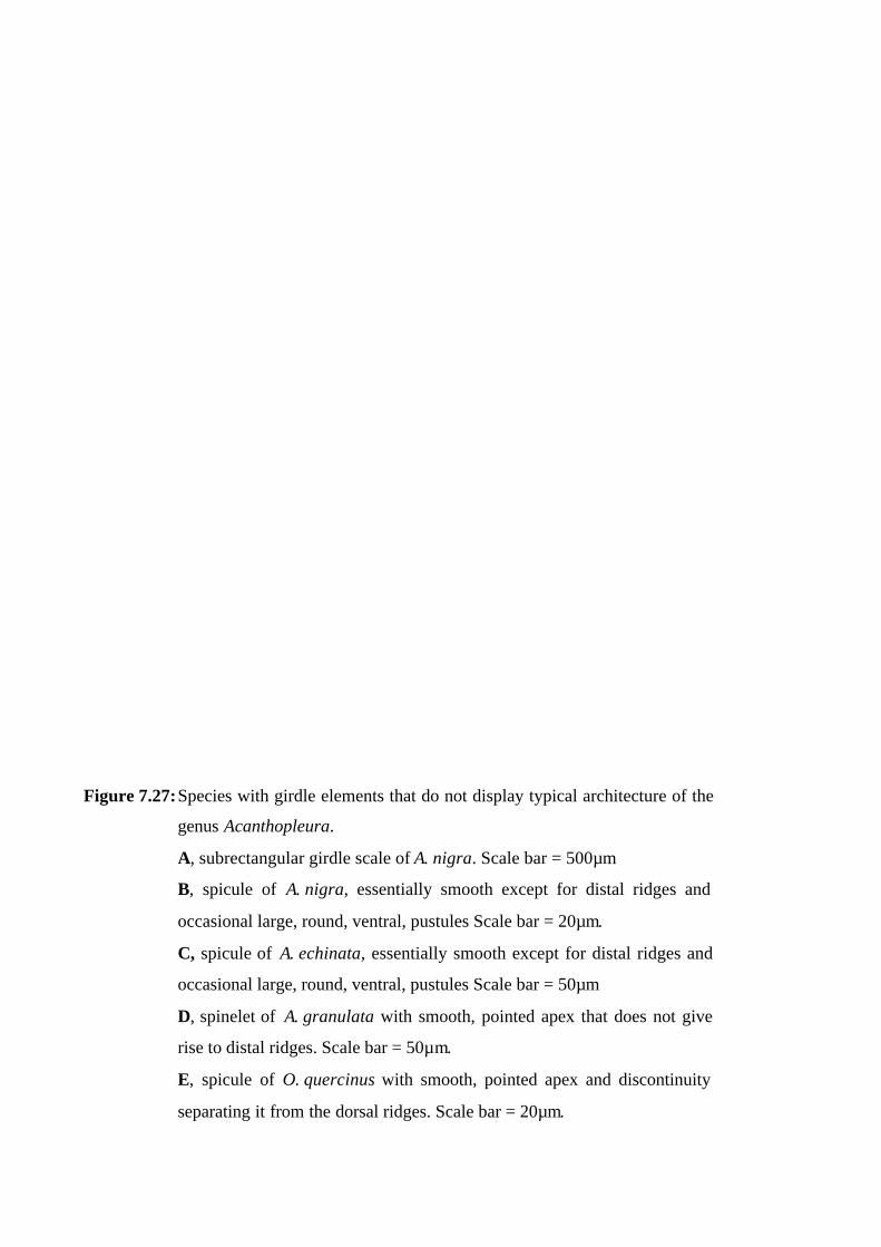
Figure 7.27: Species with girdle elements that do not display typical architecture of the
genus Acanthopleura.
A, subrectangular girdle scale of A. nigra. Scale bar = 500µm
B, spicule of A. nigra, essentially smooth except for distal ridges and
occasional large, round, ventral, pustules Scale bar = 20µm.
C, spicule of A. echinata, essentially smooth except for distal ridges and
occasional large, round, ventral, pustules Scale bar = 50µm
D, spinelet of A. granulata with smooth, pointed apex that does not give
rise to distal ridges. Scale bar = 50µm.
E, spicule of O. quercinus with smooth, pointed apex and discontinuity
separating it from the dorsal ridges. Scale bar = 20µm.

Figure 8.3: Scanning electron micrographs of the radula of Acanthopleura spinosa.
A-C, central and centrolateral teeth, top, anterior and lateral views
respectively.
A, indicating quadrilobed base of central teeth (solid arrows), and the
lateral and medial keels of centrolateral teeth (dashed arrows).
Scale bar = 100µm.
B, showing smooth, rounded anterior surface of central teeth.
Scale bar = 100µm.
C, indicating the central posterior keel of the central tooth (solid arrow),
and concave platform below the distal cusp (dashed arrow).
Scale bar = 100µm.
D, top view of radula showing major lateral teeth with chisel-shaped
medial wings (arrows). Scale bar = 500µm.
E, posterior view of major lateral teeth Scale bar = 100µm.
F, lateral view of spatulate uncinals (arrowed) showing extent of curled
distal and lateral margins. Scale bar = 200µm.
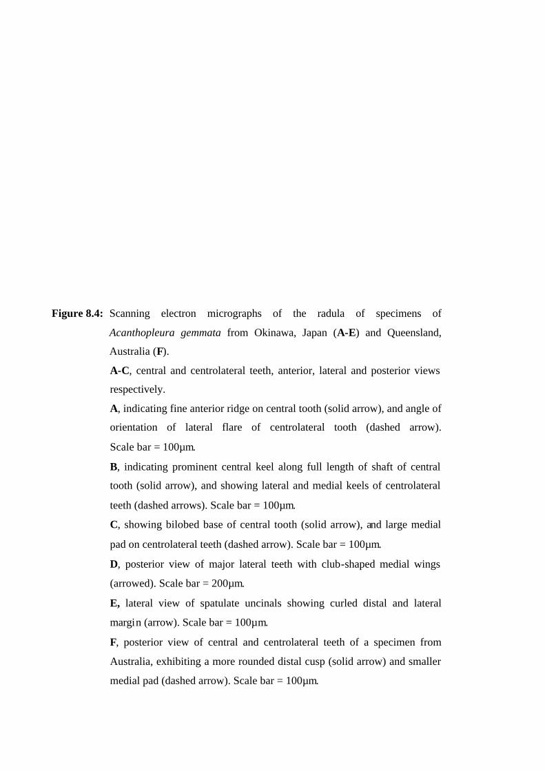
Figure 8.4: Scanning electron micrographs of the radula of specimens of
Acanthopleura gemmata from Okinawa, Japan (A-E) and Queensland,
Australia (F).
A-C, central and centrolateral teeth, anterior, lateral and posterior views
respectively.
A, indicating fine anterior ridge on central tooth (solid arrow), and angle of
orientation of lateral flare of centrolateral tooth (dashed arrow).
Scale bar = 100µm.
B, indicating prominent central keel along full length of shaft of central
tooth (solid arrow), and showing lateral and medial keels of centrolateral
teeth (dashed arrows). Scale bar = 100µm.
C, showing bilobed base of central tooth (solid arrow), and large medial
pad on centrolateral teeth (dashed arrow). Scale bar = 100µm.
D, posterior view of major lateral teeth with club-shaped medial wings
(arrowed). Scale bar = 200µm.
E, lateral view of spatulate uncinals showing curled distal and lateral
margin (arrow). Scale bar = 100µm.
F, posterior view of central and centrolateral teeth of a specimen from
Australia, exhibiting a more rounded distal cusp (solid arrow) and smaller
medial pad (dashed arrow). Scale bar = 100µm.
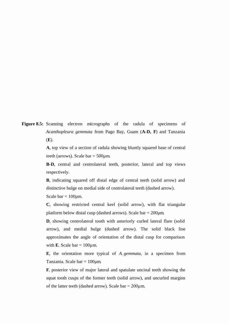
Figure 8.5: Scanning electron micrographs of the radula of specimens of
Acanthopleura gemmata from Pago Bay, Guam (A-D, F) and Tanzania
(E).
A, top view of a section of radula showing bluntly squared base of central
teeth (arrows). Scale bar = 500µm.
B-D, central and centrolateral teeth, posterior, lateral and top views
respectively.
B, indicating squared off distal edge of central teeth (solid arrow) and
distinctive bulge on medial side of centrolateral teeth (dashed arrow).
Scale bar = 100µm.
C, showing restricted central keel (solid arrow), with flat triangular
platform below distal cusp (dashed arrows). Scale bar = 200µm.
D, showing centrolateral tooth with anteriorly curled lateral flare (solid
arrow), and medial bulge (dashed arrow). The solid black line
approximates the angle of orientation of the distal cusp for comparison
with E. Scale bar = 100µm.
E, the orientation more typical of A. gemmata, in a specimen from
Tanzania. Scale bar = 100µm.
F, posterior view of major lateral and spatulate uncinal teeth showing the
squat tooth cusps of the former teeth (solid arrow), and uncurled margins
of the latter teeth (dashed arrow). Scale bar = 200µm.

Figure 8.7: Scanning electron micrographs of the radula of Acanthopleura testudo
from Djibouti, Gulf of Aden.
A-C, central and centrolateral teeth, posterior, top and lateral views
respectively.
A, showing the sturdy, squat central teeth with relatively straight sides,
distinctly flared base (solid arrow), and somewhat square distal cusp
(dashed arrow). Scale bar = 100µm.
B, showing the distinctive nodule proximal to the flat triangular platform
(dashed arrow), smooth anterior surface (solid arrow) and two small but
distinct anterior lobes (arrowheads). Scale bar = 100µm.
C, showing the centrolateral teeth with recurved lateral flare (solid arrow)
and lateral bulge in place of the usual pad (dashed arrow).
Scale bar = 200µm.
D, posterior view of major lateral and centrolateral teeth showing the large
medially extruded cusps of the former (solid arrow), and lateral bulge of
the latter (dashed arrow). Scale bar = 200µm.
E, top view of a section of the radula showing the broad, medially
extruded blades of the spatulate uncinal teeth. Scale bar = 500µm.
F, lateral view of the spatulate uncinals showing the recurved nature of the
margins (solid arrow). Scale bar = 200µm.
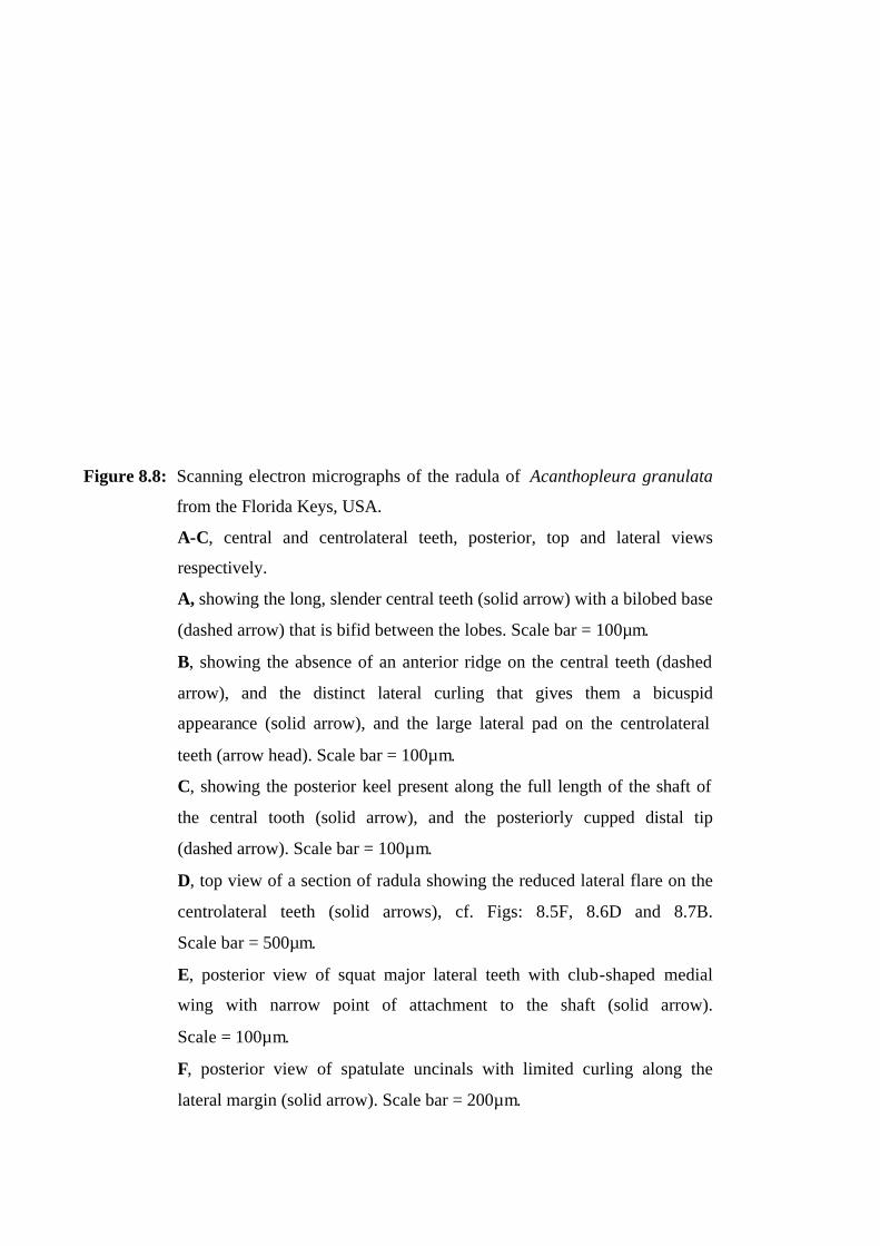
Figure 8.8: Scanning electron micrographs of the radula of Acanthopleura granulata
from the Florida Keys, USA.
A-C, central and centrolateral teeth, posterior, top and lateral views
respectively.
A, showing the long, slender central teeth (solid arrow) with a bilobed base
(dashed arrow) that is bifid between the lobes. Scale bar = 100µm.
B, showing the absence of an anterior ridge on the central teeth (dashed
arrow), and the distinct lateral curling that gives them a bicuspid
appearance (solid arrow), and the large lateral pad on the centrolateral
teeth (arrow head). Scale bar = 100µm.
C, showing the posterior keel present along the full length of the shaft of
the central tooth (solid arrow), and the posteriorly cupped distal tip
(dashed arrow). Scale bar = 100µm.
D, top view of a section of radula showing the reduced lateral flare on the
centrolateral teeth (solid arrows), cf. Figs: 8.5F, 8.6D and 8.7B.
Scale bar = 500µm.
E, posterior view of squat major lateral teeth with club-shaped medial
wing with narrow point of attachment to the shaft (solid arrow).
Scale = 100µm.
F, posterior view of spatulate uncinals with limited curling along the
lateral margin (solid arrow). Scale bar = 200µm.
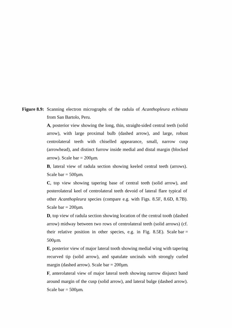
Figure 8.9: Scanning electron micrographs of the radula of Acanthopleura echinata
from San Bartolo, Peru.
A, posterior view showing the long, thin, straight-sided central teeth (solid
arrow), with large proximal bulb (dashed arrow), and large, robust
centrolateral teeth with chiselled appearance, small, narrow cusp
(arrowhead), and distinct furrow inside medial and distal margin (blocked
arrow). Scale bar = 200µm.
B, lateral view of radula section showing keeled central teeth (arrows).
Scale bar = 500µm.
C, top view showing tapering base of central teeth (solid arrow), and
posterolateral keel of centrolateral teeth devoid of lateral flare typical of
other Acanthopleura species (compare e.g. with Figs. 8.5F, 8.6D, 8.7B).
Scale bar = 200µm.
D, top view of radula section showing location of the central tooth (dashed
arrow) midway between two rows of centrolateral teeth (solid arrows) (cf.
their relative position in other species, e.g. in Fig. 8.5E). Scale bar =
500µm.
E, posterior view of major lateral tooth showing medial wing with tapering
recurved tip (solid arrow), and spatulate uncinals with strongly curled
margin (dashed arrow). Scale bar = 200µm.
F, anterolateral view of major lateral teeth showing narrow disjunct band
around margin of the cusp (solid arrow), and lateral bulge (dashed arrow).
Scale bar = 500µm.

Figure 8.10: Scanning electron micrographs of the radula of Acanthopleura
brevispinosa from Zanzibar, Tanzania.
A and B, central and centrolateral teeth, posterior, and lateral views
respectively.
A, showing the short, squat, straight-sided (solid arrow), anteroposteriorly
flattened central teeth, with a bifid base (arrowhead), and centrolaterals
with a very small lateral flare (dashed arrow). Scale bar = 100µm.
B, showing concave triangular platform (dashed arrow) below the cusp of
the central teeth that extends into a soft, central keel (arrowhead), and a
small square lateral pad on the centrolateral teeth (solid arrow).
Scale bar = 100µm.
C, oblique view of radula section showing major lateral teeth with wedge-
shaped medial wing (dashed arrows) and tapering extrusion (solid arrows),
and spatulate uncinals with folded margin half way down the shaft
(arrowhead). Scale bar = 500µm.
D, posterior view of major lateral teeth with small, medially extruded
cusps (solid arrow), and spatulate uncinals with limited marginal curling
(dashed arrow). Scale bar = 100µm.
E, anterior view of major lateral tooth cusps with distinctive lateral bulge
(arrow). Scale bar = 100µm.
F, anterior view of major lateral tooth cusp removed from shaft, showing
incomplete attachment of the lateral bulge at its base (arrow).
Scale bar = 100µm.
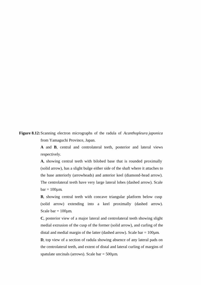
Figure 8.12: Scanning electron micrographs of the radula of Acanthopleura japonica
from Yamaguchi Province, Japan.
A and B, central and centrolateral teeth, posterior and lateral views
respectively.
A, showing central teeth with bilobed base that is rounded proximally
(solid arrow), has a slight bulge either side of the shaft where it attaches to
the base anteriorly (arrowheads) and anterior keel (diamond-head arrow).
The centrolateral teeth have very large lateral lobes (dashed arrow). Scale
bar = 100µm.
B, showing central teeth with concave triangular platform below cusp
(solid arrow) extending into a keel proximally (dashed arrow).
Scale bar = 100µm.
C, posterior view of a major lateral and centrolateral teeth showing slight
medial extrusion of the cusp of the former (solid arrow), and curling of the
distal and medial margin of the latter (dashed arrow). Scale bar = 100µm.
D, top view of a section of radula showing absence of any lateral pads on
the centrolateral teeth, and extent of distal and lateral curling of margins of
spatulate uncinals (arrows). Scale bar = 500µm.
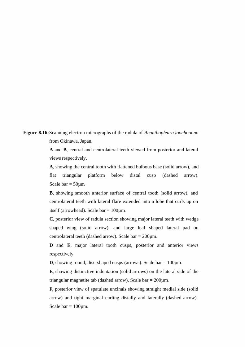
Figure 8.16: Scanning electron micrographs of the radula of Acanthopleura loochooana
from Okinawa, Japan.
A and B, central and centrolateral teeth viewed from posterior and lateral
views respectively.
A, showing the central tooth with flattened bulbous base (solid arrow), and
flat triangular platform below distal cusp (dashed arrow).
Scale bar = 50µm.
B, showing smooth anterior surface of central tooth (solid arrow), and
centrolateral teeth with lateral flare extended into a lobe that curls up on
itself (arrowhead). Scale bar = 100µm.
C, posterior view of radula section showing major lateral teeth with wedge
shaped wing (solid arrow), and large leaf shaped lateral pad on
centrolateral teeth (dashed arrow). Scale bar = 200µm.
D and E, major lateral tooth cusps, posterior and anterior views
respectively.
D, showing round, disc-shaped cusps (arrows). Scale bar = 100µm.
E, showing distinctive indentation (solid arrows) on the lateral side of the
triangular magnetite tab (dashed arrow). Scale bar = 200µm.
F, posterior view of spatulate uncinals showing straight medial side (solid
arrow) and tight marginal curling distally and laterally (dashed arrow).
Scale bar = 100µm.

Figure 8.17: Scanning electron micrographs of the radula of Acanthopleura nigra from San
Bartolo, Peru.
A-C, central and centrolateral teeth, posterior, top-anterior and top-posterior
views respectively.
A, showing the long, thin, straight-sided central tooth with bulbous base (solid
arrow) and concave region below cusp (dashed arrow) that extends into a
central keel (arrowhead) for most of the shaft. Scale bar = 200µm.
B, showing central tooth smooth and convex distally (solid arrow), but keeled
for the remainder of the shaft (dashed arrow). Scale bar = 200µm.
C, showing tapering base of central tooth (solid arrow), and centrolateral teeth
with narrow cusp (arrowhead), deeply furrowed region within the curled distal
and medial margins (dashed arrow), and absence of lateral flare. Scale bar =
200µm.
D, top view of radula section showing the location of teeth in each row,
particularly the midway position of the central teeth (solid arrow) and the
relatively large spatulate uncinals (dashed arrow). Scale bar = 500µm.
E and F, posterior and anterior views of major lateral teeth showing.
E, tapering tip of medial wing (solid arrow). Scale bar = 200µm.
F, cusps with prominent lateral bulge (solid arrow) and narrow disjunct band
(dashed arrow). Scale bar = 200µm.
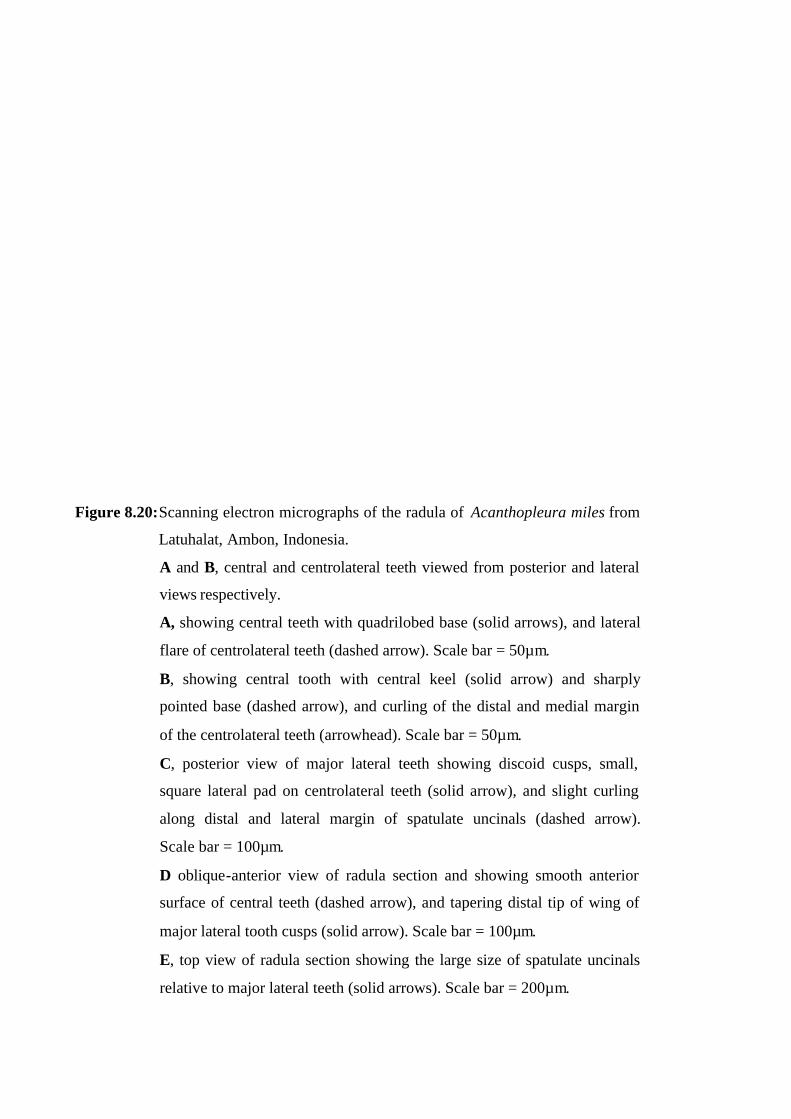
Figure 8.20: Scanning electron micrographs of the radula of Acanthopleura miles from
Latuhalat, Ambon, Indonesia.
A and B, central and centrolateral teeth viewed from posterior and lateral
views respectively.
A, showing central teeth with quadrilobed base (solid arrows), and lateral
flare of centrolateral teeth (dashed arrow). Scale bar = 50µm.
B, showing central tooth with central keel (solid arrow) and sharply
pointed base (dashed arrow), and curling of the distal and medial margin
of the centrolateral teeth (arrowhead). Scale bar = 50µm.
C, posterior view of major lateral teeth showing discoid cusps, small,
square lateral pad on centrolateral teeth (solid arrow), and slight curling
along distal and lateral margin of spatulate uncinals (dashed arrow).
Scale bar = 100µm.
D oblique-anterior view of radula section and showing smooth anterior
surface of central teeth (dashed arrow), and tapering distal tip of wing of
major lateral tooth cusps (solid arrow). Scale bar = 100µm.
E, top view of radula section showing the large size of spatulate uncinals
relative to major lateral teeth (solid arrows). Scale bar = 200µm.
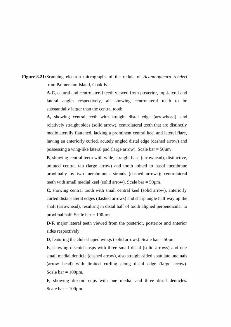
Figure 8.21: Scanning electron micrographs of the radula of Acanthopleura rehderi
from Palmerston Island, Cook Is.
A-C, central and centrolateral teeth viewed from posterior, top-lateral and
lateral angles respectively, all showing centrolateral teeth to be
substantially larger than the central tooth.
A, showing central teeth with straight distal edge (arrowhead), and
relatively straight sides (solid arrow), centrolateral teeth that are distinctly
mediolaterally flattened, lacking a prominent central keel and lateral flare,
having an anteriorly curled, acutely angled distal edge (dashed arrow) and
possessing a wing-like lateral pad (large arrow). Scale bar = 50µm.
B, showing central teeth with wide, straight base (arrowhead), distinctive,
pointed central tab (large arrow) and tooth joined to basal membrane
proximally by two membranous strands (dashed arrows); centrolateral
teeth with small medial keel (solid arrow). Scale bar = 50µm.
C, showing central tooth with small central keel (solid arrow), anteriorly
curled distal-lateral edges (dashed arrows) and sharp angle half way up the
shaft (arrowhead), resulting in distal half of tooth aligned perpendicular to
proximal half. Scale bar = 100µm.
D-F, major lateral teeth viewed from the posterior, posterior and anterior
sides respectively.
D, featuring the club-shaped wings (solid arrows). Scale bar = 50µm.
E, showing discoid cusps with three small distal (solid arrows) and one
small medial denticle (dashed arrow), also straight-sided spatulate uncinals
(arrow head) with limited curling along distal edge (large arrow).
Scale bar = 100µm.
F, showing discoid cups with one medial and three distal denticles.
Scale bar = 100µm.

Figure 8.22: Scanning electron micrographs of the radula of Onithochiton quercinus
from Rottnest Island, Western Australia.
A, central and centrolateral teeth viewed from the posterior, showing
central teeth with straight distal edge (arrowhead), straight sides and a
small central keel (solid arrow), and centrolateral teeth with a smell medial
keel (dashed arrow). Scale bar = 100µm.
B, section of the radula viewed from the anterior side showing central
teeth that are distinctly scooped distally and with a right angle bend half
way along the shaft giving the teeth a chair-shaped appearance (solid
arrows), also centrolateral teeth that are distinctly mediolaterally flattened
(dashed arrows). A single broad, stumpy spatulate uncinal does exhibit any
tapering of the shaft towards the base (arrowhead). Scale bar = 100µm.
C, major lateral teeth viewed from the posterior showing discoid cusps
(solid arrows) with one small medial denticle (dashed arrows). Many of
the club-shaped wings have broken away from the shaft of the tooth
(arrowheads). Scale bar = 200µm.
D, section of the radula viewed from the posterior side showing broad
spatulate uncinals that are slightly curled distally (solid arrows).
Scale bar = 100µm.
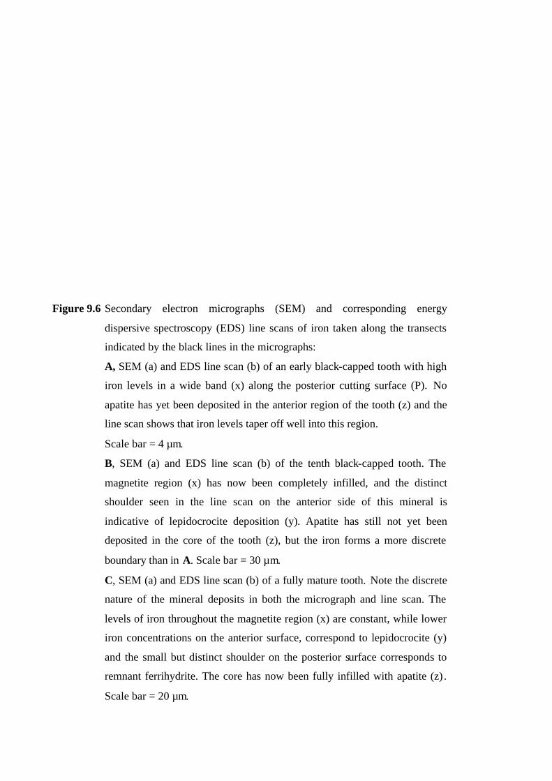
Figure 9.6 Secondary electron micrographs (SEM) and corresponding energy
dispersive spectroscopy (EDS) line scans of iron taken along the transects
indicated by the black lines in the micrographs:
A, SEM (a) and EDS line scan (b) of an early black-capped tooth with high
iron levels in a wide band (x) along the posterior cutting surface (P). No
apatite has yet been deposited in the anterior region of the tooth (z) and the
line scan shows that iron levels taper off well into this region.
Scale bar = 4 µm.
B, SEM (a) and EDS line scan (b) of the tenth black-capped tooth. The
magnetite region (x) has now been completely infilled, and the distinct
shoulder seen in the line scan on the anterior side of this mineral is
indicative of lepidocrocite deposition (y). Apatite has still not yet been
deposited in the core of the tooth (z), but the iron forms a more discrete
boundary than in A. Scale bar = 30 µm.
C, SEM (a) and EDS line scan (b) of a fully mature tooth. Note the discrete
nature of the mineral deposits in both the micrograph and line scan. The
levels of iron throughout the magnetite region (x) are constant, while lower
iron concentrations on the anterior surface, correspond to lepidocrocite (y)
and the small but distinct shoulder on the posterior surface corresponds to
remnant ferrihydrite. The core has now been fully infilled with apatite (z) .
Scale bar = 20 µm.
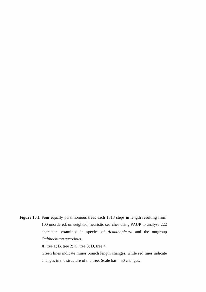
Figure 10.1 Four equally parsimonious trees each 1313 steps in length resulting from
100 unordered, unweighted, heuristic searches using PAUP to analyse 222
characters examined in species of Acanthopleura and the outgroup
Onithochiton quercinus.
A, tree 1; B, tree 2; C, tree 3; D, tree 4.
Green lines indicate minor branch length changes, while red lines indicate
changes in the structure of the tree. Scale bar = 50 changes.
