review of john gofman’s papers on lungcancer …
TRANSCRIPT
LUNGREVIEW OF JOHN GOFMAN’S PAPERS ONCANCER HAZARD FROM INHALED PLUTONIUM
M. B. Snipes
A. L. Brooks
R. G. Cuddihy
R.O.McClellan
September 1975
LF-51
UC-48
LOVELACE FOUNDATIONfor Medical Education and Research
P.O. Box 5890 Albuquerque, NM 87115
Prepared for the Division of Biomedical and Environmental Research, U.S. EnergyResearch and Development Administration Under Contract No. E(29-2)-1013
NOTICE
This report was prepared as an account of work sponsored by the UnitedStates Government. Neither the United States nor the United States EnergyResearch and Development Administration, nor any of their employees, nor anyof their contractors, subcontractors, or their employees, makes any warranty,expressed or implied, or assumes any legal liability or responsibility for theaccuracy, completeness or usefulness of any information, apparatus, product orprocess disclosed, or represents that its use would not infringe privatelyowned rights.
Available from the National Technical Information Service, U.S. Department ofCommerce, Springfield, Va. 22151.
Price: Paper Copy $4.00; Microfiche $1.45.
LF-51
Category: UC-48
REVIEW OF JOHN GOFMAN’S PAPERS ON
LUNG CANCER HAZARD FROM INHALED PLUTONIUM
September 1975
Prepared by
Dr. M. B. SnipesDr. A. L. BrooksDr. R. G. CuddihyDr. R. O. McClellan
Inhalation Toxicology Research Institute
Lovelace Foundation for Medical Education and Research
P. O. Box 5890, Albuquerque, New Mexico 87115
Prepared for the Division of Biomedical and Environmental Research under U.S.
Energy Research and Development Administration Contract E(29-2)-I013.
ABSTRACT
John W. Gofman released two papers recently on behalf of the Committee
for Nuclear Responsibility. Using what is in effect an extension of the "hot
particle" hypothesis, he defines the critical tissue in lung of humans to com-
prise i gm of bronchiolar epithelium which has a 500-day half-life for clearance
of a small portion of inhaled plutonium particles which deposit on or become
trapped on this epithelial region of the lung after clearance from alveoli.
The effect is to extend to the sensitive bronchiolar region a long-term burden
of inhaled plutonium. The slowed clearance is presumed by Gofman to be the
result of impaired ciliary activity because of smoking. His hypothesis is in-
validated by recognition that if it were true smokers would eventually accumu-
late sufficient cigarette smoke residue in the lungs to cause blockage of small
airways and cessation of ventilation. Gofman’s use of "lung cancer dose" in
his risk analyses is misleading and obscures reality; for example, the risk from
natural background radiation is projected to be considerably greater than for
fallout plutonium when analyzed using Gofman’s approach. Defining lung cancer
risk from inhaled plutonium in terms of risk per pound or ton of plutonium does
little beyond impress or confuse the average reader with large numbers. His
risk estimates for human lung cancers caused by plutonium from worldwide fallout
are based entirely on his preposterous model for long-term retention of inhaled
plutonium in bronchioles. In summary, Gofman’s approach to estimation of excess
lung cancer from inhaled plutonium is uncertain, unscientific and is not sub-
stantiated by current knowledge of the toxicity of plutonium.
TABLE OF CONTENTS
Page
ABSTRACT ................................................................. i
INTRODUCTION ............................................................. I
SUMMARY OF CNR REPORT 1975-1 AND 1975-2 .................................. 2
Specific Comments Related to CNR 1975-1 ............................. 3
Specific Comments Related to CNR 1975-2 ............................. 9
REFERENCES ............................................................... II
ii
REVIEW OF JOHN GOFMAN’S PAPERS ON
LUNG CANCER HAZARD FROM INHALED PLUTONIUM
by
M. B. Snipes, A. L. Brooks, R. G. Cuddihy and R O. McClellan
INTRODUCTION
John W. Gofman recently released two papers (1,2) on behalf of the
Committee for Nuclear Responsibility in which he developed lung cancer hazard
estimates for inhaled plutonium and estimates for numbers of human lung cancers
related to worldwide fallout of plutonium. These papers are apparently intended
for the non-scientific reader and consist mainly of assumptions, simple mathe-
matical derivations and predictions. The predictions and derived large numbers
of lung cancers predicted for fallout plutonium serve only to confuse or impress
the reader and have little scientific validity. These papers represent a very
non-constructive approach to the problem of lung cancer risk due to inhaled
plutonium.
Gofman states that his approach does not involve the "hot particle"
hypothesis which precipitated considerable controversy in 1974. However, his
approach to this problem is in fact an extension of the hot particle hypothesis.
He defines the critical tissue in lung to be bronchial epithelium comprising
I gm of sensitive epithelium. This was necessary because it then becomes pos-
sible to discuss this tissue as highly sensitive to irritants which leads to
the production of cancer of bronchiogenic origin. The next problem, irradia-
tion of this sensitive tissue for sufficient time to cause lung cancer, is
solved by Gofman by making the remarkable prediction that some plutonium par-
ticles which deposit on that sensitive epithelium or move onto it from alveoli
are trapped there with a 500-day half-life. He therefore conveniently defines
a long-term burden of plutonium or other alpha-emitting particles for a known
sensitive I gm region of lung tissue. The fallacy of this approach will be
pointed out later.
Another thing Gofman does is avoid the use of absorbed dose for relating
the amount of inhaled plutonium to possible risk. He uses the concept of
"lung cancer dose", which is very misleading in that it cannot be readily de-
fined or compared with reality. Moreover, Gofman failed to consider that
-I-
background radiation constitutes a significantly greater hazard than plutonium
in worldwide fallout if also expressed in the meaningless units of "lung can-
cer dose".
Our approach to reviewing these papers was to point out the major assump-
tions in these two papers and develop real-life tests for his hypotheses where
applicable. A test of such hypotheses is to see if predicted results make any
sense in the real world. This was done for Gofman’s prediction of a 500-day
half-life for clearance of a portion of inhaled plutonium deposited on bronchial
epithelium and for Gofman’s prediction of the carcinogenicity of a "lung cancer
dose". Bothpredictions are incompatible with the real world.
SUMMARY OF CNR REPORT 1975-1 and 1975-2
In CNR Report 1975-1, Gofman derives an estimate of lung cancer risk per
unit mass of plutonium inhaled. This calculation is similar in its initial
goals to the Radiological Effects of lonizing Radiation (BEIR) Report (3);
ever, it attempts to develop separate risk estimates for cigarette smokers as
compared to nonsmokers. Throughout the report, comparisons of the Gofman-Tamplin
risk estimates are made with the author’s extensions of the BEIR report data.
It should be clearly noted that risk values indicated as "BEIR" estimates do
not come directly from that report but represent Gofman’s personalized extension
of theBEIR report data. In general, the "Gofman" risk estimates for lung can-
cer in nonsmokers are reasonably close to his BEIR report data extensions. The
major differences occur in his modification of risk factors as applied to ciga-
rette smokers. Risk probabilities for smokers are several orders of magnitude
greater than for nonsmokers exposed to the same plutonium inhalation dose. Hethen extends these risk values from the amount of 239pu inhaled to the number
of "lung cancer doses" that would exist in a pound of 239pu or reactor grade
plutonium if it were inhaled.
In the second paper (CNR Report 1975-2), Gofman uses his plutonium lung
cancer probabilities to estimate expected lung cancer incidence in the popula-
tion exposed to nuclear weapons fallout. Because of Gofman’s estimated higher
risk to cigarette smokers compared to nonsmokers inhaling the same quantity of
plutonium, the over-whelming majority of lung cancers are projected to occur
in smokers. If cigarette smokers were excluded from Gofman’s model, plutonium
would not represent a significant hazard. Very major objections should be
-2-
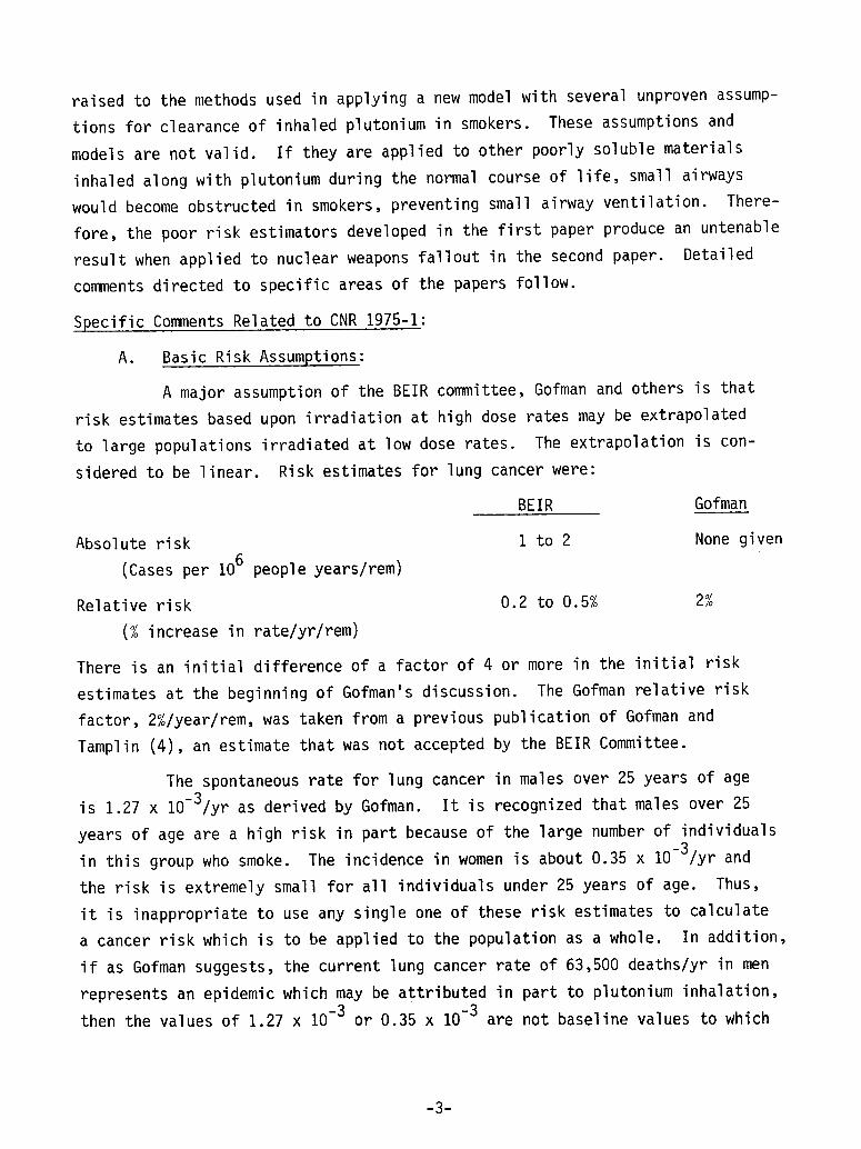
raised to the methods used in applying a new model with several unproven assump-
tions for clearance of inhaled plutonium in smokers. These assumptions and
models are not valid. If they are applied to other poorly soluble materials
inhaled along with plutonium during the normal course of life, small airways
would become obstructed in smokers, preventing small airway ventilation. There-
fore, the poor risk estimators developed in the first paper produce an untenable
result when applied to nuclear weapons fallout in the second paper. Detailed
comments directed to specific areas of the papers follow.
Specific Comments Related to CNR 1975-1:
A. Basic Risk Assumptions:
A major assumption of the BEIR committee, Gofman and others is that
risk estimates based upon irradiation at high dose rates may be extrapolated
to large populations irradiated at low dose rates. The extrapolation is con-
sidered to be linear. Risk estimates for lung cancer were:
BEIR
1 to2
Gofman
None givenAbsolute risk
(Cases per 106 people years/rem)
Relative risk 0.2 to 0.5% 2%
(% increase in rate/yr/rem)
There is an initial difference of a factor of 4 or more in the initial risk
estimates at the beginning of Gofman’s discussion. The Gofman relative risk
factor, 2%/year/rem, was taken from a previous publication of Gofman and
Tamplin (4), an estimate that was not accepted by the BEIR Committee.
The spontaneous rate for lung cancer in males over 25 years of age
is 1.27 x lO-3/yr as derived by Gofman. It is recognized that males over 25
years of age are a high risk in part because of the large number of individuals
in this group who smoke. The incidence in women is about 0.35 x lO-3/yr and
the risk is extremely small for all individuals under 25 years of age. Thus,
it is inappropriate to use any single one of these risk estimates to calculate
a cancer risk which is to be applied to the population as a whole. In addition,
if as Gofman suggests, the current lung cancer rate of 63,500 deaths/yr in men
represents an epidemic which may be attributed in part to plutonium inhalation,
then the values of 1.27 x 10⁻³ or 0.35 x 10⁻³ are not baseline values to which
-3-
the relative risk escalation due to plutonium inhalation may be added. It is
already represented in these values. A more representative rate for spontaneous
cancer induction in the total U.S. population is 4.0 x 10⁻⁴ deaths/yr (63,000
deaths/yr for men + 17,600 deaths/yr for females/200 million people). Use of
relative risk of O.5%/year/rem would lead to 2 additional cases per 106 people
years/rem. Curiously enough, Gofman back-calculates this number for the "BEIR"
estimate from his own data to be 6.3 cases per 106 people years/rem to lung
(Gofman’s page 6 - CNR Report 1975-1).
B. "Lung Cancer Dose":
Gofman’s definition of "lung cancer dose" is I/risk, where risk is
the spontaneous lung cancer incidence multiplied by 2% per year per rem of
exposure. He next multiplies by 30 to derive a lifetime risk estimate, which
he takes the reciprocal of to derive "lung cancer dose" in units of man-rem.
He next uses the assumption that 1 ~Ci delivers a cumulative infinite dose of
2000 rems. Dividing his lung cancer dose in man-rems by 2000 rems yields
values for lung cancer dose in units of ~Ci. The concept of "lung cancer dose"
for inhaled plutonium, which may be considered similar to a "drowning dose" for
water or a "burning death dose" for fire, is not truly scientific. It is more
akin to a quantity which if deposited in lung would give a certain probability
for lung cancer among members of a given population. Its use does have a practi-
cal advantage for Gofman’s point of view in that it avoids the problem of re-
lating absorbed radiation dose to anticipated biological effects. Thereby, it
obscures comparisons with background radiation dose levels which exceed lung
irradiation due to past plutonium inhalation by several orders of magnitude.
C. Lung Cancer Dose per Unit Mass:
Tables 1 and 2 on page I0 yield estimates for lung cancer risk due
to deposited 239pu or reactor grade plutonium. The "Gofman-Tamplin" and "BEIR"
estimates differ by a factor of 4, for reasons previously stated. Since the
BEIR Committee estimate is the currently accepted estimate of the scientific
community, it is the only meaningful comparison to the "Gofman-Tamplin" esti-
mates. Further, extrapolations of data to numbers of cancers per pound or ton
of plutonium or other toxicant have little significance. These estimates pro-
vide no additional useful data and serve only to confuse or impress the reader
with large numbers. It should be emphasized that lung cancer risk is associated
-4-
with radioactive material deposited in the lung; any expression of risk in
terms of tons of something is essentially useless.
D. Determination of Relevant Tissue at Risk and Dose Calculations:
The mass of a tissue used for radiation dose calculations is the
total mass in which the energy is deposited. Since the range of alpha particles
in tissue of unit density is on the order of 40 um, energy is deposited in
blood and lymphatic fluid, insensitive components of lung structure and,
especially in the case of smokers, debris contained in the lung. Selecting a
lung weight of 570 gm (an estimate for mass of bloodless lung for man) for
energy deposition will not produce a result greatly different from using I000
gm. Further reduction to a weight for sensitive tissue of I gm is inconsistent
with biological and physical properties of the lung. Gofman assumes a sensitive
tissue mass of I gm which naturally leads to an extremely large radiation dose
estimation per unit mass of plutonium inhaled.
The origin of tumors which might be produced in man due to inhalation
and retention in lung of 239pu is uncertain as no such tumors have been identi-
fied to date. Cigarette smokers do develop bronchial tumors, but the site of
irritation or exposure for carcinogens derived from cigarette smoke may be
substantially different from 239pu. Long-term retention of 239pu in lung tis-
sue primarily occurs in alveoli. Gofman assumes the critical tissue for 239pu
is the bronchial epithelium whereby a very small lung volume and related tissue
mass becomes the sensitive region of interest. This was represented by about
I gm of tissue. Plutonium-239 trapped in this small volume was assigned a
clearance half-time of 500 days due to destruction of cilia in this region
from contact with tobacco residues in smokers.
E. Testin 9 the Gofman Hypothesis in Human Smokers:
Gofman emphasizes the role of cilia in bronchi as the sole determinant
for clearance of inhaled particles which initially deposit there or pass dur-
ing lung clearance from alveoli. There may be a reduction in active cilia in
smokers, slowing movement, but lung clearance still continues in these bronchi.
Albert et al. (5) reported on bronchial deposition and clearance of aerosols
for cigarette smoking and nonsmoking humans. Their conclusion was that "It
is disappointing...that there is so little effect on the overall time for
bronchial clearance in cigarette smokers .... But this is understandable since
-5-
backup mechanisms for bronchial clearance must operate effectively to maintain
airway patency." In essence, clearance mechanisms were unaltered by smoking.
Lourenco et al. (6) noted essentially the same results, with a delay of tracheo-
bronchial clearance of 1-4 hours after inhalation in smokers. The notion that
cilia are absolutely necessary for clearance is not substantiated. All bronchial
areas in the human lung have goblet cells which secrete mucus. To maintain
airway patency the mucus thus produced must move continuously and towards the
pharynx or small airways will become obstructed and ventilation will cease.
Lung bronchial regions clear constantly in healthy humans and smokers since
airways must be patent to allow respiration. At most, only a slight delay in
clearance rate could be possible, otherwise the small bronchioles would plug
and death would soon follow.
Assuming Gofman’s model did apply to cigarette smokers, then the
inert material inhaled in smoking will be retained in the same manner as he
projects for plutonium particles. The amount of smoke inhaled by a person
smoking one filter cigarette is approximately 16.1 mg (7). If a smoker con-
sumed 20 cigarettes per day each day of a year, then 118 gm would be inhaled
each year. Assuming Gofman’s value of 2.7% deposition in the sensitive region,
3.2 gm of material would collect each year, with a 500-day retention half-time
representing a 720-day (~ 2 yr) mean residence time, about 6.4 gm would accumu-
late in this volume of 20 cm3 surrounded by Gofman’s sensitive bronchiolar
tissue of 1-gm mass. Thus, ventilation would cease or be seriously impaired
in heavy smokers. Other environmental contaminants would also contribute to
such a blockage. The fact that smokers may live a nearly normal lifespan
compromises such a model of bronchiolar clearance - it is not compatible with
life. This potential respiratory blockage would also serve two functions in
altering the dose to the bronchial epithelium: (a) to provide considerable
self-absorption for plutonium co-deposited with the smoke residue and (b)
minimize further deposition of inhaled plutonium and provide a protective
function.
Gofman presents a risk estimate on page 19 of 1.27 x lO-3/year for
"spontaneous" lung cancer rate for men (USA) over 25 years of age. This value
does not represent a spontaneous rate, rather a base rate already affected by
lung cancer deaths due to cigarette smoking and inhalation of industrial and
environmental toxicants, either naturally occurring or man-made. Two addi-
-6-
tional risk estimates, one for smokers and one for nonsmokers, are also derived.
The value of 2.3 x lO-4/year for male nonsmokers over 25 years of age would
more closely represent the spontaneous incidence rate (without the influence
of cigarette smoking) than the combined rate of 1.27 x lO-3/year.
In the "Step 3 Calculation..." on page 20, Gofman ignores the fact
that there are no goblet cells in alveoli but they are present in ciliated
regions of the lung and that mucus will move in airways with or without local
ciliary action. The pulmonary region does not fill up with the normally oc-
curring naturally generated aerosols of dust, etc. because large particles
deposit in the upper airways where they are readily cleared with the mucus
flow. Small particles which reach the alveoli may be retained there with rela-
tively long half-times.
Data included in Table i0 (page 24) are probably reasonable for both
the "Gofman-Tamplin" and "BEIR" estimates of micrograms of plutonium per gram
of lung needed to induce or initiate lung cancer in nonsmokers. These values
should also represent the risk for smokers, excluding the added risk due to
cigarette smoking. The difference between the Gofman-Tamplin and BEIR esti-
mates, only a factor of 4, is much less than biological variability observed
in many experiments.
On the bottom of page 24 and top of page 25, Dr. Gofman states that
"Since ciliary function is the mechanism counted upon for differentiating rapid
clearance in the bronchi versus slow clearance in the pulmonary region, the
absence of effective ciliary function makes it reasonable,...,to expect clearance
times to become identical. If there is any intrinsic more rapid clearance
mechanism (aside from cilia) for bronchial cells than for pulmonary cells,
such mechanism is totally hypothetical. Indeed, the effect can be such as to
worsen the estimates." This statement ignores the possibility that the cough,
mechanical breathing action, surface phenomena, etc. may also serve to move
mucus in airways. That airways do not congest in smokers indicates that either
ciliary action is not seriously impaired or that ciliary action is not ex-
clusively responsible for lung clearance.
G. Comments Related to Gofman’s General Discussion:
Experimental studies in Beagle dogs have indicated that 27.1 ug of
239pu is carcinogenic (8). This compares with 28.8 ug from Gofman’s extension
-7-
of the "BEIR" data and 7.3 ug as his cancer producing dose in nonsmokers,
Table I0. These are reasonable comparisons. It is noteworthy, however, that
this amount of activity in dogs did not produce demonstrable lifespan short-
ening. It may be appropriate therefore to make a distinction between life-
shortening and the presence of a lung tumor.
In considering the risk to the public-at-large, pp. 28-30, Gofman
ignores the fact that exposure standards are modified by the "low as practicable"
criteria. Each individual will not accumulate a maximum burden - whatever the
associated hazard. For nonsmokers, Gofman presents a risk estimate of 2000
deaths per year due to lung cancer for a 20-year period of nuclear industry
operation giving uniform maximum population exposure. This estimate is quite
large because individuals in the population cannot be exposed to maximum levels
for 20 years. However, while this estimate is large, it is not large in com-
parison to other risks taken voluntarily and involuntarily by the U.S. popula-
tion and the fact that approximately 1.9 million U.S. citizens die every year
from one cause or another. His estimated risk for smokers is probably 400
times too high. A factor of 4 is due to excessive risk assumed for each rem
of population exposure and a factor of 103 due to the application of a pre-
posterous model for bronchiolar clearance of plutonium particles. Dividing by
400, the risk for smokers in the general population would be 6000 deaths per
year for a maximum permissible burden of 16 nCi in each individual. Being
mindful that
1) the U.S. population will not be exposed to the maximum permissible
burden,
2) about 84,000 U.S. citizens presently die from lung cancer each
year, 45,000 die as a result of automobile-related accidents
and 1.9 x 10⁶ deaths occur each year in total (9),
3) The cancer risk estimate given in CNR Report 1975-1 for smokers
is probably a factor of 400 too high,
4) Gofman’s approach to estimation of lung cancer risk is uncertain,
unscientific and cannot be related to radiation doses from
plutonium,
then the plutonium hazard from nuclear fallout is truly less dramatic than
Gofman might imagine.
-8-
Specific Comments Related to CNR 1975-2:
A. Introduction:
In CNR Report 1975-1, estimates were derived for lung cancer risk
per unit mass of deposited plutonium in cigarette smokers and nonsmokers. The
estimates for nonsmokers were about 4 times higher than might be developed from
data included in the BEIR report. Estimates for smokers were about 400 times
too high due mainly to application of an absurd model for describing bronchial
clearance processes. In CNR Report 1975-2, these risk estimates are applied
to nuclear weapons fallout in estimating its impact upon U.S. and world popula-
tion lung cancer incidence. In fact, Gofman’s entire argument in CNR Report
1975-2 is based on his assessment of risk in CNR Report 1975-1. That risk esti-
mates in the first report can be invalidated essentially nullifies risk esti-
mates in the second report.
Gofman suggests that the current lung cancer rate - 63,500 deaths per
year for men and 17,000 deaths per year for women - is due in part to fallout
plutonium. This may be a true statement; however, in view of the fact that no
sex-related differences in lung cancers due to radiation exposure have been
demonstrated, lung cancer patterns in the general population probably reflect
smoking patterns in males and females.
B. Fallout:
As a result of atmospheric weapons testing, an estimated 320,000 Ci
(11,500 Ibs.) of long-lived plutonium isotopes has been deposited on the surface
of the earth; of this, about 250,000 Ci deposited on the northern hemisphere
and 16,000 Ci on the United States (I0). Machta et al. (ii) has stated
deposition of environmental pollutants is approximately 90% by rain droplet
washout. Thus, of the total amount deposited, perhaps only 1150 pounds or 32,000
Ci of weapons plutonium would have reasonably been available for inhalation by
inhabitants of the northern hemisphere. Using the Bennett (12) estimate of
pCi/person for inhaled plutonium, citizens of the United States would have ac-
cumulated approximately 8.5 mCi or 5 x 10⁻⁷ of the total 16,000 Ci deposited
on the United States. This indicates a deposition factor of 10-7 to 10-8 for
that activity dispersed into the atmosphere.
C. Lung Cancer Risk from Natural Background Based on Gofman’s Model:
Gofman proceeds to equate 7.3 micrograms of 239pu deposited or 450 pCi,
-9-
to the "lung cancer dose" for nonsmokers. Using the assumption that 1 ~Ci yields
a lifetime dose in humans of 2,000 rem, 450 pCi would yield a lifetime dose of0.9 rem. Therefore, the nonsmoker would accumulate an average dose to lungs
of approximately 10 millirem per year for a 70-year lifespan. Natural back-
ground radiation contributes approximately 150 millirem per person per year.
Background radiation dose to lung would therefore be 15 times greater than from450 pCi of 239pu. Thus, using the Gofman risk evaluation, about 15 times as
many lung cancer deaths would be related to background radiation for any given
time period for the U.S. population as would be related to inhaled plutonium;
this would apply, anyway, for nonsmokers. Using lung cancer risk per unit mass
of plutonium rather than rem dose tends to obscure this comparison.
D. Human Experience:
The workers in the Manhattan Project as well as those exposed during
the Rocky Flats fire in 1965 have not developed a single lung cancer. Perhaps
the latent period is not over. In any event, this again emphasizes that latency
period and age may both be important factors in influencing the potential fordetecting lung cancer resulting from 239pu deposited in the lung. Life short-
ening has not occurred in either group and perhaps will not, even if one or
more of these workers eventually develop lung tumors relatable to their plu-
tonium exposures.
-I0-
REFERENCES
1. Gofman, J. W. "The Cancer Hazard from Inhaled Plutonium." Committee for
Nuclear Responsibility. CNR Report 1975-1, May 14, 1975.
2. Gofman, J. W. "Estimated Production of Human Lung Cancers by Plutonium
from Worldwide Fallout." Committee for Nuclear Responsibility. CNR
Report 1975-2, July I0, 1975.
3. The Effects on Populations of Exposure to Low Levels of lonizing Radiation.
Report of the Advisory Committee on the Biological Effects of lonizing
Radiations, National Academy of Sciences, Division of Medical Sciences,
Washington, D. C., 1972.
4. Gofman, J. W. and A. R. Tamplin. "Epidemiologic Studies of Carcinogenesis
by lonizing Radiation," In Proceedings of the Sixth Berkeley Symposium on
Mathematical Statistics and Probability. Statistical Laboratory, University
of California, U. C. Press, Berkeley, California, pp. 235-277, 1971.
5. Albert, R. E., M. Lippmann, H. T. Peterson, Jr., J. Berger, K. Sanborn
and D. Bohning. "Bronchial Deposition and Clearance of Aerosols." Arch.
Intern. Med. 131: 115-127, 1973.
6. Lourenco, R. V., M. F. Klimek and C. J. Borowski. "Deposition and Clear-
ance of 2p Particles in the Tracheobronchial Tree of Normal Subjects -
Smokers and Nonsmokers." J. Clin. Invest. 50: 1411-1420, 1971.
7. Hinds, W. C. and M. W. First. "Concentrations of Nicotine and Tobacco
Smoke in Public Places." New Eng. J. Med. 292: 844-845, 1975.
8. Bair, W. J. and R. C. Thompson. "Plutonium: Biomedical Research."
Science 183: 715-722, 1974.
9. Monthly Vital Statistics Report, Provisional Statistics, Annual Summary
for the United States, 1974. U.S. Department of Health, Education, and
Welfare, Vol. 23, No. 13, May 30, 1975.
10. Draft Environmental Impact Statement for the Liquid Metal Fast Breeder
Reactor Program, 1975.
ii. Machta, L., G. J. Ferber and J. L. Heffter. "Local and World-wide Pollut-
ant Concentrations and Population Exposures from a Continuous Point
Source." Air Resources Laboratories, National Oceanic and Atmospheric
Administration, Silver Spring, Maryland, 1973.
-ii-
12. Bennett, B. G. "Fallout 239pu Dose to Man." l__n_n Fallout Program Quarterly
Summary Report, Health and Safety Laboratory, U.S. Atomic Energy Report
HASL-278, pp 41-61, 1974.
-12-
0
U_._J
LF-50
UC-48
ESTIMATES OF MORTALITY DUE TO RADIATIONPNEUMONITIS AND PULMONARY FIBROSIS AFTER EXPOSURE
TO RADIONUCLIDE RELEASES IN HYPOTHETICALLIGHT WATER REACTOR ACCIDENTS
F.F.Hahn
September 1975
LOVELACE FOUNDATIONfor Medical Education and Research
P.O. BOX 5890 Albuquerque, NM 87115
Prepared for the Division of Biomedical and Environmental Research, U.S. Energ~Research and Development Administration Under Contract No. E(29-2)-101~
NOTICE
This report was prepared as an account of work sponsored by the United StatesGovernment. Neither the United States nor the United States Energy Research andDevelopment Administration, nor any of their employees, nor any of their contractors,subcontractors, or their employees, makes any warranty, expressed or implied, orassumes any legal liability or responsibility for the accuracy, completeness orusefulness of any information, apparatus, product or process disclosed, or repre-sents that its use would not infringe privately owned rights.
The research described in this report involved animals maintained in animalcare facilities fully accredited by the American Association for Accreditation ofLaboratory Animal Care.
Available from the National Technical Information Service, U. S. Department ofCommerce, Springfield, Va. 22151
Price: Paper Copy $4.00; Microfiche $1.45
LF-50
Category: UC-48
ESTIMATES OF MORTALITY DUE TO RADIATION
PNEUMONITIS AND PULMONARY FIBROSIS AFTER EXPOSURE
TO RADIONUCLIDE RELEASES IN HYPOTHETICAL
LIGHT WATER REACTOR ACCIDENTS
September 1975
by
Fletcher F. Hahn, D.V.M., Ph.D.
Inhalation Toxicology Research Institute
Lovelace Foundation for Medical Education and Research
P. O. Box 5890, Albuquerque, New Mexico 87115
Prepared for the Division of Biomedical and Environmental Research under U.S.
Energy Research and Development Administration Contract E(29-2)-I013.
ABSTRACT
Estimates of incidence of early effects in people after exposure to
radionuclide releases in hypothetical light water reactor accidents are
necessary for benefit risk analysis of various energy sources. Because no
human data exist which directly apply to this problem, estimates must be made
from experimental data in animals. This report makes such estimates based on
hypothetical releases and radiation doses estimated in WASH-1400 and experi-
mental data derived from Beagle dogs exposed to various radionuclides.
ACKNOWLEDGMENTS
The author wishes to thank Dr. Joseph Watson, University of Pittsburgh,
for supplying information about the WASH-1400 Report, Dr. Roger O. McClellan
for his suggestions and encouragement, Mr. Fred C. Rupprecht for editorial
assistance and Ms. Mildred Morgan and Ms. Judith Miller for typing assistance
in the preparation of this report.
ii
TABLE OF CONTENTS
ABSTRACT ................................................................. i
ACKNOWLEDGMENTS .......................................................... ii
LIST OF TABLES ........................................................... iv
LIST OF FIGURES .......................................................... iv
INTRODUCTION ........................................... .................. I
REVIEW ................................................................... I
REFERENCES ............................................................... 12
iii
Table
1
LIST OF TABLES
Summary of dose-response data for man and dog exposed toexternal thoracic radiation ................................
Radiation doses to lung and time of death afterexposure for dogs dying with radiation pneumonitis afterinhalation of various beta-emitting radionuclides ..........
Predicted radiation doses to lung as a result of a"maximum" release nuclear accident .........................
3
7
7
Figure
1
LIST OF FIGURES
Dose accumulation in lung for man for a hypotheticalmaximum release from a nuclear reactor accident andfor dogs exposed to aerosols of 90Y or 91Y in fusedclay particles ............................................. 9
Cumulative incidence of deaths from radiationpneumonitis and/or pulmonary fibrosis in dogsafter inhalation of 90y in fused clay particles(Doses to lung at 365 days after exposure) ................. 9
Cumulative incidence of deaths from radiationpneumonitis and/or pulmonary fibrosis in dogsafter inhalation of 91Y in fused clay particles(Doses to lung at 365 days after exposure) ................. 10
iv
ESTIMATES OF MORTALITY DUE TO RADIATION PNEUMONITIS
AND PULMONARY FIBROSIS AFTER EXPOSURE TO RADIONUCLIDE
RELEASES IN HYPOTHETICAL LIGHT WATER REACTOR ACCIDENTS
by
F. F. Hahn
INTRODUCTION
Irradiation of the lung may occur either from external sources, such as
x-ray therapy or 60Co machines, or from internally-deposited radionuclides
such as inhaled radioactive particles. The radiation dose patterns to the
lung from these two types of exposures are quite different and therefore not
easily compared, but in sufficiently high doses, either can cause radiation
pneumonitis (inflammation or irritation of the lung) which may lead to pulmo-
nary fibrosis (scarring of the lung). Death due tO cardiopulmonary insuffi-
ciency (malfunction of the heart and lungs) may occur within days after
exposure due to radiation pneumonitis or as long as many months after exposure
due to radiation pneumonitis and/or pulmonary fibrosis. The sequence of
events may proceed even though the time of actual radiation exposure is brief
and/or the radioactivity is no longer present in the body.
Factors which influence the development of radiation pneumonitis include
the total dose of radiation, the fractionation of the radiation dose, the
type of radiation, the volume of lung affected, pre-existing diseases and
modifying drugs.
REVIEW
Considerable data concerning radiation pneumonitis have been reported
from studies related to radiation therapy of tumors arising in the lung or
other thoracic structure. Radiation pneumonitis was first described as a
clinical entity by Groover et al. 1 They recognized it as an untoward effect
associated with irradiation of thoracic tumors.
Warren and Spencer2 described the pathology of radiation pneumonitis.
They divided the reaction into three stages: acute, late and late with
superimposed acute radiation pneumonitis. They also noted that probably the
earliest effect is one of mild injury to the alveolar lining cells and capil-
lary endothelium. This suggestion has since been confirmed by numerous
-I-
studies with electron microscopy. 3’4 Edema, swelling, necrosis and prolifera-
tion of endothelium and alveolar epithelium follow. With severe or repeated
injury and attempts for repair, chronic changes such as fibroblastic prolifera-
tion occur within alveolar walls. The changes are not unique to the radiation
reaction but very prominent edema and enlarged epithelial and endothelial
cells in the absence of much exudate are suggestive of radiation pneumonitis.
Rubin and Casarett 5 described the clinical course of patients receiving
thoracic irradiation and used the clinical categories of acute, subacute and
chronic. These three periods are separated mainly on the basis of time after
exposure and overlap one another. Typically, the onset occurs I to 3 months
after completion of a 4 to 6 week course of x-irradiation, but may be delayed
6 months or ~onger. The acute clinical period may be clinically silent
depending on the degree of pulmonary involvement. As the volume of injured
lung increases, dyspnea and coughing become apparent. When more than 75% of
the lung reacts to irradiation, respiratory distress is severe and death may
occur from cardiopulmonary insufficiency. The respiratory system does not
exhibit a distinctly recognizable subacute clinical syndrome in the radiation
reaction as is encountered in other organ systems, but generally it blends
into the chronic period. Symptoms may be seen in this period 6 to 12 months
after irradiation if secondary pulmonary infections are present. The chronic
clinical period occurs about I year after exposure. Symptoms are directly
related to the degree and extent of lung damage. Scarring limited to 50% of
one lung is well tolerated and rarely symptomatic. Progressive changes
involving extensive portions of the lung may lead to right heart failure
or severe chronic dyspnea.
The time dose factors involved in the production of radiation pneumonitis
in man from therapeutic irradiation of the thorax are difficult to derive
because exposure of the lesions is maximized and exposure of the normal lung
is minimized. Generally, doses to the lung cannot be accurately calculated.
Several studies, however, have been reported which involved irradiation of
the entire thorax in treatment regimens for metastatic tumors in the lung.
Baeza6 found that 9 of 39 patients with tumor doses (which approximate lung
doses) greater than 1500-2000 delivered rads in 2-3 weeks had radiation pneu-
monitis (Table I). The average time of onset after exposure, was 2.7 months
for the entire group of patients. Newton7 found that two patients receiving
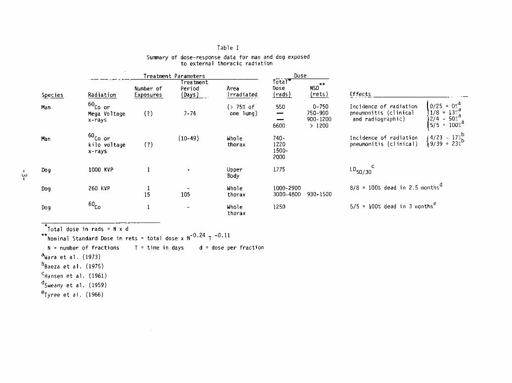
Table I
Summary of dose-response data for man and dog exposedto external thoracic radiation
Species
Man
Man
Treatment Parameters
Radiation
60Co orMega Voltagex-rays
60Co orkilo voltagex-rays
DoseTreatment Total" **
Number of Period Area Dose NSDExposures (Days) Irradiated ~rads) (rets)
(> 75% of 550 0-750(?) 7-74 one lung) ~ 750-900
900-12006600 > 1200
(10-49) Whole 740-(?) thorax 1220
1500-2000
Dog 1000 KVP I - UpperBody
Dog 260 KVP I - Whole15 105 thorax
Dog 60Co I - Wholethorax
1775
1000-29003000-4800 930-1500
1250
Effects
Incidence of radiationpneumonitis (clinicaland radiographic)
Incidence of radiationpneumonitis (clinical)
I0/25: 0%a
I/8 = 13%~2/4 = 50%°5/5 = i00%a
{4/23 = 17%~9/39 23%D
LD50/30c
8/8 = 100% dead in 2.5 monthsd
5/5 = 100% dead in 3 monthse
Total dose in rads = N x d
**Nominal Standard Dose in rets = total dose x N-0"24 T
N = number of fractions T = time in days
awara et al. (1973)
bBaeza et al. (1975)
CHansen et al, (1961)dsweany et al. (1959)
eTyree et al. (1966)
-0.11
d = dose per fraction
3000R thoracic irradiation in two weeks at a rate of 300R per day died with
radiation pneumonitis.
A quantitative approach to time dose factors for the production of
radiation pneumonitis has been published. 8 They studied the incidence of
radiation pneumonitis in a series of 51 patients treated with irradiation of
at least 75% of one whole lung for metastatic pulmonary disease. Using
probit analysis, they determined the dose response curve for development of
radiation pneumonitis as determined by clinical symptoms and radiographic
changes. These data are shown in Table I and indicate that the nominal
standard dose (NSD) which will produce radiation pneumonitis in 50% of those
exposed, is about 1050 rets. The NSD is a mathematically derived term repre-
senting the extrapolation of the dose time relationship back to a single
fraction, taking into account the number of fractions and the total time
separately 9. The NSD is expressed in "rets," or rad equivalent therapeutic.
It is used to compare doses from multiple exposures with those of single
exposures.
Only a few studies of the effects of inhaled radionuclides in man have
been reported. The exposures have either resulted in no effects being ob-
served I0’II or late effects seen and related to metaplasia and neoplasia.12’ 13
One exception is a report of a case of a radium plant worker developing
radiation pneumonitis presumably caused by inhalation of radon and radon
daughters. 14 This early report contains few details and little quantitative
information can be obtained from the paper.
Numerous animal studies describing radiation pneumonitis after external
irradiation have been published and reviewed. 15 Davis 16 was the first to
publish extensive experimental studies designed to determine the histologic
reaction of normal lung to radiation. Using dogs and rabbits, he determined
that the reactions seen in animals were similar to man. Engelstad 17 used
rabbits to study the temporal sequence of the pulmonary lesions after thora-
cic irradiation, He described stages similar to those seen in man.2 Gen-
erally, similar findings have been reported in rats. 18 dogs,19 Syrian ham-
sters 20 and mice. 21 More recent studies of the ultrastructural lesions at
early times after irradiation show focal lesions in many types of lung paren-
chymal cells,22 including the pulmonary capillaries 3 and granular pneumocytes.23
-4-
Several quantitative dose response studies have been reported in animals24
exposed to thoracic irradiation. Hansen et al. determined the LD50/30 forsingle upper body exposure of dogs to x-rays to be 1775 rads. These dogs
died with pulmonary congestion and fibrinous pneumonia but also had other
complicating factors such as neuroendocrine abnormalities and infection.
Sweany et ai.25 exposed the thorax of dogs to single doses of x-rays ranging
from I000 to 2900 R. All 8 of the dogs were dead within 2.5 months. Dogs
were also exposed to 15 fractions in 105 days resulting in a radiation dose
of 3000 to 4800 R. Four of 7 dogs died within 6 months. The NSD calculated
for these dogs was 930-1500 rets. Tyree et ai.26 reported a study in which 5
of 5 dogs exposed to 1250 rads thoracic radiation were dead within 3 months.
They commented, however, that secondary infections were an important compli-
cating factor in the death of these dogs. These data are shown in Table I.
Phillips and Margolis 21 have developed a dose-response relationship for
mice exposed to thoracic x-radiation. The LD50/160 was determined to be 1350± 50 rads. They also used fractionated doses over a period of days to more
closely compare the findings with radiation therapy patients. In these
studies the NSD ranged from 1438 to 1596 rets. Field and Hornsey27 reported
an LD50/180 for thoracic irradiated mice to be about 1170 ± I00 rads.
Experimental studies of radiation pneumonitis after inhalation of radio-
nuclides have been reported. Lesions have been described in a dog after
inhalation of 144Ce02,28 144Ce in fused clay particles,29 or 90y in fused
clay particles 30", in rats after inhalation of 144Ce OH31 or intratracheal
injection of 144CEC1332; and in dogs after inhalation of 239pu02.33 Radiation
pneumonitis induced by these internally-deposited radionuclides is, in
general, similar to that induced by external irradiation. Pulmonary lesions
consist of (I) alveolar accumulations of fibrin, red blood cells, hemosiderin,
macrophages and cellular debris, (2) alveolar septal thickening due to accumu-
lations of inflammatory cells and hypertrophy and hyperplasia of alveolar
lining cells, (3) interstitial accumulations of chronic inflammatory cells
around vessels and airways, (4) bronchiolar epithelial injury characterized
by focal denudation and hyperplasia, (5) vascular changes characterized
fibrous subintimal proliferations in elastic pulmonary arteries, fibrinoid
necrosis in muscular pulmonary arteries and perivascular fibrosis, (6) pulmonary
-5-
fibrosis characterized by fibrillar thickening of alveolar septae of pleura
and large dense scars which obliterated the normal alveolar pattern, and (7)
focal emphysema related to large scars or thrombosis of small muscular
arteries. The time and tissue distribution of these lesions may differ from
those seen in externally irradiated animals because of nonuniform localiza-
tion of the radionuclide in the lung, dose distribution of its emitted
radiation, and the half-life of the material in lung which determines the
time over which the radiation dose is delivered.
Quantitative dose response studies with dogs that have inhaled various
beta gamma emitting radionuclides have been reported. 29’34’35 Groups of
Beagle dogs received single, nose-only exposures of 90y, 91y, 144Ce or 90Sr
incorporated into aerosols of relatively insoluble particles. These particles
with their incorporated radionuclide were retained in the lung wherein the
varying physical half-lives of the 4 radioisotopes resulted in varied effective
half-lives in the lung ranging from 2.6 days for 90y to about 400 days for90Sr. Because of the protracted irradiation of the lung with 144Ce and 90Sr
very high radiation doses to lung were accumulated by the time of death.
Maximum cumulative doses ranged from 9000 rads for the short half-life isotope90y to 140,000 rads for the long half-life isotope 144Ce. In general, dogs
exposed to 90y and 91y had shorter survival times than those exposed to 144Ceor 90Sr. The doses which produced death from radiation pneumonitis and/or
pulmonary fibrosis are shown in Table 2.
Animals dying at relatively early times after inhalation exposure (< 500
days) had various degrees of radiation pneumonitis and pulmonary fibrosis and
succumbed because of respiratory insufficiency. Pulmonary lesions induced by
all 4 radionuclides were generally similar except for the short-lived isotope
(90y) which caused squamous metaplasia in bronchioles and the long-lived
isotope (90Sr) which caused generally more severe lesions.
-6-
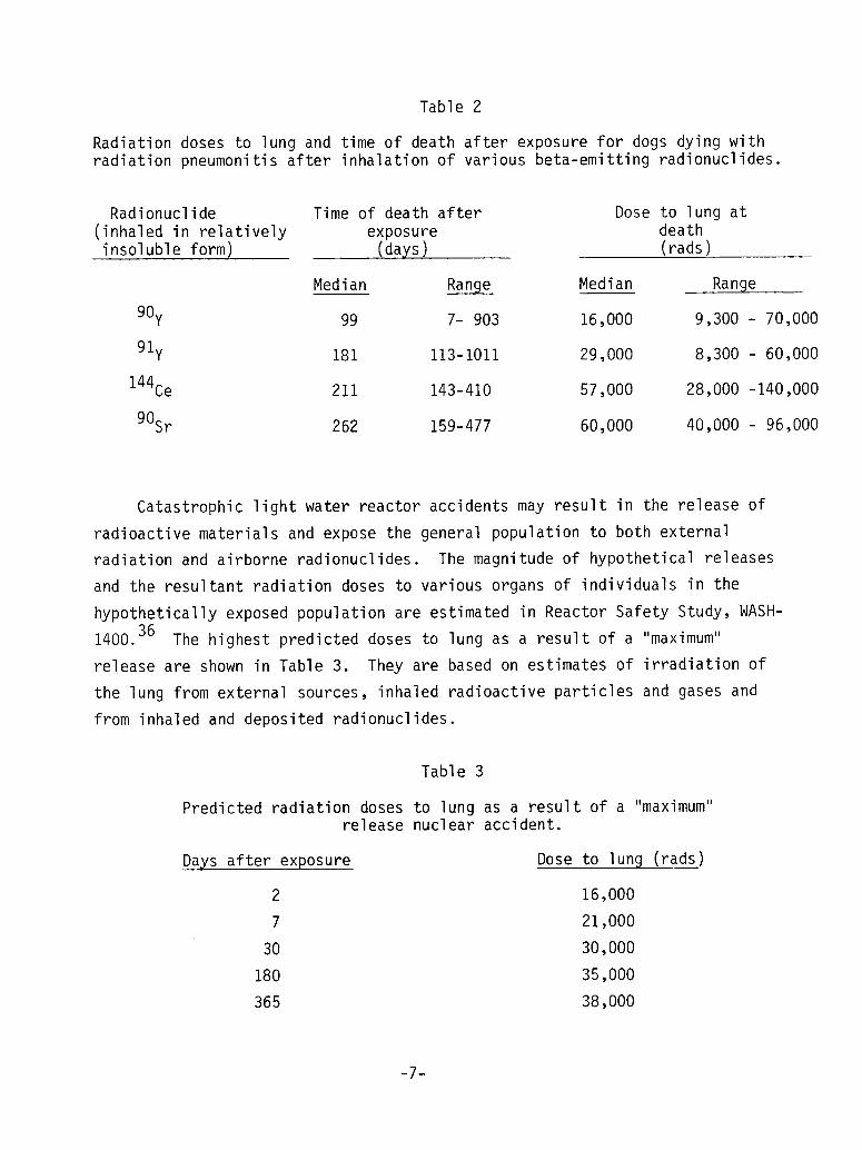
Table 2
Radiation doses to lung and time of death after exposure for dogs dying withradiation pneumonitis after inhalation of various beta-emitting radionuclides.
Radionuclide Time of death after(inhaled in relatively exposureinsoluble form) (days)
Median Range Median
90y 99 7- 903 16,000
91y 181 113-1011 29,000
144Ce 211 143-410 57,000
90Sr 262 159-477 60,000
Dose to lung atdeath(rads)
Range
9,300 - 70,000
8,300 - 60,000
28,000 -140,000
40,000 - 96,000
Catastrophic light water reactor accidents may result in the release of
radioactive materials and expose the general population to both external
radiation and airborne radionuclides. The magnitude of hypothetical releases
and the resultant radiation doses to various organs of individuals in the
hypothetically exposed population are estimated in Reactor Safety Study, WASH-
1400. 36 The highest predicted doses to lung as a result of a "maximum"
release are shown in Table 3. They are based on estimates of irradiation of
the lung from external sources, inhaled radioactive particles and gases and
from inhaled and deposited radionuclides.
Table 3
Predicted radiation doses to lung as a result of a "maximum"release nuclear accident.
Days after exposure Dose to lung (rads)
2 16,000
7 21,000
30 30,000
180 35,000
365 38,000
-7-
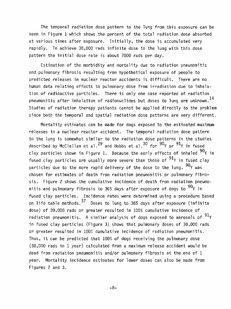
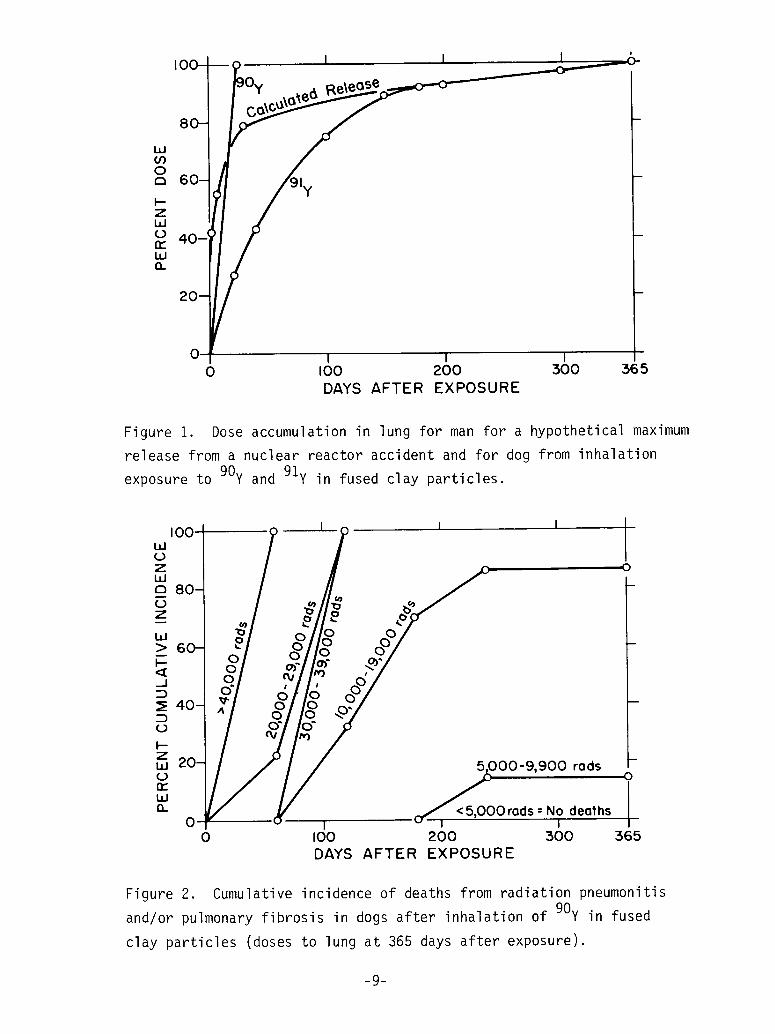
The temporal radiation dose pattern to the lung from this exposure can be
seen in Figure 1 which shows the percent of the total radiation dose absorbed
at various times after exposure. Initially, the dose is accumulated very
rapidly. To achieve 38,000 rads infinite dose to the lung with this dose
pattern the initial dose rate is about 7000 rads per day.
Estimation of the morbidity and mortality due to radiation pneumonitis
and pulmonary fibrosis resulting from hypothetical exposure of people to
predicted releases in nuclear reactor accidents is difficult. There are no
human data relating effects to pulmonary dose from irradiation due to inhala-
tion of radioactive particles. There is only one case reported of radiation14pneumonitis after inhalation of radionuclides but doses to lung are unknown.
Studies of radiation therapy patients cannot be applied directly to the problem
since both the temporal and spatial radiation dose patterns are very different.
Mortality estimates can be made for dogs exposed to the estimated maximum
releases in a nuclear reactor accident. The temporal radiation dose pattern
to the lung is somewhat similar to the radiation dose patterns in the studies
described by McClellan et ai.29 and Hobbs et ai.30 for 90¥ or 91y in fused
clay particles shown in Figure 1. Because the early effects of inhaled 90y in
fused clay particles are usually more severe than those of 91y in fused clay
particles due to the more rapid delivery of the dose to the lung, 90y waschosen for estimates of death from radiation pneumonitis or pulmonary fibro-
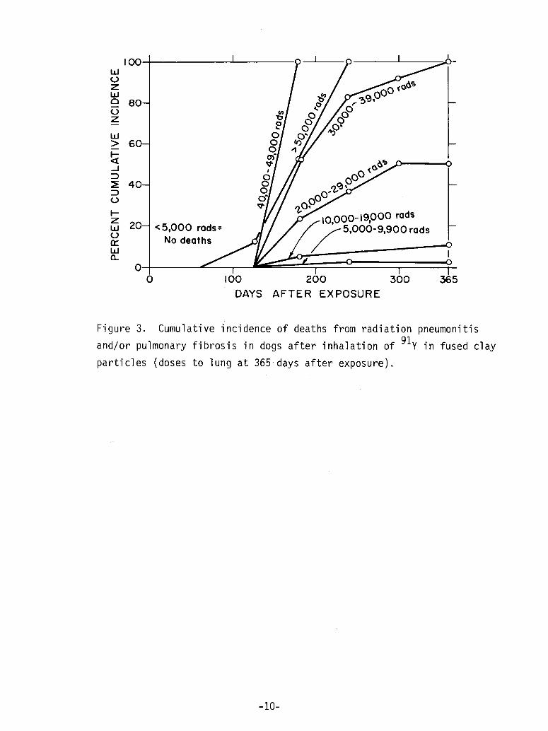
sis. Figure 2 shows the cumulative incidence of death from radiation pneumo-
nitis and pulmonary fibrosis to 365 days after exposure of dogs to 90y in
fused clay particles. Incidence rates were determined using a procedure based
on life table methods.37 Doses to lung to 365 days after exposure (infinite
dose) of 20,000 rads or greater resulted in 100% cumulative incidence of
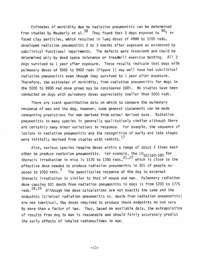
radiation pneumonitis. A similar analysis of dogs exposed to aerosols of 91yin fused clay particles (Figure 3) shows that pulmonary doses of 30,000 rads
or greater resulted in 100% cumulative incidence of radiation pneumonitis.
Thus, it can be predicted that 100% of dogs receiving the pulmonary dose
(38,000 rads in I year) calculated from a maximum release accident would
dead from radiation pneumonitis and/or pulmonary fibrosis at the end of I
year. Mortality incidence estimates for lower doses can also be made from
Figures 2 and 3.
-8-
I I I ’
I--ZW
n~
LIJQ_
I I I0 I00 200 300 365
DAYS AFTER EXPOSURE
Figure 1. Dose accumulation in lung for man for a hypothetical maximum
release from a nuclear reactor accident and for dog from inhalation
exposure to 90y and 91y in fused clay particles.
I
000
.0
~~< 5,~~00-9,900 radsOrods = No deaths
/
200 300
0
I0 I00 365
DAYS AFTER EXPOSURE
Figure 2. Cumulative incidence of deaths from radiation pneumonitisand/or pulmonary fibrosis in dogs after inhalation of 90y in fused
clay particles (doses to lung at 365 days after exposure).
-9-
I O0W(JZbJo 80-(Jz
W> 60-t--.J
:~ 40-
L~
I--Zw 20-L)rrWCL
00
<5,000 rods=No deaths
000-19,000 rods900 rods
I 1I00 200 300
DAYS AFTER EXPOSURE
I
0
..--...oI
0I
365
Figure 3. Cumulative incidence of deaths from radiation pneumonitis
and/or pulmonary fibrosis in dogs after inhalation of 91y in fused clay
particles (doses to lung at 365’days after exposure).
-I0-
Estimates of morbidity due to radiation pneumonitis can be determined
from studies by Mauderly et al. 38 They found that 3 dogs exposed to 90y in
fused clay particles, which resulted in lung doses of 4900 to 5700 rads,
developed radiation pneumonitis 2 to 3 months after exposure as evidenced by
subclinical functional impairments. The defects were transient and could be
determined only by dead space tolerance or treadmill exercise testing. All 3
dogs survived to I year after exposure. These results indicate that dogs with
pulmonary doses of 5000 to 9900 rads (Figure I) may well have had subclinical
radiation pneumonitis even though they survived to I year after exposure.
Therefore, the estimates of morbidity, from radiation pneumonitis for dogs in
the 5000 to 9900 rad dose group may be considered 100%. No studies have been
conducted on dogs with pulmonary doses appreciably smaller than 5000 rads.
There are scant quantitative data on which to compare the pulmonary
response of man and the dog, however, some general statements can be made
concerning predictions for man derived from animal derived data. Radiation
pneumonitis in many species is generally qualitatively similar although there
are certainly many minor variations in response. For example, the sequence of
lesions in radiation pneumonitis and the recognition of early and late stages
were initially derived from studies with rabbits.17
Also, various species require doses within a range of about 2 times each
other to produce radiation pneumonitis. For example, the LD50/160_180 forthoracic irradiation in mice is 1170 to 1350 rads,21’27 which is close to the
effective dose needed to produce radiation pneumonitis in 50% of people ex-
posed to 1050 rets. 7 The quantitative response of the dog to external
thoracic irradiation is similar to that of mouse and man. Pulmonary radiation
dose causing 50% death from radiation pneumonitis in dogs is from 1200 to 1775
rads. 24’25 Although the dose calculations are not exactly the same and the
endpoints (clinical radiation pneumonitis vs. death from radiation pneumonitis)
are not identical, the doses required to produce these endpoints do not vary
by more than a factor of two. Thus, based on available data, the extrapolation
of results from dog to man is reasonable and should fairly accurately predict
the early effects of inhaled radionuclides in man.
-Ii-
.
2
.
.
,
,
,
,
.
I0.
II.
12.
13.
REFERENCES
Groover, T. A., Christie, A. C. and Merritt, E. A., "Observations on theUse of the Copper Filter in the Roentgen Treatment of Deep-Seated Malig-nancy," South. Med. J. 15: 440, 1922.
Warren, S. and Spencer, J., "Radiation Reaction in the Lung," Amer. J.Roentgenol. Radium Ther. 43: 682-701, 1940.
Phillips, T. L., "An Ultrastructural Study of the Development of RadiationInjury in the Lung," Radiology 8_]_7: 49-54, 1966.
Goldenberg, V. E., Warren, S., Chute, R. and Besen, M., "Radiation Pneu-monitis in Single and Parabiotic Rats," Lab. Invest. 18: 215-226, 1968.
Rubin, P. and Casarett, G. W., "Respiratory System - Chapter 12," in Clini-cal Radiation Pathology, W. B. Saunders Co., Philadelphia, Vol. I, 1968.
Baeza, M. R., Barkley, H. T., Jr. and Fernandez, C. H., "Total Lung Irra-diation in the Treatment of Pulmonary Metastases," Radiology 116: 151-154,1975.
Newton, K. A., "Total Thoracic Irradiation Combined with Intravenous In-jection of Autogenous Marrow," Clinical Radiol. 11-12: 14-21, 1960-61.
Wara, W. M., Phillips, T. L., Margolis, L. W. and Smith, V., "RadiationPneumonitis - A New Approach to the Derivation of Time-Dose Factors,"Cancer 32: 547-552, 1973.
Ellis, F., "The Relationship of Biological Effect of Dose-Time Fractiona-tion Factors in Radiotherapy," in Current Topics in Radiation Research(Ebert, M. and Howard, A., editors), American Elsevier Publishing Co.,New York, Vol. 4, pp. 359-397, 1968.
Ross, D. M., "Summary of United States Atomic Energy Commission Contrac-tors Internal Exposure Experience," in Diagnosis and Treatment of DepositedRadionuclides (Kornberg, H. A. and Norwood, W. D., Editors), ExcerptaMedica Foundation, pp. 427-434, 1968.
Roeder, J. R., Statistical Summary of United States Atomic Energy Com-mission "Licensee’s Internal Exposure Experience 1957-1966," inDiagnosis and Treatment of Deposited Radionulcides (Kornberg, H. A.,Norwood, W. D., Editors), Excerpta Medica Foundation, pp. 435-459, 1968.
National Research Council Advisory Committee on the Biological Effectsof Ionizing Radiation (BEIR), The Effects of Populations of Exposure toLow Levels of lonizing Radiation, National Academy of Sciences, NationalResearch Council, Washington, D. C., pp. 145-156, 1972.
United Nations Scientific Committee on the Effects of Atomic Radiation(UNSCAR), lonizing Radiation: Levels and Effects, Vol. 2, pp. 417-420,1972, United Nations, Publisher, New York.
-12-
14. Rajewsky, B., "Researchers in the Problem of Radium Poisoning and theTolerance Dose of Radium," Radiology 32: 57-62, 1939.
15. Weir, C. J. and Michaelson, S. M., Pulmonary Radiation Reactions, C CThomas, Springfield, III., 1971.
16. Davis, K. S., "Intrathoracic Changes Following X-ray Treatment: AClinical and Experimental Study," Radiology 3: 301-322, 1924.
17. Englestad, R. B., "Pulmonary Lesions after Roentgen and Radium Irradia-tion," Amer. J. Roentgenol. 43: 676-681, 1940.
18. Jennings, F. L. and Arden, A., "Development of Experimental RadiationPneumonitis," Arch. Pathol. 71: 437-446, 1961.
19. Michaelson S M. and Schreiner, B F. Jr " ", . . , ., Cardlopulmonary Effects ofUpper-Body X-lrradiation in the Dog," Radiat. Res. 47: 168-181, 1971.
20. de Villiers, A. J. and Gross, P., "Radiation Pneumonitis X-Ray InducedLesions in Hamsters and Rats," Arch. Environ. Health 15: 650, 1967.
21. Phillips, T. H. L. and Margolis, L., Radiation Pathology and the ClinicalResponse of Lung and Esophagus," in Front, Radiation Ther. Onc., Karger.Basel and University Park Press, Baltimore, Md., Vol. 6, 1972.
22. Maisin, J. R., "The Ultrastructure of the Lung of Mice Exposed to aSupra Lethal Dose of Ionizing Radiation," Radiat. Res. 44: 545-564, 1970.
23. Faulkner, C. S., II and Connally, K. S., "The Ultrastructure of 60CoRadiation Pneumonitis in Rats," Lab. Invest. 28: 545-553, 1973.
24. Hansen, C. L., Jr., Michaelson, S. M. and Howland, J. W., "Lethality onUpper Body Exposure to X-Radiation in Beagles," Public Health Reports 76:242-246, 1961.
25. Sweany, S. K., Moss, W. T. and Haddy, F. J., The Effects of Chest Irradia-tion on Pulmonary Function," J. Clin. Invest. 38: 587-593, 1959.
26. Tyree, E. B., Glicksman, A. S. and Nickson, J. J., "Effect of L-Triiodothyronine on Radiation-lnduced Pulmonary Fibrosis in Dogs," Radiat.Res. 28: 30-36, 1966.
27. Field, S. B. and Hornsey, S., "Damage to Mouse Lung with Neutrons andX-Rays," Eur. J. Cancer I0: 621-627, 1974.
28. Stuart, B. 0., Casey, H. W. and Bair, W. J., "Acute and Chronic Effectsof Inhaled 144Ce02 in Dogs," Health Phys. 10: 1203-1209, 1964.
29. McClellan, R. 0., Barnes, J. E., Boecker, B. B., Chiffelle, T. L., Hobbs,C. H., Jones, R. K., Mauderly, J. L., Pickrell, J. A. and Redman, H. C.,"Toxicity of Beta-Emitting Radionuclides Inhaled in Fused Clay Particles -An Experimental Approach," in Morphology of Experimental RespiratoryCarcinogenesis, (Nettesheim, P., Hanna, M. G., Jr. and Deatherage, J. W.,Jr. Editors), AEC Symposium Series No. 21 (CONF-700501, pp. 395-415, 1970.
-13-
30.
31.
32.
33.
34.
35.
36.
37.
38.
Hobbs, C. H., Barnes, J. E., McClellan, R. 0., Chiffelle, T. L., Jones,R. K., Lundgren, D. L., Mauderly, J. L., Pickrell, J. A. and Rypka,E. W., "Toxicity in the Dog of Inhaled 90y in Fused Clay Particles:Early Biological Effects," Radiat. Res. 4__99: 430-460, 1972.
Thomas, R. L., Scott, J. K. and Chiffelle, T. L., "Metabolism andToxicity of Inhaled 144Ce in Rats," Radiat. Res. 49: 589-610, 1972.
Cember, H. and Stemmer, K., "Lung Cancer from Radioactive Cerium Chloride,"Health Phys. I0: 43-48, 1964.
Clarke, W. J. and Bair, W. J., "Plutonium Inhalation Studies VI. PathologicEffects of Inhaled Plutonium Particles in Dogs," Health Phys. I0: 391-398, 1964.
McClellan, R. O. and Rupprecht, F. C. (Editors), "Inhalation ToxicologyResearch Institute Annual Report - 1973-1974," LF-49, Lovelace Foundation,Albuquerque, New Mexico, 1974.
Jones, R. K., Hahn, F. F., Hobbs, C. H., Benjamin, S. A., Boecker, B. B.,McClellan, R. O. and Slauson, D. 0., "Pulmonary Carcinogenesis and ChronicBeta Irradiation of Lung," in Experimental Lun9 Cancer. Carcinogenesisand Bioassays, Springer-Verlag, Berlin, 1974.
Watson, Joseph: Personal Communication, University of Pittsburgh, Pitts-burgh, Pa., 1975.
Rosenblatt, L. S., Hetherington, N. H., Goldman, M. and Bustad, L. K.,"Evaluation of Tumor Incidence Following Exposure to Internal Emittersby Application of the Logistic Dose-Response Surface," Health Phys. 21:869-875, 1971.
Mauderly, J. L., Pickrell, J. A., Hobbs, C. H., Benjamin, S. A., Hahn,F. F., Jones, R. K. and Barnes, J. E., "The Effects of Inhaled 90yFused Clay Aerosol on Pulmonary Function and Related Parameters of theBeagle Dog," Radiat. Res. 56: 83-96, 1973.
-14-