review c urrent and prospective applications of metal ion–protein binding · 2015-03-30 · ......
TRANSCRIPT
Journal of Chromatography A, 988 (2003) 1–23www.elsevier.com/ locate/chroma
Review
C urrent and prospective applications of metal ion–protein bindinga b a ,*E.K.M. Ueda , P.W. Gout , L. Morganti
a ´Department of Biotechnology, Institute of Nuclear and Energy Research (IPEN-CNEN), Travessa R, 400, Cidade Universitaria,˜05508-900 Sao Paulo, SP, Brazil
bDepartment of Cancer Endocrinology, British Columbia Cancer Agency, Vancouver, Canada
Received 29 July 2002; received in revised form 9 December 2002; accepted 10 December 2002
Abstract
Since immobilized metal ion affinity chromatography (IMAC) was first introduced, several variants of this method andmany other metal affinity-based techniques have been devised. IMAC quickly established itself as a highly reliablepurification procedure, showing rapid expansion in the number of preparative and analytical applications while not remainingconfined to protein separation. It was soon applied to protein refolding (matrix-assisted refolding), evaluation of proteinfolding status, protein surface topography studies and biosensor development. In this review, applications in proteinprocessing are described of IMAC as well as other metal affinity-based technologies. 2002 Elsevier Science B.V. All rights reserved.
Keywords: Immobilized metal affinity chromatography; Reviews; Affinity chromatography; Proteins
Contents
1 . Introduction ............................................................................................................................................................................ 22 . Immobilized metal ion affinity chromatography: utilization of differences in protein–metal ion affinity ......................................... 2
2 .1. Principles and procedures ................................................................................................................................................ 22 .2. Commonly used metal ions.............................................................................................................................................. 32 .3. Factors contributing to protein–metal binding ................................................................................................................... 32 .4. Metal chelators and chelating polymeric matrices .............................................................................................................. 52 .5. Factors governing adsorption and desorption of proteins .................................................................................................... 6
2 .5.1. Chelate structure and metal ions ........................................................................................................................... 62 .5.2. Mobile phase: pH, buffers and ionic strength......................................................................................................... 62 .5.3. Additives and protein displacers ........................................................................................................................... 8
3 . Immobilized metal ion affinity chromatography as a preparative technique .................................................................................. 83 .1. Purification by immobilized metal ion affinity chromatography: pros and cons .................................................................... 83 .2. Purification of proteins fused with poly-histidine tags and other engineered metal-binding sites ............................................ 113 .3. Purification of naturally occurring metal-binding proteins .................................................................................................. 113 .4. Immobilized metal ion affinity chromatography: up-scaling and industrial use..................................................................... 123 .5. Use of immobilized metal ion affinity chromatography in conjunction with other chromatographic techniques....................... 13
*Corresponding author. Tel.:155-11-3816-9230; fax:155-11-3816-9232.E-mail address: [email protected](L. Morganti).
0021-9673/02/$ – see front matter 2002 Elsevier Science B.V. All rights reserved.doi:10.1016/S0021-9673(02)02057-5
2 E.K.M. Ueda et al. / J. Chromatogr. A 988 (2003) 1–23
4 . Metal affinity tags: advantages and applications......................................................................................................................... 135 . Metal affinity tagging: pitfalls and limitations............................................................................................................................ 146 . Beyond immobilized metal ion affinity chromatography: other applications of metal affinity......................................................... 15
6 .1. Immobilized metal ion affinity chromatography and protein refolding: matrix-assisted refolding........................................... 156 .2. Site-specific immobilization of proteins and biosensors...................................................................................................... 166 .3. Immobilized metal ion affinity chromatography as an analytical and characterization tool in protein chemistry ...................... 186 .4. Immobilized metal ion affinity chromatography and processing of phosphoproteins and phosphopeptides.............................. 196 .5. Metal binding and protein stability ................................................................................................................................... 19
7 . Conclusions ............................................................................................................................................................................ 19Acknowledgements...................................................................................................................................................................... 20References .................................................................................................................................................................................. 20
1 . Introduction Whereas many of the recently devised metal affinity-based methodologies in protein chemistry are based
In the mid 1970s, Porath et al. [1] introduced a on IMAC, a number of them are not. The latter rangenew type of chromatography which was first desig- from protein purity analysis [42] and protein refold-nated ‘‘metal chelate chromatography’’, but was later ing through matrix-assisted refolding [43] to moretermed ‘‘immobilized metal (ion) affinity chromatog- complex procedures such as the Biacore analysis ofraphy’’ (IMAC). While the basis of this method was His-tagged proteins using a chelating sensor chipnot new [2], Porath et al. [1] focused on its use for [44] and use of a metal-chelating microscopy tip forthe separation and isolation of proteins. The tech- single molecule force measurement by atomic forcenique is based on differences in the affinity of microscopy [45]. This wide range of applications canproteins for metal ions bound to a metal-chelating also be observed within the IMAC realm, where thissubstance which is immobilized on a chromatograph- type of chromatography can be employed not just foric support (resin) [3]. This ‘‘bio-affinity’’ feature protein or peptide purification, but also for separationsoon caught attention of biochemists and biologists of His -tagged oligonucleotides [46], inactivation of6
who employed IMAC and enhanced it to suit their viruses [47] and endotoxin removal from proteinparticular applications. The best known improvement preparations [48]. It would be safe to state thatis the application of histidine tags for separation of although IMAC comprises a vast area of the metalrecombinant polypeptides [4–14], which consists of affinity applications field, it is not the only meth-the insertion of a short tail of histidine residues on odology based on metal–protein interactions. Manyeither the N- or C-terminus of a protein or peptide other methods make use of the same principle and[15]. The use of such histidine and other metal hence play an important role in this developing areaaffinity tags in IMAC [19–24] proved to be a of protein biochemistry.powerful tool for protein recovery, especially whenhigh production and maximum yield of the proteinsof interest were essential [25]. Consequently, IMAC 2 . Immobilized metal ion affinityemerged as one of the major preparative methodolo- chromatography: utilization of differences ingies used for protein purification, ranging from protein–metal ion affinitybench [4–14] to pilot / industrial scale [16–20,22–32]. 2 .1. Principles and procedures
Many reviews on IMAC have been published sofar [33–41] some of them dealt with new IMAC Binding of proteins (or peptides) to metal ions isapplications and even introduced new possible ter- based on interaction between an electron-donatingritories where IMAC could be exploited. In light of group present on a protein surface and a metal ionthe new IMAC applications this review tried to go presenting one or more accessible coordination sites.deeper into this new dimension of IMAC based In IMAC, use is made of a sorbent, or matrix, totechniques and their implications in life sciences. which metal-chelating groups are covalently attached
E.K.M. Ueda et al. / J. Chromatogr. A 988 (2003) 1–23 3
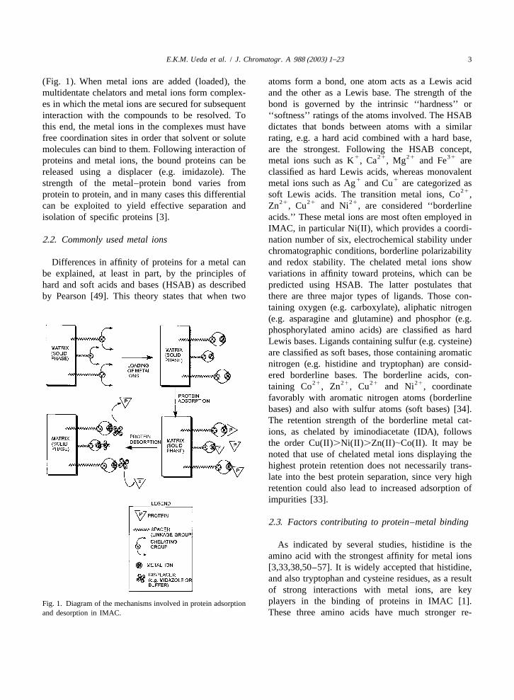
(Fig. 1). When metal ions are added (loaded), the atoms form a bond, one atom acts as a Lewis acidmultidentate chelators and metal ions form complex- and the other as a Lewis base. The strength of thees in which the metal ions are secured for subsequent bond is governed by the intrinsic ‘‘hardness’’ orinteraction with the compounds to be resolved. To ‘‘softness’’ ratings of the atoms involved. The HSABthis end, the metal ions in the complexes must have dictates that bonds between atoms with a similarfree coordination sites in order that solvent or solute rating, e.g. a hard acid combined with a hard base,molecules can bind to them. Following interaction of are the strongest. Following the HSAB concept,
1 21 21 31proteins and metal ions, the bound proteins can be metal ions such as K , Ca , Mg and Fe arereleased using a displacer (e.g. imidazole). The classified as hard Lewis acids, whereas monovalent
1 1strength of the metal–protein bond varies from metal ions such as Ag and Cu are categorized as21protein to protein, and in many cases this differential soft Lewis acids. The transition metal ions, Co ,
21 21 21can be exploited to yield effective separation and Zn , Cu and Ni , are considered ‘‘borderlineisolation of specific proteins [3]. acids.’’ These metal ions are most often employed in
IMAC, in particular Ni(II), which provides a coordi-2 .2. Commonly used metal ions nation number of six, electrochemical stability under
chromatographic conditions, borderline polarizabilityDifferences in affinity of proteins for a metal can and redox stability. The chelated metal ions show
be explained, at least in part, by the principles of variations in affinity toward proteins, which can behard and soft acids and bases (HSAB) as described predicted using HSAB. The latter postulates thatby Pearson [49]. This theory states that when two there are three major types of ligands. Those con-
taining oxygen (e.g. carboxylate), aliphatic nitrogen(e.g. asparagine and glutamine) and phosphor (e.g.phosphorylated amino acids) are classified as hardLewis bases. Ligands containing sulfur (e.g. cysteine)are classified as soft bases, those containing aromaticnitrogen (e.g. histidine and tryptophan) are consid-ered borderline bases. The borderline acids, con-
21 21 21 21taining Co , Zn , Cu and Ni , coordinatefavorably with aromatic nitrogen atoms (borderlinebases) and also with sulfur atoms (soft bases) [34].The retention strength of the borderline metal cat-ions, as chelated by iminodiacetate (IDA), followsthe order Cu(II).Ni(II).Zn(II)|Co(II). It may benoted that use of chelated metal ions displaying thehighest protein retention does not necessarily trans-late into the best protein separation, since very highretention could also lead to increased adsorption ofimpurities [33].
2 .3. Factors contributing to protein–metal binding
As indicated by several studies, histidine is theamino acid with the strongest affinity for metal ions[3,33,38,50–57]. It is widely accepted that histidine,and also tryptophan and cysteine residues, as a resultof strong interactions with metal ions, are keyplayers in the binding of proteins in IMAC [1].Fig. 1. Diagram of the mechanisms involved in protein adsorption
and desorption in IMAC. These three amino acids have much stronger re-
4 E.K.M. Ueda et al. / J. Chromatogr. A 988 (2003) 1–23
Table 2tentions, for example, than glutamate and aspartateIndividual contributions of amino acids involved in proteinresidues, which show essentially no retentionretention
[56,57]. Yip et al. [50] have correlated the retentionsFunctional group Retention strengthof a large number of synthetic, biologically active
peptides on Ni(II), Zn(II) and Cu(II) chelated in Histidine 1111aCysteine 1111IDA, with their amino acid profiles in order to
Aspartic acid, glutamic acid 2evaluate the adsorption properties of amino acidsLysine, arginine 1
involved in the retention of the peptides. Other Tryptophan, tyrosine, phenylalanine 1investigations revealed that the retention behavior of N-Terminus 11
proteins is largely governed by exposed histidine a Cysteine is the reduced form.residues on the protein surface [52,58].
Cysteine also displays strong metal affinity, al-though to a somewhat lesser extent than histidine ions. Carboxyl and phosphate functional groups are[33,38,59]. The metal affinity of these two amino the main targets for hard metal ions such as Fe(III)acids can be largely attributed to their functional and Mg(II). A well-accepted concept is that thegroups, in particular the imidazole and thiol groups, spatial distribution of histidine residues over aof histidine and cysteine, respectively. In the case of protein surface and their accessibility would influ-histidine, other functional groups, such as carboxyl ence the retention behavior of the protein moleculeand a-amino groups, also play a role in the metal- [33]. Several high metal affinity peptides have beenbinding process [60,61]. Other amino acids may also described in the literature [63,64] as well as non-have substantial metal affinity, including tryptophan, contiguous metal-binding motifs such as those foundphenylalanine and tyrosine acting directly via their in prolactin [65–70], and growth hormone [65,71–aromatic side chains [33] and arginine, lysine, as- 73]. It is notable that the possession of a metal-paragine, glutamine and methionine, acting indirectly binding motif is not always a guarantee for success-via individual or combined effects on histidine ful IMAC, since there are a number of enzymes withaccessibility [33,62]. Although the retention of pro- metal affinity that bind to the chelating matrix via ateins or peptides is primarily due to the metal site that is not the metal-binding catalytic site [3,33].affinities of their individual amino acids, other Some metal-binding sections of a protein, even whenfactors also contribute profoundly toward their metal exposed, may contribute very little to the net re-affinity, including their amino acid sequences, fold- tention strength of the protein in IMAC [33].ing, and surface properties. In view of the latter, the When the amino acid composition of a protein, orretention behavior of proteins in IMAC is not easily peptide, of interest is known, the rule presented inpredictable [33]. Table 1 can be used to predict what kind of metal
In interactions between immobilized Cu(II), ions will lead to its retention in IMAC [3]. TheNi(II), Co(II) or Zn(II) ions and amino acid residues retention strengths of the main amino acids involvedon protein surfaces, imidazolyl, thiol and indolyl in protein adsorption in IMAC are shown in Table 2functional groups are the main targets for the metal [33].
Table 1Protein metal affinity prediction based on accessible histidine and tryptophan residues on the protein surface
Occurrence of accessible histidine or Metal ions providing retentiontryptophan residues on protein surface
No His/Trp –1 His Cu(II).1 His Cu(II), Ni(II)His clusters Cu(II), Ni(II), Zn(II), Co(II)Several Trp, no His Cu(II)
E.K.M. Ueda et al. / J. Chromatogr. A 988 (2003) 1–23 5
2 .4. Metal chelators and chelating polymericmatrices
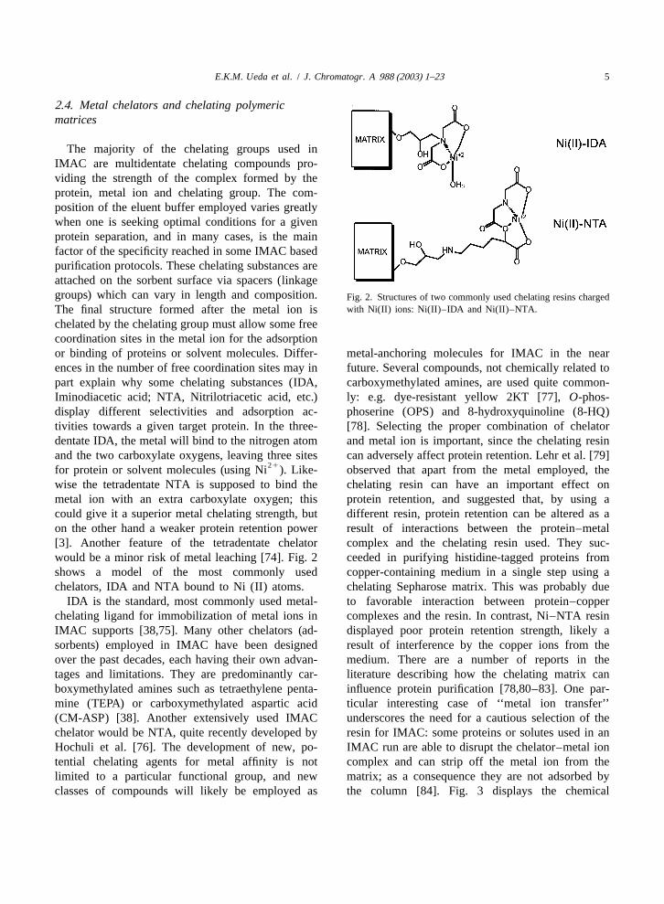
The majority of the chelating groups used inIMAC are multidentate chelating compounds pro-viding the strength of the complex formed by theprotein, metal ion and chelating group. The com-position of the eluent buffer employed varies greatlywhen one is seeking optimal conditions for a givenprotein separation, and in many cases, is the mainfactor of the specificity reached in some IMAC basedpurification protocols. These chelating substances areattached on the sorbent surface via spacers (linkagegroups) which can vary in length and composition. Fig. 2. Structures of two commonly used chelating resins charged
with Ni(II) ions: Ni(II)–IDA and Ni(II)–NTA.The final structure formed after the metal ion ischelated by the chelating group must allow some freecoordination sites in the metal ion for the adsorptionor binding of proteins or solvent molecules. Differ- metal-anchoring molecules for IMAC in the nearences in the number of free coordination sites may in future. Several compounds, not chemically related topart explain why some chelating substances (IDA, carboxymethylated amines, are used quite common-Iminodiacetic acid; NTA, Nitrilotriacetic acid, etc.) ly: e.g. dye-resistant yellow 2KT [77],O-phos-display different selectivities and adsorption ac- phoserine (OPS) and 8-hydroxyquinoline (8-HQ)tivities towards a given target protein. In the three- [78]. Selecting the proper combination of chelatordentate IDA, the metal will bind to the nitrogen atom and metal ion is important, since the chelating resinand the two carboxylate oxygens, leaving three sites can adversely affect protein retention. Lehr et al. [79]
21for protein or solvent molecules (using Ni ). Like- observed that apart from the metal employed, thewise the tetradentate NTA is supposed to bind the chelating resin can have an important effect onmetal ion with an extra carboxylate oxygen; this protein retention, and suggested that, by using acould give it a superior metal chelating strength, but different resin, protein retention can be altered as aon the other hand a weaker protein retention power result of interactions between the protein–metal[3]. Another feature of the tetradentate chelator complex and the chelating resin used. They suc-would be a minor risk of metal leaching [74]. Fig. 2 ceeded in purifying histidine-tagged proteins fromshows a model of the most commonly used copper-containing medium in a single step using achelators, IDA and NTA bound to Ni (II) atoms. chelating Sepharose matrix. This was probably due
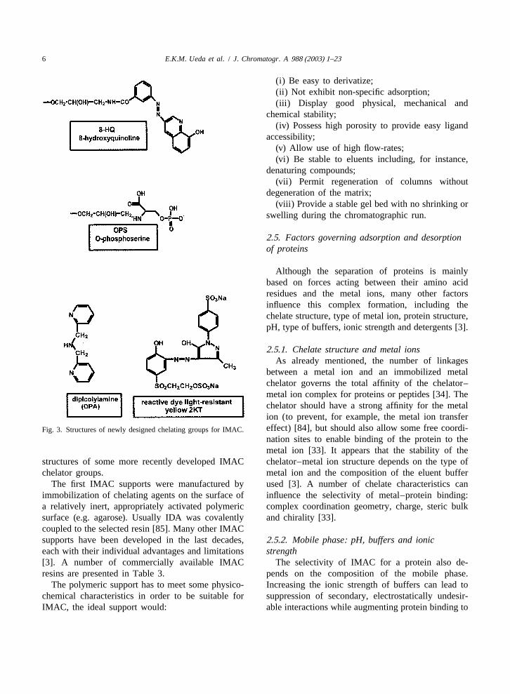
IDA is the standard, most commonly used metal- to favorable interaction between protein–copperchelating ligand for immobilization of metal ions in complexes and the resin. In contrast, Ni–NTA resinIMAC supports [38,75]. Many other chelators (ad- displayed poor protein retention strength, likely asorbents) employed in IMAC have been designed result of interference by the copper ions from theover the past decades, each having their own advan- medium. There are a number of reports in thetages and limitations. They are predominantly car- literature describing how the chelating matrix canboxymethylated amines such as tetraethylene penta- influence protein purification [78,80–83]. One par-mine (TEPA) or carboxymethylated aspartic acid ticular interesting case of ‘‘metal ion transfer’’(CM-ASP) [38]. Another extensively used IMAC underscores the need for a cautious selection of thechelator would be NTA, quite recently developed by resin for IMAC: some proteins or solutes used in anHochuli et al. [76]. The development of new, po- IMAC run are able to disrupt the chelator–metal iontential chelating agents for metal affinity is not complex and can strip off the metal ion from thelimited to a particular functional group, and new matrix; as a consequence they are not adsorbed byclasses of compounds will likely be employed as the column [84]. Fig. 3 displays the chemical
6 E.K.M. Ueda et al. / J. Chromatogr. A 988 (2003) 1–23
(i) Be easy to derivatize;(ii) Not exhibit non-specific adsorption;(iii) Display good physical, mechanical and
chemical stability;(iv) Possess high porosity to provide easy ligand
accessibility;(v) Allow use of high flow-rates;(vi) Be stable to eluents including, for instance,
denaturing compounds;(vii) Permit regeneration of columns without
degeneration of the matrix;(viii) Provide a stable gel bed with no shrinking or
swelling during the chromatographic run.
2 .5. Factors governing adsorption and desorptionof proteins
Although the separation of proteins is mainlybased on forces acting between their amino acidresidues and the metal ions, many other factorsinfluence this complex formation, including thechelate structure, type of metal ion, protein structure,pH, type of buffers, ionic strength and detergents [3].
2 .5.1. Chelate structure and metal ionsAs already mentioned, the number of linkages
between a metal ion and an immobilized metalchelator governs the total affinity of the chelator–metal ion complex for proteins or peptides [34]. Thechelator should have a strong affinity for the metalion (to prevent, for example, the metal ion transfereffect) [84], but should also allow some free coordi-Fig. 3. Structures of newly designed chelating groups for IMAC.nation sites to enable binding of the protein to themetal ion [33]. It appears that the stability of the
structures of some more recently developed IMAC chelator–metal ion structure depends on the type ofchelator groups. metal ion and the composition of the eluent buffer
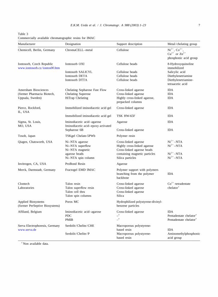
The first IMAC supports were manufactured by used [3]. A number of chelate characteristics canimmobilization of chelating agents on the surface of influence the selectivity of metal–protein binding:a relatively inert, appropriately activated polymeric complex coordination geometry, charge, steric bulksurface (e.g. agarose). Usually IDA was covalently and chirality [33].coupled to the selected resin [85]. Many other IMACsupports have been developed in the last decades,2 .5.2. Mobile phase: pH, buffers and ioniceach with their individual advantages and limitations strength[3]. A number of commercially available IMAC The selectivity of IMAC for a protein also de-resins are presented in Table 3. pends on the composition of the mobile phase.
The polymeric support has to meet some physico- Increasing the ionic strength of buffers can lead tochemical characteristics in order to be suitable for suppression of secondary, electrostatically undesir-IMAC, the ideal support would: able interactions while augmenting protein binding to
E.K.M. Ueda et al. / J. Chromatogr. A 988 (2003) 1–23 7
Table 3Commercially available chromatographic resins for IMAC
Manufacturer Designation Support description Metal /chelating group21 21Chemicell, Berlin, Germany ChromaCELL–metal Cellulose Ni , Co ,21 21Cu or Zn
phosphonic acid group
Iontosorb, Czech Republic Iontosorb OXI Cellulose beads 8-Hydroxyquinolinewww.iontosorb.cz/ iontos00.htm immobilized
Iontosorb SALICYL Cellulose beads Salicylic acidIontosorb DETA Cellulose beads DiethylenetriamineIontosorb DTTA Cellulose beads Diethylenetriamine-
tetraacetic acid
Amersham Biosciences Chelating Sepharose Fast Flow Cross-linked agarose IDA(former Pharmacia Biotech, Chelating Superose Cross-linked agarose IDAUppsala, Sweden) HiTrap Chelating Highly cross-linked agarose, IDA
prepacked columns
Pierce, Rockford, Immobilized iminodiacetic acid gel Cross-linked agarose IDAIL, USA
Immobilized iminodiacetic acid gel TSK HW-65F IDA
Sigma, St. Louis, Iminodiacetic acid–agarose Agarose IDAMO, USA Iminodiacetic acid–epoxy activated
Sepharose 6B Cross-linked agarose IDA
Tosoh, Japan TSKgel Chelate-5PWb Polymer resin21Qiagen, Chatsworth, USA Ni–NTA agarose Cross-linked agarose Ni –NTA21Ni–NTA superflow Highly cross-linked agarose Ni –NTA
Ni–NTA magnetic Cross-linked agarose beads21agarose beads containing magnetic particles Ni –NTA21Ni–NTA spin column Silica particles Ni –NTA
Invitrogen, CA, USA ProBond Resin Agarose
Merck, Darmstadt, Germany Fractogel EMD IMAC Polymer support with polymersbranching from the polymer IDAbackbone
21Clontech Talon resin Cross-linked agarose Co -tetradentateaLaboratories Talon superflow resin Cross-linked agarose chelator
Talon cell thru Cross-linked agaroseTalon spin columns Silica
Applied Biosystems Poros MC Hydrophilized polystyrene-divinyl-(former PerSeptive Biosystems) benzene particles
Affiland, Belgium Iminodiacetic acid–agarose Cross-linked agarose IDAa aPDC – Pentadentate chelatora aPMIB – Pentadentate chelator
Serva Electrophoresis, Germany Serdolit /Chelite /CHE Macroporous polystyrene-www.serva.de based resin IDA
Serdolit /Chelite /P Macroporous polystyrene- Aminomethylphosphonicbased resin acid group
a Non available data.
8 E.K.M. Ueda et al. / J. Chromatogr. A 988 (2003) 1–23
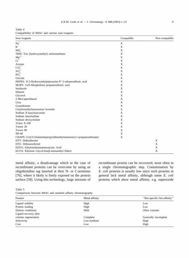
the chelate complexes, since decreasing the salt nant human prolactin [88,89] and primate prolactinconcentration can even result in a poorer protein [90], and proved to be useful for diminishing unde-adsorption [33,38]. Sodium chloride (at 0.1–1.0M) sirable interactions. Organic solvent was added to theis commonly included in IMAC buffers to suppress buffer to aid in the removal of endotoxins withoutionic interactions between sample and matrix, as deleterious effects on the correct folding of proteinswell as between proteins [3]. The effect of elec- [48], or to strip off colored contaminants, as used introlytes on protein retention is related to the affinity the isolation of albumin from a Cohn extract [3].of the metal ion for its solvated water molecules. Elution of the protein can also be achieved, althoughWeakening of the forces between the metal ions and with poor resolution, by extraction of the metal ionswater molecules by increasing the ionic strength of using a strong chelator (e.g. EDTA). Another methodthe buffers, favors the protein adsorption processes. to achieve good resolution, and hence selectiveThe use of high pH values (7 or 8) and high ionic elution, is the incorporation in the eluent of com-strength makes IMAC different from other ion-ex- pounds known to present a higher affinity for thechange chromatography. IMAC best resembles hy- adsorption sites than the proteins. Addition of his-drophobic interaction chromatography (HIC) be- tidine or imidazole has been used in gradient modecause of the use of high salt concentration buffers elution as an effective way for selective protein[38]. If desirable, one could easily elute the target elution [38]. Recently, improved displacers haveprotein by applying a continuous decreasing pH been devised, including a polymeric imidazole com-gradient rather than an increasing imidazole gradient. pound which showed superior eluting strength com-Some elution protocols involving the simultaneous pared to monomeric imidazole [91]. IMAC is com-use of both imidazole addition and pH decreasing patible with most buffer constituents required togradient in order to achieve a better chromatographic maintain protein integrity (e.g. protease inhibitors,resolution are also described [86,87]. Alternatively, glycerol), prevent protein aggregation (e.g. 2-mer-the protein can be eluted by a chelating compound captoethanol) or merely improve resolution (e.g. NP-such as EDTA, but the elution will elute all proteins 40). In Table 4, a list is presented of substancesretained along with the metal ion used, thus dimin- showing various degrees of compatibility with IMACishing the resolution and selectivity [3]. [58,92,93].
Adsorption of protein in IMAC is carried out at agiven pH at which the electron donor groups on theprotein surface are partially unprotonated. It is 3 . Immobilized metal ion affinitycommon practice to induce protein adsorption on the chromatography as a preparative techniqueIMAC support under a weakly alkaline pH in thepresence of high ionic strength buffers. Phosphate 3 .1. Purification by immobilized metal ion affinityand acetate buffers are most commonly used in chromatography: pros and consIMAC. The role of the pH is fairly complex in theelution and adsorption of proteins, because it in- IMAC is primarily based on affinity adsorptionfluences a number of properties, including the nu- and therefore possesses advantages as well as dis-cleophilic behavior of the buffer components, the advantages associated with this type of separationelectron-donor acceptor properties of the solutes and technology. Notably, the main feature of affinitymetal stability. The pH range 6–8 favors retention of chromatography is the specificity of the interactionhistidine and cysteine residues; at a more alkaline between the protein of interest and the ligand (e.g.range, coordinations with amino functional groups chelated metal ion, antibody, dye etc.). However,are favored, thus decreasing selectivity [38]. when compared to other types of affinity chromatog-
raphy, in particular immunoaffinity chromatography,2 .5.3. Additives and protein displacers IMAC exhibits some noteworthy advantages, as
Detergents can be utilized in IMAC as selectivity described in Table 5 and discussed below [33].enhancers [3]. Tween 80 at a concentration of 0.01% Purification of proteins by IMAC is based on their(v /v) has been used in the purification of recombi- affinity for metal ions. Many proteins, however, lack
E.K.M. Ueda et al. / J. Chromatogr. A 988 (2003) 1–23 9
Table 4Compatibility of IMAC and various ions/ reagents
Ions/ reagents Compatible Non-compatible1Na X
1K X1NH X4
TRIS: Tris (hydroxymethyl) aminomethane X21Mg X
2Cl XAcetate X
22CO X322SO X432PO X4
Glycine XHEPES:N-2-Hydroxyethylpiperazine-N9-2-ethanesulfonic acid XMOPS: 3-(N-Morpholino) propanesulfonic acid XImidazole XEthanol XGlycerol X2-Mercaptoethanol XUrea XGuanidinium XCetyltrimethylammonium bromide XSodiumN-laurylsarcosine XSodium laurylsulfate XSodium deoxycholate XTriton X-100 XTween 20 XTween 80 XNP-40 XCHAPS: (3-[(3-Cholamidopropyl)dimethylammonio]-1-propanesulfonate) XDTT: Dithiothreitol XDTE: Dithioerythritol XEDTA: Ethylenediaminetetraacetic Acid XEGTA: Ethylene Glycol-bis(b-aminoethyl Ether) X
metal affinity, a disadvantage which in the case of recombinant protein can be recovered, most often inrecombinant proteins can be overcome by using an a single chromatographic step. Contamination byoligohistidine tag inserted at their N- or C-terminus E. coli proteins is usually low since such proteins in[76], where it likely is freely exposed on the protein general lack metal affinity, although someE. colisurface [58]. Using this technology, large amounts of proteins which show metal affinity, e.g. superoxide
Table 5Comparisons between IMAC and standard affinity chromatography
Feature Metal affinity ‘‘Bio-specific/bio-affinity’’
Ligand stability High LowProtein loading High LowElution conditions Mild Often extremeLigand recovery aftercolumn regeneration Complete Generally incompleteSelectivity Low-medium HighCost Low High
10 E.K.M. Ueda et al. / J. Chromatogr. A 988 (2003) 1–23
dismutase, could interfere with the purification pro- Some potential IMAC disadvantages are:cess [58]. (i) Metal ion transfer (MIT) phenomena leading to
Since there is no need for extreme pHs in the protein loss [74,84,94,96] and low protein yields aremobile phase, elution conditions are usually mild. not the only concerns about this subject. AnotherHigh imidazole and salt concentrations, however, problem would be if the target protein retains thecould create a problem, in particular if the isolated capturing metal ion within its structure. In this caseprotein is required for structural analysis (e.g. crys- the trapped ion could hinder or even abolish thetallography). protein’s bioactivity, but MIT could also be carried
The most outstanding features of the use of out to charge a given protein with a desired metalhistidine tail tags are the highly selective binding [74,96]. The metal ion sequestrated by the proteinbetween the tag and the chelated metal ion and the could also be hazardous if the protein is intended forrelatively low interference of other compounds dur- therapeutics since some metals commonly used ining a purification run [58]. IMAC has some unique, IMAC are considered carcinogens. Metal ions Co(II)attractive characteristics: or Ni(II), widely used in IMAC, are known carcino-
(i) IMAC often allows single step purification genic agents, although they are considered weak[94]; mutagens when compared with arsenic or hexavalent
(ii) The protein loading capacity is relatively high chromium. Nickel has been linked to the anomalouswhen compared with other affinity chromatographic expression of a very large number of genes related totechniques (0.1–10mM /ml gel) [33,94]; cancer induction [97]. One could strip off the
(iii) The metal ions can be easily removed from undesired metal from the protein using a metal-freethe resin with a strong chelating agent such as EDTA chelating column packed with a strong chelatingor EGTA. Different metal ions can therefore be adsorbent such as TED [tris(carboxymethyl)-tested, using the same chelating resin, to determine ethylenediamine] [40,98].the best ligand for separation of a protein of interest; (ii) Metal ion leakage from the resin leading to
(iv) IMAC up-scaling is fairly easy and reproduc- metal ion contamination of the final product, aible, also for industrial applications [94]; problem which would be especially important in the
(v) IMAC is useful for concentrating dilute protein use of IMAC for the preparation of therapeutics [42].solutions [94]; Apart from the obvious threat of metal contamination
(vi) IMAC is compatible with a number of buffers of the final product, a major problem would be thecontaining high ionic force or chaotropic components shelf life of the therapeutic protein since metal ions[94]; are known to trigger not only oxidative reactions as
(vii) IMAC in general does not adversely affect catalysts [94,95,99,100] but also disrupt stabilizationthe structure of proteins. A few cases have been of lyophilized protein preparations [100], althoughreported in which metalloenzymes had their essential metallic ions have also been used as additives alongmetal ion stripped off. A case has also been reported with sugars to enhance protein stability uponwhere a protein was damaged by IMAC on a Cu(II)– lyophilization [100–102]. Some authors, aware ofIDA column, but the damage was triggered by the stabilizing effects of metal ions on proteins, havereducing agents which caused oxidative proteolysis proposed their use to enhance protein stability [77].catalysed by Cu(II) [95]; In cases when the metal is a destabilizing factor,
(viii) Upon passage through a non-charged IMAC chelating agents can be added for stability improve-column, solutions become transiently sterile since all ment [103]. Protein samples tainted with metalsmetal ions essential for bacterial growth are removed should also raise some concern, since metal displace-by chelation [94]; ment can occur during an IMAC step [40,104], and
(ix) An IMAC resin can be regenerated several metal toxicity is undesirable when it comes tohundreds of times without loss of chromatographic therapeutic proteins, but purification protocols usual-characteristics [34]. IMAC gels are also extremely ly have multiple steps after IMAC and this sub-sturdy provided that pH values below 4 are not used sequent polishing generally yields a metal free final[94]. preparation [42]. In case one would like to rest
E.K.M. Ueda et al. / J. Chromatogr. A 988 (2003) 1–23 11
assured that all metal traces were banished, one affinity tag (His–Asn–Arg–Tyr–Gly–Cys–Gly–could rely on the same post treatment recommended Cys–Cys) was produced which exhibited a strongerfor MIT prone samples and carry out an IMAC with metal-binding capacity and higher yields when com-an uncharged TED column to capture the con- pared to the regular hexa-histidine tag. Schmidt et al.taminating metal ions from the protein mixture [116] engineered a Zn(II)-binding site from the[40,98]. Another elegant way to circumvent the active center of human carbonic anhydrase II into ametal leaching problem is to use either chelating or retinol-binding protein, giving it selectivity formetal immobilizing supports with strong ion binding Zn(II) when compared to Cu(II) and Ni(II), anconstants that prevent such metal loss [105]. advantage which cannot be obtained with regular
(iii) Use of oxidative and catalytic conditions oligo-histidine tags which exhibited a less rigidduring a run, when the immobilized metal ions are conformation when compared with the engineeredprone to undergo redox reactions [94,95,99]. zinc-binding site. The design of synthetic tags with
an affinity for Cu(II) ions higher than that of hexa-3 .2. Purification of proteins fused with poly- histidine tags has also been described [117].histidine tags and other engineered metal-bindingsites 3 .3. Purification of naturally occurring metal-
binding proteinsA reliable and easy way to obtain a highly purified
recombinant protein is to synthesize the protein fused In general, histidine residues in proteins are rela-to a metal affinity site and then purify it using IMAC tively rare, amounting to about 2% of the amino acid[106]. The use of a poly-histidine tag as a metal content of globular proteins; in addition, only half ofaffinity site, applied at the C- or N-terminus of the them are exposed on protein surfaces [33]. In viewprotein to be purified, proved to be a highly effective of this, only a low number of naturally occurringapproach, usually enabling purification of the protein proteins would present some metal affinity and henceby IMAC in a one-step procedure [79,92,107–110]. be potentially suitable for purification by this type ofConsequently, this methodology has been widely chromatography. Natural possession of metal affinityused for protein recovery and purification. Proteins by a protein poses a great advantage for its purifica-such as human glutathioneS-transferase P1-1 [111], tion, since there is no need for inclusion of anmurine interleukin 12 [79], cytochromeb5 [20], affinity tag or metal-binding scaffold engineering. Asgreen fluorescent protein [109], chicken lactate dehy- previously mentioned, these naturally occurringdrogenase [92], mitochondrial ADP/ATP carrier metal affinity domains have selectivity for certainprotein [27], HTLV-I surface envelope glycoprotein metals, a characteristic not shared by hexa-histidinefragment [110], and many others, have been success- tags which are able to bind to most divalent metalfully purified making use of poly-histidine tagging. ions, thereby increasing the selectivity of the sepa-High affinity for metal ions can also be achieved ration obtained via IMAC [118]. Many proteinsusing other metal-binding peptide tags or via en- showing metal affinity have been successfullygineering of metal affinity sites on protein structures purified by IMAC without prior modification, includ-[33,112,113]. Such tags/sites were devised to suit ing growth hormone and prolactin [85,89,118–120],specific demands not met by regular histidine tags. two pituitary protein hormones with zinc affinityPasquinelli et al. [114] identified several fusion tags domains [66–73]. Prolactin also exhibits a relativelythat shifted the elution of the target protein to the high affinity for Ni(II), and Ni(II)-based IMAC oflow background region of the Zn(II)–IDA elution prolactin proved to be superior to other chromato-profile, thus facilitating purification under mild con- graphic techniques previously utilized [89]. An-ditions. Gaberc-Porekar et al. [115] developed a new guenot et al. [121] have described the purification ofhistidine tag which enabled the purification of a tomato sucrose synthase isoforms by Fe(III)-IMAC;multimeric protein (TNF-a). Enzelberger et al. [22] Boden et al. [122] reported a one-step IMAC purifi-indicated the need for newly designed metal affinity cation for goat immunoglobulins. Other proteinstags, offering more choices of selectivity; a new such as human protein C [29], calcium-binding
12 E.K.M. Ueda et al. / J. Chromatogr. A 988 (2003) 1–23
proteins [123], recombinant human interferong [42] resin bed. Chromatography using fluidized beds canand pore-forming protein (perforin) [124] have also be scaled up and there are several reports describingbeen purified or fractionated by IMAC without prior applications of expanded bed adsorption chromatog-modification. It may be noted that the ability of a raphy in large-scale protein separation [127–131].protein to bind a metal ion in solution is not Use of EBA for purification of His-tagged proteinsnecessarily a guarantee that it can be purified by [132–134] and for non-modified protein [135] hasIMAC. Thus it was found for many metalloenzymes been reported. Streamline expanded bed adsorptionthat the sites, which bind metals for catalytic func- is a technique based on fluidized bed chromatog-tion, were not available for use in IMAC, turning this raphy, in which the expanded bed has been stabi-potential binding site useless with regard to applica- lized; this renders features to the method of regulartion of this type of separation technology [3]. packed bed chromatography, in terms of adsorption
and flow-rates, but still allows processing of un-3 .4. Immobilized metal ion affinity clarified extracts [129]. Another protein separationchromatography: up-scaling and industrial use system, based on metal affinity of proteins and often
used in industrial scale methodologies, is the ‘‘aque-Metal affinity chromatography has compelling ous two-phase system,’’ also known as ‘‘affinity
properties encouraging its application in large scale partitioning of protein’’ [33]. This methodology isprotein processing. Among the major advantages of based on the partitioning of proteins of a giventhe use of IMAC for protein purification at an mixture between two aqueous phases, one of them aindustrial scale is the ease with which its bench level PEG-based metal chelate phase which selectivelyprotocols can be scaled up and the high repro- pulls metal-binding proteins into this phase. Theducibility of this technology [94]. A potential draw- affinity partitioning of proteins and peptides whichback of IMAC, however, would be metal ion leakage makes use of metal-chelating polymers is termedfrom its resins leading to metal contamination of the ‘‘immobilized metal affinity partitioning’’ (IMAP)final product [38,94]. This shortcoming could be and has been described in several studies on proteincircumvented by the use of an additional chelating isolation [77,136–139]. Another non-chromatograph-gel column to capture the metal ion contaminants ic purification approach, described by O’Brien et al.[38]. This extra step, besides the additional costs to [140], is based on the use of magnetic chelatorthe process, would bring other concerns such as particles charged with Cu(II) ions for the recovery ofenvironmental problems associated with the disposal His-tagged proteins, e.g. T4 lysozyme. As indicatedof the metal residues [38]. by the authors, the high recovery yield and the
An alternative to regular IMAC, which basically is avoidance of a clarification step are major advan-packed-bed chromatography, is ‘‘expanded bed ad- tages of this method when compared to standardsorption’’ (EBA) chromatography. This technique IMAC. As pointed out in a comprehensive review byallows separation of soluble target proteins in whole Wong et al. [38], many parameters, including pH,mammalian cell culture broth from the crude extract buffer ionic force, flow-rate and protein structure arein a single step, by adsorption to chelator–metal ion major players in large-scale IMAC. A better under-complexes on resin particles packed in a column standing of their roles could lead to improvements in(e.g. metal affinity adsorption). By applying an the up-scaling of IMAC and in its industrial applica-upward flow of the broth the sorbent gel is expanded, tion. Other important chromatographic parameters,creating a void space between the sorbent resin beads including adsorption rate constants, mass transferwhich allows cell debris, whole cells and other coefficients and equilibrium parameters also needmatter to flow through the EBA column as the attention [38]. To date, there are only a few reportsprotein is adsorbed [125,126]. Thus the adsorption of on the use of IMAC for purification of proteins to bethe protein of interest to the adsorbent gel in a used in clinical therapy [141]. This is likely due to‘‘fluidized’’ bed eliminates the need for an extra step concern over potential metal ion leakage interferingfor particle removal. During elution of the protein, with the very stringent purity requirements of thera-the liquid flow is reversed, leading to packing of the peutics [115], although data are available showing
E.K.M. Ueda et al. / J. Chromatogr. A 988 (2003) 1–23 13
that metal ion tainting of IMAC-purified products is streptavidin–horseradish peroxidase conjugate; itsnot likely to occur [42]. emitted chemiluminescence can be detected using
X-ray film. Using this technique, as little as 0.113 .5. Use of immobilized metal ion affinity pmol of polyhistidine-taggedEscherichia coli RNA
70chromatography in conjunction with other polymerases subunit could be detected on nitro-chromatographic techniques cellulose membranes [145]. Similarly, Ni–NTA–al-
kaline phosphatase conjugate (commercially avail-IMAC is frequently carried out with high ionic able from Qiagen, Hilden, Germany) was used to
strength running buffers. As such, IMAC could analyze polyhistidyl peptides on bacterial surfacessupplement other chromatographic procedures where [146]. Another labeling system used for visualizationa high salt concentration is needed or does not of his-tagged protein structures was developed byinterfere with the separation, such as HIC [34]. Buchel et al. [147] and is based on the use ofAnother possible combination described in the litera- Ni(II)–NTA gold clusters for imaging via electronture is the development of a new class of chromato- microscopy.graphic methods termed ‘‘adsorptive size exclusion,’’ (ii) Obviously, the major advantage of metalwhere size exclusion chromatography is combined affinity or other affinity tags is that they speed up thewith adsorption chromatography such as IMAC or purification process. The expressed fusion proteinHIC [129]. Use of sequential IMAC columns, can be rapidly purified using a chelating resin,charged with different metal ions, would make it charged with the metal ion of choice, which capturespossible to retain a variety of target proteins on the and retains the engineered protein; the purificationbasis of their particular metal affinity characteristics can usually be carried out in a single step operation[34]. Another methodology devised to improve res- [79,92,109,110,144]. The specific interaction be-olution is the use of IMAC followed by affinity tween the target protein and the support is highlychromatography employing anti-His-tag monoclonal critical when the protein of interest is present inantibodies [142]. crude extract at low levels due, for example, to poor
expression. There are some examples where the useof affinity tags enables the purification of poorly
4 . Metal affinity tags: advantages and expressed proteins without the need of upstreamapplications regulation, since protein expression control adjust-
ments are more cumbersome than downstream pro-Application of metal affinity tags as well as other cessing set-ups. Another advantage is that the metal
tags, e.g. based on avidin affinity, such as the biotin affinity tag can most of the times be used for aacceptor peptide [143], have proven highly advan- variety of proteins, eliminating the necessity oftageous to protein purification: elaborating individual downstream processes [144].
(i) Identification of fusion proteins, incorporating (iii) Metal affinity tags can be used to immobilizea metal affinity peptide tag (e.g. hexa-histidine), has proteins on a surface, as required for certain assays.become possible with the use of antibodies directed This approach would permit rapid screening ofagainst the tag in e.g. an immunodetection assay bioactivity or affinity towards a given analyte [144].[16]. It allowed step-by-step monitoring of a fusion Nieba et al. [44] demonstrated that His tags can beprotein, by western blots or enzyme-linked immuno- used not only to facilitate purification and detectionsorbent assay (ELISA), during procedures aimed at of proteins but also to ease kinetic studies throughoptimizing the expression of the protein [144]. biosensor analysis. It would also be useful forMcMahan and Burgess [145] synthesized a bifunc- immobilization of enzymes, since it has been foundtional compound for the detection of nitrocellulose- that such bio-affinity based immobilization proce-bound his-tagged proteins. It has a biotin as one dures often yield preparations exhibiting high cata-functional group and a Ni(II)-charged nitrilotriacetic lytic activity and enhanced stability against denatura-acid as the other. Following binding of the latter to a tion. Bio-affinity immobilization is reversiblehistidine tag, the biotin group can be detected using a (facilitating reuse of the support matrix), orients the
14 E.K.M. Ueda et al. / J. Chromatogr. A 988 (2003) 1–23
enzymes favorably and permits immobilization of can have their affinity tags removed when required,target enzymes from crude extracts or cell lysates usually via enzymatic cleavage by a site-specific[148]. The same orientation or immobilization meth- protease [25,107,154], although in many instancesodology can be applied to the manufacturing of the histidine tail does not impair activity or foldingaffinity columns [149] and to other processes where of the protein [111,155–158]. Metal-binding tags canmolecule orientation or anchoring is required, such be combined with other affinity tags in the sameas in atomic force microscopy [45], surface plasmon target molecule. This dual affinity approach canresonance (Biacore detection technology) and the bring many advantages, including increased proteinscintillation proximity format [44,149]. The impor- stability, higher purity due to increased selectivitytance of site-specific immobilization of proteins is and the option of employing different methodologiesaccentuated by the problems encountered with ran- for detection, quantification, immobilization anddom immobilization of enzymes: i.e. structural de- adsorption of the target fusion protein [154].formations, inability to control and predict structure–function relationships and impaired catalytic activity[150]. 5 . Metal affinity tagging: pitfalls and limitations
(iv) The use of metal affinity tags can in manyinstances circumvent problems resulting from incor- Although use of metal-binding tags has exception-rect protein expression. InE. coli expression sys- al advantages, it is not free of problems and in manytems, for example, truncated forms of the target cases other approaches may have to be considered. Itprotein can be produced due to errors such as is worth emphasizing, however, that the limitationsinternal initiation of translation or premature termi- associated with the tag insertion approach have notnations of protein synthesis. Since these truncated blocked its increasing application, as can be val-forms share many common physico-chemical charac- idated by the growing number of manuscripts pub-teristics with the non-modified molecule, they can lished on the subject [144]. As use of tag technologylead to contamination of the final product. Inserting a escalated over the past few years, some importantmetal affinity tag at a terminus of the molecule of limitations have been spotted. A major probleminterest could enhance isolation of only the correctly arises if the attached peptide tag disrupts proteinexpressed proteins. To which terminus the tag would bioactivity or function by altering the folding of thehave to be added would depend on the type of protein [144,154]. This apparently is the case when atruncation encountered [144]. polyhistidine tag is introduced inL-lactate dehydro-
(v) Affinity tags can be helpful in other ways than genase at the C-terminus. The resulting fusion pro-facilitating protein screening. A number of authors tein has decreased bioactivity when compared bothhave reported improvements in protein stability, and to the wild-type enzyme or the N-terminally taggedprotein expression levels following insertion of a variant [159]. Interference with antigen-bindingmetal affinity tag [21,151]. Supattapone et al. [152] property was also detected in histidine tag-containinghave reported increased resistance of histidine-tagged antibodies where again the C-terminal position of thePrP proteins against protease activity, resulting from his-tail adversely affected the binding properties ofa protease-resistant conformation of the tagged pro- the construct [160]. Problems associated with func-teins. The protein stabilization induced by insertion tionality have also been reported. Histidine tags wereof histidine or other metal affinity tags could be a found to modify the interaction of a DNA-bindingresult of disruption of the natural protein turnover as protein with DNA [161]. Another case concerning acontrolled by the N-terminus of the protein, since the DNA-binding protein [protein pi(30.5)], showed thatN-end rule states that insertion of an N-terminal insertion of histidine tails induced dimerization ofhistidine before a destabilizing amino acid would the fused proteins [28]. Insertion of metal-bindingincrease the in vivo half life of the protein [153]. tags can also lead to unstable or degraded proteinsHowever, it is possible that the protein stabilization [162], or interfere with both the expression andis in fact related to the increased speed of the assembly of a protein complex, e.g. the PsaK1 [163].purification process rather than to a change induced Ni(II)-induced oligomerization was observed in pro-by insertion of the metal tag [58]. Fusion proteins teins containing 10-mer histidine tags [164]. Whereas
E.K.M. Ueda et al. / J. Chromatogr. A 988 (2003) 1–23 15
a new metal affinity tag (heli -tag) has a higher tography, applied for the separation of proteins, canwt
metal-binding strength than the frequently used his - also be used for other protein processing methods.6
tag, the much higher imidazole concentrations, re- For example, biomolecular recognition techniques,quired for eluting a heli -tagged protein, can lead to initially employed in affinity chromatography forwt
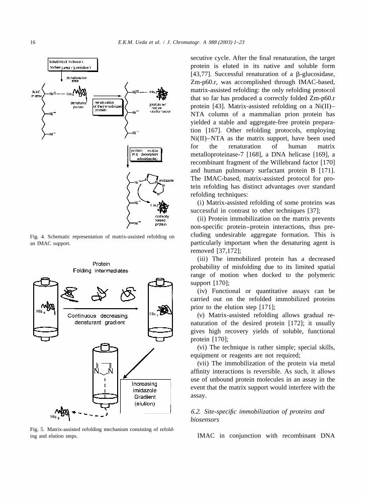
protein denaturation [22]. Following a decision to detection and quantification of proteins or peptides,adopt tag technology in the purification of proteins, have been applied as the basis for several biosensorcareful evaluation of the fusion protein design is techniques [144]. Immobilized ligand-based tag tech-required for each individual protein. The more nology has also been adapted for plate assays andinformation on the structure and folding of the biosensor methods [144]. In this section, the adapta-protein or peptide of interest can be obtained, the tion of IMAC-related methodology in other areas ofmore successful the use of the tag will be; ideally, protein processing is discussed.the tag would produce minimal changes in theactivity or structure of the protein. In the tag 6 .1. Immobilized metal ion affinity chromatographyselection process, details such as the type of metal and protein refolding: matrix-assisted refoldingaffinity tag, the terminus at which the tag will beinserted and the structure of the protein should be Production of recombinant proteins in bacteria ingiven utmost attention to ensure a successful applica- the form of cytoplasmic inclusion bodies is associ-tion [144]. ated with serious problems with regard to the proper
Other problems, associated with the use of metal structure and function of the proteins resulting fromaffinity tags, may arise when the target protein is protein misfolding. The latter is especially a problemgenerated for studies of its properties that could be when the proteins are composed of multiple subunits,affected by the tag, including structural, physiologi- have several disulfide bridges, or contain prostheticcal and pharmacological features. In such a case, groups. In the inclusion bodies, the protein is presentremoval of the tag following purification of the in an insoluble form, with reduced disulfide bondsprotein would be essential. In general, however, and no or impaired bioactivity. This worst scenariothere is no need for removal of histidine tags of misfolded proteins in particular occurs if the[107,144]. Removal of the tag can be achieved by bacterial synthesis of the protein is not supported bychemical cleavage or by enzymatic action. Chemical chaperones assisting in the folding of the protein.cleavage has several drawbacks such as harshness of However, even when use is made of protein foldingthe conditions used and the generation of toxic aid devices in the production of the protein (co-byproducts [107]. Enzymatic cleavage via endo- expression of chaperones, optimization of growthproteases that recognize specific amino acid se- conditions), a refolding step is usually needed forquences within the protein, has distinct advantages maximum recovery of the desired protein in theover the chemical method, including greater spe- native form [165,166]. In many cases, however, thecificity and less harsh conditions. However, en- folding step is ineffective, resulting in poor recoveryzymatic cleavage can be incorrect due to cleavage of and high operation costs [166]. Recently, a reliableinternal, non-canonical cleavage sites and may not technique, based on affinity tagging and designatedalways be effective, as found for the his-tag removal ‘‘matrix-assisted refolding,’’ has been developed forfrom human prolactin by factor Xa, which was only the production of soluble and functional proteinspartially successful. The latter was presumably a from inclusion bodies (Figs. 4 and 5). Briefly,result of poor accessible cleavage sites displayed by following production of the tagged protein in inclu-the protein [107,155]. sion bodies, it is solubilized, denatured and then
immobilized on a charged chelating resin [e.g.Ni(II)–NTA]. The subsequent renaturation step can
6 . Beyond immobilized metal ion affinity be carried out by applying a linear gradient spanningchromatography: other applications of metal denaturing to renaturing conditions or by iterativeaffinity refolding, a technique based on repeated cycles of
renaturation and denaturation with a decreasingThe principles of metal or other affinity chroma- concentration of the denaturing agent in each con-
16 E.K.M. Ueda et al. / J. Chromatogr. A 988 (2003) 1–23
secutive cycle. After the final renaturation, the targetprotein is eluted in its native and soluble form[43,77]. Successful renaturation of ab-glucosidase,Zm-p60.r, was accomplished through IMAC-based,matrix-assisted refolding: the only refolding protocolthat so far has produced a correctly folded Zm-p60.rprotein [43]. Matrix-assisted refolding on a Ni(II)–NTA column of a mammalian prion protein hasyielded a stable and aggregate-free protein prepara-tion [167]. Other refolding protocols, employingNi(II)–NTA as the matrix support, have been usedfor the renaturation of human matrixmetalloproteinase-7 [168], a DNA helicase [169], arecombinant fragment of the Willebrand factor [170]and human pulmonary surfactant protein B [171].The IMAC-based, matrix-assisted protocol for pro-tein refolding has distinct advantages over standardrefolding techniques:
(i) Matrix-assisted refolding of some proteins wassuccessful in contrast to other techniques [37];
(ii) Protein immobilization on the matrix preventsnon-specific protein–protein interactions, thus pre-cluding undesirable aggregate formation. This isFig. 4. Schematic representation of matrix-assisted refolding onparticularly important when the denaturing agent isan IMAC support.
removed [37,172];(iii) The immobilized protein has a decreased
probability of misfolding due to its limited spatialrange of motion when docked to the polymericsupport [170];
(iv) Functional or quantitative assays can becarried out on the refolded immobilized proteinsprior to the elution step [171];
(v) Matrix-assisted refolding allows gradual re-naturation of the desired protein [172]; it usuallygives high recovery yields of soluble, functionalprotein [170];
(vi) The technique is rather simple; special skills,equipment or reagents are not required;
(vii) The immobilization of the protein via metalaffinity interactions is reversible. As such, it allowsuse of unbound protein molecules in an assay in theevent that the matrix support would interfere with theassay.
6 .2. Site-specific immobilization of proteins andbiosensors
Fig. 5. Matrix-assisted refolding mechanism consisting of refold-ing and elution steps. IMAC in conjunction with recombinant DNA
E.K.M. Ueda et al. / J. Chromatogr. A 988 (2003) 1–23 17
technology allows efficient immobilization of a membrane can be used for the preparation of thetagged protein to a matrix with a consistent and so-called bio-functional membranes, entities indefined spatial orientation. The directed reversible which a biomolecule is immobilized on polymericimmobilization of proteins on surfaces by use of matrices cast on porous membranes. They have beenmetal complexes opens new ways for structural used in catalysis (membrane-based enzyme bio-investigation of proteins and receptor–ligand inter- reactors), separations (affinity membranes), analysisaction. Site-specific immobilization of proteins can (biosensors, metal ion-specific separations) and artifi-be used for analysis of interactions between bio- cial organs [150]. One of the applications of metalmolecules, such as protein–protein, protein–lipids, affinity-based protein adsorption is in biosensorprotein–drugs and protein–DNA interactions, by techniques. Biosensors are analytical tools in which aimmobilizing the protein of interest via an affinity molecule (e.g. an enzyme), covalently and irrever-tag on a solid-phase for subsequent interaction with sibly bound through cyanogen bromide activation ofthe second molecule. Site-specific immobilization of the support or reversibly through the exchange of theproteins has a number of advantages when compared metal ions of the IDA to an electrode surface, isto random immobilization of proteins, including utilized for the measurement of an analyte, or seriesgood steric accessibilities of active binding sites of of analytes, within a specimen. Sosnitza et al. [175]the attached proteins (e.g. enzymatic activity) and described application of reversible immobilizationincreased stability. Stability can be perceived not techniques for biosensors in which enzymes such asonly as an increased enzyme or protein shelf life but glucose oxidase, invertase and peroxidase werealso as the feasibility of reutilization of the immobil- immobilized on a metal ion-charged Chelatingized enzyme for several cycles without significant Sepharose Fast Flow resin (Pharmacia Biotech,bioactivity loss [173]. One major disadvantage of Sweden), a set-up with analytical capability compar-random immobilization of protein on polymeric able to that of regular biosensor systems. Use wasmembranes is that the bioactivity of the immobilized made of the Biacore system, a biosensor instrumentprotein is often significantly decreased [174], in using plasmon resonance detection for assessment ofsome cases because the active site may be blocked binding constants of protein and non-proteinaceousupon ligation to the support [149,150]. molecules to investigate how many histidine tags are
A useful application of IMAC technology has needed in a protein for stable binding to a metalbeen described [58] for isolation and characterization ion-charged, chelating NTA sensor chip. Two his-of protein partners, i.e. protein complexes in which tidines are usually sufficient for stable binding to the
21one subunit contains an oligohistidine tag. This Ni –NTA surface [44]. In a case described by Zhusystem can be used to test in vitro and also in vivo et al. [176], hexahistidine-tagged proteins whereassembled protein complexes containing a histidine- immobilized on a nickel-coated glass slide. Thistagged subunit. Immobilization of a his-tagged pro- procedure yielded superior quality protein chipstein can also be used for identification of protein when compared to regular protein arrays (e.g. alde-partners displaying affinity for a protein which is hyde-treated slides). Studies of the yeast proteomeimmobilized on a metal ion-charged chelating col- where performed by cloning 5800 open readingumn. This methodology has the option to either elute frames; the corresponding proteins where purifiedthe protein–protein complex by elution with buffers and spotted onto nickel-coated slides and screenedcontaining high concentrations of imidazole or, for the ability to interact with proteins and phos-alternatively, the interacting partner can be selective- pholipids. Using this technology, many new cal-ly eluted by increasing the ionic strength of the modulin and phospholipid-interacting proteins wherebuffers, or by addition of detergents, depending on identified. Fig. 6 shows a diagram of protein printingthe nature of the interaction. It was also pointed out on the glass slides. It appears that the lipid-basedthat this method is not limited to protein–protein immobilization of proteins via metal affinity sites is ainteractions, since it can be applied to identification promising method for the attachment of proteins toof any molecule displaying affinity for the immobil- an interface in an orderly fashion without functionalized protein [58]. Immobilization of proteins on a or structural losses [177–179]. Metal affinity-based
18 E.K.M. Ueda et al. / J. Chromatogr. A 988 (2003) 1–23
metals such as Cd(II). These engineered metal-bind-ing bacteria can be immobilized for the generation ofwhole-cell microbial tools for bioremediation pur-poses. One way could be the co-expression of ametal-binding protein with a cellulose-binding do-main (CDB), the latter used as the anchor site forcell immobilization on cellulose fibers[146,182,183].
6 .3. Immobilized metal ion affinity chromatographyas an analytical and characterization tool inprotein chemistry
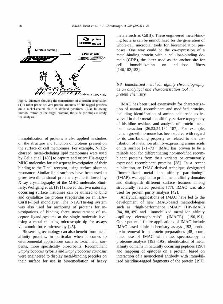
Fig. 6. Diagram showing the construction of a protein array slide:IMAC has been used extensively for characteriza-(1) a robot probe delivers precise amounts of His-tagged proteins
on a nickel-coated plate at defined positions; (2,3) following tion of natural, recombinant and modified proteins,immobilization of the target proteins, the slide (or chip) is ready including identification of amino acid residues in-for analysis. volved in their metal ion affinity, surface topography
of histidine residues and analysis of protein–metalion interaction [26,52,54,184–187]. For example,human growth hormone has been studied with regard
immobilization of proteins is also applied in studies to its zinc-binding property as related to the dis-on the structure and function of proteins present on tribution of metal ion affinity-expressing amino acidsthe surface of cell membranes. For example, Ni(II)- on its surface [71–73]. IMAC has proven to be acharged, metal-chelating lipid membranes were used reliable tool for differentiating non-modified recom-by Celia et al. [180] to capture and orient His-tagged binant proteins from their variants or erroneouslyMHC molecules for subsequent investigation of their expressed recombinant proteins [38]. In a recentbinding to the T cell receptor, using surface plasmon publication, an IMAC-derived technique, designatedresonance. Similar lipid surfaces have been used to ‘‘immobilized metal ion affinity partitioning’’grow two-dimensional protein crystals followed by (IMAP), was applied to probe metal affinity domainsX-ray crystallography of the MHC molecule. Simi- and distinguish different surface features amonglarly, Wolfgang et al. [181] showed that two naturally structurally related proteins [77]. IMAC was alsooccurring surface histidines can be utilized to bind used for protein purity analysis [42].and crystallize the protein streptavidin on an IDA– Analytical applications of IMAC have led to theCu(II)–lipid monolayer. The NTA/His-tag system development of new IMAC-based methodologieswas also used for anchoring of proteins for in- such as ‘‘high-performance IMAC’’ (HP-IMAC)vestigations of binding force measurement of re- [84,188,189] and ‘‘immobilized metal ion affinityceptor–ligand systems at the single molecule level capillary electrophoresis’’ (IMACE) [190,191].using a metal-chelating microscopic tip for assays Other potential future applications of IMAC includevia atomic force microscopy [45]. IMAC-based clinical chemistry assays [192], endo-
Biosensing technology can also benefit from metal toxin removal from protein preparations [48], com-affinity proteins, in particular when it comes to bined use of IMAC with mass spectroscopy inenvironmental applications such as toxic metal sor- proteome analysis [193–195], identification of metalbents, more specifically biosorbents. Recombinant affinity domains in naturally occurring peptides [196]Staphylococcus xylosus andStaphylococcus carnosus and mapping of epitopes on a protein, based onwere engineered to display metal-binding peptides on interaction of a monoclonal antibody with immobil-their surface for use in bioremediation of heavy ized histidine-tagged fragments of the protein [197].
E.K.M. Ueda et al. / J. Chromatogr. A 988 (2003) 1–23 19
6 .4. Immobilized metal ion affinity chromatography prolactin is more stable when stored as oligomers inand processing of phosphoproteins and secretory granules than in the monomeric formphosphopeptides [67,68]. Addition of Zn(II) to growth hormone, a
prolactin-related protein, has led to greater stabilityPhosphorylation and dephosphorylation of proteins of the hormone and this step has potential for use in
form a major mechanism in the control of protein pharmaceutical preparations [71]. In fact, engineeredactivity and regulation of biochemical pathways in metal-binding sites can lead to enhancement ofcells [198]. IMAC was found to provide a specific protein stability: a zinc-binding site insertion intomethod for identification of phosphoproteins, based proteina an engineered four-helix-bundle con-4,
on metal affinity of phosphopeptides, and Fe(III)- nected by three identical loops, rendered increasedIMAC in particular, has been used to selectively resistance against chemical denaturation probablypurify and concentrate phosphorylated proteins and due to ‘‘tightening’’ of the protein structure [207].peptides [121,187,198–200]. IMAC is often carried Other reports also demonstrate increased proteinout in conjunction with mass spectroscopy and other stabilization, e.g. increased thermal stability, con-techniques, such as reversed-phase high-performance formational stability and lower proteolytic degra-liquid chromatography and capillary electrophoresis, dation as a result of insertion of engineered metalto evaluate the effectiveness of the separation meth- affinity chelating sites, an approach which is easy tood [195,198,201–203]. implement and can be applied to a wide range of
proteins [112,113,208,209]. It is a well-accepted fact6 .5. Metal binding and protein stability that certain metal ions (calcium, magnesium and
zinc) can stabilize a protein and are used as stabiliz-Aggregation of protein hormones, such as prolac- ing excipients in pharmaceutical preparations. These
tin and growth hormone, is an important step in their metal ions stabilize the target protein through turningconcentration into secretory granules in the pituitary. its structure more rigid and compact, due to stabiliza-Such aggregation can be induced by interaction of tion of the folding [77,100].the proteins with metal ions, and zinc, for example,has a role in the formation and stabilization ofprolactin oligomers within the secretory granules 7 . Conclusions[68,77]. Proteins containing multiple accessible his-tidine residues on their surface can be precipitated This article reviews the rapidly increasing use ofusing metal ion-charged chelators (i.e. bis-copper IMAC for protein purification as well as proteinchelate). A precipitation technique analogous to the characterization. It also highlights use of IMAC inantibody–antigen precipitin reaction has been de- protein refolding, protein recognition and IMAC-veloped termed ‘‘metal affinity precipitation’’. It derived or -related methodologies such as orientedinvolves the formation of insoluble protein complex- immobilization of proteins and protein stabilizationes, bound together into a net-like structure and via metal affinity engineered sites. The currentcrosslinked via metal chelates [204]. The complexes literature indicates clear advantages of IMAC andcan be resolubilized with either a strong chelator (i.e. IMAC-derived techniques in protein or peptide pro-EDTA, EGTA) or a displacer such as imidazole cessing, when compared to other methodologies, in[205]. Affinity precipitation of proteins can also be terms of resolution, reliability, reproducibility andobtained using reversibly soluble–insoluble polymers processing time. Protein–metal affinity has a centralwhich have either a natural (chitosan) or synthetic role in a wide variety of applications in the process-(isopropylacrylamide) origin [205,206]. ing of non-modified or engineered proteins, includ-
Metal binding also provides methods for protein ing matrix-assisted refolding, protein partners identi-stabilization. Thus prolactin has a higher resistance fication, protein characterization, phosphoproteinto the enzyme, kallikrein, when complexed with zinc analysis and fractionation, protein purity assessment,ions than in the non-complexed state [69]; also, protein-folding evaluation, site-specific protein im-
20 E.K.M. Ueda et al. / J. Chromatogr. A 988 (2003) 1–23
[23] G. Chaga, M. Widersten, L. Andersson, J. Porath, U.H.mobilization (biosensors, functional assay plates andDanielson, B. Mannervik, Protein Eng. 7 (1994) 1115.molecular interaction studies), protein detection and
[24] H. Chaouk, M.T. Hearn, J. Biochem. Biophys. Methods 39quantification, protein extraction (two phase metal (1999) 161.affinity partitioning or IMAP) and IMAC coupled to [25] L. Drake, T. Barnett, Biotechniques 12 (1992) 645.other chromatographic and non-chromatographic [26] S. Sharma, G.P. Agarwal, Anal. Biochem. 288 (2001) 126.
ˆ[27] C. Fiore, V. Trezeguet, P. Roux, A. Le Saux, F. Noel, C.protein separation techniques.Schwimmer, D. Arlot, A.C. Dianoux, G.J. Lauquin, G.Brandolin, Protein Expr. Purif. 19 (2000) 57.
[28] J. Wu, M. Filutowicz, Acta Biochim. Pol. 46 (1999) 591.A cknowledgements [29] J.C. Dalton, D.F. Bruley, K.A. Kang, W.N. Drohan, Adv.
Exp. Med. Biol. 411 (1997) 419.[30] W. He, D.F. Bruley, W.N. Drohan, Adv. Exp. Med. Biol. 454EKMU is the recipient of a graduate scholarship
(1998) 689.awarded by CNPq, Brazil.[31] N.T. Mrabet, Biochemistry 31 (1992) 2690.[32] H. Yeomans-Reina, A. Ruiz-Manriquez, B.R. Wong, A.T.
Mansir, Biotechnol. Prog. 17 (2001) 729.R eferences [33] F.H. Arnold, Biotechnology 9 (1991) 151.
[34] J. Porath, Trends Anal. Chem. 7 (1988) 254.[35] T.T. Yip, T.W. Hutchens, Mol. Biotechnol. 1 (1994) 151.[1] J. Porath, J. Carlsson, I. Olsson, G. Belfrage, Nature 258[36] S.A. Lopatin, V.P. Varlamov, Prikl. Biokhim. Mikrobiol. 31(1975) 598.
(1995) 259.[2] F. Hellferich, Nature 189 (1961) 1001.[37] J. Zouhar, Chem. Listy 93 (1999) 683.˚ ´[3] L. Kagedal, in: J.-C. Janson, L. Ryden (Eds.), Protein[38] J.W. Wong, R.L. Albright, N.-H. Wang, Sep. Purif. MethodsPurification: Principles, High-Resolution Methods, and Ap-
20 (1991) 49.plications, 2nd ed, Wiley–VCH, New York, 1998, p. 311,[39] V. Gaberc-Porekar,V. Menart, J. Biochem. Biophys. MethodsChapter 8.
49 (2001) 335.´[4] B. Nilsson, G. Forsberg, T. Moks, M. Hartmanis, M. Uhlen,[40] G.S. Chaga, J. Biochem. Biophys. Methods 49 (2001) 313.Curr. Opin. Struct. Biol. 2 (1992) 569.[41] N.T. Mrabet, M.A. Vijayalakshmi, in: M.A. Vijayalakshmi[5] A. Seidler, Protein Eng. 7 (1994) 1277.
(Ed.), Biochromatography, Taylor and Francis, London,[6] T. Hopp, K.S. Pricket, V.L. Price, R.T. Libby, C.J. March,2002, p. 272.D.P. Cerretti, D.L. Urdal, P.J. Conlon, Biotechnology 6
[42] Z. Zhang, K.-T. Tong, M. Belew, T. Pettersson, J.-C. Janson,(1988) 1204.J. Chromatogr. 604 (1992) 143.[7] J.-S. Kim, R.T. Raines, Protein Sci. 2 (1993) 348.
[43] D. Sinha, M. Bakhshi, R.Vora, Biotechniques 17 (1994) 509.[8] T.G. Schmidt, A. Skerra, Protein Eng. 6 (1993) 109.¨ ¨ ¨[44] L. Nieba, S.E. Nieba-Axmann, A. Persson, M. Hamalainen,[9] D.B. Smith, K.S. Johnson, Gene 67 (1988) 31.
˚F. Edebratt, A. Hansson, J. Lidholm, K. Magnusson, A.F.¨[10] C. Lauritzen, E. Tuchsen, P.E. Hansen, O. Skovgaard,¨Karlsson, A. Pluckthun, Anal. Biochem. 252 (1997) 217.Protein Expr. Purif. 2 (1991) 372.
´[45] L. Schmitt, M. Ludwig, H.E. Gaub, R. Tampe, Biophys. J.[11] E.R. LaVallie, E.A. DiBlasio, S. Kovacic, K.L. Grant, P.F.78 (2000) 3275.Schendel, J.M. McCoy, Biotechnology 11 (1993) 187.
[46] C. Min, G.L. Verdine, Nucleic Acids Res. 24 (1996) 3806.¨[12] E. Hochuli, H. Dobeli, A. Schacher, J. Chromatogr. 411[47] P.L. Roberts, C.P. Walker, P.A. Feldman, Vox Sang. 67(1987) 177.
(Suppl. 1) (1994) 69.[13] R.C. Hockney, Trends Biotechnol. 12 (1994) 456.[48] K.L. Franken, H.S. Hiemstra, K.E. van Meijgaarden, Y.[14] E. Flaschel, K. Friehs, Biotechnol. Adv. 11 (1993) 31.
Subronto, J. den Hartigh, T.H. Ottenhoff, J.W. Drijfhout,[15] E. Hochuli, Genet. Eng. (NY) 12 (1990) 87.Protein Expr. Purif. 18 (2000) 95.[16] D.B. Evans, A.F. Vosters, J.B. Carter, S.K. Sharma, J.
[49] R.G. Pearson, J. Chem. Educ. 45 (1968) 581.Immunol. Methods 156 (1992) 231.[50] T.-T. Yip, Y. Nakagawa, J. Porath, Anal. Biochem. 183¨[17] C. Wulfing, J. Lombardero, A. Pluckthun, J. Biol. Chem. 269
(1989) 159.(1994) 2895.[51] G. Chaga, L. Andersson, B. Ersson, J. Porath, Biotechnol.[18] T.W. Hutchens, R.W. Nelson, C.M. Li, T.T. Yip, J. Chroma-
Appl. Biochem. 11 (1989) 424.togr. 604 (1992) 125.[52] E.S. Hemdan, Y.J. Zhao, E. Sulkowski, J. Porath, Proc. Natl.[19] T.W. Hutchens, T.T. Yip, J. Chromatogr. 604 (1992) 133.
Acad. Sci. USA 86 (1989) 1811.[20] R.R. Begum, R.J. Newbold, D. Whitford, J. Chromatogr. B[53] R.J. Todd, M.E. Van Dam, D. Casimiro, B.L. Haymore, F.H.737 (2000) 119.
Arnold, Proteins 10 (1991) 156.[21] T. Oswald, W. Wende, A. Pingound, U. Rinas, Appl. Mi-[54] S.-S. Suh, B.L. Haymore, F.H. Arnold, Protein Eng. 4 (1991)crobiol. Biotechnol. 42 (1994) 73.
301.[22] M.M. Enzelberger, S. Minning, R.D. Schmid, J. Chromatogr.[55] E. Leporati, J. Chem. Soc. Dalton Trans. (1986) 199.A 898 (2000) 83.
E.K.M. Ueda et al. / J. Chromatogr. A 988 (2003) 1–23 21
[56] E.S. Hemdan, J. Porath, J. Chromatogr. 323 (1985) 247. [89] E.K. Ueda, P.W. Gout, L. Morganti, J. Chromatogr. A 922[57] Z. El Rassi, C. Horvath, J. Chromatogr. 359 (1986) 241. (2001) 165.[58] M. Sawadogo, M.W. Van Dyke, Genet. Eng. 17 (1995) 53. [90] E.S. Cole, E.H. Nichols, K. Lauziere, T. Edmunds, J.M.[59] P. Hansen, L. Andersson, G. Lindeberg, J. Chromatogr. A McPherson, Endocrinology 129 (1991) 2639.
723 (1996) 51. [91] P. Arvidsson, A.E. Ivanov, I.Yu. Galaev, B. Mattiasson, J.[60] P. Hansen, G. Lindeberg, J. Chromatogr. A 690 (1995) 155. Chromatogr. B 753 (2001) 279.[61] D.R. Williams, The Metals of Life: The Solution Chemistry [92] G. Chaga, J. Hopp, P. Nelson, Biotechnol. Appl. Biochem.
of Metal Ions in Biological Systems,Van Nostrand Reinhold, 29 (1999) 19.London, 1971. [93] G. Chaga, D.E. Bochkariov, G.G. Jokhadze, J. Hopp, P.
[62] M. Zachariou, M.T. Hearn, J. Protein Chem. 14 (1995) 419. Nelson, J. Chromatogr. A 864 (1999) 247.[63] M. DiDonato, S. Narindrasorasak, J.R. Forbes, D.W. Cox, B. [94] J. Porath, Trends Anal. Chem. 7 (1992) 254.
Sarkar, J. Biol. Chem. 272 (1997) 33279. [95] K.D. Bush, J.A. Lumpkin, Biotechnol. Prog. 14 (1998) 943.[64] P.E. Dykema, P.R. Sipes, A. Marie, B.J. Biermann, D. N [96] E. Sulkowski, Bioessays 10 (1989) 170.
Crowell, S.K. Randall, Plant Mol. Biol. 41 (1999) 139. [97] D. Beyersmann, Toxicol. Lett. 127 (2002) 63.[65] J.M. Berg, Y. Shi, Science 271 (1996) 1081. [98] G. Muszynska, Y.-J. Zhao, J. Porta, J. Immunoreact. Bio-[66] S. Kinet, S. Bernichtein, P.A. Kelly, J.A. Martial,V. Goffin, J. chem. 26 (1986) 127.
Biol. Chem. 274 (1999) 26033. [99] J.R. Requena, D. Groth, G. Legname, E.R. Stadtman, S.B.[67] M.Y. Lorenson, T. Patel, J.-W. Liu, A.M. Walker, Endocrinol- Prusiner, R.L. Levine, Proc. Natl. Acad. Sci. USA 98 (2001)
ogy 137 (1996) 809. 7170.[68] H.B. Lowman, B.C. Cunningham, J.A. wells, J. Biol. Chem. [100] W. Wang, Int. J. Pharm. 185 (1999) 129.
266 (1991) 10982. [101] H.R. Constantino, R. Langer, A.M. klibanov, Pharm. Res.[69] E.A. Permyakov, D.B. Veprintsev, G.Y. Deikus, S.E. 11 (1994) 21.
Permyakov, L.P. Kalinichenko, V.M. Grishchenko, C.L. [102] B. Chen, H.R. Constantino, J. Liu, C.C. Hsu, S.J. Shire, J.Brooks, FEBS Lett. 405 (1997) 273. Pharm. Sci. 88 (1999) 477.
[70] Z. Sun, M.S. Lee, H.K. Rhee, J.M. Arrandale, P.S. Dannies, [103] M.S. Kamat, G.L. Tolman, J.M. Brown, Pharm. Biotechnol.Mol. Endocrinol. 11 (1997) 1544. 9 (1996) 343.
[71] B.C. Cunningham, M.G. Mulkerrin, J.A. Wells, Science 253 [104] T. Oswald, G. Hornbostel, U. Rinas, F.B. Anspach,(1991) 545. Biotechnol. Appl. Biochem. 25 (1997) 109.
[72] B.C. Cunningham, D.J. Henner, J.A. Wells, Science 247 [105] J. Xiao, C.E. Kibbey, D.E. Coutant, G.B. Martin, M.E.(1990) 1461. Meyerhoff, J. Liq. Chromatogr. 19 (1996) 2901.
[73] B.C. Cunningham, S. Bass, G. Fuh, J.A. Wells, Science 250 [106] I.M. Rosenberg, Protein Analysis and Purification: Bench-¨(1990) 1709. top Techniques, Birkhauser, Boston, 1996, Chapter 10, p.
[74] J. Porath, Protein Expr. Purif 3 (1992) 263. 345.[75] G.E. Lindgren, Am. Biotechnol. Lab. 12 (1994) 36. [107] J. Pedersen, C. Lauritzen, M.T. Madsen, S. Weis Dahl,[76] E. Hochuli, W. Bannwarth, H. Dobeli, R. Gentz, D. Stuber, Protein Expr. Purif. 15 (1999) 389.
Biotechnology (1988) 1321. [108] J. Crowe, H. Dobeli, R. Gentz, E. Hochuli, D. Stuber, K.[77] M. Zaveckas, B. Baskeviciute, V. Luksa, G. Zvirblis, V. Henco, Methods Mol. Biol. 31 (1994) 371.
Chmieliauskaite, V. Bumelis, H. Pesliakas, J. Chromatogr. A [109] H.J. Cha, N.G. Dalal, M.Q. Pham, W.E. Bentley, Biotech-904 (2000) 145. nol. Prog. 15 (1999) 283.
[78] M. Zachariou, M.T. Hearn, J. Chromatogr. A 890 (2000) 95. [110] B. Tallet, T. Astier-Gin, M. Castroviejo, X. Santarelli, J.[79] R.V. Lehr, L.C. Elefante, K.K. Kikly, S.P. O’Brien, R.B. Chromatogr. B 753 (2001) 17.
Kirkpatrick, Protein Expr. Purif. 19 (2000) 362. [111] M. Chang, J.L. Bolton, S.Y. Blond, Protein Expr. Purif. 17[80] D. Leibler, A. Rabinkov, M. Wilchek, J. Mol. Recogn. 9 (1999) 443.
(1996) 375. [112] F.H. Arnold, B.L. Haymore, Science 252 (1991) 1796.[81] C. Mateo, G. Fernandez-Lorente, B.C. Pessela, A. Vian, A.V. [113] H.W. Hellinga, Curr. Opin. Biotechnol. 7 (1996) 437.
Carrascosa, J.L. Garcia, R. Fernandez-Lafuente, J.M. Guisan, [114] R.S. Pasquinelli, R.E. Shepherd, R.R. Koepsel, A. Zhao,J. Chromatogr. A 915 (2001) 97. M.M. Ataai, Biotechnol. Prog. 16 (2000) 86.
[82] Q. Zeng, J. Xu, R. Fu, Q. Ye, J. Chromatogr. A 921 (2001) [115] V. Gaberc-Porekar, V. Menart, S. Jevsevar, A. Vidensek, A.197. Stalc, J. Chromatogr. A 852 (1999) 117.
[83] R.J. Todd, R.D. Johnson, F.H. Arnold, J. Chromatogr. A 662 [116] A.M. Schmidt, H.N. Muller, A. Skerra, Chem. Biol. 3(1994) 13. (1996) 645.
[84] M. Belew, J. Porath, J. Chromatogr. 516 (1990) 333. [117] V.V. Kronina, H.-J. Wirth, M.T. Hearn, J. Chromatogr. A[85] F. Maisano, S.A. Testori, G. Grandi, J. Chromatogr. 472 852 (1999) 261.
´(1989) 422. [118] J. Liesiene, K. Racaityte, M. Morkeviciene, P. Valancius, V.[86] E. Sulkowski, J. Mol. Recogn. 9 (1996) 389. Bumelis, J. Chromatogr. A 764 (1997) 27.[87] E. Sulkowski, J. Mol. Recogn. 9 (1996) 498. [119] S.A. Lopatin, A.V. Il’ina, A.A. Shul’ga, O.S. Grinchenko,[88] A.E. Price, K.B. Logvinenko, E.A. Higgins, E.S. Cole, S.M. V.P. Varlamov, K.G. Skriabin, M.P. Kirpichnikov,
Richards, Endocrinology 136 (1995) 4827. Biokhimiia 59 (1994) 226.
22 E.K.M. Ueda et al. / J. Chromatogr. A 988 (2003) 1–23
[120] T.M. McNerney, S.K. Watson, J.-H. Sim, R.L. Briden- [151] C.F. Ford, I. Suominen, C.E. Glatz, Protein Expr. Purif. 2baugh, J. Chromatogr. A 744 (1996) 223. (1991) 95.
[121] R. Anguenot, S. Yelle, B. Nguyen-Quoc, Arch. Biochem. [152] S. Supattapone, H.O. Nguyen, T. Muramoto, F.E. Cohen,Biophys. 365 (1999) 163. S.J. DeArmond, S.B. Prusiner, M. Scott, J. Virol. 74 (2000)
[122] V. Boden, J.J. Winzerling, M. Vijayalakshmi, J. Porath, J. 11928.Immunol. Methods 181 (1995) 225. [153] A. Varshavsky, Cell 69 (1992) 725.
˚ ´[123] G.S. Chaga, B. Ersson, J.O. Porath, J. Chromatogr. A 732 [154] J. Nilsson, S. Stahl, J. Lundeberg, M. Uhlen, P.A. Nygren,(1996) 261. Protein Expr. Purif. 11 (1997) 1.
[124] U. Winkler, T.M. Pickett, D. Hudig, J. Immunol. Methods [155] L. Morganti, M. Huyer, P.W. Gout, P. Bartolini, Biotechnol.191 (1996) 11. Appl. Biochem. 23 (1996) 67.
[125] D.K. McCormick, Biotechnology 11 (1993) 1059. [156] Z. Li, E. Crooke, Protein Expr. Purif. 17 (1999) 41.[126] B.C. Batt, V.M. Yabannavar, V. Singh, Bioseparation 5 [157] J.P. Jin, A. Chen, O. Ogut, Q.Q. Huang, Am. J. Physiol.
(1995) 41. Cell Physiol. 279 (2000) C1067.[127] F.P. Gailliot, C. Gleason, J.J. Wilson, J. Zwarick, Biotech- [158] R. Janknecht, G. de Martynoff, J. Lou, R.A. Hipskind, A.
nol. Prog. 6 (1990) 370. Nordheim, H.G. Stunnenberg, Proc. Natl. Acad. Sci. USA¨[128] H.J. Johansson, C. Jagersten, J. Shiloach, J. Biotechnol. 48 88 (1991) 8972.
(1996) 9. [159] C.M. Halliwell, G. Morgan, C.-P. Ou, A.E. Cass, Anal.[129] A.-K. Barnfield Frej, H.J. Johansson, S. Johansson, P. Biochem. 295 (2001) 257.
Leijon, Bioprocess Eng. 16 (1997) 57. [160] A. Goel, D. Colcher, J.-S. Koo, B.J. Booth, G. Pavlinkova,[130] R. Hjorth, Trends Biotechnol. 15 (1997) 230. S.K. Batra, Biochem. Biophys. Acta 1523 (2000) 13.
¨ ¨ ¨[131] N. Ameskamp, C. Priesner, J. Lehmann, D. Lutkemeyer, [161] H. Buning, U. Gartner, D. von Schack, P.A. Baeuerle, H.Bioseparation 8 (1999) 169. Zorbas, Anal. Biochem. 234 (1996) 227.
[132] R.H. Clemmitt, H.A. Chase, Biotechnol. Bioeng. 67 (2000) [162] J.L. Rosales, K.Y. Lee, Biochem. Biophys. Res. Commun.206. 273 (2000) 1058.
[133] R.H. Clemmitt, L.J. Bruce, H.A. Chase, Bioseparation 8 [163] H. Tang, P.R. Chitnis, Indian J. Biochem. Biophys. 37(1999) 53. (2000) 433.
[134] S. Noronha, J. Kaufman, J. Shiloach, Bioseparation 8 [164] T. Sprules, N. Green, M. Featherstone, K. Gehring,(1999) 145. Biotechniques 25 (1998) 20.
[135] N.A. Willoughby, T. Kirschner, M.P. Smith, R. Hjorth, N.J. [165] G. Georgiou, P. Valax, Curr. Opin. Biotechnol. 7 (1996)Titchener-Hooker, J. Chromatogr. A 840 (1999) 195. 190.
[136] S. Plunket, F.H. Arnold, Biotechnol. Biotech. 4 (1990) 45. [166] D.R. Thatcher, A. Hitchcock, in: R.H. Pain (Ed.), Mecha-[137] A. Otto, G. Birkenmeier, J. Chromatogr. 644 (1993) 25. nisms of Protein Folding, Oxford University Press, New[138] U. Sivars, J. Abramson, S. Iwata, F. Tjerneld, J. Chroma- York, 1996, p. 232, Chapter 9.
togr. B 743 (2000) 307. [167] R. Zahn, C. von Schroetter, K. Wuthrich, FEBS Lett. 417[139] H. Pesliakas,V. Zutautas, B. Baskeviciute, J. Chromatogr. A (1997) 400.
678 (1994) 25. [168] M. Itoh, K. Masuda, Y. Ito, T. Akizawa, M. Yoshioka, K.[140] S.M. O’Brien, R.P. Sloane, O.R. Thomas, P. Dunnill, J. Imai, Y. Okada, H. Sato, M. Seiki, J. Biochem. 119 (1996)
Biotechnol. 54 (1997) 53. 667.[141] J. Porath, J. Protein Chem. 16 (1997) 463. [169] S.F. Beresten, R. Stan, A.J. van Brabant, T. Ye, S. Naurec-
¨ ¨[142] K.M. Muller, K.M. Arndt, K. Bauer, A. Pluckthun, Anal. kiene, N.A. Ellis, Protein Expr. Purif. 17 (1999) 239.´Biochem. 259 (1998) 54. [170] J. Zouhar, E. Nanak, B. Brzobohaty, Protein Expr. Purif. 17
[143] T.G. Consler, B.L. Persson, H. Jung, K.H. Zen, K. Jung, (1999) 153.G.G. Prive, G.E. Verner, H.R. Kaback, Proc. Natl. Acad. [171] A. Holzinger, K.S. Phillips, T.E. Weaver, Biotechniques 20Sci. USA 90 (1993) 6934. (1996) 804.
[144] C. Jones, A. Patel, S. Griffin, J. Martin, P. Young, K. [172] P.-Y. Shi, N. Maizels, A.M. Weiner, Biotechniques 23O’Donnell, C. Silverman, T. Porter, I. Chaiken, J. Chroma- (1997) 1036.togr. A 707 (1995) 3. [173] E. Ordaz, A. garrido-Pertierra, M. Gallego, A. Puyet,
[145] S.A. McMahan, R.R. Burgess, Anal. Biochem. 236 (1996) Biotechnol. Prog. 16 (2000) 287.101. [174] R. Lawung, B. Danielsson, V. Prachayasittikul, L. Bulow,
´ ¨ ˚[146] H. Wernerus, J. Lehtio, T. Teeri, P.-A. Nygren, S. Stahl, Anal. Biochem. 296 (2001) 57.Appl. Environ. Microbiol. 67 (2001) 4678. [175] P. Sosnitza, M. Farooqui, M. Saleemuddin, R. Ulber, T.
[147] C. Buchel, E. Morris, E. Orlova, J. Barber, J. Mol. Biol. Scheper, Anal. Chim. Acta 368 (1998) 197.312 (2001) 371. [176] H. Zhu, M. Bilgin, R. Bangham, D. Hall, A. Casamayor, P.
[148] M. Saleemuddin, Adv. Biochem. Eng. Biotechnol. 64 Bertone, N. Lan, R. Jansen, S. Bidlingmaier, T. Houfek, T.(1999) 203. Mitchell, P. Miller, R.A. Dean, M. Gerstein, M. Snyder,
´[149] J. Turkova, J. Chromatogr. B 722 (1999) 11. Science 293 (2001) 2101.[150] D.A. Butterfield, D. Bhattacharyya, S. Daunert, L. Bachas, [177] C. Dietrich, O. Boscheinen, K.D. Scharf, L. Schmitt, R.
´J. Membr. Sci. 181 (2001) 29. Tampe, Biochemistry 35 (1996) 1100.
E.K.M. Ueda et al. / J. Chromatogr. A 988 (2003) 1–23 23
´[178] U. Radler, J. Mack, N. Persike, G. Jung, R. Tampe, [194] D. Figeys, S.P. Gygi, Y. Zhang, J. Watts, M. Gu, R.Biophys. J. 79 (2000) 3144. Aebersold, Electrophoresis 19 (1998) 1811.
´[179] C. Dietrich, L. Schmitt, R. Tampe, Proc. Natl. Acad. Sci. [195] X. Qian, W. Zhou, M.G. Khaledi, K.B. Tomer, Anal.USA 92 (1995) 9014. Biochem. 274 (1999) 174.
[180] H. Celia, E. Wilson-Kubalek, R.A. Milligan, L. Teyton, [196] A.V. Patwardhan, G.N. Goud, R.R. Koepsel, M.M. Ataai, J.Proc. Natl. Acad. Sci. USA 96 (1999) 5634. Chromatogr. A 787 (1997) 91.
[181] F. Wolfgang, W.R. Schief, D.W. Pack, C.-T. Chen, A. [197] L. Rao, D.P. Jones, L.H. Nguyen, S.A. McMahan, R.R.Chilkoti, P. Stayton, V. Vogel, F.H. Arnold, Proc. Natl. Burgess, Anal. Biochem. 241 (1996) 173.Acad. Sci. USA 93 (1996) 4937. [198] P. Cao, J.T. Stults, J. Chromatogr. A 853 (1999) 225.
´[182] J. Lehtio, H. Wernerus, P. Samuelson, T.T. Teeri, S. Stahl, [199] M.C. Posewitz, P. Tempst, Anal. Chem. 71 (1999) 2883.FEMS Microbiol. Lett. 195 (2001) 197. [200] M. Quadroni, P. James, Experientia Supplementum 88
´ ˚[183] P. Samuelson, H. Wernerus, M. Svedberg, S. Stahl, Appl. (2000) 199.Environ. Microbiol. 66 (2000) 1243. [201] S. Li, C. Dass, Anal. Biochem. 270 (1999) 9.
[184] P. Burba, B. Jakubowski, R. Kuckuk, K. Kullmer, K.G. [202] P. Cao, J.T. Stults, Rapid Commun. Mass Spectrom. 14Heumann, Fresenius J. Anal. Chem. 368 (2000) 689. (2000) 1600.
[185] R. Kuckuk, P. Burba, Fresenius J. Anal. Chem. 366 (2000) [203] A. Stensballe, S. Andersen, O.N. Jensen, Proteomics 195. (2001) 207.
[186] F. Bianchi, R. Rousseaux-Prevost, P. Hublau, J. Rousseaux, [204] M.E. Van Dam, G.E. Wuenschell, F.H. Arnold, Biotechnol.Int. J. Pept. Protein Res. 43 (1994) 410. Appl. Biochem. 11 (1989) 492.
[187] D.C. Neville, C.R. Rozanas, E.M. Price, D.B. Gruis, A.S. [205] A. Kumar, I.Y. Galaev, B. Mattiasson, Biotechnol. Bioeng.Verkman, R.R. Townsend, Protein Sci. 6 (1997) 2436. 59 (1998) 695.
[188] J. Porath, J. Chromatogr. 443 (1988) 3. [206] B. Mattiasson, A. Kumar, I.Yu. Galaev, J. Mol. Recogn. 11[189] M. Belew, T.T. Yip, L. Andersson, R. Ehrnstrom, Anal. (1998) 211.
Biochem. 164 (1987) 457. [207] L. Regan, Trends Biochem. Sci. 20 (1995) 280.[190] K. Haupt, F. Roy, M.A.Vijayalakshmi, Anal. Biochem. 234 [208] F.H. Arnold, J.H. Zhang, Trends Biotechnol. 12 (1994)
(1996) 149. 189.[191] K.-Y. Jiang, O. Pitiot, M. Anissimova, H. Adenier, M.A. [209] J.T. Kellis Jr., R.J. Todd, F.H. Arnold, Biotechnology 9
Vijayalakshmi, Biochim. Biophys. Acta 1433 (1999) 198. (1991) 994.[192] D.S. Hage, Clin. Chem. 45 (1999) 593.[193] J. Ji, A. Chakraborty, M. Geng, X. Zhang, A. Amini, M.
Bina, F. Regnier, J. Chromatogr. B 745 (2000) 197.