review article vasodilators during cerebral aneurysm...
TRANSCRIPT

775
Kazuo Abe MD
Review Article
Vasodilators duringcerebral aneurysmsurgery
The objective of this review is to review the anaesthetic im-plications of vasoactive compounds particularly with regard tothe cerebral circulation and their clinical importance for thepracticing anaesthetist. Material was selected on the basis ofvalidity and application to clinical practice and animal studieswere selected only if human studies were lacking. Hypotensivedrugs have been used to induce hypotension and in the treat-ment of intraoperative hypertension during cerebral aneurysmsurgery. After subarachnoid haemorrhage, cerebral blood flowis reduced and cerebral vasoreactivity is disturbed which maylead to brain ischaemia. Also, cerebral arterial vasospasm de-creases cerebral blood flow, and may lead to delayed ischaemicbrain damage which is a major problem after subarachnoidhaemorrhage. Recently, the use of induced hypotension has de-creased although it is still useful in patients with intraoperativeaneurysm rupture, giant cerebral aneurysm, fragile aneurysmsand multiple cerebral aneurysms. In this review, a variety ofvasodilating agents, prostaglandin Et, sodium nitroprusside, ni-troglycerin, trimetaphan, adenosine, calcium antagonists, andinhalational anaesthetics, are discussed for their clinical use-fulness. Sodium nitroprusside, nitroglycerin and isoflurane are
Key wordsANAESTHETICS, VOLATILES: isoflurane;ANAESTHETIC TECHNIQUES: hypotension;BLOOD PRESSURE: hypotension;BRAIN: blood flow, carbon dioxide tension;HORMONES: prostaglandin E,SYMPATHETIC NERVOUS SYSTEM: ganglionic blockade,
trimetaphan;PHARMACOLOGY: adenosine, nitroglycerin, nitroprusside,
calcium channel blockers, hydralazine, diazoxide,labetalol, esmolol;
COMPLICATIONS: intraoperative hypertension, brainischaemia, rebleeding.
From the Department of Anaesthesia, Osaka Police Hospital.Address correspondence to: Dr. Kazuo Abe, Department of
Anaesthesia, Osaka Police Hospital, 10-31 Kitayama, Tennouji,Osaka 543, Japan.
Accepted for publication 17th April, 1993.
the drugs of choice for induced hypotension. Prostaglandin Et,nicardipine and nitroglycerin have the advantage that they donot alter carbon dioxide reactivity. Local cerebral blood flowis increased with nitroglycerin, decreased with trimetaphan andunchanged with prostaglandin Ej. Intraoperative hypertensionis a dangerous complication occurring during cerebral aneu-rysm surgery, but its treatment in association with subarachnoidhaemorrhage is complicated in cases of cerebral arterial va-sospasm because fluctuations in cerebral blood flow may beexacerbated. Hypertension should be treated immediately to re-duce the risk of rebleeding and intraoperative aneurysmal rup-ture and the choice of drugs is discussed. Although the useof induced hypotension has declined, the control of arterialblood pressure with vasoactive drugs to reduce the risk of in-traoperative cerebral aneurysm rupture is a useful technique.Intraoperative hypertension should be treated immediately butthe cerebral vascular effects of each vasodilator should be un-derstood before their use as hypotensive agents.
L'objectif de eel article est de revoir les implications anesthe-siques des composes vasoactifs particulierement en rapport avecla circulation cerebrale ainsi que leur importance clinique pourI'anesthesiste. Les agents ont ete choisis sur la base de leurvalidite et de leur application a la pratique clinique et des etudesanimales ont ete choisies seulement en I'absence delude surIhomme. Les agents hypotenseurs ont ete utilises pour induirede Ihypotension etpour trailer Ihypertension preoperatoire pen-dant la chirurgie d'anevrisme cerebrale. Apres une hemorragiesous-arachnoidienne, le debit sanguin cerebrale reduit et la va-somotricite cerebrale perturbee peuvent entrainer une ischemiecerebrale. Ainsi, le vasospasme arteriel cerebral diminue le debitsanguin cerebral et peut conduire a des dommages cerebrauxretardes d'ischemie, probleme majeur apres une hemorragiesous-arachnoidienne. Recemment, Vutilisation de Ihypotensioncontrolee s'est rarefiee bien qu'elle soit encore utile chez lespatients avec une rupture peroperatoire d'anevrisme, en casd'anevrisme cerebral geant, d'anevrismes fragiles et d'an-evrismes cerebraux multiples. Dans cet article, une variete devasodilatateurs, le prostaglandine Ej, le nitroprussiate de so-dium, la nitroglycerine, le trimetaphan, I'adenosine, les anta-gonistes caldques et les agents d'inhalation son discutes pour
CAN J ANAESTH 1993 / 40: 8 / pp 775-90

776 CANADIAN JOURNAL OF ANAESTHESIA
leur utilite clinique. Le nitroprussiate de sodium, la nitrogly-cerine et I'isoflurane sont les agents de choixpour I'hypotensioncontrolee. La prostaglandine Ej, la nicardipine et la nitrogly-cerine ont I'avantage de ne pas alterer la reactivite au dioxydede carbone. Le debit sanguin cerebral local est augmente avecla nitroglycerine, diminue avec le trimetaphan et inchange avecla prostaglandine Et. L'hypertension peroperatoire est une com-plication dangereuse aux cours dune chirurgie d'anevrismecerebral, mais son traitement lors d'hemorragie sous-arachnoidienne se complique dans les cas de vasospasme ar-teriel cerebral parce que les fluctuations du debit sanguincerebralpeuvent s'exacerber. L'hypertension devrait etre traiteeimmediatement pour reduire le risque de resaignement et derupture peroperatoire de I'anevrisme. Le choix des agents estdiscute. Bien que I'utilisation de I'hypotension controlee estmoins frequente, le controle de la pression arterielle avec desagents vasoactifs dans le but de reduire le risque de ruptureperoperatoire d'anevrisme cerebral est une technique courante.L'hypertension peroperatoire devrait etre traitee immediatementmais les effets vasculaires cerebraux de chaque vasodilatateursdevraient etre compris avant qu'ils soient utilises comme agentshypotenseurs.
ContentsIntroductionPathophysiology- Subarachnoid haemorrhage- Cerebral vasospasmCerebral haemodynamics- Carbon dioxide reactivity- Autoregulation- Cerebral blood flow- Cerebral arterial blood flow velocityInduced hypotensionHypotensive drugs- Prostaglandin E,- Sodium nitroprusside- Adenosine- Nitroglycerin- Trimetaphan- Calcium channel blockers- Isoflurane- Other vasodilatorsClinical implications
During the clipping of cerebral arterial aneurysms, in-duced hypotension is often used to reduce the aneurysmalwall tension to minimize the risk of premature rupture.'A primary concern with the use of hypotension has beenthat cerebral blood flow (CBF) may decrease to criticallylow levels, resulting in ischaemic damage to the brain.Patients with subarachnoid haemorrhage (SAH) may de-
velop abnormal cerebrovascular reactivity and impairedautoregulation23 and they may be more susceptible tosevere flow reductions during hypotension than normalindividuals,4 resulting in global and or focal ischaemiaof the brain.5 During the first two weeks after SAH,several events may make heavy demands on the capacityof the cerebral vasculature to react adequately. Cerebralarterial vasospasm and rebleeding are major problemsand cerebral aneurysm surgery is often performed withinthe same period. Another problem during general anaes-thesia for aneurysm clipping is intraoperative hyperten-sion. Intraoperative hypertension may increase the riskof rebleeding and intraoperative aneurysm rupture beforeits exposure so that intraoperative blood pressure shouldbe well controlled.
Pathophysiology
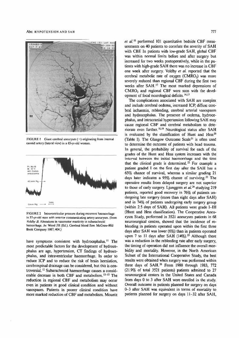
Subarachnoid haemorrhageTen percent of all strokes result from subarachnoid haem-orrhage (SAH) which is caused most often by the suddenrupture of an intracranial saccular aneurysm. Subarach-noid haemorrhage chiefly afflicts patients between 40 and60 yr of age and women are affected more often thanmen. Rupture of an intracranial arterial aneurysm pro-duces severe focal and generalized disturbances in brainfunction (Figure 1). Depending on the surgical risk, pa-tients may present a broad spectrum of clinical conditions,ranging from non-ruptured aneurysm (grade 0) to deepcoma (Grade V).6 Delayed cerebral ischaemia is themajor cause of poor outcome or death after SAH. Therisk of delayed ischaemia and cerebral infarction afterSAH is dependent on many factors, the most importantof which are the amount of blood in the basal cisterns,the presence of intracranial haematoma and the patient'sclinical condition.7 Subarachnoid haemorrhage from rup-ture of an intracranial arterial aneurysm produces severefocal and generalized disturbances in brain function. Rup-ture of a cerebral aneurysm with subsequent arterialbleeding may cause dramatic changes in intracranial pres-sure (ICP)8 (Figure 2). In severe haemorrhage, cerebralperfusion pressure may decrease to low levels with a con-comitant decrease in cerebral blood flow. Severely ele-vated ICP may also lead to brain herniation. FollowingSAH, dilated ventricles can be seen with computerizedtomographic (CT) and scanning at admission can be usedto classify the severity of haematoma. Computerized to-mographic scanning is valuable in identifying the patient'srisk of brain ischaemia after SAH.9
Brain damage may be caused by a marked increasein intracranial pressure and mechanical distortion of in-tracranial structures resulting from the sudden injectionof blood into the subarachnoid space.10 Some patients
Abe: HYPOTENSION AND SAH 777
I
I I
FIGURE 1 Giant cerebral aneurysm (—) originating from internalcarotid artery (lateral view) in a 65-yr-old woman.
Omm Hg
FIGURE 2 Intraventricular pressure during recurrent haemorrhagein 57-yr-old man with anterior communicating artery aneurysm. (fromVoldby B. Alterations in vasomotor reactivity in subarachnoidhemorrhage. In: Wood JH (Ed.). Cerebral blood flow. McGraw-HillBook Company 1987; 404.)
have symptoms consistent with hydrocephalus.'' Themost predictable factors for the development of hydroce-phalus are age, hypertension, CT findings of hydroce-phalus, and intraventricular haemorrhage. In order toreduce ICP and to reduce the risk of brain herniation,cerebrospinal drainage can be considered, but this is con-troversial. n Subarachnoid haemorrhage causes a consid-erable decrease in both CBF and metabolism. l3~15 Thereduction in regional CBF and metabolism may occureven in patients in good clinical condition and withoutvasospasm. Patients in poorer clinical condition havemore marked reduction of CBF and metabolism. Mountz
et al.15 performed 101 quantitative bedside CBF meas-urements on 40 patients to correlate the severity of SAHwith CBF In patients with low-grade SAH, global CBFwas within normal limits before and after surgery butincreased for two weeks postoperatively, while in the pa-tients with high-grade SAH there was no increase in CBFone week after surgery. Voldby et al. reported that thecerebral metabolic rate of oxygen (CMRO2) was moreseverely reduced than regional CBF during the first twoweeks after SAH.17 The most marked depressions ofCMR02 and regional CBF were seen with the devel-opment of focal neurological deficits.14'17
The complications associated with SAH are complexand include cerebral oedema, increased ICP, diffuse cere-bral ischaemia, rebleeding, cerebral arterial vasospasmand hydrocephalus. The presence of oedema, hydroce-phalus, and intracranial hypertension following SAH maycause regional CBF and cerebral metabolism to dete-riorate even further.18-19 Neurological status after SAHis evaluated by the classification of Hunt and Hess20
(Table I). The Glasgow Outcome Scale21 is often usedto determine the outcome of patients with head trauma.In general, the probability of survival for each of thegrades of the Hunt and Hess system increases with theinterval between the initial haemorrhage and the timethat the clinical grade is determined.22 For example apatient graded I on the first day after the SAH has a65% chance of survival, whereas a similar grading 21days later indicates a 95% chance of surviving.23 Theoperative results from delayed surgery are not superiorto those of early surgery. Ljunggren et al2it studying 219patients, reported good recovery in 76% of patients un-dergoing late surgery (more than eight days after SAH)and in 74% of patients undergoing early surgery group(within 2.5 days of SAH). All patients were grade I—III(Hunt and Hess classification). The Cooperative Aneu-rysm Study, performed in 3521 aneurysm patients in 68neurosurgical centres, showed that the incidence of re-bleeding in patients operated upon within the first threedays after SAH was lower (6%) than in patients operatedupon 7 to 11 days after SAH (14%).25 Although therewas a reduction in the rebleeding rate after early surgery,the timing of operation did not influence the overall mor-bidity and mortality. However, in the North AmericanSubset of the International Cooperative Study, the bestresults were obtained when surgery was performed withinthree days of SAH.26 From 1980 through 1983, 772(21.9% of total 3521 patients) patients admitted to 27neurosurgical centers in the United States and Canadafrom days 0 to 3 after SAH were enrolled in the study.Overall outcome in patients planned for surgery on days0-3 after SAH was equivalent in terms of mortality topatients planned for surgery on days 11-32 after SAH,

778 CANADIAN JOURNAL OF ANAESTHESIA
TABLE I Neurological grading system for patients with SAH
Grade Criteria
Grade I Asymptomatic or minimal headache and slight nuchal rigidityGrade II Moderate to severe headache, nuchal rigidity, no neurologic deficit other than cranial nerve palsyGrade III Drowsiness, confusion, or mild focal deficitGrade IV Stupor, moderate to severe hemiparesisGrade V Deep coma, decerebrate rigidity, moribund appearance
From Hunt WE, Hess RM. Surgical risk as related to time of intervention in the repair of intracranialaneurysms. J Neurosurg 1968; 28: 14.
but early surgery showed improved recovery. Patientsplanned for surgery 7-10 days after SAH had nearlytwice the mortality of other intervals. These results arguefor early diagnosis and surgical intervention after SAH.
Cerebral vasospasmIn the past, rebleeding was considered to be one of themajor causes of morbidity and mortality of SAH. Of2265 patients admitted to the Cooperative AneurysmStudy, Kassell et al. reported that 4.1% of patients rebledduring the first 24 hr after the initial haemorrhage. Re-bleeding was documented by bloody spinal fluid or CTscanning. They concluded that rebleeding occurred mostcommonly during the first 24 hr with a cumulative rateof approximately 20% during the first two weeks afterthe initial SAH.27 Recently, Kassell et a/.28 demonstratedthat cerebral vasospasm following aneurysmal SAH wasone of the most important causes of cerebral ischaemiaand was the main cause of death and disability in patientswith SAH. Vasospasm, especially of the severe and dif-fuse variety, was associated with a reduction in both re-gional CBF and CMRO2.1417 Angiographic vasospasmis defined as a narrowing of the column of dye in themajor cerebral arteries. The narrowing is time-dependent,rarely pronounced before the fourth day after the initialhaemorrhage and reaches a peak at about the seventhday.29"31 Clinical cerebral vasospasm, the syndrome ofthe ischaemic consequences of cerebral artery narrowing,is characterized by an insidious onset of confusion anddecreased level of consciousness followed by focal motorand speech impairment and is often heralded by wor-sening headache and increasing blood pressure.28 Cer-ebral arterial spasm, as revealed by angiography, is amajor cause of delayed cerebral ischaemia.32 Using pos-itron emission tomography, Carpenter et a/.33 measuredthe regional CMRO2, oxygen extraction fraction andCBF, and concluded that the initial aneurysm ruptureproduced a primary reduction in CMRO2 and that sub-sequent vasospasm caused ischaemia.
Delayed cerebral ischaemia is the major cause of deathand disability in patients with SAH. The treatment ofcerebral vasospasm may be considered in the following
categories: (1) prevention or reversal of the arterial nar-rowing, (2) prevention or reversal of the ischaemic neu-rological deficits, and (3) protection from infarction.28 Di-rect pharmacological dilatation of spastic vessels has notbeen demonstrated but therapy may be directed to preventits development or ameliorate the ischaemic consequen-ces.34"36 Several agents have been used to prevent or re-verse the arterial narrowing. Calcium channel blockingagents have been used because the contraction of cerebralarterial smooth muscle cells is calcium-dependent andcan be blocked by preventing the influx of extracellularcalcium. Nimodipine has proved effective both in reduc-ing the mortality rate and in diminishing the severity andincidence of permanent deficits caused by delayed ischae-mic deterioration.34-36"38
Cerebral haemodynamics
Carbon dioxide reactivityCerebral arteries and arterioles react quickly to changesin PaCO2. In the PaCO2 range 25 to 60 mmHg the re-lationship between CBF and PaCO2 is linear, with a flowchange of about 4% per millimeter of mercury.39 Thevascular reactivity to carbon dioxide is considered to bethe predominant factor in the regulation of regional CBFand seems to be a stable phenomenon. Even after severehead trauma or stroke, carbon dioxide reactivity is atleast partially preserved,4041 but Schaldn et al. reportedthat loss of carbon dioxide reactivity is a good predictorof outcome from head injury.42 Reduction of cerebralblood flow by hyperventilation is used to control acuteincrease of intracranial pressure such as epidural haema-toma. Voldby et al.43 studied cerebral vasomotor reactivityto hypocapnia in 34 patients between the 3rd and 13thday after rupture of an intracranial aneurysm and re-ported that the cerebrovascular response to hyperventi-lation was generally preserved. Clinical grading of SAH(Hunt and Hess classification) correlates with the disturb-ance of carbon dioxide reactivity, autoregulation and in-tracranial pressure (Table II). In animals, Artru et al.**reported that carbon dioxide responsiveness was abol-ished during profound hypotension induced with trimet-

Abe: HYPOTENSION AND SAH 779
TABLE II
Grade
IIIIIIV-V
ICP and vascular responsiveness after subarachnoid haemorrhage
ICPmmHg)
10 ±318 ±629 ±6
CO2 responseACBF/& PCO2
1.61 ± 0.671.02 ± 0.50.61 ± 0.42
AutoregulationACBF/&%MABP
-0.042 ±0.194-0.230 ±0.514-0.344 ±0.219
Data are mean ± SD. Units for CO2 response are ml • 100 g"1 • min~' change in CBF- mmHg"1 change in CO2
Autoregulation is expressed as ml • 100 g"1 • min"1 change in CBF- % change in MAP (a value of 0.000 wouldbe perfect autoregulation, and a more negative number indicates progresssive autoregulatory impairment.From Voldby B, Enevoldsen EM, Jensen FT. Cerebrovascular reactivity in patients with ruptured intracranialaneurysm. J Neurosurg 1985; 62: 59, and Voldby B, Enevoldsen EM. Intracranial pressure changes followinganeurysm rupture. Part I: clinical and angjographic correlations, J Neurosurg 1982; 56: 186.And from Todd MM, Warner DS. Neuroanesthesia: a critical review. In: Rogers MC, Tinker JH, Covino BG,Longnecker DE (Eds.). Principles and Practice of Anesthesiology. Mosby Year Book 1992; 1633.
aphan or sodium nitroprusside. We evaluated carbon di-oxide reactivity during deliberate hypotension induced byprostaglandin E, (PGE,), nitroglycerin (TNG), nicardi-pine in patients during cerebral aneurysm surgery.45"47
Nitroglycerin- or nicardipine-induced hypotension did notchange carbon dioxide reactivity during hypotension inour studies.46"47
AutoregulationAutoregulation protects the brain against ischaemiacaused by decreased blood pressure. The exact mech-anism of autoregulation is unknown but cerebral resist-ance vessels dilate in response to a decrease in arterialblood pressure or an increase in intracranial pressure.2'48
Cerebral autoregulation is a vulnerable mechanism: im-pairment of autoregulation has been found in patientswith SAH,3-49"50 and brain tumour.39 Severe head injuryis accompanied by marked disturbances of autoregula-tion.51 Loss of autoregulation often correlates withchanges in cerebral blood flow in the resting conditionwith either hypo- or hyperperfusion.52 In intracranialtumours, autoregulation is impaired in the diseased areaand its surroundings.53 After SAH, cerebral blood flowmay be reduced in response to small decreases in bloodpressure in patients with cerebral vasospasm. In the firstweek after SAH, a 10-20% reduction of mean arterialblood pressure using trimetaphan or sodium nitroprussidedecreased CBF in patients with angiographic cerebral ar-terial vasospasm.54 Using the intraarterial '"Xenon in-jection method, Voldby et al.43 studied the effects of hyp-otension on CMRO2, AVDO2, cerebral spinal fluidlactate and intraventricular pressure in 34 patients withinfirst 13 days of SAH. Mean arterial blood pressure wasreduced between 10 and 20% for five minutes by rv tri-metaphan or sodium nitroprusside. The severity of an-giographic cerebral vasospasm was measured by angi-ography immediately after the CBF study and there
was a close correlation between the degree of cerebralvasospasm and the impairment of cerebral autoregula-tion, and patients with slight vasospasm and normal rest-ing cerebral blood flow pattern frequently showed focalchanges during induced hypotension. Diffuse severe va-sospasm was accompanied by global impairment of au-toregulation. There was a correlation between the pres-ence of cerebral vasospasm and impaired autoregulationand between the degree, extension of vasospasm and theseverity of autoregulatory impairment.
Cerebral blood flowSeveral methods are available to measure CBF includingl33Xenon inhalation,55 I33Xenon clearance,56"57 xenoncomputed tomographic blood flow mapping,58 positronemission tomography,59 and the hydrogen clearance tech-nique.60"61 Ishii13 and Pickard et al.62 reported that, afterSAH, regional CBF was reduced by 25 and 50%, de-pending on the impairment of consciousness. Values forregional CBF below 20 but higher 12 ml • 100 g"1 • min"1
were associated with clinical neurological deficits thatwere reversible. The values for regional CBF of 12ml • 100 g"1 • min"1 or less were associated with clinicaldeficits that were not reversible when the vasospasm re-solved. Bell et al.63 studied ischaemic cerebral oedemaand regional cerebral blood flow in 41 baboons and re-ported that the threshold of ischaemia is 40.5% of normalcerebral blood flow in cortex and 34.4% of normal flowin subcortical white matter. They concluded that reversalof the neurological deficit and prevention of ischaemicoedema formation can be expected if cerebral blood flowcan be restored to above the 40% threshold within 30minutes. Their results suggest the risk of brain ischaemiaduring induced hypotension. In our studies, prostaglandinE, (PGEO did not change LCBF after the induction ofhypotension.45"46 Trimetaphan decreased LCBF at 30minutes after the start of agent but increased to the pre-

780 CANADIAN JOURNAL OF ANAESTHESIA
treatment level after its discontinuation,64 whereas nitro-glycerin increased LCBF at 30 min after the start anddecreased to the pretreatment level after its discontinua-tion.46
Cerebral metabolic rate (CMR) is the rate at whichthe brain used or produced metabolic substrates or by-products, oxygen (CMRO2), glucose (CMR-Glu), or lac-tate (CMR-Lact). Cerebral metabolic rate plays a majorrole in the control of cerebral blood flow and is alteredby anaesthetics.
Cerebral arterial blood flow velocityThe normal value of mean middle cerebral artery (MCA)blood flow velocity varies from 35 to 90 cm • sec"1 andan average value is about 60 cm • sec"1 during awakeand resting states.65 Elevation of mean MCA velocities> 120 cm • sec~' has been widely used as the criteria forvasospasm.6667 The degree of MCA velocity elevation hasbeen correlated with the clinical symptom caused by de-layed ischaemia.66 A rapid increase of velocity may pre-dict neurological deterioration, but high velocities areoften unaccompanied by neurological symptoms.66'68
Davis et al. compared serial arterial velocities and neu-rological deficits in 34 patients after SAH, and reportedthat eight of 16 patients without delayed ischaemiahad evidence of vasospasm (MCA velocity >120cm • sec~')-69 They concluded that concordant vasospasmand hypoperfusion were most often present in patientswith delayed ischaemia and lateralizing neurological def-icits.
Induced hypotension during cerebral aneurysm surgeryInduced hypotension has been used during cerebral aneu-rysm surgery to reduce the risk of intraoperative aneur-ysmal rupture. Intraoperative rupture of a cerebral aneu-rysm dramatically interrupts a microsurgical procedureand jeopardizes the outcome for the patient. However,it is controversial whether induced hypotension is usefulin the prevention and management of intraoperativeaneurysmal rupture. Giannotta et al. performed a retro-spective analysis in 276 consecutive surgical proceduresfor 317 intracranial aneurysms to determine the factorsthat governed the outcome from intraoperative ruptureof aneurysms. There were 16 intraoperative aneurysmalruptures in 108 operations without induced hypotensionand 20 ruptures in 168 operations with hypotension, butin cases of induced hypotension, 11 of the 20 patientssuffered from permanent deficits or died. However, all16 patients of intraoperative ruptures without inducedhypotension made a good recovery. They concluded thatinduced hypotension may not be necessary in the man-agement of intraoperative rupture of aneurysm.70 Themajor argument against induced hypotension is that SAH
and cerebral vasospasm may disrupt cerebral autoreg-ulation, especially in patients with low classification grade.The safety of systemic hypotension during cerebral aneu-rysm surgery depends on preservation of adequate CBEHitchcock et al. studied the outcome in 112 patients op-erated upon for clipping of intracranial aneurysms andconcluded that the incidence of postoperative neurologicaldeficits was higher in those patients subjected to intraop-erative hypotension below 60 mmHg mean arterial bloodpressure and the duration of hypotension.71 The marginof safety is reduced during induced hypotension andtherefore the technique should be used only when it maybenefit the patient and only by those trained and ex-perienced in its use.5 The competency of cerebral au-toregulation and CBF in aneurysm patients during hyp-otensive anaesthesia is variable. Considerable effort hasbeen directed to determine the ideal drug for inducedhypotension. The three drugs in the most common useare TNG, SNP and isoflurane, but there are no pro-spective studies demonstrating an improved neurologicoutcome with any agents. Lam et al. performed a retro-spective study in 85 patients receiving a combination ofhalothane (0.5-1%), fentanyl and SNP, and in 105 pa-tients with isoflurane-induced hypotension. Outcome wasevaluated as - good: complete recovery except for minorcranial nerve dysfunction; satisfactory: major focal neu-rological deficit; poor: vegetative state or death. They con-cluded that there were no differences in outcome betweenthe hypotensive methods.72
Marked reduction in arterial pressure diminishes bleed-ing and on occasion allows the neurosurgeon to regaincontrol of an irreversible situation but the neurologicaland systemic effects of severe hypotension coupled withextreme blood loss should be borne in mind, and normo-tension, normovolaemia, administration of cerebral pro-tective agents, and appropriate temporary clipping canbe utilized to minimize the risk of cerebral ischaemia.73
Ausman et al.1A concluded that it may be unreasonableto make the whole brain hypotensive when only the vesselwith the aneurysm needs to be controlled and they rec-ommended the use of temporary vascular clips. Althoughno clinical comparisons between induced hypotensionand temporary vascular clipping have been done, thereare some advantages to temporary vascular clipping. Inpatients with impaired cerebral autoregulation and de-creased CBF after SAH, temporary vascular clipping maybe safer than induced hypotension, because it decreasesflow only to that portion of the brain supplied by thetemporarily occluded vessels and maintains collateralflow, rather than decreasing CBF to the entire brainthrough induced hypotension.
Extreme intraoperative hypertension is one of themajor problems during cerebral aneurysm surgery. The

Abe: HYPOTENSION AND SAH 781
treatment of hypertension in association with SAH iscomplicated in cases of cerebral vasospasm that may ex-acerbate fluctuations in CBE It has been reported thatantihypertensive therapy should be withheld except whenelevations in blood pressure are extreme because the clearbenefits have not been shown from reducing CBE75
Hypotensive drugs
Prostaglandin Et
The prostaglandins are a large family of naturally oc-curring substances with a variety of biological actions.Prostaglandin E| (PGE|) reduces blood pressure by re-laxing vascular smooth muscle, mainly by dilatation ofresistance vessels, but in large doses may exert a pre-dominant vasodilator action on the systemic arterial cir-culation so that it induces systemic hypotension.76-77 Inour study, PGE, (initial dose: 0.1 jig • kg"1 • min~') wasinfused continuously to induced systemic hypotension,and PGE, did not change LCBF and carbon dioxidereactivity during surgery, but the hypotensive effects ofPGE| persisted after its discontinuation.45'46 Goto et al.also reported prolongation of the hypotensive effect dur-ing general anaesthesia, although the blood concentrationof PGE, decreased to preadministration levels about tenminutes after the end of infusion.78 Although PGE, in-hibits platelet aggregation79 Carlson reported that inhi-bition did not occur at clinically used doses of PGE,.80
Sodium nitroprussideSodium nitroprusside (SNP) has been used to inducehypotension during cerebral surgery because it has arapid onset and a short half-life. Its onset of action iswithin 30 sec, and peak hypotensive effect occurs withintwo minutes and hypotensive effect disappears withinthree minutes after its discontinuation. Sodium nitroprus-side primarily dilates resistance vessels, and the haemo-dynamic response to its administration results from acombination of venous pooling and reduced arterial im-pedance. Sodium nitroprusside must be administered asa continuous infusion. The initial dose is 0.5-1.5|ig • kg"1 • min~' and higher rates are necessary to inducehypotension during surgery. The adverse effects of SNPinclude cyanide and thiocynate toxicity, rebound hyper-tension, intracranial hypertension and blood coagulationabnormalities. The principal metabolite of nitroprusside,cyanide, is converted to thiocyanate in the liver and mayaccumulate in patients with liver disease. Accumulationof cyanide can occur if SNP is infused at rates greaterthan 2 fig • kg"1 • min~'. The risk of thiocynate toxicityincreases when SNP is infused for more than 24 hr, es-pecially if renal function is impaired. Toxic effects maybe prevented or reversed by the administration of sodium
nitrate, sodium thiosulphate, or hydroxycobalamin. Thecombination of SNP with captopril reduces the dose re-quirement of SNP.81 Sodium nitroprusside markedly in-creases the intracranial pressure in patients with low in-tracranial compliance82"83 and the effect is even greaterthan nitroglycerin.84 Cerebral vasodilatation induced bySNP is unlikely to affect regional cerebral blood flowwhich remains unchanged during the hypotension andgross cerebral metabolic disturbance have not been ob-served.85 Sodium nitroprusside may induce coagulationdisturbances and SNP-induced coagulation abnormalitiescan induce increased bleeding caused by vasodilatation.86
Cerebral perfusion is better maintained during drug-induced hypotension; of the drugs commonly used, per-fusion is maintained best with SNP.5 Cerebral blood flowis maintained during SNP-induced hypotension.87 How-ever, high organ blood flow alone may not guaranteeadequate tissue oxygenation and this may be particularlyrelevant with SNP because it has no effect on CMRO2.
88
AdenosineAdenosine is an endogenous vasodilatator and is involvedin several vascular beds. It is normally salvaged fromthe tissue and rephosphorylated by ATP-dependent kinasereactions.89"90 It has been used to induce hypotension incerebral aneurysm surgery due to its rapid onset andstable hypotensive action without rebound hypertension,and its favourable cardiovascular effects91-92 with onlyminor decreases in whole body oxygen consumption.89
The effects of this agent are rapidly and spontaneouslyreversed when its administration is discontinued and ithas no haematological or biochemical toxicity.89 Lager-kranser et al. studied the effects of adenosine-inducedhypotension on CBF, CMRO2 and cerebral lactate pro-duction in ten patients undergoing cerebral aneurysmsurgery and reported that adenosine-induced hypotensionat MAP between 40-50 mmHg (5.3-6.7 kPa) did notcause any adverse effects on cerebral circulation or oxy-genation.93 Zall et al.94 reported that adenosine-inducedhypotension during cerebral aneurysm surgery inhibitsrenin release and induces profound decreases in renalblood flow and glomerular filtration rate caused predom-inantly by afferent glomerular arterilar vasoconstriction.When the adenosine infusion was discontinued, glomer-ular filtration rate returned to baseline levels. These re-sults suggest that its use should be limited to brief periodsof hypotension, and it should not be used in patientswith impaired renal function.
NitroglycerinOrganic nitrates are polyesters of nitric acid, whereas or-ganic nitrites are esters of nitrous acids. Nitrate estersare characterized by a sequence of carbon-oxygen-

782 CANADIAN JOURNAL OF ANAESTHESIA
nitrogen. On the other hand, nitro compounds possesscarbon-nitrogen bonds. Organic nitrates, nitrites and sev-eral other compounds that are capable of conversion tonitric oxide have been termed nitro-vasodilators. Nitro-glycerin (TNG) dilates both veins and arteries directly,and has little effect on the smaller resistance vessels ofthe body. Nitroglycerin's relaxation of vascular smoothmuscles stems from the intracellular reaction of organicnitrates with a sulfydryl moiety on the nitrate receptorto form inorganic nitrite. Nitrite is then oxidized to formnitric oxide. Nitric oxide in combination with tissue thiolsforms an activator of guanylate cyclase, the enzyme thatcatalyzes the formation of cyclic guanylic acid (cGMP)(Figure 3). The increase in cGMP with guanylate cyclaseactivator is associated with relaxation of vascular smoothmuscle. Nitric oxide is thought to be the active inter-mediator for the action of this broad class of agents.95"96
Perioperatively, intravenous nitroglycerin infusion maybe used to reduce blood pressure during cerebral aneu-rysm surgery. Cottrell studied the changes in intracranialpressure in hyperventilated patients undergoing craniot-omy and reported that intracranial pressure increasedfrom 14.2 to 30.8 mmHg and cerebral perfusion pressuredecreased from 90.2 to 38.2 mmHg.83 Low intracranialcompliance contraindicates the use of TNG prior to duralopening. Langerkranser97 reported that TNG may causeincreased intracranial pressure, especially in patients withintracranial hypertension, by its venodilatating effects onthe cerebral circulation and concluded that, during neu-rosurgical operations, the administration of TNG should,if possible, be restricted to the period when the dura isopen, and the lungs should be moderately hyperventi-lated. Maktabi et a/.98 studied the effects of SNP-, TNG-and isoflurane-induced hypotensive anaesthesia on thecardiovascular system and intrapulmonary shunting in 30patients and reported that cardiac index was decreasedmore by TNG and isoflurane than with SNP at a meanarterial blood pressure of 40 mmHg.
DrimetaphanTrimetaphan (TMP) acts mainly by sympathetic gangli-onic blockade as well as by histamine release.99'100 A dis-tinct advantage of TMP is that automatic reflexes areblocked. Knight et al. demonstrated that norepinephrine,epinephrine, plasma renin activity, and angiotensin II didnot increase as much after TMP as after SNP.101 Usinga thermal gradient blood flow meter, we measured localcerebral blood flow in 19 patients undergoing cerebralaneurysm clipping during TMP-induced hypotension andfound that local cerebral blood flow was reduced withTMP.63 Turner et al reported that TMP has little dilatoraction on the cerebral vessels, as indicated by the lackof increase in intracranial pressure with this drug.l02 Tri-
Vascular smooth muscle cell
T\GC
(Guanytale cycfeM) I
Cyclic GMP
cGMPKINASE
P-MyosinLight chain
_JJ P-prolein
. MyosinLight chain
1relaxation
FIGURE 3 Schematic diagram of cellular mechanism of action ofnitrogen oxide-containing vasodilators (nitrovasodilators). Theconversion of guanosine triphosphate (GTP) is catalyzed by guanylatecyclase. Nitric oxide (NO) derived from the nitrovasodilators activatesthe soluble isoenzyme form of guanylate cyclase and results inincreased cyclic guanylic acid (cGMP) synthesis and cyclic GMPdependent protein kinase activation. These events result in thedephosphorylation of myosin light chain and relaxation. From IgnarroLJ, Lippton H, Edwards JC, et al. J Pharmacol Exp Ther 1981; 218:739, and Murad F. Cyclic guanosine monophosphate as a mediator ofvasodilation. J Clin Invest 1986; 78: 3).
metaphan is rarely used because of the high incidenceof adverse effects such as bowel and bladder atony andthe rapid development of tachyphylaxis.
Calcium antagonistsThe use of calcium channel blockers has enabled markedprogress to be made in studies concerning the role ofextracellular Ca+ + influx in cardiac and smooth musclecontraction. Calcium channel blockers, structurally re-lated to nifedipine, are beneficial in the treatment of hy-pertension, myocardial ischaemia, and cerebral and cor-onary vasospasm during anaesthesia.103 The majorantihypertensive mechanism of calcium antagonists is bydecreasing systemic vascular resistance, modified by thecounter-regulatory responses of the baroreflexes and therenin-angiotensin-aldosterone system.104 However, thecalcium channel blockers, verapamil105 and diltiazem106
may produce severe negative chronotropic and dromo-tropic effects when used to induce systemic hypotension.Recently, it has been shown that calcium channel blockersalso affect the cerebral circulation. Nicardipine,l07>108 ni-modipine,109 verapamil107 and diltiazem110 increased cer-ebral blood flow in animals. Nicardipine is a water-soluble, photoresistant di-hydropyridine calcium channelblocker that causes potent systemic and coronary vaso-dilatation but does not result in negative inotropic, chro-notropic, or dromotropic effects.11 It has been reportedthat nicardipine increased cerebral blood flow in humans

Abe: HYPOTENSION AND SAH 783
and animals.108'"2 In our study, nicardipine did notchange LCBF or carbon dioxide reactivity, but LCBFwas improved in patients with good presurgical neuro-logical status than in those with poor neurological status.The hypotensive effect of nicardipine persisted after itsdiscontinuation which is similar to PGEi47 The elimi-nation half-life of nicardipine increased to four to eighthours when a continuous intravenous infusion was ad-ministered. "2 Anaesthesia and operation may interferewith drug disposition, and lead to a decrease in systemicclearance and an increase in plasma concentration."3
Consequently, the continuous administration of nicardi-pine to induce hypotension during anaesthesia may resultin cumulative effects that persist after discontinuation ofthe infusion. It has been reported that the calcium an-tagonists dilate the cerebral resistance vessels and increasecerebral blood flow. "4
Diltiazem is a benzothiazepine derivative calcium an-tagonist which acts by interfering with calcium-mediatedevents in excitation-contraction coupling in cardiac andsmooth muscle. The effectiveness of diltiazem in patientswith mild to moderate hypertension has been proved indouble-blind comparison with placebo.115"6 Also, dilti-azem was effective when given intravenously to patientswith hypertensive emergencies117 and/or with postopera-tive hypertension. "8 Intravenous diltiazem has been usedto prevent ischaemia in patients with coronary artery dis-ease during non-cardiac surgery."9 There is some infor-mation about the cerebral vascular effects of diltiazemwhich appears to block receptors and potential dependentcalcium channels without blocking stretch-induced cal-cium influx.120 It has been reported that diltiazem pre-vents or at least minimizes Ca+ + entry into the vascularmuscle and endothelial cells in the cerebral arteriesthrough potential-sensitive and receptor-operated mech-anisms, and that this effect has some selectivity for thecerebral arteries.121
Verapamil is a less potent vasodilator than nicardipine.Prompt reduction of blood pressure can be achieved afterthe intravenous administration of verapamil. The cur-rently recommended dosage is 5 to 10 mg, given as anrv bolus over two minutes. Intravenous administrationof verapamil causes a decrease in arterial blood pressuredue to a decrease in vascular resistance, but the reflextachycardia is blunted by the direct negative chronotropiceffect of verapamil. "3
IsofluraneThe volatile anaesthetic, isoflurane, has been used ex-tensively to induce hypotension during cerebral aneurysmsurgery because of its rapid onset of action, easy con-trollability, and rapid reversal of the cardiovascular effectson discontinuation. It causes peripheral vasodilatation
with little effect on pulmonary gas exchange or cardiacoutput.122-123 Newman et al. studied the effect ofisoflurane-induced hypotension on CBF and CMR02 in12 patients undergoing cerebral aneurysm surgery andconcluded that, with regard to global cerebral oxy-genation, isoflurane was a safe agent with which to inducehypotension.88 Using thermal diffusion probe, Roth etal.124 measured cerebral cortical blood flow and CMRO2
in patients undergoing cerebral aneurysm surgery andconcluded that CBF and oxygen delivery were main-tained during isoflurane-induced hypotension duringfentanyl-nitrous oxide anaesthesia. Haraldsted et al.125
studying the cerebral arteriovenous O2 difference duringin 20 patients undergoing cerebral aneurysm surgery, con-cluded that cerebral blood flow and oxygen demand/supply ratios were maintained favourably during induc-tion of hypotension with isoflurane at concentrations<2.5 MAC. Isoflurane causes an increase in intracranialpressure,126 plasma epinephrine is decreased but plasmaepinephrine and norepinephrine concentrations andplasma renin activity increased during induced hypoten-sion in halothane-SNP combined group. It was concludedthat isoflurane-induced hypotension with isofluraneanaesthesia, unlike SNP-induced hypotension with halo-thane anaesthesia, attenuated the stress response.127 Therole of the endothelium in the vascular response to volatileanaesthetics remains controversial. Several studies suggestthat the volatile anaesthetics may induce endothelium-dependent relaxation in isolated vascular rings.128-129
Stone et al. studied the endothelium-dependent vasculareffects of isoflurane using isolated ring preparations ofrat thoracic aorta and reported that isoflurane causes va-soconstriction through inhibition of basal EDRF produc-tion or stimulation of the release of an endothelium-derived vasoconstriction factor at low concentrations andthat at higher concentrations a direct vasodilating effectof anaesthetic predominates.l29 However, the role of theendothelium in the vascular response to volatile anaes-thetics remains uncertain.
Other vasodilators
HYDRALAZINE
Hydralazine causes direct relaxation of arteriolar smoothmuscle and most of its effects are confined to the car-diovascular system. The decrease in blood pressure is as-sociated with a decrease in vascular resistance in the cor-onary, cerebral and renal circulations. Hydralazine is wellabsorbed through the gastrointestinal tract and the half-life is one hour. Hydralazine is administered in doses of20 to 40 mg rv when there is an urgent need to decreaseblood pressure, but the response is very unpredictableand prolonged hypotension is not unusual even with doses
784 CANADIAN JOURNAL OF ANAESTHESIA
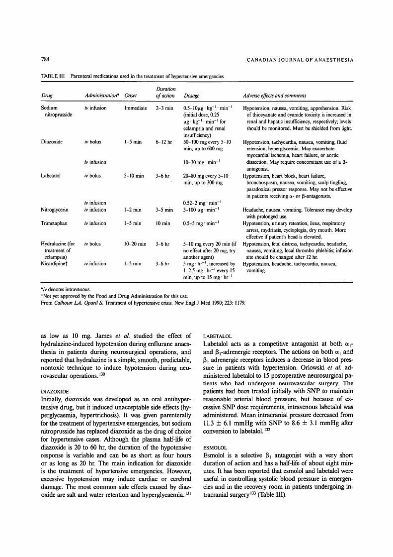
TABLE III Parenteral medications used in the treatment of hypertensive emergencies
Drug Administration* OnsetDurationof action Dosage Adverse effects and comments
Sodium rv infusionnitroprusside
Diazoxide rv bolus
rv infusion
Labetalol rv bolus
rv infusionNitroglycerin rv infusion
Trimetaphan rv infusion
Hydralazine (for rv bolustreatment ofeclampsia)
Nicardipinet rv infusion
Immediate 2-3 min
1-5 min
1-5 min
6-12 hr
5-10 min 3-6 hr
(initial dose, 0.25|ig- kg"1 • min"1 foreclampsia and renalinsufficiency)50-100 mg every 5-10min, up to 600 mg
10-30 mg-min-1
20-80 mg every 5-10min, up to 300 mg
0.52-2 mg-min"1
1-2 min 3-5 min 5-100 ng- min"1
1-5 min 10 min 0.5-5 mg • min"1
10-20 min 3-6 hr
3-6 hr
5-10 mg every 20 min (ifno effect after 20 mg, tryanother agent)5 mg • hr"1, increased by1-2.5 mg • hr"1 every 15min, up to 15 mg • hr"1
Hypotension, nausea, vomiting, apprehension. Riskof thiocyanate and cyanide toxicity is increased inrenal and hepatic insufficiency, respectively; levelsshould be monitored. Must be shielded from light.
Hypotension, tachycardia, nausea, vomiting, fluidretension, hyperglycemia. May exacerbatemyocardial ischemia, heart failure, or aorticdissection. May require concomitant use of a 3-antagonist.
Hypotension, heart block, heart failure,bronchospasm, nausea, vomiting, scalp tingling,paradoxical pressor response. May not be effectivein patients receiving a- or 3-antagonists.
Headache, nausea, vomiting. Tolerance may developwith prolonged use.
Hypotension, urinary retention, ileus, respiratoryarrest, mydriasis, cycloplegia, dry mouth. Moreeffective if patient's head is elevated.
Hypotension, fetal distress, tachycardia, headache,nausea, vomiting, local thrombo phlebitis; infusionsite should be changed after 12 hr.
Hypotension, headache, tachycardia, nausea,vomiting.
*/v denotes intravenous.|Not yet approved by the Food and Drug Administration for this use.From Calhoun LA, Oparil S. Treatment of hypertensive crisis. New Engl J Med 1990; 223: 1179.
as low as 10 mg. James et al. studied the effect ofhydralazine-induced hypotension during enflurane anaes-thesia in patients during neurosurgical operations, andreported that hydralazine is a simple, smooth, predictable,nontoxic technique to induce hypotension during neu-rovascular operations. l3°
DIAZOXIDE
Initially, diazoxide was developed as an oral antihyper-tensive drug, but it induced unacceptable side effects (hy-perglycaemia, hypertrichosis). It was given parenterallyfor the treatment of hypertensive emergencies, but sodiumnitroprusside has replaced diazoxide as the drug of choicefor hypertensive cases. Although the plasma half-life ofdiazoxide is 20 to 60 hr, the duration of the hypotensiveresponse is variable and can be as short as four hoursor as long as 20 hr. The main indication for diazoxideis the treatment of hypertensive emergencies. However,excessive hypotension may induce cardiac or cerebraldamage. The most common side effects caused by diaz-oxide are salt and water retention and hyperglycaemia.131
LABETALOL
Labetalol acts as a competitive antagonist at both a r
and 3,-adrenergic receptors. The actions on both a, andj3j adrenergic receptors induces a decrease in blood pres-sure in patients with hypertension. Orlowski et al. ad-ministered labetalol to 15 postoperative neurosurgical pa-tients who had undergone neurovascular surgery. Thepatients had been treated initially with SNP to maintainreasonable arterial blood pressure, but because of ex-cessive SNP dose requirements, intravenous labetalol wasadministered. Mean intracranial pressure decreased from11.3 ± 6.1 mmHg with SNP to 8.6 ± 3.1 mmHg afterconversion to labetalol.132
ESMOLOL
Esmolol is a selective f$i antagonist with a very shortduration of action and has a half-life of about eight min-utes. It has been reported that esmolol and labetalol wereuseful in controlling systolic blood pressure in emergen-cies and in the recovery room in patients undergoing in-tracranial surgery133 (Table III).

Abe: HYPOTENSION AND SAH 785
Clinical implicationsThe use of induced hypotension has declined becauseit may induce or exacerbate brain ischaemia during aneu-rysm surgery, but intraoperative rupture of a cerebralaneurysm, especially if rupture occurs before its exposuremay induce catastrophic result. A recent retrospectivestudy suggests the usefulness of temporal clipping thaninduced hypotension.70 The use of temporal clipping todecompress aneurysms, or to treat intraoperative aneu-rysm rupture has been preferred to induced hypotensionrecently. However, it is unclear whether improvement inoutcome results from the use of temporary clips or theincrease in surgical experience and expertise.134 Prolongedtemporary occlusion can be unforgiving when the oc-cluded vessel supplies numerous small perforators suchas the carotid birfucation, the Ml segment of middlecerebral artery, or the anterior communicating artery.These arterial segments supply perforating vessels withoutcollateral circulation. The other difficulty that arises fromtemporal clipping is the physical and anatomical limi-tations of applying multiple clips in a confined space,particularly with basilar aneurysms in which space is usu-ally the limiting factor. Complete dissection of an aneu-rysm neck by surgical clipping and without injury tosurrounding brain or blood vessels, is the goal of aneu-rysm surgery. In cases of giant cerebral aneurysm, fragileaneurysm or multiple cerebral aneurysms, induced hyp-otension may be recommended to prevent the intraop-erative rupture, but long-term hypotension or profoundhypotension may induce cerebral ischaemia after surgeryand provoke end-organ ischaemia or infarction. Thus,profound and long-term hypotension should be avoided.During cerebral aneurysm surgery, the effect of vasod-ilators on cerebral vasculature should be kept in mind.Sodium nitroprusside, nitroglycerin and isoflurane havebeen recommended for induced hypotension because oftheir rapid onset and recovery and the stability of thecerebral vasculature.
Intraoperative hypertension during cerebral aneurysmsurgery should be treated immediately because it maydamage the cardiovascular, renal and central nervous sys-tems and it may induce rebleeding and cerebral aneur-ysmal rupture during surgery. Any drug used for thetreatment of intraoperative hypertension carries the riskof decreasing cerebral blood flow below the lower limitof autoregulation to induce cerebral ischaemia or infarc-tion. Vasoactive drugs have less effects in the cerebralcirculation than in other vascular beds, in part becauseof the protective effect of the blood-brain barrier, butdrugs that penetrate the blood-brain barrier and dilatethe cerebral vessels (e.g., hydralazine, sodium nitroprus-side, nifedipine and verapamil) may lead to uneven cer-ebral perfusion due to an "intracranial steal" effect, and
drugs that dilate cerebral vessels and increase cerebralblood flow may also cause an immediate increase in in-tracranial pressure and create the potential for cerebralherniation.135
In our studies, arterial blood pressure was reduced im-mediately after the start of hypotensive drugs, but thehypotensive effect of PGE, and nicardipine infusion per-sisted after their discontinuation. PGE, and nicardipinedo not change LCBF; trimetaphan decreases LCBF andnitroglycerine increases LCBE We confirmed that PGEl5
TMP, TNG and nicardipine did not change carbon di-oxide reactivity during surgery but carbon dioxide reac-tivity had a close correlation with presurgical neurologicalstatus.
ConclusionInduced hypotension has been used to decrease the riskof intraoperative aneurysmal rupture but the use of itis declining because of brain ischaemia. But intraoper-ative hypertension should be treated immediately to re-duce the risk of rebleeding. Profound hypotension shouldbe avoided. A drug that does not change CBF duringcerebral aneurysm surgery should be used to induce hyp-otension.
AcknowledgementsI thank Professor Ikuto Yoshiya MD, PhD and YasuhiroShimada MD, PhD, Dept of Anaesthesia Osaka UniversityMedical School and Nagoya University Medical Schoolfor their valuable advice.
References1 Lam AM. Induced hypotension. Can Anaesth Soc J 1984;
31: S56-S62.2 Grubb RL Jr, Raichle ME, Eichling JO, Gado MH.
Effects of subarachnoid hemorrhage on cerebral blood vol-ume, blood flow, and oxygen utilization in humans. J Neu-rosurg 1977; 46: 446-53.
3 Batjer HH, Frankfurt AI, Purdy PD, Smith SS, SamsonDS. Use of etomidate, temporary arterial occlusion, andintraoperative angiography in surgical treatment of largeand giant cerebral aneurysms. J Neurosurg 1988; 68:234-40.
4 Symon L. Disordered cerebro-vascular physiology inaneurysmal subarachnoid haemorrhage. Acta Neurochir(Wien) 1978; 41: 7-22.
5 McDowall DG. Induced hypotension and brain ischaemia.BrJ Anaesth 1985; 57: 110-9.
6 Hunt WE, Kosnik EJ. Timing and perioperative care inintracranial aneurysm surgery. Clin Neurosurg 1974; 21:78-89.
7 Disney L, Weir B, Grace M. Factors influencing the out-come of aneurysm rupture in poor grade patients: a pro-spective series. Neurosurgery 1988; 23: 1-9.

786 CANADIAN JOURNAL OF ANAESTHESIA
8 Dorsch NWC, Branston NM, Harris RJ, Bentivoglio P,Symon L. An experimental study of the effect of nimodi-pine in primate subarachnoid haemorrhage. Acta Neuro-chir (Wien) 1989; 99: 65-75.
9 Adams HP Jr, Kassell NF, Tomer JC, Haley EC Jr.Predicting cerebral ischemia after aneurysmal subarachnoidhemorrhage: influences of clinical condition, CT results,and antifibrinolytic therapy. A report of the CooperativeAneurysm Study. Neurology 1987; 37: 1586-91.
10 Fein JM. Cerebral energy metabolism after subarachnoidhemorrhage. Stroke 1975; 6: 1-8.
11 Graff-Radford NR, Tomer J, Adams HP, Kassell NF.Factors associated with hydrocephalus after subarachnoidhemorrhage. A report of the Cooperative Aneurysm Study.Arch Neurol 1989; 46: 744-52.
12 Mohr G, Ferguson G, Khan M, et al. Intraventricularhemorrhage from ruptured aneurysm. J Neurosurg 1983;58: 482-7.
13 Ishii R. Regional cerebral blood flow in patients with rup-tured intracranial aneurysms. J Neurosurg 1979; 50:587-94.
14 Powers WJ, Grubb RL Jr, Baker RP, Mintun MA, RaichleME. Regional cerebral blood flow and metabolism in re-versible ischemia due to vasospasm. J Neurosurg 1985; 62:539-46.
15 Weir B, Menon D, Overton T. Regional cerebral bloodflow in patients with aneurysms: estimation by Xenon 133inhalation. Can J Neurol Sci 1978; 5: 301-5.
16 Mountz JM, McGillicuddy JE, Wilson MW, Bartold SP,Siegal EM. Pre- and post-operative cerebral blood flowchanges in subarachnoid haemorrhage. Acta Neurochir(Wien) 1991; 109: 30-3.
17 Voldby B, Enevoldsen EM, Jensen FT. Regional CBF, in-traventricular pressure, and cerebral metabolism in patientswith ruptured intracranial aneurysms. J Neurosurg 1985;62: 48-58.
18 Menon D, Weir B, Overton T. Ventricular size and cerebralblood flow following subarachnoid hemorrhage. Journal ofComputer Assisted Tomography 1981; 5: 328-33.
19 Mat hew NT, Meyer JS, Hartmann A. Diagnosis andtreatment of factors complicating subarachnoid hemor-rhage. Neuroradiology 1974; 6: 237-45.
20 Hunt WE, Hess RM. Surgical risk as related to time of in-tervention in the repair of intracranial aneurysms. J Neuro-surg 1968; 28: 14-20.
21 Jennett B, Bond M. Assessment of outcome after severebrain damage. Lancet 1975; 1: 480-4.
22 Alvord EC Jr, Loeser JD, Bailey WL, Copass MK.Subarachnoid hemorrhage due to ruptured aneurysms. Asimple method of estimating prognosis. Arch Neurol 1972;27: 273-84.
23 Archer DP, Shaw DA, Leblanc RL, Tranmer BI.Haemodynamic considerations in the management of pa-
tients with subarachnoid haemorrhage. Can J Anaesth1991; 38: 454-70.
24 Ljunggren B, Brandt L, Kagstrom E, Sundbarg G.Results of early operations for ruptured aneurysms.J Neurosurg 1981; 54: 473-9.
25 Kassell NF, Tomer JC, Haley EC Jr, Jane JA, Adams HP,Kongable GL. The International Cooperative Study on theTiming of Aneurysm Surgery. Part 1: Overall managementresults. J Neurosurg 1990; 73: 18-36.
26 Haley EC Jr, Kassell NF, Tomer JC. The InternationalCooperative Study on The Timing of Aneurysm Surgery.The North American experience. Stroke 1992; 23: 205-14.
27 Kassell NJ, Tomer JC. Aneurysmal rebleeding: a prelimi-nary report from the Cooperative Aneurysm Study. Neuro-surgery. 1983; 13: 479-81.
28 Kassell NF, Sasaki T, Colohan ART, Nazar G. Cerebralvasospasm following aneurysmal subarachnoid hemor-rhage. Stroke 1985; 16: 562-72.
29 Graf CJ, Nibbelink DW. Cooperative study of intracranialaneurysms and subarachnoid hemorrhage. Report on arandomized treatment study III. Intra-cranial surgery.Stroke 1974; 5: 559-610.
30 Kwak R, Niizuma H, Ohi T, Suzuki J. Angiographicstudy of cerebral vasospasm following rupture of intracran-ial aneurysms: Part 1. Time of the appearance. SurgNeurol 1979; 11: 257-62.
31 Weir B, Grace M, Hansen J, Rothberg C. Time course ofvasospasm in man. J Neurosurg 1978; 48: 173-8.
32 Hews RC, Zervas NT, Varsos V. Cerebral vasospasm aftersubarachnoid hemorrhage: an update. Ann Neurol 1983;14: 599-608.
33 Carpenter DA, Grubb RL Jr, Tempel LW, Powers WJ.Cerebral oxygen metabolism after aneurysmal subarach-noid hemorrhage. J Cereb Blood Row Metab 1991; 11:837-44.
34 Ohman J, Heiskanen O. Effect of nimodipine on the out-come of patients after aneurysmal subarachnoid hemor-rhage and surgery. J Neurosurg 1988; 69: 683-6.
35 Petruk KC, West M, Mohr G et al. Nimodipine treatmentin poor-grade aneurysm patients. Results of a multicenterdouble-blind placebo-controlled trial. J Neurosurg 1988; 68:505-17.
36 Philippon J, Grob R, Dagreou F, Guggiari M, Rivierez M,Viars P. Prevention of vasospasm in subarachnoid haemor-rhage. A controlled study with nimodipine. Acta Neurochir(Wien) 1986; 82: 110-4.
37 Allen GS, Ahn HS, Preziosi TJ et al. Cerebral arterial va-sospasm - a controlled trial of nimodipine in patients withsubarachnoid hemorrhage. N Engl J Med 1983; 308:619-24.
38 Origitano TC, Wascher TM, Reichman OH, AndersonDE. Sustained increased cerebral blood flow with prophy-lactic hypertensive hypervolemic hemodilution ("triple-H"

Abe: HYPOTENSION AND SAH 787
therapy) after subarachnoid hemorrhage. Neurosurgery1990; 27: 729-39.
39 Olesen J. Quantitive evaluation of normal and pathologiccerebral blood flow regulation to perfusion pressurechanges in man. Arch Neurol 1973; 28: 143-9.
40 Enevoldsen EM, Jensen FT Autoregulation and CO2 re-sponses of cerebral blood flow in patients with acute severehead injury. J Neurosurg 1978; 48: 689-703.
41 Paulson OB. Cerebral apoplexy (stroke): pathogenesis, pa-thophysiology and therapy as illustrated by regional bloodflow measurements in the brain. Stroke 1971; 2: 327-60.
42 Schalen W, Messeter K, Nordstrom C. Cerebral vasoreac-tivity and the prediction of outcome in severe traumaticbrain lesions. Acta Anaesthesiol Scand 1991; 35: 113-22.
43 Voldby B, Enevoldsen EM, Jensen FT. Cerebrovascularreactivity in patients with ruptured intracranial aneurysms.J Neurosurg 1985; 62: 59-67.
44 Artru AA, Colley PS. Cerebral blood flow responses tohypocapnia during hypotension. Stroke 1984; 15: 878-83.
45 Abe K, Demizu A, Mima T, Kamada K, Yoshiya I.Carbon dioxide reactivity during prostaglandin Ej inducedhypotension for cerebral aneurysm surgery. Can J Anaesth1992: 39: 253-9.
46 Abe K, Iwanaga H, Yoshiya I. Carbon dioxide reactivityand local cerebral blood flow during prostaglandin Ej ornitroglycerine-induced hypotension. Can J Anaesth 1992;39: 799-804.
47 Abe K, Shimada Y, Iwanaga H, Yoshiya I. Effect of nicar-dipine on local cerebral blood flow, carbon dioxide reactiv-ity and blood flow velocity of internal carotid artery duringcerebral aneurysm surgery. Anesth Anal (in press).
48 Nomes H. The role of intracranial pressure in the arrest ofhemorrhage in patients with ruptured intracranial aneu-rysm. J Neurosurg 1973; 51: 226-34.
49 FarrarJK, Gamache FW Jr, Ferguson GG, Barker J, Var-key GP, Drake CG. Effects of profound hypotension oncerebral blood flow during surgery for intracranial aneu-rysms. J Neurosurgery 1981; 55: 857-64.
50 Messeter K, Brandt L, Ljunggren B, et al. Prediction andprevention of delayed ischemic dysfunction after aneurys-mal subarachnoid haemorrhage and early operation. Neu-rosurgery 1987; 20: 548-53.
51 Cold GE, Jensen FT Cerebral autoregulation in uncon-scious patients with brain injury. Acta Anaesthesiol Scand1978; 22: 270-80.
52 Muizelaar JP, Ward JD, Marmarou A, Newlon PG,Wachi A. Cerebral blood flow and metabolism in severelyhead-injured children. Part 2: Autoregulation. J Neurosurg1989; 71: 72-6.
53 Smith DR, Jacobson J, Kobrine AI, Rizzoli HV. Regionalcerebral blood flow with intracranial mass lesions. Part II:Autoregulation in localized mass lesions. Surg Neurol 1977;7: 238-40.
54 Dembach PD, Little JR, Jones SC, Ebrahim ZY. Alteredcerebral autoregulation and CO2 reactivity after aneurys-mal subarachnoid hemorrhage. Neurosurgery 1988; 22:822-6.
55 Obrist WD, Thompson HKJr, King CH, Wang HS.Determination of regional cerebral blood flow by inhala-tion of 133-Xenon. Circ Res 1967; 20: 124-35.
56 Lassen NA, Ingvar DH. Regional cerebral blood flowmeasurement in man. Arch Neurol 1963; 9: 615-22.
57 Olsen TS, Larsen B, Skriver EB, Herning M, EnevoldsenE, Lassen NA. Focal cerebral hyperemia in acute stroke.Incidence, pathophysiology and clinical significance. Stroke1981; 12: 598-607.
58 Yonas H, Wolfson SK Jr, Gur D, et al. Clinical experiencewith the use of xenon-enhanced CT blood flow mapping incerebral vascular disease. Stroke 1984; 15: 443-9.
59 Powers WJ, Raichle MW. Positron emission tomographyand its application to the study of cerebrovascular diseasein man. Stroke 1985; 16: 361-76.
60 Aukland K, Bower BF, Berliner RW. Measurement oflocal blood flow with hydrogen gas. Circ Res 1964; 14:164-87.
61 Young W. H2 clearance measurement of blood flow: a re-view of technique and polarographic principles. Stroke1980; 11: 552-64.
62 Pickard JD, Matheson M, Patterson J, Wyper D.Prediction of late ischemic complications after cerebralaneurysms surgery by the intraoperative measurement ofcerebral blood flow. J Neurosurg 1980; 63: 305-8.
63 Bell BA, Symon L, Branston NM. CBF and time thres-holds for the formation of ischemic cerebral edema, and ef-fect of reperfusion in baboons. J Neurosurg 1985; 62:31-41.
64 Abe K, Demizu A, Kamada K, Morimoto T, Sakaki T,Yoshiya I. Local cerebral blood flow with prostaglandin E!or trimethaphan during cerebral aneursym clip ligation.Can J Anaesth 1991; 38: 831-6.
65 Aaslid R, Huber P, Nornes H. Evaluation of cerebrovascu-lar spasm with transcranial Doppler ultrasound. J Neuro-surg 1984; 60: 37-42.
66 Seiler RW, Grolimund P, Aaslid R, Huber R, Nornes H.Cerebral vasospasm evaluated by transcranial ultrasoundcorrelated with clinical grade and CT-visualized subarach-noid hemorrhage. J Neurosurg 1986; 64: 594-600.
67 Sloan MA, Haley EC Jr, Kassell NF, et al. Sensitivity andspecificity of transcranial Doppler ultrasonography in thediagnosis of vasospasm following subarachnoid hemor-rhage. Neurology 1989; 39: 1514-8.
68 Caplan LR, Brass LM, DeWitt LD, et al. TranscranialDoppler ultrasound: present status. Neurology 1990; 40:696-700.
69 Davis SM, Andrews JT, Lichtenstein M, Rossiter SC,Kaye AH, Hopper J. Correlations between cerebral arter-

788 CANADIAN JOURNAL OF ANAESTHESIA
ial velocities, blood flow, and delayed ischemia after sub-arachnoid hemorrhage. Stroke 1992; 23: 492-7.
70 Giannotta SL, Oppenheimer JH, Levy ML, Zelman V.Management of intraoperative rupture of aneurysm with-out hypotension. Neurosurgery 1991; 28: 531-6.
71 Hitchcock ER, Tsementzis SA, Dow A A. Short- andlong-term prognosis of patients with subarachnoid haemor-rhage in relation to intraoperative period of hypotension.Acta Neurochir 1984; 70: 235-41.
72 Lam AM, Manninen PH. Induced hypotension for cere-bral aneurysm - isoflurane or sodium nitroprusside? Can JAnaesth 1987; 34: S121-S122.
73 Batjer H, Samson D Intraoperative aneurysmal rupture:incidence, outcome and suggestions for surgical manage-ment. Neurosurgery 1986; 18: 701-17.
74 Amman JI, Diaz FG, Malik GM, Fielding AS, Son CS.Current management of cerebral aneurysms: is it based onfacts or myths? Surg Neurol 1985; 24: 625-35.
75 Calhoun DA, Oparil S. Treatment of hypertensive crisis.N Engl J Med 1990; 223: 1177-83.
76 Carlson LA, Ekelund L-G, Oro L. Circulatory and respi-ratory effects of different doses of prostaglandin E( in man.Acta Physiol Scand 1969; 75: 161-9.
77 D'Ambra MN, LaRaia PJ, Philbin DM, Watkins WD,Hilgenberg AD, Buckley Ml Prostaglandin Ej. A newtherapy for refractory right heart failure and pulmonaryhypertension after mitral valve replacement. J Thorac Car-diovasc Surg 1985; 89: 567-72.
78 Goto F, Otani E, Fujita T. Antihypertensive activity andmetabolic rate of prostaglandin Ej in surgical patientsunder general anesthesia. Prostaglandins Leukot EssentFatty Acid 1985; 18: 359-66.
79 Sinha AK, Colman RW. Prostaglandin E] inhibits plateletaggregation by a pathway independent of adenosine 3', 5'-monophosphate. Science 1978; 200: 202-3.
80 Carbon LA, Irion E, Oro L. Effect of infusion of prosta-glandin Ej on the aggregation of blood platelets in man.Life Sci 1968; 7: 85-90.
81 Woodside J, Gamer L, Bedford RF, et al. Captopril redu-ces the dose requirement for sodium nitroprusside inducedhypotension. Anesthesiology 1984; 60: 413-7.
82 Marsh ML Shapiro HM, Smith RW, Marshall LF.Changes in neurologic status and intracranial pressure as-sociated with nitroprusside administration. Anesthesiology1979; 51: 336-8.
83 CottrellJE, Gupta B, Rappaport H, Tumdorf H, Ranso-hoffJ, Flamm ES. Intracranial pressure during nitro-glycerin-induced hypotension. J Neurosurg 1980; 53:309-11.
84 Rogers MC, Traystman RJ. Cerebral hemodynamic effectsof nitroglycerin and nitroprusside. Acta Neurol ScandSuppl 1979; 62: 600-1.
85 Pinaud M, Souron R, Lelausque JN, Gazeau M-F, Lajat
X Dixneuf B. Cerebral blood flow and cerebral oxygenconsumption during nitroprusside-induced hypotension toless than 50 mmHg. Anesthesiology 1989; 70: 255-60.
86 Hines R, Barash PG. Infusion of sodium nitroprusside in-duces platelet dysfunction in vitro. Anesthesiology 1989; 70:611-5.
87 Larsen R, Teichmann J, Hilftker O, Busse C, Sonntag H.Nitroprusside-hypotension: cerebral blood flow and cere-bral oxygen consumption in neurosurgical patients. ActaAnaesthesiol Scand 1982; 26: 327-30.
88 Newman B, Gelb AW, Lam AM. The effect of isoflurane-induced hypotension on cerebral blood flow and cerebralmetabolic rate for oxygen in humans. Anesthesiology 1986;64: 307-10.
89 Sollevi A, Lagerkranser M, Irestedt L, Gordon E, Lind-quist C. Controlled hypotension with adenosine in cerebralaneurysm surgery. Anesthesiology 1984; 61: 400-5.
90 Winn HR, Welsh JE, Rubio R, Berne RM. Brain adeno-sine production in rat during sustained alteration in sys-temic blood pressure. Am J Physiol 1980; 239:H636-H641.
91 Owall A, Gordon E, Lagerkranser M, Lindquist C, Rude-hill A, Sollevi A. Clinical experience with adenosine forcontrolled hypotension during cerebral aneurysm surgery.Anesth Analg 1987; 66: 229-34.
92 Owall A, Lagerkranser M, Sollevi A. Effects of adenosine-induced hypotension on myocardial hemodynamics andmetabolism during cerebral aneurysm surgery. AnesthAnalg 1988; 67: 228-32.
93 Lagerkranser M, Bergstrand G, Gordon E, et al. Cerebralblood flow and metabolism during adenosine-induced hyp-otension in patients undergoing cerebral aneurysm surgery.Acta Anaesthesiol Scand 1989; 33: 15-20.
94 lall, S, Eden E, Winso I, Volkmann R, Sollevi A,Ricksten SE. Controlled hypotension with adenosine orsodium nitroprusside during cerebral aneurysm surgery: ef-fects on renal hemodynamics, excretory function, and reninrelease. Anesth Analg 1990; 71: 631-6.
95 Murad F. Cyclic guanosine monophosphate as a mediatorof vasodilation. J Clin Invest 1986; 78: 1-5.
96 Ignarro LJ, Lippton H, Edwards JC, et al. Mechanism ofvascular smooth muscle relaxation by organic nitrates, ni-trites, nitroprusside and nitric oxide: evidence for the in-volvement of S-nitrosothiols as active intermediates. JPharmacol Exp Ther 1981; 218: 739-49.
97 Langerkranser M. Effects of nitroglycerin on intracranialpressure and cerebral blood flow. Acta Anaesthesiol Scand1992; Suppl 36: 34-6.
98 Maktabi M, Warner D, Sokoll M, et al. Comparison ofnitroprusside, nitroglycerin, and deep isoflurane anesthesiafor induced hypotension. Neurosurgery 1986; 19: 350-5.
99 Larson AG. Deliberate hypotension. Anesthesiology 1964;24: 682-706.

Abe: HYPOTENSION AND SAH 789
100 Miller ED Jr. Deliberate hypotension. In: Miller RD Jr(Ed.). Anesthesia, 2nd ed, New York: Churchill Living-stone, 1986; 1949-70.
101 Knight PR, Lane GA, Hensinger RN, Bolles RS, Bjo-raker DG. Catecholamine and resin-angiotensin responseduring hypotensive anesthesia induced by sodium nitro-prusside or trimethaphan camsylate. Anesthesiology 1983;59: 248-53.
102 Turner JM, Powell D, Gibson RM, McDowall DG.Intracranial pressure changes in neurosurgical patientsduring hypotension induced with sodium nitroprusside ortrimetaphan. Br J Anaesth 1977; 49: 419-25.
103 Reves JG, Kissin I, Lell WA, Tosone S. Calcium entryblockers: uses and implications for anesthesiologists.Anesthesiology 1982; 57; 504-18.
104 Opie LH. Calcium channel antagonists. Part III: Use andcomparative efficacy in hypertension and supraventriculararrhythmias. Minor indications. Cardiovascular Drugsand Therapy 1988; 1: 625-56.
105 Zimpfer M, Fitzal S, Tonczar L. Verapamil as a hypoten-sive agent during neuroleptanaesthesia. Br J Anaesth1981; 53: 885-9.
106 Bernard JM, Pinaud M, Carteau S, Hubert C, SouronR. Hypotensive actions of diltiazem and nitroprussidecompared during fentanyl anaesthesia for total hip arthro-plasty. Can Anaesth Soc J 1986; 33: 308-14.
107 HofRP Calcium antagonists and the peripheral circula-tion: differences and similarities between PY 108-068, ni-cardipine, verapamil and diltiazem. Br J Pharmacol 1983;78: 375-94.
108 Sorkin EM, Clissold SP. Nicardipine. A review of itspharmacodynamic and pharmacokinetic properties andtherapeutic efficacy, in the treatment of angina pectoris,hypertension and related cardiovascular disorders. Drugs1987; 33: 296-345.
109 Milde LN, Milde JH, Michenfelder JD. Delayed treat-ment with nimodipine improves cerebral blood flow aftercomplete cerebral ischaemia in the dog. J Cereb BloodFlow Metab 1986; 6: 332-7.
110 Pearce WJ, Bevan JA. Diltiazem and autoregulation ofcanine cerebral blood flow. J Pharmacol Exp Ther 1987;242: 812-7.
111 Frishman WH. New therapeutic modalities in hyperten-sion: focus on a new calcium antagonist-nicardipine. JClin Pharmacol 1989; 29: 481-7.
112 Yamamoto M, Ohta T, Toda N. Mechanisms of relaxantaction of nicardipine, a new Ca++-agonist, on isolateddog cerebral and mesenteric arteries. Stroke 1983; 14:270-5.
113 Norman J. The IV administration of drugs (Editorial). BrJ Anaesth 1983; 55: 1049-52.
114 Bertel O, Conen D, Radii EW, Muller J, Lang C, DubachUC. Nifedipine in hypertensive emergencies. B M J 1983;286: 19-21.
115 Pool PE, Massie BM, Venkataraman K, et al. Diltiazemas monotherapy for systemic hypertension: a multicenter,randomized, placebo-controlled trial. Am J Cardiol 1986;57: 212-7.
116 Weber MA, Cheung DG, Graettinger WF, Lipson JL.Characterization of antihypertensive therapy by whole-dayblood pressure monitoring. JAMA 1988; 259: 3281-5.
117 Onoyama K, Omae T, Ilmura O, et al. Effects of a dripinfusion of intravenous diltiazem on severe systemic hyper-tension. Current Therapeutic Research 1988; 43: 361-8.
118 Mullen JC, Miller DR, Weisel RD, et al. Postoperativehypertension: a comparison of diltiazem, nifedipine, andnitroprusside. J Thorac Cardiovasc Surg 1988; 96: 122-32.
119 Godet G, Coriat P, Baron JF, et al. Prevention of in-traoperative myocardial ischemia during noncardiacsurgery with intravenous diltiazem: a randomized trial ver-sus placebo. Anesthesiology 1987; 66: 241-5.
120 Bevan JA. Selective action of diltiazem on the cerebralvascular smooth muscle in the rabbit: antagonism of ex-trinsic but not intrinsic maintained tone. Am J Cardiol1982; 49: 519-24.
121 Bevan JA, Bevan RD, Frazee JG. Experimental chroniccerebrovascular spasm in the monkey: an assessment ofthe functional changes in the cerebral arteries and theirprotection by diltiazem. Am J Cardiol 1985; 56:15H-20H.
122 Lam AM, Gelb AW. Cardiovascular effects of isoflurane-induced hypotension for cerebral aneurysm surgery.Anesth analg 1983; 62: 742-8.
123 Nicholas JF, Lam AM. Isofurane-induced hypotensiondoes not cause impairment in pulmonary gas exchange.Can Anaesth Soc J 1984; 31: 352-8.
124 Roth S, Jones SC, Ebrahim ZY, Friel H, Little JR. Localcortical blood flow and oxygen consumption duringisoflurane-induced hypotension. Results in patients under-going intracranial aneurysm clipping. Clevel Clin J Med1989; 56: 766-70.
125 Haraldstedt VY, Asmussen J, Herlevsen P, Cold GE.Cerebral arteriovenous difference of oxygen during grad-ual and sudden increase of the concentration of isofluranefor induction of deliberate hypotension. Acta AnaesthesiolScand 1992; 36: 142-44.
126 Grosslight K, Foster R, Colohan AR, Bedford REIsoflurane for neuroanesthesia: risk factors for increases inintracranial pressure. Anesthesiology 1985; 65: 533-6.
127 Macnab MSP, Manninen PH, Lam AM, Gelb AW. Thestress response to induced hypotension for cerebral aneu-rysm surgery: a comparison of two hypotensive tech-niques. Can J Anaesth 1988; 35: 111-5.
128 Blaise G, Still JC, Nugent M, Van Dyke RA, VanhouttePM. Isoflurane causes endothelium-dependent inhibitionof contractile responses of canine coronary arteries. Anes-thesiology 1987; 67: 513-7.

790 CANADIAN JOURNAL OF ANAESTHESIA
129 Stone DJ, Johns RA. Endothelium-dependent effects ofhalothane, enflurane and isoflurane on isolated rat aorticvascular rings. Anesthesiology 1989; 71: 126-32.
130 James DJ, Bedford RE Hydralazine for controlled hypo-tension during neurosurgical operations. Anesth Analg1982; 61: 1016-9.
131 Gerber JG, Nies AS. Antihypertensive agents and thedrug therapy of hypertension. In: Gilman AG (Ed.). ThePharmacological Basis of Therapeutics. 8th ed. New York:Pergamon Press, 1990; 784-813.
132 OrlowskiJP, Shiesley D, Vidt DG, Barnett GH, Little/ALabetalol to control blood pressure after cerebrovascu-lar surgery. Crit Care Med 1988; 16: 765-8.
133 Muzzi DA, Black S, Losasso TJ, Cucchiara RE Labet-alol and esmolol in the control of hypertension after intra-cranial surgery. Anesth Analg 1990; 70: 68-71.
134 Chong KY, Gelb AW. Management of intracranial aneu-rysms and subarachnoid hemorrhage. Current Opinion inAnaesthesiology 1992; 5: 620-5.
135 Bauer JH, Reams GP. The role of calcium entry blockersin hypertensive emergencies. Circulation 1987; 75:V174-V180.